Embed Size (px)
Citation preview

8/2/2019 Neurophysiological Effects of Spinal Manipulation
http://slidepdf.com/reader/full/neurophysiological-effects-of-spinal-manipulation 1/15
The Spine Journal 2 (2002) 357–371
1529-9430/02/$ – see front matter © 2002 Elsevier Science Inc. All rights reserved.
PII: S1529-9430(02) 00400-X
Review Article
Neurophysiological effects of spinal manipulationJoel G. Pickar, DC, PhD*
Palmer Center for Chiropractic Research, 1000 Brady Street, Davenport, IA 52803, USA
Received 23 February 2001; accepted 15 May 2002
Abstract Background context:
Despite clinical evidence for the benefits of spinal maniputation and the appar-
ent wide usage of it, the biological mechanisms underlying the effects of spinal manipulation are not
known. Although this does not negate the clinical effects of spinal manipulation, it hinders acceptance
by the wider scientific and health-care communities and hinders rational strategies for improving the
delivery of spinal manipulation.
Purpose: The purpose of this review article is to examine the neurophysiological basis for the effectsof spinal manipulation.
Study design: A review article discussing primarily basic science literature and clinically oriented
basic science studies.
Methods: This review article draws primarily from the peer-reviewed literature available on Med-
line. Several textbook publications and reports are referenced. A theoretical model is presented de-
scribing the relationships between spinal manipulation, segmental biomechanics, the nervous system
and end-organ physiology. Experimental data for these relationships are presented.
Results: Biomechanical changes caused by spinal manipulation are thought to have physiological con-
sequences by means of their effects on the inflow of sensory information to the central nervous system.
Muscle spindle afferents and Golgi tendon organ afferents are stimulated by spinal manipulation.
Smaller-diameter sensory nerve fibers are likely activated, although this has not been demonstrated di-
rectly. Mechanical and chemical changes in the intervertebral foramen caused by a herniated interver-
tebral disc can affect the dorsal roots and dorsal root ganglia, but it is not known if spinal manipulation
directly affects these changes. Individuals with herniated lumbar discs have shown clinical improve-ment in response to spinal manipulation. The phenomenon of central facilitation is known to increase
the receptive field of central neurons, enabling either subthreshold or innocuous stimuli access to cen-
tral pain pathways. Numerous studies show that spinal manipulation increases pain tolerance or its
threshold. One mechanism underlying the effects of spinal manipulation may, therefore, be the manip-
ulation’s ability to alter central sensory processing by removing subthreshold mechanical or chemical
stimuli from paraspinal tissues. Spinal manipulation is also thought to affect reflex neural outputs to
both muscle and visceral organs. Substantial evidence demonstrates that spinal manipulation evokes
paraspinal muscle reflexes and alters motoneuron excitability. The effects of spinal manipulation on
these somatosomatic reflexes may be quite complex, producing excitatory and inhibitory effects.
Whereas substantial information also shows that sensory input, especially noxious input, from
paraspinal tissues can reflexively elicit sympathetic nerve activity, knowledge about spinal manipu-
lation’s effects on these reflexes and on end-organ function is more limited.
Conclusions: A theoretical framework exists from which hypotheses about the neurophysiologicaleffects of spinal manipulation can be developed. An experimental body of evidence exists indicating
that spinal manipulation impacts primary afferent neurons from paraspinal tissues, the motor control
system and pain processing. Experimental work in this area is warranted and should be encouraged to
help better understand mechanisms underlying the therapeutic scope of spinal manipulation. © 2002
Elsevier Science Inc. All rights reserved.
Keywords: Spinal manipulation; Neurophysiology; Manual therapy; Manual medicine; Chiropractic; Osteopathy
FDA device/drug status: not applicable.Nothing of value received from a commercial entity related to this research.This study was supported by the Consortial Center for Chiropractic
Research through cooperative agreement 1-U24AR5166 funded by NationalInstitutes of Health, National Center for Complementary and Alternative
Medicine and by the National Institute of Neurological Disease and StrokeGrant NS35300 on behalf of the Office of Alternative Medicine.
* Corresponding author. Palmer Center for Chiropractic Research,
1000 Brady Street, Davenport, IA 52803, USA. Tel.: (563) 884-5219; fax:
(563) 884-5227.
E-mail address: [email protected] (J.G. Pickar)

8/2/2019 Neurophysiological Effects of Spinal Manipulation
http://slidepdf.com/reader/full/neurophysiological-effects-of-spinal-manipulation 2/15
358
J.G. Pickar / The Spine Journal 2 (2002) 357–371
Introduction
Recent reports estimate that 7.7% to 8.3% of the US pop-
ulation uses some form of complementary or alternative
medicine [1–3]. Approximately 30% to 40% of these indi-
viduals likely receive spinal manipulation [1]. Strong evi-
dence supports using spinal manipulation to help patients
with acute low back pain and neck pain [4,5]. The benefitsof spinal manipulation for other disorders, such as chronic
low back pain and visceral disorders, are less clear, although
benefits have been noted [4,6–8]. Despite the clinical evi-
dence for the benefits of and the apparent wide usage of spi-
nal manipulation, the biological mechanisms underlying the
effects of spinal manipulation are not known. Although this
does not negate the clinical effects of spinal manipulation, it
hinders acceptance by the wider scientific and health-care
communities and hinders rational strategies for improving
the delivery of spinal manipulation. The purpose of this re-
view article is to examine the neurophysiological basis for
and the neurophysiological effects of spinal manipulation.
Biomechanical considerations of spinal manipulation
Spinal manipulation by its very nature is a mechanical
input to tissues of the vertebral column. Chiropractors de-
liver more than 90% of these manipulations in the United
States [9]. Spinal manipulation is distinguished from spinal
mobilization in several ways [10]. During spinal manipula-
tion, the practitioner delivers a dynamic thrust (impulse) to
a specific vertebra. The clinician controls the velocity, mag-
nitude and direction of the impulse [11]. The art or skill of
spinal manipulation lies in the clinician’s ability to controlthese three factors once the specific contact with a vertebra
is made. Mobilization techniques are sometimes used prepa-
ratory to the manipulation. Manipulation is also distinguished
from mobilization in that it is delivered at or near the end of
the physiological range of motion (the so-called paraphysio-
logical range [12]) but not exceeding the anatomical limits of
motion. A cracking or popping sound often, but not necessar-
ily, accompanies the manipulation, because gapping the joint
creates fluid cavitation [13,14].
The most common form of spinal manipulation used by
chiropractors is the short-lever, high-velocity and low-
amplitude thrust [15]. The clinician usually delivers the
dynamic thrust through a short-lever arm by manually con-
tacting paraspinal tissues overlying the spinous, transverse
or mammillary processes of the vertebra being manipulated.
Alternatively, the clinician contacts tissues overlying the
lamina or articular pillar of the vertebra. To manipulate the
pelvis, the iliac spine or the ischial spine is used [10]. Spinal
manipulation may also be delivered through a long-lever
arm. While one hand may contact a specific area over the
vertebra being manipulated, the second hand contacts an
area of the body distant from the specific contact. Force is
developed through this long-lever arm. However, using a
short-lever arm applied directly over the vertebra minimizes
the force necessary to accomplish the manipulation [10] by
reducing the amount of compliant tissue through which the
force must be transmitted.
Several laboratories have studied biomechanical features
of short-lever, high-velocity and low-amplitude manipula-
tion. Herzog’s group [16] was the first to report the biome-
chanical features of a spinal manipulation in an indexed
journal. They identified two characteristics common to the
delivery of a spinal manipulation: 1) a preload force fol-
lowed by 2) a larger impulse force. Using two chiropractors,
they quantified the preload and peak impulse forces applied
perpendicular to the contact point and the impulse duration
during manipulation of the sacroiliac joint. Preload load
forces ranged from 20 to 180 N, and peak forces ranged from
220 to 550 N. Often the preload was approximately 25% of
impulse load. The duration of the high-velocity impulse
ranged from 200 to 420 ms.
A number of studies have confirmed the force–time pro-
file initially described by Hessel et al. [16]. Herzog et al.
[17] showed the time to peak impulse was similar duringmanipulation of the thoracic spine and sacroiliac joint (ap-
proximately 150 ms
77 ms, mean
SD). The perpendicu-
larly applied preload and peak impulse forces were also similar
during spinal manipulations applied to the thoracic (139
46 N
vs. 88
78 N, respectively) and sacroiliac (328
78 N vs.
399
119 N, respectively) regions. Studies of the cervical
spine indicate that preload, peak impulse force and time to
peak impulse are less compared with the thoracic and lum-
bosacral spine [17–19]. Depending upon the type of cervical
manipulative technique used, preload forces range from 0 to
approximately 50 N, and peak impulse forces range from
approximately 40 N to approximately 120 N. The forces de-livered during cervical manipulations develop faster than
during manipulation of the thoracic spine and sacroiliac joint.
Impulse duration lasts from approximately 30 ms to approxi-
mately 120 ms. The large variability in the applied forces and
durations should be recognized. The impact of this variability
on the biological mechanisms that could contribute to the
clinical effects of manipulation is unknown.
A complete understanding of the biomechanics of spi-
nal manipulation requires knowing the manner in which
manipulative loads are transmitted to a specific vertebra.
Experimentally, this is substantially more difficult and more
complex compared with measuring applied loads. Transmit-
ted loads may be different from applied loads because of the
effects of patient positioning and the contributions from in-
ertial loads, loading moments and the active and passive
properties of the intervening connective and muscle tissues.
Triano and Schultz [20] calculated peak transmitted loads at
a lumbar segment by measuring loads transmitted to a force
plate placed under the subject. The force plate was capable
of transducing forces and moments about three orthogonal
axes. Peak forces transmitted to a lumbar segment during a
side posture spinal manipulation tended to be higher than
peak forces applied during a prone thoracic or sacroiliac
manipulation measured by Herzog et al. [17]. Transmitted

8/2/2019 Neurophysiological Effects of Spinal Manipulation
http://slidepdf.com/reader/full/neurophysiological-effects-of-spinal-manipulation 3/15
J.G. Pickar / The Spine Journal 2 (2002) 357–371
359
impulse durations were similar to applied impulse durations
measured by Herzog et al. [17]. Peak transmitted moments
were approximately three to four times less than peak trans-
mitted forces. The transmitted loads were considered below
a threshold level capable of injuring the lumbar spine (see
[20] for further discussion).
In addition to applied and transmitted loads, the relative dis-
placement or movement between contiguous vertebrae during
a spinal manipulation has been studied. Nathan and Keller [21]
measured intervertebral lumbar motion using pins inserted into
lumbar spinous processes. Manipulations were delivered using
a mechanical adjusting device (Activator Adjusting Instru-
ment, Activator Methods International, Ltd., Phoenix, AZ
[22]). Impulse duration using this device is approximately
5 ms, an impulse duration shorter than that from manual ma-
nipulation. Impulses delivered to the L2 spinous produced 1.62
mm1.06 mm peak axial displacement (in the longitudinal
plane), 0.480.1 mm shear displacement (in the transverse
plane) and 0.890.49° of rotation between L3 and L4 [21].
Smith et al. [23] measured similar vertebral displacements inthe lumbar spine of the dog. L2 translated 0.710.03 mm and
rotated 0.530.15° on L3 with impulse loads of 53 N. Gal et
al. [24] performed measurements in the thoracic spine, but their
results are difficult to compare with those reported above for
the lumbar spine. Nonetheless, the movements induced during
a spinal manipulative load suggest that mechanical processes
may play a role in the biological effects of spinal manipulation.
Neurophysiological and biomechanical mechanisms
underlying the effects of spinal manipulation
Numerous theories have been proposed to explain the ef-fects of spinal manipulation [25,26]. A thread common to
many of these theories is that changes in the normal anatomi-
cal, physiological or biomechanical dynamics of contiguous
vertebrae can adversely affect function of the nervous system.
[27,28]. Spinal manipulation is thought to correct these changes.
Accordingly, a number of biomechanical changes pro-
duced by vertebral movement during a spinal manipulation
have been hypothesized. The mechanical force introduced
into the vertebral column during a spinal manipulation may
directly alter segmental biomechanics by releasing trapped
meniscoids, releasing adhesions or by reducing distortion of
the annulus fibrosus [29–33]. In addition, individual motion
segments can buckle, thereby producing relatively large ver-
tebral motions that achieve a new position of stable equilib-
rium [34]. The mechanical changes elicited by manipulation
may provide sufficient energy to restore a buckled segment to
a lower energy level, thus reducing mechanical stress or
strain on soft and hard paraspinal tissues [35]. A major con-
sequence of these hypothesized mechanical changes elicited
by manipulation could be the restoration of zygapophyseal
joint mobility and joint play [31]. In fact, authoritative dis-
cussion of spinal manipulation considers “the goal of ma-
nipulation to restore maximal, pain-free movement of the
musculoskeletal system” (from [35a] and see [31,36,37]).
Biomechanical changes caused by the manipulation are
thought to have physiological consequences by means of
their effects on the inflow of sensory information to the cen-
tral nervous system [25,36]. By releasing trapped meniscoids,
discal material or segmental adhesions, or by normalizing a
buckled segment, the mechanical input may ultimately re-
duce nociceptive input from receptive nerve endings in inner-
vated paraspinal tissues. This would be consistent with the
observation that spinal manipulation is not painful when ad-
ministered correctly. In addition, the mechanical thrust could
either stimulate or silence nonnociceptive, mechanosensitive
receptive nerve endings in paraspinal tissues, including skin,
muscle, tendons, ligaments, facet joints and intervertebral
disc [28,38,39]. These neural inputs may influence pain-
producing mechanisms as well as other physiological sys-
tems controlled or influenced by the nervous system.
Fig. 1 diagrams the theoretical relationships between spi-
nal manipulation, segmental biomechanics, the nervous sys-
tem and end-organ physiology. A biomechanical alteration
between vertebral segments hypothetically produces a bio-mechanical overload the effects of which may alter the sig-
naling properties of mechanically or chemically sensitive
neurons in paraspinal tissues. These changes in sensory in-
put are thought to modify neural integration either by di-
rectly affecting reflex activity and/or by affecting central
neural integration within motor, nociceptive and possibly
autonomic neuronal pools. Either of these changes in sen-
sory input may elicit changes in efferent somatomotor and
visceromotor activity. Pain, discomfort, altered muscle func-
tion or altered visceromotor activities comprise the signs or
symptoms that might cause patients to seek spinal manipula-
tion. Spinal manipulation, then, theoretically alters the inflowof sensory signals from paraspinal tissues in a manner that
improves physiological function. This explanation comprises
one of the most rational neurophysiological bases for the
mechanisms underlying the effects of spinal manipulation.
Experimental efforts to understand sensory processing from
paraspinal tissues and the effects of spinal manipulation on
this sensory processing is receiving increasing attention, as
described below. Each of the following sections addresses a
component of the theoretical relationship depicted in Fig. 1
with each section’s number corresponding to a numbered
component in the figure.
1. The effects of spinal manipulation on sensory receptors
in paraspinal tissues
Group I and II afferents (proprioceptive afferents)
Korr [36] proposed that spinal manipulation increases
joint mobility by producing a barrage of impulses in muscle
spindle afferents and smaller-diameter afferents ultimately
silencing facilitated
motoneurons. Fig. 2 shows the neural
circuitry of the
loop. He hypothesized that
-motoneuron
discharge is elevated in muscles of vertebral segments re-
sponding to spinal manipulation. The high gain of the

8/2/2019 Neurophysiological Effects of Spinal Manipulation
http://slidepdf.com/reader/full/neurophysiological-effects-of-spinal-manipulation 4/15
360
J.G. Pickar / The Spine Journal 2 (2002) 357–371
loop would impair joint mobility by sensitizing the stretch
reflex to abnormally small changes in muscle length. Korr
further hypothesized that spinal manipulation stimulates
muscle spindle afferents, that is, Group Ia and possibly
Group II afferents (Table 1). The barrage of impulses from
these afferents produced by the spinal manipulation would
reduce the gain of the loop through an undetermined neu-
ral pathway. Although portions of this mechanism remainspeculative, the contribution of proprioceptive afferents to
spine function and the neurophysiological effects of spinal
manipulation on these afferents are receiving increasing at-
tention.
The importance of paraspinal proprioceptive input to the
function of the vertebral column, and of the lumbar spine in
particular, has been demonstrated recently in humans. Several
studies indicate that muscle spindle input from the lumbar
multifidus helps to accurately position the pelvis and lum-
bosacral spine. Healthy individuals can accurately reposition
their lumbosacral spine, but their repositioning ability is im-
paired when the multifidus muscle is vibrated [40]. Vibration
stimulates muscle spindles and creates a sensory illusion that
the multifidus is stretched and therefore that the spine is
flexed more than it actually is. The repositioning error occurs
because of the misperception of vertebral position. Interest-
ingly, lumbosacral-repositioning ability is impaired in indi-
viduals with a history of low back pain, even in the absence
of vibration [41]. This finding was associated with altered
proprioceptive input from muscle spindles [41]. In addition,
paraspinal muscles in individuals with a history of low back
pain also have longer response times to sudden loads, which
also suggests the presence of abnormal paraspinal proprio-
ceptive input in these individuals [42–44].
Two experimental models have been developed recently
that should enhance our neurophysiological understandingof the lumbar and cervical spine in general and of spinal ma-
nipulation specifically [45,46]. The experimental preparations
enable the recording of neural activity from paraspinal tissues
under conditions where controlled mechanical loads can be
applied to an individual vertebra. The discharge properties of
primary afferents with receptive fields in paraspinal tissues
and the effects of these sensory inputs on neurons in the spi-
nal cord can be determined. The preparations isolate the
spinous process of a cervical [45] or lumbar [46] vertebra and
use a servo-driven motor to control the displacement of or
force applied to the spinous process. These preparations will
enable neurophysiological studies not possible in humans.Recent findings using one of the experimental models
described above [46] demonstrate that spinal manipulation
modifies the discharge of Group I and II afferents. Pickar
and Wheeler [47] recorded single-unit activity from muscle
spindle and Golgi tendon organ afferents having receptive
fields in the lumbar multifidus and longissimus muscles
Fig. 1. A theoretical model showing components that describe the relation-
ships between spinal manipulation, segmental biomechanics, the nervoussystem and physiology. The neurophysiological effects of spinal manipula-
tion could be mediated at any of the numbered boxes.
Fig. 2. Schematic showing the sensory pathways that could modulate motoneuron discharge. High-frequency discharge from muscle spindles input may
affect descending input to the motoneurons. In addition, input from the smaller-diameter Group III and IV neurons may affect motoneurons.

8/2/2019 Neurophysiological Effects of Spinal Manipulation
http://slidepdf.com/reader/full/neurophysiological-effects-of-spinal-manipulation 5/15
J.G. Pickar / The Spine Journal 2 (2002) 357–371
361
while applying a spinal manipulative-like load to a lumbar
vertebra. Golgi tendon organ afferents were generally silent
at rest and were activated more by the impulsive thrust of aspinal manipulation than by the static load preparatory to
the thrust. Their silence resumed at the end of the manipula-
tion. Muscle spindles generally had a resting discharge that
also increased more to the impulse than to the preload
(200% compared with 30%). The spindles were silenced for
1.3 seconds on average after the manipulative impulse. In
addition, a presumed Pacinian corpuscle responded to the im-
pulse of a manipulative-like load but not to loads with a slower
force–time profile. The discharge of these three types of affer-
ents may represent a portion of the neural discharge recorded
by Colloca et al. [48] during spinal manipulation in an anesthe-
tized human patient undergoing an L4–L5 laminectomy. Theyrecorded multiunit activity from the intact S1 nerve root
during spinal manipulations of the lumbosacral region using
a low-force, short-duration (impulse) loading apparatus (ie,
Activator Adjusting Instrument [22]).
Group III and IV afferents
Electrophysiological recordings from Group III and IV
afferents innervating the spine of rats, rabbits and cats have
helped us understand the mechanical and chemical stimuli
that can excite the receptive endings of sensory paraspinal
neurons. Cavanaugh et al. [49] recorded afferent activity from
the medial branch of the dorsal primary rami after removing
the superficial and deep lower back muscles in the rat. Gentle
probing of the facet capsule elicited a slowly adapting dis-
charge, whereas forceful pulling on the supraspinous ligament
elicited a slowly adapting discharge from afferents in the lum-
bar spine. The forces applied to these tissues were not quanti-
fied. The afferents from which Cavanaugh et al. [49] recorded
were likely slowly conducting, that is, Group III and/or Group
IV afferents, but classification based on single-unit conduction
velocities was not obtained. Pickar and McLain [50] recorded
single-unit activity from Group III (conduction velocity,
9.0
6.6 m/s) and Group IV (conduction velocity, 1.5
0.5 m/s)
afferents with receptive fields in feline lumbar paraspinal tis-
sues. They measured the response of these small-diameter
neurons to movement of the L5–L6 facet joint. The majority
of afferents, including seven with receptive fields in or nearthe facet joint capsule, responded in a graded fashion to the
direction of a nonnoxious load applied to the joint. Yamashita
et al. [51] found that only 20% of Group III afferents in and
around the lumbar facet joint had high mechanical thresholds
(greater than 8.5 gm), as determined with von Frey–like hairs.
This latter finding contrasts with afferents studied in the cervi-
cal spine, where almost all Group III afferents studied had
high mechanical thresholds [52]. However, Yamashita et al.
[51] further showed that substance P increases the resting dis-
charge and decreases the von Frey threshold by
80% and
30%, respectively, of the afferents in and around the lumbar
facet joint. This suggests that inflammation may decreasethe mechanical thresholds of receptive endings around a lum-
bar facet joint. Again, this contrasts with the discharge proper-
ties of Group III afferents in the cervical region, which were
not sensitive to the inflammatory mediator bradykinin [52]. To
date, there have been no studies investigating the effects of spi-
nal manipulation on the discharge properties of small-diameter,
thinly myelinated and unmyelinated sensory neurons innervat-
ing paraspinal tissues.
The studies cited above make it reasonable to think that
spinal manipulation may add a novel sensory input or remove
a source of aberrant input. Gillette [28] presented a speculative
yet comprehensive analysis of the receptive nerve endings po-
tentially affected by spinal manipulation. He suggested that
40 types of mechanoreceptive endings in the skin and deep
tissues of the paraspinal region could be activated, because
they have mechanical thresholds below the level of mechanical
force applied during a manipulation. The mechanoreceptors
include proprioceptors (muscle spindles, both primary and sec-
ondary endings and Golgi tendon organs), low-threshold mech-
anoreceptors, high-threshold mechanoreceptors, high-threshold
mechanonociceptors and high-threshold polymodal nociceptors
[28]. Thus, all classifications of sensory neurons, that is, Group
Ia, Ib, II, III and IV fibers (Table 1), could be affected, theoret-
ically, by spinal manipulation.
Table 1
Classification of receptive nerve endings and their parent nerve fibers
Type of receptive ending Location Innervation
Conduction
velocity (m/s)
Proprioceptors (specifically, muscle
spindles and Golgi tendon organs)
Muscle
Muscle
Group Ia, Ib (A
) afferents
Group II (A
) afferents
80–120
35–65
Low-threshold mechanoreceptors Muscles, joints, skin
Muscles, joints, ligaments, skinMuscles, joints, ligaments, skin
Group II (A
) afferents
Group III (A
) afferentsPossibly Group IV (C) afferents
35–65
2.6–30
2.5
High-threshold mechanoreceptors Muscles, joints, skin
Muscles, joints, ligaments, skin
Muscles, joints, ligaments, skin
Group II afferents
Group III (A
) afferents
Group IV (C) afferents
35–65
2.6–30
2.5
Chemoreceptors and thermoreceptors Muscles, joints, skin
Muscles, joints, ligaments, skin
Group III (A
) afferents
Group IV (C) afferents
2.6–30
2.5
8/2/2019 Neurophysiological Effects of Spinal Manipulation
http://slidepdf.com/reader/full/neurophysiological-effects-of-spinal-manipulation 6/15
362
J.G. Pickar / The Spine Journal 2 (2002) 357–371
2. The effects of spinal manipulation on neural tissue
within the intervertebral foramen
The spinal roots within the intervertebral foramen (IVF)
possess unusual anatomical properties, having less connec-
tive tissue support and protection compared with peripheral
nerve [53,54]. As the peripheral nerve trunk enters the IVF,
its epineurium separates from the trunk and becomes con-tinuous with the dura mater. Perineurium surrounding indi-
vidual fascicles is lost as the fascicles separate into ventral
and dorsal roots. Endoneurium surrounding the individual
Schwann cells that ensheath both the myelinated and unmyeli-
nated axons continue into the nerve roots, but the endoneu-
rium’s collagen content becomes less dense and is no longer
organized as a protective sheath [55]. In addition, the density
of Na
+
channels in the soma and initial segment of dorsal
root ganglia cells is relatively high, suggesting these regions
may be unusually excitable [56]. These properties may render
neural tissue within the IVF vulnerable to effects of mechanical
compression and the chemical environment produced bychanges in the intervertebral disc or facet joints [57].
Substantial evidence demonstrates that the dorsal roots
(DRs) and dorsal root ganglia (DRG) are more susceptible to
the effects of mechanical compression than are the axons of
peripheral nerves, because impaired or altered function is
produced at substantially lower pressures [57,58]. Compres-
sive loads as low as 10 mg applied rapidly to the DRs slightly
increases the discharge of Group I, II, III and IV afferents
[58]. Slowly repeated loads or gradually increasing loads
produce conduction block [58,59]. Maintained compressive
pressures as low as 20 mm Hg applied to the DRs cause con-
duction block [60]. Although the DRs are not as sensitive as
the DRGs to mechanical pressure, prior mechanical injury
greatly increases resting DR discharge. In contrast, only
slight mechanical compression applied to the DRG is suffi-
cient to produce large, prolonged increases in the discharge of
Group I, II, III and IV afferents even in the absence of prior
mechanical injury [58,59,61].
Mechanical compression of the DRs or DRG, in addition to
altering impulse-based neural transmission (ie, action poten-
tials), may alter non–impulse-based mechanisms (eg, axoplas-
mic transport). This biological concept was introduced into the
literature of spinal manipulation nearly a quarter century ago
[60]. Applying as little as 10 mm Hg pressure to the DRs re-
duces by 20% to 30% nutritional transport to the peripheralaxons as measured by tracer-labeled glucose [62]. DR com-
pression reduces the transport rate of the neuropeptide sub-
tance P but not vasointestinal peptide [63]. In addition, DRG
compression increases endoneurial fluid pressure and is ac-
companied by edema and hemorrhage within the DRG [64].
Compression studies, like those described above, laid ex-
perimental groundwork for investigating how herniated in-
tervertebral discs affect nerve root function. Clearly, the
idea that a herniated disc could directly compress the DRs
or DRG is straightforward. Recently, pressure between a
herniated disc and the nerve root was measured in 34 hu-
mans undergoing surgery for lumbar disc herniation [65].
Mean pressures of 53 mm Hg (range, 7 to 256 mm Hg) were
measured. A second idea describing how herniated interverte-
bral discs could affect nerve root function suggests that its ef-
fects are mediated indirectly by the release of neuroactive
chemicals [66]. This mechanism would help explain the
common observation that, even in the absence of compres-
sion, herniated discs are accompanied by neurological find-
ings. Recent studies demonstrate that the application of nu-
cleus pulposus to a lumbar nerve root causes mechanical
hyperalgesia in the distal limb and causes swelling in and de-
creased blood flow to the DRG [67,68]. In addition, phospho-
lipase A
2
(PLA
2
), an inflammatory mediator associated with
disc herniation [66,69], is neurotoxic in high doses to Group I,
II, III and IV [61]. In moderate doses it increases mechanical
sensitivity of the DRs, producing long-lasting discharge, and it
increases the discharge of previously silent DRG cells [61,70].
Whereas increasing evidence demonstrates that the mechan-
ical and chemical consequences of a herniated disc can affect
neural tissue within the IVF, no studies were found investigat-ing the effects of spinal manipulation on the mechanical or
chemical environment of the IVF. Whether spinal manipulation
can alter neural function by mechanically changing compres-
sional pressures or reducing the concentration of metabolites in
the IVF is unknown. However, several case studies [35,71,72]
and randomized clinical studies [73,74] show that spinal ma-
nipulation of patients with herniated intervertebral discs can
be followed by clinical improvements. These findings war-
rant further investigation. Without adequate basic science
studies, it will be difficult to determine the mechanism of
action underlying observed clinical improvements.
3. The effects of spinal manipulation on
central facilitation
Central facilitation (also called central sensitization) refers to
the increased excitability or enhanced responsiveness of dorsal
horn neurons to an afferent input. Central facilitation can be
manifested by increased spontaneous central neural activity, by
enhanced discharge of central neurons to an afferent input or by
a change in the receptive field properties of central neurons [75].
Denslow et al. [76] were one of the first groups of inves-
tigators to systematically study the neural organization of
tender areas in paraspinal tissues. Their findings lead to oneof the predominant rationales for the clinical use of spinal
manipulation, namely, the premise that persistent alterations
in normal sensory input from a functional spinal unit increases
the excitability of neuronal cells or circuits in the spinal cord
[25,36,76]. They observed that muscles with firm texture,
which accompany postural abnormalities, show electromyo-
graphic (EMG) characteristics different from muscles with
normal texture. Either spontaneous EMG activity was present
or EMG activity could be induced unlike the normal area
[77,78]. In subsequent studies, Denslow et al. [76,79] showed
that reflex erector spinae activity evoked by pressure placed

8/2/2019 Neurophysiological Effects of Spinal Manipulation
http://slidepdf.com/reader/full/neurophysiological-effects-of-spinal-manipulation 7/15
J.G. Pickar / The Spine Journal 2 (2002) 357–371
363
against paraspinal tissues varied between subjects and be-
tween vertebral segments. The patterns they observed sug-
gested that
motoneurons could be held in a facilitated state
because of sensory bombardment from segmentally related
paraspinal structures. The motor reflex thresholds also corre-
lated with pain thresholds, further suggesting that some sensory
pathways were also sensitized or facilitated in the abnormal
segment [76].
We currently know that the phenomenon of central facil-
itation increases the receptive field of central neurons and
allows innocuous mechanical stimuli access to central pain
pathways [80]. In other words, subthreshold mechanical
stimuli may initiate pain, because central neurons have be-
come sensitized. Removal of these subthreshold stimuli
should be clinically beneficial. One mechanism underlying
the clinical effects of spinal manipulation may be the re-
moval of subthreshold stimuli induced by changes in joint
movement or joint play (see previous section: Neurophysio-
logical and biomechanical mechanisms underlying the ef-
fects of spinal manipulation). In addition, nonnoxious me-chanical inputs themselves can also have therapeutic effect.
The gate control theory of Melzack and Wall [81] drew at-
tention to the active role of the dorsal horn of the spinal
cord. The dorsal horn is not simply a passive relay station
for sensory messages but can modulate the messages as
well. Numerous studies inspired by Melzack and Wall’s
theory clearly demonstrate that nonnoxious mechanical in-
puts travelling by means of the large, myelinated A fiber
neurons can inhibit the response of dorsal horn neurons to
nociceptive stimuli from C fibers (reviewed in [82]). Natu-
ral activation of A-
and A-
fibers (Table 1) has been
shown to reduce chronic pain and increase pain thresholdlevels (reviewed in [82]). If such a gate mechanism contrib-
utes to the effects of spinal manipulation, the means by
which such a short-lasting nonnoxious mechanical input
produces a long-lasting effect needs to be understood.
Effects on pain and pain processing
Numerous studies suggest that spinal manipulation alters
central processing of innocuous, mechanical stimuli, be-
cause pain tolerance or threshold levels increase. In patients
with low back pain, Glover et al. [83] examined areas of
lumbar skin that were painful to a pinprick. Fifteen minutes
after spinal manipulation of the lumbar region, the size of the area from which the pinpricks evoked pain was reduced
compared with the control group receiving detuned short-wave
therapy. Terrett and Vernon [84] quantified the reduction in
pain sensitivity after spinal manipulation. They established a
model of pain sensation using graded, electrical stimulation of
cutaneous paraspinal tissues. A blinded observer assessed the
minimal current necessary to evoke pain (pain threshold)
and the maximal tolerable current that evoked pain (pain
tolerance) in subjects with tender regions of the thoracic
spine. Spinal manipulation significantly increased (1.5-fold)
pain tolerance levels within 30 seconds. Over the next 9.5
minutes, tolerance levels progressively increased (up to 2.4-
fold; Fig. 3).
Continued efforts to determine and quantify the effects of
spinal manipulation on nociceptive processing have made use
of the pressure algometer. The reliability and validity of this
pressure gauge have been demonstrated [85,86]. Vernon [87]
measured changes in the pressure/pain threshold after spinal
manipulation using this device sensation. The pressure/pain
threshold represents the magnitude of the pressure at which
the subject reports that the sensation of pain changes to a
sensation of tenderness. In this case study, spinal manipula-
tion increased the average pressure/pain threshold of six
tender spots in the neck region by approximately 50% (from
2 kg/cm
2
to 2.9 kg/cm
2
). In a study of the lumbar spine, nei-
ther spinal manipulation nor spinal mobilization changed
the pressure/pain thresholds at three standardized locations
in patients with chronic mechanical low back pain [88]. The
standardized locations were myofascial trigger points asso-
ciated with low back pain but were not necessarily clinically
relevant (ie, tender) to the patient. These latter results, whencompared with those from Vernon’s study [87], could sug-
gest that physiological responses to spinal manipulation are
specific to regions of the vertebral column. Alternatively, the
results suggest that the neurophysiological effects of spinal
manipulation on pain processing will be understood only
when symptomatic sites are chosen based on their degree of
tenderness or painfulness to the patient. Overall, the findings
are provocative and warrant continued investigation. If spi-
nal manipulation initiates changes in the central facilitatory
state of the spinal cord, then understanding the relationship
between biomechanical inputs to and the neurophysiologi-
cal responses from paraspinal tissues will enable us to opti-mize the delivery of these manipulations.
The effect of spinal manipulation on pain could also be me-
diated by the neuroendocrine system. The endogenous opiate
Fig. 3. Increased pain tolerance threshold after spinal manipulation.
Threshold determined by the electrical current necessary to evoke maximal
endurable pain. (From [84], Reprinted with permission).

8/2/2019 Neurophysiological Effects of Spinal Manipulation
http://slidepdf.com/reader/full/neurophysiological-effects-of-spinal-manipulation 8/15
364
J.G. Pickar / The Spine Journal 2 (2002) 357–371
system is known to modify pain processes [89], and a number
of therapeutic modalities, including acupuncture [90], transcu-
taneous nerve stimulation [91] and exercise [92], are thought to
exert pain-relieving effects through activation of this system.
Several studies have investigated the effect on spinal manipula-
tion on circulating levels of
-endorphin. The findings have
been inconsistent for possible reasons discussed by Rosner
[93]. Vernon et al. [94] reported an 8% increase in plasma
-endorphin levels 5 minutes after spinal manipulation but
not after control interventions. Christian et al. [95] did not
find any change in plasma
-endorphin levels, but their as-
say would have been unable to detect an 8% increase be-
cause their between-assay variation was greater than the
8%. On the other hand, Sanders et al. [96] did not find any
change in plasma
-endorphin levels despite a reduction in
the visual analog pain scale in the group receiving spinal ma-
nipulation. Anti–pain-producing effects of
-endorphin can
be mediated by their ability to bind to membrane-bound re-
ceptors on sensory nerve endings in the periphery as well as
to receptors in the spinal cord and brain. However, the rela-tionship between circulating levels of
-endorphin and the
release of
-endorphin in the spinal cord is not known [97].
Thus, while the experiments cited may indicate a response
mediated by peripheral receptors, the effects of spinal ma-
nipulation on
-endorphin release within the central ner-
vous system are unknown.
4. The effects of spinal manipulation on somatosomatic
(muscle) reflexes
Substantial evidence demonstrates that spinal manipula-
tion evokes paraspinal muscle reflexes and alters motoneu-ron excitability. In asymptomatic patients. Herzog’s group
[98,99] showed that posterior to anterior spinal manipula-
tive treatments applied to the cervical, thoracic lumbar and
sacroiliac regions increased paraspinal EMG activity in a
pattern related to the region of the spine that was manipu-
lated. The EMG response latencies occur within 50 to 200
ms after initiation of the manipulative thrust. Similarly, spi-
nal manipulation using an Activator Adjusting Instrument
applied to a transverse process elicits paraspinal EMG ac-
tivity at the same segmental level but within 2 to 3 ms [22].
Colloca and Keller [100] confirmed these latter findings in
symptomatic patients with low back pain. In addition, they
reported that the increased EMG activity, while beginning
within 2 to 3 ms of the manipulation, reached its peak within
50 to 100 ms. EMG activity representing a strong reflex re-
sponse in terms of peak amplitude was relatively long lived
(greater than 273 ms), whereas EMG activity representing
weak reflex responses was more short lived (less than 273 ms).
Paraspinal EMG responses were greatest in magnitude
when the manipulation was delivered close to the electrode
site and, interestingly, the more chronic the low back pain,
the less the EMG response. It is important to note that the
EMG electrodes were not placed relative to any physical
finding associated with the low back, for example, a pre-
sumed site of muscle spasm or a site of muscle pain or ten-
derness.
The effect of spinal manipulation on paraspinal muscle
activity is not only excitatory. In one symptomatic patient
with spontaneous muscle activity in the thoracic spine,
Suter et al. [99] observed reduced paraspinal EMG activity
within 1 second after a thoracic spinal manipulation. De-
Vocht obtained similar findings in a symptomatic patient
with low back pain (Fig. 4, unpublished observations). He
placed EMG electrodes over palpably taut lumbar paraspi-
nal muscles and often observed a decrease in spontaneous
EMG activity after spinal manipulation using an Activator
Adjusting Instrument and treatment protocol. The decreased
muscle activity did not occur instantaneously.
The effects of spinal manipulation on somatomotor activity
may be quite complex, producing excitatory and inhibitory ef-
fects. It is worth noting that many of the individual human
studies cited above were performed on either symptomatic or
asymptomatic individuals but not both. In addition, EMG re-
cordings were sometimes obtained from standardized sitesand in other studies were obtained relative to clinical findings
of taut muscle fibers. Paradoxical findings may be reconciled if
future studies compare the effects of spinal manipulation on
symptomatic versus asymptomatic subjects and on anatomical
sites with clinically identified or quantified signs. Clearly, the
potential for spinal manipulation to inhibit motor activity can
Fig. 4. Original data from a subject showing spontaneous paraspinal mus-
cle activity in the lower lumbar spine and its response to spinal manipula-
tion. Electromyographic (EMG) activity (top trace) decreased in response
to spinal manipulation using an Activator Adjusting Instrument and treat-
ment protocol. A tripolar, disposable, adhesive EMG electrode was placed
approximately 2 to 3 cm to the right of the L4 spinous process over
paraspinal muscle that was taut when palpated. Bottom trace is the output
from an accelerometer attached to the head of the Activator Adjusting
Instrument. Large accelerometer spikes represent the onset of each spinal
manipulative impulse. Spinal manipulation was applied three times to the
sacral base (third manipulation not obtained from the accelerometer) and
successively applied to the L5, L4 and L3 mammillary processes. The large
spikes in the top trace are likely mechanical artifacts from the manipulative
impulse. (Unpublished observations.)
8/2/2019 Neurophysiological Effects of Spinal Manipulation
http://slidepdf.com/reader/full/neurophysiological-effects-of-spinal-manipulation 9/15
J.G. Pickar / The Spine Journal 2 (2002) 357–371
365
be determined only under experimental conditions where mus-
cle activity is spontaneously present or has been evoked.
The effects of spinal manipulation on paraspinal EMG
activity may be associated with increases in muscle strength
measured after spinal manipulation. Suter et al. [101] stud-
ied symptomatic patients with sacroiliac joint dysfunction,
anterior knee pain and evidence of motor inhibition to knee
extensor muscles. A side posture spinal manipulation ap-
plied to the sacroiliac joint significantly decreased the inhi-
bition of the knee extensors on the side of the body to which
the manipulation was applied. Similarly, Keller and Colloca
found that erector spinae isometric strength (assessed using
EMG activity) was increased after spinal manipulation com-
pared with sham manipulation [102]. In neurophysiological
terms, these two studies indicate that spinal manipulation im-
proves muscle function either through facilitation or disinhibi-
tion of neural pathways.
A series of studies has sought to understand how spinal
manipulation affects central processing of motor control in-
formation. The studies indicate that spinal manipulation canboth increase the excitability of motor pathways in the spi-
nal cord and depress the inflow of sensory information from
muscle spindles. In asymptomatic patients, Dishman et al.
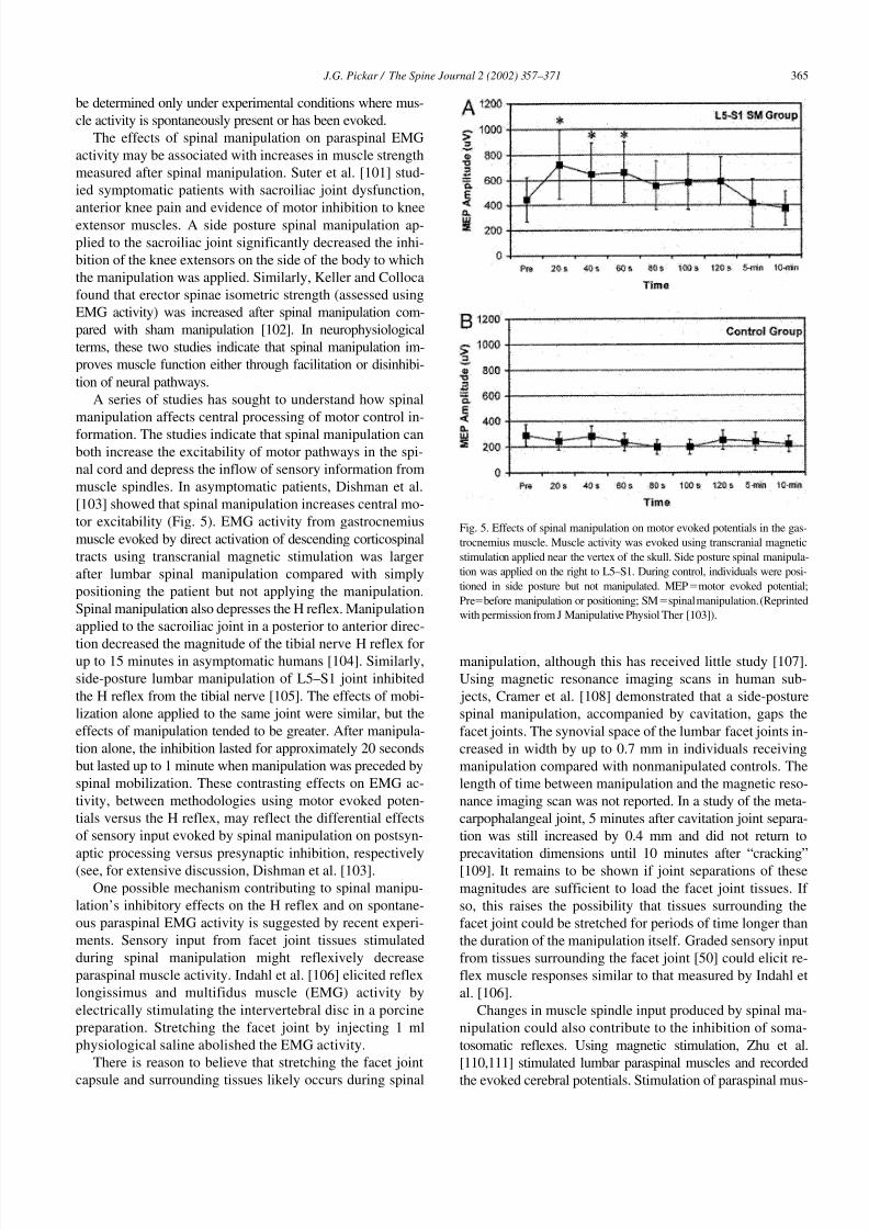
[103] showed that spinal manipulation increases central mo-
tor excitability (Fig. 5). EMG activity from gastrocnemius
muscle evoked by direct activation of descending corticospinal
tracts using transcranial magnetic stimulation was larger
after lumbar spinal manipulation compared with simply
positioning the patient but not applying the manipulation.
Spinal manipulation also depresses the H reflex. Manipulation
applied to the sacroiliac joint in a posterior to anterior direc-
tion decreased the magnitude of the tibial nerve H reflex forup to 15 minutes in asymptomatic humans [104]. Similarly,
side-posture lumbar manipulation of L5–S1 joint inhibited
the H reflex from the tibial nerve [105]. The effects of mobi-
lization alone applied to the same joint were similar, but the
effects of manipulation tended to be greater. After manipula-
tion alone, the inhibition lasted for approximately 20 seconds
but lasted up to 1 minute when manipulation was preceded by
spinal mobilization. These contrasting effects on EMG ac-
tivity, between methodologies using motor evoked poten-
tials versus the H reflex, may reflect the differential effects
of sensory input evoked by spinal manipulation on postsyn-
aptic processing versus presynaptic inhibition, respectively
(see, for extensive discussion, Dishman et al. [103].
One possible mechanism contributing to spinal manipu-
lation’s inhibitory effects on the H reflex and on spontane-
ous paraspinal EMG activity is suggested by recent experi-
ments. Sensory input from facet joint tissues stimulated
during spinal manipulation might reflexively decrease
paraspinal muscle activity. Indahl et al. [106] elicited reflex
longissimus and multifidus muscle (EMG) activity by
electrically stimulating the intervertebral disc in a porcine
preparation. Stretching the facet joint by injecting 1 ml
physiological saline abolished the EMG activity.
There is reason to believe that stretching the facet joint
capsule and surrounding tissues likely occurs during spinal
manipulation, although this has received little study [107].
Using magnetic resonance imaging scans in human sub-
jects, Cramer et al. [108] demonstrated that a side-posture
spinal manipulation, accompanied by cavitation, gaps the
facet joints. The synovial space of the lumbar facet joints in-
creased in width by up to 0.7 mm in individuals receiving
manipulation compared with nonmanipulated controls. The
length of time between manipulation and the magnetic reso-
nance imaging scan was not reported. In a study of the meta-
carpophalangeal joint, 5 minutes after cavitation joint separa-
tion was still increased by 0.4 mm and did not return to
precavitation dimensions until 10 minutes after “cracking”
[109]. It remains to be shown if joint separations of these
magnitudes are sufficient to load the facet joint tissues. If
so, this raises the possibility that tissues surrounding the
facet joint could be stretched for periods of time longer than
the duration of the manipulation itself. Graded sensory input
from tissues surrounding the facet joint [50] could elicit re-
flex muscle responses similar to that measured by Indahl et
al. [106].
Changes in muscle spindle input produced by spinal ma-
nipulation could also contribute to the inhibition of soma-
tosomatic reflexes. Using magnetic stimulation, Zhu et al.
[110,111] stimulated lumbar paraspinal muscles and recorded
the evoked cerebral potentials. Stimulation of paraspinal mus-
Fig. 5. Effects of spinal manipulation on motor evoked potentials in the gas-
trocnemius muscle. Muscle activity was evoked using transcranial magnetic
stimulation applied near the vertex of the skull. Side posture spinal manipula-
tion was applied on the right to L5–S1. During control, individuals were posi-
tioned in side posture but not manipulated. MEPmotor evoked potential;
Prebefore manipulation or positioning; SMspinal manipulation. (Reprinted
with permission from J Manipulative Physiol Ther [103]).

8/2/2019 Neurophysiological Effects of Spinal Manipulation
http://slidepdf.com/reader/full/neurophysiological-effects-of-spinal-manipulation 10/15
366
J.G. Pickar / The Spine Journal 2 (2002) 357–371
cle spindles using vibration reduced the magnitude of the
cerebral potentials. Similarly, muscle spasm in human patients
reduced the magnitude of the paraspinal muscle–evoked cere-
bral potentials. Spinal manipulation reversed these effects,
improving muscle spasm and restoring the magnitude of the
evoked cerebral potentials [111], suggesting that increased
sensory input from paraspinal muscle spindles during mus-
cle spasm may contribute to the reduced magnitude of the
evoked cerebral potentials. It is worthwhile recalling Korr’s
ideas [36] that spinal manipulation increases joint mobility
by producing a barrage of impulses in muscle spindle afferents
and smaller-diameter afferents, ultimately silencing facilitated
motoneurons (see previous section: The effects of spinal ma-
nipulation on sensory neurons innervating paraspinal tissues;
Group I and II afferents [proprioceptive afferents]).
At first it seems counterintuitive that muscle spindle dis-
charge is increased during muscle spasm, because one could
anticipate muscle shortening and spindle unloading during
spasm. However, extensive studies from Proske’s laboratory
(reviewed in [112]) show that a maintained joint position ormaintained muscle shortening, even for short durations, alters
muscle spindle sensitivity to subsequent joint movement or
muscle stretch. For example, from a given muscle length, mus-
cle spindles respond more to a slow stretch when a leg muscle
has previously been held at a shortened length compared with
having been previously held at a long length for as little as 10
seconds [113]. Recently, Pickar and Kang [114] observed the
same phenomenon in the lumbar longissimus and multifidus
muscles (Fig. 6). Muscle spindle activity in response to a
slow vertebral translation that stretched the muscle spindle
depended on whether the muscle had previously been short-
ened for as little as 5 seconds (by linearly displacing the L6
vertebra dorsalward) or had previously been stretched (by lin-
early displacing the L6 vertebra ventralward). If paraspinal
muscle spasm results in muscle shortening, or if segmental
buckling results in muscle shortening ipsilaterally and mus-
cle lengthening contralaterally, then for the same change in
muscle length subsequent stretch or vibration of the affected
muscles would increase spindle discharge more than ex-
pected. Because spinal manipulation has been shown to
stimulate muscle spindles (Fig. 7), spinal manipulation may
normalize spindle biomechanics and return muscle spindle
discharge to normal.
5. The effects of spinal manipulation on
somatovisceral reflexes
A number of animal experiments provide evidence sup-
porting the link between altered paraspinal sensory input
and a somatovisceral change shown in Fig. 1. Sensory input
from paraspinal tissues can evoke visceral reflexes affectingthe sympathetic nervous system and may alter end-organ
function. In general, nonnoxious paraspinal sensory input
appears to have an inhibitory effect on sympathetic outflow,
whereas noxious input appears to have an excitatory effect.
However, insufficient experiments have been conducted to
determine the regional variation of this effect, that is, the
change in sympathetic outflow to different organs. Nonethe-
less, the data are provocative, indicating that neural input
from axial tissues can evoke somatovisceral reflexes.
Sato and Swenson [115] applied a nonnoxious mechani-
cal stimulus to several vertebrae in the thoracic and lumbar
Fig. 6. Paraspinal muscle spindle sensitivity to identical changes in vertebral displacement is determined by the previous short-term history of its muscle’s length. The
receptive field of the muscle spindle was in the lumbar multifidus muscle. Top panel shows the discharge frequency of the muscle spindle afferent recorded from the
L6 dorsal root. Instantaneous discharge frequency was averaged into 25-ms bins. The bottom panel shows the amount the L6 vertebra was displaced during flexion
(negative displacement) and extension (positive displacement). These displacements shortened and stretched the multifidus muscle, respectively, because holding the
L6 vertebra in an extended position (12 to 15 seconds) increased spindle discharge frequency compared with control. Conversely, holding the L6 vertebra in a flexed
position (11 to 15 seconds) decreased spindle discharge frequency compared with control. Spindle discharge was the same at the start of the protocols (control, 0 to 2
seconds). The rapid extensions and flexions at the start of each protocol provided the same initial conditions. (Spindle discharge is not shown during these displace-
ments). Top panel shows the change in muscle spindle sensitivity to the slow vertebral extension (18.5 to 40 seconds) after holding the multifidus muscle in shortened
and stretched positions. Note that vertebral position was held for as little as 5 seconds. (Reprinted with permission from J Neuromusculoskel Sys, Data Trace
Publishing Company [114]).

8/2/2019 Neurophysiological Effects of Spinal Manipulation
http://slidepdf.com/reader/full/neurophysiological-effects-of-spinal-manipulation 11/15
J.G. Pickar / The Spine Journal 2 (2002) 357–371
367
spine of rats by applying a force to the lateral aspects of theirspinous processes. Renal and adrenal sympathetic nerve ac-
tivities were recorded. Because the paraspinal musculature
was removed, the sensory input was derived presumably
from the facet joints, intervertebral discs and/or interverte-
bral ligaments. The mechanical stimulus reflexively decreased
the level of renal and adrenal sympathetic nerve activity by
25% to 40%. The stimuli were short in duration (approxi-
mately 30 seconds), and the responses attenuated rapidly.
The sensory input from the paraspinal tissues had access to
centers at least as high as the upper cervical spinal cord,
because C1–C2 spinal cord transection abolished the inhibi-
tion. Sato and Swenson concluded that nonnoxious mechani-cal stimuli applied to the spine reflexively inhibit the level of
sympathetic nerve activity by means of a supraspinal reflex.
Budgell et al. [116,117] also stimulated paraspinal structures
using noxious and nonnoxious chemical stimuli. Injections
were placed into the lumbar facet joints or lumbar interspinous
tissues. Blood pressure and sciatic nerve blood flow were mea-
sured [116]. A small volume (20 ul) of a nonnoxious chemical
(physiological saline 0.9%) injected into the interspinous
ligament produced a depressor response and a concomitant
decrease in sciatic nerve blood flow. A similar volume of
low-dose capsaicin (2 ug), which activates nociceptive neurons
[118], caused an initial increase in blood pressure and sciatic
nerve blood flow. However, when injected into the facet joint,
capsaicin produced a depressor response. The results from the
interspinous ligament are consistent with the suggestion of-
fered by Sato and Swenson [115] that stimulation of recep-
tive endings sensitive to innocuous mechanical stimuli in
the paraspinal tissues produce inhibitory somaticsympa-
thetic reflexes. The findings from the facet joints suggested
to the authors that capsaicin might more effectively produce
innocuous mechanical changes in the facet joint compared
with the interspinous ligament by increasing the permeabil-
ity of the synovial membrane’s microvasculature. Similar to
the cardiovascular effects produced by capsaicin injection
into the lumbar interspinous ligament, capsaicin injection
into the lumbar interspinous tissues also increased adrenal
sympathetic nerve activity and catecholamine secretion
[117], whereas physiological saline injection had no effect.
Thus, noxious stimulation of paraspinal tissues can produce
excitatory somatic-sympathetic reflexes.
More recently, Pickar et al. [119], in a preliminary re-
port, showed that mustard oil, a nociceptive substance that
also produces inflammation, injected into the lumbar multi-
fidus muscle increases the discharge of sympathetic nerves
to the kidney and spleen. The response is a reflex mediated
by segmental branches of the dorsal ramus and is integrated
by centers at least as high as the upper cervical spinal cord.
This reflex organization is similar to that found by Sato and
Swenson [115] for the sympathetic nerves to the kidney and
adrenal gland. Interestingly, animal studies have also shown
that increased splenic sympathetic nerve discharge is immu-
nosuppressive, decreasing the number of natural killer cells
released. Somatovisceral reflex stimulation of the sympa-
thetic outflow to the spleen may contribute to the depressedlevels of natural killer cells measured in individuals with
low back pain [120].
Mechanical stimulation of paraspinal tissues can be suf-
ficient to inhibit gastric motility. Myoelectric activity from
the wall of the gastrointestinal tract in conscious rabbits was
decreased by sustained (2.5 minutes) mechanical inputs
[121]. In these experiments, it was unclear if the mechanical
stimulation was noxious or innocuous, but the inhibition of
gastric motility was greatest when the mechanical stimulation
was applied to the sixth thoracic vertebra, and it decreased as
the mechanical stimulation was applied further cranial or cau-
dal. These results were confirmed by Budgell and Suzuki[122]. Noxious chemical stimulation inhibited gastric motil-
ity, and the effect tended to be greater when the stimulus was
applied to the mid-thoracic region compared with the lumbar
region. In addition, the inhibitory response was shown to be
a reflex predominated by changes in sympathetic outflow
and to a lesser extent vagal outflow.
It is important to note that these studies do not provide evi-
dence for the unique potential of paraspinal tissues to elicit so-
matosympathetic reflexes. Substantial evidence shows that
noxious stimulation of tissues in the appendicular skeleton also
evokes somatosympathetic reflexes [123], but nothing is
known about the relative magnitudes of somatosympathetic re-
flexes elicited by axial versus appendicular tissues. Although
the data on gastric motility suggest segmental specificity, it is
not certain the degree to which segmental input from paraspi-
nal tissues produce regionally specific changes in sympathetic
nerve activity.
Very few laboratory or clinically oriented basic science
studies have been conducted to determine the effects of spi-
nal manipulation on the sympathetic nervous system. Re-
cently, Budgell and Hirano [124] measured changes in heart
rate variability after upper cervical versus sham spinal ma-
nipulation. Power spectral analysis of heart rate variability
showed that manipulation increased the ratio of low fre-
Fig. 7. Original tracing of a muscle spindle’s response to a spinal manipu-
lative-like load. The single unit activity was obtained from a muscle spin-
dle afferent in the L6 dorsal root. The muscle spindle was located in the
lumbar paraspinal muscles. Inset shows the spindle’s discharge on an
expanded time scale immediately before, during and shortly after the
impulse. (From [47] Reprinted with permission.)

8/2/2019 Neurophysiological Effects of Spinal Manipulation
http://slidepdf.com/reader/full/neurophysiological-effects-of-spinal-manipulation 12/15
368
J.G. Pickar / The Spine Journal 2 (2002) 357–371
quency to high frequency components indicating a possible
shift in the balance of autonomic control of the heart toward
the parasympathetic nervous system.
Spinal manipulation may alter the response of immunologic
cells as well as the production of immunomodulatory and neu-
romodulatory cytokines. In a series of studies on human sub- jects in the 1990s, Brennan et al. [120,125,126] showed that
spinal manipulation but not sham manipulation nor soft tissue
massage primed polymorphonuclear leukocytes (PMNs) and
monocytes. Spinal manipulation enhanced the respiratory
burst (a marker for phagocytic activity) of these white blood
cells to a particulate challenge. The mechanism is unclear,
although speculation on the role of substance P was discussed.
Spinal manipulation also primed the polymorphonuclear leu-
kocytes for enhanced production of cytokines as determined by
the release of tumor necrosis factor in response to endotoxin
challenge. The priming effect was short lived, being greater
15 minutes after manipulation compared with 30 and 45 min-utes. The biological consequence of these changes have yet to
be investigated, but their changes suggested their potential use,
at least, as markers of successful spinal manipulation.
Conclusion
A theoretical framework has been presented for under-
standing the neurophysiological effects of spinal manipula-
tion. The reasons underlying the biomechanical changes in
the vertebral column are hypothesized to affect neural input,
subsequently altering central processing and affecting reflex
somatomotor or somatovisceral output. Table 2 summarizes
the evidence for the theoretical relationships presented in this
review. Spinal manipulation evokes changes in the neuromus-
culoskeletal system. The experimental evidence indicates that
the impulse load of a spinal manipulation impacts propriocep-
tive primary afferent neurons from paraspinal tissues. In addi-
tion, spinal manipulation can affect pain processing, possibly
by altering the central facilitated state of the spinal cord, and
can affect the motor control system. Animal experiments show
that sensory input from paraspinal tissues has the capacity to
reflexively alter the neural outflow to the autonomic nervous
system. However, the effects of spinal manipulation on the
autonomic nervous system are less well investigated The
neurophysiological evidence demonstrating physiological
effects produced by spinal manipulation is growing. More
than one mechanism likely explains the effects of spinal
manipulation. During the past 10 to 20 years, novel experi-
mental approaches have been developed to investigate both
the effects of and the mechanisms underlying spinal manip-ulation. Neurophysiological studies of the spine using ani-
mal models are difficult, if for no other reason than the
paraspinal tissues of interest directly overlie the central ner-
vous system and the distances between paraspinal tissues
and the spinal cord are short. Several experimental models
have offered solutions to this difficulty. Continued work in
this area will help us better understand the therapeutic
mechanisms impacted through spinal manipulation.
References
[1] Druss BG, Rosenheck RA. Association between use of uncoven-tional therapies and conventional medical services. JAMA 1999;
282:651–6.
[2] Altman B, Lynn M. Use of alternative care providers by the adult
population: utilization patterns and expenditures. Workshop presen-
tation. National Center for Complementary and Alternative Medi-
cine, June 14, 2000.
[3] Eisenberg DM, Davis RB, Ettner SL, et al. Trends in alternative
medicine use in the United States, 1990–1997: results of a follow-up
national survery. JAMA 2002;280:1569–75.
[4] Bigos S, Bowyer O, Braen G, et al. Acute low-back problems in
adults. AHCPR Publication No. 95-0643. Rockville, MD, US Dept
of Health and Human Services, Public Health Service, Agency for
Health Care and Policy and Research. Clinical Practice Guideline,
Quick Reference Guide Number 14, 1994.
[5] Hurwitz EL, Aker PD, Adams AH, Meeker WC, Shekelle PG. Ma-nipulation and mobilization of the cervical spine: a systematic re-
view of the literature. Spine 1996;21(15):1746–60.
[6] Budgell B. Spinal manipulative therapy and visceral disorders. Chi-
ropractic J Austral 1999;29:123–8.
[7] Bronfort G, Assendelft WJ, Evans R, Haas M, Bouter L. Efficacy of
spinal manipulation for chronic headache: a systematic review. J
Manipulative Physiol Ther 2001;24:457–66.
[8] Masarsky CS, Todres-Masarsky C. Somatovisceral aspects of chiro-
practic: an evidence-based approach. New York: Churchill-Living-
stone, 2001.
[9] Shekelle PG, Adams AH, Chassin MR, Hurwitz EL, Brook RH.
Spinal manipulation for low-back pain. Ann Intern Med 1992;117:
590–8.
[10] Grice A, Vernon H. Basic principles in the performance of chiro-
Table 2
Current evidence for the neurophysiological mechanisms underlying the effects of spinal manipulation
Mechanism
Current evidence
In favor Negative Unknown
Alters Group Ia and Group II mechanoreceptor discharge X
Alters Group III and Group IV mechanoreceptor or chemoreceptor discharge X
Alters mechanical environment of the IVF X
Alters chemical environment of the IVF XInfluences sensory processing in the spinal cord (ie, central facilitation) X
Affects neuroendocrine system X X
Impacts control of skeletal muscle reflexes (ie, somatosomatic reflexes) X
Impacts control of autonomic reflexes (ie, somatovisceral reflexes) X
IVF
intervertebral foramen.

8/2/2019 Neurophysiological Effects of Spinal Manipulation
http://slidepdf.com/reader/full/neurophysiological-effects-of-spinal-manipulation 13/15
J.G. Pickar / The Spine Journal 2 (2002) 357–371
369
practic adjusting: historical review, classification, and objectives.
In: Haldeman S, editor. Principles and practice of chiropractic, 2nd
ed. Norwalk: Appleton and Lange, 1992:443–58.
[11] Bergmann TF. Short lever, specific contact articular chiropractic
technique. J Manipulative Physiol Ther 1992;15:591–5.
[12] Bartol KM. Osseous manual thrust techniques. In: Gatterman MI,
editor. Foundations of chiropractic, 1st ed. St. Louis: Mosby, 1995:
88–104.
[13] Conway PJW, Herzog W, Zhang Y, Hasler EM, Ladly K. Forces re-quired to cause cavitation during spinal manipulation of the thoracic
spine. Clin Biomech 1993;8:210–4.
[14] Brodeur R. The audible release associated with joint manipulation. J
Manipulative Physiol Ther 1995;18:155–64.
[15] Haldeman S. Spinal manipulative therapy; a status report. Clin Or-
thop 1983;179:62–70.
[16] Hessel BW, Herzog W, Conway PJW, McEwen MC. Experimental
measurement of the force exerted during spinal manipulation using
the Thompson technique. J Manipulative Physiol Ther 1990;13:
448–53.
[17] Herzog W, Conway PJ, Kawchuk GN, Zhang Y, Hasler EM. Forces
exerted during spinal manipulative therapy. Spine 1993;18:1206–12.
[18] Kawchuk GN, Herzog W, Hasler EM. Forces generated during spi-
nal manipulative therapy of the cervical spine: a pilot study. J Ma-
nipulative Physiol Ther 1992;15:275–8.
[19] Kawchuk GN, Herzog W. Biomechanical characterization (finger-
printing) of five novel methods of cervical spine manipulation. J
Manipulative Physiol Ther 1993;16:573–7.
[20] Triano J, Schultz AB. Loads transmitted during lumbosacral spinal
manipulative therapy. Spine 1997;22:1955–64.
[21] Nathan M, Keller TS. Measurement and analysis of the in vivo pos-
teroanterior impulse response of the human thoracolumbar spine: a
feasibility study. J Manipulative Physiol Ther 1994;17:431–41.
[22] Fuhr AW, Smith DC. Accuracy of piezoelectric accelerometers
measuring displacement of a spinal adjusting instrument. J Manipu-
lative Physiol Ther 1986;9:15–21.
[23] Smith DB, Fuhr AW, Davis BP. Skin accelerometer displacement
and relative bone movement of adjacent vertebrae in response to
chiropractic percussion thrusts. J Manipulative Physiol Ther 1989;
12:26–37.
[24] Gal J, Herzog W, Kawchuk G, Conway P, Zhang YT. Biomechani-
cal studies of spinal manipulative therapy (SMT): quantifying the
movements of vertebral bodies during SMT. J CCA 1994;38:11–24.
[25] Leach RA. The chiropractic theories, 3rd ed. Baltimore: Williams
and Wilkins, 1994.
[26] Gatterman MI. What’s in a word? In: Gatterman MI, editor. Foun-
dations of chiropractic, 1st ed. St. Louis: Mosby, 1995:6–17.
[27] Triano J. Interaction of spinal biomechanics and physiology. In:
Anonymous principles and practice of chiropractic, 2nd ed. Nor-
walk: Appleton and Lange, 1992:225–57.
[28] Gillette RG. A speculative argument for the coactivation of diverse
somatic receptor populations by forceful chiropractic adjustments.
Manual Med 1987;3:1–14.
[29] Farfan HF. The scientific basis of manipulation procedures. In:Buchanan WW, Kahn MF, Laine V, Rodnan GP, Scott JT, Zvaifler
NJ, Grahame R, editors. Clinics in rheumatic diseases. London: WB
Saunders Company, Ltd., 1980:159–77.
[30] Giles LGF. Anatomical basis of low back pain. Baltimore: Williams
and Wilkins, 1989.
[31] Lewit K. Manipulative therapy in rehabilitation of the locomotor
system. Oxford: Butterworth-Heinemann, 1991.
[32] Haldeman S. The clinical basis for discussion of mechanisms of ma-
nipulative therapy. In: Korr IM, editor. The neurobiologic mecha-
nisms in manipulative therapy. New York: Plenum, 1978:53–75.
[33] Vernon H. Biological rationale for possible benefits of spinal ma-
nipulation. Cherkin DC, Mootz RD. AHCPR Publication No. 98-
N002, 105–115. 1997. Chiropractic in the United States: training,
practice and research.
[34] Wilder DG, Pope MH, Frymoyer JW. The biomechanics of lumbar
disc herniation and the effect of overload and instability. J Spinal
Disord 1988;1:16–32.
[35] Triano J. The mechanics of spinal manipulation. In: Herzog W, edi-
tor. Clinical biomechanics of spinal manipulation. New York:
Churchill Livingstone, 2001:92–190.
[35a] Greenman PE. Principles of Manual Medicine. Baltimore: Williams
and Wilkins, 1989, p 4.
[36] Korr IM. Proprioceptors and somatic dysfunction. J Am OsteopathAssoc 1975;74:638–50.
[37] Whittingham W, Nilsson N. Active range of motion in the cervical
spine increases after spinal manipulation (toggle recoil). J Manipu-
lative Physiol Ther 2001;24:552–5.
[38] Eldred E, Hutton RS, Smith JL. Nature of the persisting changes in
afferent discharge from muscle following its contraction. In:
Homma S, editor. Understanding the stretch reflex. New York:
Elsevier Scientific Publishing Company, 1976:157–83.
[39] Buerger AA. Experimental neuromuscular models of spinal manual
techniques. Manual Med 1983;1:10–7.
[40] Brumagne S, Cordo P, Lysens R, Verschueren S, Swinnen S. The
role of paraspinal muscle spindles in lumbosacral position sense in
individuals with and without low back pain. Spine 2000;25:989–94.
[41] Brumagne S, Lysens R, Swinnen S, Verschueren S. Effect of
paraspinal muscle vibration on position sense of the lumbosacral
spine. Spine 1999;24:1328–31.
[42] Wilder DG, Aleksiev AR, Magnusson ML, Pope MH, Spratt KF,
Goel VK. Muscular response to sudden load. A tool to evaluate fa-
tigue and rehabilitation. Spine 1996;21:2628–39.
[43] Radebold A, Cholewicki J, Panjabi MM, Patel TCh. Muscle re-
sponse pattern to sudden trunk loading in healthy individuals and in
patients with chronic low back pain. Spine 2000;25:947–54.
[44] Radebold A, Cholewicki J, Polzhofer GK, Greene HS. Impaired
postural control of the lumbar spine is associated with delayed mus-
cle response times in patients with chronic idiopathic low back pain.
Spine 2001;26:724–30.
[45] Bolton PS, Holland CT. An in vivo method for studying afferent fi-
bre activity from cervical paravertebral tissue during vertebral mo-
tion in anaesthetised cats. J Neurosci Methods 1998;85:211–8.
[46] Pickar JG. An in vivo preparation for investigating neural responses
to controlled loading of a lumbar vertebra in the anesthetized cat. J
Neurosci Methods 1999;89:87–96.
[47] Pickar JG, Wheeler JD. Response of muscle proprioceptors to spinal
manipulative-like loads in the anesthetized cat. J Manipulative
Physiol Ther 2001;24:2–11.
[48] Colloca CJ, Keller TS, Gunzburg R, Vandeputte K, Fuhr AW. Neu-
rophysiologic response to intraoperative lumbosacral spinal manip-
ulation. J Manipulative Physiol Ther 2000;23:447–56.
[49] Cavanaugh JM, El-Bohy A, Hardy WN, Getchell TV, Getchell ML,
King AI. Sensory innervation of soft tissues of the lumbar spine in
the rat. J Orthop Res 1989;7:378–88.
[50] Pickar JG, McLain RF. Responses of mechanosensitive afferents to
manipulation of the lumbar facet in the cat. Spine 1995;20:2379–85.
[51] Yamashita T, Cavanaugh JM, Ozaktay CA, Avramov AI, King AI.Effect of substance P on mechanosensitive units of tissues around
and in the lumbar facet joint. J Orthop Res 1993;11:205–14.
[52] Abrahams VC, Lynn B, Richmond FJR. Organization and sensory
properties of small myelinated fibers in the dorsal cervical rami of
the cat. J Physiol 1984;347:177–87.
[53] Beel JA, Stodieck LS, Luttges MW. Structural properties of spinal
nerve roots: biomechanics. Exp Neurol 1986;91:30–40.
[54] Berthold CH, Carlstedt T, Corneliuson O. Anatomy of the nerve
root at the central-peripheral transitional region. In: Dyck PJ, editor.
Peripheral neuropathy, 1st ed. Philadelphia: WB Saunders Com-
pany, 1984:156–70.
[55] Thomas PK, Berthold CH, Ochoa J. Microscopic anatomy of the pe-
ripheral nervous system. In: Dyck PJ, editor. Peripheral neuropathy,
1st ed. Philadelphia: WB Saunders Company, 1993:28–91.

8/2/2019 Neurophysiological Effects of Spinal Manipulation
http://slidepdf.com/reader/full/neurophysiological-effects-of-spinal-manipulation 14/15
370
J.G. Pickar / The Spine Journal 2 (2002) 357–371
[56] Devor M, Obermayer M. Membrane differentiation in rat dorsal root
ganglia and possible consequences for back pain. Neurosci Letters
1984;51:341–6.
[57] Rydevik BL. The effects of compression on the physiology of nerve
roots. J Manipulative Physiol Ther 1992;15:62–6.
[58] Howe JF, Loeser JD, Calvin WH. Mechanosensitivity of dorsal root
ganglia and chronically injured axons:a physiological basis for the
radicular pain of nerve root compression. Pain 1977;3:27–41.
[59] Wall PD, Devor M. Sensory afferent impulses originate from dorsalroot ganglia as well as from the periphery in normal and nerve in-
jured rats. Pain 1983;17:321–39.
[60] Sharpless SK. Susceptibility of spinal roots to compression block.
Goldstein M. (No. 15), 155–161. 1975. NINCDS-Research Status of
Spinal Manipulation Therapy.
[61] Ozaktay AC, Kallakuri S, Cavanaugh JM. Phospholipase A
2
sensi-
tivity of the dorsal root and dorsal root ganglion. Spine 1998;23:
1297–306.
[62] Olmarker K, Rydevik B, Hansson T, Holm S. Compression-induced
changes of the nutritional supply to the porcine cauda equina. J Spi-
nal Disord 1990;3:25–9.
[63] Cornefjord M, Olmarker K, Farley DB, Weinstein JN, Rydevik B.
Neuropeptide changes in compressed spinal nerve roots. Spine
1995;20:670–3.
[64] Rydevik BL, Myers RR, Powell HC. Pressure increase in the dorsal
root ganglion following mechanical compression. Closed compart-
ment syndrome in nerve roots. Spine 1989;14:574–6.
[65] Takahashi K, Shima I, Porter RW. Nerve root pressure in lumbar
disc herniation. Spine 1999;24:2003–6.
[66] McCarron RF, Wimpee MW, Hudkins PG, Laros GS. The inflam-
matory effect of nucleus pulposus. A possible element in the patho-
genesis of low-back pain. Spine 1987;12:760–4.
[67] Kawakami M, Tamaki T, Hayashi N, et al. Mechanical compression
of the lumber nerve root alters pain-related behaviors induced by the
nucleus pulposus in the rat. J Orthop Res 2000;18:257–64.
[68] Yabuki S, Igarashi T, Kikuchi S. Application of nucleus pulposus to
the nerve root simultaneously reduces blood flow in dorsal root gan-
glion and corresponding hindpaw in the rat. Spine 2000;25:1471–6.
[69] Nygaard OP, Mellgren SI, Osterud B. The inflammatory properties
of contained and noncontained lumbar disc herniation. Spine 1997;
22:2484–8.
[70] Chen C, Cavanaugh JM, Ozaktay AC, Kallakuri S, King AI. Effects
of phospholipase A
2
on lumbar nerve root structure and function.
Spine 1997;22:1057–64.
[71] Floman Y, Liram N, Gilai AN. Spinal manipulation results in imme-
diate H-reflex changes in patients with unilateral disc herniation.
Eur Spine J 1997;6:398–401.
[72] Stern PJ, Côte P, Cassidy JD. A series of consecutive cases of low
back pain with radiating leg pain treated by chiropractors. J Manipu-
lative Physiol Ther 1995;18:335–42.
[73] Nwuga VC. Relative therapeutic efficacy of vertebral manipulation
and conventional treatment in back pain management. Am J Phys
Med 1982;61:273–8.
[74] Burton AK, Tillotson KM, Cleary J. Single-blind randomised con-trolled trial of chemonucleolysis and manipulation in the treatment
of symptomatic lumbar disc herniation. Eur Spine J 2000;9:202–7.
[75] Cook AJ, Woolf CJ, Wall PD, McMahon SB. Dynamic receptive
field plasticity in rat spinal cord dorsal horn following C-primary af-
ferent input. Nature 1987;325:151–3.
[76] Denslow JS, Korr IM, Krems AD. Quantitative studies of chronic
facilitation in human motoneuron pools. Am J Physiol 1947;150:
229–38.
[77] Denslow JS, Clough GH. Reflex activity in the spinal extensors. J
Neurophysiol 1941;4:430–7.
[78] Denslow JS, Hassett CC. The central excitatory state associated
with postural abnormalities. J Neurophysiol 1942;5:393–401.
[79] Denslow JS. An analysis of the variability of spinal reflex thresh-
olds. J Neurophysiol 1944;7:207–15.
[80] Woolf CJ. The dorsal horn: state-dependent sensory processing and
the generation of pain. In: Wall PD, Melzack R, editors. Textbook
of pain, 3rd ed. Edinburgh: Churchill Livingstone, 1994:101–12.
[81] Melzack R, Wall PD. Pain mechanisms: a new theory. Science
1965;150:971–9.
[82] Besson J-M, Chaouch A. Peripheral and spinal mechanisms of noci-
ception. Physiol Rev 1987;67(1):67–186.
[83] Glover JR, Morris JG, Khosla T. Back pain: a randomized clinical
trial of rotational manipulation of the trunk. Br J Indust Med 1974;31:59–64.
[84] Terrett ACJ, Vernon HT. Manipulation and pain tolerance: a con-
trolled study of the effect of spinal manipulation on paraspinal cuta-
neous pain tolerance levels. Am J Phys Med 1984;63:217–25.
[85] Fischer AA. Pressure algometry over normal muscles. Standard val-
ues, validity and reproduceability of pressure threshold. Pain 1986;
30:115–26.
[86] Reeves JL, Jaeger B, Graff-Radford SB. Reliability of the pressure
algometer as a measure of myofascial trigger point sensitivity. Pain
1986;24:313–21.
[87] Vernon HT. Pressure pain threshold evaluation of the effect of spi-
nal manipulation on chronic neck pain: a single case study. J CCA
1988;32:191–4.
[88] Cote P, Silvano AM, Vernon H, Mior SA. The short-term effect of a
spinal manipulation on pain/pressure threshold in patients with
chronic mechanical low back pain. J Manipulative Physiol Ther
1994;17:364–8.
[89] Fields HL, Basbaum AI. Central nervous system mechanisms of
pain modulation. In: Wall PD, Melzack R, editors. Textbook of
pain, 3rd ed. Edinburgh: Churchill Livingstone, 1994:101–12.
[90] Szczudlik A, Lypka A. Plasma immunoreactive beta-endorphin and
enkephalin concentration in healthy subjects before and after elec-
troacupuncture. Acupunct Electro-Ther Res 1983;8:127–37.
[91] Hughes GSJ, Lichstein PR, Whitlock D, Harker C. Response of
plasma beta-endorphins to trancutaneous electrical nerve stimula-
tion in healthy subjects. Phys Ther 1984;64:1062–6.
[92] Harber VJ, Sutton JR. Endorphins and exercise. Sports Med 1984;1:
154–71.
[93] Rosner A. Endocrine disorders. In: Masarsky CS, Todres-Masarsky
C, editors. Somatovisceral aspects of chiropractic: an evidence-
based approach. New York: Churchill-Livingstone, 2001:187–202.
[94] Vernon HT, Dhami MSI, Howley TP, Annett R. Spinal manipula-
tion and beta-endorphin: a controlled study of the effect of a spinal
manipulation on plasma beta-endorphin levels in normal males. J
Manipulative Physiol Ther 1986;9:115–23.
[95] Christian GF, Stanton GJ, Sissons D, et al. Immunoreactive acth,
b-endorphin, and cortisol levels in plasma following spinal manipu-
lative therapy. Spine 1988;13:1411–7.
[96] Sanders GE, Reinert O, Tepe R, Maloney P. Chiropractic adjustive
manipulation on subjects with acute low back pain: visual analog
pain scores and plasma b-endorphin levels. J Manipulative Physiol
Ther 1990;13:391–5.
[97] Clement-Jones V, Lowry PJ, Rees LH, Besser GM. Development
of a specific extracted radioimmunoassay for methionine enkepha-lin in human plasma and cerebrospinal fluid. J Endocrinol 1980;86:
231–43.
[98] Herzog W, Scheele D, Conway PJ. Electromyographic responses of
back and limb muscles associated with spinal manipulative therapy.
Spine 1999;24:146–53.
[99] Suter E, Herzog W, Conway PJ, Zhang YT. Reflex response associ-
ated with manipulative treatment of the thoracic spine. J Neuromus-
culoskel Sys 1994;2:124–30.
[100] Colloca CJ, Keller TS. Stiffness and neuromuscular reflex response
of the human spine to posteroanterior manipulative thrusts in pa-
tients with low back pain. J Manipulative Physiol Ther 2001;24:
489–500.
[101] Suter E, McMorland G, Herzog W, Bray R. Conservative lower
back treatment reduces inhibition in knee-extensor muscles: a ran-

8/2/2019 Neurophysiological Effects of Spinal Manipulation
http://slidepdf.com/reader/full/neurophysiological-effects-of-spinal-manipulation 15/15
J.G. Pickar / The Spine Journal 2 (2002) 357–371
371
domized controlled trial. J Manipulative Physiol Ther 2000;23:
76–80.
[102] Keller TS, Colloca CJ. Mechanical force spinal manipulation in-
creases trunk muscle strength assessed by electromyography: a
comparative clinical trial. J Manipulative Physiol Ther 2000;23:
585–95.
[103] Dishman JD, Ball KA, Burke J. Central motor excitability changes
after spinal manipulation, a transcranial magnetic stimulation study.
J Manipulative Physiol Ther 2002;25:1–9.[104] Murphy BA, Dawson NJ, Slack JR. Sacroiliac joint manipulation
decreases the H-reflex. Electromyog Clin Neurophysiol 1995;35:
87–94.
[105] Dishman JD, Bulbulian R. Spinal reflex attenuation associated with
spinal manipulation. Spine 2000;25:2519–25.
[106] Indahl A, Kaigle AM, Reikeras O, Holm SH. Interaction between
the porcine lumbar intervertebral disc, zygapophysial joints, and
paraspinal muscles. Spine 1997;22:2834–40.
[107] El-Bohy AA, Goldberg SJ, King AI. Measurement of facet capsular
stretch [abstract]. Biomechanics Symposium 1987; American Soci-
ety of Mechanical Engineers: ASME, NY, NY; 161–4.
[108] Cramer GD, Tuck NR, Knudsen JT, et al. Effects of side-posture
positioning and side-posture adjusting on the lumbar zygapophyseal
joints as evaluated by magnetic resonance imaging: a before and af-
ter study with randomization. J Manipulative Physiol Ther 2000;23:
380–94.
[109] Unsworth A, Dowson D, Wright V. “Cracking joints.” Ann Rheum
Dis 1971;30:348–58.
[110] Zhu Y, Haldeman S, Starr A, Seffinger MA, Su S. Paraspinal mus-
cle evoked cerebral potentials in patients with unilateral low back
pain. Spine 1993;18:1096–102.
[111] Zhu Y, Haldeman S, Hsieh C-YJ, Wu P, Starr A. Do cerebral poten-
tials to magnetic stimulation of paraspinal muscles reflect changes
in palpable muscle spasm, low back pain, and activity scores? J Ma-
nipulative Physiol Ther 2000;23:458–64.
[112] Proske U, Morgan DL, Gregory JE. Thixotropy in skeletal muscle
and in muscle spindles: a review. Prog Neurobiol 1993;41:705–21.
[113] Gregory JE, Mark RF, Morgan DL, Patak A, Polus B, Proske U. Ef-
fects of muscle history on the stretch reflex in cat and man. J Neuro-
physiol 1990;424:93–107.
[114] Pickar JG, Kang YM. Short-lasting zstretch of lumbar paraspinal
muscle decreases muscle spindle sensitivity to subsequent musclez
stretch. J Neuromusculoskel Sys 2001;9:88–96.
[115] Sato A, Swenson RS. Sympathetic nervous system response to me-
chanical stress of the spinal column in rats. J Manipulative Physiol
Ther 1984;7:141–7.
[116] Budgell B, Hotta H, Sato A. Spinovisceral reflexes evoked by nox-
ious and innocuous stimulation of the lumbar spine. J Neuromuscu-
loskel Sys 1995;3:122–30.
[117] Budgell B, Sato A, Suzuki A, Uchida S. Responses of adrenal func-
tion to stimulation of lumbar and thoracic interspinous tissues in the
rat. Neurosci Res 1997;28:33–40.[118] Fitzgerald M. Capsaicin and sensory neurons—a review. Pain 1983;
15:109–30.
[119] Pickar JG, Kang YM, Cobb T, Kenney MJ. Mustard oil injected into
lumbar multifidus muscle increases sympathetic nerve activity to
spleen and kidney via a suprasegmental reflex [abstract]. Soc Neu-
rosci Abstr 2001;27(818):9.
[120] Brennan PC, Graham MA, Triano JJ, Hondras MA, Anderson RJ.
Lymphocyte profiles in patients with chronic low back pain enrolled
in a clinical trial. J Manipulative Physiol Ther 1994;17:219–27.
[121] Deboer KF, Schutz M, McKnight ME. Acute effects of spinal ma-
nipulation on gastrointestinal myoelectric activity in conscious rab-
bits. Manual Med 1988;3:85–94.
[122] Budgell B, Suzuki A. Inhibition of gastric motility by noxious
chemical stimulation of interspinous tissues in the rat. J Autonom
Nerv Sys 2000;80:162–8.
[123] Sato A, Sato Y, Schmidt RF. The impact of somatosensory input on
autonomic functions. In: Blaustein MP, Grunicke H, Pette D,
Schultz G, Schweiger M, editors. Reviews of physiology, biochem-
istry and pharmacology. Tokyo: Springer, 1997.
[124] Budgell B, Hirano F. Innocuous mechanical stimulation of the neck
and alterations in heart-rate variability in healthy young adults. Au-
ton Neurosci Basic Clin 2001;91:96–9.
[125] Brennan PC, Kokjohn K, Kaltinger CJ, et al. Enhanced phagocytic
cell respiratory burst induced by spinal manipulation: potential role
of substance P. J Manipulative Physiol Ther 1991;14:399–407.
[126] Brennan PC, Triano JJ, McGregor M, Kokjohn K, Hondras MA,
Brennan DC. Enhanced neutrophil respiratory burst as a biological
marker for manipulation forces: duration of the effect and associa-
tion with substance P and tumor necrosis factor. J Manipulative
Physiol Ther 1992;15:83–9.
[127] Habler H-J, Eschenfelder S, Liu X-G, Janig W. Sympathetic-sensory
coupling after L5 spinal nerve lesion in the rat and its relation to
changes in dorsal root ganglion blood flow. Pain 2000;87:335–45.