Embed Size (px)
Citation preview
W0.001.08N
Neuropsychology
Workshop
LEUVEN 18-19 November 2010

This workshop is supported by research grants from the Fund for Scientific Research (FWO Flanders, WO.001.08N) and from the Methusalem program grant by the Flemish Government awarded to Johan Wagemans (METH/08/02).

1
Preface
Welcome to our Neuropsychology Workshop in Leuven!
Neuropsychology in the original sense deals with the understanding of brain-behaviour relationships
through the study of brain damage. It has a clinical component, focused on the diagnosis and
rehabilitation of individual patients with brain damage, as well as a cognitive, research-oriented
component, testing theories about brain-behaviour relationships by applying specific experimental
paradigms to groups of patients with similar pathologies or sometimes individual cases with unique,
focal lesions. In this sense neuropsychology is one of many approaches to study brain-behaviour
relationships, complementary to psychophysics, experimental psychology, brain imaging, modeling,
etc. – and collectively constituting the interdisciplinary field of affective, behavioural or cognitive
neuroscience. While this broad domain is currently booming in Flanders, as anywhere else in the
world, the narrower domain of neuropsychology in the original sense has lagged behind somewhat,
especially in comparison to its role in the French-speaking part of Belgium, The Netherlands, the
United Kingdom, Germany, France, Italy, etc. At the same time, the clinical field of neuropsychology
is gaining much interest, also among students and practitioners in clinical psychology, and more
cognitively oriented research has continued to evolve significantly with the more widely applied
usage of novel techniques, more elaborated theories, etc. We are very pleased, therefore, that we
are able to organize this two-day workshop in Leuven to inform all those who are interested in these
new developments, with the generous support from FWO-Flanders, in the context of a Scientific
Research Network, involving all experimental psychology research groups in Flanders (Brussels,
Ghent, Leuven), and centered on the linkage with the cognitive neurosciences. We are also very
pleased that all of these groups and several of its associated partners beyond Flanders are actively
participating in this workshop.
The first day of this workshop seeks to show case some of the most critical methodological issues and
techniques in neuropsychology. From a historical review of single case studies to an exploration of
voxel-based lesion mapping techniques of group studies, this day will bring us up to date on some of
the most important tools and techniques employed within neuropsychology.
The second day of this workshop will see these methods and techniques applied to critical questions
in the study of visual perception from the neural underpinnings of consciousness and attention to
the study of the human brains unparalleled ability to recognize objects and faces.
From our invited speakers to our general audience this workshop brings together some of the
world’s most respected researchers in the field of neuropsychology. We hope this workshop will
provide a fruitful format to advance our understanding of what brain damage can tell us about brain
function. And we hope it will inspire some of you to seek collaboration with some of these experts to
foster this field further in Flanders too!
Sincerely,
Lee de-Wit, Kathleen Vancleef, and Johan Wagemans

2
Program
Thursday, 18 November 2010
METHODS DAY
8:30-9:00 Registration and hanging posters
GEN
ERA
L M
ETH
OD
S
9:00-9:15 Welcome
9:15-10:00 Jane Riddoch: Single case analyses in the 21st century
10:00-10:45 Peter Mariën: The role of cerebello-cerebral diaschisis in the development of the concept ‘cerebellar neurocognition’
10:45-11:15 Coffee break
11:15-11:45 Gilles Pourtois: Intracranial recordings from category-selective regions of the visual cortex during object recognition
11:45-12:30 Hans-Otto Karnath: Principles of modern lesion analysis
12:30-13:00 Lunch
13:00-14:00 Lunch and poster session
NEU
RO
PSY
CH
OLO
GY
AN
D
fMR
I
14:00-14:30 David Milner: Combining evidence from fMRI and visual agnosia to argue for separate form, texture and colour processing channels
14:30-15:00 Glyn Humphreys: Learning about functional interactivity by functional imaging patients
15:00-15:30 Coffee break
15:30-16:15 Rik Vandenberghe: Parcellation of parietal cortex: Convergence between lesion-symptom mapping and mapping of the intact functioning brain
16:15-16:45 Nele Demeyere: Differentiating subitizing and counting: A voxel-based correlational study
16:45-17:15 Poster session

3
Friday, 19 November 2010
VISUAL PERCEPTION DAY 8:30-9:00 Registration and hanging posters
09:00-09:45 David Milner: Is visual processing for action in the dorsal stream accessible to consciousness?
ATTEN
TION
AN
D
CO
NSC
IOU
SNESS
9:45-10:15 Jean-Philippe van Dijck: The role of spatial attention in number processing: Evidence from neglect
10:15-10:45 Coffee break
10:45-11:15 Jane Riddoch: What extinction tells us about perception
11:15-11:45 Céline Gillebert: Lesions restricted to the IPS and SPL impair attentional selection and spatial shifting
11:45-12:15 Gilles Pourtois: The role of the superior colliculus in audio-visual integration
12:15-12:45 Lunch
12:45-13:45 Lunch and poster session
13:45-14:30 Glyn Humphreys: Direct routes in object recognition
OB
JECT A
ND
FAC
E R
ECO
GN
ITION
14:30-15:00 Cathleen Grimsen/Manfred Fahle: Behavioural correlates of impaired object recognition after stroke
15:00-15:30 Coffee break
15:30-16:00 Goedele Van Belle: Whole or hole: Expert face recognition requires holistic perception
16:00-16:30 Sharon Gilaie-Dotan: What can developmental visual integrative agnosia tell us about brain processes?
16:30-17:00 Hans-Otto Karnath: Neural structures involved in visual recognition
17:00-… Reception
4
POSTERS
Lina Aimola1 Igor Schindler2, Annalena Venneri2,3
1Durham University, UK;
2University of Hull, UK;
3San Camillo Hospital, Venice, Italy
Near and far space processing: insight from a single case study of neglect Studies of patients with unilateral neglect showing selective impairment in either near (within reaching) or far (beyond reaching) space suggest that these two spatial areas are coded differently by the brain. It is not clear, however, whether any difference in performance between these spatial domains is a task independent phenomenon or modulated by the requirement for a motor response. A 32 year old right brain damaged patient (MF) and a group of healthy controls (N = 10) were assessed with a serial visual search task and a Landmark line bisection paradigm requiring either a directional or non-directional motor response. In the perceptual version they were asked to make a button press response whereas in the motor version a direct ballistic movement towards the stimulus was required. MF and the controls were assessed with both tasks and response modes in near (57 cm) and far space (114 cm). Compared to the controls, MF showed left neglect in the visual search task only for the perceptual condition in far space [t(11) = 3.54, p < 0.01] and for the motor condition in near space [t(11) = 4.48, p < 0.01]. In contrast MF was not impaired in both versions of the Landmark task irrespective of spatial distance [p > .05]. A morphometric assessment of MF’s brain lesion showed marked damage in the ventro-temporal cortex, Fusiform Gyrus, Superior Temporal Gyrus, Insula, Angular Gyrus and posterior Cingulate of the right hemisphere. The anatomical evidence in MF showed that damage of the ventral stream results in far space neglect only when space is coded through perceptual information (i.e. visual), while the presence of neglect deficits when acting within near space might be associated to parietal and posterior cingulate damage. Overall the findings suggested that there is a complex interaction between task and mode of response underlying dissociations in neglect for near and far space. In addition, the specificity of these space related deficits was crucially related to damage of particular brain structures which could be especially recruited for near or far space coding.
Jutta Billino Justus-Liebig-Universität Giessen, Germany
Ageing as a window to networks for visual motion processing The visual system doubtlessly represents the most important sensory system in humans. During ageing a wide range of visual abilities seems to decline, however, there have also been descriptions of stable or minimally affected functions. Since senescent optics of the eye cannot sufficiently explain functional decline and stability, cortical and subcortical changes appear to determine specific visual resources and vulnerabilities. In order to gain insights into functional correlates of neuronal changes during ageing, we studied processing of visual motion information which involves a network of extrastriate brain areas. Motion perception provides a crucial prerequisite for proper interactions with our environment, for precise motor actions, and for appropriate interpretation of social actions. Although neuronal processing mechanisms underlying visual motion perception are extraordinarily well understood, there are still controversies about observed ageing effects. Using psychophysical methods, we studied perception for different types of motion information in a large sample comprising 123 participants, ranging in age between 18 and 82 years. Motion types differed in their signal complexity, but also in their environmental relevance. We determined detection thresholds by varying signal-to-noise levels and fitting psychometric functions to the data. We found pronounced
5
differences in age-related sensitivity decline for specific motion types. Relative threshold changes over the given age range varied between doubling and stability. Results raise many questions on how differential resources and vulnerabilities can occur within one sensory system. We discuss our findings in context of neurophysiological and imaging data on age-related degradation of neuronal response properties and structural changes. We propose a network of visual subsystems that allows for adaptive processes and plasticity during ageing. This capacity seems especially useful for processing visual information of high complexity.
Julie Debrabant1,2 (18th only) Hilde Van Waelvelde1,2, Guy Vingerhoets1
1Ghent University (UGent), Belgium;
2University College Artevelde Ghent, Belgium
Development of response timing in continuous reaction time performance Procedural learning refers to an implicit process in which a behavioral response is refined through repetitive performance. The neural substrate of this process includes frontostriatal systems that enable the anticipation and precise timing of planned motor responses. Given the evidence of continued development in these systems during childhood, this study explores possible age-related differences in the procedural learning of a timed motor response. Eighty typically developing children in 4 age groups (5-6, 7-8, 9-10 and 11-12 year olds) performed a continuous reaction time task and an explicit timing control task. Speeded responses at temporally regular relative to irregular stimuli were present at the ages of 9-10 and 11-12. Also, by 7-8 years of age, anticipatory responses occurred with a significant increase between the ages of 9-10 and 11-12. These developmental trends were not related to simple motor performance effects and explicit timing measures. Consequently, age-related differences in reaction time indices of response timing suggest a linkage to changes in frontostriatal chronometric systems.
Hyo Jung De Smet1 Peter Mariën2,3,4
1Ghent University (UGent), Belgium;
2Vrije Universiteit Brussel (VUB), Belgium;
3ZNA AZ Middelheim, Antwerp,
Belgium;4 Institute Born-Bunge, Antwerp, Belgium
Cerebello-cerebral diaschisis as possible pathophysiological substrate of cognitive and behavioural symptoms During the past decades neuroanatomical studies convincingly demonstrated cerebellar connectivity with the supratentorial association areas involved in higher cognitive functioning, while functional neuroimaging studies provided evidence of cerebellar activation during a variety of cognitive tasks such as executive functioning, memory, learning, attention, visuo-spatial regulation, language and behavioral-affective modulation. In addition, cerebellar syndromes such as the cerebellar cognitive affective syndrome (CCAS) and the posterior fossa syndrome (PFS) are well-known clinical entities that may occur following cerebellar damage. Although extensive research has substantially broadened the insights in the cognitive and affective role of the cerebellum, the precise nature of the cerebellar contribution to cognitive, linguistic and affective processing is not clear yet. In this contribution, the phenomenon of cerebello-cerebral diaschisis is suggested as a possible functional substrate of cognitive, linguistic and behavioural deficits in patients with cerebellar lesions. Cerebello-cerebral diaschisis reflects the metabolic impact of a cerebellar lesion on a distant, but anatomically and functionally connected supratentorial region.
6
Kris Evers1
Jean Steyaert1, Ilse L.J. Noens1,2, Johan Wagemans1
1University of Leuven (K.U.Leuven), Belgium;
2Massachusetts General Hospital, Boston, MA, USA
No difficulties in extracting subtle emotional cues from social contexts in children with an autism spectrum disorder Background: Children with an autism spectrum disorder (ASD) are known to have difficulties in interpreting social interactions. They are generally found to have problems with processing facial emotional expressions, certainly when dynamic or more complex and naturalistic emotions are used. Since eye-tracking studies revealed atypical viewing behavior in ASD, good performances in some emotion processing tasks could be due to compensatory, more emotionally neutral mechanisms in individuals with ASD. Objectives: We wanted to investigate the ability of children with ASD in extracting subtle emotional cues from a context and reading these expressions from dynamic facial expressions or word labels. Methods: Two groups of 24 boys, individually matched for age (M = 11.79 y, range between 9.38 y and 14.04 y) and full scale IQ (M = 107.96, range between 83.25 and 132.25) were tested. Both groups were matched on group level for verbal IQ en performal IQ. One group had received a clinical diagnosis of ASD based on a multidisciplinary assessment and met DSM-IV-TR PDD criteria. Children with attention deficits or using medication were excluded. The typically developing group was representative for the general population. Each trial consisted of an auditory and visually presented social context (‘story’), eliciting either a subtle or an intense expression of anger or happiness. This story was followed by a test screen, consisting of either two dynamic facial expressions or two word labels (stimulus type). The answer possibilities always consisted of a match and a mismatch item. The mismatch item had either the correct intensity, but incorrect emotion (emotion-mismatch-trial) or the incorrect intensity, but correct emotion (intensity-mismatch-trial). Participants had to indicate how the protagonist in the story felt. Results: No large group differences were found. The ASD group performed more slowly, but not worse than the typically developing group. Task difficulty largely depended on the specific task requirements. As expected, subtle emotions and intensity-mismatch-trials were more difficult than intense emotions and emotion-mismatch-trials. A trend for a three-way-interaction between mismatch, intensity and group was found. Further analysis pointed out that in the subtle trials mismatch only seemed to have an effect in the typically developing group and not in the ASD group. Happiness was responded to more accurately than anger but only in the more difficult subtle trials, not in the intense trials. Dynamic facial expressions were responded to less accurately than word labels. Older children performed better than younger children. Conclusions: We did not find large difficulties in extracting subtle emotions from an auditory and visually presented social context. Task difficulty and group differences depended on the specific task requirements. Slower responses and slightly less influences of task difficulty on performance could indicate rule-based response strategy in the ASD group.
Elfi Goesaert
Marc Van Baelen, Werner Spileers, Johan Wagemans, Hans P. Op de Beeck University of Leuven (K.U.Leuven), Belgium
Preferences to insensible 'phantom' stimuli in low vision patients as measured with fMRI Several forms of retinal degeneration exist, each associated with a characteristic loss of receptors in the retina. For example, in retinitis pigmentosa (RP), input to the peripheral retina is lost, while in macular degeneration (MD), input to the fovea is affected. If the loss of functioning receptors is total, then it is theoretically impossible that stimuli presented at the affected retinal locations would yield any responses anywhere in the brain. Such stimuli should be ‘insensible’. To illustrate this
7
seemingly trivial point, we scanned a juvenile MD (JMD) patient with a standard phase-encoding design, existing of contracting/expanding rings to investigate activations in the primary visual cortex (V1) and the foveal confluence. In addition, we looked at responses in the ventral visual stream, as there is a strong link between central activation and fusiform face area (FFA), and peripheral activation and parahippocampal place area (PPA) (Hasson et al, 2002). Surprisingly, we found that the V1 eccentricity map of the JMD patient was dominated by a preference for (para)foveal stimuli, despite the subject's large central scotoma. At first sight, the term 'phantom' stimuli seems in place here, in analogy to somatosensory experiences associated with a phantom limb - in which case no functioning receptors are left, yet a clear physiological response is present in the central nervous system. However, a comparison of visual responses with a no-stimulus fixation baseline reveals that all seemingly impossible responses to insensible stimuli are due to massive de-activations for the contrasted perceivable stimuli. De-activations throughout most of retinotopic cortex for (sensible) peripheral stimuli explain the counterintuitive findings in the JMD patient. Ventrally, we found a pattern linked to eccentric activation in FFA as well as in PPA for JMD, linked to peripheral input. This is also caused by the lack of central activation, as well as the fact that in normal subjects all ventral regions respond to all stimuli despite their retinal position. E.g., even though FFA normally prefers foveal stimuli, retinotopic maps will reveal a preference for peripheral stimuli if the foveal stimuli are insensible due to receptor dysfunction. The morph and block data used to define FFA and PPA, lastly, matched up nicely with each other. This illustrates the pittfalls when trying to interpret relative preference maps (one condition versus another one). Furthermore, they provide a powerful illustration of the danger of over-interpreting evidence for relative preferences, also termed 'sensitivity' or 'informativeness', as evidence that particular regions are involved in stimulus interpretation - a topic that has received much attention in the context of face representations in high-level visual cortex (see e.g. Haxby et al., 2001; Spiridon & Kanwisher, 2002).
Mijke O. Hartendorp Stefan Van der Stigchel, Albert Postma Utrecht University, The Netherlands
Selection of response candidates during the process of object categorization is based on similarity in intrinsic part structure It is well known that multiple interpretations are activated when an object needs to be categorized. It has been suggested that distinct interpretation may share large similarities in visual appearance. It is not clear, however, which aspect of perceptual similarity is most important for response selection. We therefore investigated the similarity between response candidates by conducting a double-naming experiment. Observers were asked to name two possible interpretations of a morphed figure. Information about the degree of similarity between the objects used for morphing (i.e. extreme objects of a morph series) was previously collected (Hartendorp et al., 2010) for the aspects of shape, number of parts, intrinsic part structure, semantics and phonology. We used this information to reveal which aspect of similarity was shared between the two responses on the double-naming task. The findings showed that two extremes of a morph series showing high similarity on the aspect of intrinsic part structure resulted in morphed figures being interpreted as both the dominant and non-dominant object. The other similarity aspects had no influence on the interpretations. We conclude that selection of response candidates is based on similarity in intrinsic part structure. These findings are discussed in terms of the skeletal representation of an object.
8
Ines Ann Heber Johanna Hauke, Dominik Stefan Rausch, Torsten Kuhlen, Bruno Fimm, Jochen Müsseler RWTH Aachen University, Germany
Perception and attention towards moving stimuli in peri- and extrapersonal virtual space Objective: Attention towards peri- and extrapersonal space is processed by the dorsal and ventral visual pathways in the brain. Accordingly, attentional phenomena like pseudoneglect or neglect differ depending on the spatial depth of presentation. However, those phenomena are usually examined with stationary stimuli. We sought to explore whether visuo-spatial attention and perception of moving stimuli differ depending on spatial depth. Participants and Methods: We used a Virtual Reality (VR) setup and presented moving stimuli (matched for retinal size) in peripersonal (40 cm) and extrapersonal (200 cm) virtual space using a rear projection screen and a stereo projector. 16 participants (10 male, 6 female) took part in the study. The task was to indicate the onset position (on the x-axis) of horizontally moving stimuli (either in foveofugal or foveopetal direction) with a mouse click. The dependent variable was computed as the difference between perceived and actual onset position of the stimulus. Results: We found a general effect of mislocalization of stimulus onset, as well as a dissociation of onset mislocalization depending on viewing distance: While in extrapersonal space, participants’ misjudgements were more pronounced with foveofugal than with foveopetal motions, no such difference occurred in peripersonal space. Differences in magnitude between the left and right visual hemifield were not observed. Conclusion: The results indicate that moving stimuli are processed depending on spatial depth of presentation and that the processing of motion within the dorsal and ventral visual streams seems to be directionally biased. Spatial attention and perception differ subject to depth of presentation. Thus, differences in misjudgments across depth and direction apparently reflect distinct attentional mechanisms within the dorsal and ventral visual streams.
David Henderickx (19th only) Kathleen Maetens, Natacha Deroost, Eric Soetens Vrije Universiteit Brussel (VUB), Belgium
Visual saliency processing elicits IOR in endogenous and exogenous orienting People are faster at detecting a visual target that appears at a previously cued location as compared to an uncued location. This cueing effect is thought to reflect a mechanism of attentional facilitation. However, when the cue-target interval exceeds approximately 250ms in exogenous cueing, the reaction time pattern is reversed: inhibition at the cued location sets in, while detection at the uncued locations is facilitated. This phenomenon is known as Inhibition of Return (IOR, Posner & Cohen, 1984). In general, IOR was found after exogenous and scarcely after endogenous cueing. We suggest that the absence of IOR is due to the fact that usually no bottom-up saliency-based orienting processes are claimed with endogenous orienting. We developed a split-cue task, in which we were able to manipulate the use of saliency processes in endogenous orienting. A central colour cue was presented before a peripheral location cue, consisting of coloured squares left and right from fixation, with participants having to orient to the colour of the central cue. In control conditions, the central cue was presented together with or after the peripheral cues, to prevent saliency-based orienting. IOR was observed in the endogenous orienting condition that claimed saliency processes and not in the control condition. The results suggest that the use of low-level saliency processing is essential for IOR to occur either in endogenous or exogenous visual orienting.
9
Olivera Ilic Vanja Kovic University of Novi Sad, Serbia
Controlling for homogeneity takes animacy effect away! RT studies regarding animate vs. inanimate object processing often report that animates are processed quicker and more accurately than inanimates. However, more recent fMRI and ERP studies report contradictory evidence and hence, the contemporary theories in the field disagree of how animates and inanimates are processed and represented on the brain level. Two factors which could have been cause of these discrepancies namely, procedure and homogeneity of the stimulus category, were controlled in the present study. Thus, animates and inanimates were presented in match/mismatch paradigm under three commonly used procedures (naming task, categorization task and cross modal procedure), controlling for the homogeneity of the categories (by introducing subcategories with small and large within-group variability). Results revealed no main effect of animacy, but main effect of homogeneity and significant homogeneity by animacy interaction (and homogeneity by animacy by match condition interaction). They demonstrate that differences in processing and representations of animates and inanimates can be due to the lack of control of variability of selected objects within each of the categories and are more in agreement with the distributed, rather than modality specific account of semantic representation, that explains processing differences by structural differences between animate and inanimate objects.
Christophe Lafosse (18th only) Rehabilitation Hospital Hof ter Schelde, Belgium; University of Leuven (K.U.Leuven), Belgium
Evidence for a graviceptive misperception and a pathologically re-aligned body orientation in contraversive pushing Background: Contraversive Pushing (CP) is characterised by a postural imbalance due to a ‘pushing away’ reaction of the body towards the contralesional (and often hemiplegic) side of space. We have aimed to demonstrate the existence of a biased postural body scheme in patients with contraversive pushing (CP). The similarity of the presence of contraversive pushing and the syndrome of spatial hemineglect together with a gender-related differentiation suggest the existence of a “pusher syndrome”, in which the pathophysiology points in the direction of a spatial higher-order motor processing deficit resulting in a higher frequency and severity of contraversive pushing after right brain lesions (Lafosse et al., 2005). Methods: We have systematically studied the subjective postural vertical (SPV), the subjective straight ahead (SSA) and the location and lateral shift of the center-of-gravity (COG) in 43 right brain damaged stroke patients, classified according to the severity of their neglect and the all or no presence of CP. Results: The perceived localisation of the body saggital midplane in space (SPV) is ipsilesionally displaced in patients with hemispatial neglect and without CP. These patients lean towards the side of space where they feel aligned with their ipsilesionally displaced SPV, resulting in a ipsilesional deviation of the COG. However, in the neglect patients with CP, we still noticed an ipsilesional deviation of the SPV (with increased intra-individual variability) but we noticed a displaced COG towards the contralesional side of space. These results were confirmed by the time courses of changes in a case developing neglect and CP after RBD atrophy. No additional lesions were found in the (postero-lateral) thalamic regions. Conclusion: The results indicate that the perceived localisation of the body saggital midplane in space is displaced in patients with hemispatial neglect. Consequently, patients try to align their altered egocentric reference by leaning towards the side of space where they feel aligned with their perceived postural verticality, resulting in a ipsilesional deviation of the COG. However, when the hemispatial graviceptive misperception becomes excessive, an increasing mismatch arises between
10
the perceived body orientation and the direction of the gravitational force (Lafosse, 2004). The results indicated that this conflict is compensated by a shift of the COG towards the contraversive side in order to re-align the egocentric framework with gravitational vertical, resulting in an ipsilesional ‘pushing away’ reaction or resistance when the body is corrected towards or over the objective midsagittal plane. In this respect, our results favour the interpretation that the underlying cause of contraversive pushing is a pathologically (contraversive) re-aligned body orientation after a severe (ipsilesionally) misperception of the body orientation in relation to gravity.
Ann Lavrysen1 Elke Heremans1, Nici Wenderoth1, Peter Feys1,2, Stephan P. Swinnen1, Werner F. Helsen1
1University of Leuven (K.U.Leuven), Belgium;
2Hasselt University (UHasselt), Belgium
Manual asymmetries in eye-hand coordination: An fMRI study Asymmetries in the kinematics and neural substrates of eye-hand coordinated behaviour have been accredited to differential hemispheric specializations. An alternative explanation of observed differences between left and right hand movements could be hand preference related effects. In order to test both assumptions, an experiment was conducted with left- and right-handers performing left and right hand-eye coordinated movements in time with a metronome. Spatiotemporal accuracy was comparable for both hands, whereas hand peak velocity was reached earlier when moving with the left than the right hand. The underlying brain activation pattern showed that, similar to right-handers (Lavrysen et al., 2008), left-handers activated more perceptuomotor areas when using their left compared to the right hand. Altogether, these results confirm a specialty of the right brain/left hand system in perceptuomotor processing independent of hand preference.
Tanja C.W. Nijboer1
Gudrun M.S. Nys2, Maarten J. van der Smagt1, Stefan Van der Stigchel1, Chris Dijkerman1
1Utrecht University, The Netherlands;
2Ghent University (UGent), Belgium
Repetitive long-term prism adaptation permanently improves the detection of contralesional visual stimuli in a patient with chronic neglect The aim of the current study was to investigate long-term effects in spatial awareness after daily exposure to prism adaptation during three months in a patient with hemispatial neglect. Results showed improvement in the detection of stimuli in the contralesional visual field, as measured with perimetry, in the contralesional visual field up to 24 months after ending prism adaptation. These perimetrical results suggests that compensatory eye movements are an unlikely candidate for an underlying mechanism.
Ervin Poljac University of Leuven (K.U.Leuven), Belgium
Perception of facial expressions in high Autism-spectrum Quotient (AQ) individuals Autism is a disorder characterized by specific impairments in emotional processing such as interpreting emotional expressions. We employed The Autism Spectrum Quotient (Baron-Cohen et al. 2006) to quantify autistic traits in a group of 260 students to investigate whether this measure is related to the perception of facial emotional expressions. The emotional processing of a group of twelve students scoring significantly higher than the average on the AQ was compared to a group of twelve students with significantly lower scores using The Facial Recognition Task (Montagne et al. 2007). This task consists of short video clips of a neutral face
11
changing into the full-blown expression of one of the six basic emotions (anger, disgust, fear, happiness, sadness and surprise). We found differences between the two groups in accuracy and sensitivity of the perception of emotional facial expressions. The group with the higher AQ score were less accurate and needed higher emotional content to recognize emotions of anger, disgust, happiness and sadness.
Maren Praß1
Cathleen Grimsen1, Antje Kraft2, Andreas Kastrup3, Manfred Fahle1
University of Bremen, Germany; 2Charité Berlin, Germany;
3Klinikum Bremen-Mitte, Germany
Neural correlates of impaired object recognition after stroke While severe object agnosia occurs predominantly after bilateral occipito-temporal lesions, performance in object recognition is impaired in some patients suffering from unilateral lesions (see Grimsen et al., ECVP 2010), while not in others. To study how processing of object identification after stroke is mediated on a neural level, we used functional magnetic resonance imaging to compare the brain activity in ventral occipital areas between healthy participants and stroke patients. Patients had unilateral lesions in occipito-temporal cortex with intact calcarine sulcus and normal visual acuity and visual fields. Using a rapid event-related paradigm (animal/non-animal categorization), where images were presented left or right of a fixation point, we obtained category specific activation in different areas of ventral occipital cortex in normal observers. For animals bilateral activity was found both in the lateral occipital cortex and the fusiform gyrus. Activity for non-animals was found more medial and anterior of fusiform gyrus. However, in patients the pattern of activity differed between the lesioned and intact hemisphere, and from that of healthy participants. The effect of unilateral lesions in occipito-temporal cortex on neural responses in a categorization task is discussed under aspects of plasticity and hemispheric differentiation.
Marc Schipper
Udo Ernst, Heiko Stecher, Vanessa Teiwes, Manfred Fahle University of Bremen, Germany
A horizontal bias in contour integration: Evidence from psychophysics, electrophysiology and functional MRI Perception of shapes in natural scenes is strongly modulated by global context interacting with local integration processes, to construct a representation from local image features. An example is contour integration, grouping nearly collinear, aligned edge elements into coherent forms. We investigate how global and local integration processes interact, and whether they are separate or combined processes. We combined EEG recordings with psychophysical experiments. Observers had to detect contours of locally aligned edge elements, embedded in a background of randomly oriented distractors. The global form of the contours was either a horizontal or a vertical ellipse. For horizontal ellipses, both reaction times and detection thresholds are smaller than for vertical ellipses (horizontal bias). Ellipses aligned radially to the fixation spot are easier detected than tangentially aligned ellipses. The relative strengths of these effects depend on the eccentricity of the contours. The horizontal bias is confirmed by electrophysiological data, showing a significant difference in the occipital and parietal ERPs between horizontal versus vertical ellipses starting 150 ms after stimulus onset. ERP differences in frontal electrodes appear later starting 220 ms after stimulus onset. Hence the horizontal bias emerges already during contour integration, suggesting that local and global form integration interact very early.
12
To investigate which brain areas are involved in this effect and how the corresponding neuronal processes propagate from lower to higher visual areas (or vice versa) a functional MRI study is in progress. Supported by BMBF / National Bernstein Network for Computational Neuroscience (Bernstein Group Bremen, Förderkennzeichen 01GQ0705)
Yamaya Sosa Mark E. McCourt North Dakota State University, ND, USA
Attentional scanning effects on visuospatial attention in tachistoscopic line bisection Purpose: Normal observers display a modest but significant leftward bias of visuospatial attention, i.e. pseudoneglect, and bisect horizontal lines systematically leftward of veridical center. Pseudoneglect is theorized to reflect the specialization of the right hemisphere for the deployment of visuospatial attention. The direction in which lines are scanned powerfully modulates this tonic bias such that leftward error commonly increases with rightward scanning and decreases with leftward scanning. Despite its powerful influence on visuospatial attention, little is currently known concerning the origin of this effect. Using eye-tracking and a tachistoscopic line bisection protocol we ask whether the tonic leftward bias of visuospatial attention is modulated by the type (saccadic vs. smooth pursuit) and direction (leftward vs. rightward) of attentional scanning, executed with or without eye movements (overt vs. covert). Method: Observers (N=13) overtly or covertly attended to the smooth (7.5o/s) or sudden displacement of a tracking dot moving leftward or rightward from a lateral screen location toward the center of the display, whereupon the tracking dot disappeared. Contingent on appropriate gaze location, 60 ms later pre-transected lines were presented for 150 ms, jittered in position (±0.75o) around the current gaze location, and subjects made forced-choice judgments of transector location relative to line midpoint. A non-scanning control condition was included, as well as a condition in which subjects manually bisected lines by adjusting the transector uniformly rightward or leftward from one line endpoint until it was judged to be centered. Results: A 2 (scanning direction) x 2 (eye movement type) x 2 (attention type) repeated-measures ANOVA revealed significant effects of all three variables. Leftward scanning caused relatively leftward bisection errors and vice versa. Smooth pursuit scanning caused greater leftward error than did saccadic scanning. Overt scanning led to larger leftward error than did covert scanning. These main effects were moderated by significant 2-way interactions such that scanning direction had a larger effect on bisection error for smooth vs. saccadic shifts of eye position/attention; scanning direction had a larger effect on bisection error for overt vs. covert attention; and, irrespective of scanning direction, the effect of smooth vs. saccadic shifts of eye position/attention was larger in the overt attention condition. The 3-way interaction was not significant. The effect of scanning in the manual bisection condition was similar: leftward scanning led to significantly larger leftward bisection errors than rightward scanning. Mean bisection error in the non-scanning control condition was leftward of veridical, as was mean bisection error averaged across scanning conditions in the main experiment. Conclusions: Attentional scanning significantly modulates visuospatial attention. Overt scanning is significantly more potent than covert scanning, and smooth scanning is significantly more potent than saccadic scanning. Contrary to most previous reports we find that rightward scanning leads to rightward bisection error, and vice versa. Our results imply that visuospatial attention is deployed asymmetrically around a scanned target such that the larger portion is deployed ahead of the target.
13
Nathalie Vaes1,3 Gudrun M.S. Nys1, Guy Vingerhoets1, Christophe Lafosse2,3
1Ghent University (UGent), Belgium;
2University of Leuven (K.U.Leuven), Belgium;
3Rehabilitation Hospital ‘Hof
ter Schelde’, Antwerp, Belgium
Contraversive Neglect? A potential modulation of spatial neglect in association with contraversive pushing Patients with spatial neglect usually show a postural deviation towards the ipsilesional side. However, in some cases neglect can be accompanied by contraversive pushing (CP), which is characterized by a postural deviation towards the contralesional side. In the present study, we compare neglect performance between neglect patients with and without CP. Our hypothesis is that patients with CP show ‘contraversive neglect’ (CN), i.e. a spatial shift consisting of a cross-over effect on line bisection and a decreased contralesional neglect, in comparison to neglect patients without CP. First, neglect patients with and without CP are compared on the Schenkenberg’s Line Bisection Test and the Bells Test. Subsequently, the evolution of spatial neglect is observed in relation to postural characteristics in a follow-up case study. Significantly less contralesional neglect and more contraversive cross-over is demonstrated on line bisection in neglect patients with CP compared to neglect patients without CP. This trend also is reflected in the case study. With respect to cancellation performance, no significant relation with postural characteristics was found. The outcome of this study gives initial indications for the presence of CN in association with CP.
Jehanne Van Boxstael (18th only) Ghent University (UGent), Belgium
Neural correlates of space and time in narrative comprehension During narrative comprehension, one unwittingly constructs what has become known as a situation model (van Dijk & Kintsch, 1983) or a mental model (Johnson-Laird, 1983): a global mental representation of the situation that is being described in the narrative, adding general world knowledge and prior discourse context to the local text coherence. This study set out to shed light on the neural correlates of the temporal and spatial dimensions of this construct: do there really exist separate neural underpinnings for the different aspects of the supratextual processing of simple narratives? Twenty healthy volunteers listened to 160 short Dutch stories while being scanned with fMRI at 3T. As in the study by Ferstl and colleagues (2005), the inconsistency paradigm was used to filter out lower levels of language processing. Each consistent story thus also had an inconsistent counterpart, containing an erroneous spatial or temporal story element. Subjects judged on the consistency of every story. Processing an inconsistent story elicited classical error-detection areas such as the anterior cingulate, the spatial inconsistencies adding more to this contrast than the temporal ones. The differential content-specific activations are discussed in relation to the existing neuroimaging literature. Furthermore, the results are considered pertaining to out of scanner assessments of transportability, vividness of mental imagery, verbal and spatial working memory, and comprehension of the stories measured through situational level memory questions.
14
Jochen Vandenbossche1 Natacha Deroost1, Eric Soetens1, Joke Spildooren2, Sarah Vercruysse2, Alice Nieuwboer2, Eric Kerckhofs2
1Vrije Universiteit Brussel (VUB), Belgium;
2University of Leuven (K.U.Leuven), Belgium
Executive dysfunction in parkinson’s disease: freezing of gait associated with impaired conflict resolution Background: Freezing of gait (FOG) is a major and disturbing symptom in Parkinson’s disease (PD). Executive dysfunction could possibly be involved in the aetiology of FOG. In this study we wanted to examine whether conflict resolution is more affected in patients with FOG compared to those without and to determine whether this process is influenced by medication intake. Methods: Eleven PD patients with FOG, eleven PD patients without FOG and ten healthy control subjects, matched for age, gender and education, participated in the study. General motor, mental and cognitive screening tests, as well as specific neuropsychological assessment of executive functions and the Attention Network Test (ANT) were administered. The ANT, providing an estimate of executive control, alerting and orienting attention, was conducted in both ON and OFF phase (counterbalanced design) in order to determine medication-specific effects. Results: FOG showed a clear association with impairment in the executive control network of the ANT determining conflict resolution, compared to non-freezers and healthy controls, F(2,28) = 5.41, p=.01. Other executive functions, like abstract problem solving, mental flexibility and verbal fluency did not show any relationship with FOG (p>.10). Conventional antiparkinson medication did not have an ameliorating effect on conflict resolution (p>.10), although orienting attention improved with medication, F(1,17) = 9.81, p<.01. Conclusions: This study supports a crucial role for conflict resolution as a contributing factor to FOG. The association with impaired conflict resolution is important in understanding the interplay between cognitive and motor problems in FOG.
Stefan van der Stigchel1
Tanja C.W. Nijboer1, Douwe P. Bergsma1, Mathias Abegg2, Jason J.S. Barton2 1Utrecht University, The Netherlands;
2University of British Columbia, Vancouver, BC, Canada
Anomalous global effects induced by 'blind' distractors in visual hemifield defects Previous research has revealed that a stimulus presented in the blind visual field of participants with visual hemifield defects can evoke oculomotor competition, in the absence of awareness. Here we studied three cases to determine whether a distractor in a blind hemifield would be capable of inducing a global effect, a shift of saccade endpoint when target and distractor are close to each other, in participants with lesions of the optic radiations or striate cortex. We found that blind field distractors significantly shifted saccadic endpoints in two of three participants with lesions of either the striate cortex or distal optic radiations. The direction of the effect was paradoxical, however, in that saccadic endpoints shifted away from blind field distractors, whereas endpoints shifted towards distractors in the visible hemifields, which is the normal global effect. These results provide further evidence that elements presented in the blind visual field can generate modulatory interactions in the oculomotor system, which may differ from interactions in normal vision.
15
List of participants
Lina Aimola Christophe Lafosse
Jutta Billino Ann Lavrysen
Wouter Braet Bart Machilsen
Marijke Brants Peter Mariën
Karen Caeyenberghs Jason Martin
Natalie Caspari Mark McCourt
Evelien Coppens Rudmer Menger
Alex Davila Davila Urszula Mihulowicz
Wouter De Baene David Milner
Julie Debrabant Tanja Nijboer
Stefaan Decorte Maarten Plessers
Maarten Demeyer Ervin Poljac
Nele Demeyere Gilles Pourtois
Hyo Jung De Smet Maren Praß
Lee de-Wit Paola Previtali
Chris Dijkerman Jane Riddoch
Kris Evers Marc Schipper
Manfred Fahle Katrien Torfs
Wim Gevers Nathalie Vaes
Elena Gheorghiu Goedele Van Belle
Sharon Gilaie-Dotan Jehanne Van Boxstael
Céline Gillebert Kathleen Vancleef
Véronique Ginsburg Rik Vandenberghe
Elfi Goesaert Jochen Vandenbossche
Cathleen Grimsen Jan Van den Stock
Mijke Hartendorp Stefan van der Stigchel
Ines Ann Heber Jean-Phillippe van Dijck
David Henderickx Raymond van Ee
Elke Heremans Lien Van Eylen
Frouke Hermens Martine van Zandvoort
Glyn Humphreys Karl Verfaillie
Olivera Ilic Nele Verreyt
Hans-Otto Karnath Guy Vingerhoets
Anouk Keizer Johan Wagemans
Ralf Krampe
16
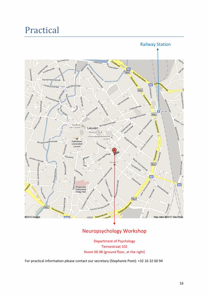
Practical
For practical information please contact our secretary (Stephanie Poot): +32 16 32 60 94
Neuropsychology Workshop
Department of Psychology
Tiensestraat 102
Room 00.98 (ground floor, at the right)
Railway Station