Embed Size (px)
Citation preview
RESEARCH ARTICLE SUMMARY◥
NEUROSCIENCE
A global brain state underliesC. elegans sleep behaviorAnnika L. A. Nichols, Tomáš Eichler, Richard Latham, Manuel Zimmer*
INTRODUCTION:Global brain states such assleep and wakefulness involve reconfigurationsof neural circuit activity across the entire ner-vous system. Yet it is not understood how thebrain can effectively switch between and main-tain different states. Do dedicated brain centerscontrol states via top-down mechanisms? Andto what extent do self-organizing principlesof neuronal networks play a role? To addressthese questions, it would be ideal to measurethe contributions of all individual neurons toa global brain state. Unfortunately, this is cur-rently not possible in mammals or other largeorganisms. Every animal thoroughly studiedexhibits sleeplike behaviors, implying that sleepis an essential, primordial, and common func-tion of neural networks. In mammals, sleep is
defined at the physiological level by a charac-teristic electroencephalography (EEG) signal.Such a definition is missing for invertebratemodels, which primarily rely on behavioraldefinitions.
RATIONALE: The nematode Caenorhabditiselegans is a tractable model organism with thepotential to overcome these limitations: It has astereotypic and mapped nervous system of only302 neurons. Sleep is developmentally timedand occurs predominantly during lethargusperiods of ~2 hours at the end of each larvalstage. During wakefulness, the worm brainexhibits neuronal population dynamics that in-volve a large fraction (~40%) of neurons. Theseneuronal activities are highly coordinated across
the neuronal population; that is, they sharecommon activity patterns. This feature can bequantified with computational methods andvisualized in low-dimensional brain state phaseplots. The resulting brain state trajectory repre-
sents the action sequenceof the animals. To controlsensory-evoked switchingbetween sleep and wake-fulness, we established abehavioral genetics para-digm combined with con-
trolled changes in oxygen (O2) concentration.This method, together with whole-brain imag-ing at single-cell resolution, enables us to ob-serve brainwide neuronal activity dynamicsduring brain state transitions.
RESULTS:During lethargus, wild C. elegansstrains prefer to sleep in social aggregates, andlocal O2 concentrations are a key underlyingcue. In this study, we have shown that a neu-ropeptide receptor (NPR-1) expressed in a hubinterneuron regulates information processingof the arousal cue. We could recapitulate theseswitches between sleep and wakefulness inimmobilized animals while recording the ac-tivity of nearly all neurons in the brain via Ca2+
imaging. We found that sleep in C. elegans is aglobal brain state in which about 75% of neu-rons displaying activity during wakefulnessbecome inactive. However, a few specific neu-rons retained activity during sleep, notablyg-aminobutyric acid–producing (GABAergic)and peptidergic head neurons such as the sleep-promoting interneuron RIS. Chemosensory cir-cuits activated by atmospheric O2 rapidly evokedtransitions to wakefulness by effectively activat-ing neuronal population dynamics. In contrast,entries into sleep occurred spontaneously inthe absence of arousing cues via convergenceof neuronal activities toward the global qui-escent state. Here, the sleep-active neuronsretained stationary high activity.
CONCLUSION: Using computational analy-sis, we have shown that sleep is an emergentproperty of neuronal networks. When lethargusanimals are in a favorable environment suchas a social aggregate, sleep can evolve sponta-neously in the absence of arousing cues; these,however, can rapidly reactivate dynamical brainactivity. Our analysis reveals that neuronalnetworks feature properties of dynamic attrac-tors during wakefulness, whereas during sleepthese dynamics converge toward a fixed point.This attractor state mechanism could be ameans to effectively switch between and main-tain global brain states.▪
RESEARCH
Nichols et al., Science 356, 1247 (2017) 23 June 2017 1 of 1
Research Institute of Molecular Pathology (IMP), Vienna Biocenter(VBC), Campus-Vienna-Biocenter 1, 1030 Vienna, Austria.*Corresponding author. Email: [email protected] this article as A. L. A. Nichols et al., Science 356,eaam6851 (2017). DOI: 10.1126/science.aam6851
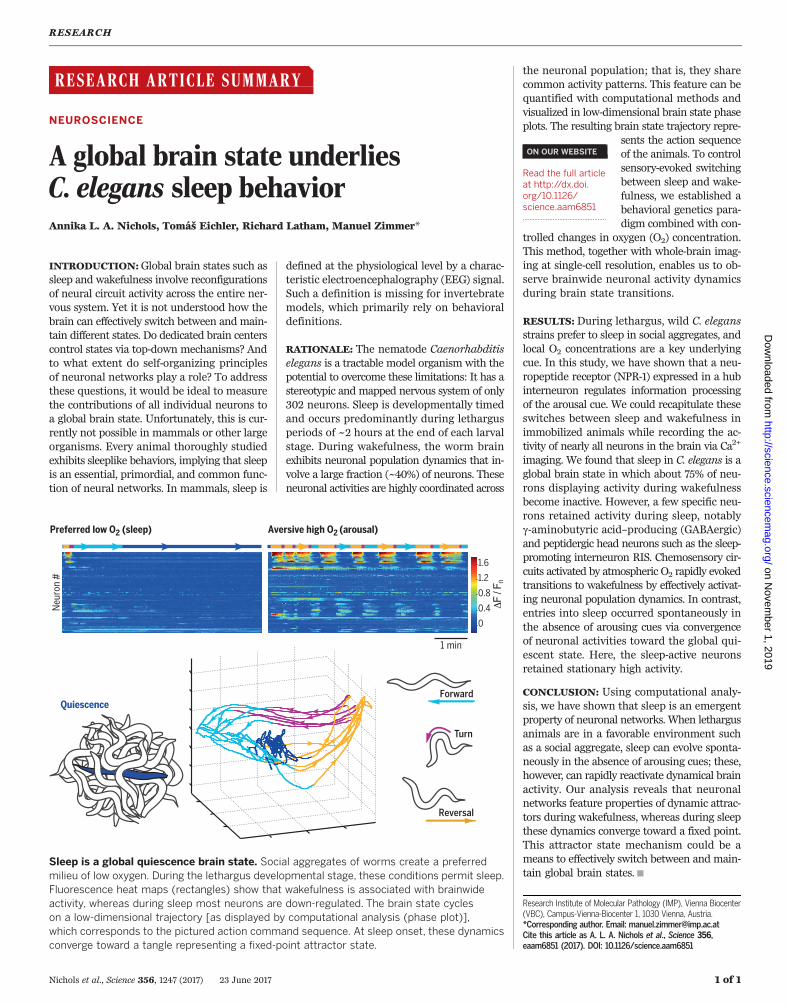
Sleep is a global quiescence brain state. Social aggregates of worms create a preferredmilieu of low oxygen. During the lethargus developmental stage, these conditions permit sleep.Fluorescence heat maps (rectangles) show that wakefulness is associated with brainwideactivity, whereas during sleep most neurons are down-regulated. The brain state cycleson a low-dimensional trajectory [as displayed by computational analysis (phase plot)],which corresponds to the pictured action command sequence. At sleep onset, these dynamicsconverge toward a tangle representing a fixed-point attractor state.
ON OUR WEBSITE◥
Read the full articleat http://dx.doi.org/10.1126/science.aam6851..................................................
on Novem
ber 1, 2019
http://science.sciencemag.org/
Dow
nloaded from

RESEARCH ARTICLE◥
NEUROSCIENCE
A global brain state underliesC. elegans sleep behaviorAnnika L. A. Nichols, Tomáš Eichler, Richard Latham, Manuel Zimmer*
How the brain effectively switches between and maintains global states, such as sleep andwakefulness, is not yet understood. We used brainwide functional imaging at single-cellresolution to show that during the developmental stage of lethargus, the Caenorhabditiselegans brain is predisposed to global quiescence, characterized by systemic down-regulationof neuronal activity. Only a few specific neurons are exempt from this effect. In the absenceof external arousing cues, this quiescent brain state arises by the convergence of neuronalactivities toward a fixed-point attractor embedded in an otherwise dynamic neural statespace. We observed efficient spontaneous and sensory-evoked exits from quiescence.Our data support the hypothesis that during global states such as sleep, neuronalnetworks are drawn to a baseline mode and can be effectively reactivated by signalingfrom arousing circuits.
Behavioral states resembling mammaliansleep have been described across the Ani-malia phyla (1). The ubiquity of such obser-vations suggests that some aspects of sleepobserved in mammals might be fundamen-
tal to all nervous systems. Nonetheless, sleep isincompletely understood in terms of mechanism,regulation, and function. There are two majorhypotheses concerning how this global brain statearises in the brain. One is a top-down regulatorymechanism implicating sleep-promoting centersin inducing sleep upon the rest of the brain (2).Alternatively, sleep onset may be a bottom-upmechanism, an emergent property of neuronalcircuits, which is then spatially and temporallycoordinated by sleep-regulatory circuits (3–6). In-triguingly, cultured mouse cortical neurons spon-taneously display sleeplike activity but can beinduced into wakelike activity by wake-promotingsubstances; this finding indicates that sleep maybe the default state of neuronal networks (7). Inmammals, there are cases in which behavior andlocal brain state are uncorrelated. For example,cetaceans such as dolphins can be awake andalert while their brains are sleeping unihemi-spherically (8). Furthermore, local cortical net-works in awake rats can transiently go “offline”and display sleeplike properties (6). Similarly, sub-populations of Kenyon cells in Drosophila displaysleeplike properties upon prolonged sleep dep-rivation (9). These studies highlight the need forcomprehensively measuring the activity of localcircuits and individual neurons across the entirebrain to understand its global state.Although great leaps in our understanding of
neuronal activity during sleep have been madeby recording local activity (6, 10–14) and global
activity at low resolution (15–18), currently avail-able recording techniques do not have the spatial-temporal resolution to measure brainwide activityof single neurons in mammals. Therefore, wecannot determine how individual neurons acrossthe entire brain are contributing to the emergentproperties of global brain states.The behavioral quiescence seen during the de-
velopmentally timed lethargus periods of the soil-dwelling nematode Caenorhabditis elegans hasrecently been shown to fulfill behavioral criteriafor sleep [i.e., increased arousal thresholds (19),reversibility (19), specific posture (20–22), and ho-meostasis (19, 20, 23)]. During the lethargus stage,animals switch between behavioral quiescence(i.e., absence of any motion) and short, spontane-ous periods of motion (19, 20). Furthermore, thereis wide molecular conservation of regulation, in-cluding control of timing during development viaPERIOD (which regulates circadian timing in otherorganisms); wakefulness-promoting signaling viapigment-dispersing factor (PDF), cyclic adeno-sine monophosphate (cAMP), and dopamine; andquiescence-promoting signaling via epidermalgrowth factor (EGF) (24–28). The g-aminobutyricacid–producing (GABAergic) and peptidergic in-terneuron termed RIS has been shown to exhibitelevated activity during lethargus and to induceimmobility when activated optogenetically (29).Furthermore, both sensory and downstream neu-rons have been shown to have dampened activityduring quiescence (30–32). However, a compre-hensive view of global nervous system activitychanges during behavioral quiescence with single-cell resolution has been lacking.
Low-O2 environments promotebehavioral quiescence in npr-1 animals
We first sought to establish an experimental par-adigm for effective switching between quiescentand aroused brain states. Previous reports suggest
that during the domestication of the standardlaboratory strain N2, several mutations wereacquired and fixed in its genome (33). One ofthese mutations is a gain-of-function mutationin the G protein–coupled receptor neuropeptidereceptor 1 (npr-1) (34). All true wild C. elegansisolates tested so far have a low-activity allele ofnpr-1, called g320 in the wild strain Hawaiian(33). These animals, as well as animals containingan npr-1 loss-of-function mutant (ad609) in theN2 background (henceforth npr-1 animals), accu-mulate at the border of bacterial lawns, wherefood is enriched. Here, they feed in groups byforming social aggregates, whereas standard N2animals exhibit solitary feeding behavior. There-fore, the behavior of npr-1 animals often servesas a proxy for behavior of wild strains (33, 34).Previous behavioral studies on animals in thelethargus period have reported that npr-1 animalsand wild strains have low levels of behavioralquiescence relative to N2 animals (25). However,when animals are left unperturbed in highlycontrolled environments for an extended time,npr-1 shows only slightly less quiescence relativeto N2 (23, 35). Because these studies were done onisolated animals, we aimed to measure the quies-cence of animals in groups. We generated trans-genic npr-1 animals with fluorescent pharyngesto easily detect and video-track individuals insocial aggregates (Fig. 1, A and B, fig. S1A, andmovie S1). We found that lethargus [as describedin this study, representing late larval stage 4 (L4)]npr-1 animals within social aggregates showperiods of locomotor quiescence, whereas animalsin the mid-L4 stage (prelethargus) generally donot. The same difference in activity betweenlethargus and prelethargus animals was seen forisolated npr-1 animals that dwell in the bacterialborder (Fig. 1B and fig. S1B). Thus, lethargusnpr-1 animals exhibit quiescence behavior inaggregates and bacterial lawn borders.Low O2 tension is one of the key environmen-
tal features found in both bacterial lawn bordersand social aggregates (36, 37). To test whetherO2 levels alone could permit quiescence in npr-1animals, we modified a previously reported be-havioral assay in which worms were placed in anO2 flow arena for direct video tracking (38, 39);behavior was observed on a homogeneous foodlawn lacking borders. The animals were kept at21% (atmospheric) O2 for 10 min, then shifted to10% O2, which is within the preferred concen-tration range of npr-1 animals (36), for 10 min.This assay enables quantitative imaging of be-havior, so that we could apply stringent criteriafor defining quiescence versus active behavior—that is, prolonged absence versus occurrenceof detectable movement such as locomotion orhead motions (fig. S1, C to I, and supplementarymaterials). We found that prelethargus andlethargus npr-1 animals were seldom quiescentat 21% O2, which is consistent with the studieson isolated animals. At 10% O2, only a few shortbouts of quiescence could be observed in pre-lethargus npr-1 animals; however, lethargusnpr-1 animals displayed long quiescence inter-vals with short interrupting bouts of activity
RESEARCH
Nichols et al., Science 356, eaam6851 (2017) 23 June 2017 1 of 9
Research Institute of Molecular Pathology (IMP), Vienna Biocenter(VBC), Campus-Vienna-Biocenter 1, 1030 Vienna, Austria.*Corresponding author. Email: [email protected]
on Novem
ber 1, 2019
http://science.sciencemag.org/
Dow
nloaded from
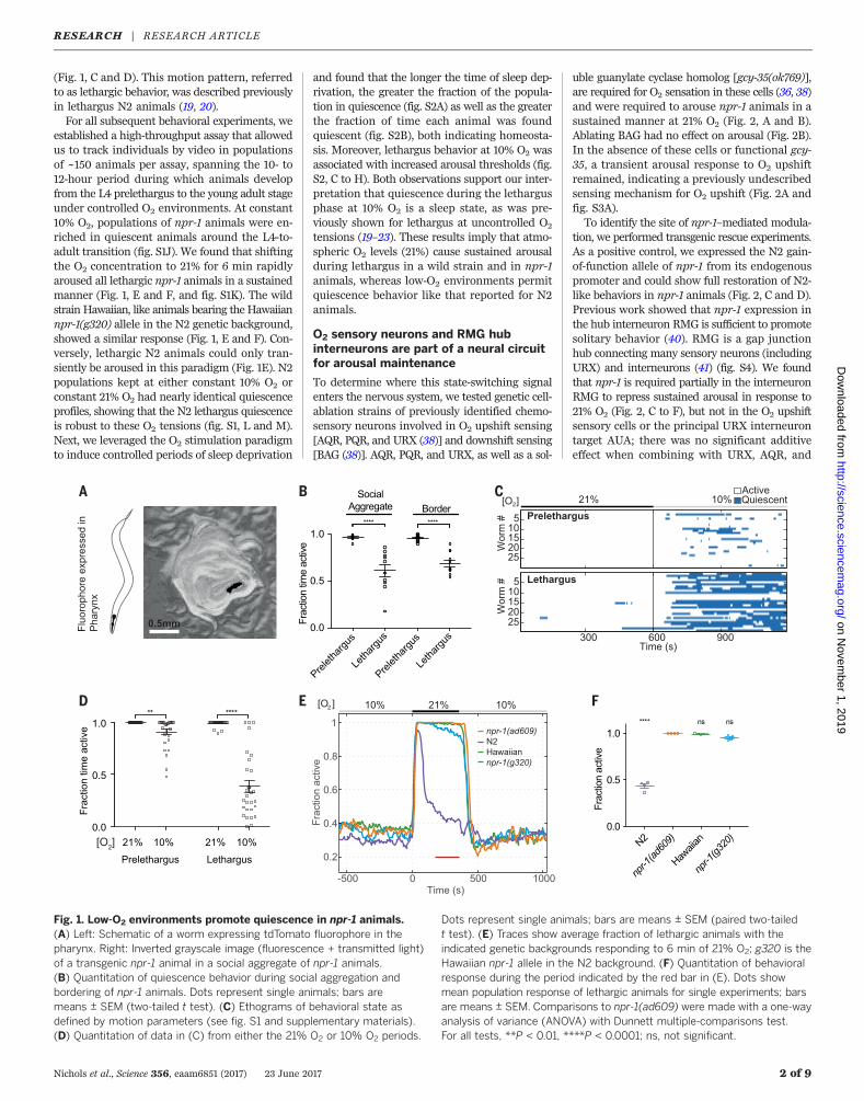
(Fig. 1, C and D). This motion pattern, referredto as lethargic behavior, was described previouslyin lethargus N2 animals (19, 20).For all subsequent behavioral experiments, we
established a high-throughput assay that allowedus to track individuals by video in populationsof ~150 animals per assay, spanning the 10- to12-hour period during which animals developfrom the L4 prelethargus to the young adult stageunder controlled O2 environments. At constant10% O2, populations of npr-1 animals were en-riched in quiescent animals around the L4-to-adult transition (fig. S1J). We found that shiftingthe O2 concentration to 21% for 6 min rapidlyaroused all lethargic npr-1 animals in a sustainedmanner (Fig. 1, E and F, and fig. S1K). The wildstrain Hawaiian, like animals bearing the Hawaiiannpr-1(g320) allele in the N2 genetic background,showed a similar response (Fig. 1, E and F). Con-versely, lethargic N2 animals could only tran-siently be aroused in this paradigm (Fig. 1E). N2populations kept at either constant 10% O2 orconstant 21% O2 had nearly identical quiescenceprofiles, showing that the N2 lethargus quiescenceis robust to these O2 tensions (fig. S1, L and M).Next, we leveraged the O2 stimulation paradigmto induce controlled periods of sleep deprivation
and found that the longer the time of sleep dep-rivation, the greater the fraction of the popula-tion in quiescence (fig. S2A) as well as the greaterthe fraction of time each animal was foundquiescent (fig. S2B), both indicating homeosta-sis. Moreover, lethargus behavior at 10% O2 wasassociated with increased arousal thresholds (fig.S2, C to H). Both observations support our inter-pretation that quiescence during the lethargusphase at 10% O2 is a sleep state, as was pre-viously shown for lethargus at uncontrolled O2
tensions (19–23). These results imply that atmo-spheric O2 levels (21%) cause sustained arousalduring lethargus in a wild strain and in npr-1animals, whereas low-O2 environments permitquiescence behavior like that reported for N2animals.
O2 sensory neurons and RMG hubinterneurons are part of a neural circuitfor arousal maintenance
To determine where this state-switching signalenters the nervous system, we tested genetic cell-ablation strains of previously identified chemo-sensory neurons involved in O2 upshift sensing[AQR, PQR, and URX (38)] and downshift sensing[BAG (38)]. AQR, PQR, and URX, as well as a sol-
uble guanylate cyclase homolog [gcy-35(ok769)],are required for O2 sensation in these cells (36, 38)and were required to arouse npr-1 animals in asustained manner at 21% O2 (Fig. 2, A and B).Ablating BAG had no effect on arousal (Fig. 2B).In the absence of these cells or functional gcy-35, a transient arousal response to O2 upshiftremained, indicating a previously undescribedsensing mechanism for O2 upshift (Fig. 2A andfig. S3A).To identify the site of npr-1–mediated modula-
tion, we performed transgenic rescue experiments.As a positive control, we expressed the N2 gain-of-function allele of npr-1 from its endogenouspromoter and could show full restoration of N2-like behaviors in npr-1 animals (Fig. 2, C and D).Previous work showed that npr-1 expression inthe hub interneuron RMG is sufficient to promotesolitary behavior (40). RMG is a gap junctionhub connecting many sensory neurons (includingURX) and interneurons (41) (fig. S4). We foundthat npr-1 is required partially in the interneuronRMG to repress sustained arousal in response to21% O2 (Fig. 2, C to F), but not in the O2 upshiftsensory cells or the principal URX interneurontarget AUA; there was no significant additiveeffect when combining with URX, AQR, and
Nichols et al., Science 356, eaam6851 (2017) 23 June 2017 2 of 9
Fig. 1. Low-O2 environments promote quiescence in npr-1 animals.(A) Left: Schematic of a worm expressing tdTomato fluorophore in thepharynx. Right: Inverted grayscale image (fluorescence + transmitted light)of a transgenic npr-1 animal in a social aggregate of npr-1 animals.(B) Quantitation of quiescence behavior during social aggregation andbordering of npr-1 animals. Dots represent single animals; bars aremeans ± SEM (two-tailed t test). (C) Ethograms of behavioral state asdefined by motion parameters (see fig. S1 and supplementary materials).(D) Quantitation of data in (C) from either the 21% O2 or 10% O2 periods.
Dots represent single animals; bars are means ± SEM (paired two-tailedt test). (E) Traces show average fraction of lethargic animals with theindicated genetic backgrounds responding to 6 min of 21% O2; g320 is theHawaiian npr-1 allele in the N2 background. (F) Quantitation of behavioralresponse during the period indicated by the red bar in (E). Dots showmean population response of lethargic animals for single experiments; barsare means ± SEM. Comparisons to npr-1(ad609) were made with a one-wayanalysis of variance (ANOVA) with Dunnett multiple-comparisons test.For all tests, **P < 0.01, ****P < 0.0001; ns, not significant.
RESEARCH | RESEARCH ARTICLEon N
ovember 1, 2019
http://science.sciencem
ag.org/D
ownloaded from
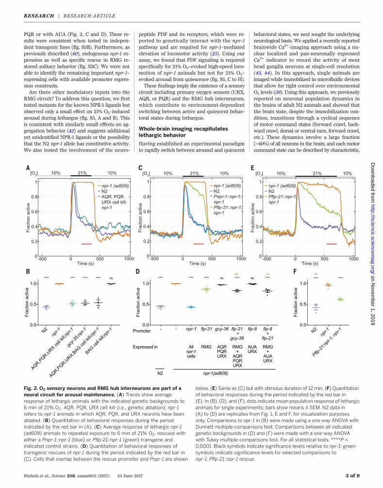
PQR or with AUA (Fig. 2, C and D). These re-sults were consistent when tested in indepen-dent transgenic lines (fig. S3B). Furthermore, aspreviously described (40), endogenous npr-1 ex-pression as well as specific rescue in RMG re-stored solitary behavior (fig. S3C). We were notable to identify the remaining important npr-1–expressing cells with available promoter expres-sion constructs.Are there other modulatory inputs into the
RMG circuit? To address this question, we firsttested mutants for the known NPR-1 ligands butobserved only a small effect on 21% O2–inducedarousal during lethargus (fig. S5, A and B). Thisis consistent with similarly small effects on ag-gregation behavior (42) and suggests additionalyet unidentified NPR-1 ligands or the possibilitythat the N2 npr-1 allele has constitutive activity.We also tested the involvement of the neuro-
peptide PDF and its receptors, which were re-ported to genetically interact with the npr-1pathway and are required for npr-1–mediatedelevation of locomotor activity (25). Using ourassay, we found that PDF signaling is requiredspecifically for 21% O2–evoked high-speed loco-motion of npr-1 animals but not for 21% O2–evoked arousal from quiescence (fig. S5, C to H).These findings imply the existence of a sensory
circuit including primary oxygen sensors (URX,AQR, or PQR) and the RMG hub interneurons,which contribute to environment-dependentswitching between active and quiescent behav-ioral states during lethargus.
Whole-brain imaging recapitulateslethargic behavior
Having established an experimental paradigmto rapidly switch between aroused and quiescent
behavioral states, we next sought the underlyingneurological basis. We applied a recently reportedbrainwide Ca2+-imaging approach using a nu-clear localized and pan-neuronally expressedCa2+ indicator to record the activity of mosthead ganglia neurons at single-cell resolution(43, 44). In this approach, single animals areimaged while immobilized in microfluidic devicesthat allow for tight control over environmentalO2 levels (38). Using this approach, we previouslyreported on neuronal population dynamics inthe brains of adult N2 animals and showed thatthe brain state, despite the immobilization con-dition, transitions through a cyclical sequenceof motor command states (forward crawl, back-ward crawl, dorsal or ventral turn, forward crawl,etc.). These dynamics involve a large fraction(~40%) of all neurons in the brain, and each motorcommand state can be described by characteristic,
Nichols et al., Science 356, eaam6851 (2017) 23 June 2017 3 of 9
Fig. 2. O2 sensory neurons and RMG hub interneurons are part of aneural circuit for arousal maintenance. (A) Traces show averageresponse of lethargic animals with the indicated genetic backgrounds to6 min of 21% O2. AQR, PQR, URX cell kill (i.e., genetic ablation); npr-1refers to npr-1 animals in which AQR, PQR, and URX neurons have beenablated. (B) Quantitation of behavioral responses during the periodindicated by the red bar in (A). (C) Average response of lethargic npr-1(ad609) animals to repeated exposure to 6 min of 21% O2, rescued witheither a Pnpr-1::npr-1 (blue) or Pflp-21::npr-1 (green) transgene andindicated control strains. (D) Quantitation of behavioral responses oftransgenic rescues of npr-1 during the period indicated by the red bar in(C). Cells that overlap between the rescue promoter and Pnpr-1 are shown
below. (E) Same as (C) but with stimulus duration of 12 min. (F) Quantitationof behavioral responses during the period indicated by the red bar in(E). In (B), (D), and (F), dots indicate mean population response of lethargicanimals for single experiments; bars show means ± SEM. N2 data in(A) to (D) are replicates from Fig. 1, E and F, for visualization purposesonly. Comparisons to npr-1 in (B) were made using a one-way ANOVA withDunnett multiple-comparisons test. Comparisons between all indicatedgenetic backgrounds in (D) and (F) were made with a one-way ANOVAwith Tukey multiple-comparisons test. For all statistical tests, ****P <0.0001. Black symbols indicate significance levels relative to npr-1; greensymbols indicate significance levels for selected comparisons tonpr-1; Pflp-21::npr-1 rescue.
RESEARCH | RESEARCH ARTICLEon N
ovember 1, 2019
http://science.sciencem
ag.org/D
ownloaded from
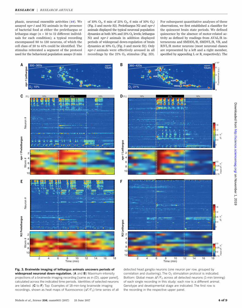
phasic, neuronal ensemble activities (44). Weassayed npr-1 and N2 animals in the presenceof bacterial food at either the prelethargus orlethargus stage (n = 10 to 12 different individ-uals for each condition); a typical recordingencompassed 80 to 130 neurons, of which thecell class of 20 to 41% could be identified. Thestimulus reiterated a segment of the protocolused for the behavioral population assays (6 min
of 10% O2, 6 min of 21% O2, 6 min of 10% O2)(Fig. 3 and movie S2). Prelethargus N2 and npr-1animals displayed the typical neuronal populationdynamics at both 10% and 21% O2 levels; lethargusN2 and npr-1 animals in addition displayedperiods of widespread down-regulation of braindynamics at 10% O2 (Fig. 3 and movie S2). Onlynpr-1 animals were effectively aroused in allrecordings by the 21% O2 stimulus (Fig. 3D).
For subsequent quantitative analyses of theseobservations, we first established a classifier forthe quiescent brain state periods. We definedquiescence by the absence of motor-related ac-tivity as defined by readings from AVAL/R in-terneurons and SMDDL/R, SMDVL/R, VB, andRIVL/R motor neurons (most neuronal classesare represented by a left and a right member,specified by appending L or R, respectively). The
Nichols et al., Science 356, eaam6851 (2017) 23 June 2017 4 of 9
Fig. 3. Brainwide imaging of lethargus animals uncovers periods ofwidespread neuronal down-regulation. (A and B) Maximum-intensityprojections of a brainwide imaging recording [same as in (D), upper panel],calculated across the indicated time periods. Identities of selected neuronsare labeled. (C to F) Top: Examples of 18-min-long brainwide imagingrecordings, shown as heat maps of fluorescence (DF/F0) time series of all
detected head ganglia neurons (one neuron per row, grouped bycorrelation and clustering). The O2 stimulation protocol is indicated.Bottom: Global mean DF/F0 across all detected neurons (1-min binning)of each single recording in this study; each row is a different animal.Genotype and developmental stage are indicated. The first row isthe recording in the respective upper panel.
RESEARCH | RESEARCH ARTICLEon N
ovember 1, 2019
http://science.sciencem
ag.org/D
ownloaded from
activity of these neurons in freely behaving wormscorresponds to the execution of one of the motorcommand states (44, 45) (Fig. 4, A and B, andfig. S6) (see supplementary materials for details).Prelethargus animals had very few periods ofquiescence, whereas both N2 and npr-1 lethargusanimals displayed quiescence at baseline 10%O2 (Fig. 4, C and D), like the levels seen in ourbehavioral assays (Fig. 1E). Furthermore, all npr-1lethargus animals exited quiescence when shiftedto 21% O2 and on average maintained the activestate for the duration of the 6-min stimulus (Fig.4C). Consistent with our behavioral results (forcontrols of transgenic lines, see fig. S7, A and B),the fraction of lethargus N2 animals in an activebrain state transiently increased upon O2 upshift(Fig. 4D). These data show that our resultsobtained in the behavioral paradigm can be re-capitulated by whole-brain imaging in immobi-lized animals.
Quiescence during lethargus is a globalbrain state
We next quantified how the above-defined qui-escence state correlates with global neuronal ac-tivity levels by calculating the mean cumulativefrequency distributions of the change in fluores-cence intensity (DF/F0) across all neurons (exceptneurons that encode O2 sensory information).According to a DF/F0 cutoff for defining activeversus inactive neurons during the aroused statein either prelethargus or lethargus animals, ~40%of all neurons were active; this number droppedto ~10% during quiescence in both lethargus N2and lethargus npr-1 animals (Fig. 4, E and F).Thus, during quiescence, about three-fourths ofall neurons that are normally found to be activewere down-regulated.To investigate this observation further, we
focused our subsequent analyses on all neuronsfrom cell classes that could be reproducibly iden-tified in enough recordings (n ≥ 3). For example,the RIM and AVB interneurons, which are activeduring the reversal and forward motor commandstates, respectively (44), were both found to belargely inactive during quiescence (Fig. 4, G andH). We made the same observation for the grossmajority of the other neurons belonging to thereversal or forward ensembles (Fig. 4, I and J).Also, a large fraction of neurons whose activitywas not exclusive to the forward or reversalstates was significantly down-regulated duringquiescence; notable among these were sensoryneurons that show spontaneous activity duringactive brain states (Fig. 4K). In summary, qui-escence during lethargus affects many neuronsof both the sensory and premotor domains.
Npr-1 animals exhibit an enhancedsensory-motor transformation
How could npr-1 control the arousal thresholdsof animals? To address this question, we inter-rogated our data for possible correlations betweensensory circuit activity and genotype, develop-mental stage, or brain state. Mechanosensory andnociceptive neurons exhibit weaker sensory re-sponses during lethargus in N2 animals but not
in npr-1 animals (25, 30–32). In agreement withthis, npr-1 had nearly identical O2-evoked re-sponses in URX and AQR in prelethargus versuslethargus, but N2 had a larger variability in URXresponses during lethargus, with some neuronsnot responding (fig. S8, A and B). Lethargusnpr-1 animals showed increased URX peak re-sponses relative to lethargus N2 animals (fig.S8B). Although URX peak responses correlatedwith transient arousal (0 to 1 min after stimulus),we did not find such a correlation with sustainedarousal (3 to 6 min after stimulus) (fig. S8, Cand D). However, N2 AQRs responded similarlyin prelethargus versus lethargus. Neither tran-sient nor tonic responses in AQR (46) correlatedwith transient or sustained arousal (fig. S8, Eto I). In summary, we did not find a strikingmodulation of AQR and URX that could explainthe sustained arousal phenotype of npr-1 animals.We found that npr-1 animals might have more
complex O2 sensory representations than N2, aswe more frequently observed responses fromAUA (a principal interneuron target of URX)and RMG interneurons (fig. S9, A and B). More-over, we found more putative IL2 sensory neuronresponses to O2 upshift in npr-1 animals (fig. S9C).O2 sensory activity in IL2s, or in any other neuronin the anterior ganglion, has not been reportedpreviously.How do motor behavior–related brain dynam-
ics change in response to the stimulus? Thefrequency of reversal command states, whichcorresponds to the frequency of cycles throughthe motor command sequence, was up-regulatedat 21% O2 in npr-1 animals but not in N2 animals(fig. S10), showing that in comparison to N2,npr-1 animals exhibit a more effective sensory-motor transformation in both prelethargus andlethargus stages. This is consistent with findingsin adult animals (47). This effect could be causedby a more complex sensory representation of O2
stimuli. As npr-1–mediated sustained arousal canbe rescued in RMG neurons, RMG modulationmight dampen the excitability of sensory circuitsin an npr-1–dependent manner, preventing O2-evoked sustained motor dynamics in N2 animals.
Quiescence displays features of afixed-point attractor in neuronalpopulation dynamics
We next focused our analysis on the neurons thatwere exempt from down-regulation during qui-escence. Most prominent among these was theGABAergic and peptidergic sleep-active inter-neuron RIS, which exhibited Ca2+ plateaus duringthe forward state and remained active duringquiescence (Fig. 4, B, L, and M); this observationis consistent with previous RIS recordings atlow temporal resolution in unconstrained ani-mals (29). Relative to prelethargus animals, RISactivity is elevated during the forward period inlethargus animals, which suggests that elevatedRIS activity increases the likelihood of enteringthe quiescent state and that RIS activity duringthe forward state may reflect sleep pressure(Fig. 4, L and M). Besides RIS, other GABAergichead neurons such as RMED and RMEV, which
are active during the forward state (44) (Fig. 4,A and I, and fig. S11), showed residual (i.e.,reduced-amplitude) Ca2+ plateaus during qui-escence. This is consistent with increased GABAsignaling during mammalian sleep (2). Similar-ly, the neuropeptidergic GABA uptake neuronALA, which is required for stress-induced qui-escence in adults (26, 48–50), showed some spon-taneous activity and was not down-regulatedduring quiescence (Fig. 4K). In addition, the GABAuptake neuron AVF (50) was only slightly down-regulated during quiescence (Fig. 4K). Thus, al-though RIS, RMED, and RMEV decrease activitywhen the brain transitions from forward into areversal state (44), they maintain tonic (i.e., sus-tained) activity when the system transitions intothe quiescent state.We next sought to characterize how quies-
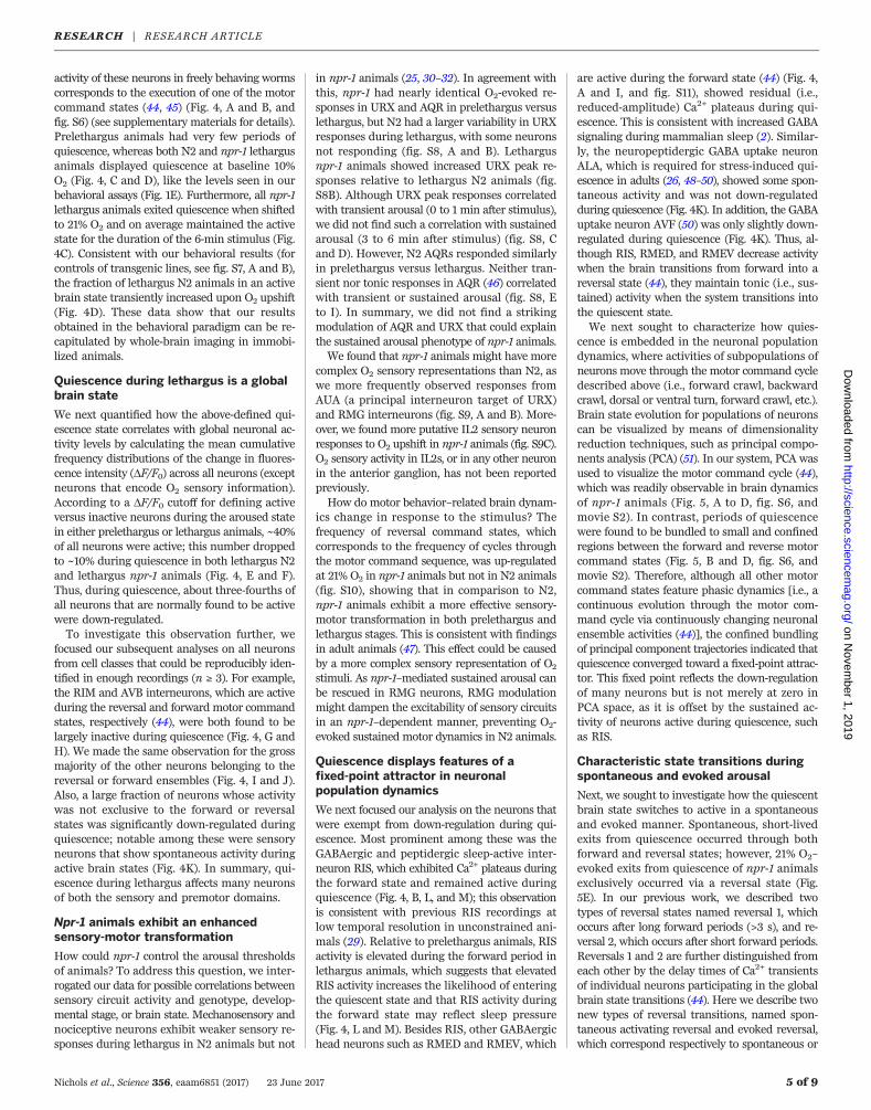
cence is embedded in the neuronal populationdynamics, where activities of subpopulations ofneurons move through the motor command cycledescribed above (i.e., forward crawl, backwardcrawl, dorsal or ventral turn, forward crawl, etc.).Brain state evolution for populations of neuronscan be visualized by means of dimensionalityreduction techniques, such as principal compo-nents analysis (PCA) (51). In our system, PCA wasused to visualize the motor command cycle (44),which was readily observable in brain dynamicsof npr-1 animals (Fig. 5, A to D, fig. S6, andmovie S2). In contrast, periods of quiescencewere found to be bundled to small and confinedregions between the forward and reverse motorcommand states (Fig. 5, B and D, fig. S6, andmovie S2). Therefore, although all other motorcommand states feature phasic dynamics [i.e., acontinuous evolution through the motor com-mand cycle via continuously changing neuronalensemble activities (44)], the confined bundlingof principal component trajectories indicated thatquiescence converged toward a fixed-point attrac-tor. This fixed point reflects the down-regulationof many neurons but is not merely at zero inPCA space, as it is offset by the sustained ac-tivity of neurons active during quiescence, suchas RIS.
Characteristic state transitions duringspontaneous and evoked arousal
Next, we sought to investigate how the quiescentbrain state switches to active in a spontaneousand evoked manner. Spontaneous, short-livedexits from quiescence occurred through bothforward and reversal states; however, 21% O2–evoked exits from quiescence of npr-1 animalsexclusively occurred via a reversal state (Fig.5E). In our previous work, we described twotypes of reversal states named reversal 1, whichoccurs after long forward periods (>3 s), and re-versal 2, which occurs after short forward periods.Reversals 1 and 2 are further distinguished fromeach other by the delay times of Ca2+ transientsof individual neurons participating in the globalbrain state transitions (44). Here we describe twonew types of reversal transitions, named spon-taneous activating reversal and evoked reversal,which correspond respectively to spontaneous or
Nichols et al., Science 356, eaam6851 (2017) 23 June 2017 5 of 9
RESEARCH | RESEARCH ARTICLEon N
ovember 1, 2019
http://science.sciencem
ag.org/D
ownloaded from

Nichols et al., Science 356, eaam6851 (2017) 23 June 2017 6 of 9
Fig. 4. Quiescent brain states have reduced activity across multipleneuron classes. Quantitative data corresponding to the brainwideimaging recordings in Fig. 3 are shown for npr-1 (prelethargus n = 10,lethargus n = 11) and N2 (prelethargus n = 10, lethargus n = 12). (A and B)Example traces showing some of the reversal interneuron AVAL/R, theVB02 forward motor neuron SMDDL/R, and RIVL/R ventral turningneurons that are used to define quiescent (blue background) brain states(see supplementary materials for classification rules), as well as tracesof the GABAergic neurons RIS and RMED/V. (C and D) Traces show meanfraction of time spent active in 1-min bins for npr-1 (C) and N2 (D) animals.(E and F) Mean cumulative frequencies (±SEM) of DF/F0 across allneurons (excluding O2 sensory neurons) in each recording for npr-1(E) and N2 (F) animals. Significant differences between lethargus activeand lethargus quiescent were determined by permutation test. (G andH) Mean (±SEM) fractional histograms (log scale) of all measured DF/F0
value distributions for selected neurons during forward/turn, reversal, orquiescent brain states in lethargus: (G) RIML reversal interneuron, (H)AVBL forward interneuron. (I to K) Relative activity of the indicatedneurons in the quiescent state compared to the neuron’s principal stateduring activity, calculated as the mean difference between distributionslike those shown in (G) and (H). Dots indicate difference within a singlerecording; bars show means ± SEM, with N indicated in parentheses.Significance was determined by a permutation test. Neuron names shownin gray rather than black were used to classify the quiescent state.Data are from npr-1 lethargus recordings: (I) forward/turn neurons, (J)reversal neurons, (K) all other active neurons. In (I) and (J), ambiguousneuron cell class identities are followed by f; other possible identitiesare shown in the supplementary materials. (L and M) RIS neuron as in (G)and (H). For all statistical tests, *P < 0.05, **P < 0.01, ***P < 0.001,****P < 0.0001.
RESEARCH | RESEARCH ARTICLEon N
ovember 1, 2019
http://science.sciencem
ag.org/D
ownloaded from
Nichols et al., Science 356, eaam6851 (2017) 23 June 2017 7 of 9
Fig. 5. Characterization of brain state transitions. (A to D) Phase plotsof the first three temporal principal components (TPCs), which are thetime integrals of principal components calculated from time derivativesof neuronal activity traces from brainwide imaging recordings. Developmentalstage and genotype are indicated. Coloring indicates the motor commandstate; arrows indicate direction of trajectory. (E) Exits from quiescence boutsoccur through forward or reverse brain states. “Evoked” indicates eventsinduced by the shift to 21% O2. (F and G) Summaries of phase timinganalyses. Median phase delays of neuron classes with respect to AVAL (t = 0)for different reversal types are shown. Neuron polarity is designated by (+)or (–) indicating whether the neuron’s rises or falls in activity were used. The
raw data are shown in fig. S12. See table S1 for statistical analysis. Ambiguousneuron identities are denoted as in Fig. 4, I and J. (H) Quiescent brainstates are frequently preceded by a forward state. (I to P) Traces (right axes)show frequency of state transitions as a function of current forwardduration (time from AVA activity fall period) for npr-1 [(I) to (L)] and N2 [(M)to (P)] for the indicated O2 tension and developmental stage. Histograms(left axes) show number of forward periods per bin. Monotonic increases infractions are indicated significant as determined by a permutation test; **P <0.01, ***P < 0.001 (see supplementary materials). Number of recordings(same as Fig. 3): npr-1 (prelethargus n = 10, lethargus n = 11) and N2(prelethargus n = 10, lethargus n = 12).
RESEARCH | RESEARCH ARTICLEon N
ovember 1, 2019
http://science.sciencem
ag.org/D
ownloaded from

21% O2–evoked events that terminate quiescence.Figure 5, F and G, summarizes results; fig. S12shows details. Spontaneous activating reversalsfeature early recruitment of some neurons (e.g.,interneurons AIB and RIM; head motor neuronSMDDL) but otherwise display a similar orderof Ca2+ signals as in reversal 1 (compare Fig. 5, Fand G; compare fig. S12, B and C). In contrast,evoked reversals in npr-1 animals are rapid tran-sitions exhibiting short phase delays betweenneurons, a feature shared with reversal 2 (com-pare Fig. 5, F and G; compare fig. S12, A and D).This observation highlights a difference betweenO2-evoked responses and a previously reportednociceptive response, which during lethargus isaccompanied by a decorrelation of interneuronactivity (31). During evoked reversals, Ca2+ signalsof O2 sensory neurons URX, AQR, IL2, andURX’s principal postsynaptic interneuron tar-get AUA rose consistently earlier than all otherparticipating neurons (fig. S12, A and E). Thus,spontaneous arousal from quiescence evolvesthrough a characteristic sequence of neuronalensemble activity, and O2 sensory circuits effec-tively and rapidly recruit motor command ac-tivity similar to that observed in already activeanimals. Table S1 shows statistical tests for allrelevant comparisons.
Prolonged forward states transitionto quiescence
Next, we investigated how the active brain stateswitches to quiescence. The great majority oftransitions into quiescence occurred through for-ward states (Fig. 5H). We calculated the fractionsof reversal and quiescence transitions as a func-tion of forward duration, which we define as thetime passed since AVA activity fall (Fig. 5, I to P).Behavioral studies have shown that the duration offorward episodes decays with two exponential timeconstants (52, 53). We made a consistent obser-vation with forward state durations in our im-mobilized preparation: In both N2 and npr-1prelethargus animals, most forward initiationswere terminated by a reversal command within3 s, leading to continuation of active states (Fig. 5,I, J, M, and N). In contrast, in lethargus animalsthe distributions of forward lengths shifted tolonger durations (>3 s). Furthermore, instead ofobserving constant transition rates into quies-cence, we observed that the fraction of forwardperiods ending in quiescence increased over for-ward state duration, thereby exceeding the fractionof reversal commands after 30 s (Fig. 5, K, L, O,and P). This was the case for both N2 and npr-1animals, except for npr-1 animals experiencing21% O2 stimulation, where reversal commandprobability remained relatively high throughoutthe length of forward periods (Fig. 5L). Thesedata show that quiescence entry during lethargusdepends on the prior time that animals spent inthe forward period and that arousing sensory cuescan maintain the active brain state by triggeringreversal commands, thereby resetting the systemto another active period.Our findings indicate that the low activity of
the NPR-1 neuropeptide receptor in wild C. elegans
confers regulation of a quiescent behavioral stateduring lethargus in response to environmentalO2 conditions. This might be a self-protectivestrategy in which low O2 could signify safer en-vironments such as a social aggregate (37). EEGsignatures define the various stages of sleep inmammals, although with low spatial resolution(14). In contrast, sleep states in invertebrates haveso far mostly relied on the classical behavioraldefinition of sleep (19–22, 54–56); however, localfield potential recordings and calcium imagingin flies show neuronal down-regulation duringsleep (9, 57). Our work reveals a global neuronalsignature of an invertebrate sleep state, whichmodulates the activity of most individual activeneurons. Although active C. elegans exhibit pha-sic neuronal population dynamics that repre-sent the motor command states (44), quiescencecorresponds to a more stationary region arounda fixed point in neuronal state space. This attrac-tor feature implies that the quiescent state isan intrinsic network property and that sleep-promoting neurons and brain centers—as reportedacross many organisms (2, 29, 58–60)—might beintegral parts of neural networks, as opposed tohierarchically organized top-down controllers. Thiscould enable an efficient means of changing theglobal state of the brain via neuromodulators bysubtly changing its state bias, as opposed to in-stantaneous reprogramming of all its individualnetwork components. During lethargus, the qui-escence attractor can thus be seen as a defaultstate of the network that emerges in the absenceof arousing inputs. Dedicated sensory circuitscan rapidly switch the brain state to active byrecruiting behavior-related neuronal populationdynamics. Conversely, during nonlethargic phasessuch as prelethargus, the putative default state ismaintained activity (switching among forward,reverse, and turning) regardless of the sensoryinput. Building on recent studies reporting therequirement for neuropeptides, we suggest thatthese neuromodulators are crucial for establish-ing the propensity for the quiescent network stateduring lethargus (19, 29, 49, 61–63). Therefore,global brain states are likely controlled by multi-ple signals antagonizing or promoting arousal andquiescence, and which originate from both theenvironment and the internal state of the animal.It is possible that the quiescence brain state in
C. elegans serves a function equivalent to the de-fault mode in the human brain, which correspondsto intrinsic functional activity of the awake brainat rest (64, 65). Moreover, we find differences aswell as parallels between mammalian deep sleepand C. elegans lethargus. Ensembles of corticalneurons exhibit periodic high-amplitude oscilla-tions (delta wave) during deep sleep (10), whereasthe C. elegans brain during lethargus nonperiodi-cally fluctuates between quiescence and shortactive bouts. However, both have periods of al-most complete down-regulation of neuronal ac-tivity: the trough of the delta wave oscillation inmammals (10–12) and brainwide quiescence inC. elegans. In addition, the short active bouts re-semble micro-arousals occurring during mam-malian deep sleep (12, 66). We thus provide a
neuronal imaging paradigm to study the en-docrine control, network mechanisms, evolu-tion, and basic functions of global brain statessuch as active wakefulness, rest, and sleep.
REFERENCES AND NOTES
1. R. Allada, J. M. Siegel, Unearthing the phylogenetic roots ofsleep. Curr. Biol. 18, R670–R679 (2008). doi: 10.1016/j.cub.2008.06.033; pmid: 18682212
2. C. B. Saper, P. M. Fuller, N. P. Pedersen, J. Lu, T. E. Scammell,Sleep state switching. Neuron 68, 1023–1042 (2010).doi: 10.1016/j.neuron.2010.11.032; pmid: 21172606
3. J. M. Krueger, F. Obál Jr., A neuronal group theory ofsleep function. J. Sleep Res. 2, 63–69 (1993). doi: 10.1111/j.1365-2869.1993.tb00064.x; pmid: 10607073
4. J. M. Krueger et al., Sleep as a fundamental property ofneuronal assemblies. Nat. Rev. Neurosci. 9, 910–919 (2008).doi: 10.1038/nrn2521; pmid: 18985047
5. I. N. Pigarev, H. C. Nothdurft, S. Kastner, Evidence forasynchronous development of sleep in cortical areas. Neuroreport8, 2557–2560 (1997). doi: 10.1097/00001756-199707280-00027;pmid: 9261826
6. V. V. Vyazovskiy et al., Local sleep in awake rats. Nature 472,443–447 (2011). doi: 10.1038/nature10009; pmid: 21525926
7. V. Hinard et al., Key electrophysiological, molecular, andmetabolic signatures of sleep and wakefulness revealed inprimary cortical cultures. J. Neurosci. 32, 12506–12517 (2012).doi: 10.1523/JNEUROSCI.2306-12.2012; pmid: 22956841
8. L. M. Mukhametov, A. Y. Supin, I. G. Polyakova,Interhemispheric asymmetry of the electroencephalographicsleep patterns in dolphins. Brain Res. 134, 581–584 (1977).doi: 10.1016/0006-8993(77)90835-6; pmid: 902119
9. D. Bushey, G. Tononi, C. Cirelli, Sleep- and wake-dependentchanges in neuronal activity and reactivity demonstrated in flyneurons using in vivo calcium imaging. Proc. Natl. Acad. Sci.U.S.A. 112, 4785–4790 (2015). doi: 10.1073/pnas.1419603112;pmid: 25825756
10. M. Steriade, D. A. McCormick, T. J. Sejnowski, Thalamocorticaloscillations in the sleeping and aroused brain. Science 262,679–685 (1993). doi: 10.1126/science.8235588;pmid: 8235588
11. M. Steriade, I. Timofeev, F. Grenier, Natural waking and sleepstates: A view from inside neocortical neurons. J. Neurophysiol.85, 1969–1985 (2001). pmid: 11353014
12. B. O. Watson, D. Levenstein, J. P. Greene, J. N. Gelinas,G. Buzsáki, Network homeostasis and state dynamics ofneocortical sleep. Neuron 90, 839–852 (2016). doi: 10.1016/j.neuron.2016.03.036; pmid: 27133462
13. V. V. Vyazovskiy, K. D. Harris, Sleep and the single neuron: Therole of global slow oscillations in individual cell rest. Nat. Rev.Neurosci. 14, 443–451 (2013). doi: 10.1038/nrn3494;pmid: 23635871
14. R. E. Brown, R. Basheer, J. T. McKenna, R. E. Strecker,R. W. McCarley, Control of sleep and wakefulness. Physiol. Rev.92, 1087–1187 (2012). doi: 10.1152/physrev.00032.2011;pmid: 22811426
15. A. L. Loomis, E. N. Harvey, G. Hobart, Further observations onthe potential rhythms of the cerebral cortex during sleep.Science 82, 198–200 (1935). doi: 10.1126/science.82.2122.198;pmid: 17844579
16. E. Aserinsky, N. Kleitman, Regularly occurring periods ofeye motility, and concomitant phenomena, during sleep. Science118, 273–274 (1953). doi: 10.1126/science.118.3062.273;pmid: 13089671
17. L. A. Finelli, H. Baumann, A. A. Borbély, P. Achermann, Dualelectroencephalogram markers of human sleep homeostasis:Correlation between theta activity in waking and slow-waveactivity in sleep. Neuroscience 101, 523–529 (2000).doi: 10.1016/S0306-4522(00)00409-7; pmid: 11113301
18. A. Suzuki, C. M. Sinton, R. W. Greene, M. Yanagisawa, Behavioraland biochemical dissociation of arousal and homeostatic sleepneed influenced by prior wakeful experience in mice. Proc. Natl.Acad. Sci. U.S.A. 110, 10288–10293 (2013). doi: 10.1073/pnas.1308295110; pmid: 23716651
19. D. M. Raizen et al., Lethargus is a Caenorhabditis eleganssleep-like state. Nature 451, 569–572 (2008). doi: 10.1038/nature06535; pmid: 18185515
20. S. Iwanir et al., The microarchitecture of C. elegans behaviorduring lethargus: Homeostatic bout dynamics, a typicalbody posture, and regulation by a central neuron. Sleep 36,385–395 (2013). doi: 10.5665/sleep.2456; pmid: 23449971
Nichols et al., Science 356, eaam6851 (2017) 23 June 2017 8 of 9
RESEARCH | RESEARCH ARTICLEon N
ovember 1, 2019
http://science.sciencem
ag.org/D
ownloaded from
21. N. Tramm, N. Oppenheimer, S. Nagy, E. Efrati, D. Biron, Why dosleeping nematodes adopt a hockey-stick-like posture?PLOS ONE 9, e101162 (2014). doi: 10.1371/journal.pone.0101162;pmid: 25025212
22. J. Schwarz, J.-P. Spies, H. Bringmann, Reduced musclecontraction and a relaxed posture during sleep-like lethargus.Worm 1, 12–14 (2012). doi: 10.4161/worm.19499;pmid: 24058817
23. S. Nagy et al., Homeostasis in C. elegans sleep is characterizedby two behaviorally and genetically distinct mechanisms.eLife 3, e04380 (2014). doi: 10.7554/eLife.04380;pmid: 25474127
24. G. C. Monsalve, C. Van Buskirk, A. R. Frand, LIN-42/PERIODcontrols cyclical and developmental progression of C. elegansmolts. Curr. Biol. 21, 2033–2045 (2011). doi: 10.1016/j.cub.2011.10.054; pmid: 22137474
25. S. Choi, M. Chatzigeorgiou, K. P. Taylor, W. R. Schafer,J. M. Kaplan, Analysis of NPR-1 reveals a circuit mechanism forbehavioral quiescence in C. elegans. Neuron 78, 869–880(2013). doi: 10.1016/j.neuron.2013.04.002; pmid: 23764289
26. C. Van Buskirk, P. W. Sternberg, Epidermal growth factorsignaling induces behavioral quiescence in Caenorhabditiselegans. Nat. Neurosci. 10, 1300–1307 (2007). doi: 10.1038/nn1981; pmid: 17891142
27. K. Singh, J. Y. Ju, M. B. Walsh, M. A. DiIorio, A. C. Hart, Deepconservation of genes required for both Drosophilamelanogaster and Caenorhabditis elegans sleep includes a rolefor dopaminergic signaling. Sleep 37, 1439–1451 (2014).doi: 10.5665/sleep.3990; pmid: 25142568
28. N. F. Trojanowski, D. M. Raizen, Call it worm sleep. TrendsNeurosci. 39, 54–62 (2016). doi: 10.1016/j.tins.2015.12.005;pmid: 26747654
29. M. Turek, I. Lewandrowski, H. Bringmann, An AP2 transcriptionfactor is required for a sleep-active neuron to induce sleep-likequiescence in C. elegans. Curr. Biol. 23, 2215–2223 (2013).doi: 10.1016/j.cub.2013.09.028; pmid: 24184105
30. J. Schwarz, I. Lewandrowski, H. Bringmann, Reduced activity ofa sensory neuron during a sleep-like state in Caenorhabditiselegans. Curr. Biol. 21, R983–R984 (2011). doi: 10.1016/j.cub.2011.10.046; pmid: 22192827
31. J. Y. Cho, P. W. Sternberg, Multilevel modulation of a sensorymotor circuit during C. elegans sleep and arousal. Cell 156,249–260 (2014). doi: 10.1016/j.cell.2013.11.036;pmid: 24439380
32. S. Choi et al., Sensory neurons arouse C. elegans locomotionvia both glutamate and neuropeptide release. PLOS Genet. 11,e1005359 (2015). doi: 10.1371/journal.pgen.1005359;pmid: 26154367
33. P. T. McGrath et al., Quantitative mapping of a digenicbehavioral trait implicates globin variation in C. eleganssensory behaviors. Neuron 61, 692–699 (2009). doi: 10.1016/j.neuron.2009.02.012; pmid: 19285466
34. M. de Bono, C. I. Bargmann, Natural variation in a neuropeptideY receptor homolog modifies social behavior and foodresponse in C. elegans. Cell 94, 679–689 (1998). doi: 10.1016/S0092-8674(00)81609-8; pmid: 9741632
35. S. Nagy, D. M. Raizen, D. Biron, Measurements of behavioralquiescence in Caenorhabditis elegans. Methods 68, 500–507(2014). doi: 10.1016/j.ymeth.2014.03.009; pmid: 24642199
36. J. M. Gray et al., Oxygen sensation and social feeding mediatedby a C. elegans guanylate cyclase homologue. Nature 430,317–322 (2004). doi: 10.1038/nature02714; pmid: 15220933
37. C. Rogers, A. Persson, B. Cheung, M. de Bono, Behavioralmotifs and neural pathways coordinating O2 responses andaggregation in C. elegans. Curr. Biol. 16, 649–659 (2006).doi: 10.1016/j.cub.2006.03.023; pmid: 16581509
38. M. Zimmer et al., Neurons detect increases and decreasesin oxygen levels using distinct guanylate cyclases. Neuron 61,
865–879 (2009). doi: 10.1016/j.neuron.2009.02.013;pmid: 19323996
39. I. Hums et al., Regulation of two motor patterns enablesthe gradual adjustment of locomotion strategy in Caenorhabditiselegans. eLife 5, e14116 (2016). doi: 10.7554/eLife.14116;pmid: 27222228
40. E. Z. Macosko et al., A hub-and-spoke circuit drives pheromoneattraction and social behaviour in C. elegans. Nature 458,1171–1175 (2009). doi: 10.1038/nature07886; pmid: 19349961
41. J. G. White, E. Southgate, J. N. Thomson, S. Brenner, Thestructure of the nervous system of the nematode Caenorhabditiselegans. Philos. Trans. R. Soc. London Ser. B 314, 1–340(1986). doi: 10.1098/rstb.1986.0056; pmid: 22462104
42. C. Rogers et al., Inhibition of Caenorhabditis elegans social feedingby FMRFamide-related peptide activation of NPR-1. Nat. Neurosci.6, 1178–1185 (2003). doi: 10.1038/nn1140; pmid: 14555955
43. T. Schrödel, R. Prevedel, K. Aumayr, M. Zimmer, A. Vaziri,Brain-wide 3D imaging of neuronal activity in Caenorhabditiselegans with sculpted light. Nat. Methods 10, 1013–1020(2013). doi: 10.1038/nmeth.2637; pmid: 24013820
44. S. Kato et al., Global brain dynamics embed the motor commandsequence of Caenorhabditis elegans. Cell 163, 656–669 (2015).doi: 10.1016/j.cell.2015.09.034; pmid: 26478179
45. T. Kawano et al., An imbalancing act: Gap junctions reduce thebackward motor circuit activity to bias C. elegans for forwardlocomotion. Neuron 72, 572–586 (2011). doi: 10.1016/j.neuron.2011.09.005; pmid: 22099460
46. K. E. Busch et al., Tonic signaling from O2 sensors sets neuralcircuit activity and behavioral state. Nat. Neurosci. 15, 581–591(2012). doi: 10.1038/nn.3061; pmid: 22388961
47. H. Jang et al., Dissection of neuronal gap junction circuits thatregulate social behavior in Caenorhabditis elegans. Proc. Natl.Acad. Sci. U.S.A. 114, E1263–E1272 (2017). doi: 10.1073/pnas.1621274114; pmid: 28143932
48. A. J. Hill, R. Mansfield, J. M. N. G. Lopez, D. M. Raizen,C. Van Buskirk, Cellular stress induces a protective sleep-likestate in C. elegans. Curr. Biol. 24, 2399–2405 (2014).doi: 10.1016/j.cub.2014.08.040; pmid: 25264259
49. M. D. Nelson et al., FMRFamide-like FLP-13 neuropeptidespromote quiescence following heat stress in Caenorhabditiselegans. Curr. Biol. 24, 2406–2410 (2014). doi: 10.1016/j.cub.2014.08.037; pmid: 25264253
50. M. Gendrel, E. G. Atlas, O. Hobert, A cellular and regulatorymap of the GABAergic nervous system of C. elegans. eLife 5,e17686 (2016). doi: 10.7554/eLife.17686; pmid: 27740909
51. J. P. Cunningham, B. M. Yu, Dimensionality reduction forlarge-scale neural recordings. Nat. Neurosci. 17, 1500–1509(2014). doi: 10.1038/nn.3776; pmid: 25151264
52. R. Shingai, Durations and frequencies of free locomotion in wildtype and GABAergic mutants of Caenorhabditis elegans.Neurosci. Res. 38, 71–83 (2000). doi: 10.1016/S0168-0102(00)00148-6; pmid: 10997580
53. N. Srivastava, D. A. Clark, A. D. T. Samuel, Temporal analysisof stochastic turning behavior of swimming C. elegans.J. Neurophysiol. 102, 1172–1179 (2009). doi: 10.1152/jn.90952.2008; pmid: 19535479
54. J. C. Hendricks et al., Rest in Drosophila is a sleep-like state.Neuron 25, 129–138 (2000). doi: 10.1016/S0896-6273(00)80877-6; pmid: 10707978
55. J. C. Hendricks, A. Sehgal, A. I. Pack, The need for a simpleanimal model to understand sleep. Prog. Neurobiol. 61,339–351 (2000). doi: 10.1016/S0301-0082(99)00048-9;pmid: 10727779
56. S. S. Campbell, I. Tobler, Animal sleep: A review of sleep durationacross phylogeny. Neurosci. Biobehav. Rev. 8, 269–300 (1984).doi: 10.1016/0149-7634(84)90054-X; pmid: 6504414
57. D. A. Nitz, B. van Swinderen, G. Tononi, R. J. Greenspan,Electrophysiological correlates of rest and activity in Drosophila
melanogaster. Curr. Biol. 12, 1934–1940 (2002). doi: 10.1016/S0960-9822(02)01300-3; pmid: 12445387
58. M. Xu et al., Basal forebrain circuit for sleep-wake control.Nat. Neurosci. 18, 1641–1647 (2015). doi: 10.1038/nn.4143;pmid: 26457552
59. S. Potdar, V. Sheeba, Lessons from sleeping flies: Insightsfrom Drosophila melanogaster on the neuronal circuitry andimportance of sleep. J. Neurogenet. 27, 23–42 (2013).doi: 10.3109/01677063.2013.791692; pmid: 23701413
60. J. M. Donlea, D. Pimentel, G. Miesenböck, Neuronal machineryof sleep homeostasis in Drosophila. Neuron 81, 860–872(2014). doi: 10.1016/j.neuron.2013.12.013; pmid: 24559676
61. M. D. Nelson et al., The neuropeptide NLP-22 regulates asleep-like state in Caenorhabditis elegans. Nat. Commun. 4,2846 (2013). doi: 10.1038/ncomms3846; pmid: 24301180
62. M. Turek, J. Besseling, J. P. Spies, S. König, H. Bringmann,Sleep-active neuron specification and sleep induction requireFLP-11 neuropeptides to systemically induce sleep. eLife 5,e12499 (2016). doi: 10.7554/eLife.12499; pmid: 26949257
63. R. D. Nath, E. S. Chow, H. Wang, E. M. Schwarz,P. W. Sternberg, C. elegans stress-induced sleep emergesfrom the collective action of multiple neuropeptides. Curr. Biol.26, 2446–2455 (2016). doi: 10.1016/j.cub.2016.07.048;pmid: 27546573
64. M. E. Raichle et al., A default mode of brain function.Proc. Natl. Acad. Sci. U.S.A. 98, 676–682 (2001). doi: 10.1073/pnas.98.2.676; pmid: 11209064
65. M. E. Raichle, A. Z. Snyder, A default mode of brain function: Abrief history of an evolving idea. Neuroimage 37, 1083–1090(2007). doi: 10.1016/j.neuroimage.2007.02.041;pmid: 17719799
66. P. Halász, O. Kundra, P. Rajna, I. Pál, M. Vargha, Micro-arousalsduring nocturnal sleep. Acta Physiol. Acad. Sci. Hung. 54, 1–12(1979). pmid: 232612
ACKNOWLEDGMENTS
We thank L. Vosshall, H. Kaplan, and M. Andrione for criticallyreading the manuscript; members of the Zimmer laboratory foradvice and discussion; M. Suplata and M. Sonntag for writing somescripts; G. Rath for IT support; and C. Bargmann for strains.Some strains were provided by the Caenorhabditis Genetics Center,which is funded by NIH Office of Research Infrastructure Programsgrant P40 OD010440. The research leading to these results hasreceived funding from the European Community’s SeventhFramework Programme (FP7/2007-2013)/ERC grant agreement281869 and the Research Institute of Molecular Pathology (IMP).M.Z. is supported by Simons Foundation grant 324958; T.E.was supported by a Ph.D. fellowship of the Boehringer IngelheimFonds. The IMP is funded by Boehringer Ingelheim. Authorcontributions: A.L.A.N. designed experiments, developed analyticalmethods, performed experiments, and analyzed data; T.E.designed and developed methods for lethargus population assaysand performed initial behavioral experiments; R.L. performedaggregation assays; M.Z. designed experiments, developed analyticalmethods, and led the project; and A.L.A.N. and M.Z. wrote themanuscript. Raw data and codes are available upon reasonablerequest.
SUPPLEMENTARY MATERIALS
www.sciencemag.org/content/356/6344/eaam6851/suppl/DC1Materials and MethodsFigs. S1 to S12Table S1Movies S1 and S2References (67–79)
30 December 2016; accepted 12 May 201710.1126/science.aam6851
Nichols et al., Science 356, eaam6851 (2017) 23 June 2017 9 of 9
RESEARCH | RESEARCH ARTICLEon N
ovember 1, 2019
http://science.sciencem
ag.org/D
ownloaded from
sleep behaviorC. elegansA global brain state underlies Annika L. A. Nichols, Tomás Eichler, Richard Latham and Manuel Zimmer
DOI: 10.1126/science.aam6851 (6344), eaam6851.356Science
, this issue p. eaam6851Scienceinto the quiescent mode.maintained active network dynamics. Conversely, in the absence of such arousing cues, network dynamics converged
sensory neurons rapidly evoked and2by the systemic down-regulation of neuronal network dynamics. Signaling from Oworms between behaviorally quiescent and active states. They observed a global quiescence brain state characterized
concentrations, they could rapidly switch the2 imaging at single-neuron resolution in nematodes. By changing O+Ca2 studied this question using brain-wideet al.How does the brain switch between wakefulness and sleep? Nichols
Neuronal basis of lethargy in worms
ARTICLE TOOLS http://science.sciencemag.org/content/356/6344/eaam6851
MATERIALSSUPPLEMENTARY http://science.sciencemag.org/content/suppl/2017/06/23/356.6344.eaam6851.DC1
CONTENTRELATED
http://stm.sciencemag.org/content/scitransmed/5/179/179ra44.fullhttp://stm.sciencemag.org/content/scitransmed/4/129/129ra43.fullhttp://stm.sciencemag.org/content/scitransmed/4/150/150ra122.fullhttp://stm.sciencemag.org/content/scitransmed/2/31/31ra33.fullhttp://stm.sciencemag.org/content/scitransmed/4/161/161ra151.full
REFERENCES
http://science.sciencemag.org/content/356/6344/eaam6851#BIBLThis article cites 79 articles, 9 of which you can access for free
PERMISSIONS http://www.sciencemag.org/help/reprints-and-permissions
Terms of ServiceUse of this article is subject to the
is a registered trademark of AAAS.ScienceScience, 1200 New York Avenue NW, Washington, DC 20005. The title (print ISSN 0036-8075; online ISSN 1095-9203) is published by the American Association for the Advancement ofScience
Science. No claim to original U.S. Government WorksCopyright © 2017 The Authors, some rights reserved; exclusive licensee American Association for the Advancement of
on Novem
ber 1, 2019
http://science.sciencemag.org/
Dow
nloaded from