Embed Size (px)
Citation preview
TOXICOLOGICAL SCIENCES 99(2), 553–565 (2007)
doi:10.1093/toxsci/kfm171
Advance Access publication July 16, 2007
Neurotoxic Potential of Depleted Uranium—Effects in Primary CorticalNeuron Cultures and in Caenorhabditis elegans
George C.-T. Jiang,* Kristen Tidwell,† Beth Ann McLaughlin,† Jiyang Cai,‡ Ramesh C. Gupta,x Dejan Milatovic,{
Richard Nass,{ and Michael Aschner{,1
*Department of Physiology and Pharmacology, Wake Forest University School of Medicine, Winston-Salem, North Carolina 27157-1083; †Department of
Neurology; ‡Vanderbilt Eye Institute, Vanderbilt University, Nashville, Tennessee 37232; xToxicology Department, Murray State University, Hopkinsville,Kentucky 42240; and {Department of Pediatrics, Vanderbilt University, Nashville, Tennessee 37232
Received April 17, 2007; accepted June 13, 2007
Depleted uranium (DU) is an extremely dense metal that is
used in radiation shielding, counterbalances, armor, and ammu-
nition. In light of the public concerns about exposure to DU and
its potential role in Gulf War Syndrome (GWS), this study
evaluated the neurotoxic potential of DU using focused studies on
primary rat cortical neurons and the nematode Caenorhabditis
elegans. We examined cell viability, cellular energy metabolism,
thiol metabolite oxidation, and lipid peroxidation following
exposure of cultured neurons to DU, in the form of uranyl
acetate. We concurrently evaluated the neurotoxicity of uranyl
acetate in C. elegans using various neuronal–green fluourescent
protein reporter strains to visualize neurodegeneration. Our stud-
ies indicate that uranyl acetate has low cytotoxic potential, and
uranium exposure does not result in significant changes in cellular
energy metabolism, thiol metabolite oxidation, or lipid peroxida-
tion. Furthermore, our C. elegans studies do not show any sig-
nificant neurodegeneration following uranyl acetate exposure.
Together, these studies suggest that DU, in the form of uranyl
acetate, has low neurotoxic potential. These findings should
alleviate the some of public concerns regarding DU as an etiologic
agent of neurodegenerative conditions associated with GWS.
Key Words: depleted uranium; primary neurons; neurotoxicity;
Gulf War Syndrome; C. elegans.
Depleted uranium (DU) is a by-product of the enrichment of
naturally occurring uranium for its most radioactive isotope,235U. The extremely dense and pyrophoric properties of DU
make it an excellent metallic substrate for radiation shielding,
counterbalances, and in armor and ammunition (Jiang and
Aschner, 2006). As a heavy metal, internalized DU is cleared
by the kidneys, and numerous studies have demonstrated
nephrotoxicity after exposure to high levels of DU (Andrews
and Bates, 1987; Carriere et al., 2005; Goldman et al., 2006;
Kobayashi et al., 1984; Taulan et al., 2004). Other than the
effects on the kidneys, DU exposure is thought to result in
neurologic sequelae. Indeed, it has been hypothesized that DU
may contribute to the etiology of Gulf War Syndrome (GWS)
(Abu-Qare and Abou-Donia, 2002; Bem and Bou-Rabee, 2004;
Doucet, 1994; Durakovic, 2003; Gronseth, 2005; Jamal et al.,1996; Jiang and Aschner, 2006). Follow-up studies on Gulf
War veterans exposed to DU demonstrated decreased cognitive
performance compared to unexposed veterans, which provided
evidence for such a theory (McDiarmid et al., 2000). The
increased usage and health concerns have led researchers to
scrutinize the effects of DU exposure on the central nervous
system (CNS).
The recent interest in the effects of DU exposure on the CNS
has led to a number of studies with small animals. Such studies
have shown that uranium (U) indeed crosses the blood–brain
barrier (Abou-Donia et al., 2002; Barber et al., 2005; Briner
and Murray, 2005; Fitsanakis et al., 2006; Houpert et al., 2004;
Leggett and Pellmar, 2003; Lestaevel et al., 2005; Paquet et al.,2006; Pellmar et al., 1999a,b), accumulates in a dose-
dependent manner in specific brain structures (Fitsanakis et al.,2006; Pellmar et al., 1999a), and results in increased lipid
oxidation (Briner and Murray, 2005), nitric oxide generation
(Abou-Donia et al., 2002), and sensorimotor deficits (Abou-
Donia et al., 2002). These studies have attempted to correlate the
observed neurobiological changes with potential functional
changes in cognitive behavior (Abou-Donia et al., 2002; Belles
et al., 2005; Briner and Murray, 2005; Houpert et al., 2005).
To date, however, there remains a significant gap in understand-
ing the specific effects of uranium on cells of the CNS, and the
potential molecular changes involved upon DU exposure.
The cellular effects of DU have only been evaluated in
a limited number of cell culture models. Studies in Chinese
hamster ovary cells have demonstrated cytogenetic toxicity of
uranium (Lin et al., 1993), and induction of hypoxanthine
(guanine) phosphoribosyltransferase (hprt) mutations and
DNA adducts (Albertini et al., 2003; Stearns et al., 2005).
Studies with immortalized human osteoblast cells to evaluate
1 To whom correspondence should be addressed at Departments of Pedi-
atrics and Pharmacology, and the Vanderbilt Kennedy Center for Research
on Human Development, Vanderbilt University, 6110 MRBIII, 465 21st Ave.
S, Nashville, TN 37232-2495. Fax: (615) 322-6541. E-mail: michael.
� The Author 2007. Published by Oxford University Press on behalf of the Society of Toxicology. All rights reserved.For Permissions, please email: [email protected]
Dow
nloaded from https://academ
ic.oup.com/toxsci/article/99/2/553/1680540 by guest on 23 January 2022
the effects of DU have corroborated this finding, further dem-
onstrating that DU results in genotoxicity, and that it can be
neoplastic (Miller et al., 1998a, 2001, 2002, 2003). Uranium
has also been shown to induce activation of stress gene expres-
sion in human liver carcinoma cells (HepG2) (Miller et al.,2004). In the mouse macrophage cell line, J774, uranium treat-
ment resulted in time- and concentration-dependent uptake
of uranium, cytotoxicity, and induction of apoptosis (Kalinich
et al., 2002). Concentration-dependent cytotoxicity was also
observed in NRK-52E cells, another immortalized cell culture
model representative of rat kidney proximal epithelium cells
(Carriere et al., 2004). Researchers have also evaluated the
transcriptomic and proteomic responses of HEK293 kidney
cells, and renal tissue from rats exposed to DU, and found that
there were several oxidative-response–related transcripts that
were upregulated, and significantly increased peroxide levels
that support the implication of oxidative stress (Prat et al.,2005; Taulan et al., 2004, 2006). In rat brain endothelial cells,
the closest in vitro model to cells of CNS origin, researchers
demonstrated that uranium did not result in significant cyto-
toxicity (Dobson et al., 2006).
To date, researchers have not undertaken focused studies to
determine the effects of DU on cells of CNS origin. Numerous
CNS cell models are available for study, including primary
cultures and immortalized cell lines. Although primary cultures
have a finite life span compared to immortalized cell lines, the
former offer many advantages as cell lines will often show
numerous changes in cell cycle and proliferation, morphology,
and chromosomal variations. Furthermore, primary are cultured
in the context of their naturally occurring neighboring cell
types. In these studies, we have attempted to fill the gap in the
knowledge of DU neurotoxicity by performing focused studies
using primary rat cortical neurons to examine the acute
neurotoxic potential of DU and the specific cellular effects in
neurons. We are testing the hypothesis that DU results in
significant concentration-dependent cytotoxicity, and oxidative
stress, as has been previously seen in other cell culture models.
The nematode, Caenorhabditis elegans, is an excellent
model organism that has been used in a number of
toxicological studies (Anderson and Wild, 1994; Dhawan
et al., 1999; Reichert and Menzel, 2005; Swain et al., 2004).
The worms are easily grown and maintained, and have a
rapid replication cycle, allowing for thousands of worms to be
evaluated within a number of days (Brenner, 1974). The
nematode is a model organism, with its complete genome
determined, numerous genetic mutants freely available, and
multicolor reporter constructs, e.g., green fluorescent protein
(GFP), can be easily introduced into the system (Hobert and
Loria, 2006; Link and Johnson, 2002; Miller, et al., 1999).
Furthermore, there are only 302 neurons in the nematode,
in which all the projection pathways have been determined
(Gally and Bessereau, 2003; Wadsworth and Hedgecock,
1992). All of these C. elegans characteristics make it a powerful
organism to evaluate the toxicological potential of a wide array
of compounds. For our studies, C. elegans is an organism in
which we can evaluate the in vivo effects of uranium on CNS
cells. We tested the hypothesis that uranium exposure results
in significant concentration-dependent neurotoxicity as can be
visualized by neurodegeneration.
In light of the public concerns regarding DU, this study
sought to evaluate the neurotoxicity of DU, in the form of
uranyl acetate, using focused studies of a relatively homoge-
neous cell population of CNS origin. Here, we investigate the
cytotoxic effects of U in primary rat neuronal cultures,
subsequent changes in cellular metabolism, and concurrently
evaluate the neurotoxicity of U in C. elegans using neuronal-
GFP reporter strains.
MATERIALS AND METHODS
Materials. Uranyl acetate (UO2(CH3COO)2�2H2O) was purchased from
Ted Pella, Inc. (Redding, CA). All other chemicals were purchased from Sigma
(St Louis, MO) unless otherwise stated. Coverslips for cell culture were
purchased from Carolina Biological Supply (Burlington, NC). All tissue culture
media and supplements were purchased from Invitrogen (Carlsbad, CA), except
for Hyclone Fetal Bovine Serum and Hyclone F12, which were purchased from
VWR (Suwanee, GA). Nematode growth reagents and plasticware were
purchased from VWR.
Cell culture conditions and uranyl acetate treatments. All experiments
were approved by the Institutional Animal Care and Use Committee of
Vanderbilt University and were performed according to Guidelines for
Animal Experimentation as set forth by Vanderbilt University. Rat cortical
neuron cultures were prepared from E17 rat pups, as previously described
(McLaughlin et al., 1998). Briefly, E17 Harlan Sprague–Dawley rat embryos
were decapitated, and the brains rapidly removed and placed in a 35-mm petri-
dishes with cold Hank’s balanced salt solution (HBSS). The cortices were
dissected under a dissection microscope and then were placed in another dish
containing HBSS to further remove blood vessels and meninges from cortical
tissues. The isolated cortices were then transferred to a petri-dish containing
0.6% (wt/vol) trypsin in HBSS for 30 min. After two washes in HBSS, the
cortical tissues were mechanically dissociated with a glass Pasteur pipette.
Dissociated cortical cells were plated on poly-L-ornithine-treated glass cover-
slips in six-well plates, using a plating medium of glutamine-free Dulbecco’s
modified Eagle’s medium–Eagle’s salts (Invitrogen), supplemented with Ham’s
F12 (Hyclone, Logan, UT), heat-inactivated fetal bovine serum (Hyclone), and
penicillin/streptomycin (Sigma), at a density of 700,000 cells per well. After
2 days in vitro, nonneuronal cell division was halted by a 1-day exposure
to 10lM cytosine arabinoside (Sigma), and cultures were shifted to Neuro-
basal media (Invitrogen), supplemented with B27 (Invitrogen) and penicillin/
streptomycin. Cells were maintained by changing the media every 2–3 days and
grown at 37�C in a humidified atmosphere of 5% CO2 in air.
Cells were treated 3 weeks after isolation with DU (uranyl acetate), prepared
as sterile solutions in treatment buffer, for 24 h, at 37�C in a humidified
atmosphere of 5% CO2 in air. Treatment buffer consisted of minimal essential
media (Invitrogen) supplemented with 25mM 4-(2-hydroxyethyl)-1-piperazi-
neethanesulfonic acid 10 ml N2 media supplement (Invitrogen), 0.001% BSA
(Sigma). N-methyl-D-aspartate (NMDA, Sigma) was used as a positive control
for cytotoxicity at a final concentration of 100lM in conjunction with 10lM
glycine.
Cell viability determinations. Primary rat cortical neuron viability was
determined by fluorescence activated cell sorting (FACS) using the LIVE/
DEAD viability/cytotoxicity kit (Molecular Probes, Eugene, OR). Both floating
and attached cells were collected and stained with 2 ll of calcein and 8 ll of
554 JIANG ET AL.
Dow
nloaded from https://academ
ic.oup.com/toxsci/article/99/2/553/1680540 by guest on 23 January 2022
ethidium homodimer in phosphate buffered saline (PBS) as previously
described (Chen et al., 2002). The percentage of viable cells was analyzed
by flow cytometry (BD Immunocytometry Systems, San Jose, CA). For each
sample, at least 10,000 cells were counted on a BD FACScan (Becton
Dickinson, San Jose, CA). Data analyses were performed with WinMDI
(Windows Multiple Document Interafce for Flow Cytometry) (http://
facs.scripps.edu).
Cell viability and proliferation were evaluated by lactate dehydrogenase
(LDH) (Sigma) and MTT (3-(4,5-Dimethylthiazol-2-yl)-2,5-diphenyltetrazo-
lium bromide, a tetrazole) (Sigma) assays. LDH release was measured with an
in vitro toxicology assay kit (Sigma) by assaying 40 ll sample medium
spectrophotometrically (490:630 nm) according to the manufacturer’s protocol,
to obtain a measure of cytoplasmic LDH released from dead and dying neurons
(Legrand et al., 1992). MTT is yellow until reduced to purple formazan in the
mitochondria of living cells. The reduction of MTT to formazan occurs only
when mitochondrial reductase enzymes are active, and thus conversion is
a measurement of mitochondrial inhibition, and can be correlated to the number
of viable (living) cells. LDH release and MTT analyses were determined
according to manufacturer’s instructions. LDH release results were confirmed
qualitatively by visual inspection of the cells and, in several instances,
quantitatively by cell counts by the method of Rosenberg and Aizenman
(1989).
Thiol metabolite determination. Quantification of levels of glutathione
(GSH) and its related products were performed by high-performance liquid
chromatography (HPLC) as previously described (Jones, 2002; Jones et al.,1998; Nelson et al., 1999). Briefly, treated cells were washed with PBS, and
resuspended in 0.5% perchloric acid with 0.2M boric acid and 10lM c-Glu–Glu
(internal standard), and sonicated with a Sonics Vibra-Cell, two times for 20 s at
25% power. Extracts were derivatized with iodoacetic acid and dansyl chloride.
The acid soluble cysteine (Cys), cystine (CySS), GSH, and oxidized glutathione
(GSSG) were analyzed by HPLC using fluorescence detection on a Waters 2695
Alliance HPLC system (Waters, Milford, MA). Samples were loaded onto an
YMC Pack NH2 (amino) column (Waters) and were eluted with a gradient of
sodium acetate. The solvent used for mobile phase was 80% methanol. The peaks
were quantified by integration relative to the internal standard. Using this method,
samples were analyzed for Cys, CySS, GSH, and GSSG content. Redox status for
the GSH/GSSG redox couple (Eh GSH), and the Cys/CySS redox couple (Eh Cys)
were calculated using the Nernst equation.
Total adenosine nucleotides determination. Changes in adenosine
nucleotides were measured by isocratic reversed-phase HPLC as previously
described (Yang et al., 2004). For HPLC analysis, treatment media was
removed from the cell samples before adding 950 ll of chilled 0.3M perchloric
acid with 1mM disodium ethylenediaminetetraacetate to each well to harvest
cell extracts into microcentrifuge tubes. An aliquot of 2M potassium hydroxide
(170 ll) was then added to each sample, followed by centrifugation at 9000 3 g
to remove precipitates of KClO4. The supernatant was then stored at � 80�Cuntil HPLC analysis on a Waters HPLC system (Waters), coupled with a dual
k-absorbance UV detector (Model 2487) equipped to a computer system with
Waters Millennium software program (Workstation v. 4.0) for data processing.
The mobile phase used was 0.1M ammonium dihydrogen phosphate (pH 6.0)
with 1% methanol. Using the Symmetry Shield C-18 column and a flow rate of
0.6 ml/min, the peaks of adenosine triphosphate (ATP), adenosine diphosphate
(ADP), and adenosine monophosphate (AMP) were eluted at retention times
of 3.462, 3.868, and 5.694 min, respectively, with a variation window of
0.2 min in both standard and sample extracts. The peak height responses for all
three nucleotides were recorded at 206 nm. The concentration of each nucleotide
was determined in a 15-ll sample extract injected to HPLC and finally expressed
in terms of nmol nucleotide per ml extract. The total adenosine nucleotides (TAN)
content was calculated by TAN ¼ ATP þ ADP þ AMP, while the energy
charge potential (ECP) was calculated by the equation ECP ¼ [ATP þ0.5 (ADP)]/TAN, as previously described (Yang et al., 2004).
F2-IsoP quantitation. Quantification of F2-isoprostanes (F2-IsoP) levels
was determined using a stable isotope dilution method with detection by gas
chromatography/mass spectrometry and selective ion monitoring as previously
described (Milatovic et al., 2005; Morrow and Roberts, 1991; Roberts and
Morrow, 1994). Briefly, samples were extracted and saponified, a stable isotope
internal standard added, and then prepared for gas chromatography through
a series of purifications by C-18 and Silica Sep-Pak cartridges and thin layer
chromatography (TLC). Gas chromatography was performed using a 15 m
long, 0.25 mm diameter, 0.25-lm film thickness, DB1701 fused silica capillary
column (Fisons, Folsom, CA). The injector temperature was 265�C and oven
(column) temperature was programmed from 200�C to 300�C at 15�C/min.
Helium was used as the carrier gas at a flow rate of 1 ml/min. Ion source
temperature was 250�C, electron energy was 70 eV, and filament current was
0.25 mA. For analysis, compounds were dissolved in undecane that was dried
over a bed of calcium hydride. Negative ion chemical ionization mass
spectrometry was performed using an Agilent Technologies G1789A GC/MSD
instrument with a Hewlett–Packard computer system with ChemStation-NT.
Total protein content was determined by BCA assay (Pierce, Rockford, IL)
with bovine serum albumin as the standard (Smith et al., 1985).
Strains and maintenance. Caenorhabditis elegans strains were cultured
on bacterial lawns of either NA-22 or OP-50, seeded on 8P or nematode growth
medium (NGM) plates respectively, at 20�C according to standard methods
(Brenner, 1974). Caenorhabditis elegans strain N2 (var. Bristol) is the wild-
type strain, and was a gift of Dr Richard Nass (Vanderbilt University,
Nashville, TN). The BY250 strain was developed and obtained from
Dr Richard Nass (Vanderbilt University, Nashville, TN). Strain NW1229
(dpy-20(e1362) IV; evIs111) was obtained from the C. elegans Genetics Center
(CGC, University of Minnesota, Minneapolis, MN).
Exposure of C. elegans to uranyl acetate. Embryos were obtained by
hypochlorite treatment of gravid adults (Lewis and Fleming, 1995). After
17–24 h incubation in M9 buffer to obtain synchronized L1s, such that all
nematodes are at the same point in their life cycle, the worms were washed once
in 10 ml of dH2O, and then diluted to 50 worms per ll. L1 worms were treated
with DU (uranyl acetate), prepared from a 1M stock solution in water. Five
thousand worms were used in each siliconized microcentrifuge tube (Denver
Scientific Inc., Metuchen, NJ) per treatment assay, and incubated with gentle
shaking at 800 rpm for 30 min on a VWR Digital Mini Vortex Mixer (VWR
Scientific, Suwanee, GA). Worms were then spread on NGM/OP-50 plates and
incubated for 24 h at 20�C before further evaluation. For quantitative analyses
of uranyl acetate-induced changes in worm viability, total number of live
worms was determined for each concentration by counting each plate under
a Stemi-2000 dissecting microscope (Zeiss, Thornwood, NY).
Photomicroscopy. Cell morphology was visually inspected on a Zeiss
Axiovert 40 inverted microscope (Zeiss, Thornwood, NY). Cortical culture
images were captured on an inverted Nomarski microscope (Zeiss Axiovert
200M) with AxioCam and AxioVision 4.4 software (Zeiss), using fixed
exposures for all image captures between different treatments. Nematode
images were captured on a Zeiss upright LSM510 confocal microscope (Zeiss),
using laser scanning fluorescence and DIC (Nomarski) imaging. Worms were
photographed under oil immersion with a 403/1.30 Plan-Neofluar objective
using fixed exposure settings for all image captures between different treat-
ments. Images were exported using the Zeiss LSM Image Browser. Images
were quantified for their fluorescence using Adobe Photoshop 6.0 (Adobe, San
Jose, CA) and NIH ImageJ software. The fluorescent intensities were subse-
quently used to test if the levels of fluorescence were decreased upon treatment
with U. With BY250 worms, cell bodies and dendrites were also manually
scored as present if fluorescence could be seen. Dendrites were scored as
abnormal if they had breaks or were barely visible. The ratio of abnormal:normal
dendrites was used to calculate ratios for the different treatments, which were then
compared for significance as previously described (Nass and Blakely, 2003).
Data analysis. All results are given as mean ± standard error of the mean.
Differences between groups were analyzed statistically with one-way ANOVA
followed by post hoc tests for multiple comparisons with p < 0.05 considered
statistically significant.
LOW NEUROTOXIC POTENTIAL OF DU 555
Dow
nloaded from https://academ
ic.oup.com/toxsci/article/99/2/553/1680540 by guest on 23 January 2022
RESULTS
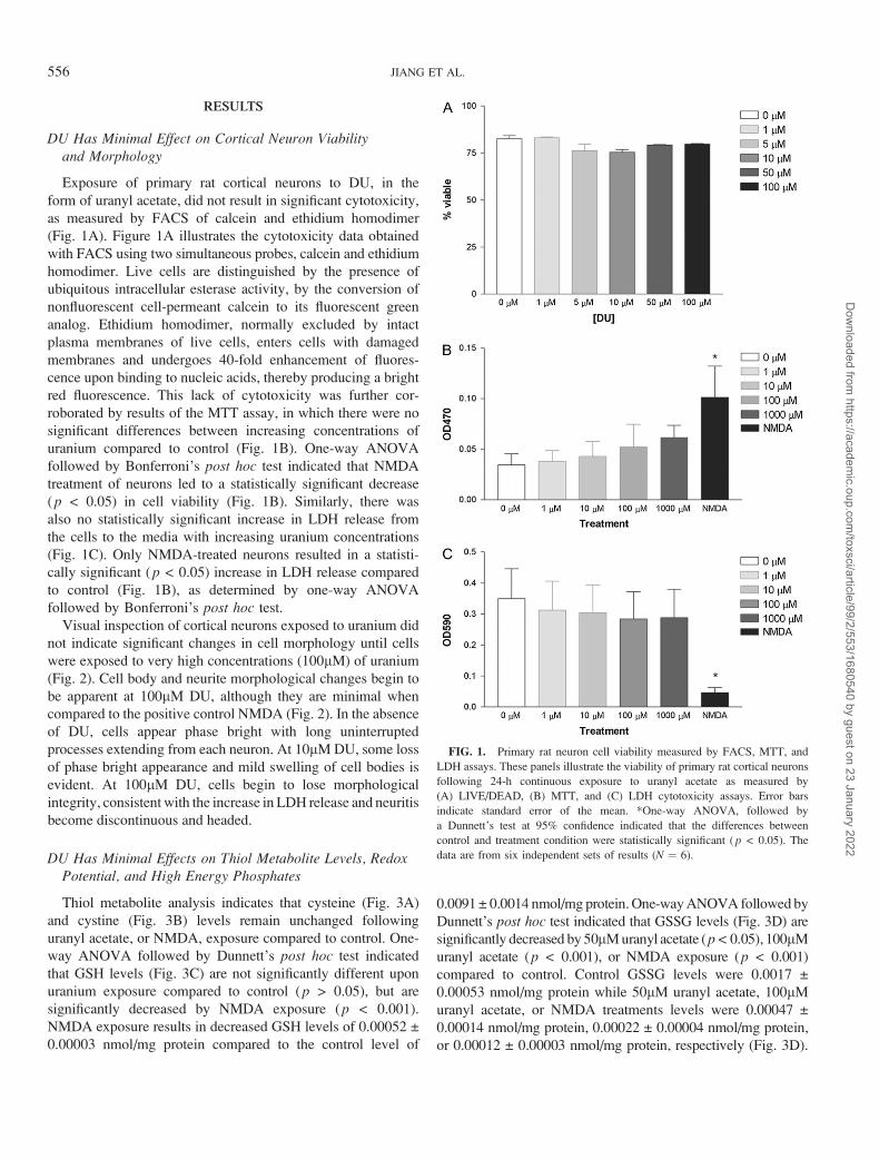
DU Has Minimal Effect on Cortical Neuron Viabilityand Morphology
Exposure of primary rat cortical neurons to DU, in the
form of uranyl acetate, did not result in significant cytotoxicity,
as measured by FACS of calcein and ethidium homodimer
(Fig. 1A). Figure 1A illustrates the cytotoxicity data obtained
with FACS using two simultaneous probes, calcein and ethidium
homodimer. Live cells are distinguished by the presence of
ubiquitous intracellular esterase activity, by the conversion of
nonfluorescent cell-permeant calcein to its fluorescent green
analog. Ethidium homodimer, normally excluded by intact
plasma membranes of live cells, enters cells with damaged
membranes and undergoes 40-fold enhancement of fluores-
cence upon binding to nucleic acids, thereby producing a bright
red fluorescence. This lack of cytotoxicity was further cor-
roborated by results of the MTT assay, in which there were no
significant differences between increasing concentrations of
uranium compared to control (Fig. 1B). One-way ANOVA
followed by Bonferroni’s post hoc test indicated that NMDA
treatment of neurons led to a statistically significant decrease
(p < 0.05) in cell viability (Fig. 1B). Similarly, there was
also no statistically significant increase in LDH release from
the cells to the media with increasing uranium concentrations
(Fig. 1C). Only NMDA-treated neurons resulted in a statisti-
cally significant (p < 0.05) increase in LDH release compared
to control (Fig. 1B), as determined by one-way ANOVA
followed by Bonferroni’s post hoc test.
Visual inspection of cortical neurons exposed to uranium did
not indicate significant changes in cell morphology until cells
were exposed to very high concentrations (100lM) of uranium
(Fig. 2). Cell body and neurite morphological changes begin to
be apparent at 100lM DU, although they are minimal when
compared to the positive control NMDA (Fig. 2). In the absence
of DU, cells appear phase bright with long uninterrupted
processes extending from each neuron. At 10lM DU, some loss
of phase bright appearance and mild swelling of cell bodies is
evident. At 100lM DU, cells begin to lose morphological
integrity, consistent with the increase in LDH release and neuritis
become discontinuous and headed.
DU Has Minimal Effects on Thiol Metabolite Levels, RedoxPotential, and High Energy Phosphates
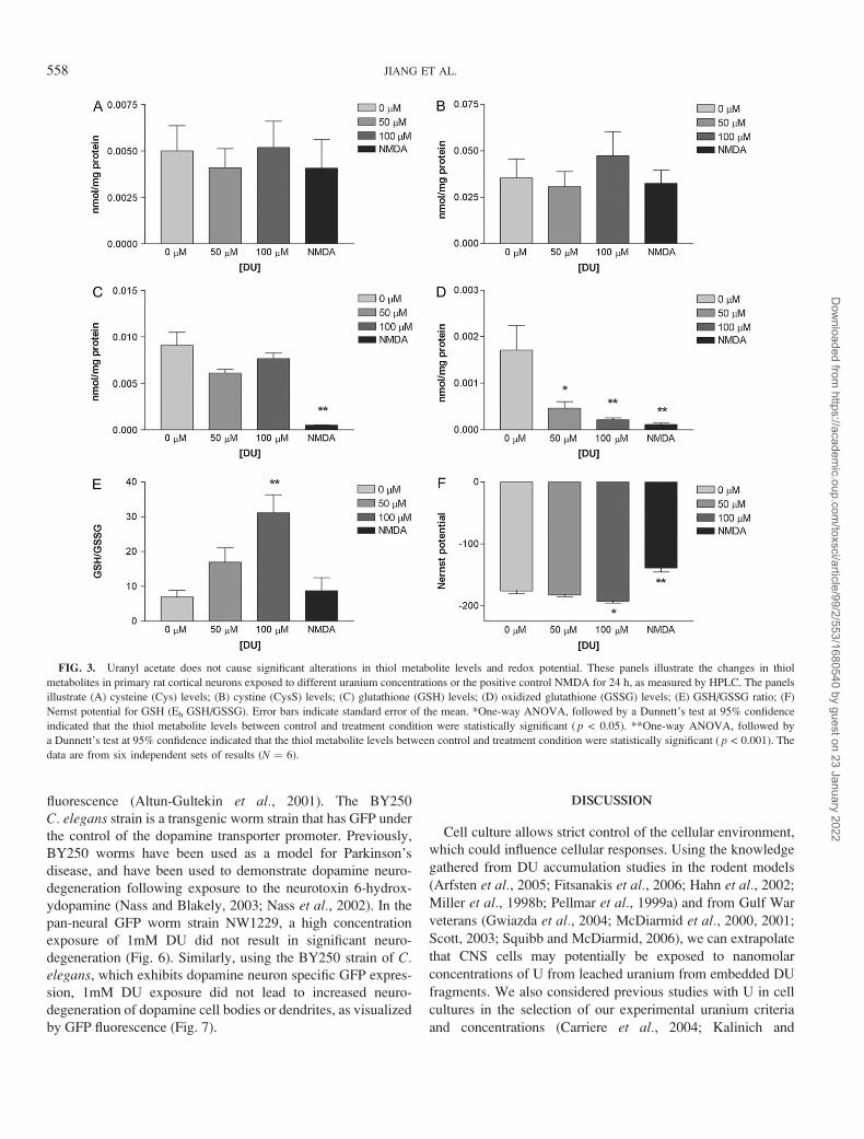
Thiol metabolite analysis indicates that cysteine (Fig. 3A)
and cystine (Fig. 3B) levels remain unchanged following
uranyl acetate, or NMDA, exposure compared to control. One-
way ANOVA followed by Dunnett’s post hoc test indicated
that GSH levels (Fig. 3C) are not significantly different upon
uranium exposure compared to control (p > 0.05), but are
significantly decreased by NMDA exposure (p < 0.001).
NMDA exposure results in decreased GSH levels of 0.00052 ±0.00003 nmol/mg protein compared to the control level of
0.0091± 0.0014 nmol/mg protein. One-way ANOVA followed by
Dunnett’s post hoc test indicated that GSSG levels (Fig. 3D) are
significantly decreased by 50lM uranyl acetate (p< 0.05), 100lM
uranyl acetate (p < 0.001), or NMDA exposure (p < 0.001)
compared to control. Control GSSG levels were 0.0017 ±0.00053 nmol/mg protein while 50lM uranyl acetate, 100lM
uranyl acetate, or NMDA treatments levels were 0.00047 ±0.00014 nmol/mg protein, 0.00022 ± 0.00004 nmol/mg protein,
or 0.00012 ± 0.00003 nmol/mg protein, respectively (Fig. 3D).
FIG. 1. Primary rat neuron cell viability measured by FACS, MTT, and
LDH assays. These panels illustrate the viability of primary rat cortical neurons
following 24-h continuous exposure to uranyl acetate as measured by
(A) LIVE/DEAD, (B) MTT, and (C) LDH cytotoxicity assays. Error bars
indicate standard error of the mean. *One-way ANOVA, followed by
a Dunnett’s test at 95% confidence indicated that the differences between
control and treatment condition were statistically significant ( p < 0.05). The
data are from six independent sets of results (N ¼ 6).
556 JIANG ET AL.
Dow
nloaded from https://academ
ic.oup.com/toxsci/article/99/2/553/1680540 by guest on 23 January 2022
One-way ANOVA analysis followed by Dunnett’s post hoc test
indicated that the GSH/GSSG ratio of 100lM uranyl acetate
exposed neurons was 31.16 ± 5.23, which was significantly
higher (p< 0.001) than the control ratio of 6.98 ± 1.83 (Fig. 3E).
There was no statistical difference between GSH/GSSG ratios
(Fig. 3E) for 50lM uranyl acetate, or NMDA treatments compared
to control (p > 0.05). Overall the Nernst potential for GSH
(Eh GSH/GSSG, Fig. 3F) is � 176.0 ± 4.62 for control, and �182.4 ± 3.08 in 50lM uranyl acetate exposed neurons, which
is not statistically significant (p > 0.05). Eh GSH/GSSG
for 100lM uranyl acetate exposure (Fig. 3F) is � 192.7 ±2.94, and is statistically significant compared to control (p <0.05). The Eh GSH/GSSG following NMDA exposure (Fig. 3F)
is� 138.9± 6.46 and exhibits greater statistical significance (p<0.001).
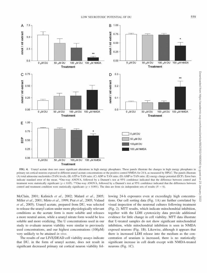
DU exposure of neurons did not result in a statistically
significant decrease in TAN compared to control, although
there was a statistically significant ( p < 0.001) decrease
in TAN in NMDA-exposed neurons to control (Fig. 4A),
as determined by one-way ANOVA followed by Dunnett’s
test at 95% confidence interval. NMDA treatment also yielded
an overall decrease in the ATP to TAN ratio from 0.75 ± 0.029
in controls to 0.42 ± 0.098 (Fig. 4B), which was statistically
significant ( p < 0.05). The ADP to TAN ratio does not result
in statistically significant differences with increasing DU
concentrations, or with NMDA treatment, compared to
control (Fig. 4C). Like the ATP to TAN ratio, the ratio of
AMP to TAN ratio is only significantly different ( p < 0.001)
in NMDA treated cells, but not in DU exposed (Fig. 4D).
Overall, the ECP of the control primary rat cortical neurons is
0.84 ± 0.016, and did not result in statistically significant
changes upon DU exposure, but was significantly decreased
( p < 0.05) in the NMDA treated cultures 0.63 ± 0.050
(Fig. 4E).
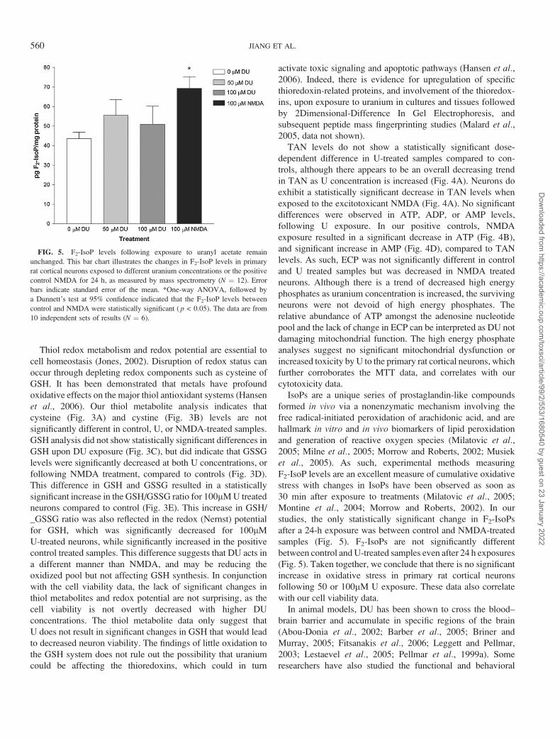
DU Exposure Does Not Significantly Change F2-IsoPLevels in Primary Rat Cortical Neurons
One-way ANOVA indicates that DU exposure did not result
in a significant increase in the level of F2-IsoP, products of
lipid peroxidation, between controls and treatments (Fig. 5).
After a 24 h 50lM DU exposure, neurons demonstrated
F2-IsoP levels of 55.5 ± 8.1 pg/mg total protein, while 24 h
100lM DU exposed neurons exhibited 51.0 ± 9.2 pg/mg total
protein F2-IsoP. The only statistically significant difference be-
tween samples occurred between the control (43.6 ± 3.3 pg/mg
total protein) and the positive control NMDA treated neurons,
which increased F2-IsoP levels to 69.3 ± 5.9 pg/mg total protein
(p < 0.05).
DU Exposure Does Not Cause Neurodegenerationin C. elegans
DU exposure does lead to increased uranium accumulation
in the different C. elegans strains, with 100lM DU treated N2
worms exhibiting 10.9 ± 1.10 ng 238U/lg total protein (data not
shown). The NW1229 strain is a transgenic C. elegans strain
that is a fusion of the GFP gene to the promoter of the F25B3.3gene, the C. elegans ortholog of the Ca2þ-regulated rasnucleotide exchange factor CalDAG-GEFII/RasGRP, which is
ubiquitously expressed in the vertebrate nervous system (Ebinu
et al., 1998; Kawasaki et al., 1998). The resulting transgenic
strain produces exclusive pan-neural GFP expression, in which
all neurons express GFP, and can be easily visualized with
FIG. 2. Uranyl acetate exposure results in minimal morphological changes in primary rat cortical neurons. These panels are representative images to illustrate the
cellular morphology of primary rat cortical neurons exposed to 0-, 1-, 10-, or 100lM uranyl acetate or the excitotoxic positive control (100lM NMDA with 10lM
glycine). Changes in cell body and dendrite morphology begin to be noticeable at 100lM uranyl acetate, and are readily apparent in the positive NMDA control.
LOW NEUROTOXIC POTENTIAL OF DU 557
Dow
nloaded from https://academ
ic.oup.com/toxsci/article/99/2/553/1680540 by guest on 23 January 2022
fluorescence (Altun-Gultekin et al., 2001). The BY250
C. elegans strain is a transgenic worm strain that has GFP under
the control of the dopamine transporter promoter. Previously,
BY250 worms have been used as a model for Parkinson’s
disease, and have been used to demonstrate dopamine neuro-
degeneration following exposure to the neurotoxin 6-hydrox-
ydopamine (Nass and Blakely, 2003; Nass et al., 2002). In the
pan-neural GFP worm strain NW1229, a high concentration
exposure of 1mM DU did not result in significant neuro-
degeneration (Fig. 6). Similarly, using the BY250 strain of C.elegans, which exhibits dopamine neuron specific GFP expres-
sion, 1mM DU exposure did not lead to increased neuro-
degeneration of dopamine cell bodies or dendrites, as visualized
by GFP fluorescence (Fig. 7).
DISCUSSION
Cell culture allows strict control of the cellular environment,
which could influence cellular responses. Using the knowledge
gathered from DU accumulation studies in the rodent models
(Arfsten et al., 2005; Fitsanakis et al., 2006; Hahn et al., 2002;
Miller et al., 1998b; Pellmar et al., 1999a) and from Gulf War
veterans (Gwiazda et al., 2004; McDiarmid et al., 2000, 2001;
Scott, 2003; Squibb and McDiarmid, 2006), we can extrapolate
that CNS cells may potentially be exposed to nanomolar
concentrations of U from leached uranium from embedded DU
fragments. We also considered previous studies with U in cell
cultures in the selection of our experimental uranium criteria
and concentrations (Carriere et al., 2004; Kalinich and
FIG. 3. Uranyl acetate does not cause significant alterations in thiol metabolite levels and redox potential. These panels illustrate the changes in thiol
metabolites in primary rat cortical neurons exposed to different uranium concentrations or the positive control NMDA for 24 h, as measured by HPLC. The panels
illustrate (A) cysteine (Cys) levels; (B) cystine (CysS) levels; (C) glutathione (GSH) levels; (D) oxidized glutathione (GSSG) levels; (E) GSH/GSSG ratio; (F)
Nernst potential for GSH (Eh GSH/GSSG). Error bars indicate standard error of the mean. *One-way ANOVA, followed by a Dunnett’s test at 95% confidence
indicated that the thiol metabolite levels between control and treatment condition were statistically significant ( p < 0.05). **One-way ANOVA, followed by
a Dunnett’s test at 95% confidence indicated that the thiol metabolite levels between control and treatment condition were statistically significant ( p < 0.001). The
data are from six independent sets of results (N ¼ 6).
558 JIANG ET AL.
Dow
nloaded from https://academ
ic.oup.com/toxsci/article/99/2/553/1680540 by guest on 23 January 2022
McClain, 2001; Kalinich et al., 2002; Malard et al., 2005;
Miller et al., 2001; Mirto et al., 1999; Prat et al., 2005; Vidaud
et al., 2005). Uranyl acetate, prepared from DU, was selected
to release the uranyl cation under more physiologically relevant
conditions as the acetate form is more soluble and releases
a more neutral anion, while a uranyl nitrate form would be less
soluble and more oxidizing. The U concentrations used in our
study to evaluate neuron viability were similar to previously
used concentrations, and our highest concentrations (100lM)
very unlikely to be attained in vivo.
The results of our LIVE/DEAD cell viability assays indicate
that DU, in the form of uranyl acetate, does not result in
significant decreased primary rat cortical neuron viability fol-
lowing 24-h exposures even at exceedingly high concentra-
tions. Our cell sorting data (Fig. 1A) are further correlated by
visual inspection of the neuronal cultures following treatment
(Fig. 2). MTT results, which indicate mitochondrial inhibition,
together with the LDH cytotoxicity data provide additional
evidence for little change in cell viability. MTT data illustrate
that U-treated samples do not show significant mitochondrial
inhibition, while mitochondrial inhibition is seen in NMDA
exposed neurons (Fig. 1B). Likewise, although it appears that
there is increased LDH release into the medium as the con-
centration of uranium is increased, there is no statistically
significant increase in cell death except with NMDA-treated
neurons (Fig. 1C).
FIG. 4. Uranyl acetate does not cause significant alterations in high energy phosphates. These panels illustrate the changes in high energy phosphates in
primary rat cortical neurons exposed to different uranyl acetate concentrations or the positive control NMDA for 24 h, as measured by HPLC. The panels illustrate
(A) total adenosine nucleotides (TAN) levels; (B) ATP to TAN ratio; (C) ADP to TAN ratio; (D) AMP to TAN ratio; (E) energy charge potential (ECP). Error bars
indicate standard error of the mean. *One-way ANOVA, followed by a Dunnett’s test at 95% confidence indicated that the difference between control and
treatment were statistically significant ( p < 0.05). **One-way ANOVA, followed by a Dunnett’s test at 95% confidence indicated that the differences between
control and treatment condition were statistically significant ( p < 0.001). The data are from six independent sets of results (N ¼ 6).
LOW NEUROTOXIC POTENTIAL OF DU 559
Dow
nloaded from https://academ
ic.oup.com/toxsci/article/99/2/553/1680540 by guest on 23 January 2022
Thiol redox metabolism and redox potential are essential to
cell homeostasis (Jones, 2002). Disruption of redox status can
occur through depleting redox components such as cysteine of
GSH. It has been demonstrated that metals have profound
oxidative effects on the major thiol antioxidant systems (Hansen
et al., 2006). Our thiol metabolite analysis indicates that
cysteine (Fig. 3A) and cystine (Fig. 3B) levels are not
significantly different in control, U, or NMDA-treated samples.
GSH analysis did not show statistically significant differences in
GSH upon DU exposure (Fig. 3C), but did indicate that GSSG
levels were significantly decreased at both U concentrations, or
following NMDA treatment, compared to controls (Fig. 3D).
This difference in GSH and GSSG resulted in a statistically
significant increase in the GSH/GSSG ratio for 100lM U treated
neurons compared to control (Fig. 3E). This increase in GSH/
_GSSG ratio was also reflected in the redox (Nernst) potential
for GSH, which was significantly decreased for 100lM
U-treated neurons, while significantly increased in the positive
control treated samples. This difference suggests that DU acts in
a different manner than NMDA, and may be reducing the
oxidized pool but not affecting GSH synthesis. In conjunction
with the cell viability data, the lack of significant changes in
thiol metabolites and redox potential are not surprising, as the
cell viability is not overtly decreased with higher DU
concentrations. The thiol metabolite data only suggest that
U does not result in significant changes in GSH that would lead
to decreased neuron viability. The findings of little oxidation to
the GSH system does not rule out the possibility that uranium
could be affecting the thioredoxins, which could in turn
activate toxic signaling and apoptotic pathways (Hansen et al.,2006). Indeed, there is evidence for upregulation of specific
thioredoxin-related proteins, and involvement of the thioredox-
ins, upon exposure to uranium in cultures and tissues followed
by 2Dimensional-Difference In Gel Electrophoresis, and
subsequent peptide mass fingerprinting studies (Malard et al.,2005, data not shown).
TAN levels do not show a statistically significant dose-
dependent difference in U-treated samples compared to con-
trols, although there appears to be an overall decreasing trend
in TAN as U concentration is increased (Fig. 4A). Neurons do
exhibit a statistically significant decrease in TAN levels when
exposed to the excitotoxicant NMDA (Fig. 4A). No significant
differences were observed in ATP, ADP, or AMP levels,
following U exposure. In our positive controls, NMDA
exposure resulted in a significant decrease in ATP (Fig. 4B),
and significant increase in AMP (Fig. 4D), compared to TAN
levels. As such, ECP was not significantly different in control
and U treated samples but was decreased in NMDA treated
neurons. Although there is a trend of decreased high energy
phosphates as uranium concentration is increased, the surviving
neurons were not devoid of high energy phosphates. The
relative abundance of ATP amongst the adenosine nucleotide
pool and the lack of change in ECP can be interpreted as DU not
damaging mitochondrial function. The high energy phosphate
analyses suggest no significant mitochondrial dysfunction or
increased toxicity by U to the primary rat cortical neurons, which
further corroborates the MTT data, and correlates with our
cytotoxicity data.
IsoPs are a unique series of prostaglandin-like compounds
formed in vivo via a nonenzymatic mechanism involving the
free radical-initiated peroxidation of arachidonic acid, and are
hallmark in vitro and in vivo biomarkers of lipid peroxidation
and generation of reactive oxygen species (Milatovic et al.,2005; Milne et al., 2005; Morrow and Roberts, 2002; Musiek
et al., 2005). As such, experimental methods measuring
F2-IsoP levels are an excellent measure of cumulative oxidative
stress with changes in IsoPs have been observed as soon as
30 min after exposure to treatments (Milatovic et al., 2005;
Montine et al., 2004; Morrow and Roberts, 2002). In our
studies, the only statistically significant change in F2-IsoPs
after a 24-h exposure was between control and NMDA-treated
samples (Fig. 5). F2-IsoPs are not significantly different
between control and U-treated samples even after 24 h exposures
(Fig. 5). Taken together, we conclude that there is no significant
increase in oxidative stress in primary rat cortical neurons
following 50 or 100lM U exposure. These data also correlate
with our cell viability data.
In animal models, DU has been shown to cross the blood–
brain barrier and accumulate in specific regions of the brain
(Abou-Donia et al., 2002; Barber et al., 2005; Briner and
Murray, 2005; Fitsanakis et al., 2006; Leggett and Pellmar,
2003; Lestaevel et al., 2005; Pellmar et al., 1999a). Some
researchers have also studied the functional and behavioral
FIG. 5. F2-IsoP levels following exposure to uranyl acetate remain
unchanged. This bar chart illustrates the changes in F2-IsoP levels in primary
rat cortical neurons exposed to different uranium concentrations or the positive
control NMDA for 24 h, as measured by mass spectrometry (N ¼ 12). Error
bars indicate standard error of the mean. *One-way ANOVA, followed by
a Dunnett’s test at 95% confidence indicated that the F2-IsoP levels between
control and NMDA were statistically significant ( p < 0.05). The data are from
10 independent sets of results (N ¼ 6).
560 JIANG ET AL.
Dow
nloaded from https://academ
ic.oup.com/toxsci/article/99/2/553/1680540 by guest on 23 January 2022

changes associated with increased DU accumulation to in-
vestigate if the cognitive defects that were seen in Gulf War
veterans could be correlated to specific neurotransmitters
(Abou-Donia et al., 2002; Belles et al., 2005; Briner and
Murray, 2005; Houpert et al., 2004, 2005; Pellmar et al.,1999b). While studies have shown increased oxidative stress in
certain brain regions, it remains unclear whether DU accumu-
lation in the brain results in any significant neurodegeneration.
It would be logical to surmise that there is very little degener-
ation occurring considering the minimal functional changes
that have been shown following DU administration. The nem-
atode C. elegans provides an excellent model to visualize
neurodegeneration, as there is no blood–brain barrier that
neurotoxicants must traverse, and neurons can be easily labeled
with fluorescent markers. The ease of growth, maintenance,
and manipulation also allows researchers to evaluate the
chemotoxic effects on a whole organism, which helps in further
extrapolation of data to human health and disease. Considering
these factors, we utilized two transgenic strains to determine
if DU exposure led to any neurodegeneration. Nematodes do
accumulate U with increasing U exposure (data not shown)
and in the pan-neural GFP-expressing strain NW1229, our
experiments demonstrate that there is no significant degener-
ation of neurons following U exposure (Fig. 6). Several pieces
of data from previously published articles have suggested
potential involvement of the dopaminergic system following
DU exposure including increased uranium uptake in the
midbrain of rats implanted with DU pellets (Houpert et al.,2004; Pellmar et al., 1999a), and increased lipid peroxidation
and nitric oxide generation in the midbrain of rats exposed to
DU (Abou-Donia et al., 2002). Confocal microscopic in-
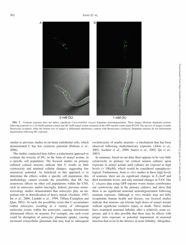
spection of BY250 worms did not demonstrate significant
degeneration of dopamine neurons following U exposure
(Fig. 7).
Researchers have demonstrated increased accumulation of
DU in specific brain regions following increased DU exposure
as previously described (Jiang and Aschner, 2006). In our
nematode experiments, we have evaluated the effects of U on
C. elegans neurons in an attempt to evaluate neurons in brain
regions that have been shown to accumulate DU in rats. Our
data demonstrate that there is a dose-dependent increase in
uranium accumulation (data not shown) but not a corresponding
increase in neurodegeneration (Figs. 6 and 7). These results
correlate with the results from our experiments with primary rat
cortical neuron cultures which indicate that the neurons can
tolerate U without significant cell death (Fig. 1A). Our data are
FIG. 6. Uranium exposure does not result in significant neurodegeneration in Caenorhabditis elegans. These images illustrate the pan-neural GFP reporter
strain of C. elegans (NW1229) exposed to a (A) 0mM control and (B) 1mM treatment dose of uranyl acetate. The top row of images is under fluorescent excitation,
while the bottom row of images is differential interference contrast with fluorescence overlayed. There is no visual evidence for neuronal degeneration following
DU exposure.
LOW NEUROTOXIC POTENTIAL OF DU 561
Dow
nloaded from https://academ
ic.oup.com/toxsci/article/99/2/553/1680540 by guest on 23 January 2022
similar to previous studies in rat brain endothelial cells, which
demonstrated U has low cytotoxic potential (Dobson et al.,2006).
The studies conducted here follow a reductionist approach to
evaluate the toxicity of DU, in the form of uranyl acetate, in
a specific cell population. The focused studies on primary
cultured cortical neurons indicate that U results in little
cytotoxicity and minimal cellular changes, suggesting low
neurotoxic potential. As beneficial as this approach is to
determine the effects within a specific cell population, this
methodology cannot exclude the possibility that DU has
neurotoxic effects on other cell populations within the CNS,
such as astrocytes and/or microglia. Indeed, previous neuro-
toxicology studies demonstrated that astrocytes play an im-
portant role in detoxification of heavy metals (Aschner, 1997;
Im et al., 2006; Lindahl et al., 1999; Tiffany-Castiglion and
Qian, 2001). As such, the possibility exists that U accumulates
within astrocytes, resulting in a variety of subsequent
molecular events within the astrocytes, causing downstream
detrimental effects on neurons. For example, one such event
could be disruption of astrocytic glutamate uptake, causing
increased extracellular glutamate that may lead to subsequent
excitotoxicity of nearby neurons—a mechanism that has been
observed following methylmercury exposure (Allen et al.,2002; Aschner et al., 2000; Juarez et al., 2002; Qu et al.,2003).
In summary, based on our data, there appears to be very little
cytotoxicity in primary rat cortical neuron cultures upon
exposure to uranyl acetate until cultures are exposed to high
levels (> 100lM), which would be considered supraphysio-
logical. Furthermore, from in vitro studies at these high levels
of uranium, there are no significant changes in F2-IsoP and
thiol metabolite levels, and only minimal changes in TAN. Our
C. elegans data using GFP reporter worm strains corroborates
our cytotoxicity data in the primary cultures, and show that
there is no significant neuronal neurodegeneration following
uranium exposure. Although in vitro models may not fully
recapitulate human health and disease, our focused studies
indicate that neurons can tolerate high doses of uranyl acetate
without significant oxidative injury or death. These studies
have examined neuronal lethality after a relatively acute ex-
posure, and it is also possible that there may be effects with
longer term exposure or potential impairment of neuronal
function that occur in the absence of acute lethality. Altogether,
FIG. 7. Uranium exposure does not induce significant Caenorhabditis elegans dopamine neurodegeneration. These images illustrate dopamine neurons
following exposure to a (A) 0mM uranium control and (B) 1mM uranyl acetate treatment in the GFP reporter worm strain BY250. The top row of images is under
fluorescent excitation, while the bottom row of images is differential interference contrast with fluorescence overlayed. Dopamine neurons do not demonstrate
degeneration following DU exposure.
562 JIANG ET AL.
Dow
nloaded from https://academ
ic.oup.com/toxsci/article/99/2/553/1680540 by guest on 23 January 2022
although our reductionist approach cannot exclude the
possibility of DU as a neurotoxic agent to other CNS cell
populations, the results of our studies in primary rat cortical
neurons and in C. elegans demonstrate low acute neurotoxic
potential of uranyl acetate, and should alleviate some of the
concern surrounding DU as a neurotoxin, and as a chemical
that may be responsible for GWS. These results support the
emerging agreement among workers in the field that DU
neurotoxicity is not a component of primary GWS, though it
may constitute a separate entity in the cluster of Gulf War
illnesses.
FUNDING
D.O.D. grant (DAMD 17-01-1-0685) to M.A.
REFERENCES
Abou-Donia, M. B., Dechkovskaia, A. M., Goldstein, L. B., Shah, D. U.,
Bullman, S. L., and Khan, W. A. (2002). Uranyl acetate-induced
sensorimotor deficit and increased nitric oxide generation in the central
nervous system in rats. Pharmacol. Biochem. Behav. 99(2), 553–565.
Abu-Qare, A. W., and Abou-Donia, M. B. (2002). Depleted uranium—The
growing concern. J. Appl. Toxicol. 22(3), 149–152.
Albertini, R. J., Jacobson-Kram, D., Sullivan, L. M., Gucer, P., and
McDiarmid, M. A. (2003). HPRT mutations in T-cells in Gulf War veterans
exposed to depleted uranium (DU). Toxicol. Sci. 72(1), 280.
Allen, J. W., Shanker, G., Tan, K. H., and Aschner, M. (2002). The
consequences of methylmercury exposure on interactive functions between
astrocytes and neurons. Neurotoxicology 23(6), 755–759.
Altun-Gultekin, Z., Andachi, Y., Tsalik, E. L., Pilgrim, D., Kohara, Y., and
Hobert, O. (2001). A regulatory cascade of three homeobox genes, ceh-10,
ttx-3 and ceh-23, controls cell fate specification of a defined interneuron class
in C. elegans. Development 128(11), 1951–1969.
Anderson, S. L., and Wild, G. C. (1994). Linking genotoxic responses and
reproductive success in ecotoxicology. Environ. Health Perspect.
102((Suppl. 12), 9–12.
Andrews, P. M., and Bates, S. B. (1987). Effects of dietary protein on uranyl-
nitrate-induced acute renal failure. Nephron 45(4), 296–301.
Arfsten, D. P., Schaeffer, D. J., Johnson, E. W., Cunningham, J. R., Still, K. R.,
and Wilfong, E. R. (2006). Evaluation of the effect of implanted depleted
uranium on male reproductive success, sperm concentration, and sperm
velocity. Environ. Res. 100(2), 205–215.
Aschner, M. (1997). Astrocyte metallothioneins (MTs) and their neuro-
protective role. Ann. N.Y. Acad. Sci. 825, 334–347.
Aschner, M., Yao, C. P., Allen, J. W., and Tan, K. H. (2000). Methylmercury
alters glutamate transport in astrocytes. Neurochem. Int. 37(2–3), 199–206.
Barber, D. S., Ehrich, M. F., and Jortner, B. S. (2005). The effect of stress on
the temporal and regional distribution of uranium in rat brain after acute
uranyl acetate exposure. J. Toxicol. Environ. Health A 68(2), 99–111.
Belles, M., Albina, M. L., Linares, V., Gomez, M., Sanchez, D. J., and
Domingo, J. L. (2005). Combined action of uranium and stress in the rat - I.
Behavioral effects. Toxicol. Lett. 158(3), 176–185.
Bem, H., and Bou-Rabee, F. (2004). Environmental and health consequences of
depleted uranium use in the 1991 Gulf War. Environ. Int. 30(1), 123–134.
Brenner, S. (1974). The genetics of Caenorhabditis elegans. Genetics 77(1),
71–94.
Briner, W., and Murray, J. (2005). Effects of short-term and long-term depleted
uranium exposure on open-field behavior and brain lipid oxidation in rats.
Neurotoxicol. Teratol. 27(1), 135–144.
Carriere, M., Avoscan, L., Collins, R., Carrot, F., Khodja, H., Ansoborlo, E.,
and Gouget, B. (2004). Influence of uranium speciation on normal rat kidney
(NRK-52E) proximal cell cytotoxicity. Chem. Res. Toxicol. 17(3), 446–452.
Carriere, M., Gouget, B., Gallien, J. P., Avoscan, L., Gobin, R.,
Verbavatz, J. M., and Khodja, H. (2005). Cellular distribution of uranium
after acute exposure of renal epithelial cells: SEM, TEM and nuclear
microscopy analysis. Nucl. Instr. Methods Phys. Res. B Beam Interact.
Mater. Atoms 231, 268–273.
Chen, M., Yang, Z., Wu, R., and Nadler, J. L. (2002). Lisofylline, a novel
antiinflammatory agent, protects pancreatic beta-cells from proinflammatory
cytokine damage by promoting mitochondrial metabolism. Endocrinology
143(6), 2341–2348.
Dhawan, R., Dusenbery, D. B., and Williams, P. L. (1999). Comparison
of lethality, reproduction, and behavior as toxicological endpoints in
the nematode Caenorhabditis elegans. J. Toxicol. Environ. Health A 58(7),
451–462.
Dobson, A. W., Lack, A. K., Erikson, K. M., and Aschner, M. (2006). Depleted
uranium is not toxic to rat brain endothelial (RBE4) cells. Biol. Trace Elem.
Res. 110(1), 61–72.
Doucet, I. (1994). Desert Storm syndrome: Sick soldiers and dead children?
Med. War 10(3), 183–194.
Durakovic, A. (2003). Undiagnosed illnesses and radioactive warfare. Croat.
Med. J. 44(5), 520–532.
Ebinu, J. O., Bottorff, D. A., Chan, E. Y., Stang, S. L., Dunn, R. J., and
Stone, J. C. (1998). RasGRP, a Ras guanyl nucleotide-releasing protein with
calcium- and diacylglycerol-binding motifs. Science 280(5366), 1082–1086.
Fitsanakis, V. A., Erikson, K. M., Garcia, S. J., Evje, L., Syversen, T., and
Aschner, M. (2006). Brain accumulation of depleted uranium in rats
following 3- or 6-month treatment with implanted depleted uranium pellets.
Biol. Trace Elem. Res. 111(1–3), 185–197.
Gally, C., and Bessereau, J. L. (2003). C. elegans: Of neurons and genes]. Med.
Sci. (Paris) 19(6–7), 725–734.
Goldman, M., Yaari, A., Doshnitzki, Z., Cohen-Luria, R., and Moran, A.
(2006). Nephrotoxicity of uranyl acetate: effect on rat kidney brush border
membrane vesicles. Arch. Toxicol. 80(7), 387–393.
Gronseth, G. S. (2005). Gulf War syndrome: A toxic exposure? A systematic
review. Neurol. Clin.23(2), 523-þ.
Gwiazda, R. H., Squibb, K., McDiarmid, M., and Smith, D. (2004). Detection
of depleted uranium in urine of veterans from the 1991 Gulf War. Health
Phys. 86(1), 12–18.
Hahn, F. F., Guilmette, R. A., and Hoover, M. D. (2002). Implanted depleted
uranium fragments cause soft tissue sarcomas in the muscles of rats. Environ.
Health Perspect. 110(1), 51–59.
Hansen, J. M., Zhang, H., and Jones, D. P. (2006). Differential oxidation
of thioredoxin-1, thioredoxin-2, and glutathione by metal ions. Free Radic.
Biol. Med. 40(1), 138–145.
Hobert, O., and Loria, P. (2006). Uses of GFP in Caenorhabditis elegans.
Methods Biochem. Anal. 47, 203–226.
Houpert, P., Lestaevel, P., Amourette, C., Dhieux, B., Bussy, C., and Paquet, F.
(2004). Effect of U and 137Cs chronic contamination on dopamine and
serotonin metabolism in the central nervous system of the rat. Can.
J. Physiol. Pharmacol. 82(2), 161–166.
Houpert, P., Lestaevel, P., Bussy, C., Paquet, F., and Gourmelon, P. (2005).
Enriched but not depleted uranium affects central nervous system in long-
term exposed rat. Neurotoxicology. 26(2), 1015–1020.
Im, J. Y., Paik, S. G., and Han, P. L. (2006). Cadmium-induced astroglial death
proceeds via glutathione depletion. J. Neurosci. Res. 83(2), 301–308.
LOW NEUROTOXIC POTENTIAL OF DU 563
Dow
nloaded from https://academ
ic.oup.com/toxsci/article/99/2/553/1680540 by guest on 23 January 2022
Jamal, G. A., Hansen, S., Apartopoulos, F., and Peden, A. (1996). The ‘‘Gulf
War syndrome’’. Is there evidence of dysfunction in the nervous system?
J. Neurol. Neurosurg. Psychiatr. 60(4), 449–451.
Jiang, G. C., and Aschner, M. (2006). Neurotoxicity of depleted uranium:
Reasons for increased concern. Biol. Trace Elem. Res. 110(1), 1–18.
Jones, D. P. (2002). Redox potential of GSH/GSSG couple: Assay and
biological significance. Methods Enzymol. 348, 93–112.
Jones, D. P., Carlson, J. L., Samiec, P. S., Sternberg, P., Jr, Mody, V. C., Jr,
Reed, R. L., and Brown, L. A. (1998). Glutathione measurement in human
plasma. Evaluation of sample collection, storage and derivatization con-
ditions for analysis of dansyl derivatives by HPLC. Clin. Chim. Acta 275(2),
175–184.
Juarez, B. I., Martinez, M. L., Montante, M., Dufour, L., Garcia, E., and
Jimenez-Capdeville, M. E. (2002). Methylmercury increases glutamate
extracellular levels in frontal cortex of awake rats. Neurotoxicol. Teratol.
24(6), 767–771.
Kalinich, J. F., and McClain, D. E. (2001). Staining of intracellular deposits of
uranium in cultured murine macrophages. Biotech. Histochem. 76(5–6),
247–252.
Kalinich, J. F., Ramakrishnan, N., Villa, V., and McClain, D. E. (2002).
Depleted uranium-uranyl chloride induces apoptosis in mouse J774 macro-
phages. Toxicology 179(1–2), 105–114.
Kawasaki, H., Springett, G. M., Toki, S., Canales, J. J., Harlan, P.,
Blumenstiel, J. P., Chen, E. J., Bany, I. A., Mochizuki, N., Ashbacher, A.,
et al. (1998). A Rap guanine nucleotide exchange factor enriched highly
in the basal ganglia. Proc. Natl. Acad. Sci. U.S.A. 95(22), 13278–13283.
Kobayashi, S., Nagase, M., Honda, N., and Hishida, A. (1984). Glomerular
alterations in uranyl acetate-induced acute renal failure in rabbits. Kidney Int.
26(6), 808–815.
Leggett, R. W., and Pellmar, T. C. (2003). The biokinetics of uranium
migrating from embedded DU fragments. J. Environ. Radioact. 64(2–3),
205–225.
Legrand, C., Bour, J. M., Jacob, C., Capiaumont, J., Martial, A., Marc, A.,
Wudtke, M., Kretzmer, G., Demangel, C., Duval, D., et al. (1992). Lactate
dehydrogenase (LDH) activity of cultured eukaryotic cells as marker of the
number of dead cells in the medium. J. Biotechnol. 25(3), 231–243.
Lestaevel, P., Houpert, P., Bussy, C., Dhieux, B., Gourmelon, P., and
Paquet, F. (2005). The brain is a target organ after acute exposure to depleted
uranium. Toxicology 212(2–3), 219–226.
Lewis, J. A., and Fleming, J. T. (1995). Basic culture methods. Methods Cell
Biol. 48, 3–29.
Lin, R. H., Wu, L. J., Lee, C. H., and Lin-Shiau, S. Y. (1993). Cytogenetic
toxicity of uranyl nitrate in Chinese hamster ovary cells. Mutat. Res. 319(3),
197–203.
Lindahl, L. S., Bird, L., Legare, M. E., Mikeska, G., Bratton, G. R., and
Tiffany-Castiglioni, E. (1999). Differential ability of astroglia and neuronal
cells to accumulate lead: Dependence on cell type and on degree of
differentiation. Toxicol. Sci. 50(2), 236–243.
Link, C. D., and Johnson, C. J. (2002). Reporter transgenes for study of oxidant
stress in Caenorhabditis elegans. Methods Enzymol. 353, 497–505.
Malard, V., Prat, O., Darrouzet, E., Berenguer, F., Sage, N., and Quemeneur, E.
(2005). Proteomic analysis of the response of human lung cells to uranium.
Proteomics 5(17), 4568–4580.
McDiarmid, M. A., Engelhardt, S. M., and Oliver, M. (2001). Urinary uranium
concentrations in an enlarged Gulf War veteran cohort. Health Phys. 80(3),
270–273.
McDiarmid, M. A., Keogh, J. P., Hooper, F. J., McPhaul, K., Squibb, K.,
Kane, R., DiPino, R., Kabat, M., Kaup, B., Anderson, L., et al. (2000).
Health effects of depleted uranium on exposed Gulf War veterans. Environ.
Res. 82(2), 168–180.
McLaughlin, B. A., Nelson, D., Silver, I. A., Erecinska, M., and
Chesselet, M. F. (1998). Methylmalonate toxicity in primary neuronal
cultures. Neuroscience 86(1), 279–290.
Milatovic, D., Gupta, R. C., Dekundy, A., Montine, T. J., and Dettbarn, W. D.
(2005). Carbofuran-induced oxidative stress in slow and fast skeletal
muscles: Prevention by memantine and atropine. Toxicology 208(1), 13–24.
Miller, A. C., Blakely, W. F., Livengood, D., Whittaker, T., Xu, J. Q.,
Ejnik, J. W., Hamilton, M. M., Parlette, E., St John, T., Gerstenberg, H. M.,
et al. (1998a). Transformation of human osteoblast cells to the tumorigenic
phenotype by depleted uranium uranyl chloride. Environ. Health Perspect.
106(8), 465–471.
Miller, A. C., Brooks, K., Smith, J., and Page, N. (2004). Effect of the
militarily-relevant heavy metals, depleted uranium and heavy metal tungsten-
alloy on gene expression in human liver carcinoma cells (HepG2). Molecular
and Cellular Biochemistry 255(1–2), 247–256.
Miller, A. C., Brooks, K., Stewart, M., Anderson, B., Shi, L., McClain, D., and
Page, N. (2003). Genomic instability in human osteoblast cells after exposure
to depleted uranium: Delayed lethality and micronuclei formation. Journal
of Environmental Radioactivity 64(2–3), 247–259.
Miller, A. C., Fuciarelli, A. F., Jackson, W. E., Ejnik, E. J., Emond, C.,
Strocko, S., Hogan, J., Page, N., and Pellmar, T. (1998b). Urinary and serum
mutagenicity studies with rats implanted with depleted uranium or tantalum
pellets. Mutagenesis 13(6), 643–648.
Miller, A. C., Mog, S., McKinney, L., Luo, L., Allen, J., Xu, J. Q., and Page, N.
(2001). Neoplastic transformation of human osteoblast cells to the tumor-
igenic phenotype by heavy metal-tungsten alloy particles: Induction of
genotoxic effects. Carcinogenesis 22(1), 115–125.
Miller, A. C., Stewart, M., Brooks, K., Shi, L., and Page, N. (2002). Depleted
uranium-catalyzed oxidative DNA damage: Absence of significant alpha
particle decay. J. Inorg. Biochem. 91(1), 246–252.
Miller, D. M., III, Desai, N. S., Hardin, D. C., Piston, D. W., Patterson, G. H.,
Fleenor, J., Xu, S., and Fire, A. (1999). Two-color GFP expression system
for C. elegans. Biotechniques 26(5), 914–1.
Milne, G. L., Musiek, E. S., and Morrow, J. D. (2005). F2-isoprostanes as
markers of oxidative stress in vivo: An overview. Biomarkers 10(Suppl. 1),
S10–S23.
Mirto, H., Barrouillet, M. P., Henge-Napoli, M. H., Ansoborlo, E.,
Fournier, M., and Cambar, J. (1999). Influence of uranium(VI) speciation
for the evaluation of in vitro uranium cytotoxicity on LLC-PK1 cells. Hum.
Exp. Toxicol. 18(3), 180–187.
Montine, K. S., Quinn, J. F., Zhang, J., Fessel, J. P., Roberts, L. J.,
Morrow, J. D., and Montine, T. J. (2004). Isoprostanes and related products
of lipid peroxidation in neurodegenerative diseases. Chem. Phys. Lipids
128(1–2), 117–124.
Morrow, J. D., and Roberts, L. J. (1991). Quantification of noncyclooxygenase
derived prostanoids as a marker of oxidative stress. Free Radic. Biol. Med.
10(3–4), 195–200.
Morrow, J. D., and Roberts, L. J. (2002). Mass spectrometric quantification of
F2-isoprostanes as indicators of oxidant stress. Methods Mol. Biol. 186, 57–66.
Musiek, E. S., Milne, G. L., McLaughlin, B., and Morrow, J. D. (2005).
Cyclopentenone eicosanoids as mediators of neurodegeneration: A patho-
genic mechanism of oxidative stress-mediated and cyclooxygenase-mediated
neurotoxicity. Brain Pathol. 15(2), 149–158.
Nass, R., and Blakely, R. D. (2003). The Caenorhabditis elegans dopaminergic
system: Opportunities for insights into dopamine transport and neuro-
degeneration. Annu. Rev. Pharmacol. Toxicol. 43, 521–544.
Nass, R., Hall, D. H., Miller, D. M., III, and Blakely, R. D. (2002). Neurotoxin-
induced degeneration of dopamine neurons in Caenorhabditis elegans. Proc.
Natl. Acad. Sci. U.S.A. 99(5), 3264–3269.
Nelson, K. C., Carlson, J. L., Newman, M. L., Sternberg, P., Jr, Jones, D. P.,
Kavanagh, T. J., Diaz, D., Cai, J., and Wu, M. (1999). Effect of dietary
564 JIANG ET AL.
Dow
nloaded from https://academ
ic.oup.com/toxsci/article/99/2/553/1680540 by guest on 23 January 2022
inducer dimethylfumarate on glutathione in cultured human retinal pigment
epithelial cells. Invest. Ophthalmol. Vis. Sci. 40(9), 1927–1935.
Paquet, F., Houpert, P., Blanchardon, E., Delissen, O., Maubert, C., Dhieux, B.,
Moreels, A. M., Frelon, S., and Gourmelon, P. (2006). Accumulation and
distribution of uranium in rats after chronic exposure by ingestion. HealthPhys. 90(2), 139–147.
Pellmar, T. C., Fuciarelli, A. F., Ejnik, J. W., Hamilton, M., Hogan, J.,
Strocko, S., Emond, C., Mottaz, H. M., and Landauer, M. R. (1999a).
Distribution of uranium in rats implanted with depleted uranium pellets.
Toxicol. Sci. 49(1), 29–39.
Pellmar, T. C., Keyser, D. O., Emery, C., and Hogan, J. B. (1999b).
Electrophysiological changes in hippocampal slices isolated from rats
embedded with depleted uranium fragments. Neurotoxicology 20(5),
785–792.
Prat, O., Berenguer, F., Malard, V., Tavan, E., Sage, N., Steinmetz, G., and
Quemeneur, E. (2005). Transcriptomic and proteomic responses of
human renal HEK293 cells to uranium toxicity. Proteomics 5(1),
297–306.
Qu, H., Syversen, T., Aschner, M., and Sonnewald, U. (2003). Effect of
methylmercury on glutamate metabolism in cerebellar astrocytes in culture.
Neurochem. Int. 43(4–5), 411–416.
Reichert, K., and Menzel, R. (2005). Expression profiling of five different
xenobiotics using a Caenorhabditis elegans whole genome microarray.
Chemosphere 61(2), 229–237.
Roberts, L. J., and Morrow, J. D. (1994). Isoprostanes. Novel markers of
endogenous lipid peroxidation and potential mediators of oxidant injury.
Ann. N.Y. Acad. Sci. 744, 237–242.
Rosenberg, P. A., and Aizenman, E. (1989). Hundred-fold increase in neuronal
vulnerability to glutamate toxicity in astrocyte-poor cultures of rat cerebral
cortex. Neurosci. Lett. 103(2), 162–168.
Scott, K. (2003). Lung burdens of depleted uranium in Gulf War veterans. Mil.
Med. 168(11), ii.
Smith, P. K., Krohn, R. I., Hermanson, G. T., Mallia, A. K., Gartner, F. H.,
Provenzano, M. D., Fujimoto, E. K., Goeke, N. M., Olson, B. J., and
Klenk, D. C. (1985). Measurement of protein using bicinchoninic acid. Anal.
Biochem. 150(1), 76–85.
Squibb, K. S., and McDiarmid, M. A. (2006). Depleted uranium exposure and
health effects in Gulf War veterans. Philos. Trans. R. Soc. Lond. B Biol. Sci.
361(1468), 639–648.
Stearns, D. M., Yazzie, M., Bradley, A. S., Coryell, V. H., Shelley, J. T.,
Ashby, A., Asplund, C. S., and Lantz, R. C. (2005). Uranyl acetate induces
hprt mutations and uranium-DNA adducts in Chinese hamster ovary EM9
cells. Mutagenesis 20(6), 417–423.
Swain, S. C., Keusekotten, K., Baumeister, R., and Sturzenbaum, S. R. (2004).
C. elegans metallothioneins: New insights into the phenotypic effects
of cadmium toxicosis. J. Mol. Biol. 341(4), 951–959.
Taulan, M., Paquet, F., Argiles, A., Demaille, J., and Romey, M. C. (2006).
Comprehensive analysis of the renal transcriptional response to acute uranyl
nitrate exposure. BMC Genomics 7, 2.
Taulan, M., Paquet, F., Maubert, C., Delissen, O., Demaille, J., and
Romey, M. C. (2004). Renal toxicogenomic response to chronic uranyl
nitrate insult in mice. Environ. Health Perspect. 112(16), 1628–1635.
Tiffany-Castiglion, E., and Qian, Y. (2001). Astroglia as metal depots:
Molecular mechanisms for metal accumulation, storage and release. Neuro-
toxicology 22(5), 577–592.
Vidaud, C., Dedieu, A., Basset, C., Plantevin, S., Dany, I., Pible, O., and
Quemeneur, E. (2005). Screening of human serum proteins for uranium
binding. Chem. Res. Toxicol. 18(6), 946–953.
Wadsworth, W. G., and Hedgecock, E. M. (1992). Guidance of neuroblast
migrations and axonal projections in Caenorhabditis elegans. Curr. Opin.Neurobiol. 2(1), 36–41.
Yang, M. S., Yu, L. C., and Gupta, R. C. (2004). Analysis of changes in energy
and redox states in HepG2 hepatoma and C6 glioma cells upon exposure to
cadmium. Toxicology 201(1–3), 105–113.
LOW NEUROTOXIC POTENTIAL OF DU 565
Dow
nloaded from https://academ
ic.oup.com/toxsci/article/99/2/553/1680540 by guest on 23 January 2022
![A Case of Acute Myocardial Infarction in a Neurotoxic ... Though myocardial infarction following Russell’s viper bite has been previously reported [4], neurotoxic bite following](https://img.pdfslide.net/doc/110x75/5f4d33cb9f8d3b244f66047d/a-case-of-acute-myocardial-infarction-in-a-neurotoxic-though-myocardial-infarction.jpg)