Embed Size (px)
Citation preview
Introduction
Sweet’s syndrome is an entity characterized by a con-stellation of symptoms, physical features, and pathologicalfindings which include fever, leukocytosis with neutrophi-lia, tender erythematous to violaceous skin lesions, anda diffuse infiltrate consisting predominantly of mature neu-trophils typically located in the upper dermis. Neutrophilicdermatosis of the dorsal hands is a variant of this syndromewith a morphology and response to treatment of the lesionssimilar to those of Sweet’s syndrome, but with the lesionsmainly located on the dorsal part of the hands. Sweet’s syn-drome is associated with a variety of underlying conditionsin as many as 50 percent of patients (5) and this dermato-sis may occur as a paraneoplastic syndrome. The most com-mon underlying neoplasm is acute myelogenous leukaemia.Among solid tumours, carcinomas of the genitourinary or-gans, breast, and gastrointestinal tract, are the most com-mon (2). We present a case of neutrophilic dermatosis ofthe dorsal hands in a patient with a “myeloproliferative neo-plasm, unclassifiable” and a simultaneous cancer of colon.
Case report
In November, 2007, an 83-year-old woman was ad-mitted to the hospital for evaluation of haematochezia. Inher medical history, with the exception of hypertension thatwas treated with valsartan, no prior pathological conditionswere reported. She did not refer anorexia, weight loss or
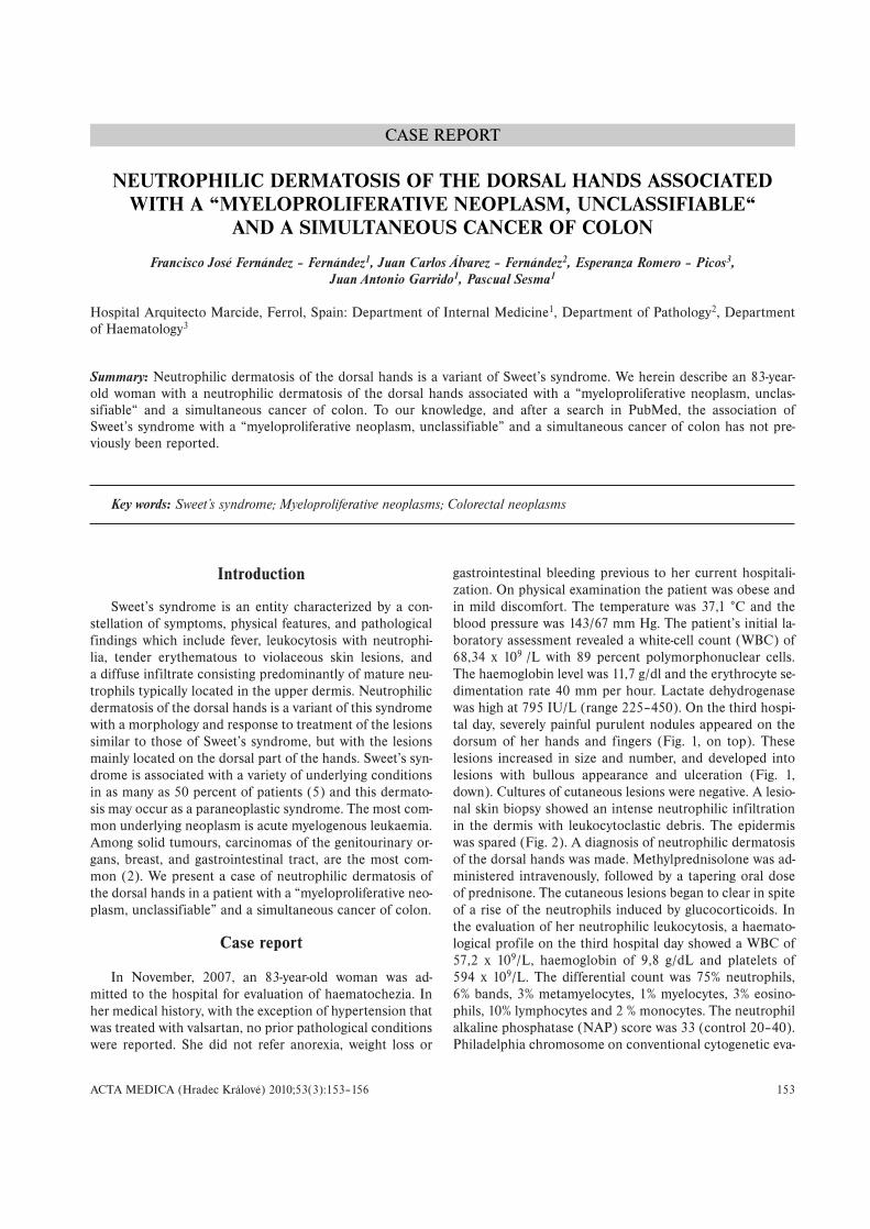
gastrointestinal bleeding previous to her current hospitali-zation. On physical examination the patient was obese andin mild discomfort. The temperature was 37,1 °C and theblood pressure was 143/67 mm Hg. The patient’s initial la-boratory assessment revealed a white-cell count (WBC) of68,34 x 109 /L with 89 percent polymorphonuclear cells.The haemoglobin level was 11,7 g/dl and the erythrocyte se-dimentation rate 40 mm per hour. Lactate dehydrogenasewas high at 795 IU/L (range 225–450). On the third hospi-tal day, severely painful purulent nodules appeared on thedorsum of her hands and fingers (Fig. 1, on top). Theselesions increased in size and number, and developed intolesions with bullous appearance and ulceration (Fig. 1,down). Cultures of cutaneous lesions were negative. A lesio-nal skin biopsy showed an intense neutrophilic infiltrationin the dermis with leukocytoclastic debris. The epidermiswas spared (Fig. 2). A diagnosis of neutrophilic dermatosisof the dorsal hands was made. Methylprednisolone was ad-ministered intravenously, followed by a tapering oral doseof prednisone. The cutaneous lesions began to clear in spiteof a rise of the neutrophils induced by glucocorticoids. Inthe evaluation of her neutrophilic leukocytosis, a haemato-logical profile on the third hospital day showed a WBC of57,2 x 109/L, haemoglobin of 9,8 g/dL and platelets of594 x 109/L. The differential count was 75% neutrophils,6% bands, 3% metamyelocytes, 1% myelocytes, 3% eosino-phils, 10% lymphocytes and 2 % monocytes. The neutrophilalkaline phosphatase (NAP) score was 33 (control 20–40).Philadelphia chromosome on conventional cytogenetic eva-
153
CASE REPORT
NEUTROPHILIC DERMATOSIS OF THE DORSAL HANDS ASSOCIATEDWITH A “MYELOPROLIFERATIVE NEOPLASM, UNCLASSIFIABLE“
AND A SIMULTANEOUS CANCER OF COLON
Francisco José Fernández – Fernández1, Juan Carlos Álvarez – Fernández2, Esperanza Romero – Picos3,
Juan Antonio Garrido1, Pascual Sesma1
Hospital Arquitecto Marcide, Ferrol, Spain: Department of Internal Medicine1, Department of Pathology2, Departmentof Haematology3
Summary: Neutrophilic dermatosis of the dorsal hands is a variant of Sweet’s syndrome. We herein describe an 83-year-old woman with a neutrophilic dermatosis of the dorsal hands associated with a “myeloproliferative neoplasm, unclas-sifiable“ and a simultaneous cancer of colon. To our knowledge, and after a search in PubMed, the association ofSweet’s syndrome with a “myeloproliferative neoplasm, unclassifiable” and a simultaneous cancer of colon has not pre-viously been reported.
Key words: Sweet’s syndrome; Myeloproliferative neoplasms; Colorectal neoplasms
ACTA MEDICA (Hradec Králové) 2010;53(3):153–156
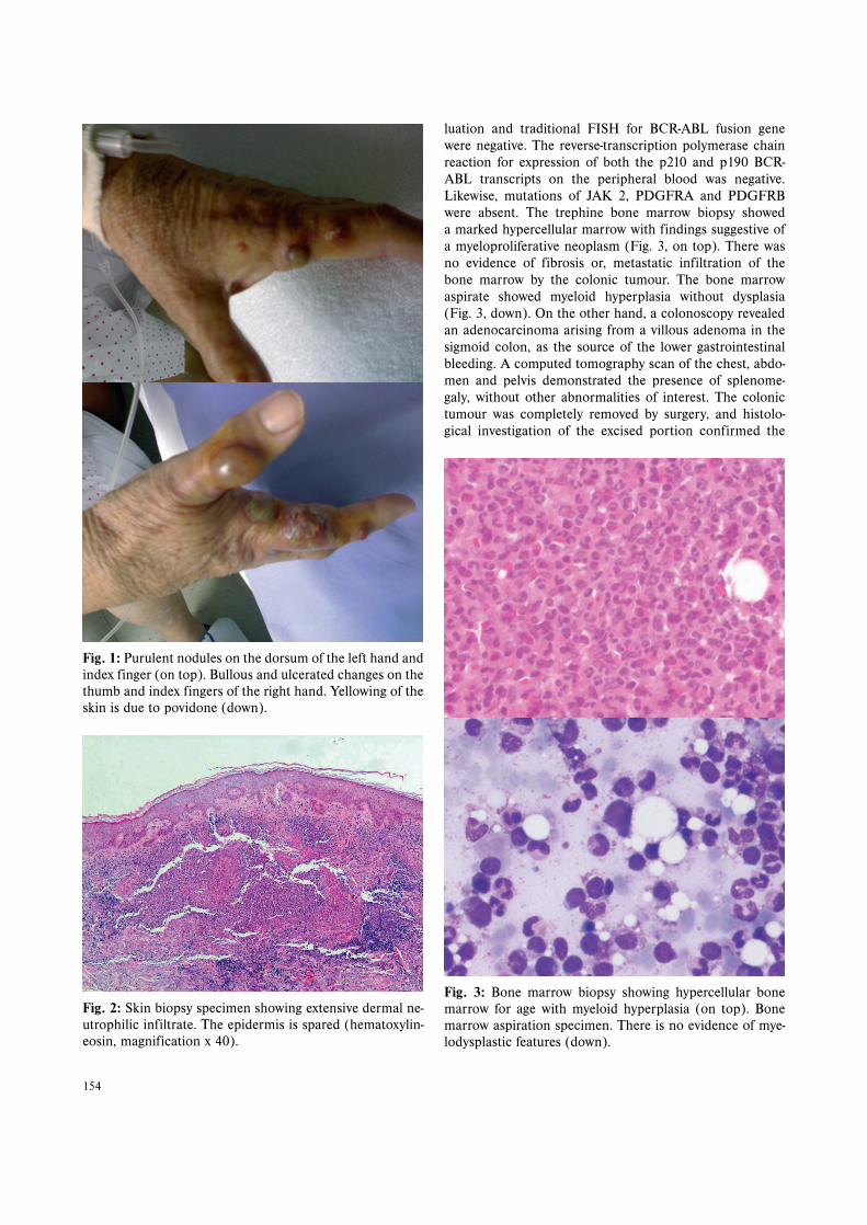
luation and traditional FISH for BCR-ABL fusion genewere negative. The reverse-transcription polymerase chainreaction for expression of both the p210 and p190 BCR-ABL transcripts on the peripheral blood was negative.Likewise, mutations of JAK 2, PDGFRA and PDGFRBwere absent. The trephine bone marrow biopsy showeda marked hypercellular marrow with findings suggestive ofa myeloproliferative neoplasm (Fig. 3, on top). There wasno evidence of fibrosis or, metastatic infiltration of thebone marrow by the colonic tumour. The bone marrowaspirate showed myeloid hyperplasia without dysplasia(Fig. 3, down). On the other hand, a colonoscopy revealedan adenocarcinoma arising from a villous adenoma in thesigmoid colon, as the source of the lower gastrointestinalbleeding. A computed tomography scan of the chest, abdo-men and pelvis demonstrated the presence of splenome-galy, without other abnormalities of interest. The colonictumour was completely removed by surgery, and histolo-gical investigation of the excised portion confirmed the
154
Fig. 1: Purulent nodules on the dorsum of the left hand andindex finger (on top). Bullous and ulcerated changes on thethumb and index fingers of the right hand. Yellowing of theskin is due to povidone (down).
Fig. 3: Bone marrow biopsy showing hypercellular bonemarrow for age with myeloid hyperplasia (on top). Bonemarrow aspiration specimen. There is no evidence of mye-lodysplastic features (down).
Fig. 2: Skin biopsy specimen showing extensive dermal ne-utrophilic infiltrate. The epidermis is spared (hematoxylin-eosin, magnification x 40).
existence of a well differentiated adenocarcinoma invadingthe adipose perivisceral tissue with metastatic involvementin two of the 7 nodes removed. After surgery, haematologi-cal abnormalities persisted and the NAP score decreasedmarkedly to 3 (range 20–40), persisting markedly decreasedin the follow-up studies. Likewise, there was no monocyto-sis in peripheral blood studies over a longer span of time. Inimaging studies at 2 and 6 months of the colonic tumour re-section there was no evidence of metastatic disease of hercancer of colon, presenting only splenomegaly. These alte-rations after surgery allowed us to exclude that the peri-pheral blood neutrophilia was reactive to the cancer ofcolon. In November 2007, an initial diagnosis of Ph-negati-ve chronic myeloid leukaemia was made. According to the2008 World Health Organization classification system forhaematological malignancies, and based on the clinical,molecular and pathological findings, we believe that our pa-tient had a “myeloproliferative neoplasm, unclassifiable”.Treatment with hydroxyurea was used to control her symp-toms and WBC count. She died about a year after her firstpresentation, probably by a pulmonary embolism, and therewas no evidence of an accelerated phase or blastic crisis inher last revision. Necropsy was not performed.
Discussion
Sweet’s syndrome (the eponym for acute febrile neu-trophilic dermatosis) was described by Robert D. Sweet in1964 (10). Since 1995, several authors have reported a va-riant of this syndrome with a morphology and response totreatment of the lesions similar to those of Sweet’s syndro-me, but with the lesions mainly located on the dorsal partof the hands. This variant was named “pustular vasculitis ofthe dorsal hands” or “neutrophilic dermatosis of the dorsalhands”(4,9). Sweet’s syndrome can be idiopathic or as-sociated, among other disorders, with myeloproliferativeprocesses, solid tumours, autoimmune diseases, infectionsand drugs. Cohen et al. (2) noted that approximately 10–20percent of the patients with Sweet’s syndrome had eithera haematological malignancy or a solid malignant tumour.Most of the cancers were hematopoietic, especially acutemyelogenous leukaemia and myelodysplastic syndromes,but about 15 percent were due to solid tumours. Further-more, the drug-induced variant of the dermatosis has beenobserved in patients following the administration of granu-locyte-colony stimulating factor (G-CSF). The pathogenesisof Sweet’s syndrome is poorly understood. Currently, it isthought that cytokine dysregulation accounts for most ofthe clinical, pathological, and laboratory changes of thissyndrome. Among the mainly involved cytokines are inter-leukin (IL)-1, IL-6, IL-8, granulocyte colony-stimulatingfactor (G-CSF) and granulocyte-macrophage colony stimu-lating factor (GM-CSF). Several observations have shownthat interleukin-1 (IL-1) may be an important mediator inSweet’s syndrome. Thus, acute myelogenous leukaemia is
the malignancy most commonly associated with Sweet’ssyndrome and this syndrome can occur after G-CSF treat-ment. IL-1 is spontaneously produced by acute myeloblas-tic leukaemia (AML) cells (8), and this cytokine inducessynthesis of GM-CSF and G-CSF (7). Likewise, polymor-phonuclear leukocytes may induce IL-8 gene expression inresponse to GM-CSF, and IL-8 is a potent chemotacticfactor for polymorphonuclear leukocytes (6), amplifyingthe inflammatory response by recruiting additional po-lymorphonuclear leukocytes into inflammatory sites. Inagreement with this pathogenic mechanism, a recent casereport showed that anakinra, an IL-1 receptor antagonist,was effective in a patient with refractory Sweet’s syndro-me (3).
In our patient, in spite of a rise of the neutrophils in-duced by glucocorticoids, the skin lesions resolved quicklyafter glucocorticoids introduction. It is known that gluco-corticoids inhibit the synthesis of almost all known cytoki-nes (1), and this is primarily achieved through their actionon the function of transcription factors, such as nuclearfactor kappa B (NF-kB) and activator protein-1 (AP-1)that are required for transcription of proinflammatory me-diators. The association of Sweet’s syndrome with two si-multaneous tumours is not frequent. Thus, in a review of theliterature Cohen et al. (2) reported only four patients withsolid tumours who also had a haematological malignancy.To our knowledge, and after a search in PubMed, the as-sociation of Sweet’s syndrome with a “myeloproliferativeneoplasm, unclassifiable” and a simultaneous cancer of co-lon has not previously been reported. In both idiopathicand malignancy-associated Sweet’s syndrome there are usu-ally a good response to corticosteroid therapy. In our pa-tient, the myeloproliferative neoplasm and cancer of colonwere simultaneously diagnosed, and regression of the skinlesions was obtained with glucocorticoids before the speci-fic treatment of the tumours.
Acknowledgements
We thank Drs. T. González and C. Quinteiro from theMolecular Medicine Unit – Fundación Pública Galega deMedicina Xenómica. Hospital Clínico Universitario de San-tiago de Compostela for help with the molecular analysis ofthe patient.
References
1. Auphan N, DiDonato JA, Rosette C, Helmberg A, Karin M. Immunosuppressionby glucocorticoids: inhibition of NF-Kappa B activity through induction of IKappa B synthesis. Science 1995;270:286–90.
2. Cohen PR, Holder WR, Tucker SB, Kono S, Kurzrock R. Sweet syndrome in pa-tients with solid tumors. Cancer 1993;72:2723–31.
3. Delluc A, Limal N, Puéchal X, Frances C, Piette JC, Cacoub P. Efficacy of ana-kinra, an IL1 receptor antagonist, in refractory Sweet syndrome. Ann Rheum Dis2008;67:278–9.
4. DiCaudo DJ, Connolly SM. Neutrophilic dermatosis (pustular vasculitis) of thedorsal hands: a report of 7 cases and review of the literature. Arch Dermatol2002;138:361–5
5. Kemmett D, Hunter JA. Sweet’s syndrome: A clinicopathologic review of twenty-nine cases. J Am Acad Dermatol 1990;23:503–7.
155

6. McCain RW, Dessypris EN, Christman JW. Granulocyte/macrophage colony-sti-mulating factor stimulates human polymorphonuclear leukocytes to produce in-terleukin-8 in vitro. Am J Respir Cell Mol Biol 1993;8:28–34.
7. Ogawa Y, Yonekura S, Nagao T. Granulocyte colony-stimulating factor produc-tion by human bone marrow fibroblasts stimulated with interleukins. Am JHematol 1996;52:71–6.
8. Rambaldi A, Torcia M, Bettoni S, et al. Modulation of cell proliferation and
cytokine production in acute myeloblastic leukemia by interleukin-1 receptorantagonist and lack of its expression by leukemic cells. Blood 1991;78:3248–53.
9. Strutton G, Weedon D, Robertson I. Pustular vasculitis of the hands. J Am AcadDermatol 1995;32:192–8.
10. Sweet RD. An acute febrile neutrophilic dermatosis. Br J Dermatol 1964;76:349–56.
156
Corresponding author:
Dr. F. J. Fernández – Fernández, Department of Internal Medicine, Hospital Arquitecto Marcide, 15405 Ferrol, Spain; e-mail: [email protected]
Received: 17/02/2010.Accepted in revised form: 18/06/2010.