Embed Size (px)
Citation preview
Page 1/13
A comparative study of the characteristics andclinical signi�cance of extracellular traps ofneutrophils in gingival samples of periodontitis andgingivitis patientsFei Zhang
Linyi Central HospitalXi-Mei Yang
Linyi Central HospitalShu-Yu Jia ( [email protected] )
linyi central hospital https://orcid.org/0000-0002-9054-8584
Research article
Keywords: periodontitis; gingivitis; neutrophils; extracellular traps of neutrophils
Posted Date: September 12th, 2019
DOI: https://doi.org/10.21203/rs.2.14324/v1
License: This work is licensed under a Creative Commons Attribution 4.0 International License. Read Full License
Page 2/13
AbstractBackground: We sought to compare the characteristics and clinical signi�cance of extracellular traps ofneutrophils in gingival samples of periodontitis and gingivitis. Methods: The clinical indexes of gingivalsamples from periodontitis and gingivitis were measured; the expression of TNF-alpha and IL-8 weremeasured by real-time �uorescent quantitative PCR; the expression of TLR-8 and MMP-9 were measuredby Western blotting assays Chemotaxis, phagocytosis and phagocytic activity of neutrophils weremeasured. Results: The periodontal clinical indexes of periodontitis and gingivitis samples weresigni�cantly higher than those of healthy people (P<0.05). There were signi�cant differences in BOP, PDand CAL between periodontitis and gingivitis samples (P<0.05), but no differences in gingival index (GI)(P>0.05). Compared with the healthy group, the expressions of TNF-α and IL-8 in the periodontitis groupand the gingivitis group increased signi�cantly (P<0.05), and TNF-α in the gingivitis group wassigni�cantly lower than that in the healthy group (P<0.05). The expression of IL-8 in the periodontitisgroup was signi�cantly higher than that in the periodontitis group (P<0.05); the expression of TLR-8 andMMP-9 in the periodontitis group was different from that in the gingivitis group and the healthy peoplegroup, and the expression of TLR-8 and MMP-9 in the gingivitis group was signi�cantly different fromthat in the healthy people group (P<0.05); the neutrophil mobility index in healthy people was (3.02±0.53),and that in the periodontitis group was (2.21±0.13), and (2.31±0.12) in the gingiva. There were signi�cantdifferences between the periodontitis group and the gingivitis group (P<0.05, P<0.05). The phagocyticability and activity of neutrophils in healthy people were (392.03±20.04), (89.34±10.12), the periodontitisgroup was (360.32±18.23), (80.02±10.21), and the gingivitis group was (359.32±20.11), (78.93±17.32),respectively. There were signi�cant differences between the periodontitis group and the gingivitis groupand healthy people group (P<0.05, P<0.05). Conclusion: The chemotaxis of neutrophils in gingivalsamples of periodontitis and gingivitis patients was decreased, the phagocytosis ability and activity ofneutrophils were reduced, and the release of extracellular trap-releasing inducible factors TNF-alpha andIL-8 also declined.
BackgroundPeriodontitis is an infectious disease caused by plaque microorganisms and their metabolites(1). Themain clinical manifestations are loss of periodontal attachments and gradual destruction and absorptionof the alveolar bone(2). In this process, the immune response to the pathogenic microorganism plays animportant role. The gum is one of the periodontal tissues (the gum, periodontal ligament, alveolar bone,and cementum) and is directly exposed to the mouth and consists of keratinized epithelium andconnective tissue covering the alveolar bone and roots. Gum disease is a disease that is con�ned to thegum tissue, the most common being chronic marginal gingivitis. Plaque microorganisms are the initiatingfactor of gingivitis and periodontitis. Among them, non-adherent gingival plaque is closely related to theoccurrence and development of periodontitis and is considered to be the "development frontier" ofperiodontitis, which is related to the rapid destruction of the alveolar bone. A large number of activatedimmune cells cannot only trigger the host's immune response to periodontal pathogen infection, but also
Page 3/13
release a cascade of in�ammatory factors, including various acute phase reaction proteins, cytokines,etc., leading to the destruction of periodontal soft tissue and hard tissue(3). Neutrophils have a rod-shaped nucleus or lobulated nucleus and �ne particles. The cytoplasm is colorless or reddish. It haschemotaxis, phagocytosis and bactericidal action. When the body is attacked by pathogens, neutrophilsare under the action of chemokines. They quickly gather near the site of infection, enter the infectedtissue through the vessel wall, and kill pathogenic microorganisms(4) by phagocytosis and release ofbactericidal particles. Neutrophil extracellular reticular traps (NETs) are a new defense mechanism(5),and unlike apoptosis and necrosis, release a variety of bactericidal substances to remove pathogenicmicroorganisms, including neutrophil primary granule release such as protease or secondary particlebacteriostatic peptide LL-37, etc.; tertiary particles such as protease-containing matrix metalloproteinase9 (MMP-9), etc(6, 7). Many factors can stimulate neutrophils to produce neutrophil extracellular traps. Forexample, plasma TNF-α and interleukin-8 can also stimulate neutrophil release of NETs(8, 9).
Methods1.1 General Information
Between July 2015 and September 2016, we randomly selected 27 patients (14 males and 13 females)with periodontitis (CP), 17 patients (7 males and 10 females) with gingivitis (cg) and 20 patients (8 malesand 12 females) with no periodontal disease (PH). Subjects were 24-60 years old with an average age of(33.24 ± 6.93) years. Periodontitis and gingivitis were diagnosed according to the diagnostic criteria ofthe Periodontal Disease Association
The inclusion criteria were: at least 20 teeth in the mouth, 4-6 teeth with severe periodontitis in allperiodontal patients; no periodontal treatment in the preceding 6 months; no smoking history in thepreceding 6 months; no history of head and neck radiotherapy; no antibiotics, phenytoin sodium,cyclosporine, calcium channel blockers, oral contraceptives, atropine and other drugs within the month;no serious systemic diseases and serious infections in other areas. Pregnant or lactating women wereexcluded. The study protocol was approved by the Ethics Committee of Linyi Central Hospital. All studyparticipants provided written informed consent.
1.2 Experimental methods
1.2.1 Determination of clinical indicators
The researchers who received professional training in periodontal examination and passed the standardconformance test (Kappa=0.73) used the Williams probe to detect periodontal clinical indicators in allsubjects, including the gingival index (GI), detection of bleeding on probing (BOP), depth of detection(PD), and clinical attachment level (CAL). Among them, PD and CAL were detected at 6 sites on thebuccal (lip) side and at the distal, middle and proximal sites of the lingual side.
1.2.2 Collection of peripheral blood samples
Page 4/13
Two mL of peripheral venous blood of all subjects was extracted and placed in an anticoagulation tubecontaining K2EDTA. Peripheral blood samples were collected by centrifugation (4000 r/min, 6 min) at4°C. The supernatant was placed in a sterile Eppendorf tube and stored at -80 ° C until use.
1.2.3 Extraction of total RNA from peripheral blood samples and RT-PCR determination
One ml of Trizol was added in the serum and repeatedly pipetted, and the cell lysate was transferred to a1.5 ml Eppendorf tube, left for 5 min, and fully lysed. Two hundred μL of chloroform was added per 1 mlof Trizol, mixed for 15 s, and left at room temperature for 3 min. After centrifugation at 4o C 12000 g for15 min, the upper aqueous phase was aspirated and transferred to another new Eppendorf tube. Then,0.5 ml of isopropanol was added per 1 ml of Trizol, mix, and left at room temperature for 10 min. Aftercentrifugation at 42000g of 4oC for 10 min, the supernatant was discarded, and RNA was precipitated onthe bottom of the tube. One ml of 75% ethanol was added per 1 ml of Trizol and mix vigorously. Aftercentrifugation at 4o C at 7500 g for 5 min, the supernatant was discarded. RNA was precipitated at roomtemperature for 5-10 min, dissolved in 30 μL of EPC treated water, and stored at -80oC for use. RNA purityand concentration were measured by spectrophotometry at A260 and A260/280. Total RNA wasextracted using a total RNA rapid extraction kit and reverse transcribed with Super M-MLV reversetranscriptase using miRNA-speci�c reverse transcription primers (Table 1). RT-PCR was performed in areal-time quantitative system. The ampli�cation conditions are shown in Table 2, the primer design isshown in Table 3, and the reaction system is shown in Table 4.
1.2.4 Western blotting assays
The expression of TLR-8 and MMP-9 in cells was detected by Western blotting assays. The protein wasresolved by a 4%-10% polyacrylamide gel electrophoresis and the gel was transferred to a PVDFmembrane using SDS polyacrylamide gel electrophoresis. After blocking with 5% non-fat dry milk for 1 hat room temperature, the membrane was �rst incubated with 1:500 diluted primary antibody overnight,and after wash with TBS-T buffer, the membrane was incubated with secondary antibody for 1 h.Following rinse with TBS-T solution, enhanced chemiluminescence was carried out. The PVDF membranewas placed in a developing solution in a dark room and exposed to an X-ray �lm for exposure anddevelopment. Protein densitometry was performed using Image 1.6 software.
1.2.5 Preparation of leukocyte suspension
After extracting 3 mL of venous blood from all subjects, we used heparin as an anticoagulant toprecipitate red blood cells, and white blood cells were taken. After wash, 1×10⁶ / mL white blood cellsuspension was prepared with RPMI1640 medium containing 10% bovine serum.
1.2.6 Determination of neutrophil chemotaxis
Using agarose glass plate method: agarose solution was prepared with 1640 solution, and after additionof 10% inactivated AB serum, the glass plate was poured ,. Each group consisted of three circular holes
Page 5/13
arranged in a straight line with a diameter and a pitch of 2.5 mm. Five μL of a white cell suspension(1×10⁶/mL) was added in triplicates to the mesopores and Escherichia coli was added to the side wellsfor 24 hours, and 5 μL of the �ltrate was used as the chemokine, and the other side was treated with 5 μLof the 1640 solution. The agarose glass plate was placed in a 37 ° C incubator, incubated with saturatedhumidity and 5% CO2 for 4 h, removed, �xed with methanol and formaldehyde. After agarose gelelectrophoresis, the gel was stained with Wright, and measured under an optical microscope. Thedistance a cell moved from the edge of the hole to the hole on either side was measured.
1.2.7 Determination of phagocytic function of neutrophils
Staphylococcus aureus was grown on agar slants for 24 h, colonies were washed with sterile isotonicsaline, washed twice with PBS, and the cells were suspended at 5×10⁷ /ml by speci�c concentrationmethod. Three drops of heparinized blood were taken on the concave slide, and three drops ofStaphylococcus aureus suspension were added and after thorough mixing, they were placed in a sealedwet box. After incubation at 37 ° C for 30 min, the box was shaken once every 10 min; 1 drop was takenwith a capillary suction tube, pushed onto a slide on a glass slide, �xed in methanol, stained with Giemsa,and counted by light microscopy.
1.3 Data processing
Data were expressed as mean ± standard deviation. One-way analysis of variance was used forcomparison between groups. P < 0.05 was considered to have a signi�cant statistical difference.
Results2.1 Clinical indicators
The clinical indicators in the groups are shown in Table 5. The periodontal clinical indexes ofperiodontitis and gingivitis samples were signi�cantly higher than those of healthy people (P <0.05).Comparison of periodontitis with gingivitis samples showed that there were signi�cant differences in theblood index, depth of detection and CAL (P <0.05), and there was no difference in the gingival index (P>0.05).
2.2 RNA expression measurement results
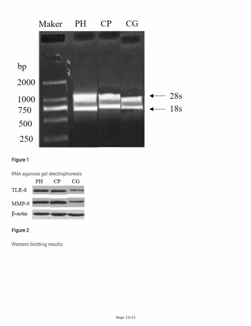
The purity of extracted total RNA was measured by agarose gel electrophoresis, and the results areshown in Fig. 1. As can be seen from the �gure, RNA was of high purity and can be used for PCRampli�cation.
The expression levels of RNA in each group were measured by RT-PCR. The results are shown in Table 6.The results showed that the expression of TNF-alpha and IL-8 in the periodontitis group and the gingivitisgroup increased signi�cantly compared with the healthy control group (P < 0.05), and the expression of
Page 6/13
TNF-alpha in the gingivitis group was signi�cantly lower than that in the periodontitis group (P < 0.05),and the expression of IL-8 was signi�cantly higher than that in the periodontitis group (P < 0.05).
2.3 Western blotting results
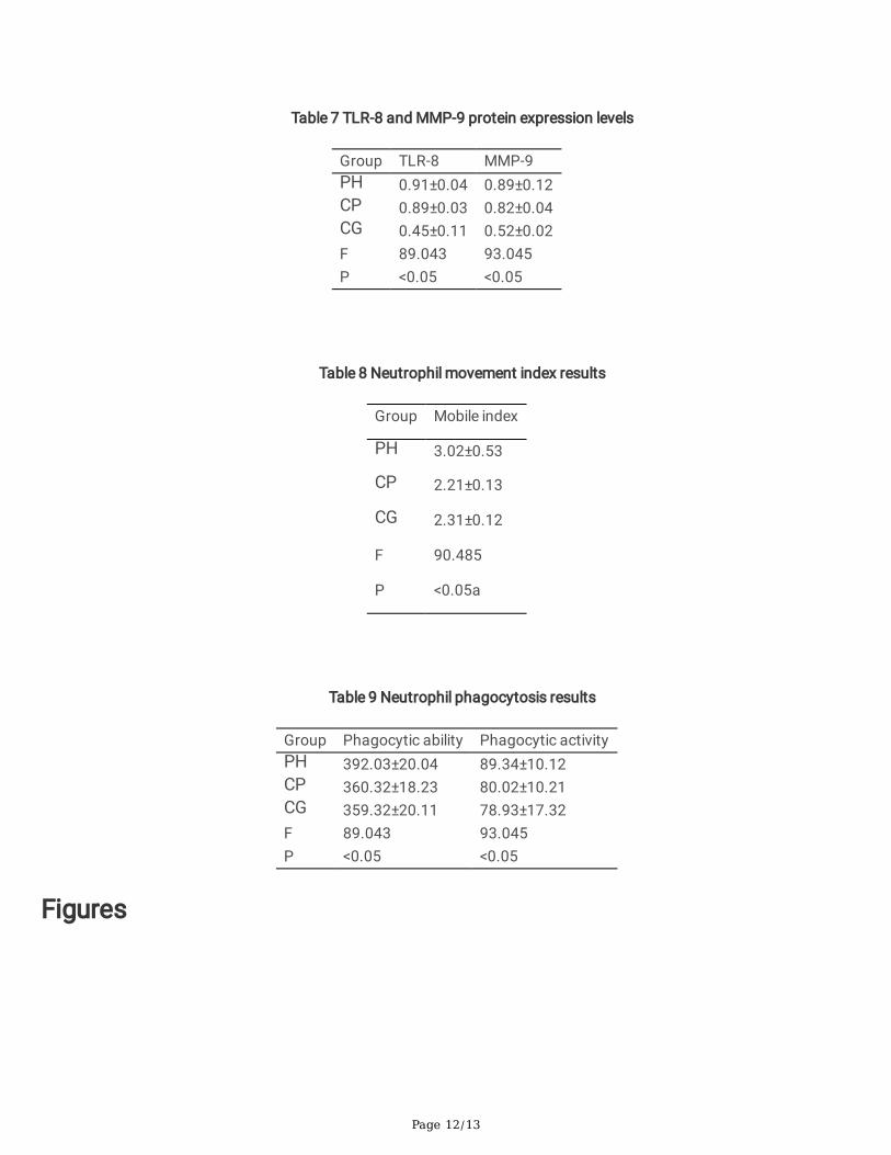
The plasma expression levels of TLR-8 and MMP-9 were determined by Western blotting assays. Theresults are shown in Fig. 2 and Table 7. The expression of TLR-8 and MMP-9 in the periodontitis groupwas different from that in the gingivitis group and the healthy control group, and the difference betweenthe gingivitis group and the healthy control group was signi�cant (P < 0.05). The expression of TLR-8 andMMP-9 in the periodontitis group was higher than that in the gingivitis group, but there was no signi�cantdifference (P > 0.05). It indicated that the neutrophil chemotactic function in periodontitis and gingivitispatients was signi�cantly lower than that in healthy subjects.
2.4 Neutrophil chemotaxis
The neutrophil shift index was (3.02±0.53) in healthy subjects, (2.21±0.13) in the periodontitis group, and(2.31±0.12) in the periodontitis group. There were signi�cant differences between the periodontitis groupand the gingivitis group and the healthy control group (P < 0.05, P < 0.05). This indicated that thechemotactic function of neutrophils in patients with periodontitis and gingivitis is signi�cantly lower thanthat in healthy subjects.
2.5 Determination of neutrophil phagocytosis
The phagocytic ability and activity of neutrophils were (392.03 ±20.04), and (89.34 ±10.12) in healthysubjects, and (360.32 ±18.23) and (80.02 ±10.21) in the periodontitis group, and (359.32 ±20.11) and(78.93 ±17.32) in the gingivitis group. There were signi�cant differences between the periodontitis groupand the gingivitis group and the healthy control group (P < 0.05, P < 0.05). The results showed that thephagocytic ability and activity of neutrophils in periodontitis and gingivitis patients were signi�cantlylower than those in healthy subjects.
DiscussionPeriodontal disease is a chronic in�ammatory disease of the tooth-supporting tissue caused by plaqueinfection, including gingivitis and periodontitis. The main manifestation is periodontal support tissuedestruction(10).
In 2004, Brinkmann et al. �rst proposed another method for killing pathogenic microorganisms, neutrophilextracellular reticular traps (NETs), which differ from apoptosis and necrosis(11). NETs are closely relatedto human diseases and play a unique role in the development of disease(12, 13). In most cases, theproduction of NETs is conducive to the body's innate immunity(14), but recent studies have found thatthe network is also involved in the pathological process of human disease. When a mesh is produced, alarge amount of autoantigens(15) is produced. If the removal is not timely, a large amount of antigenswill accumulate, which may induce the body to produce a large amount of autoantibodies, leading to
Page 7/13
autoimmune diseases. Neural network research focuses on infectious diseases and autoimmunediseases, but it is also closely related to thrombotic diseases (acute myocardial infarction)(16).
NETs is a newly discovered neutrophil bactericidal mechanism that activates neutrophils to releasechromatin and then combines with the gingival sulcus to form a new defense network against gingivalplaque bacteria(17). It has been reported(18, 19) that under the induction of IL-8, LPS and interferon, theneutrophil nucleus is deformed, the chromatin is homogeneous, the nuclear membrane is broken, thenuclear material is directly in contact with the particles, forming a mixture, and �nally the cell membraneis broken, NETs are released. This study found that compared with the healthy control group, theexpression of TNF-α and IL-8 in the periodontitis group and the gingivitis group was signi�cantlyincreased (P<0.05), and TNF-α in the gingivitis group was signi�cantly lower than that in the periodontitisgroup. The expression of IL-8 was signi�cantly higher in the xx group (P<0.05) than in the periodontitisgroup (P<0.05).
MMP-8 secreted by neutrophils, also known as neutrophil collagenase, is one of the most importantMMPs in in�ammatory periodontal tissues. Type I collagen �bers are the main components of theperiodontal ligament and alveolar bone. Studies have shown that the main role of MMP-8 is to destroytype I collagen �bers. MMP-8 is also present in normal periodontal tissues. In the absence ofin�ammation, the ratio of MMP-8 and its endogenous inhibitor (TIMP-1) is close to 1:1, which is involvedin normal metabolism of the extracellular matrix. However, when pathogens invade, the expression ofMMP-8 is abnormally regulated by cytokines, which disrupts the balance between MMP-8 and TIMP-1,leading to damage of type I collagen in periodontal tissues. This study showed that there was adifference in the expression of TLR-8 and MMP-9 between the periodontitis group and the gingivitis groupand the healthy control group, and the difference between the gingivitis group and the healthy controlgroup was signi�cant (P< 0.05). The expression of TLR-8 and MMP-9 in the periodontitis group washigher than that in the gingivitis group, but there was no signi�cant difference (P>0.05), indicating thatthe chemotactic function of neutrophils in patients with periodontitis and gingivitis is signi�cantly lowerthan that in healthy controls.
Neutrophils are important cells to prevent periodontal infection. The study on the role of neutrophils injuvenile periodontitis shows that chemotaxis of neutrophils decreases and the phagocytosis function ofneutrophils is normal. This report proves that the weakening of neutrophil chemotaxis makes juvenileperiodontitis patients susceptible to speci�c local bacteria and causes rapid destruction of theperiodontal tissue(20). This study indicated that there were signi�cant differences between theperiodontitis group and the gingivitis group and healthy control group (P < 0.05). The results showed thatthe neutrophil chemotactic function of periodontitis and gingivitis patients was signi�cantly lower thanthat of healthy controls. There were signi�cant differences between the periodontitis group and thegingivitis group and the healthy control group (P < 0.05, P < 0.05). The results showed that the phagocyticability and activity of neutrophils in periodontitis and gingivitis patients were signi�cantly lower thanthose in healthy controls.
Page 8/13
In summary, the chemotaxis of neutrophils in gingival samples of periodontitis and gingivitis decreased,the ability of phagocytosis and activity of neutrophils were reduced, and the release of extracellular trap-releasing inducible factors TNF-alpha and IL-8 declined.
List Of Abbreviationsneutrophil extracellular reticular traps: NETs
matrix metalloproteinase 9: MMP-9
periodontal disease: PH
gingival index: GI
bleeding on probing: BOP
depth of detection: PD
clinical attachment level: CAL
DeclarationsEthics approval and consent to participate The study protocol was approved by the Ethics Committee ofLinyi Central Hospital. All study participants provided written informed consent.
Consent for publication: All study participants provided written informed consent.
Availability of data and material Data could be available to readers at reasonable request from thecorresponding author .
Competing interests: The authors declare that they have no competing interests.
Funding: No funding was received for this study.
Authors' contributions FZ contributed to the conception and design of the study; XY contributed to theacquisition of data; SJ performed the experiments; SJ contributed to the analysis of data; FZ wrote themanuscript; All authors reviewed and approved the �nal version of the manuscript.
Acknowledgements: None.
References1. Hajishengallis G. Periodontitis: from microbial immune subversion to systemic in�ammation. Nature
reviews Immunology. 2015;15(1):30-44.
Page 9/13
2. Eke PI, Dye BA, Wei L, Slade GD, Thornton-Evans GO, Borgnakke WS, et al. Update on Prevalence ofPeriodontitis in Adults in the United States: NHANES 2009 to 2012. Journal of periodontology.2015;86(5):611-22.
3. Zambon JJ, Haraszthy VI, Hariharan G, Lally ET, Demuth DR. The Microbiology of Early-OnsetPeriodontitis: Association of Highly Toxic Actinobacillus actinomycetemcomitans Strains WithLocalized Juvenile Periodontitis. Journal of periodontology. 1996;67 Suppl 3S:282-90.
4. Jaillon S, Galdiero MR, Del Prete D, Cassatella MA, Garlanda C, Mantovani A. Neutrophils in innateand adaptive immunity. Seminars in immunopathology. 2013;35(4):377-94.
5. Zawrotniak M, Rapala-Kozik M. Neutrophil extracellular traps (NETs) - formation and implications.Acta biochimica Polonica. 2013;60(3):277-84.
�. Hermosilla C, Caro TM, Silva LM, Ruiz A, Taubert A. The intriguing host innate immune response:novel anti-parasitic defence by neutrophil extracellular traps. Parasitology. 2014;141(11):1489-98.
7. Manda A, Pruchniak MP, Arazna M, Demkow UA. Neutrophil extracellular traps in physiology andpathology. Central-European journal of immunology. 2014;39(1):116-21.
�. Demers M, Wagner DD. Neutrophil extracellular traps: A new link to cancer-associated thrombosisand potential implications for tumor progression. Oncoimmunology. 2013;2(2):e22946.
9. White PC, Chicca IJ, Cooper PR, Milward MR, Chapple IL. Neutrophil Extracellular Traps inPeriodontitis: A Web of Intrigue. Journal of dental research. 2016;95(1):26-34.
10. Buduneli N, Kinane DF. Host-derived diagnostic markers related to soft tissue destruction and bonedegradation in periodontitis. Journal of clinical periodontology. 2011;38 Suppl 11:85-105.
11. Warnatsch A, Ioannou M, Wang Q, Papayannopoulos V. In�ammation. Neutrophil extracellular trapslicense macrophages for cytokine production in atherosclerosis. Science. 2015;349(6245):316-20.
12. Arazna M, Pruchniak MP, Zycinska K, Demkow U. Neutrophil extracellular trap in human diseases.Advances in experimental medicine and biology. 2013;756:1-8.
13. Vorobjeva NV, Pinegin BV. Neutrophil extracellular traps: mechanisms of formation and role in healthand disease. Biochemistry Biokhimiia. 2014;79(12):1286-96.
14. Park SY, Shrestha S, Youn YJ, Kim JK, Kim SY, Kim HJ, et al. Autophagy Primes Neutrophils forNeutrophil Extracellular Trap Formation during Sepsis. American journal of respiratory and criticalcare medicine. 2017;196(5):577-89.
15. Sorensen OE, Borregaard N. Neutrophil extracellular traps - the dark side of neutrophils. The Journalof clinical investigation. 2016;126(5):1612-20.
1�. Ginley B, Emmons T, Sasankan P, Urban C, Segal BH, Sarder P. Identi�cation and characterization ofneutrophil extracellular trap shapes in �ow cytometry: SPIE; 2017.
17. Lightfoot YL, Kaplan MJ. Disentangling the role of neutrophil extracellular traps in rheumaticdiseases. Current opinion in rheumatology. 2017;29(1):65-70.
1�. Stakos DA, Kambas K, Konstantinidis T, Mitroulis I, Apostolidou E, Arelaki S, et al. Expression offunctional tissue factor by neutrophil extracellular traps in culprit artery of acute myocardial
Page 10/13
infarction. European heart journal. 2015;36(22):1405-14.
19. Meher AK, Spinosa M, Davis JP, Pope N, Laubach VE, Su G, et al. Novel Role of IL (Interleukin)-1betain Neutrophil Extracellular Trap Formation and Abdominal Aortic Aneurysms. Arteriosclerosis,thrombosis, and vascular biology. 2018;38(4):843-53.
20. Sochalska M, Potempa J. Manipulation of Neutrophils by Porphyromonas gingivalis in theDevelopment of Periodontitis. Frontiers in cellular and infection microbiology. 2017;7:197.
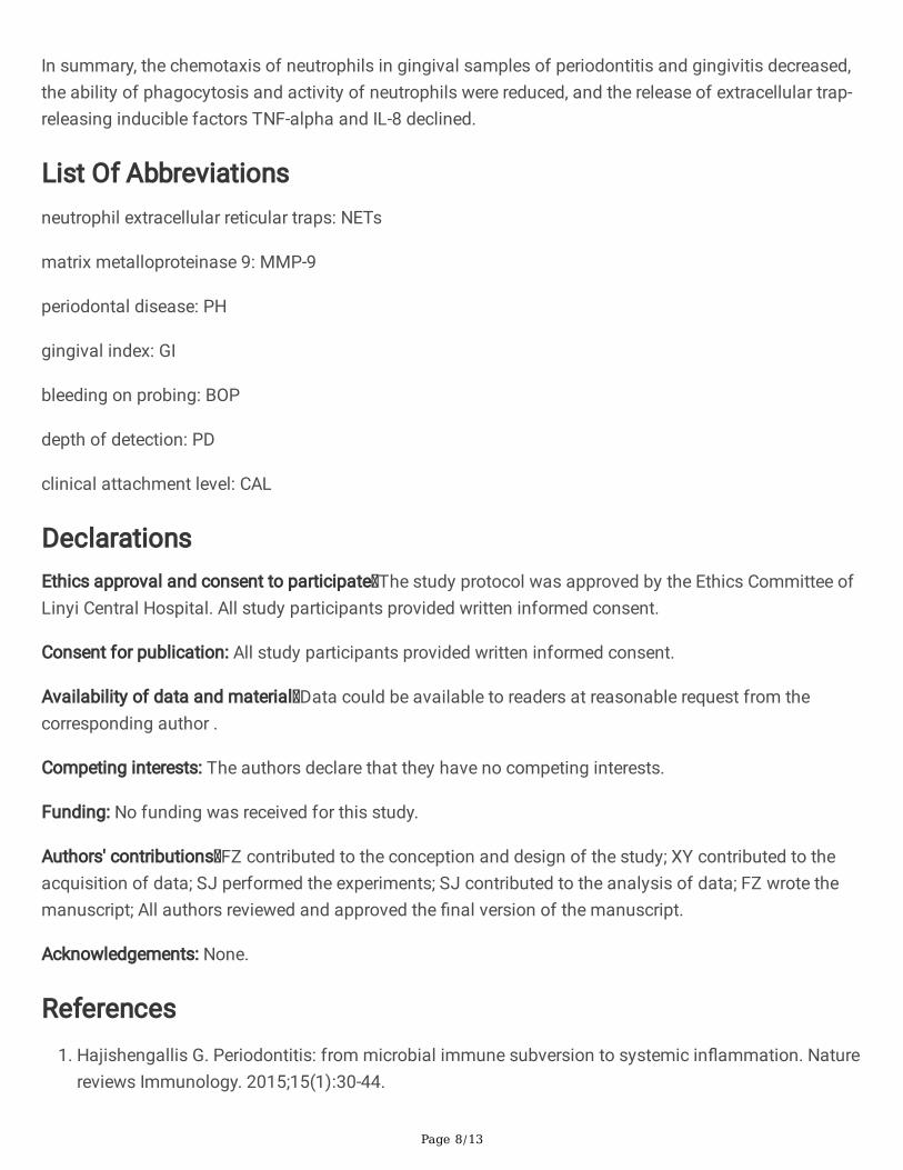
TablesTable 1 Reverse transcription system
5×PrimeScript Buffer 4μL1×PrimeScript RT Enzyme Mix I 1μLOligo dT Primer (50μM) 1μL (25pmol)Random 6 mers (100μM) 1μL (50pmol)Total RNA lμLRNase Free dH2O 12μL
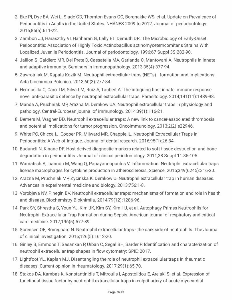
Table 2 PCR conditions
step temperature time
Pre-denaturation 95℃ 10 min
transsexual 94℃ 30 s 40 cycles
Annealing/extension 60℃ 1 min
Melting curve analysis
95℃ 15 s
60℃ 1 min
94℃ 15 s
60℃ 15 s
Page 11/13
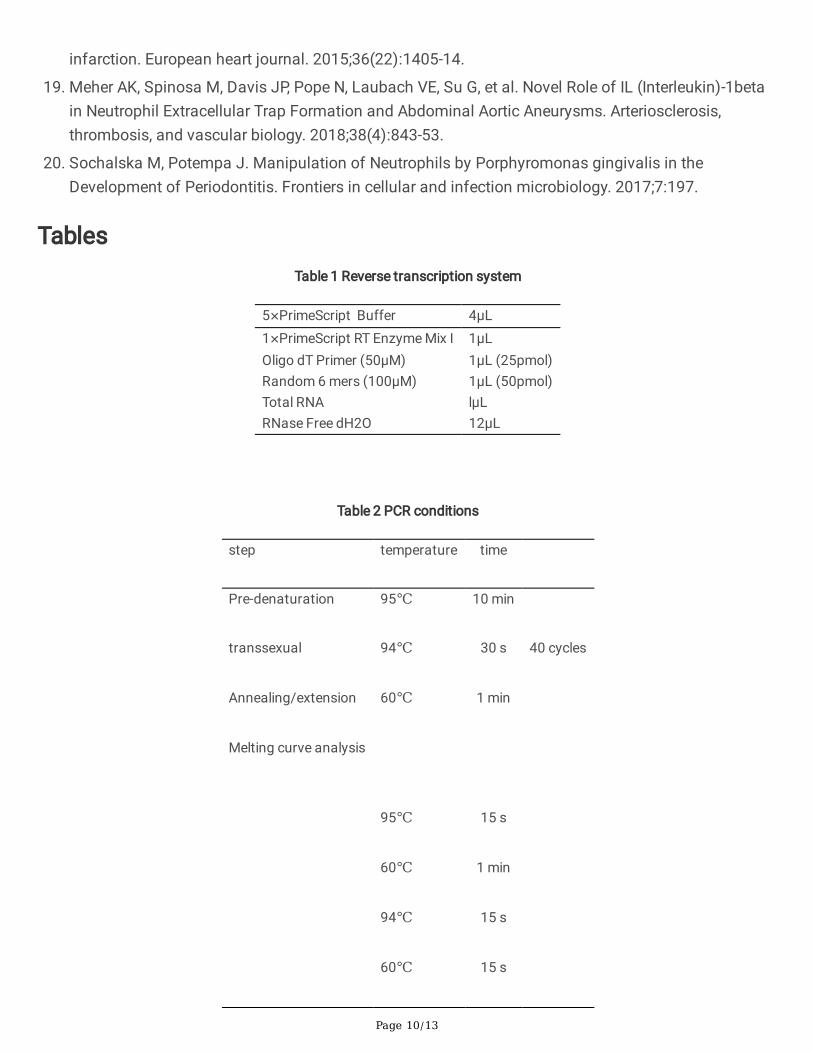
Table 3 Sequences of primers used in the study
RNA Upstream Downstream
TNF-α 5 '-TGTTCTTACACCCCCTCCCCTTTT-3' 5’-TATAAGTGTCAGCCGGCTGAGAA-3’
IL-8 5′ -GGCCGAUUGUGAACAUGGATT-3′ 5′-UCCAUGUUCACAAUCGGCCGC-3′
β-actin 5 '-TGGCACCCAGCACAATGAA-3' 5'-CTAAGTCATAGTCCGCCTAGAAGCA-3'
Table 4 PCR system
5×PrimeEx Taq TM II (2×) 12.5μLPCR Forward Primer (10μM) 1μL 0.4μMPCR Reverse Primer(10μM) 1μL 0.4μMDNA template 2μLdH2O (sterilized distilled water) 8.5μLTotal 25μL
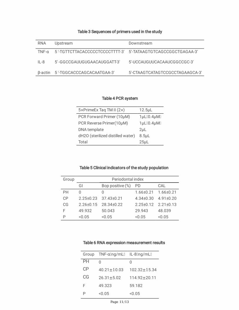
Table 5 Clinical indicators of the study population
Group Periodontal indexGI Bop positive (%) PD CAL
PH 0 0 1.66±0.21 1.66±0.21CP 2.25±0.23 37.43±0.21 4.34±0.30 4.91±0.20CG 2.26±0.15 28.34±0.22 2.25±0.12 2.21±0.13F 49.932 50.043 29.943 48.039P <0.05 <0.05 <0.05 <0.05
Table 6 RNA expression measurement results
Group TNF-α ng/mL IL-8 ng/mL
PH 0 0
CP 40.21±10.03 102.32±15.34
CG 26.31±5.02 114.92±20.11
F 49.323 59.182
P <0.05 <0.05
Page 12/13
Table 7 TLR-8 and MMP-9 protein expression levels
Group TLR-8 MMP-9PH 0.91±0.04 0.89±0.12CP 0.89±0.03 0.82±0.04CG 0.45±0.11 0.52±0.02F 89.043 93.045P <0.05 <0.05
Table 8 Neutrophil movement index results
Group Mobile index
PH 3.02±0.53
CP 2.21±0.13
CG 2.31±0.12
F 90.485
P <0.05a
Table 9 Neutrophil phagocytosis results
Group Phagocytic ability Phagocytic activityPH 392.03±20.04 89.34±10.12CP 360.32±18.23 80.02±10.21CG 359.32±20.11 78.93±17.32F 89.043 93.045P <0.05 <0.05
Figures
Page 13/13
Figure 1
RNA agarose gel electrophoresis
Figure 2
Western blotting results


![Diabetes and Periodontal Diseases: An Established Two-Way ... · Silness gingival index (with values from 0 to 3, where 2 or 3 indicates bleeding) and [8] of periodontitis (assessed](https://img.pdfslide.net/doc/110x75/5e6babd75005771ca5638355/diabetes-and-periodontal-diseases-an-established-two-way-silness-gingival-index.jpg)