Embed Size (px)
Citation preview
CLINICAL MICROBIOLOGY REVIEWS, Apr. 2008, p. 380–401 Vol. 21, No. 20893-8512/08/$08.00�0 doi:10.1128/CMR.00050-07Copyright © 2008, American Society for Microbiology. All Rights Reserved.
New Aspects of Neotropical Polycystic (Echinococcus vogeli) andUnicystic (Echinococcus oligarthrus) Echinococcosis
Antonio D’Alessandro1* and Robert L. Rausch2
Department of Tropical Medicine, School of Public Health and Tropical Medicine SL-29, Tulane University, 1440 Canal Street,Suite 2210, New Orleans, Louisiana 70112-2824,1 and Department of Comparative Medicine, School of Medicine,
University of Washington, Box 357190, Seattle, Washington 98195-71902
INTRODUCTION .......................................................................................................................................................380Taxonomy of Echinococcus spp...........................................................................................................................................381Neotropical Species of Echinococcus in the Final Host .....................................................................................381Characteristics of the Strobilar Stages of E. vogeli and E. oligarthrus............................................................382The Metacestode of E. vogeli in the Natural Intermediate Host and in Humans .........................................382Histogenesis and Pathogenesis of the Metacestode of E. vogeli in Polycystic Echinococcosis.....................384The Metacestode of E. oligarthrus in the Natural Intermediate Host .............................................................384Histogenesis and Pathogenesis of the Metacestode of E. oligarthrus in Unicystic Echinococcosis .............385
MEDICAL ASPECTS OF NEOTROPICAL ECHINOCOCCOSES ....................................................................385Clinical Characteristics of Polycystic and Unicystic Echinococcoses..............................................................386Clinical Characteristics of Polycystic Echinococcosis (E. vogeli) (Types I to V) ...........................................386
Type I....................................................................................................................................................................386Type II ..................................................................................................................................................................388Type III.................................................................................................................................................................389Type IV .................................................................................................................................................................390Type V...................................................................................................................................................................390
Clinical Characteristics of Unicystic Echinococcosis (E. oligarthrus) (Types VI and VII)...........................391Type VI .................................................................................................................................................................391Type VII................................................................................................................................................................391
Consequences of Medical Treatment ...................................................................................................................391DIAGNOSIS OF POLYCYSTIC ECHINOCOCCOSIS (E. VOGELI).................................................................392
Patients’ Histories...................................................................................................................................................393Sex and Age Distribution.......................................................................................................................................393Laboratory Findings ...............................................................................................................................................393Physical Examination of the Abdomen................................................................................................................393Radiological Imaging..............................................................................................................................................393Serological Tests .....................................................................................................................................................394Parasitological Diagnosis.......................................................................................................................................395
EPIDEMIOLOGY.......................................................................................................................................................395Geographical Distribution of Neotropical Echinococcoses ...............................................................................395Natural Hosts of E. vogeli and E. oligarthrus ......................................................................................................396
CONCLUSIONS .........................................................................................................................................................399ACKNOWLEDGMENTS ...........................................................................................................................................400REFERENCES ............................................................................................................................................................400
INTRODUCTION
The most pathogenic of cestodes in the human host arethose of the family Taeniidae Ludwig 1886 (suborder TaeniataSkriabin et Schulz 1937), when the organism at the asexuallyreproducing stage, the metacestode, develops in the liverand/or lungs and elsewhere. In addition to the metacestode(cysticercus) of Taenia solium L. 1758, the metacestodes of thefour species of the genus Echinococcus Rudolphi 1801 are themost important cause of severe to fatal disease in humans.Investigations in the Neotropics during recent decades haveshown that Echinococcus vogeli, the causative agent of polycys-
tic echinococcosis, the main subject of the present discussion,may be recognized as the most pathogenic species of Echino-coccus.
The genus Echinococcus contains four species readily distin-guished on the basis of morphological characters in both thestrobilar and metacestode stages. They also differ markedly inbiological characteristics. The strobilar stage of each occurs inthe small intestine of a carnivore; the metacestodes developin organs of an herbivorous intermediate host that is the typicalprey of the respective final hosts. Each of the four species canbe distinguished morphologically and biologically produce adistinctive form of echinococcosis in humans with the degreeof pathogenicity depending on the process involved in asexualreproduction in the metacestode. The species of Echinococcusare E. granulosus (Batsch 1786), which causes cystic echino-coccosis; E. oligarthrus (Diesing 1863), which causes unicystic
* Corresponding author. Present address: Arenales 2303, 3er piso,Buenos Aires 1124, Argentina. Phone: (54 11) 4824 1446. E-mail:[email protected].
380
on March 16, 2020 by guest
http://cmr.asm
.org/D
ownloaded from
echinococcosis; E. multilocularis Leuckart 1863, which causesalveolar echinococcosis; and E. vogeli Rausch et Bernstein1972, which causes polycystic echinococcosis.
Taxonomy of Echinococcus spp.
Two species, E. granulosus and E. multilocularis, occur innatural hosts in the northern hemisphere. The former now hasa cosmopolitan distribution in livestock-raising countries, hav-ing been introduced with animals from Europe beginning inthe early 16th century (55). The taxonomy of cestodes of thegenus Echinococcus has been complicated by typological de-scriptions (1) of numerous taxa, the majority now recognizedas E. granulosus.
Several genotypes, ostensibly having metacestodes adaptedto the various species of domestic ungulates, have been distin-guished by molecular-genetic methods (84), but otherwise theyare morphologically and biologically like E. granulosus. Suchgenotypes appear to have arisen after the domestication ofungulates, beginning about 8,000 years before the present (BP)(35). Echinococcus granulosus and its natural hosts (wolf [Canislupus L.] and deer [family Cervidae]) appear to represent anancient assemblage in the holarctic zones of tundra and taiga.By contrast, E. multilocularis shows little genetic variation (68),but it also, like E. granulosus, exhibits morphological variationin the strobila, and host-induced variation in the metacestodeoccurs (62). The recently described E. shiquicus Xiao et al.2005 resembles E. multilocularis morphologically; the metaces-tode was found in pikas (family Ochotonidae) and differs mor-phologically from that developing in the typical intermediatehosts of E. multilocularis, rodents of the family Arvicolidae.Such rodents are sympatric with pikas, from which the meta-cestode of E. shiquicus was studied. Of the four species ofEchinococcus recognized here, the metacestode of E. mul-tilocularis exhibits the least degree of host specificity, havingbeen recorded in natural infections in small, herbivorous mam-
mals of at least eight families (56). A major obstacle to a betterunderstanding of the taxonomic status of genotypes of cestodesin the genus Echinococcus has been the lack of investigationsinvolving experimental infections of ungulates and rodents thatserve as intermediate hosts. Thus far, no genotype has beenshown to be reproductively isolated, and the status of thevarious nominal taxa therefore remains uncertain.
Neotropical Species of Echinococcus in the Final Host
The two neotropical species and their respective hosts ap-pear also to represent assemblages of ancient origin, as indi-cated by the typical occurrence of their metacestodes in hys-tricognath rodents. Those rodents were the dominant smallterrestrial herbivores in South America by the Miocene epoch,from ca. 22 million to 5 million years BP (25). The familyTaeniidae is represented in South America by a few species ofthe genus Taenia, in addition to the indigenous E. vogeli and E.oligarthrus. During the Pliocene epoch, formation of the Pan-amanian isthmus enabled numerous faunal exchanges betweenNorth America and South America; various species of Taenia,but none of Echinococcus, dispersed into South America withtheir hosts by way of the isthmus of Panama. The cougar, Pumaconcolor (L.), for example, of North American origin and nowhaving an extensive geographic range in South America (90),harbors at least one nearctic species of Taenia and has becomea common final host of the neotropical E. oligarthrus. It isevident that E. oligarthrus is capable of development in anyspecies of felid in the neotropical region and probably in theNearctic as well.
Of the four species of Echinococcus considered in this re-view, only E. oligarthrus has wild cats (Felidae) as a final host(60). The strobilar stages of the three others occur in carni-vores of the family Canidae, each in a characteristic species offinal host under natural conditions, but the domestic dog, Ca-nis lupus forma familiaris, can readily replace their natural final
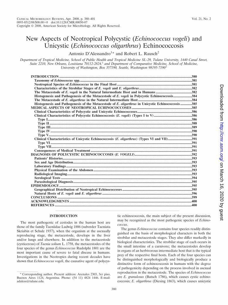
FIG. 1. Life cycle of Echinococcus vogeli in neotropical forests and the course of domiciliary transmission to humans.
VOL. 21, 2008 NEW ASPECTS OF NEOTROPICAL ECHINOCOCCOSIS 381
on March 16, 2020 by guest
http://cmr.asm
.org/D
ownloaded from
hosts. The domestic dog is the most significant source of in-fection of humans by the etiologic agents of cystic, alveolar,and polycystic forms of echinococcosis.
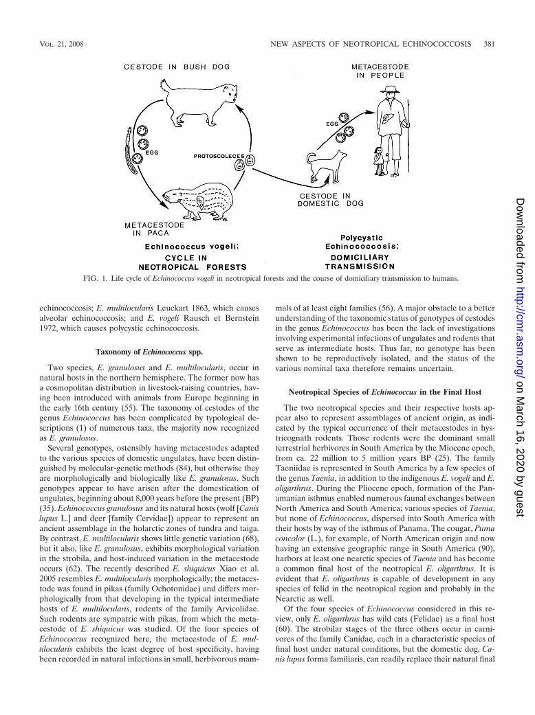
The natural cycle of E. vogeli was first reported by Cabreraet al. (8), who noted that the bush dog, Speothos venaticus(Lund), the natural final host of that cestode, hunted its pre-ferred prey, the paca, Cuniculus paca (L.), in packs, pursuing iton land and in water (Fig. 1, 2, and 3). Although bush dogs andpacas are of similar sizes, single bush dogs may sometimes becapable of overcoming the rodents, as shown by Deutsch (21).A fox-like canid, Cerdocyon thous (L.), which occurs widely inSouth America, was infected experimentally by D’Alessandro(unpublished data). That canid, an omnivore that feeds also onsmall mammals (67), does not hunt in packs and thereforeappears to be unable to capture mammals as large as a paca. InArgentina, fox-like canids of the genus Dusicyon are ofteninfected by E. granulosus (5, 74). Those animals feed mainly onsmaller mammals but evidently become infected by that ces-tode from scavenging on carcasses of sheep (67).
At the present, the bush dog is the only known natural finalhost of E. vogeli. Few bush dogs have been examined for thepresence of intestinal helminths, but the high prevalence of themetacestode of E. vogeli in pacas is indicative of a much higherrate in the final hosts. Such a differential between the respec-
tive final and intermediate hosts is typical also for E. granulosusand E. multilocularis (57, 61).
Characteristics of the Strobilar Stages of E. vogeli andE. oligarthrus
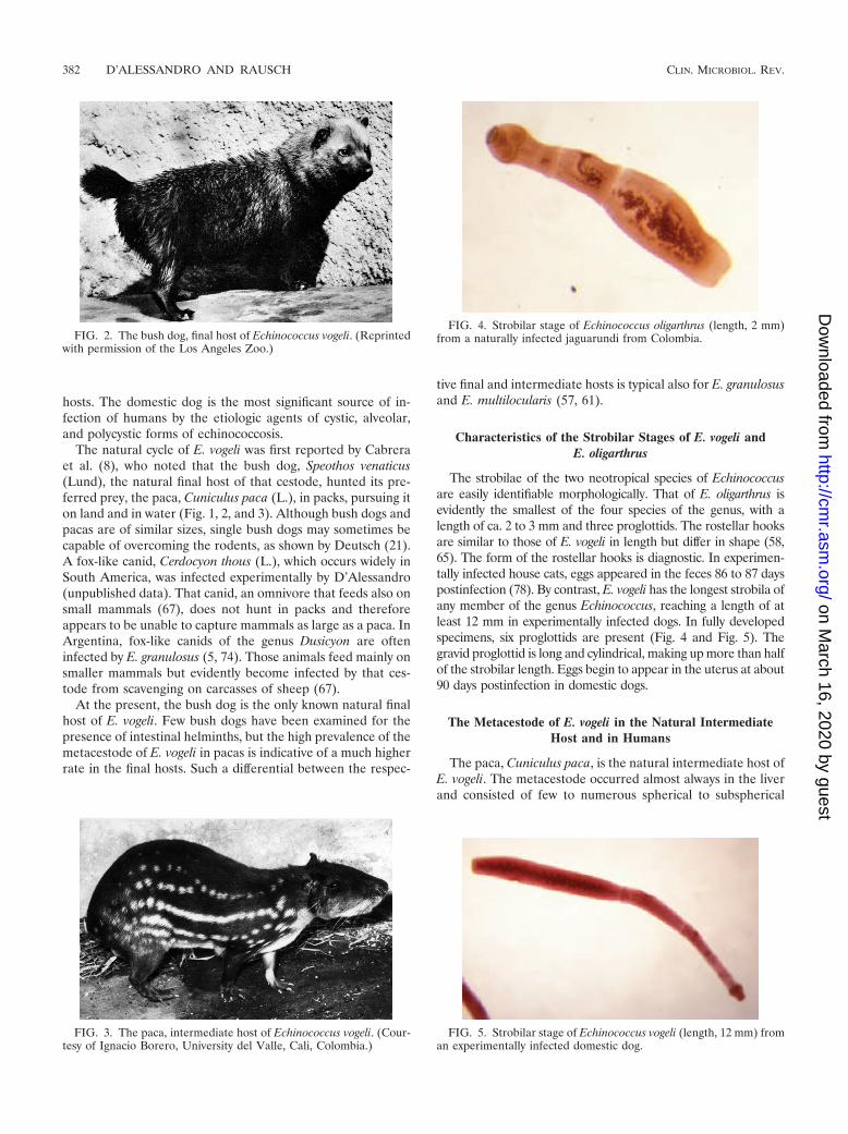
The strobilae of the two neotropical species of Echinococcusare easily identifiable morphologically. That of E. oligarthrus isevidently the smallest of the four species of the genus, with alength of ca. 2 to 3 mm and three proglottids. The rostellar hooksare similar to those of E. vogeli in length but differ in shape (58,65). The form of the rostellar hooks is diagnostic. In experimen-tally infected house cats, eggs appeared in the feces 86 to 87 dayspostinfection (78). By contrast, E. vogeli has the longest strobila ofany member of the genus Echinococcus, reaching a length of atleast 12 mm in experimentally infected dogs. In fully developedspecimens, six proglottids are present (Fig. 4 and Fig. 5). Thegravid proglottid is long and cylindrical, making up more than halfof the strobilar length. Eggs begin to appear in the uterus at about90 days postinfection in domestic dogs.
The Metacestode of E. vogeli in the Natural IntermediateHost and in Humans
The paca, Cuniculus paca, is the natural intermediate host ofE. vogeli. The metacestode occurred almost always in the liverand consisted of few to numerous spherical to subspherical
FIG. 2. The bush dog, final host of Echinococcus vogeli. (Reprintedwith permission of the Los Angeles Zoo.)
FIG. 3. The paca, intermediate host of Echinococcus vogeli. (Cour-tesy of Ignacio Borero, University del Valle, Cali, Colombia.)
FIG. 4. Strobilar stage of Echinococcus oligarthrus (length, 2 mm)from a naturally infected jaguarundi from Colombia.
FIG. 5. Strobilar stage of Echinococcus vogeli (length, 12 mm) froman experimentally infected domestic dog.
382 D’ALESSANDRO AND RAUSCH CLIN. MICROBIOL. REV.
on March 16, 2020 by guest
http://cmr.asm
.org/D
ownloaded from
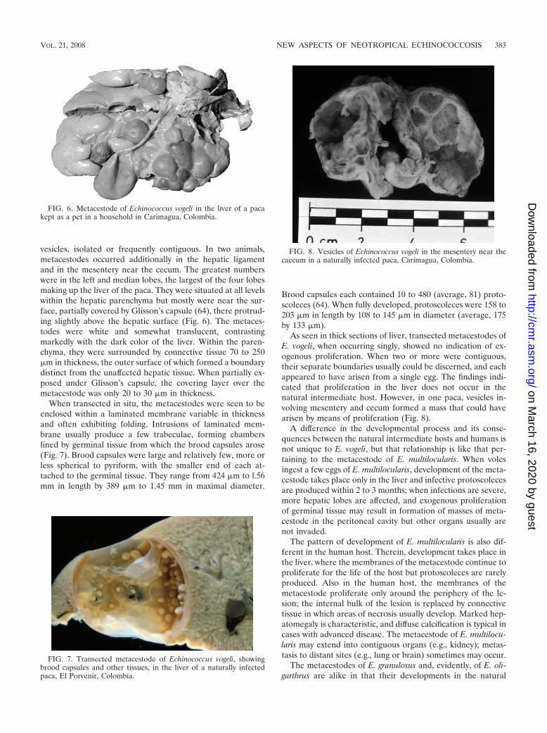
vesicles, isolated or frequently contiguous. In two animals,metacestodes occurred additionally in the hepatic ligamentand in the mesentery near the cecum. The greatest numberswere in the left and median lobes, the largest of the four lobesmaking up the liver of the paca. They were situated at all levelswithin the hepatic parenchyma but mostly were near the sur-face, partially covered by Glisson’s capsule (64), there protrud-ing slightly above the hepatic surface (Fig. 6). The metaces-todes were white and somewhat translucent, contrastingmarkedly with the dark color of the liver. Within the paren-chyma, they were surrounded by connective tissue 70 to 250�m in thickness, the outer surface of which formed a boundarydistinct from the unaffected hepatic tissue. When partially ex-posed under Glisson’s capsule, the covering layer over themetacestode was only 20 to 30 �m in thickness.
When transected in situ, the metacestodes were seen to beenclosed within a laminated membrane variable in thicknessand often exhibiting folding. Intrusions of laminated mem-brane usually produce a few trabeculae, forming chamberslined by germinal tissue from which the brood capsules arose(Fig. 7). Brood capsules were large and relatively few, more orless spherical to pyriform, with the smaller end of each at-tached to the germinal tissue. They range from 424 �m to l.56mm in length by 389 �m to 1.45 mm in maximal diameter.
Brood capsules each contained 10 to 480 (average, 81) proto-scoleces (64). When fully developed, protoscoleces were 158 to203 �m in length by 108 to 145 �m in diameter (average, 175by 133 �m).
As seen in thick sections of liver, transected metacestodes ofE. vogeli, when occurring singly, showed no indication of ex-ogenous proliferation. When two or more were contiguous,their separate boundaries usually could be discerned, and eachappeared to have arisen from a single egg. The findings indi-cated that proliferation in the liver does not occur in thenatural intermediate host. However, in one paca, vesicles in-volving mesentery and cecum formed a mass that could havearisen by means of proliferation (Fig. 8).
A difference in the developmental process and its conse-quences between the natural intermediate hosts and humans isnot unique to E. vogeli, but that relationship is like that per-taining to the metacestode of E. multilocularis. When volesingest a few eggs of E. multilocularis, development of the meta-cestode takes place only in the liver and infective protoscolecesare produced within 2 to 3 months; when infections are severe,more hepatic lobes are affected, and exogenous proliferationof germinal tissue may result in formation of masses of meta-cestode in the peritoneal cavity but other organs usually arenot invaded.
The pattern of development of E. multilocularis is also dif-ferent in the human host. Therein, development takes place inthe liver, where the membranes of the metacestode continue toproliferate for the life of the host but protoscoleces are rarelyproduced. Also in the human host, the membranes of themetacestode proliferate only around the periphery of the le-sion; the internal bulk of the lesion is replaced by connectivetissue in which areas of necrosis usually develop. Marked hep-atomegaly is characteristic, and diffuse calcification is typical incases with advanced disease. The metacestode of E. multilocu-laris may extend into contiguous organs (e.g., kidney); metas-tasis to distant sites (e.g., lung or brain) sometimes may occur.
The metacestodes of E. granulosus and, evidently, of E. oli-garthrus are alike in that their developments in the natural
FIG. 6. Metacestode of Echinococcus vogeli in the liver of a pacakept as a pet in a household in Carimagua, Colombia.
FIG. 7. Transected metacestode of Echinococcus vogeli, showingbrood capsules and other tissues, in the liver of a naturally infectedpaca, El Porvenir, Colombia.
FIG. 8. Vesicles of Echinococcus vogeli in the mesentery near thecaecum in a naturally infected paca, Carimagua, Colombia.
VOL. 21, 2008 NEW ASPECTS OF NEOTROPICAL ECHINOCOCCOSIS 383
on March 16, 2020 by guest
http://cmr.asm
.org/D
ownloaded from
intermediate hosts and their developments in humans do notdiffer. In their respective intermediate hosts, the metacestodesof both species enlarge concentrically, and many protoscolecesare produced.
Histogenesis and Pathogenesis of the Metacestode ofE. vogeli in Polycystic Echinococcosis
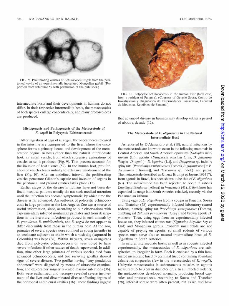
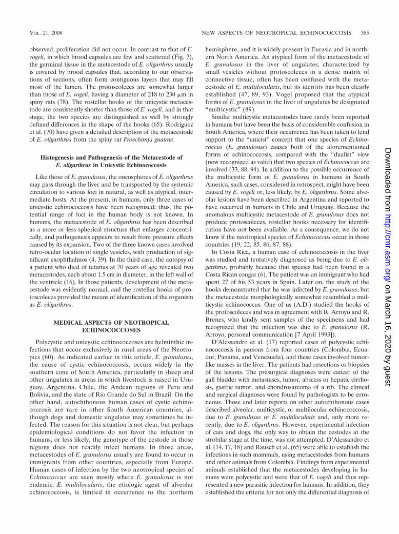
After ingestion of eggs of E. vogeli, the oncospheres releasedin the intestine are transported to the liver, where the onco-sphere forms a primary lacuna and development of the meta-cestode begins. In hosts other than the natural intermediatehost, an initial vesicle, from which successive generations ofvesicles arise, is produced (Fig. 9). That process accounts forthe invasion of host tissues (59). In the human host, prolifer-ation of vesicles leads initially to extensive involvement of theliver (Fig. 10). After an undefined interval, the proliferatingvesicles penetrate Glisson’s capsule and invasion of organs inthe peritoneal and pleural cavities takes place (12).
Earlier stages of the disease in humans have not been de-fined, because patients usually do not seek medical attentionuntil the infection has become symptomatic, by which time thedisease is far advanced. An outbreak of polycystic echinococ-cosis in large primates at the Los Angeles Zoo was a source ofuseful information, since, according to our observations withexperimentally infected nonhuman primates and from descrip-tions in the literature, infections produced in such animals byE. granulosus, E. multilocularis, and E. vogeli do not appear todiffer discernibly from those in the human host. At the zoo,primates of several species were confined as young juveniles inan enclosure adjacent to one in which a bush dog (captured inColombia) was kept (36). Within 10 years, seven orangutansdied from polycystic echinococcosis or were noted to havesevere infections if other causes of death supervened. In addi-tion, nine other large primates of various species died fromadvanced echinococcosis, and two surviving gorillas showedsigns of severe disease. Two gorillas having “very pendulousabdomens” were diagnosed by means of ultrasonic examina-tion, and exploratory surgery revealed massive infections (36).Both were euthanized, and necropsy revealed severe involve-ment of the liver and disseminated disease affecting organs ofthe peritoneal and pleural cavities (36). Those findings suggest
that advanced disease in humans may develop within a periodof about a decade (12).
The Metacestode of E. oligarthrus in the NaturalIntermediate Host
As reported by D’Alessandro et al. (18), natural infections bythe metacestode are known to occur in the following mammals inCentral America and South America: opossums [Didelphis mar-supialis (L.)]; agoutis {Dasyprocta punctata Gray, D. fuliginosaWagler, D. aguti [� D. leporina (L.)], and Dasyprocta sp. indet.};spiny rats {Proechimys semispinosus (Tomes), P. guyannensis [� P.decumanus (Thomas)], and Proechimys sp. indet.}; and pacas.The metacestode described as E. cruzi Brumpt et Joyeux 1924 (7),from agoutis in Brazil, has been shown to be that of E. oligarthrus(63). The metacestode has been reported to occur in rabbits(Sylvilagus floridanus (Allen)) in Venezuela (41). S. floridanus hasexpanded its range into South America relatively recently, via thePanamanian isthmus.
Using eggs of E. oligarthrus from a cougar in Panama, Sousaand Thatcher (78) experimentally infected laboratory-rearedrodents, namely, spiny rat Proechimys semispinosus (Tomes),climbing rat Tylomys panamensis (Gray), and brown agouti D.punctata. Then, using eggs from an experimentally infectedhouse cat, they infected cotton rats (Sigmodon hispidus Say etOrd) and Mongolian gerbils. Probably small felids are notcapable of preying on agoutis, so small rodents of variousspecies must serve also as natural intermediate hosts of E.oligarthrus in South America.
In natural intermediate hosts, as well as in rodents infectedexperimentally, the metacestodes of E. oligarthrus are sub-spherical to irregular in form. Each is enclosed by a thin lam-inated membrane lined by germinal tissue containing abundantcalcareous corpuscles (few in the metacestodes of E. vogeli).Unicystic metacestodes in subcutaneous muscles in agoutismeasured 0.5 to 3 cm in diameter (78). In all infected rodents,the metacestodes developed normally, producing brood cap-sules and protoscoleces. According to Sousa and Thatcher(78), internal septae were often present, but as we also have
FIG. 10. Polycystic echinococcosis in the human liver (fatal case,from a resident of Panama). (Courtesy of Octavio Sousa, Centro deInvestigacion y Diagnostico de Enfermedades Parasitarias, Facultadde Medicina, Republica de Panama.)
FIG. 9. Proliferating vesicles of Echinococcus vogeli from the peri-toneal cavity of an experimentally inoculated Mongolian gerbil. (Re-printed from reference 59 with permission of the publisher.)
384 D’ALESSANDRO AND RAUSCH CLIN. MICROBIOL. REV.
on March 16, 2020 by guest
http://cmr.asm
.org/D
ownloaded from
observed, proliferation did not occur. In contrast to that of E.vogeli, in which brood capsules are few and scattered (Fig. 7),the germinal tissue in the metacestode of E. oligarthrus usuallyis covered by brood capsules that, according to our observa-tions of sections, often form contiguous layers that may fillmost of the lumen. The protoscoleces are somewhat largerthan those of E. vogeli, having a diameter of 218 to 230 �m inspiny rats (78). The rostellar hooks of the unicystic metaces-tode are consistently shorter than those of E. vogeli, and in thatstage, the two species are distinguished as well by stronglydefined differences in the shape of the hooks (65). Rodriguezet al. (70) have given a detailed description of the metacestodeof E. oligarthrus from the spiny rat Proechimys guairae.
Histogenesis and Pathogenesis of the Metacestode ofE. oligarthrus in Unicystic Echinococcosis
Like those of E. granulosus, the oncospheres of E. oligarthrusmay pass through the liver and be transported by the systemiccirculation to various loci in natural, as well as atypical, inter-mediate hosts. At the present, in humans, only three cases ofunicystic echinococcosis have been recognized; thus, the po-tential range of loci in the human body is not known. Inhumans, the metacestode of E. oligarthrus has been describedas a more or less spherical structure that enlarges concentri-cally, and pathogenesis appears to result from pressure effectscaused by its expansion. Two of the three known cases involvedretro-ocular location of single vesicles, with production of sig-nificant exophthalmos (4, 39). In the third case, the autopsy ofa patient who died of tetanus at 70 years of age revealed twometacestodes, each about 1.5 cm in diameter, in the left wall ofthe ventricle (16). In those patients, development of the meta-cestode was evidently normal, and the rostellar hooks of pro-toscoleces provided the means of identification of the organismas E. oligarthrus.
MEDICAL ASPECTS OF NEOTROPICALECHINOCOCCOSES
Polycystic and unicystic echinococcoses are helminthic in-fections that occur exclusively in rural areas of the Neotro-pics (60). As indicated earlier in this article, E. granulosus,the cause of cystic echinococcosis, occurs widely in thesouthern cone of South America, particularly in sheep andother ungulates in areas in which livestock is raised in Uru-guay, Argentina, Chile, the Andean regions of Peru andBolivia, and the state of Rio Grande do Sul in Brazil. On theother hand, autochthonous human cases of cystic echino-coccosis are rare in other South American countries, al-though dogs and domestic ungulates may sometimes be in-fected. The reason for this situation is not clear, but perhapsepidemiological conditions do not favor the infection inhumans, or less likely, the genotype of the cestode in thoseregions does not readily infect humans. In those areas,metacestodes of E. granulosus usually are found to occur inimmigrants from other countries, especially from Europe.Human cases of infection by the two neotropical species ofEchinococcus are seen mostly where E. granulosus is notendemic. E. multilocularis, the etiologic agent of alveolarechinococcosis, is limited in occurrence to the northern
hemisphere, and it is widely present in Eurasia and in north-ern North America. An atypical form of the metacestode ofE. granulosus in the liver of ungulates, characterized bysmall vesicles without protoscoleces in a dense matrix ofconnective tissue, often has been confused with the meta-cestode of E. multilocularis, but its identity has been clearlyestablished (47, 89, 93). Vogel proposed that the atypicalforms of E. granulosus in the liver of ungulates be designated“multicystic” (89).
Similar multicystic metacestodes have rarely been reportedin humans but have been the basis of considerable confusion inSouth America, where their occurrence has been taken to lendsupport to the “unicist” concept that one species of Echino-coccus (E. granulosus) causes both of the aforementionedforms of echinococcosis, compared with the “dualist” view(now recognized as valid) that two species of Echinococcus areinvolved (33, 88, 94). In addition to the possible occurrence ofthe multicystic form of E. granulosus in humans in SouthAmerica, such cases, considered in retrospect, might have beencaused by E. vogeli or, less likely, by E. oligarthrus. Some alve-olar lesions have been described in Argentina and reported tohave occurred in humans in Chile and Uruguay. Because theanomalous multicystic metacestode of E. granulosus does notproduce protoscoleces, rostellar hooks necessary for identifi-cation have not been available. As a consequence, we do notknow if the neotropical species of Echinococcus occur in thosecountries (19, 22, 85, 86, 87, 88).
In Costa Rica, a human case of echinococcosis in the liverwas studied and tentatively diagnosed as being due to E. oli-garthrus, probably because that species had been found in aCosta Rican cougar (6). The patient was an immigrant who hadspent 27 of his 53 years in Spain. Later on, the study of thehooks demonstrated that he was infected by E. granulosus, butthe metacestode morphologically somewhat resembled a mul-ticystic echinococcus. One of us (A.D.) studied the hooks ofthe protoscoleces and was in agreement with R. Arroyo and R.Brenes, who kindly sent samples of the specimens and hadrecognized that the infection was due to E. granulosus (R.Arroyo, personal communication [7 April 1993]).
D’Alessandro et al. (17) reported cases of polycystic echi-nococcosis in persons from four countries (Colombia, Ecua-dor, Panama, and Venezuela), and these cases involved tumor-like masses in the liver. The patients had resections or biopsiesof the lesions. The presurgical diagnoses were cancer of thegall bladder with metastases, tumor, abscess or hepatic cirrho-sis, gastric tumor, and chondrosarcoma of a rib. The clinicaland surgical diagnoses were found by pathologists to be erro-neous. Those and later reports on other autochthonous casesdescribed alveolar, multicystic, or multilocular echinococcosis,due to E. granulosus or E. multilocularis and, only more re-cently, due to E. oligarthrus. However, experimental infectionof cats and dogs, the only way to obtain the cestodes at thestrobilar stage at the time, was not attempted. D’Alessandro etal. (14, 17, 18) and Rausch et al. (65) were able to establish theinfections in such mammals, using metacestodes from humansand other animals from Colombia. Findings from experimentalanimals established that the metacestodes developing in hu-mans were polycystic and were that of E. vogeli and thus rep-resented a new parasitic infection for humans. In addition, theyestablished the criteria for not only the differential diagnosis of
VOL. 21, 2008 NEW ASPECTS OF NEOTROPICAL ECHINOCOCCOSIS 385
on March 16, 2020 by guest
http://cmr.asm
.org/D
ownloaded from
infection with the two neotropical species, E. vogeli and E.oligarthrus, but also criteria separating the causative agentsfrom the other two recognized species, E. granulosus and E.multilocularis.
Fortunately, in recent years, investigators have become in-creasingly aware of and interested in the two zoonoses causedby the neotropical cestodes and have improved their capabil-ities for recognition and diagnosis of such infections. Due to itsfrequency, polycystic echinococcosis is no longer a medicalcuriosity, and it should be considered in the differential diag-nosis of polycystic masses in humans. On the other hand,human infections with the unicystic metacestode of E. oliga-rthrus have been rarely diagnosed.
Clinical Characteristics of Polycystic andUnicystic Echinococcoses
The clinical characteristics of polycystic echinococcosis inpatients depend on the location of the metacestode as well asthe extent of invasion of tissues, particularly the liver (Table 1).
D’Alessandro et al. (17) published observations on all re-ported or known cases of polycystic echinococcosis. In addi-tion, D’Alessandro invited senior parasitologists of CentralAmerican and South American countries to report, duringvarious meetings of the Federacion Latinoamericana de Para-sitologos, the status of echinococcosis in their countries, inparticular concerning polycystic echinococcosis in humans andother animals. Their contributions have been reported (12).The present report includes all data, published and unpub-lished, obtained from reliable sources since that time.
The total number of known cases of neotropical echinococ-cosis, as of March 2007, is 172. Included in that number are 18new cases observed by Ulysses Meneghelli (personal commu-nication [October 2006]). In addition, two serological surveysin Brazil demonstrated that 41 of 1,064 and 19 of 40 persons(total, 60 of 1,104 [5%]) showed antibodies for Echinococcus inthe counterelectrophoresis test. Whether all those 60 personshad asymptomatic echinococcosis is not known, because onesingle test may overlook or involve a cross-reaction with otherconditions or parasitic infections. Nonetheless, it seems logical
to consider that some of those persons from areas in which theinfection is endemic may have had asymptomatic infections byE. vogeli. Therefore, the number of infected persons in theneotropical countries may be 232 and, in Brazil alone, 160 (52;U. Meneghelli, personal communication [2006]). Forty Brazil-ian cases of polycystic echinococcosis occurring in the states ofPara and Amapa, in the western Amazonian region, were sum-marized by Soares et al. (76). Included in that group were 14cases reported by Orlando Fonseca and Aurelio Costa in 1995(12).
As of March 2007, the clinical information from 81 patientsis available among the 172 known cases (Table 1). By far, theliver was the organ most frequently affected. Indeed, in 81% ofthe cases, metacestodes were found in the liver alone or withvesicles situated in the abdomen, in the liver and the lungs/pleural cavities, or only as calcified vesicles in the liver. Organsinvolved in the abdomen included the diaphragm, spleen, pan-creas, omentum, mesenteries, rectal-vesical sac, ovaries,uterus, abdominal wall, psoas muscle, and vertebra. In thechest, the lungs were infiltrated in 11 (14%) cases; also in-volved were ribs, intercostals and subscapular muscle, pleura,pericardium, auricle, vena cava, and other large mediastinalvessels. The remaining 13 polycystic lesions were located onlyin the mesentery. Of unicystic lesions, due to metacestodes ofE. oligarthrus, one was located in the heart and two were in theorbit. Those single-site infections were reported as being notconcomitant or secondary to any other lesions located else-where in the body.
Clinical Characteristics of Polycystic Echinococcosis(E. vogeli) (Types I to V)
Examination of the patients’ case histories offered the op-portunity to group them in types so that frequency of clinicalfeatures, severity of the illness, complications, types of treat-ments used, and mortality could be better assessed.
Type I. The most common type of presentation, type I,included lesions in the liver (Fig. 11) and in the abdomen, seenin 30 of 81 (37%) cases (Table 1). The patients presented withpalpable, hard, and rounded masses, painful or not painful,
TABLE 1. Frequency of clinical characteristics of polycystic and unicystic neotropical echinococcoses according to species
Clinical typeof echinococcosis Clinical presentation
No. of cases
E. vogeli E. cf. vogelia E. oligarthrus E. granulosus Total (%)c
PolycysticI Cysts in the liver and abdominal cavity 19 11 0 1 30 (37)II Cysts in the liver and abdominal cavity plus hepatic insufficiency 9 12 0 0 21 (26)III Cysts in the liver and lung/chest 4 7 0 0 11 (14)IV Cysts only in the mesentery of the intestine or of the stomach 9 4 0 0 13 (16)V Calcified cysts in the liver and lungb 1 2 0 0 3 (4)
UnicysticVI Cysts only in the orbit 0 0 2 0 2 (2)VII Cysts only in the heart 0 0 1 0 1 (1)
Total 42 36 3 1 81 (100)
a E. cf. vogeli causes infections with polycystic lesions but with no hooks found.b The total number of these patients with liver cysts was 66/81 (81%), and the number with lung cysts was 11/81 (14%).c Excludes the E. granulosus case.
386 D’ALESSANDRO AND RAUSCH CLIN. MICROBIOL. REV.
on March 16, 2020 by guest
http://cmr.asm
.org/D
ownloaded from
usually within or connected with the liver (the hepatic massesand size of abdomen progressively enlarging), abdominal pain,gastrointestinal problems, marked weight loss, and fever. Theillness was first treated with analgesics or other symptomaticdrugs. For seven of the patients in this group, a diagnosticlaparotomy/biopsy (Table 2) was the only invasive procedurecarried out; these patients usually were considered to haveintractable disease (carcinoma) and left alone. With recogni-tion of the nature of this tropical illness, surgical interventionwas begun. Surgery was well tolerated, and none of 17 casesdied during the surgery or as a direct consequence. For at least4 of those 17 cases, a partial hepatectomy was carried out; forothers, the entire metacestode was resected or a partial cys-tectomy was performed. The entire metacestode could notalways be removed. At times, some spilling of its fluid contentinto the abdominal cavity occurred, with the risk of secondaryproliferation causing recurrent disease. Polycystic echinococ-cosis in most cases takes a chronic course, sometimes overmany years. However, surgery that failed to resect the entiremetacestode was usually beneficial to the patient, who mightthen live for a long time with minimal disturbances.
A typical case of type I disease is as follows (12): a 58-year-old female who was born in a rural area and relocated to the
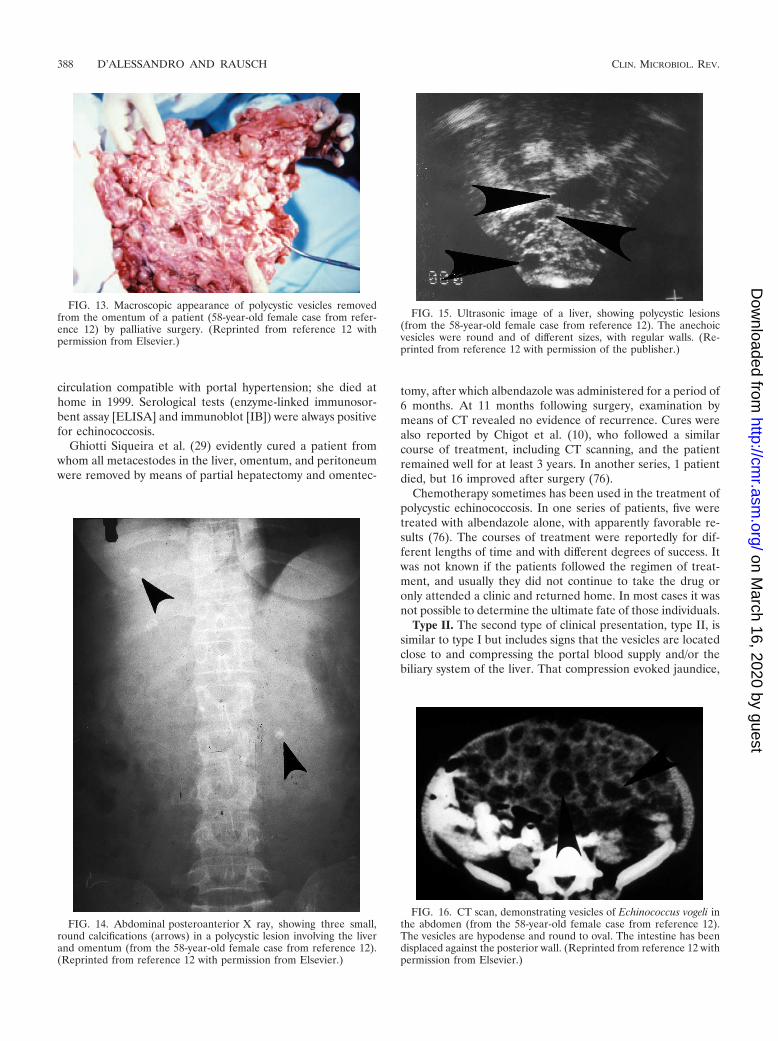
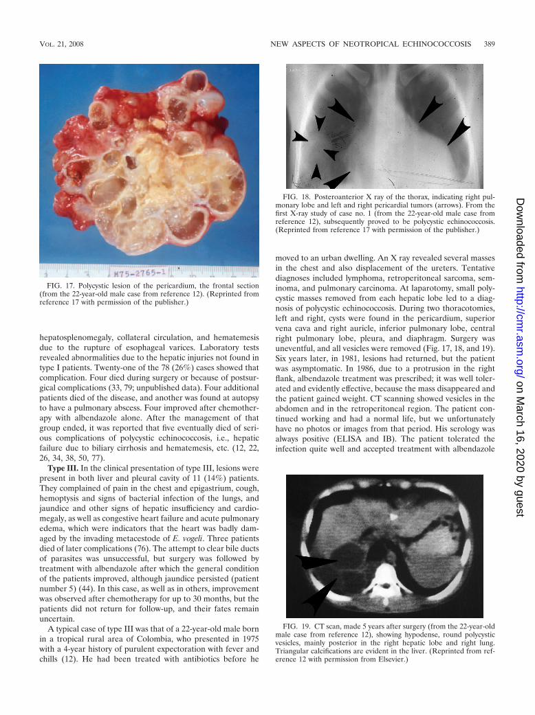
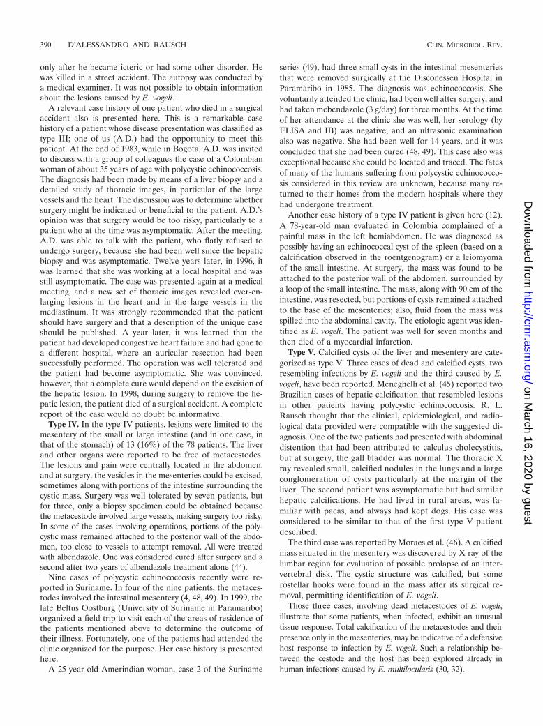
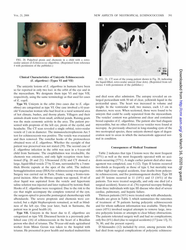
cities of Buenaventura and Cali, Colombia, had a 15-year his-tory of an abdominal mass as of presentation in 1981. Withinone year, she lost 8 to 10 kg in body weight and the mass hadbecome larger, compressing the thorax, and was movable byhand. The enlarged abdomen gave the impression of advancedpregnancy. The mass was hard and painless; laboratory resultswere normal. Imaging showed round calcifications. Laparos-copy revealed polycystic masses in the abdominal cavity, liver,omentum, and abdominal wall. Simple X rays, computerizedtomography (CT) scan, and ultrasonic examination were per-formed (Fig. 12, 13, 14, 15, and 16). We recommended pallia-tive surgery because the abdominal cysts were interfering withbreathing and eating. The surgeon excised only the vesiclesfrom the sites mentioned. The liver, stomach, and spleenformed a unified mass that was not disturbed. With the re-moval of 2 kg of cysts, the patient had a short, uneventfulrecovery after surgery, with relief of intra-abdominal pressure.A year later, she again became symptomatic and for 28 dayswas treated with albendazole, which was well tolerated. Sheimproved clinically for several months and did not return for afollow-up. Subsequently, it was learned that the patient be-came cachectic and developed ascites and collateral abdominal
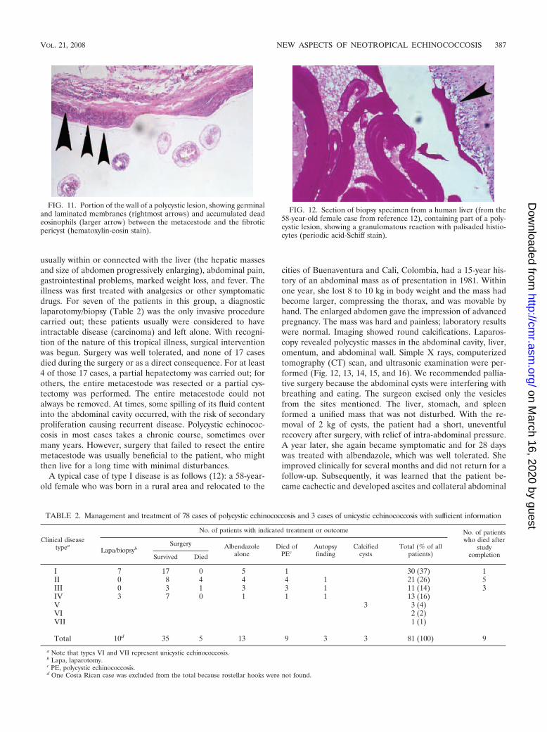
FIG. 11. Portion of the wall of a polycystic lesion, showing germinaland laminated membranes (rightmost arrows) and accumulated deadeosinophils (larger arrow) between the metacestode and the fibroticpericyst (hematoxylin-eosin stain).
TABLE 2. Management and treatment of 78 cases of polycystic echinococcosis and 3 cases of unicystic echinococcosis with sufficient information
Clinical diseasetypea
No. of patients with indicated treatment or outcome No. of patientswho died after
studycompletion
Lapa/biopsybSurgery Albendazole
aloneDied of
PEcAutopsyfinding
Calcifiedcysts
Total (% of allpatients)Survived Died
I 7 17 0 5 1 30 (37) 1II 0 8 4 4 4 1 21 (26) 5III 0 3 1 3 3 1 11 (14) 3IV 3 7 0 1 1 1 13 (16)V 3 3 (4)VI 2 (2)VII 1 (1)
Total 10d 35 5 13 9 3 3 81 (100) 9
a Note that types VI and VII represent unicystic echinococcosis.b Lapa, laparotomy.c PE, polycystic echinococcosis.d One Costa Rican case was excluded from the total because rostellar hooks were not found.
FIG. 12. Section of biopsy specimen from a human liver (from the58-year-old female case from reference 12), containing part of a poly-cystic lesion, showing a granulomatous reaction with palisaded histio-cytes (periodic acid-Schiff stain).
VOL. 21, 2008 NEW ASPECTS OF NEOTROPICAL ECHINOCOCCOSIS 387
on March 16, 2020 by guest
http://cmr.asm
.org/D
ownloaded from
circulation compatible with portal hypertension; she died athome in 1999. Serological tests (enzyme-linked immunosor-bent assay [ELISA] and immunoblot [IB]) were always positivefor echinococcosis.
Ghiotti Siqueira et al. (29) evidently cured a patient fromwhom all metacestodes in the liver, omentum, and peritoneumwere removed by means of partial hepatectomy and omentec-
tomy, after which albendazole was administered for a period of6 months. At 11 months following surgery, examination bymeans of CT revealed no evidence of recurrence. Cures werealso reported by Chigot et al. (10), who followed a similarcourse of treatment, including CT scanning, and the patientremained well for at least 3 years. In another series, 1 patientdied, but 16 improved after surgery (76).
Chemotherapy sometimes has been used in the treatment ofpolycystic echinococcosis. In one series of patients, five weretreated with albendazole alone, with apparently favorable re-sults (76). The courses of treatment were reportedly for dif-ferent lengths of time and with different degrees of success. Itwas not known if the patients followed the regimen of treat-ment, and usually they did not continue to take the drug oronly attended a clinic and returned home. In most cases it wasnot possible to determine the ultimate fate of those individuals.
Type II. The second type of clinical presentation, type II, issimilar to type I but includes signs that the vesicles are locatedclose to and compressing the portal blood supply and/or thebiliary system of the liver. That compression evoked jaundice,
FIG. 13. Macroscopic appearance of polycystic vesicles removedfrom the omentum of a patient (58-year-old female case from refer-ence 12) by palliative surgery. (Reprinted from reference 12 withpermission from Elsevier.)
FIG. 14. Abdominal posteroanterior X ray, showing three small,round calcifications (arrows) in a polycystic lesion involving the liverand omentum (from the 58-year-old female case from reference 12).(Reprinted from reference 12 with permission from Elsevier.)
FIG. 15. Ultrasonic image of a liver, showing polycystic lesions(from the 58-year-old female case from reference 12). The anechoicvesicles were round and of different sizes, with regular walls. (Re-printed from reference 12 with permission of the publisher.)
FIG. 16. CT scan, demonstrating vesicles of Echinococcus vogeli inthe abdomen (from the 58-year-old female case from reference 12).The vesicles are hypodense and round to oval. The intestine has beendisplaced against the posterior wall. (Reprinted from reference 12 withpermission from Elsevier.)
388 D’ALESSANDRO AND RAUSCH CLIN. MICROBIOL. REV.
on March 16, 2020 by guest
http://cmr.asm
.org/D
ownloaded from
hepatosplenomegaly, collateral circulation, and hematemesisdue to the rupture of esophageal varices. Laboratory testsrevealed abnormalities due to the hepatic injuries not found intype I patients. Twenty-one of the 78 (26%) cases showed thatcomplication. Four died during surgery or because of postsur-gical complications (33, 79; unpublished data). Four additionalpatients died of the disease, and another was found at autopsyto have a pulmonary abscess. Four improved after chemother-apy with albendazole alone. After the management of thatgroup ended, it was reported that five eventually died of seri-ous complications of polycystic echinococcosis, i.e., hepaticfailure due to biliary cirrhosis and hematemesis, etc. (12, 22,26, 34, 38, 50, 77).
Type III. In the clinical presentation of type III, lesions werepresent in both liver and pleural cavity of 11 (14%) patients.They complained of pain in the chest and epigastrium, cough,hemoptysis and signs of bacterial infection of the lungs, andjaundice and other signs of hepatic insufficiency and cardio-megaly, as well as congestive heart failure and acute pulmonaryedema, which were indicators that the heart was badly dam-aged by the invading metacestode of E. vogeli. Three patientsdied of later complications (76). The attempt to clear bile ductsof parasites was unsuccessful, but surgery was followed bytreatment with albendazole after which the general conditionof the patients improved, although jaundice persisted (patientnumber 5) (44). In this case, as well as in others, improvementwas observed after chemotherapy for up to 30 months, but thepatients did not return for follow-up, and their fates remainuncertain.
A typical case of type III was that of a 22-year-old male bornin a tropical rural area of Colombia, who presented in 1975with a 4-year history of purulent expectoration with fever andchills (12). He had been treated with antibiotics before he
moved to an urban dwelling. An X ray revealed several massesin the chest and also displacement of the ureters. Tentativediagnoses included lymphoma, retroperitoneal sarcoma, sem-inoma, and pulmonary carcinoma. At laparotomy, small poly-cystic masses removed from each hepatic lobe led to a diag-nosis of polycystic echinococcosis. During two thoracotomies,left and right, cysts were found in the pericardium, superiorvena cava and right auricle, inferior pulmonary lobe, centralright pulmonary lobe, pleura, and diaphragm. Surgery wasuneventful, and all vesicles were removed (Fig. 17, 18, and 19).Six years later, in 1981, lesions had returned, but the patientwas asymptomatic. In 1986, due to a protrusion in the rightflank, albendazole treatment was prescribed; it was well toler-ated and evidently effective, because the mass disappeared andthe patient gained weight. CT scanning showed vesicles in theabdomen and in the retroperitoneal region. The patient con-tinued working and had a normal life, but we unfortunatelyhave no photos or images from that period. His serology wasalways positive (ELISA and IB). The patient tolerated theinfection quite well and accepted treatment with albendazole
FIG. 17. Polycystic lesion of the pericardium, the frontal section(from the 22-year-old male case from reference 12). (Reprinted fromreference 17 with permission of the publisher.)
FIG. 19. CT scan, made 5 years after surgery (from the 22-year-oldmale case from reference 12), showing hypodense, round polycysticvesicles, mainly posterior in the right hepatic lobe and right lung.Triangular calcifications are evident in the liver. (Reprinted from ref-erence 12 with permission from Elsevier.)
FIG. 18. Posteroanterior X ray of the thorax, indicating right pul-monary lobe and left and right pericardial tumors (arrows). From thefirst X-ray study of case no. 1 (from the 22-year-old male case fromreference 12), subsequently proved to be polycystic echinococcosis.(Reprinted from reference 17 with permission of the publisher.)
VOL. 21, 2008 NEW ASPECTS OF NEOTROPICAL ECHINOCOCCOSIS 389
on March 16, 2020 by guest
http://cmr.asm
.org/D
ownloaded from
only after he became icteric or had some other disorder. Hewas killed in a street accident. The autopsy was conducted bya medical examiner. It was not possible to obtain informationabout the lesions caused by E. vogeli.
A relevant case history of one patient who died in a surgicalaccident also is presented here. This is a remarkable casehistory of a patient whose disease presentation was classified astype III; one of us (A.D.) had the opportunity to meet thispatient. At the end of 1983, while in Bogota, A.D. was invitedto discuss with a group of colleagues the case of a Colombianwoman of about 35 years of age with polycystic echinococcosis.The diagnosis had been made by means of a liver biopsy and adetailed study of thoracic images, in particular of the largevessels and the heart. The discussion was to determine whethersurgery might be indicated or beneficial to the patient. A.D.’sopinion was that surgery would be too risky, particularly to apatient who at the time was asymptomatic. After the meeting,A.D. was able to talk with the patient, who flatly refused toundergo surgery, because she had been well since the hepaticbiopsy and was asymptomatic. Twelve years later, in 1996, itwas learned that she was working at a local hospital and wasstill asymptomatic. The case was presented again at a medicalmeeting, and a new set of thoracic images revealed ever-en-larging lesions in the heart and in the large vessels in themediastinum. It was strongly recommended that the patientshould have surgery and that a description of the unique caseshould be published. A year later, it was learned that thepatient had developed congestive heart failure and had gone toa different hospital, where an auricular resection had beensuccessfully performed. The operation was well tolerated andthe patient had become asymptomatic. She was convinced,however, that a complete cure would depend on the excision ofthe hepatic lesion. In 1998, during surgery to remove the he-patic lesion, the patient died of a surgical accident. A completereport of the case would no doubt be informative.
Type IV. In the type IV patients, lesions were limited to themesentery of the small or large intestine (and in one case, inthat of the stomach) of 13 (16%) of the 78 patients. The liverand other organs were reported to be free of metacestodes.The lesions and pain were centrally located in the abdomen,and at surgery, the vesicles in the mesenteries could be excised,sometimes along with portions of the intestine surrounding thecystic mass. Surgery was well tolerated by seven patients, butfor three, only a biopsy specimen could be obtained becausethe metacestode involved large vessels, making surgery too risky.In some of the cases involving operations, portions of the poly-cystic mass remained attached to the posterior wall of the abdo-men, too close to vessels to attempt removal. All were treatedwith albendazole. One was considered cured after surgery and asecond after two years of albendazole treatment alone (44).
Nine cases of polycystic echinococcosis recently were re-ported in Suriname. In four of the nine patients, the metaces-todes involved the intestinal mesentery (4, 48, 49). In 1999, thelate Beltus Oostburg (University of Suriname in Paramaribo)organized a field trip to visit each of the areas of residence ofthe patients mentioned above to determine the outcome oftheir illness. Fortunately, one of the patients had attended theclinic organized for the purpose. Her case history is presentedhere.
A 25-year-old Amerindian woman, case 2 of the Suriname
series (49), had three small cysts in the intestinal mesenteriesthat were removed surgically at the Disconessen Hospital inParamaribo in 1985. The diagnosis was echinococcosis. Shevoluntarily attended the clinic, had been well after surgery, andhad taken mebendazole (3 g/day) for three months. At the timeof her attendance at the clinic she was well, her serology (byELISA and IB) was negative, and an ultrasonic examinationalso was negative. She had been well for 14 years, and it wasconcluded that she had been cured (48, 49). This case also wasexceptional because she could be located and traced. The fatesof many of the humans suffering from polycystic echinococco-sis considered in this review are unknown, because many re-turned to their homes from the modern hospitals where theyhad undergone treatment.
Another case history of a type IV patient is given here (12).A 78-year-old man evaluated in Colombia complained of apainful mass in the left hemiabdomen. He was diagnosed aspossibly having an echinococcal cyst of the spleen (based on acalcification observed in the roentgenogram) or a leiomyomaof the small intestine. At surgery, the mass was found to beattached to the posterior wall of the abdomen, surrounded bya loop of the small intestine. The mass, along with 90 cm of theintestine, was resected, but portions of cysts remained attachedto the base of the mesenteries; also, fluid from the mass wasspilled into the abdominal cavity. The etiologic agent was iden-tified as E. vogeli. The patient was well for seven months andthen died of a myocardial infarction.
Type V. Calcified cysts of the liver and mesentery are cate-gorized as type V. Three cases of dead and calcified cysts, tworesembling infections by E. vogeli and the third caused by E.vogeli, have been reported. Meneghelli et al. (45) reported twoBrazilian cases of hepatic calcification that resembled lesionsin other patients having polycystic echinococcosis. R. L.Rausch thought that the clinical, epidemiological, and radio-logical data provided were compatible with the suggested di-agnosis. One of the two patients had presented with abdominaldistention that had been attributed to calculus cholecystitis,but at surgery, the gall bladder was normal. The thoracic Xray revealed small, calcified nodules in the lungs and a largeconglomeration of cysts particularly at the margin of theliver. The second patient was asymptomatic but had similarhepatic calcifications. He had lived in rural areas, was fa-miliar with pacas, and always had kept dogs. His case wasconsidered to be similar to that of the first type V patientdescribed.
The third case was reported by Moraes et al. (46). A calcifiedmass situated in the mesentery was discovered by X ray of thelumbar region for evaluation of possible prolapse of an inter-vertebral disk. The cystic structure was calcified, but somerostellar hooks were found in the mass after its surgical re-moval, permitting identification of E. vogeli.
Those three cases, involving dead metacestodes of E. vogeli,illustrate that some patients, when infected, exhibit an unusualtissue response. Total calcification of the metacestodes and theirpresence only in the mesenteries, may be indicative of a defensivehost response to infection by E. vogeli. Such a relationship be-tween the cestode and the host has been explored already inhuman infections caused by E. multilocularis (30, 32).
390 D’ALESSANDRO AND RAUSCH CLIN. MICROBIOL. REV.
on March 16, 2020 by guest
http://cmr.asm
.org/D
ownloaded from
Clinical Characteristics of Unicystic Echinococcosis(E. oligarthrus) (Types VI and VII)
The unicystic lesions of E. oligarthrus in humans have beenso far reported in only two loci: in the orbit of the eye and inthe myocardium. We designate them type VI and type VII,respectively, using the same terminology as that used for casesof E. vogeli.
Type VI. Unicysts in the orbit (two cases due to E. oliga-rthrus) are categorized as type VI. One case involved a 61-year-old Venezuelan woman who had lived in a rural semiarid areaof hot climate, bushes, and thorny plants. Villagers and theiranimals drank water from small, artificial ponds. Raising goatswas the main economic activity in the area. The patient pre-sented with proptosis of the left eye, ptosis of the eyelid, andheadache. The CT scan revealed a single orbital, retro-ocularvesicle of 2 cm in diameter. The immunoelectrophoresis Arc 5test for echinococcosis was positive. The vesicle was evacuatedand then removed. The rostellar hooks of the protoscolecesobtained were of E. oligarthrus. Whether the eyesight of thatpatient was preserved was not stated (39). The second case ofE. oligarthrus infection in the orbit was seen in a 6-year-oldchild from Suriname. The exophthalmos was irreducible, thechemosis was extensive, and only light reception vison func-tioned (Fig. 20 and 21). Ultrasound (US) and CT showed alarge, liquid-filled vesicle 27 by 32 mm, surrounded by a 3-mm-thick fibrotic capsule at the apex of the orbit. The indirecthemagglutination assay (IHA) for echinococcosis was negative.Surgery was carried out in Paris, France, using a fronto-tem-poral incision. After the fibrous capsule was opened, 10 ml ofclear liquid was aspirated from the vesicle and hypertonicsaline solution was injected and later replaced by isotonic fluid.Hooks of E. oligarthrus were recognized. Due to the risk to theeye that might accompany the complete removal of the cyst,the surgeon opted to prescribe postsurgical chemotherapy withalbendazole. The severe proptosis and chemosis were cor-rected, but a slight blepharoptosis remained, as well as blind-ness of the left eye. One year later, back in Suriname, thepatient was in good health (4).
Type VII. Unicysts in the heart due to E. oligarthrus arecategorized as type VII. Discussed herein is a previously pub-lished case (16) of echinococcosis in the heart of a Brazilianperson who died of tetanus. The 70-year-old male agriculturalworker from Minas Gerais was taken to the hospital withtetanus. He presented in poor health and marked malnutrition
and died soon after admission. The autopsy revealed an en-larged pericardium with 50 ml of clear, yellowish liquid in thepericardial space. The heart was increased in volume andweight. In the ventricular wall, two masses, each 1.5 cm indiameter, were seen. When sectioned, those were found to beunicysts that could be easily separated from the myocardium.The vesicles’ content was gelatinous and clear and containedbrood capsules of E. oligarthrus. The patient also had chagasicmyocarditis, but no other Echinococcus vesicles were found atnecropsy. As previously observed in long-standing cysts of thetwo neotropical species, these unicysts showed signs of degen-eration next to areas in which the metacestode appeared nor-mal in condition.
Consequences of Medical Treatment
Table 2 indicates that type I lesions were the most frequent(37%) as well as the most frequently operated with no acci-dents occurring (57%). A single earlier patient died after man-agement was completed, case 4 (12). Type II lesions occurredtwo-thirds as frequently as those of type I, but mortality wasrather high (four surgical accidents, four deaths from polycys-tic echinococcosis, and five postmanagement deaths). Type IIIand IV lesions occurred in 11 (14%) and 13 (16%) of thepatients. Ten were treated surgically, and only one died (of asurgical accident). Soares et al. (76) reported necropsy findingsfrom three individuals with type III disease who died of severecardiac, pulmonary, and hepatic failure.
Thirteen patients (16%) were treated only with albendazole.Results are given in Table 3, which summarizes the outcomesof treatment of 78 patients having polycystic echinococcosisand for whom sufficient information was available. Thirty-fivecases (45%) involved surgical attempts to remove large, inva-sive polycystic lesions or attempts to clear biliary obstructions.The patients tolerated surgery well and had no complications;only five (6%) died (due to surgical accidents) (17 [case 13], 33,76, 80, and the present report).
D’Alessandro (12) included by error, among persons whohad died from surgical complications of polycystic echinococ-
FIG. 21. CT scan of the young patient shown in Fig. 20, indicatingthe liquid-filled, retro-ocular unicyst (four dots). (Reprinted from ref-erence 4 with permission of the publisher.)
FIG. 20. Palpebral ptosis and chemosis in a child with a retro-ocular unicyst of Echinococcus oligarthrus. (Reprinted from reference4 with permission of the publisher.)
VOL. 21, 2008 NEW ASPECTS OF NEOTROPICAL ECHINOCOCCOSIS 391
on March 16, 2020 by guest
http://cmr.asm
.org/D
ownloaded from
cosis, those who had died from E. vogeli infections; the confu-sion has been recognized and corrected.
Of the 13 patients treated only with albendazole, results forthe first six were reported as partially or wholly successful.Meneghelli et al. (44) had followed them for 10 to 30 months,the elapsed time now being about 15 years. To obtain addi-tional information, we contacted Ulysses Meneghelli (personalcorrespondence [27 October 2006]). Unfortunately, he had lostcontact with all but two patients. One had died of hemorrhagedue to the rupture of esophageal varices, and the second hadactive polycystic echinococcosis that did not respond to che-motherapy with albendazole.
The observations from the other investigations listed in Ta-ble 3 indicated results of albendazole treatment similar tothose of Meneghelli et al.: diminished size of the metacestodesand clinical improvement in the patient. It seems thereforethat the drug is not parasitocidal but rather parasitostatic.Nonetheless, it may be used to ameliorate the overall conditionof patients, both pre- and postsurgery. The experience gainedduring the last 10 years indicates that surgery is a very impor-tant part of treatment for patients who can tolerate the pro-cedure (depending on age, general health, and willingness toundergo the operation, etc.). In the group of patients evaluatedin this review, surgery was carried out in almost half of the 78cases surveyed; only five patients died of surgical accident (asmentioned above). The only five patients that seem to havebeen cured were treated surgically with the additional chemo-therapy with albendazole. CT scan and US, performed aftertreatment was terminated, were negative (10, 29, 42, 44,49, 53).
Albendazole has been administered at a dosage of 10 mg/kgof body weight/day, divided into two or three doses daily. Theusual schedule has been three months of treatment with a2-week interval between months, but higher dosages have beenused, as well as longer treatment with no interruptions. Sideeffects discerned have included increased aminotransferase,leucopenia, proteinuria, alopecia, gastrointestinal symptoms,and allergic reactions. Such side effects were transitory anddisappeared if the drug was discontinued and treatment couldthen be restarted with no ill effects. Early during treatment,appetite improved, temperature was reduced, and generalwell-being improved. After three weeks of treatment, reduc-
tion in the number and size of cysts may be evident. Abdominalpain or respiratory signs may disappear in days or weeks. Theuse of mebendazole has been limited, because it is less soluble(26, 54, 66).
To summarize Table 2 and Table 3, 23 (29%) of 78 patientsdied. Mortality by type of polycystic echinococcosis was asfollows: one patient, four patients, three patients, and onepatient had types I, II, III, and IV, respectively. Nine patientsdied after the study was completed; five of them were had typeII, the category with highest mortality, probably due to hepaticfailure and its complications. The only patients most probablycured were treated surgically in combination with chemother-apy (albendazole). Apparently, however, albendazole alonewas parasitostatic against the metacestode of E. vogeli. As aconsequence of past experience, we learned that patients arebest handled by surgical intervention followed by a few weeksof therapy with albendazole. When the invasion by the meta-cestode involves essential organs or especially vulnerable partsof organs, the judgment of the surgeon must determinewhether to leave portions of the metacestode and continuetherapy with albendazole or, in the future, perhaps to admin-ister other drugs that reduce the invasiveness or are para-sitocidal for the metacestode. Liver transplant has also beenconsidered as a possibility (44).
We consider that the metacestode of E. oligarthrus is uni-cystic, single or multiple (when multiple, each cyst is separateand independent), and that it does not proliferate in the hu-man host but that its structure in humans is evidently identicalwith that in the natural intermediate host (see above). Micro-scopic and macroscopic findings from the three known cases ofunicystic echinococcosis were unlike those from cases of infec-tion by E. vogeli. Anatomically, the unicystic metacestode ex-hibits a quite different arrangement of brood capsules, and ofcourse, the size and form of rostellar hooks of protoscolecesare diagnostic. Only a few metacestodes of E. oligarthrus, froma cardiac infection in one individual, have been studied. Onecannot certainly predict that lesions of long standing may notexhibit some greater degree of invasiveness, or other E. vogeli-like characteristics in the human host, but interspecific simi-larities of such magnitude would not be expected to occuramong species of Echinococcus.
E. vogeli (and evidently E. oligarthrus) is less organ-specificthan is the metacestode of E. multilocularis, which usuallyremains localized in the liver. E. multilocularis undergoes in-trahepatic proliferation, typically not producing protoscolecesin the human host; extension into the peritoneal cavity doesnot occur, but contiguous organs (kidney) may be invaded, andoccasionally metastasis to lung or brain may occur. E. vogeliappears to be the most pathogenic of the four species of thegenus Echinococcus.
DIAGNOSIS OF POLYCYSTIC ECHINOCOCCOSIS(E. VOGELI)
Before this illness was recognized as being due to a parasiticinfection, the intra-abdominal masses were erroneously con-sidered to represent a variety of disorders, including hepatictumor, abscess, cirrhosis or cholecystitis, gall bladder cancer,mesenteric tumor, and costal chondrosarcoma (17). With mod-ern diagnostic methods, the recognition of polycystic echino-
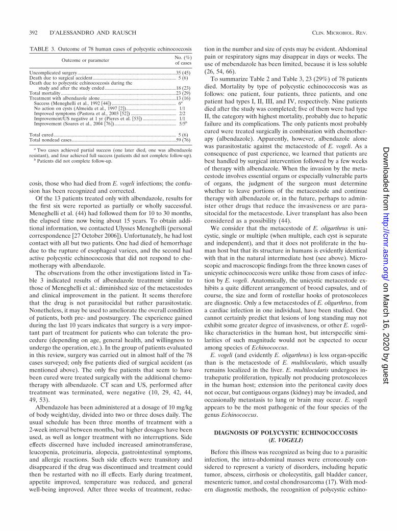
TABLE 3. Outcome of 78 human cases of polycystic echinococcosis
Outcome or parameter No. (%)of cases
Uncomplicated surgery ...................................................................................35 (45)Death due to surgical accident ...................................................................... 5 (6)Death due to polycystic echinococcosis during the
study and after the study ended ............................................................18 (23)Total mortality .................................................................................................23 (29)Treatment with albendazole alone................................................................13 (16)
Success (Meneghelli et al., 1992 �44�) ...................................................... 6a
No action on cysts (Almeida et al., 1997 �2�).......................................... 1/1Improved symptoms (Pastora et al., 2003 �52�) ...................................... 2/2Improvement/US negative at 1 yr (Pieres et al. �53�) ............................ 1/1Improvement (Soares et al., 2004 �76�).................................................... 5/5b
Total cured....................................................................................................... 5 (6)Total nondead cases........................................................................................59 (76)
a Two cases achieved partial success (one later died, one was albendazoleresistant), and four achieved full success (patients did not complete follow-up).
b Patients did not complete follow-up.
392 D’ALESSANDRO AND RAUSCH CLIN. MICROBIOL. REV.
on March 16, 2020 by guest
http://cmr.asm
.org/D
ownloaded from
coccosis has been greatly facilitated. Moreover, with an in-creasing number of published reports concerning this disease,medical personnel in tropical areas, as in Colombia and morerecently in Brazil, have been keenly interested in undertakingepidemiological surveys (51, 69).
Patients’ Histories
Geographical origin of the patients is very important. Theytypically are born, or have lived for prolonged periods, in ruraltropical areas of continental South America, particularly inregions with past or present abundance of wildlife. Familiaritywith pacas, hunting pacas, knowledge of possible presence of“water vesicles” in their livers, and whether domestic dogswere fed viscera of pacas are demographic data that are veryuseful to the clinician in reaching a correct diagnosis. Pro-longed contact with domestic dogs is equally important. (It wasmentioned earlier that neotropical echinococcosis occurs out-side the geographical area of transmission of E. granulosus tohumans.)
Sex and Age Distribution
The number of cases that has been published and thosefindings from reliable sources make a total of 172. Those forwhich information is sufficient number 81. Of those cases, 45%were males and 55% were females. The median age was 43years. That figure is the same as that obtained in our previousreview of cases 10 years ago. Ages of patients ranged from 6 to77 years. The youngest age group, 6 to 22 years, made up 16%of the total. The youngest patient from whom data were re-corded was 6 years old; a 12-year-old child died of portalhypertension 1.7 years after the onset of symptoms (26). Of the81 patients for whom adequate information is available, 6, 12,16, 19, 17, 4, and 5 patients were in the first, second, third,fourth, fifth, sixth, and seventh decades of life, respectively.
Most of the patients infected with E. vogeli were born andhad lived all their lives in rural tropical areas of Central Amer-ica and South America where wildlife was abundant, and it wasimpossible to determine the duration of their infection. If theinfected individual had moved permanently to a city dwelling,it was possible to determine the approximate age of the Echi-nococcus infection, which, for three of our patients, were 12,25, or 60 years. Symptoms had appeared at 2, 10, and 36 yearsof age, respectively, after the patients left the rural area. Themedian age at diagnosis of alveolar echinococcosis (E.multilocularis) was 53 years (91).
Laboratory Findings
General observations, including our own, indicate that lab-oratory findings, outside of the obvious significance of mor-phological features of the cestodes, are not useful in the diag-nosis of neotropical echinococcoses. However, it is importantto assess possible injury to the liver when infiltration of thebiliary system may be diagnosed. Increases of alkaline phos-phatase, bilirubin, liver transaminases, and gamma globulinand diminished albumen and hemoglobin are common findingsin cases of polycystic echinococcosis (45). Eosinophilia was
reported in about 21% of cases, ranging from 9 to 28%, but itis not considered to have specific diagnostic value.
Physical Examination of the Abdomen
In most instances, the patients affected with polycystic echi-nococcosis consult for abdominal pain and have recognized thepresence of a mass in the abdomen close to the liver. Hepa-tomegaly was usually present because that organ was certainlyinvolved in 80% of cases of polycystic echinococcosis. Whenthe metacestode is located in the mesentery, it usually can bepalpated as a mass separate from the liver.
Radiological Imaging
With the help of the newer radiological techniques, US andCT, the tridimensional form of the metacestode becomes vis-ible, making possible the detection of its polycystic character,which was not demonstrable by palpation and simple X rayonly. As a consequence, the differential diagnoses became lesscomplicated and diagnosis required consideration of fewer ill-nesses. (Definitive diagnosis, however, is accomplished bystudy of the metacestode itself, obtained by means of biopsy,surgery, or autopsy.) A simple X ray may show the parasiticmass and any calcifications in the parasite suggestive of echi-nococcosis. Calcifications are annular, 2 to 3 cm in diameter,with a radiodense halo and a clear center. They are locatedwithin the polycystic lesions in the liver or elsewhere in theabdomen if there are extensions into other organs (Fig. 14 and18). Initially, we used simple tomography that could show thelobulated character of lesions.
US of the polycystic lesions shows multiple rounded, uniloc-ular, anechoid formations with regular walls. That picture byitself does not provide the diagnosis of echinococcosis but isuseful in assessing serological findings. For those reasons, UShas become important in field studies of human populations atthe time of confirming serological tests. In surveys of popula-tions, US is considered to be more accurate than serology (27,75). Portable equipment is available at affordable prices, andtherefore, US is also being used in developing countries in-stead of the much more expensive apparatus for CT.
CT scans show multiple, hypodense, cystic structure, roundor ovoid, of various sizes, often coalescent in liver, spleen,pancreas, omentum, pelvis, and lung, etc. Calcifications of vari-able appearance, usually solid and small but at times large andbizarre, can be detected and help to make an accurate diag-nosis. Meneghelli et al. (44, 45) published excellent US and CTphotos of Brazilian patients with polycystic echinococcosis.Also included in their publications are images showing changesin size and number of cysts following albendazole treatment(Fig. 22). CT scanning is expensive but is the best procedure, sofar, for differential diagnosis of polycystic lesions (cystic echi-nococcosis, polycystic liver and kidney disease, primary or met-astatic malignancies, and hepatic amebic abscess [15], pancre-atic epithelioma, ovarian cyst and, in the lungs, dermatoid cystswith inclusions, neurinoma, aortic aneurism, primary bronchialcarcinoma, metastatic sarcoma, seminoma, and lesions in ovaryand uterus, etc.) and for assessment of albendazole treatment.We are not aware of reports on the use of magnetic resonanceimaging in diagnosing polycystic echinococcosis.
VOL. 21, 2008 NEW ASPECTS OF NEOTROPICAL ECHINOCOCCOSIS 393
on March 16, 2020 by guest
http://cmr.asm
.org/D
ownloaded from
Serological Tests
Wilson et al. (92) published a report concerned with molec-ular and immunological diagnosis of parasitic infections, basedon their experience at the Centers for Disease Control andPrevention. The authors discussed serological diagnosis as itapplies to cystic and alveolar echinococcoses, but the methodsalso apply to diagnosis of neotropical echinococcoses:
False-positive reactions may occur in persons withother helminthic infections, cancer and chronic im-mune disorders. Negative test results do not rule outechinococcosis because some cyst-carriers do nothave detectable antibodies. Whether the patient hasdetectable antibodies is dependent on the physicallocation, structural integrity, and vitality of the larvalcysts. Cysts in the liver are more likely to elicit anantibody response than those in the lung, and, re-gardless of location, tests for antibody are least sen-sitive for patients with hyaline cysts (p. 561–562 inreference 92).
A patient with senescent, calcified, or dead cysts is generallyfound to be seronegative. At the present, the best availableserological diagnosis is obtained by using a combination oftests. ELISA or IHA is used to screen all specimens; a positivereaction is confirmed by an immunoblot assay or any gel dif-fusion assay that demonstrates the presence of the echinococ-cal Arc 5. Although those confirmatory assays give false-posi-tive reactions with sera from 5 to 25% of persons withneurocysticercosis, the clinical and epidemiological presenta-tion of neurocysticercosis patients should rarely be confusedwith that of echinococcosis. Following successful radical sur-gery, antibody levels decline and sometimes disappear; anti-bodies rise again if secondary hydatid cysts develop. Tests forArc 5 or immunoglobulin E antibodies appear to reflect anantibody decline during the first 24 months postsurgery,whereas the IHA and other tests remain positive for at least 4years. Chemotherapy has not been followed by consistent de-clines in antibody levels. Consequently, the usefulness of se-rology to monitor the course of disease is limited; imagingtechniques provide a more accurate assessment of the patient’scondition.
The antigen used for serodiagnosis of infection with E.granulosus and E. multilocularis, as well as that with E. vogeliand in one case that with E. oligarthrus, has been the fluid frommetacestodes of E. granulosus. In addition, purified antigens ofall echinococcal species but E. oligarthrus have been developedand used. From crude tissues, Gottstein et al. (31) derived theE. vogeli Ev2 antigen that differentiates between E. vogeli andE. granulosus but not between E. vogeli and E. multilocularis.The latter two species, however, are not sympatric; therefore,epidemiological data are important for distinguishing them.
Unfortunately, the test using the Ev2 antigen could not beevaluated in a blind serological survey carried out in a largeindigenous human population in a South American country.As originally agreed upon, the results of the serological testswere mailed to our colleagues, but the parasitological infor-mation concerning the humans tested was never received by us.
Antigen of E. oligarthrus is not available, nor is serum frompatients with that infection.
Only the case (orbital) reported by Lopera et al. (39) waspositive for Arc 5 by immunoelectrophoresis, indicating cross-reactivity between antigens of E. oligarthrus and E. granulosus,the latter having been used in the test. It was stated 10 yearsago that separation of metacestodes of the two neotropicalspecies was not possible serologically, and that situation hasnot changed (12).
As stated by Wilson et al. (92), use of two serological tests,one very sensitive (ELISA) and another very specific (IB), isconvenient. Among the 81 patients with neotropical echino-coccosis that we evaluated, 40 were studied serologically; for 12of them, the two tests gave identical results. For a particularpatient undergoing surgery in France, the IHA and the Arc 5tests were positive, as were the ELISA and IB tests.
Among other patients tested, the results were as follows.IHA was positive for 14 of 19 persons. Of the five negativecases, from one, a metacestode of E. vogeli had been removed1.5 years earlier, while two had active E. vogeli infections andtwo were negative, having completely calcified hepatic cysts(42). Counter electrophoresis was positive in 6 of 7 cases,immunoelectophoresis Arc 5 was positive in 7 of 9, ELISA was
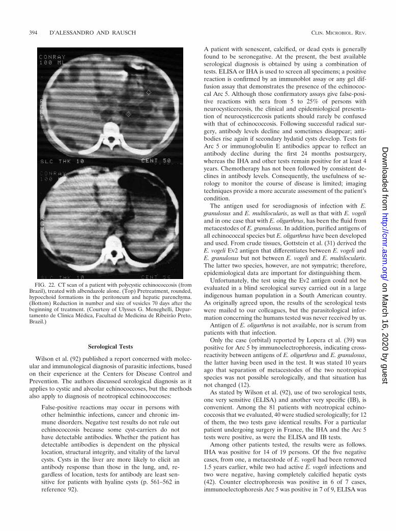
FIG. 22. CT scan of a patient with polycystic echinococcosis (fromBrazil), treated with albendazole alone. (Top) Pretreatment, rounded,hypoechoid formations in the peritoneum and hepatic parenchyma.(Bottom) Reduction in number and size of vesicles 70 days after thebeginning of treatment. (Courtesy of Ulysses G. Meneghelli, Depar-tamento de Clinica Medica, Facultad de Medicina de Ribeirao Preto,Brazil.)
394 D’ALESSANDRO AND RAUSCH CLIN. MICROBIOL. REV.
on March 16, 2020 by guest
http://cmr.asm
.org/D
ownloaded from
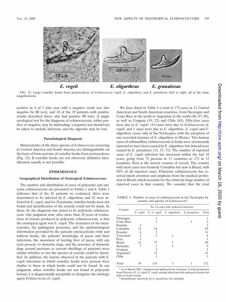
positive in 4 of 5 (the case with a negative result was alsonegative by IB test), and 18 of the 19 patients with positiveresults described above also had positive IB tests. A singleserological test for the diagnosis of echinococcosis, either pos-itive or negative, may be misleading: a negative test should notbe taken to exclude infection, and the opposite may be true.
Parasitological Diagnosis
Metacestodes of the three species of Echinococcus occurringin Central America and South America are distinguishable onthe basis of form and size of rostellar hooks from protoscoleces(Fig. 23). If rostellar hooks are not observed, definitive iden-tification usually is not possible.
EPIDEMIOLOGY
Geographical Distribution of Neotropical Echinococcoses
The number and distribution of cases of polycystic and uni-cystic echinococcosis are presented in Tables 1 and 4. Table 1indicates that of the 81 patients we evaluated, three weredetermined to be infected by E. oligarthrus and 42 were in-fected by E. vogeli, and for 36 patients, rostellar hooks were notfound and identification of the cestode could not be made. Inthose 36, the diagnosis was stated to be polycystic echinococ-cosis. Our judgment now, after more than 20 years of evalua-tions of lesions produced in polycystic echinococcosis, is thatthe etiological agent was E. vogeli. The structures of the meta-cestodes, the pathogenic processes, and the epidemiologicalinformation provided by the patients (metacestodes with andwithout hooks, the patients’ knowledge of pacas and theirinfections, the awareness of feeding liver of pacas, with anycysts present, to domestic dogs, and the presence of domesticdogs around previous or current dwellings of patients) weresimilar whether or not the species of cestode could be identi-fied. In addition, the lesions observed in the patients with E.vogeli infections in which rostellar hooks were present weresimilar to those in which hooks could not be found. In ourjudgment, when rostellar hooks are not found in polycysticlesions, it is diagnostically acceptable to designate the etiologicagent Echinococcus cf. vogeli.
We have listed in Table 4 a total of 172 cases in 12 CentralAmerican and South American countries, from Nicaragua andCosta Rica in the north to Argentina in the south (86, 87, 88),as well as Uruguay (19, 22) and Chile (85). Fifty-four caseswere due to E. vogeli, 114 cases were due to Echinococcus cf.vogeli, and 3 cases were due to E. oligarthrus. E. vogeli and E.oligarthrus occur only in the Neotropics, with the exception ofone recorded instance of E. oligarthrus in Mexico. Two humancases of submaxillary echinococcosis in India were erroneouslyreported to have been caused by E. oligarthrus but instead werecaused by E. granulosus (13, 37, 71). The number of reportedcases of E. vogeli infection has increased within the last 10years, going from 72 persons in 11 countries to 172 in 12countries. Peru is the newest country of record. The countrywith most cases was formerly Colombia but now is Brazil, with58% of all reported cases. Polycystic echinococcosis has re-ceived much attention and emphasis from the medical profes-sion in Brazil, which accounts for the relatively large number ofreported cases in that country. We consider that the total
FIG. 23. Large rostellar hooks from protoscoleces of Echinococcus vogeli, E. oligarthrus, and E. granulosus (left to right, all at the samemagnification).
TABLE 4. Number of cases of echinococcosis in the Neotropics bycountry and species of Echinococcusa
CountryNo. of cases with indicated infection
E. vogeli E. cf. vogeli E. oligarthrus E. granulosus Total
Nicaragua 1 1Costa Rica 1 1 2Panama 2 2Colombia 15 14 29Ecuador 6 5 11Venezuela 2 1 1 4Peru 1 1Brazil 21 77 1 99Suriname 7 1 1 9Uruguay 2 2Argentinab 11 11Chile 1 1
Total 54 114 3 1 172
a As of March 2007. (Adapted and updated from reference 12 with permissionfrom Elsevier.) E. cf. vogeli, E. vogeli causing infections with polycystic lesions butwith no hooks found.
b Synanthropic infections by E. granulosus not included.
VOL. 21, 2008 NEW ASPECTS OF NEOTROPICAL ECHINOCOCCOSIS 395
on March 16, 2020 by guest
http://cmr.asm
.org/D
ownloaded from
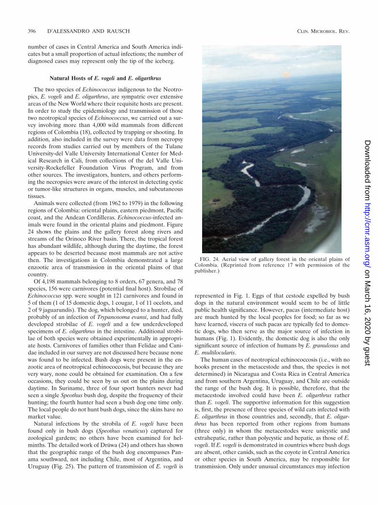
number of cases in Central America and South America indi-cates but a small proportion of actual infections; the number ofdiagnosed cases may represent only the tip of the iceberg.
Natural Hosts of E. vogeli and E. oligarthrus
The two species of Echinococcus indigenous to the Neotro-pics, E. vogeli and E. oligarthrus, are sympatric over extensiveareas of the New World where their requisite hosts are present.In order to study the epidemiology and transmission of thosetwo neotropical species of Echinococcus, we carried out a sur-vey involving more than 4,000 wild mammals from differentregions of Colombia (18), collected by trapping or shooting. Inaddition, also included in the survey were data from necropsyrecords from studies carried out by members of the TulaneUniversity-del Valle University International Center for Med-ical Research in Cali, from collections of the del Valle Uni-versity-Rockefeller Foundation Virus Program, and fromother sources. The investigators, hunters, and others perform-ing the necropsies were aware of the interest in detecting cysticor tumor-like structures in organs, muscles, and subcutaneoustissues.
Animals were collected (from 1962 to 1979) in the followingregions of Colombia: oriental plains, eastern piedmont, Pacificcoast, and the Andean Cordilleras. Echinococcus-infected an-imals were found in the oriental plains and piedmont. Figure24 shows the plains and the gallery forest along rivers andstreams of the Orinoco River basin. There, the tropical foresthas abundant wildlife, although during the daytime, the forestappears to be deserted because most mammals are not activethen. The investigations in Colombia demonstrated a largeenzootic area of transmission in the oriental plains of thatcountry.
Of 4,198 mammals belonging to 8 orders, 67 genera, and 78species, 156 were carnivores (potential final host). Strobilae ofEchinococcus spp. were sought in 121 carnivores and found in5 of them (1 of 15 domestic dogs, 1 cougar, 1 of 11 ocelots, and2 of 9 jaguarundis). The dog, which belonged to a hunter, died,probably of an infection of Trypanosoma evansi, and had fullydeveloped strobilae of E. vogeli and a few underdevelopedspecimens of E. oligarthrus in the intestine. Additional strobi-lae of both species were obtained experimentally in appropri-ate hosts. Carnivores of families other than Felidae and Cani-dae included in our survey are not discussed here because nonewas found to be infected. Bush dogs were present in the en-zootic area of neotropical echinococcosis, but because they arevery wary, none could be obtained for examination. On a fewoccasions, they could be seen by us out on the plains duringdaytime. In Suriname, three of four sport hunters never hadseen a single Speothus bush dog, despite the frequency of theirhunting; the fourth hunter had seen a bush dog one time only.The local people do not hunt bush dogs, since the skins have nomarket value.
Natural infections by the strobila of E. vogeli have beenfound only in bush dogs (Speothus venaticus) captured forzoological gardens; no others have been examined for hel-minths. The detailed work of Druwa (24) and others has shownthat the geographic range of the bush dog encompasses Pan-ama southward, not including Chile, most of Argentina, andUruguay (Fig. 25). The pattern of transmission of E. vogeli is
represented in Fig. 1. Eggs of that cestode expelled by bushdogs in the natural environment would seem to be of littlepublic health significance. However, pacas (intermediate host)are much hunted by the local peoples for food; so far as wehave learned, viscera of such pacas are typically fed to domes-tic dogs, who then serve as the major source of infection inhumans (Fig. 1). Evidently, the domestic dog is also the onlysignificant source of infection of humans by E. granulosus andE. multilocularis.
The human cases of neotropical echinococcosis (i.e., with nohooks present in the metacestode and thus, the species is notdetermined) in Nicaragua and Costa Rica in Central Americaand from southern Argentina, Uruguay, and Chile are outsidethe range of the bush dog. It is possible, therefore, that themetacestode involved could have been E. oligarthrus ratherthan E. vogeli. The supportive information for this suggestionis, first, the presence of three species of wild cats infected withE. oligarthrus in those countries and, secondly, that E. oligar-thrus has been reported from other regions from humans(three only) in whom the metacestodes were unicystic andextrahepatic, rather than polycystic and hepatic, as those of E.vogeli. If E. vogeli is demonstrated in countries where bush dogsare absent, other canids, such as the coyote in Central Americaor other species in South America, may be responsible fortransmission. Only under unusual circumstances may infection
FIG. 24. Aerial view of gallery forest in the oriental plains ofColombia. (Reprinted from reference 17 with permission of thepublisher.)
396 D’ALESSANDRO AND RAUSCH CLIN. MICROBIOL. REV.
on March 16, 2020 by guest
http://cmr.asm
.org/D
ownloaded from
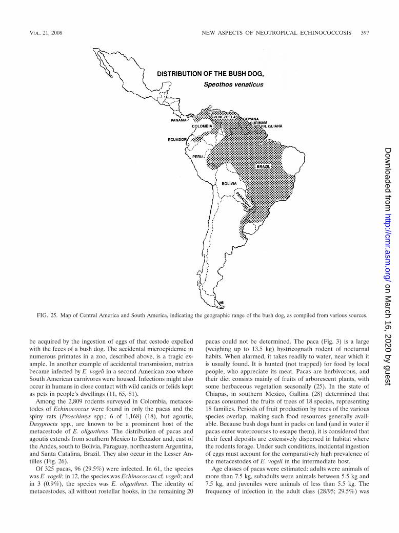
be acquired by the ingestion of eggs of that cestode expelledwith the feces of a bush dog. The accidental microepidemic innumerous primates in a zoo, described above, is a tragic ex-ample. In another example of accidental transmission, nutriasbecame infected by E. vogeli in a second American zoo whereSouth American carnivores were housed. Infections might alsooccur in humans in close contact with wild canids or felids keptas pets in people’s dwellings (11, 65, 81).
Among the 2,809 rodents surveyed in Colombia, metaces-todes of Echinococcus were found in only the pacas and thespiny rats (Proechimys spp.; 6 of 1,168) (18), but agoutis,Dasyprocta spp., are known to be a prominent host of themetacestode of E. oligarthrus. The distribution of pacas andagoutis extends from southern Mexico to Ecuador and, east ofthe Andes, south to Bolivia, Paraguay, northeastern Argentina,and Santa Catalina, Brazil. They also occur in the Lesser An-tilles (Fig. 26).
Of 325 pacas, 96 (29.5%) were infected. In 61, the specieswas E. vogeli; in 12, the species was Echinococcus cf. vogeli; andin 3 (0.9%), the species was E. oligarthrus. The identity ofmetacestodes, all without rostellar hooks, in the remaining 20
pacas could not be determined. The paca (Fig. 3) is a large(weighing up to 13.5 kg) hystricognath rodent of nocturnalhabits. When alarmed, it takes readily to water, near which itis usually found. It is hunted (not trapped) for food by localpeople, who appreciate its meat. Pacas are herbivorous, andtheir diet consists mainly of fruits of arborescent plants, withsome herbaceous vegetation seasonally (25). In the state ofChiapas, in southern Mexico, Gallina (28) determined thatpacas consumed the fruits of trees of 18 species, representing18 families. Periods of fruit production by trees of the variousspecies overlap, making such food resources generally avail-able. Because bush dogs hunt in packs on land (and in water ifpacas enter watercourses to escape them), it is considered thattheir fecal deposits are extensively dispersed in habitat wherethe rodents forage. Under such conditions, incidental ingestionof eggs must account for the comparatively high prevalence ofthe metacestodes of E. vogeli in the intermediate host.
Age classes of pacas were estimated: adults were animals ofmore than 7.5 kg, subadults were animals between 5.5 kg and7.5 kg, and juveniles were animals of less than 5.5 kg. Thefrequency of infection in the adult class (28/95; 29.5%) was
FIG. 25. Map of Central America and South America, indicating the geographic range of the bush dog, as compiled from various sources.
VOL. 21, 2008 NEW ASPECTS OF NEOTROPICAL ECHINOCOCCOSIS 397
on March 16, 2020 by guest
http://cmr.asm
.org/D
ownloaded from
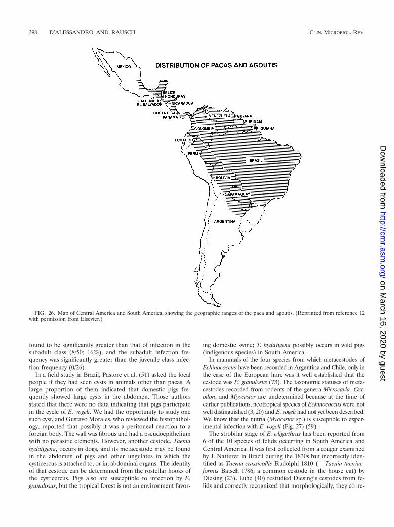
found to be significantly greater than that of infection in thesubadult class (8/50; 16%), and the subadult infection fre-quency was significantly greater than the juvenile class infec-tion frequency (0/26).
In a field study in Brazil, Pastore et al. (51) asked the localpeople if they had seen cysts in animals other than pacas. Alarge proportion of them indicated that domestic pigs fre-quently showed large cysts in the abdomen. Those authorsstated that there were no data indicating that pigs participatein the cycle of E. vogeli. We had the opportunity to study onesuch cyst, and Gustavo Morales, who reviewed the histopathol-ogy, reported that possibly it was a peritoneal reaction to aforeign body. The wall was fibrous and had a pseudoepitheliumwith no parasitic elements. However, another cestode, Taeniahydatigena, occurs in dogs, and its metacestode may be foundin the abdomen of pigs and other ungulates in which thecysticercus is attached to, or in, abdominal organs. The identityof that cestode can be determined from the rostellar hooks ofthe cysticercus. Pigs also are susceptible to infection by E.granulosus, but the tropical forest is not an environment favor-
ing domestic swine; T. hydatigena possibly occurs in wild pigs(indigenous species) in South America.
In mammals of the four species from which metacestodes ofEchinococcus have been recorded in Argentina and Chile, only inthe case of the European hare was it well established that thecestode was E. granulosus (73). The taxonomic statuses of meta-cestodes recorded from rodents of the genera Microcavia, Oct-odon, and Myocastor are undetermined because at the time ofearlier publications, neotropical species of Echinococcus were notwell distinguished (3, 20) and E. vogeli had not yet been described.We know that the nutria (Myocastor sp.) is susceptible to exper-imental infection with E. vogeli (Fig. 27) (59).
The strobilar stage of E. oligarthrus has been reported from6 of the 10 species of felids occurring in South America andCentral America. It was first collected from a cougar examinedby J. Natterer in Brazil during the 1830s but incorrectly iden-tified as Taenia crassicollis Rudolphi 1810 (� Taenia taeniae-formis Batsch 1786, a common cestode in the house cat) byDiesing (23). Luhe (40) restudied Diesing’s cestodes from fe-lids and correctly recognized that morphologically, they corre-
FIG. 26. Map of Central America and South America, showing the geographic ranges of the paca and agoutis. (Reprinted from reference 12with permission from Elsevier.)
398 D’ALESSANDRO AND RAUSCH CLIN. MICROBIOL. REV.
on March 16, 2020 by guest
http://cmr.asm
.org/D
ownloaded from
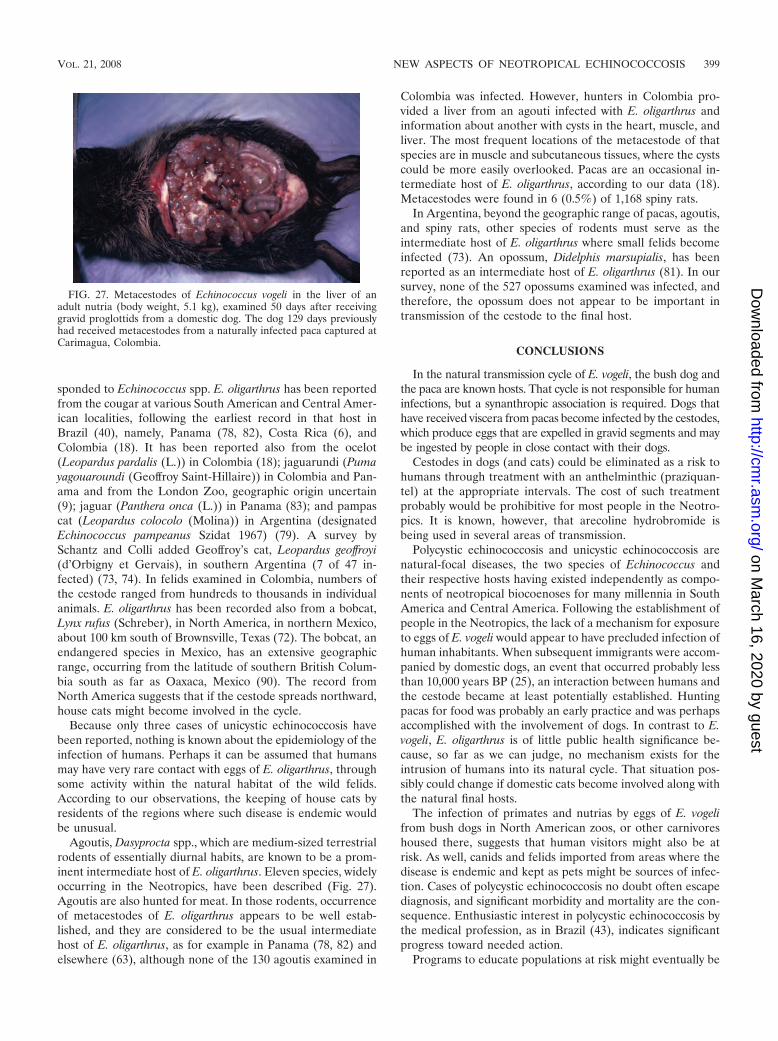
sponded to Echinococcus spp. E. oligarthrus has been reportedfrom the cougar at various South American and Central Amer-ican localities, following the earliest record in that host inBrazil (40), namely, Panama (78, 82), Costa Rica (6), andColombia (18). It has been reported also from the ocelot(Leopardus pardalis (L.)) in Colombia (18); jaguarundi (Pumayagouaroundi (Geoffroy Saint-Hillaire)) in Colombia and Pan-ama and from the London Zoo, geographic origin uncertain(9); jaguar (Panthera onca (L.)) in Panama (83); and pampascat (Leopardus colocolo (Molina)) in Argentina (designatedEchinococcus pampeanus Szidat 1967) (79). A survey bySchantz and Colli added Geoffroy’s cat, Leopardus geoffroyi(d’Orbigny et Gervais), in southern Argentina (7 of 47 in-fected) (73, 74). In felids examined in Colombia, numbers ofthe cestode ranged from hundreds to thousands in individualanimals. E. oligarthrus has been recorded also from a bobcat,Lynx rufus (Schreber), in North America, in northern Mexico,about 100 km south of Brownsville, Texas (72). The bobcat, anendangered species in Mexico, has an extensive geographicrange, occurring from the latitude of southern British Colum-bia south as far as Oaxaca, Mexico (90). The record fromNorth America suggests that if the cestode spreads northward,house cats might become involved in the cycle.
Because only three cases of unicystic echinococcosis havebeen reported, nothing is known about the epidemiology of theinfection of humans. Perhaps it can be assumed that humansmay have very rare contact with eggs of E. oligarthrus, throughsome activity within the natural habitat of the wild felids.According to our observations, the keeping of house cats byresidents of the regions where such disease is endemic wouldbe unusual.
Agoutis, Dasyprocta spp., which are medium-sized terrestrialrodents of essentially diurnal habits, are known to be a prom-inent intermediate host of E. oligarthrus. Eleven species, widelyoccurring in the Neotropics, have been described (Fig. 27).Agoutis are also hunted for meat. In those rodents, occurrenceof metacestodes of E. oligarthrus appears to be well estab-lished, and they are considered to be the usual intermediatehost of E. oligarthrus, as for example in Panama (78, 82) andelsewhere (63), although none of the 130 agoutis examined in
Colombia was infected. However, hunters in Colombia pro-vided a liver from an agouti infected with E. oligarthrus andinformation about another with cysts in the heart, muscle, andliver. The most frequent locations of the metacestode of thatspecies are in muscle and subcutaneous tissues, where the cystscould be more easily overlooked. Pacas are an occasional in-termediate host of E. oligarthrus, according to our data (18).Metacestodes were found in 6 (0.5%) of 1,168 spiny rats.
In Argentina, beyond the geographic range of pacas, agoutis,and spiny rats, other species of rodents must serve as theintermediate host of E. oligarthrus where small felids becomeinfected (73). An opossum, Didelphis marsupialis, has beenreported as an intermediate host of E. oligarthrus (81). In oursurvey, none of the 527 opossums examined was infected, andtherefore, the opossum does not appear to be important intransmission of the cestode to the final host.
CONCLUSIONS
In the natural transmission cycle of E. vogeli, the bush dog andthe paca are known hosts. That cycle is not responsible for humaninfections, but a synanthropic association is required. Dogs thathave received viscera from pacas become infected by the cestodes,which produce eggs that are expelled in gravid segments and maybe ingested by people in close contact with their dogs.
Cestodes in dogs (and cats) could be eliminated as a risk tohumans through treatment with an anthelminthic (praziquan-tel) at the appropriate intervals. The cost of such treatmentprobably would be prohibitive for most people in the Neotro-pics. It is known, however, that arecoline hydrobromide isbeing used in several areas of transmission.
Polycystic echinococcosis and unicystic echinococcosis arenatural-focal diseases, the two species of Echinococcus andtheir respective hosts having existed independently as compo-nents of neotropical biocoenoses for many millennia in SouthAmerica and Central America. Following the establishment ofpeople in the Neotropics, the lack of a mechanism for exposureto eggs of E. vogeli would appear to have precluded infection ofhuman inhabitants. When subsequent immigrants were accom-panied by domestic dogs, an event that occurred probably lessthan 10,000 years BP (25), an interaction between humans andthe cestode became at least potentially established. Huntingpacas for food was probably an early practice and was perhapsaccomplished with the involvement of dogs. In contrast to E.vogeli, E. oligarthrus is of little public health significance be-cause, so far as we can judge, no mechanism exists for theintrusion of humans into its natural cycle. That situation pos-sibly could change if domestic cats become involved along withthe natural final hosts.
The infection of primates and nutrias by eggs of E. vogelifrom bush dogs in North American zoos, or other carnivoreshoused there, suggests that human visitors might also be atrisk. As well, canids and felids imported from areas where thedisease is endemic and kept as pets might be sources of infec-tion. Cases of polycystic echinococcosis no doubt often escapediagnosis, and significant morbidity and mortality are the con-sequence. Enthusiastic interest in polycystic echinococcosis bythe medical profession, as in Brazil (43), indicates significantprogress toward needed action.
Programs to educate populations at risk might eventually be
FIG. 27. Metacestodes of Echinococcus vogeli in the liver of anadult nutria (body weight, 5.1 kg), examined 50 days after receivinggravid proglottids from a domestic dog. The dog 129 days previouslyhad received metacestodes from a naturally infected paca captured atCarimagua, Colombia.
VOL. 21, 2008 NEW ASPECTS OF NEOTROPICAL ECHINOCOCCOSIS 399
on March 16, 2020 by guest
http://cmr.asm
.org/D
ownloaded from
beneficial, but means of establishing such programs seem gen-erally to be unavailable. The risk of acquiring echinococcosis(sensu lato) is seen to be greatly diminished if dogs are notkept (57), but the ancient association between people and theirdogs is disrupted only under exceptional demands for changesin lifestyle.
ACKNOWLEDGMENTS
We express our thanks to Ulysses Meneghelli, who kindly providedinformation on the outcome of patients he had treated earlier withalbendazole from unpublished results of a human serological surveyfor echinococcosis in Brazil as well as permission to use some of theimaging from his patients. We also thank Mark Wiser, Maria Frontini,and Kris Swalm, Tulane University, School of Public Health and Trop-ical Medicine, Department of Tropical Medicine, Epidemiology andComputing Services, respectively, for their help with the preparation ofthe manuscript due to their expertise with the computer. Specialthanks to Virginia Rausch, Burke Museum of Natural History, Uni-versity of Washington, for her interest and help in this work and forpreparing the drawing of the life cycle. We are grateful as well to MarkDerby and David Wolbrecht, University of Washington, who kindlyprovided technical assistance.
REFERENCES
1. Abuladze, K. I. 1964. Teniaty—lentochnye gel’minty zhivotnykh i chelovekai vyzyvaemye imi zabolevaniia. Osnovy tsestodologii, vol. 4. Nauka, Moskva,U.S.S.R.
2. Almeida, S. C. X., R. L. Martins, A. P. Morales, C. A. Viegas, and M. Grilo.1997. Hidatidosis pulmonar policıstica mimetizando lesaos metastaticas: re-lato de caso. J. Pneumonol. 23:261–263.
3. Alvarez, V. 1961. Investigaciones sobre echinococcosis silvestre en Chile.Biologica 31:89–94.
4. Basset, D., C. Girou, I. P. Nozais, F. D’Hermies, C. Hoang, R. Gordon, andA. D’Alessandro. 1998. Neotropical echinococcosis in Suriname: Echinococ-cus oligarthrus in the orbit and Echinococcus vogeli in the abdomen. Am. J.Trop. Med. Hyg. 59:787–790.
5. Blood, B. D., and J. L. Lelijveld. 1969. Studies on sylvatic echinococcosis insouthern South America. Z. Tropenmed. Parasitol. 20:475–482.
6. Brenes Madrigal, R. R., G. Munoz Montoya, G. E. M. Ocampo, and G. R.Herrera. 1973. Presencia en Costa Rica de Echinococcus oligarthrus Diesing1863, colectado en el intestino delgado de Felis concolor costaricensis. Rev.Biol. Trop. 21:139–141.
7. Brumpt, E., and C. Joyeux. 1924. Description d’un nouvel Echinocoque:Echinococcus cruzi n. sp. Ann. Parasitol. Hum. Comp. 2:226–231.
8. Cabrera, A., J. Yepes, and C. C. Wiedner. 1960. Mamiferos sud americanos.Historia natural, vol. 1, 2nd ed. Ediar S. A. Editores, Buenos Aires, Argentina.
9. Cameron, T. W. M. 1926. Observations on the genus Echinococcus Rudolphi1801. J. Helminthol. 4:13–22.
10. Chigot, J. J., F. Menegaux, R. Hammoud, J. P. Nozias, and C. Hoang. 1995.A propos d’un cas d’hydatidose a Echinococcus vogeli ayant beneficie d’untraitement chirurgical complet. Sem. Hosp. Paris 71:429–431.
11. Cross, J., and R. M. Thomas. 1966. Hydatid disease in the nutria. J. Para-sitol. 52:1215–1216.
12. D’Alessandro, A. 1997. Polycystic echinococcosis in tropical America: Echi-nococcus vogeli and E. oligarthrus. Acta Trop. 67:43–65.
13. D’Alessandro, A., and R. L. Rausch. 2004. Erroneous report of Echinococcusoligarthrus as a cause of echinococcosis in India. J. Parasitol. 90:202–203.
14. D’Alessandro, A., H. Henao, and C. Cuello. 1978. Una caso colombianoautoctono de hidatidosis poliquistica multiple de hidago, paricardio, pul-mones, pleura y corazon. Acta Med. Valle 9:23–35.
15. D’Alessandro, A., J. Lega, and M. A. Vera. 1966. Cystic calcifications of theliver in Colombia. Echinococcosis or calcified abscesses? Am. J. Trop. Med.Hyg. 15:908–913.
16. D’Alessandro, A., L. E. Ramirez, E. Chapadeiro, E. R. Lopes, and P. M. DeMesquita. 1995. Second recorded case of human infection by Echinococcusoligarthrus. Am. J. Trop. Med. Hyg. 52:29–33.
17. D’Alessandro, A., R. L. Rausch, C. Cuello, and N. Aristibal. 1979. Echino-coccus vogeli in man, with a review of polycystic hydatid disease in Colombiaand neighboring countries. Am. J. Trop. Med. Hyg. 28:303–317.
18. D’Alessandro, A., R. L. Rausch, G. A. Morales, S. Collet, and D. Angel. 1981.Echinococcus infections in Colombian animals. Am. J. Trop. Med. Hyg.30:1263–1276.
19. Dardel, G. 1955. A proposito de la equinococosis alveolaris en el Uruguay.Arch. Urug. Med. Cir. Esp. 46:25–32.
20. De La Barrera, J. M. 1947. Echinococcus granulosus in Microcavia australis.Arch. Int. Hidat. 7:175–176.
21. Deutsch, L. A. 1983. An encounter between bush dog (Speothos venaticus)and paca (Agouti paca). J. Mammal. 64:532–533.
22. Deve, F., R. Piaggio-Blanco, and F. Garcia-Capurro. 1936. Echinococcosehepatique maligne micropolykystique infiltrate. Forme intermediara entrel’echinococcose hydatic et l’echinococcose alveolaire. Arch. Urug. Med. Cir.Est. 8:3–28.
23. Diesing, C. M. 1850. Systema helminthum. Historiae naturalis classica, vol.XI. [Reprint, H. R. Engelmann (J. Cramer), Weinheim, Germany, 1960.]Vindobonae, Braumuller, Germany.
24. Druwa, P. 1982. Perro de grulleiro, der Sudamerikanische Waldhund—einRatsel fur die Hundeforschung. Zeitsch. Kolner Zoo. 25:71–81.
25. Eisenberg, J. F. 1989. Mammals of the Neotropics. The Northern Neotro-pics, vol. 1. University of Chicago Press, Chicago, IL.
26. Ferreira, M. S., S. A. Nishioka, A. Rocha, and A. D’Alessandro. 1995. Echi-nococcus vogeli polycystic hydatid disease: report of two Brazilian casesout-side the Amazon region. Trans. Roy. Soc. Trop. Med. Hyg. 89:286–287.
27. Frider, B., J. Moguilensky, J. C. Salvitti, M. Odriozol, and E. Larrieu. 1999.Epidemiological surveillance of human hydatidosis by means of ultrasonog-raphy: Its contribution to the evaluation of control programs. Acta Trop.79:219–223.
28. Gallina, S. 1981. Contribucion al conocimiento de los habitos alimenticiosdel tapezcuintle (Aguti paca Lin.) en Lancaja-Chansayab, Chiapas, p. 58–67.In P. R. Castillo (ed.), Estudios ecologicos en el tropico Mexicano, publica-tion 6. Instituto de Ecologıa A. C., Mexico, D.F., Mexico
29. Ghiotti Siqueira, N., F. Barbosa de Almeida, S. R. Silva Chalub, J. R.Machadao-Silva, and R. Rodrigues-Silva. 2007. Successful outcome of he-patic polycystic echinococcosis managed with surgery and chemotherapy.Trans. R. Soc. Trop. Med. Hyg. 101:624–626.
30. Gottstein, B. 1992. Molecular and immunological diagnosis of echinococco-sis. Clin. Microbiol. Rev. 5:248–261.
31. Gottstein, B., A. D’Alessandro, and R. L. Rausch. 1995. Immunodiagnosis ofpolycystic hydatid disease/polycystic echinococcosis due to Echinococcus vo-geli. Am. J. Trop. Med. Hyg. 53:558–563.
32. Gottstein, B., B. Mesarina, I. Tannrer, R. D. Ammann, J. F. Wilson, J.Eckert, and A. Lanier. 1991. Specific cellular and humoral immune responsesin patients with different long-term courses of alveolar echinococcosis infec-tion with Echinococcus multilocularis. Am. J. Trop. Med. Hyg. 45:734–742.
33. Grases, P., and N. Salazar. 1970. Hidatidosis alveolar. G. E. N. 25:71–89.34. Guderian, R., K. R. Kerrigan, M. E. Chico, A. Guevara, and G. Hidalgo.
1990. Hidatidosis humana en el Oriente Ecuatoriano: informe de los prim-eros casos. Rev. Med. Vozandes 4:57–60.
35. Herre, W., and M. Rohrs. 1973. Haustiere—zoologisch gesehen. GustavFischer Verlag, Stuttgart, Germany.
36. Howard, E. B., and A. P. Gendron. 1980. Echinococcus vogeli infection inhigher primates at the Los Angeles Zoo, p. 379–382. In R. J. Montali and G.Migaki (ed.), The comparative pathology of zoo animals. Symposia of theNational Zoological Park. Smithsonian Institution Press, Washington, DC.
37. Kini, U., S. Chariff, and V. Nirmala. 1997. Aspiration cytology of Echino-coccus oligarthrus. A case report. Acta Cytol. 41:544–548.
38. Lazo, R., C. Moran, E. Plaza, and A. Mite. 1985. Hidatidosis humana porEchinococcus vogeli en el Ecuador, p. 237–244. In Memorias del VII Con-greso Latinoamericano de Parasitologıa. Trabajos Graficos Juan MontalvoCiao Ldta., Guayaquil, Ecuador.
39. Lopera, R. D., R. D. Melendez, I. Fernandez, J. Sirit, and M. P. Perera. 1989.Orbital hydatid cyst of Echinococcus oligarthrus in a human in Venezuela. J.Parasitol. 75:467–470.
40. Luhe, M. 1910. Cystotanien sudamerikanischer Feliden. Zool. Jahrb. Suppl.12:687–710.
41. Melendez, R. D., M. S. Yepez, and A. Coronado. 1984. Echinococcus oli-garthus cysts of rabbits in Venezuela. J. Parasitol. 70:1004–1005.
42. Meneghelli, U. G. 1985. Multiple hepatic calcifications resulting from polycystichydatid disease. Rev. Goiania Med. 31:53–60.
43. Meneghelli, U. G. 2002. Hidatidose policıstica: a comprovacao de sua ocorrenciae a possıvel dimensao epidemiologica da doenca no Brasil. A gastroenterologıano Brasil II. Lemos Editorial, Sao Paulo, Brazil.
44. Meneghelli, U. G., A. L. C. Martinelli, A. D. Bellucci, M. G. Villanova,M. A. S. Llorach Velludo, and J. E. Magro. 1992. Polycystic hydatid disease(Echinococcus vogeli). Treatment with albendazole. Ann. Trop. Med. Para-sitol. 86:151–156.
45. Meneghelli, U. G., A. L. C. Martinelli, M. A. S. Llorach Velludo, A. D.Bellucci, J. E. Magro, and M. L. P. Barbo. 1992. Polycystic hydatid disease(Echinococcus vogeli). Clinical, laboratory and morphological findings innine Brazilian patients. J. Hepatol. 14:203–210.
46. Moraes, M. A. P., M. N. M. Sobreira, P. Medeiros Filho, A. Cavalca Tavares,and M. I. Gomes. 2003. Polycystic hydatidosis: casual finding of calcifiedhydatid cyst simulating mesenteric neoplasm. Rev. Soc. Bras. Med. Trop.36:519–521.
47. Ohbayashi, M., R. L. Rausch, and F. H. Fay. 1971. On the ecology anddistribution of Echinococcus species (Cestoda: Taeniidae), and characteris-tics of their development in the intermediate host. II. Comparative studieson the development of E. multilocularis Leuckart, 1863, in the intermediatehost. Jpn. J. Vet. Res. 19(Suppl. 3):1–53.
400 D’ALESSANDRO AND RAUSCH CLIN. MICROBIOL. REV.
on March 16, 2020 by guest
http://cmr.asm
.org/D
ownloaded from
48. Oostburg, B. F. J., M. A. Vrede, and A. E. Bergen. 2000. The occurrence ofpolycystic echinococcosis in Suriname. Ann. Trop. Med. Parasit. 94:247–252.
49. Oostburg, B. F. J., M. Eersel, M. A. Vrede, B. Gottstein, R. L. Rausch, andA. D’Alessandro. 2004. Preliminary field observation on neotropical echino-coccosis in Suriname. Rev. Pat. Trop. (Goiania) 35:125–134.
50. Passos, D. M. M., M. Pinho Alves, O. Creuv, G. Fritas, J. P. Matushita, andE. Marchiori. 1982. Equinococcose alveolar hepatica (descricao de um caso).Radiol. Bras. 15:129–132.
51. Pastore, R., L. H. Vitali, V. O. Macedo, and A. Prata. 2003. A serologicalsurvey of the infection by Echinococcus sp. in the municipality of SenaMadureira, Acre. Rev. Soc. Bras. Med. Trop. 36:473–477.
52. Pastore, R., L. H. Vitali, J. Weirich, A. C. Tojal, V. O. Macedo, and A. Prata.2003. Hidatidosis poliquistica: relato de dois casos procedentes de SenaMadureira, Acre, na Amazonia brasileira. Rev. Soc. Bras. Med. Trop. 36:97–101.
53. Pieres, J. A. A., G. B. Pereira, R. Pastore, and L. Lauria-Pires. 2001. Doencahepatica policıstica. J. Bras. Patol. 37:90–98.
54. Pinilla, L. G., K. Zamora, and G. Gonzalez. 1982. Hidatidosis poliquıstica.Tribuna Med. 11:47–50.
55. Rausch, R. L. 1967. On the ecology and distribution of Echinococcus spp.(Cestoda: Taeniidae), and characteristics of their development in the inter-mediate host. Ann. Parasitol. Hum. Comp. 42:19–63.
56. Rausch, R. L. 1986. Life cycle patterns and geographic distribution of Echi-nococcus species, p. 44–80. In R. C. A. Thompson (ed.), The biology ofEchinococcus and hydatid disease. George Allen and Unwin, London,United Kingdom.
57. Rausch, R. L. 2003. Cystic echinococcus in the Arctic and sub-Arctic. Par-asitology 127(Suppl.):S73–S85.
58. Rausch, R. L., and J. J. Bernstein. 1972. Echinococcus vogeli sp. n. (Cestoda:Taeniidae) from the bush dog, Speothos venaticus (Lund). Z. Tropenmed.Parasitol. 23:25–34.
59. Rausch, R. L., and A. D’Alessandro. 1999. Histogenesis in the metacestodeof Echinococcus vogeli and mechanism of pathogenesis in polycystic hydatiddisease. J. Parasitol. 85:410–418.
60. Rausch, R. L., and A. D’Alessandro. 2002. The epidemiology of echinococ-cosis caused by Echinococcus oligarthrus and E. vogeli in the Neotropics, p.107–113. In P. Craig and Z. Pawlowski (ed.), Proceedings of the NATOWorkshop on Cestode Zoonoses: echinococcosis and cysticercosis—anemergent and global problem. IOS Press, Amsterdam, The Netherlands.
61. Rausch, R. L., and F. H. Fay. 2002. Epidemiology of alveolar echinococcosis,with reference to St. Lawrence Island, Bering Sea, p. 309–325. In P. Craigand Z. Pawlowski (ed.), Proceedings of the NATO Workshop on CestodeZoonoses: echinococcosis and cysticercosis—an emergent and global prob-lem. IOS Press, Amsterdam, The Netherlands.
62. Rausch, R. L., and S. R. Richards. 1971. Observations on parasite-hostrelationships of Echinococcus multilocularis Leuckart, 1863, in North Dakota.Can. J. Zool. 49:1317–1330.
63. Rausch, R. L., A. D’Alessandro, and M. Ohbayashi. 1984. The taxonomicstatus of Echinococcus cruzi Brumpt and Joyeux, 1924 (Cestoda: Taeniidae)from an agouti (Rodentia: Dasyproctidae) in Brazil. J. Parasitol. 70:295–302.
64. Rausch, R. L., A. D’Alessandro, and V. R. Rausch. 1981. Characteristics ofthe larval Echinococcus vogeli Rausch and Bernstein, 1972 in the naturalintermediate host, the paca, Cuniculus paca L. (Rodentia: Dasyproctidae).Am. J. Trop. Med. Hyg. 30:1043–1052.
65. Rausch, R. L., V. R. Rausch, and A. D’Alessandro. 1978. Discrimination ofthe larval stages of Echinococcus oligarthrus (Diesing, 1863) and E. vogeliRausch and Bernstein, 1972 (Cestoda: Taeniidae). Am. J. Trop. Med. Hyg.27:1195–1202.
66. Rausch, R. L., J. F. Wilson, B. J. McMahon, and M. A. O’Gorman. 1986.Consequences of continuous mebendazole therapy in alveolar hydatid dis-ease—with a summary of a 10-year clinical trial. Ann. Trop. Med. Parasitol.80:403–419.
67. Redford, K. H., and J. F. Eisenberg. 1992. Mammals of the Neotropics. TheSouthern Cone, vol. 2. University of Chicago Press, Chicago, IL.
68. Rinder, H., R. L. Rausch, K. Takahashi, H. Kopp, A. Thomschke, and T.Loscher. 1997. Limited range of genetic variation in Echinococcus multilocu-laris. J. Parasitol. 83:1045–1050.
69. Rodrigues-Silva, R., J. R. V. Peixoto, R. M. F. Oliveira, R. Maghalaes Pinto,and D. Correa-Gomez. 2002. An autochtonous case of Echinococcus vogeliRausch & Bernstein, 1972. Polycystic echinococcosis in the State of Ron-donia, Brazil. Mem. Inst. Oswaldo Cruz 97:123–126.
70. Rodriguez, G., M. Tamayo, and J. Boshell. 2000. Estructura del quiste
hidatico producio por Echinococcus oligarthrus en el hospedero intermedi-ario Proechimys cf. guairae (rata espinosa) en Casanare, Colombia. Bio-medica 20:238–247.
71. Sahni, J. K., M. Jain, Y. Bajaj, V. Kumar, and A. Jain. 2000. Submandibularhydatid cyst caused by Echinococcus oligarthrus. J. Laringol. Otol. 114:473–476.
72. Salinas-Lopez, N., F. Jimenez-Guzman, and A. Cruz-Reyes. 1996. Presenceof Echinococcus oligarthrus (Diesing, 1863) Lyhe, 1910 in Lynx rufus texensisAllen, 1895. from San Fernando, Tamaulipas State, in north-east Mexico.Int. J. Parasitol. 26:793–796.
73. Schantz, P. M., and C. Colli. 1973. Echinococcus oligarthrus (Diesing, 1863)from Geoffroy’s cat (Felis geoffroyi D’Orbigny and Gervais) in temperateSouth America. J. Parasitol. 59:1138–1140.
74. Schantz, P. M., A. Cruz-Reyes, C. Colli, and R. D. Lord. 1975. Sylvaticechinococcosis in Argentina. I. On the morphology and biology of strobilarEchinococcus granulosus (Batsch, 1786) from domestic and sylvatic animalhosts. Tropenmed. Parasitol. 26:334–344.
75. Shambesh, M. A., P. S. Craig, C. N. L. Macpherson, M. T. Rogan, A. B.Gusbi, and F. E. Echtuish. 1999. An extensive ultrasound and serologicalstudy to investigate the prevalence of human cystic echinococcosis in North-ern Libya. Am. J. Trop. Med. Hyg. 60:462–468.
76. Soares, M. V. C. P., C. A. Moreira-Silva, M. Moreira Alves, H. M. Nunes, I.Abracado do Amaral, L. J. M. Pereira Moia, S. R. S. Silva Conde, F. BarbosaAlmeida, R. Rodrigues-Silva, and J. A. Barletta Crescente. 2004. Equinoco-cose policıstica na Amazonia oriental brasileira: atualizacao da casuıstica.Rev. Soc. Bras. Med. Trop. 37(Suppl.):75–83.
77. Sousa, O. E., and J. Lombardo Ayala. 1965. Informe de un caso de hidatido-sis en sujeto nativo panameno. Primer caso autoctono. Arch. Med. Panama14:79–86.
78. Sousa, O. E., and V. E. Thatcher. 1969. Observations on the life-cycle ofEchinococcus oligarthrus (Diesing, 1863) in the Republic of Panama. Ann.Trop. Med. Parasitol. 63:165–175.
79. Szidat, L. 1967. Echinococcus pampeanus. Una nueva especie de la Argen-tina, parasita de Felis colocolo pajeros Desmarest, 1916 (Cestoda). Neotrop.13:90–96.
80. Szotlender, N., F. Acevedo, E. Cargonell, and E. Sogbe. 1969. Hidatidosishepatica. Presentacion de un caso. Arch. Hosp. Vargas (Caracas) 25:71–89.
81. Thatcher, V. E. 1972. Neotropical echinococcosis in Colombia. Ann. Trop.Med. Parasitol. 66:99–105.
82. Thatcher, V. E., and O. E. Sousa. 1966. Echinococcus oligarthrus Diesing,1863, in Panama and a comparison with a recent human hydatid. Ann. Trop.Med. Parasitol. 60:405–416.
83. Thatcher, V. E., and O. E. Sousa. 1967. Echinococcus oligarthrus (Diesing,1863) from a Panamanian jaguar (Felis onca L.). J. Parasitol. 53:1040.
84. Thompson, R. C. A., and D. P. McManus. 2003. Towards a taxonomicrevision of the genus Echinococcus. Trends Parasitol. 18:452–457.
85. Villaroel, L., J. Seitz, and O. Castillo. 1983. Hidatidosis pulmonar por Echi-nococcus multilocularis. Rev. Chilena Cir. 36:436–437.
86. Vinas, M. 1903. Quistes hidaticos multiloculares o alveolares. La SemanaMed. (Argentina) 10:1234–1236.
87. Vinas, M. 1904. Echinococcus alveolar. La Semana Med. (Argentina) 11:931–932.
88. Vinas, M. 1905. Parasitologia del Echinococcus alveolar. An. Depart. Nac.Hig. (Buenos Aires) 12:71–82.
89. Vogel, H. 1957. Uber den Echinococcus multilocularis Suddeutschlands. I.Das Bandwurmstadium von Stammen menschlicher und tierischer Herkunft.Z. Tropenmed. Parasit. 8:404–456.
90. Wilson, D. E., and D.-A. M. Reeder. 2005. Mammal species of the world.Johns Hopkins University Press, Baltimore, MD.
91. Wilson, J. F., R. L. Rausch, and F. R. Wilson. 1995. Alveolar hydatid disease.Review of the surgical experience in 42 cases of active disease among Alas-kan eskimos. Ann. Surg. 221:315–323.
92. Wilson, M., P. M. Schantz, and T. Nutman. 2006. Molecular and immuno-logical approaches to the diagnosis of parasitic infections, p. 557–568. In B.Detrick, R. G. Hamilton, and J. D. Folds (ed.), Manual of molecular andclinical laboratory immunology, 7th ed. American Society for Microbiology,Washington, DC.
93. Xiao, N., J. Qui, M. Nakao, K. Makaya, H. Yamashaki, Y. Sako, P. M.Mamuti, P. M. Schantz, P. Craig, and A. Ito. 2003. Identification of Echi-nococcus species from a yak in the Qinghai-Tibet Plateau region of China.Am. J. Trop. Med. Hyg. 69:445–446.
94. Zerega, F. 1965. Hidatidosis alveolar. Rev. Ecuador Hig. Med. Trop. 22:115–117.
VOL. 21, 2008 NEW ASPECTS OF NEOTROPICAL ECHINOCOCCOSIS 401
on March 16, 2020 by guest
http://cmr.asm
.org/D
ownloaded from