Embed Size (px)
Citation preview
APPLIED AND ENVIRONMENTAL MICROBIOLOGY, Sept. 1992, p. 3110-3116 Vol. 58, No. 90099-2240/92/093110-07$02.00/0Copyright © 1992, American Society for Microbiology
A New Assay for Lignin-Type Peroxidases Employingthe Dye Azure BFREDERICK S. ARCHIBALD
Pulp and Paper Research Institute of Canada, 570 St. John's Boulevard,Pointe Claire, Quebec, Canada H9R 3J9
Received 17 March 1992/Accepted 9 June 1992
The discovery in 1983 of fungal "ligninases" capable of catalyzing the peroxidation of nonphenolic aromaticlignin components has been seen as a major advance in understanding how certain basidiomycete fungi cancompletely degrade lignin. The ability of these lignin-type peroxidases to convert millimolar concentrations ofveratryl alcohol to veratraldehyde, indicated by a change in the A310 of veratraldehyde, has become thestandard assay for routine quantitation of LP activity. A new assay based on the oxidation of micromolarconcentrations of the dye Azure B is presented. Although it is as simple and rapid as the veratryl alcohol assay,it appears to overcome some of the shortcomings of that assay. In particular, interference from UV- andshort-wavelength visible-light-absorbing materials is greatly reduced and assay specificity is improved.
The discovery in 1983 of fungal lignin-type secreted per-oxidases (lignin peroxidases [LP]) (13, 34) has been heraldedas a major advance in understanding how the white-rotbasidiomycetes degrade lignin. LP has now been found inmany (but not all) white-rot fungi and in certain procaryotes(31). The initial detection and quantitation of LP activitywere made by oxidation of a 1-0-4 model compound (34).Later, a gas chromatographic assay of the ethylene releasedby the one-electron oxidation of 2-keto-4-methiolbutyricacid (KMB) by Phanerochaete chrysosporium LP was used.However, p-0-4 model compounds must be synthesized,and the KMB assay is considerably more laborious thanmost cuvette-spectrophotometer assays. As a result, when asimple assay based on the oxidation of veratryl (3,5-dimethoxybenzyl) alcohol (VA) to veratraldehyde was intro-duced in 1984 (35), it was widely adopted. To date, nearly allof the hundreds of articles appearing on the subjects of LP,ligninolytic activity, and white-rot basidiomycetes have em-ployed some variation of the VA assay for routine quantita-tion and detection of LP activity.LP activity can also be detected by cleavage or one-
electron oxidation of a wide variety of nonphenolic diaryl-propanes (13, 15, 35), ,3-0-4-linked lignin model compounds(15, 35, 37), other methoxybenzenes (19, 20), aromatic ringcleavages (17, 25, 37), and Ca-Co cleavages (15, 17, 34).However, since these methods generally offer increasedcomplexity and no apparent advantages, VA oxidation hasremained the universal assay. Certain polymeric dyes havebeen employed as LP or lignin degradation indicators inmultiday cultures (12, 14), and radiolabelled CO2 or solublematerials released from natural and synthetic lignins andlignin models likewise have been used for specific purposes(21, 29), but both methods are too slow, cumbersome, orexpensive to be used as routine assays. The best existingalternative is probably the original KMB-ethylene assaysince it is very sensitive and can be used in turbid orUV-absorbing samples where the VA assay is useless.However, it is slower, short-term rates cannot be monitored,and a number of oxidants other than LP are known tooxidize KMB, releasing ethylene. These oxidants includeliver microsomes and normal liver metabolic systems (10,24), an Mn2+-phenol-pyrophosphate-SO32- system (39), sin-
glet oxygen (23), H202 and ascorbic acid (8, 9), Mn3+chelates (3a), UV light (23), and, most notably, hydroxyl freeradicals (.OH) for which ethylene production from KMB isfrequently employed as an assay (5, 8, 23). Previous workhas also shown KMB oxidation to be characteristic of activecultures of Trametes versicolor (but not of eight nonligni-nolytic fungi), even when no LP could be detected by otherassays (2, 4).Assays employing VA are, in contrast, simple and rapid
and enable one to easily monitor reaction rates and the effectof additions on rates. However, VA assays have the follow-ing shortcomings: (i) veratric radical intermediates and theproduct veratraldehyde may be reduced by many fungal ormedium components concomitant with the peroxidative ox-idation of VA; (ii) measurement is at 310 nm, a wavelength atwhich phenolics and other aromatics typical of fungi andlignin-containing materials absorb very strongly, makingassays in complex systems difficult; (iii) optical dispersion ishigh at 310 nm, making the assay much more sensitive toturbidity than one measuring in the visible range; (iv) theassay has a relatively low sensitivity; (v) an interferingsubstrate (methyl-3-methoxy-4-hydroxybenzoate) is foundin some commercial VA preparations (36); and (vi) at leastsome white-rot basidiomycetes secrete nonperoxidizing fla-vin-cofactored VA oxidases (VAO), which also oxidize VAto veratraldehyde (6, 16, 26, 27).Work on the mechanisms of biological bleaching of kraft
brownstock by T. versicolor made the deficiencies of the VAassay very obvious; therefore, on the basis of earlier reportsof the use of N-limited P. chrysosporium and LP to degradeazo and heterocyclic textile dyes in industrial wastes (11,30), a survey of potential assay substrates was performed.This report describes the result, i.e., a new rapid cuvetteassay for LP proteins which lacks many of the problems ofVA-based assays.
MATERIALS AND METHODS
Enzyme assays. The VA assay for LP used 50 mM Natartrate buffer (pH 2.5 or 4.5)-0.1 mM H202-2 mM VA in a1.0-ml reaction volume, and activity was monitored at 310nm (35). The dye-based LP assays were run similarly, with 1ml of 50 mM Na tartrate (pH 4.5 or 2.5)-0.1 mM H202-32
3110
on March 25, 2019 by guest
http://aem.asm
.org/D
ownloaded from
NEW ASSAY FOR LIGNIN-TYPE PEROXIDASES 3111
FxM Azure B (or other dye), with activity monitored at anappropriate wavelength (a few nanometers below the km.).The Azure B LP assay, as developed here, contained (finalconcentrations) 32 1xM Azure B and 100 pM H202 in 50 mMNa tartrate buffer (pH 4.5, 25°C). The dye and H202 weremade up as 100 x stock solutions, and 10 p,l of each wasadded to the 1.0-ml final reaction volume. The opticaldensity (OD) decrease was read at exactly 651 nm. Since theabsolute A651 of 32 p.M Azure B was about 1.56, the chartrecorder span was set for 1.4 to 1.6 OD units. For monitoringextended or very rapid reactions, the offset was reduced asthe reaction proceeded or the full scale (span) was increasedto 0.4 or 1.0 OD unit.The 3,3'-diaminobenzidine assay for peroxidatic activity
used 50 mM Na phosphate (pH 7.0)-1.0 mM 3,3'-diami-nobenzidine-2.0 mM H202 in a 1-ml volume, and the activitywas monitored at 482 nm. T. versicolor laccase was quanti-tated in 1 ml of 100mM Na acetate buffer (pH 5.0) containing1 mM 2,2'-azino-bis-(3-ethyl benzthiazoline)-6-sulfonate andmeasured at 420 nm (38). Pleurotus VAO was measured in1.0 ml of 50 mM Na tartrate buffer (pH 5.0) with 1 mM VA(6). All assays, and isobestic point and absorbance peakdeterminations were done at 25°C by using a Perkin-ElmerX3 UV-visible scanning spectrophotometer, calibrated forwavelength with a holmium oxide filter and for absorbanceby using Gilford 202 OD standards at 550 nm. Most enzymeassays were run on a chart recorder at a full scale of 0.20 ODunit (200 milliabsorbance units).The manganese peroxidase (MnP) assay (total volume, 1.0
ml) contained 0.2 mM MnSO4, 0.1 mM H202, and 0.0025%phenol red, all in 50 mM buffer (pH 4.5). Buffers employedwere sodium malonate, sodium tartrate, sodium oxalate, anda mixture of 25 mM sodium lactate and sodium succinate.The reaction was monitored at 431 nm (22).Enzymes. T. versicolor LP was a mixture of at least three
isozymes prepared from T. versicolor 52 (dikaryon) (ATCC20869). The fungus was grown on a simple defined medium(TDM) (2) containing 0.1% Tween 20, limiting N (as NH4C1),and high 02 with slow shaking (70 rpm) for 15 days in 1-literaliquots in 2.8-liter Fembach flasks. The LP in the superna-tant was bulk adsorbed to DEAE-Bio-Gel equilibrated with SmM Na succinate buffer (pH 5.5), the gel was poured into acolumn and washed with the same buffer, and then peakswere eluted with a 0 to 0.5 M NaCl gradient. LP activity wasconcentrated, desalted, and run onto a Mono-Q (Pharmacia)column with the same buffer, and peaks were eluted on a 0 to0.5 M NaCl gradient. LP peaks were pooled, desalted, andconcentrated by ultrafiltration and then stored in smallaliquots at -20°C. This final preparation showed a promi-nent 407-nm Soret absorbance and contained three major LPpeaks, no MnP, and traces of laccase activity. P. chryso-sporium 439 (from ATCC 20696) LP was induced on a simpledefined medium (2) by using conditions similar to those usedfor T. versicolor, and the supernatant was filtered (pore size,0.45 p.m), dialyzed against water, and concentrated. Thiscrude preparation retained good activity for at least severalmonths at 4 and 20°C. A second P. chrysosporium LPpreparation was obtained commercially. VAO I and II werepurified as previously described (6) from Pleurotus sajor-caju 405 (Paprican collection). MnP was purified to homo-geneity on a sodium dodecyl sulfate-polyacrylamide gel fromT. versicolor 52J grown under nitrogen-sufficient conditions(when no LP could be detected) by a method nearly identicalto that used for T. versicolor LP. For LP, a crude commer-cial preparation of P. chrysosporium MnP was also obtainedand used. Coprinus macrorhizus peroxidase was purchased
25
400 440 480 520 560 600 640 700
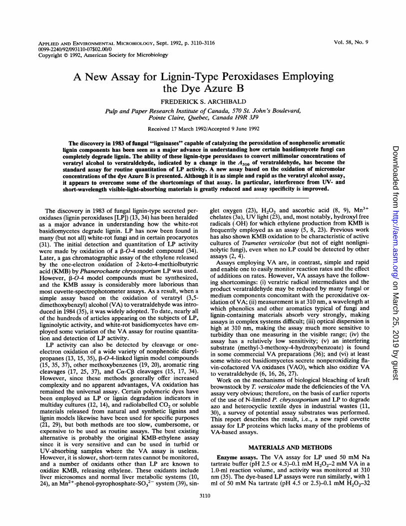
Wavelength (nm)FIG. 1. Successive visible absorption spectra recorded just be-
fore and during the first 25 min of oxidation of 32 ,uM Azure B by0.001 IU of T. versicolor LP and 0.1 mM H202 in 50 mM Na tartrate(pH 4.5).
from the Chemical Dynamics Corp., South Plainfield, N.J.,and not further purified. Arthromyces ramosus and horse-radish peroxidases were from Sigma Chemical Co., and P.chrysosporium LP and MnP were from Tienzyme, Inc.,State Park, Pa.Manganic pyrophosphate was prepared by the classical
procedure of Kenten and Mann (18) by using MnCl2, MnO2and 8-hydroxyquinoline-deferrated Na pyrophosphate; re-acted overnight; and then microfiltered and quantitated bythe A478 peak, assuming a molar extinction coefficient (EM)of 107. Such preparations always contain some Mn2+. Alldyes and other chemicals were from Sigma, Aldrich, andFisher. The Poly dyes are stable nontoxic high-molecular-weight dyes designed by the Dynapol Corp. as food color-ants and sold by Sigma.
RESULTS
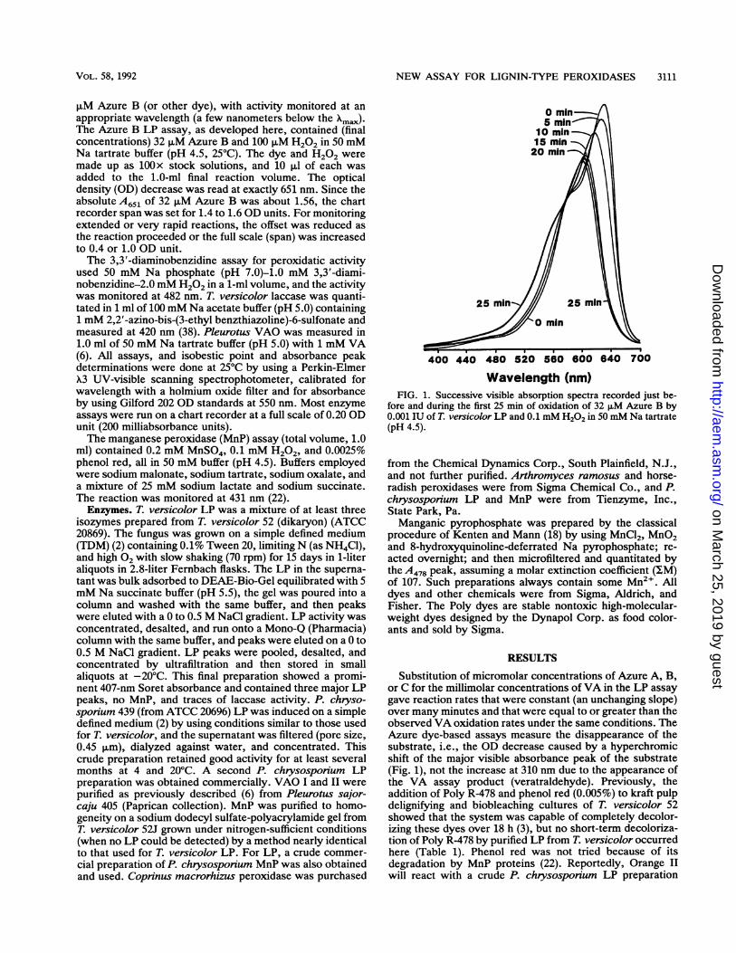
Substitution of micromolar concentrations of Azure A, B,or C for the millimolar concentrations ofVA in the LP assaygave reaction rates that were constant (an unchanging slope)over many minutes and that were equal to or greater than theobserved VA oxidation rates under the same conditions. TheAzure dye-based assays measure the disappearance of thesubstrate, i.e., the OD decrease caused by a hyperchromicshift of the major visible absorbance peak of the substrate(Fig. 1), not the increase at 310 nm due to the appearance ofthe VA assay product (veratraldehyde). Previously, theaddition of Poly R-478 and phenol red (0.005%) to kraft pulpdelignifying and biobleaching cultures of T. versicolor 52showed that the system was capable of completely decolor-izing these dyes over 18 h (3), but no short-term decoloriza-tion of Poly R-478 by purified LP from T. versicolor occurredhere (Table 1). Phenol red was not tried because of itsdegradation by MnP proteins (22). Reportedly, Orange IIwill react with a crude P. chrysosporium LP preparation
VOL. 58, 1992
on March 25, 2019 by guest
http://aem.asm
.org/D
ownloaded from
APPL. ENVIRON. MICROBIOL.
TABLE 1. Oxidation of various dyes by T. versicolor LP'
A Measured AOD (shortSubstrate Concn XmaX (nm) (nm) term)b
Poly R-478 0.005% 520 520 0Poly S 0.005% 444 444 0Poly Y 0.005% 470 470 0Orange II 57 ,uM 489 489 0Tropaeolin 0 32 ,uM 398 398 1+Azure A 32 ,uM 626 640 2+Azure Bc 32 ,uM 647 (600) 651 4+Azure C 32 ,uM 619 623 3+
a Assays using T. versicolor mixed LP (=0.002 U assay-'), 50 mM Natartrate buffer (pH 4.5, 25°C), and 0.1 mM H202 at 0.20 OD unit full scale wererun in a 1.0-ml volume.
b Reactions ranged from no detectable reaction over 10 min (0) to a rapidchange in OD (4+).
c The millimolar extinction coefficient (ImM) (651 nm) of Azure B underassay conditions is approximately 48.8, with a shoulder at 600 nm.
(11), but no reaction (over a 10-min period) was seen with thepurified mixed T. versicolor LP isozymes. Tropaeolin 0,while weakly reactive with the LP preparation, has twophenolic hydroxyl groups and therefore reacted slowly withlaccase as well. Since the goal was to find a substrate thatreacted with all LP but only LP proteins, both of these dyeswere rejected. The remainder of the work employed Azure Bonly, which was selected because it had the most rapidreaction with LP and the longest measurement wavelengthof the three Azure dyes.To be of any value, a substrate for an LP assay must show
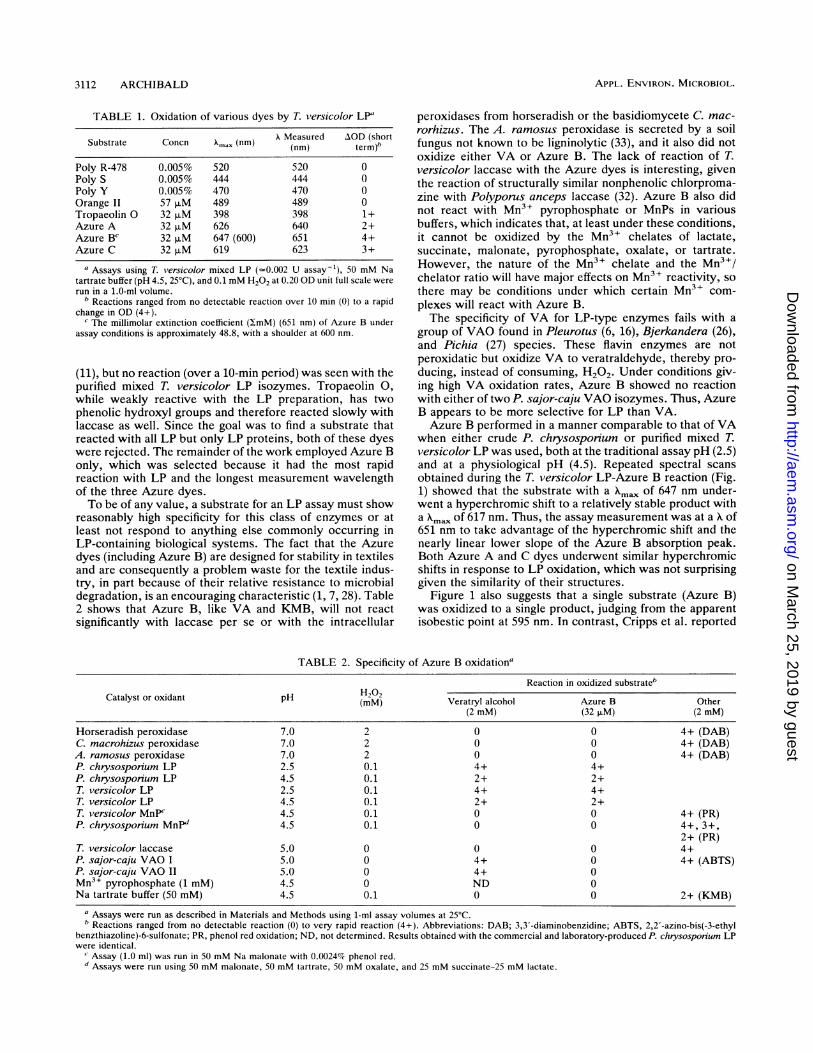
reasonably high specificity for this class of enzymes or atleast not respond to anything else commonly occurring inLP-containing biological systems. The fact that the Azuredyes (including Azure B) are designed for stability in textilesand are consequently a problem waste for the textile indus-try, in part because of their relative resistance to microbialdegradation, is an encouraging characteristic (1, 7, 28). Table2 shows that Azure B, like VA and KMB, will not reactsignificantly with laccase per se or with the intracellular
peroxidases from horseradish or the basidiomycete C. mac-
rorhizus. The A. ramosus peroxidase is secreted by a soilfungus not known to be ligninolytic (33), and it also did notoxidize either VA or Azure B. The lack of reaction of T.versicolor laccase with the Azure dyes is interesting, giventhe reaction of structurally similar nonphenolic chlorproma-zine with Polyporus anceps laccase (32). Azure B also didnot react with Mn3+ pyrophosphate or MnPs in variousbuffers, which indicates that, at least under these conditions,it cannot be oxidized by the Mn3+ chelates of lactate,succinate, malonate, pyrophosphate, oxalate, or tartrate.However, the nature of the Mn3+ chelate and the Mn3+/chelator ratio will have major effects on Mn reactivity, sothere may be conditions under which certain Mn3+ com-
plexes will react with Azure B.The specificity of VA for LP-type enzymes fails with a
group of VAO found in Pleurotus (6, 16), Bjerkandera (26),and Pichia (27) species. These flavin enzymes are notperoxidatic but oxidize VA to veratraldehyde, thereby pro-ducing, instead of consuming, H202. Under conditions giv-ing high VA oxidation rates, Azure B showed no reactionwith either of two P. sajor-caju VAO isozymes. Thus, AzureB appears to be more selective for LP than VA.Azure B performed in a manner comparable to that of VA
when either crude P. chrysosporium or purified mixed T.versicolor LP was used, both at the traditional assay pH (2.5)and at a physiological pH (4.5). Repeated spectral scans
obtained during the T. versicolor LP-Azure B reaction (Fig.1) showed that the substrate with a Xma, of 647 nm under-went a hyperchromic shift to a relatively stable product witha Xmax of 617 nm. Thus, the assay measurement was at a of651 nm to take advantage of the hyperchromic shift and thenearly linear lower slope of the Azure B absorption peak.Both Azure A and C dyes underwent similar hyperchromicshifts in response to LP oxidation, which was not surprisinggiven the similarity of their structures.
Figure 1 also suggests that a single substrate (Azure B)was oxidized to a single product, judging from the apparentisobestic point at 595 nm. In contrast, Cripps et al. reported
TABLE 2. Specificity of Azure B oxidation'
Reaction in oxidized substratebCatalyst or oxidant pH H202
(mM) Veratryl alcohol Azure B Other(2 mM) (32 ,uM) (2 mM)
Horseradish peroxidase 7.0 2 0 0 4+ (DAB)C. macrohizus peroxidase 7.0 2 0 0 4+ (DAB)A. ramosus peroxidase 7.0 2 0 0 4+ (DAB)P. chrysosporium LP 2.5 0.1 4+ 4+P. chrysosporium LP 4.5 0.1 2+ 2+T. versicolor LP 2.5 0.1 4+ 4+T. versicolor LP 4.5 0.1 2+ 2+T. versicolor MnPC 4.5 0.1 0 0 4+ (PR)P. chrysosponium MnPd 4.5 0.1 0 0 4+, 3+,
2+ (PR)T. versicolor laccase 5.0 0 0 0 4+P. sajor-caju VAO I 5.0 0 4+ 0 4+ (ABTS)P. sajor-caju VAO II 5.0 0 4+ 0Mn3+ pyrophosphate (1 mM) 4.5 0 ND 0Na tartrate buffer (50 mM) 4.5 0.1 0 0 2+ (KMB)
a Assays were run as described in Materials and Methods using 1-mI assay volumes at 25'C.b Reactions ranged from no detectable reaction (0) to very rapid reaction (4+). Abbreviations: DAB; 3,3'-diaminobenzidine; ABTS, 2,2'-azino-bis(-3-ethyl
benzthiazoline)-6-sulfonate; PR, phenol red oxidation; ND, not determined. Results obtained with the commercial and laboratory-produced P. chrysosporium LPwere identical.
' Assay (1.0 ml) was run in 50 mM Na malonate with 0.0024% phenol red.d Assays were run using 50 mM malonate, 50 mM tartrate, 50 mM oxalate, and 25 mM succinate-25 mM lactate.
3112 ARCHIBALD
on March 25, 2019 by guest
http://aem.asm
.org/D
ownloaded from
NEW ASSAY FOR LIGNIN-TYPE PEROXIDASES 3113
that an LP-containing supernatant from a P. chrysosporiumculture yielded four colored products from Azure B, asmeasured by thin-layer chromatography (11). Whether theproduct species seen in Fig. 1 was produced and then furtherdegraded by the LP isozymes or other factors in the P.chrysosporium supernatant is unknown; in any case, theassay is accurate as long as the rate measured is thedisappearance of the Azure B (substrate) and not the appear-ance of a specific product.When using a hydrophilic chromophore like Azure B as an
assay substrate, pH effects on the color Xmn and extinctioncoefficient (EM) must be considered. The A647 of Azure Breportedly increases by 15% when the pH is decreased frompH 5.0 to 3.5 (11). However, when 50 mM Na tartrate bufferat pH 4.5 (pK, 4.16) and 520% (vol/vol) enzyme or culturefluid (typically pH 4.1 to 4.6) are used, the assay pH is +0.1of the buffer pH. Figure 2A displays the ability of Azure B toinhibit VA oxidation by LP, while Fig. 2B presents theopposite experiment, the inhibition of Azure B oxidation byVA at the realistic pH of 4.5. Clearly, T. versicolor LPpreferentially oxidizes micromolar concentrations of AzureB over millimolar concentrations of VA. Both inhibitioneffects showed rather poor linearity with concentration, soKi values were not calculated.
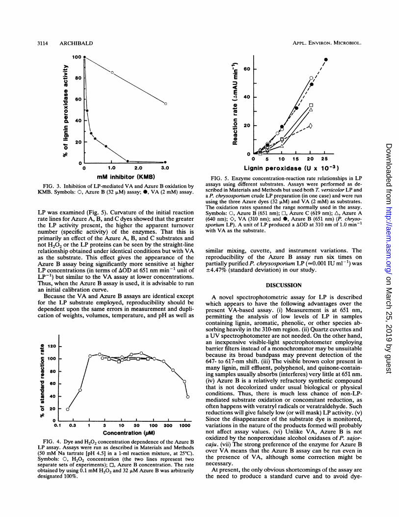
Purified LP enzymes cannot directly oxidize KMB but theoxidation proceeds rapidly in the presence of VA, as indi-cated by the evolution of ethylene (2). Increasing amounts ofKMB competitively inhibit the appearance of veratralde-hyde apparently because a peroxidase-generated veratrylintermediate is the proximal oxidant of KMB and is, in theprocess, rereduced to VA, thus preventing the appearance ofveratraldehyde (2). Substitution of Azure B for VA reducesthis KMB interference (Fig. 3). Since a relatively high-energy oxidation is required to oxidize KMB, it is notunreasonable to presume that the proximal veratryl oxidiz-ing species reduced by KMB will also be reduced by manybiological molecules and that therefore the Azure B LPassay will be less affected by many reductants interferingwith the VA-based assay.The optimal concentrations of the two LP substrates,
H202 and Azure B, employed in the assay were determined(Fig. 4). The concentration of H202 used previously in theVA assay (100 ,uM) proved to be entirely satisfactory, as didthe initial concentration of Azure B (32 ,uM).Reducing the Azure B concentration from 32 to 4 ,uM had
no effect on the initial reaction rate, indicating that theapparent Km for Azure B of the mixed T. versicolor LP wasc2 ,uM. Reducing the Azure B concentration to 4 or 8 ,Mtherefore would affect only the usable extent of the 651-nmcolor change, not the initial rate. In the case of spectropho-tometers unable to offset 1.4 OD units and to continue togive a smooth baseline at 0.2 OD unit full scale, it may bepreferable to use 8 pLM Azure B and an offset of 0.2 OD unit,or 0.4 OD unit full scale and no offset at all, although thetotal extent of measurable LP-driven oxidation will be re-duced accordingly.Although the Azure B assay wavelength (651 nm) should
permit assay of moderately turbid samples, the fact thatAzure B is a dye means that its adsorption by sampleparticulates must be considered. Hardwood kraft brown-stock (0.25% [wt/vol]) added to 32 p,M Azure B in standardassay buffer adsorbed 20.0 puM (62.5%) Azure B in 5 min.Highly purified cellulose (Solka-Floc) bound 7.5 p,M (23.4%)in 5 min, while a large mass of fresh agar plate-grown T.versicolor mycelium bound 4.0 ,uM (12.5%) Azure B underthe same conditions. Thus, at least some solids will interfere
100
80
0(U1- 60
:-c._ 400
20
01
1001
04..
0
80
60
40
A
0 4 8 12 16
(WM) azure b
20 -
0 2.0 4.0
mM veratryl alcoholFIG. 2. (A) Inhibition of the LP-catalyzed oxidation of VA by
Azure B. LP assay conditions were as described in Materials andMethods, and the reaction rate was measured at 310 nm. Symbols:0, 4 mM VA; 0, 2 mM VA. (B) Inhibition of the LP-catalyzedoxidation of Azure B by the addition of VA. Assay conditions wereas described in Materials and Methods, and the rate was measuredat 651 nm. Symbols: El, 32 p.M Azure B; 0, 16 ,uM Azure B;*,4 p,M Azure B.
with the assay via adsorption. Samples should therefore becentrifuged or filtered before assay unless the solids presentare known to not adsorb Azure B.Repeated pulsing of the assay buffer containing 32 ,uM
Azure B plus LP with 2 p,M H202 showed that the apparentKm (H202) for a mixture of three T. versicolor LP isozymeswas 0.5 ,uM, and that for each nanomole of H202 consumed,0.98 nmol of Azure B disappeared. Thus, one H202 wasconsumed, two water molecules were produced, and twoelectrons were abstracted per unit of Azure B oxidized.Since peroxidases are typically one-electron-abstracting ox-idants, the oxidation reaction has at least two steps. The LPsare not necessarily involved in both oxidative steps or in allof the H202 consumption. Since such a multistep reaction,especially one involving aryloxy radical intermediates, mightbe expected to have an enzyme concentration-reaction raterelationship other than 1:1, the response of the assay toincreasing amounts of P. chrysosponium and T. versicolor
VOL. 58, 1992
on March 25, 2019 by guest
http://aem.asm
.org/D
ownloaded from
APPL. ENVIRON. MICROBIOL.
100a
00(_0
00.
Cco01
0
80
60 .
40
20 F
0
0
v-
Ic
E
0(UI-
C.2"U(U(U
I*10
0S"
0 1.0 2.0 3.0
mM inhibitor (KMB)FIG. 3. Inhibition of LP-mediated VA and Azure B oxidation by
KMB. Symbols: 0, Azure B (32 ,uM) assay; *, VA (2 mM) assay.
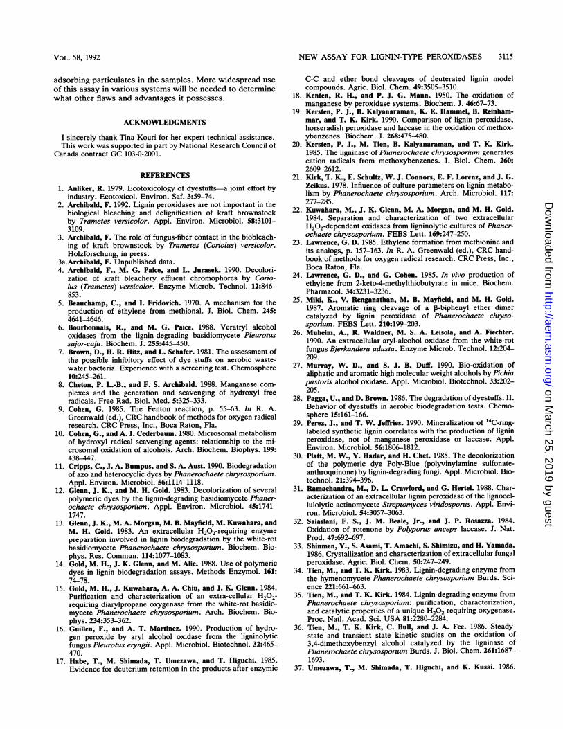
LP was examined (Fig. 5). Curvature of the initial reactionrate lines for Azure A, B, and C dyes showed that the greaterthe LP activity present, the higher the apparent turnovernumber (specific activity) of the enzymes. That this isprimarily an effect of the Azure A, B, and C substrates andnot H202 or the LP proteins can be seen by the straight-linerelationship obtained under identical conditions but with VAas the substrate. This effect gives the appearance of theAzure B assay being significantly more sensitive at higherLP concentrations (in terms of AOD at 651 nm min-1 unit ofLP-1) but similar to the VA assay at lower concentrations.Thus, when the Azure B assay is used, it is advisable to runan initial calibration curve.Because the VA and Azure B assays are identical except
for the LP substrate employed, reproducibility should bedependent upon the same errors in measurement and dupli-cation of weights, volumes, temperature, and pH as well as
0
a
0
0
la
.10
120
100
80 1-
60
40
20 _
0.1 0.3 1 3 10 30 100 300 1000
Concentration (,PM)FIG. 4. Dye and H202 concentration dependence of the Azure B
LP assay. Assays were run as described in Materials and Methods(50 mM Na tartrate [pH 4.5] in a 1-ml reaction mixture, at 25°C).Symbols: 0, H202 concentration (the two lines represent twoseparate sets of experiments); El, Azure B concentration. The rateobtained by using 0.1 mM H202 and 32 ,uM Azure B was arbitrarilydesignated 100%.
60
40
20
00 5 10 15 20 25
Lignin peroxidase (U x 10-3)FIG. 5. Enzyme concentration-reaction rate relationships in LP
assays using different substrates. Assays were performed as de-scribed in Materials and Methods but used both T. versicolor LP anda P. chrysosponium crude LP preparation (in one case) and were runusing the three Azure dyes (32 ,uM) and VA (2 mM) as substrates.The oxidation rates spanned the range normally used in the assay.Symbols: 0, Azure B (651 nm); El, Azure C (619 nm); A, Azure A(640 nm); O, VA (310 nm); and 0, Azure B (651 nm) (P. chryso-sporium LP). A unit of LP produced a AOD at 310 nm of 1.0 min-with VA as the substrate.
similar mixing, cuvette, and instrument variations. Thereproducibility of the Azure B assay run six times onpartially purified P. chrysosponum LP ('=0.001 IU ml-) was+4.47% (standard deviation) in our study.
DISCUSSION
A novel spectrophotometric assay for LP is describedwhich appears to have the following advantages over thepresent VA-based assay. (i) Measurement is at 651 nm,permitting the analysis of low levels of LP in samplescontaining lignin, aromatic, phenolic, or other species ab-sorbing heavily in the 310-nm region. (ii) Quartz cuvettes anda UV spectrophotometer are not needed. On the other hand,an inexpensive visible-light spectrophotometer employingbarrier filters instead of a monochromator may be unsuitablebecause its broad bandpass may prevent detection of the647- to 617-nm shift. (iii) The visible brown color present inmany lignin, mill effluent, polyphenol, and quinone-contain-ing samples usually absorbs (interferes) very little at 651 nm.(iv) Azure B is a relatively refractory synthetic compoundthat is not decolorized under usual biological or physicalconditions. Thus, there is much less chance of non-LP-mediated substrate oxidation or concomitant reduction, asoften happens with veratryl radicals or veratraldehyde. Suchreductions will give falsely low (or will mask) LP activity. (v)Since the disappearance of the substrate dye is monitored,variations in the nature of the products formed will probablynot affect assay values. (vi) Unlike VA, Azure B is notoxidized by the nonperoxidase alcohol oxidases of P. sajor-caju. (vii) The strong preference of the enzyme for Azure Bover VA means that the Azure B assay can be run even inthe presence of VA, although some correction might benecessary.At present, the only obvious shortcomings of the assay are
the need to produce a standard curve and to avoid dye-
3114 ARCHIBALD
on March 25, 2019 by guest
http://aem.asm
.org/D
ownloaded from
NEW ASSAY FOR LIGNIN-TYPE PEROXIDASES 3115
adsorbing particulates in the samples. More widespread useof this assay in various systems will be needed to determinewhat other flaws and advantages it possesses.
ACKNOWLEDGMENTS
I sincerely thank Tina Kouri for her expert technical assistance.This work was supported in part by National Research Council of
Canada contract GC 103-0-2001.
REFERENCES
1. Anliker, R. 1979. Ecotoxicology of dyestuffs-a joint effort byindustry. Ecotoxicol. Environ. Saf. 3:59-74.
2. Archibald, F. 1992. Lignin peroxidases are not important in thebiological bleaching and delignification of kraft brownstockby Trametes versicolor. Appl. Environ. Microbiol. 58:3101-3109.
3. Archibald, F. The role of fungus-fiber contact in the biobleach-ing of kraft brownstock by Trametes (Coriolus) versicolor.Holzforschung, in press.
3a.Archibald, F. Unpublished data.4. Archibald, F., M. G. Paice, and L. Jurasek. 1990. Decolori-
zation of kraft bleachery effluent chromophores by Corio-lus (Trametes) versicolor. Enzyme Microb. Technol. 12:846-853.
5. Beauchamp, C., and I. Fridovich. 1970. A mechanism for theproduction of ethylene from methional. J. Biol. Chem. 245:4641-4646.
6. Bourbonnais, R., and M. G. Paice. 1988. Veratryl alcoholoxidases from the lignin-degrading basidiomycete Pleurotussajor-caju. Biochem. J. 255:445-450.
7. Brown, D., H. R. Hitz, and L. Schafer. 1981. The assessment ofthe possible inhibitory effect of dye stuffs on aerobic waste-water bacteria. Experience with a screening test. Chemosphere10:245-261.
8. Cheton, P. L.-B., and F. S. Archibald. 1988. Manganese com-
plexes and the generation and scavenging of hydroxyl freeradicals. Free Rad. Biol. Med. 5:325-333.
9. Cohen, G. 1985. The Fenton reaction, p. 55-63. In R. A.Greenwald (ed.), CRC handbook of methods for oxygen radicalresearch. CRC Press, Inc., Boca Raton, Fla.
10. Cohen, G., and A. I. Cederbaum. 1980. Microsomal metabolismof hydroxyl radical scavenging agents: relationship to the mi-crosomal oxidation of alcohols. Arch. Biochem. Biophys. 199:438-447.
11. Cripps, C., J. A. Bumpus, and S. A. Aust. 1990. Biodegradationof azo and heterocyclic dyes by Phanerochaete chrysosporium.Appl. Environ. Microbiol. 56:1114-1118.
12. Glenn, J. K., and M. H. Gold. 1983. Decolorization of severalpolymeric dyes by the lignin-degrading basidiomycete Phaner-ochaete chrysosporium. Appl. Environ. Microbiol. 45:1741-1747.
13. Glenn, J. K., M. A. Morgan, M. B. Mayfield, M. Kuwahara, andM. H. Gold. 1983. An extracellular H202-requiring enzymepreparation involved in lignin biodegradation by the white-rotbasidiomycete Phanerochaete chrysosporium. Biochem. Bio-phys. Res. Commun. 114:1077-1083.
14. Gold, M. H., J. K. Glenn, and M. Alic. 1988. Use of polymericdyes in lignin biodegradation assays. Methods Enzymol. 161:74-78.
15. Gold, M. H., J. Kuwahara, A. A. Chiu, and J. K. Glenn. 1984.Purification and characterization of an extra-cellular H202-requiring diarylpropane oxygenase from the white-rot basidio-mycete Phanerochaete chrysosponium. Arch. Biochem. Bio-phys. 234:353-362.
16. Guillen, F., and A. T. Martinez. 1990. Production of hydro-gen peroxide by aryl alcohol oxidase from the ligninolyticfungus Pleurotus eryngii. Appl. Microbiol. Biotechnol. 32:465-470.
17. Habe, T., M. Shimada, T. Umezawa, and T. Higuchi. 1985.Evidence for deuterium retention in the products after enzymic
C-C and ether bond cleavages of deuterated lignin modelcompounds. Agric. Biol. Chem. 49:3505-3510.
18. Kenten, R. H., and P. J. G. Mann. 1950. The oxidation ofmanganese by peroxidase systems. Biochem. J. 46:67-73.
19. Kersten, P. J., B. Kalyanaraman, K. E. Hammel, B. Reinham-mar, and T. K. Kirlk 1990. Comparison of lignin peroxidase,horseradish peroxidase and laccase in the oxidation of methox-ybenzenes. Biochem. J. 268:475-480.
20. Kersten, P. J., M. Tien, B. Kalyanaraman, and T. K. Kirk.1985. The ligninase of Phanerochaete chrysosporium generatescation radicals from methoxybenzenes. J. Biol. Chem. 260:2609-2612.
21. Kirk, T. K., E. Schultz, W. J. Connors, E. F. Lorenz, and J. G.Zeikus. 1978. Influence of culture parameters on lignin metabo-lism by Phanerochaete chrysosporium. Arch. Microbiol. 117:277-285.
22. Kuwahara, M., J. K. Glenn, M. A. Morgan, and M. H. Gold.1984. Separation and characterization of two extracellularH202-dependent oxidases from ligninolytic cultures of Phaner-ochaete chrysosponum. FEBS Lett. 169:247-250.
23. Lawrence, G. D. 1985. Ethylene formation from methionine andits analogs, p. 157-163. In R. A. Greenwald (ed.), CRC hand-book of methods for oxygen radical research. CRC Press, Inc.,Boca Raton, Fla.
24. Lawrence, G. D., and G. Cohen. 1985. In vivo production ofethylene from 2-keto-4-methylthiobutyrate in mice. Biochem.Pharmacol. 34:3231-3236.
25. Miki, K., V. Renganathan, M. B. Mayfield, and M. H. Gold.1987. Aromatic ring cleavage of a p-biphenyl ether dimercatalyzed by lignin peroxidase of Phanerochaete chryso-sporium. FEBS Lett. 210:199-203.
26. Muheim, A., R. Waldner, M. S. A. Leisola, and A. Fiechter.1990. An extracellular aryl-alcohol oxidase from the white-rotfungus Bjerkandera adusta. Enzyme Microb. Technol. 12:204-209.
27. Murray, W. D., and S. J. B. Duff. 1990. Bio-oxidation ofaliphatic and aromatic high molecular weight alcohols by Pichiapastoris alcohol oxidase. Appl. Microbiol. Biotechnol. 33:202-205.
28. Pagga, U., and D. Brown. 1986. The degradation of dyestuffs. II.Behavior of dyestuffs in aerobic biodegradation tests. Chemo-sphere 15:161-166.
29. Perez, J., and T. W. Jeffries. 1990. Mineralization of "'C-ring-labeled synthetic lignin correlates with the production of ligninperoxidase, not of manganese peroxidase or laccase. Appl.Environ. Microbiol. 56:1806-1812.
30. Platt, M. W., Y. Hadar, and H. Chet. 1985. The decolorizationof the polymeric dye Poly-Blue (polyvinylamine sulfonate-anthroquinone) by lignin-degrading fungi. Appl. Microbiol. Bio-technol. 21:394-396.
31. Ramachandra, M., D. L. Crawford, and G. Hertel. 1988. Char-acterization of an extracellular lignin peroxidase of the lignocel-lulolytic actinomycete Streptomyces viridosporus. Appl. Envi-ron. Microbiol. 54:3057-3063.
32. Saiaslani, F. S., J. M. Beale, Jr., and J. P. Rosazza. 1984.Oxidation of rotenone by Polyporus anceps laccase. J. Nat.Prod. 47:692-697.
33. Shinmen, Y., S. Asami, T. Amachi, S. Shimizu, and H. Yamada.1986. Crystallization and characterization of extracellular fungalperoxidase. Agric. Biol. Chem. 50:247-249.
34. Tien, M., and T. K. Kirk. 1983. Lignin-degrading enzyme fromthe hymenomycete Phanerochaete chrysosponum Burds. Sci-ence 221:661-663.
35. Tien, M., and T. K. Kirk. 1984. Lignin-degrading enzyme fromPhanerochaete chrysosporium: purification, characterization,and catalytic properties of a unique H202-requiring oxygenase.Proc. Natl. Acad. Sci. USA 81:2280-2284.
36. Tien, M., T. K. Kirk, C. Bull, and J. A. Fee. 1986. Steady-state and transient state kinetic studies on the oxidation of3,4-dimethoxybenzyl alcohol catalyzed by the ligninase ofPhanerochaete chrysosporium Burds. J. Biol. Chem. 261:1687-1693.
37. Umezawa, T., M. Shimada, T. Higuchi, and K. Kusai. 1986.
VOL. 58, 1992
on March 25, 2019 by guest
http://aem.asm
.org/D
ownloaded from
3116 ARCHIBALD APPL. ENvIRON. MICROBIOL.
Aromatic ring cleavage of beta-0-4 lignin substructure modeldimers by lignin peroxidase of Phanerochaete chrysosporium.FEBS Lett. 205:287-292.
38. Wolfenden, B. S., and R. L. Willson. 1982. Radical-cations asreference chromogens in kinetic studies of one-electron transferreactions: pulse radiolysis studies of 2,2'-azinobis-(3-ethyl benz-
thiazoline-6-sulphonate). J. Chem. Soc. Perkin Trans. II:805-812.
39. Yang, S. F. 1969. Further studies on ethylene formation froma-keto-y-methylthiobutyric acid or ,B-methylthiopropionalde-hyde by peroxidase in the presence of sulfite and oxygen. J.Biol. Chem. 244:4360-4365.
on March 25, 2019 by guest
http://aem.asm
.org/D
ownloaded from