Embed Size (px)
Citation preview
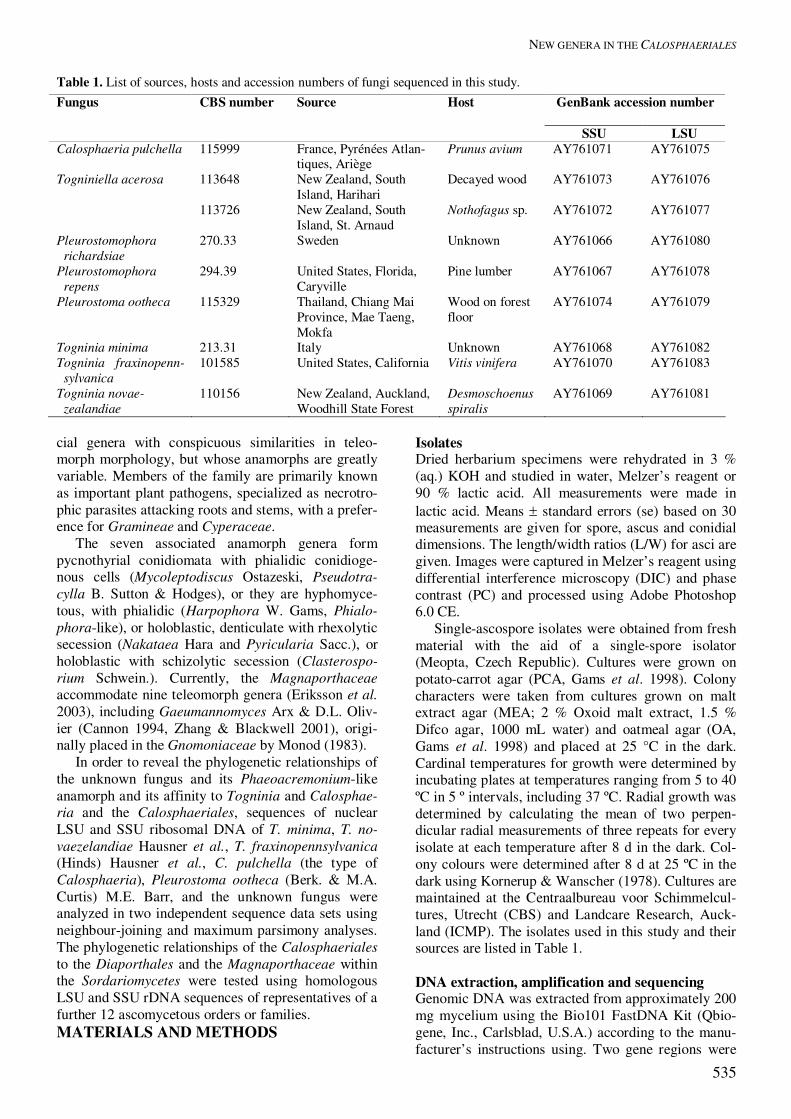
STUDIES IN MYCOLOGY 50: 533–550. 2004.
533
New genera in the Calosphaeriales: Togniniella and its anamorph Phaeocrella, and Calosphaeriophora as anamorph of Calosphaeria
Martina Réblová1*, Lizel Mostert2, Walter Gams2 and Pedro W. Crous2 1Department of Plant Taxonomy and Biosystematics, Institute of Botany, Academy of Sciences, 252 43 Pr�honice, Czech Republic; 2Centraalbureau voor Schimmelcultures, Fungal Biodiversity Centre, Uppsalalaan 8, 3584 CT Utrecht, The Netherlands *Correspondence: Martina Réblová, [email protected] Abstract: During a survey of perithecial ascomycetes in New Zealand, two collections of a Togninia-like fungus were made on decayed wood. In culture, colonies produced a Phaeoacremonium-like anamorph. In order to reveal the phylogenetic relationships of the unknown fungus and its affinity to Togninia and other genera in the Calosphaeriales, sequences of nuclear LSU and SSU ribosomal DNA were obtained of several members of this order. These data, supported by morpho-logical and cultural characteristics, confirm that the New Zealand fungus represents a new genus very close to Calosphaeria. The genus Togniniella is proposed here to accommodate these collections, while Phaeocrella is established for their ana-morphs. Furthermore, Calosphaeria pulchella was found to form a distinct Acremonium-like anamorph in culture, for which the genus Calosphaeriophora is proposed. Pleurostoma with Pleurostomophora anamorphs is a sister genus to the above two genera, forming the Pleurostomataceae. Togninia with its Phaeoacremonium anamorphs, together with Jobellisia, are closer to the Diaporthales, and deserve the rank of family, for which Togniniaceae is proposed. The presence of significantly different anamorphs in the Calosphaeriales, as well as obvious differences in teleomorph morphology of species accommo-dated in Calosphaeria, suggest that both the Calosphaeriales and Calosphaeria as presently perceived, are polyphyletic.
Taxonomic novelties: Calosphaeriophora Réblová, L. Mostert, W. Gams & Crous gen. nov., Calosphaeriophora pulchella Réblová, L. Mostert, W. Gams & Crous sp. nov., Phaeocrella Réblová, L. Mostert, W. Gams & Crous gen. nov., Phaeocrella acerosa Réblová, L. Mostert, W. Gams & Crous sp. nov., Pleurostomataceae Réblová, L. Mostert, W. Gams & Crous fam. nov., Togniniaceae Réblová, L. Mostert, W. Gams & Crous fam. nov., Togniniella Réblová, L. Mostert, W. Gams & Crous gen. nov., Togniniella acerosa Réblová, L. Mostert, W. Gams & Crous sp. nov. Key words: Acremonium, LSU and SSU rDNA phylogeny, Phaeoacremonium, Phialophora, systematics, Togninia.
INTRODUCTION Traditionally, a number of small pyrenomycetous genera with simple, dark perithecia (occasionally embedded in a stroma), unitunicate asci, hyaline to slightly pigmented, ellipsoid to allantoid ascospores, have been classified in the Calosphaeriales. These fungi occupy similar or highly specialized habitats and they have been seen by only a handful of mycologists. Munk (1957) described the Calosphaeriaceae, and assigned the family to the broadly perceived Sphaeria-les. He drew attention to the unique centrum present within ascomata of these fungi, suggesting that it could be used as basis for recognizing a separate order among perithecial ascomycetes. The order Calosphae-riales was later recognized by Barr (1983), who out-lined the history of the Calosphaeriaceae and the respective genera, and published the first modern concept of this family (Barr 1985, Barr et al. 1993). Several researchers have noted that the Calosphae-riales represent a polyphyletic group of phenotypically similar taxa that may comprise at least two phyloge-
netic lineages (Barr et al. 1993, Samuels & Candous-sau 1996, Barr 1998), viz. the putative diatrypaceous lineage (Diatrypaceae/Xylariales) on the one hand, and the diaporthaceous lineage (apparently the Gnomo-niaceae/Diaporthales) on the other hand. After exclu-sion of the stromatic calosphaeriaceous family Gra-phostromataceae, associated with Nodulosporium-like anamorphs in the Xylariales (Barr et al. 1993), the Calosphaeriales of the Diaporthales lineage encom-passed six nonstromatic genera, i.e. Calosphaeria Tul. & C. Tul., Jattaea Berl., Pleurostoma Tul. & C. Tul., Romellia Berl., Wegelina Berl., and Togninia Berl., and the stromatic Pachytrype Berl. ex M.E. Barr et al. (Barr et al. 1993). The position of Enchnoa Fr. within the Calosphaeriales is debatable (Petrak & Sydow 1936, Barr 1985). Apart from Graphostroma Piroz. (Pirozynski 1974), we do not have phylogenetic clues to the true relationships of genera within the Calos-phaeriales. Current generic concepts in the Calosphaeriales are based primarily on the arrangement of ascomata, neck lengths, presence and arrangement of stromatic tissue or subiculum, and arrangement of the asci, viz. in

RÉBLOVÁ ET AL.
534
spicate arrangement or in small fascicles (Barr 1985). The perithecia of the Calosphaeriales are superficial or immersed in wood, arise separately with separate necks, or are tightly aggregated in circinate groups on wood beneath the periderm, with radially converging beaks that may be united in a disc piercing the periderm. Genera assigned to the Calosphaeriales possess true, persistent, apically free paraphyses and hyaline, allantoid to suballantoid, aseptate or delicately 1-septate ascospores arranged 2−3-seriately or in a fascicle within the ascus. The conidiogenesis of members of the Calosphae-riales is reported as either being phialidic or holoblas-tic. Pachytrype Berl. ex M.E. Barr et al. has a Cyto-spora Ehrenb. anamorph (Barr et al. 1993); Calos-phaeria fagi Samuels & Candoussau has Ramichlorid-ium-like and Sporothrix-like synanamorphs (Samuels & Candoussau 1996); Calosphaeria pulchella (Pers. : Fr.) J. Schröt. has an Acremonium-like anamorph (this study), and Togninia has Phaeoacremonium W. Gams et al. anamorphs (Hausner et al. 1992, Mostert et al. 2003). Except these few known life histories, the connections to asexual states are little known, nor are there DNA-based phylogenies to reveal likely sexual or asexual relatives. Phaeoacremonium is a dematiaceous hyphomycete genus of approximately 17 species (Crous et al. 1996, Dupont et al. 2000, Groenewald et al. 2001, Mostert et al. 2004), introduced to include fungi that are interme-diate between Acremonium Link : Fr. and Phialophora Medlar, encompassing ecologically important fungi associated with human infections and disease symp-toms of woody hosts. The link between Phaeoacremo-nium aleophilum and Togninia minima (Tul. & C. Tul.) Berl., the type of the genus, was recently established in vitro by Mostert et al. (2003). Togninia Berl. has historically been classified in the Calosphaeriaceae of the Calosphaeriales (Berlese 1900, Barr 1985, Eriks-son et al. 2003, Mostert et al. 2003). During a survey of perithecial ascomycetes in New Zealand in March and February 2003, two collections of a minute, lignicolous, saprobic Togninia-like spe-cies were encountered. In culture, colonies produced a Phaeoacremonium-like anamorph. Herbarium material of a fungus originating in North America, Canada, whose morphological characteristics match well those of the fungus collected in New Zealand, was found in the DAOM herbarium. The unknown fungus resembles Togninia in having dark, nonstromatic perithecia with elongate necks; asci arranged in a spicate formation along elongate ascogenous hyphae; a thickened ascal apex lacking any discharge mechanism; true paraphy-ses; hyaline, aseptate, suballantoid ascospores and a dematiaceous hyphomycete anamorph with phialidic conidiogenesis. The unknown fungus differs from Togninia in the shape of the asci, which are apically obtuse, tapering conspicuously towards the base from
the sporiferous portion; the fasciculate arrangement of the ascospores in the upper part of the ascus; the pres-ence of short cells along the ascogenous hyphae, from which each ascus arises as an outgrowth, and the branching pattern of conidiophores of the Phaeoacre-monium-like anamorph vs. asci with obtuse to broadly rounded bases; 2−3-seriately arranged ascospores filling the entire ascus; elongate ascogenous hyphae with attached remnants of the basal parts of the asci after their separation, and Phaeoacremonium ana-morphs of Togninia. Based on the ecology and the teleomorph and anamorph morphology, the six nonstromatic calos-phaeriaceous genera, including the unknown fungus, can be compared with the Gnomoniaceae of the Di-aporthales and the Magnaporthaceae (order uncer-tain). The Diaporthales include either saprobes or endo-phytes fruiting on moribund tissue, or plant-pathogenic fungi divided into six well-supported phylogenetic lineages, viz. the four families Diaporthaceae, Gnomo-niaceae, Melanconidaceae and Valsaceae, and three less resolved species complexes for which no families are currently available, namely the Schizoparme com-plex, the Wuestneia complex, and the Cryphonectria-Endothia complex, as evidenced by sequences of the large-subunit nuclear ribosomal DNA (Castlebury et al. 2002, Lee et al. 2004, this volume). The order is also well-defined morphologically based on dark, beaked perithecia with opaque walls immersed in stromata or freely in host tissue; lack of true paraphy-ses; unitunicate, short-stipitate asci, rounded at the bottom and often floating free within the centrum at maturity, with a distinct, refractive ring in the ascal apex (Barr 1978, 1990). The known asexual states of members of the Diaporthales have been linked to coelomycetous fungi forming pycnidia or acervuli with or without stromata and generally with phialidic, rarely annellidic conidiogenesis. The Gnomoniaceae are a phylogenetically (Castlebury et al. 2002) and morpho-logically (Monod 1983) well-defined group within the Diaporthales, whose members bear some resemblance to taxa placed in the Calosphaeriales, especially in characters of the perithecia, asci and ascospores. The Gnomoniaceae include taxa with dark, upright perithe-cia immersed in herbaceous tissue or wood and erum-pent separately, with central, rarely eccentric necks, without stromata or surrounded by a reduced prosen-chymatous stroma, with asci basally rounded and hyaline ascospores with variable septation. The ana-morphs linked to this family have phialidic conidioge-nous cells. The Magnaporthaceae are of uncertain ordinal affiliation within the Sordariomycetes (Cannon 1994, Kirk et al. 2001, Eriksson et al. 2003). The family was erected by Cannon (1994) for six nonstromatic perithe-
NEW GENERA IN THE CALOSPHAERIALES
535
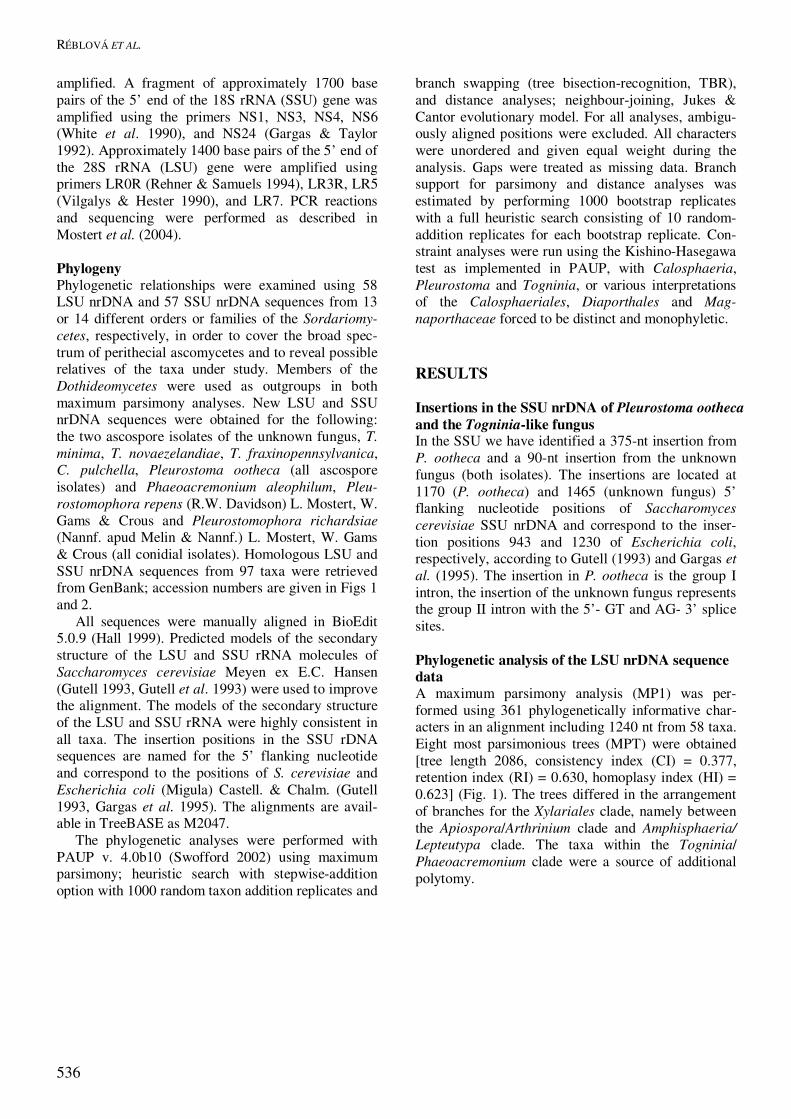
Table 1. List of sources, hosts and accession numbers of fungi sequenced in this study. Fungus CBS number Source
Host
GenBank accession number
SSU LSU Calosphaeria pulchella 115999 France, Pyrénées Atlan-
tiques, Ariège Prunus avium AY761071 AY761075
Togniniella acerosa 113648 New Zealand, South Island, Harihari
Decayed wood AY761073 AY761076
113726 New Zealand, South Island, St. Arnaud
Nothofagus sp. AY761072 AY761077
Pleurostomophora richardsiae
270.33 Sweden Unknown AY761066 AY761080
Pleurostomophora repens
294.39 United States, Florida, Caryville
Pine lumber AY761067 AY761078
Pleurostoma ootheca 115329 Thailand, Chiang Mai Province, Mae Taeng, Mokfa
Wood on forest floor
AY761074 AY761079
Togninia minima 213.31 Italy Unknown AY761068 AY761082 Togninia fraxinopenn- sylvanica
101585 United States, California Vitis vinifera AY761070 AY761083
Togninia novae- zealandiae
110156 New Zealand, Auckland, Woodhill State Forest
Desmoschoenus spiralis
AY761069 AY761081
cial genera with conspicuous similarities in teleo-morph morphology, but whose anamorphs are greatly variable. Members of the family are primarily known as important plant pathogens, specialized as necrotro-phic parasites attacking roots and stems, with a prefer-ence for Gramineae and Cyperaceae. The seven associated anamorph genera form pycnothyrial conidiomata with phialidic conidioge-nous cells (Mycoleptodiscus Ostazeski, Pseudotra-cylla B. Sutton & Hodges), or they are hyphomyce-tous, with phialidic (Harpophora W. Gams, Phialo-phora-like), or holoblastic, denticulate with rhexolytic secession (Nakataea Hara and Pyricularia Sacc.), or holoblastic with schizolytic secession (Clasterospo-rium Schwein.). Currently, the Magnaporthaceae accommodate nine teleomorph genera (Eriksson et al. 2003), including Gaeumannomyces Arx & D.L. Oliv-ier (Cannon 1994, Zhang & Blackwell 2001), origi-nally placed in the Gnomoniaceae by Monod (1983). In order to reveal the phylogenetic relationships of the unknown fungus and its Phaeoacremonium-like anamorph and its affinity to Togninia and Calosphae-ria and the Calosphaeriales, sequences of nuclear LSU and SSU ribosomal DNA of T. minima, T. no-vaezelandiae Hausner et al., T. fraxinopennsylvanica (Hinds) Hausner et al., C. pulchella (the type of Calosphaeria), Pleurostoma ootheca (Berk. & M.A. Curtis) M.E. Barr, and the unknown fungus were analyzed in two independent sequence data sets using neighbour-joining and maximum parsimony analyses. The phylogenetic relationships of the Calosphaeriales to the Diaporthales and the Magnaporthaceae within the Sordariomycetes were tested using homologous LSU and SSU rDNA sequences of representatives of a further 12 ascomycetous orders or families. MATERIALS AND METHODS
Isolates Dried herbarium specimens were rehydrated in 3 % (aq.) KOH and studied in water, Melzer’s reagent or 90 % lactic acid. All measurements were made in lactic acid. Means � standard errors (se) based on 30 measurements are given for spore, ascus and conidial dimensions. The length/width ratios (L/W) for asci are given. Images were captured in Melzer’s reagent using differential interference microscopy (DIC) and phase contrast (PC) and processed using Adobe Photoshop 6.0 CE. Single-ascospore isolates were obtained from fresh material with the aid of a single-spore isolator (Meopta, Czech Republic). Cultures were grown on potato-carrot agar (PCA, Gams et al. 1998). Colony characters were taken from cultures grown on malt extract agar (MEA; 2 % Oxoid malt extract, 1.5 % Difco agar, 1000 mL water) and oatmeal agar (OA, Gams et al. 1998) and placed at 25 °C in the dark. Cardinal temperatures for growth were determined by incubating plates at temperatures ranging from 5 to 40 ºC in 5 º intervals, including 37 ºC. Radial growth was determined by calculating the mean of two perpen-dicular radial measurements of three repeats for every isolate at each temperature after 8 d in the dark. Col-ony colours were determined after 8 d at 25 ºC in the dark using Kornerup & Wanscher (1978). Cultures are maintained at the Centraalbureau voor Schimmelcul-tures, Utrecht (CBS) and Landcare Research, Auck-land (ICMP). The isolates used in this study and their sources are listed in Table 1. DNA extraction, amplification and sequencing Genomic DNA was extracted from approximately 200 mg mycelium using the Bio101 FastDNA Kit (Qbio-gene, Inc., Carlsblad, U.S.A.) according to the manu-facturer’s instructions using. Two gene regions were
RÉBLOVÁ ET AL.
536
amplified. A fragment of approximately 1700 base pairs of the 5’ end of the 18S rRNA (SSU) gene was amplified using the primers NS1, NS3, NS4, NS6 (White et al. 1990), and NS24 (Gargas & Taylor 1992). Approximately 1400 base pairs of the 5’ end of the 28S rRNA (LSU) gene were amplified using primers LR0R (Rehner & Samuels 1994), LR3R, LR5 (Vilgalys & Hester 1990), and LR7. PCR reactions and sequencing were performed as described in Mostert et al. (2004). Phylogeny Phylogenetic relationships were examined using 58 LSU nrDNA and 57 SSU nrDNA sequences from 13 or 14 different orders or families of the Sordariomy-cetes, respectively, in order to cover the broad spec-trum of perithecial ascomycetes and to reveal possible relatives of the taxa under study. Members of the Dothideomycetes were used as outgroups in both maximum parsimony analyses. New LSU and SSU nrDNA sequences were obtained for the following: the two ascospore isolates of the unknown fungus, T. minima, T. novaezelandiae, T. fraxinopennsylvanica, C. pulchella, Pleurostoma ootheca (all ascospore isolates) and Phaeoacremonium aleophilum, Pleu-rostomophora repens (R.W. Davidson) L. Mostert, W. Gams & Crous and Pleurostomophora richardsiae (Nannf. apud Melin & Nannf.) L. Mostert, W. Gams & Crous (all conidial isolates). Homologous LSU and SSU nrDNA sequences from 97 taxa were retrieved from GenBank; accession numbers are given in Figs 1 and 2. All sequences were manually aligned in BioEdit 5.0.9 (Hall 1999). Predicted models of the secondary structure of the LSU and SSU rRNA molecules of Saccharomyces cerevisiae Meyen ex E.C. Hansen (Gutell 1993, Gutell et al. 1993) were used to improve the alignment. The models of the secondary structure of the LSU and SSU rRNA were highly consistent in all taxa. The insertion positions in the SSU rDNA sequences are named for the 5’ flanking nucleotide and correspond to the positions of S. cerevisiae and Escherichia coli (Migula) Castell. & Chalm. (Gutell 1993, Gargas et al. 1995). The alignments are avail-able in TreeBASE as M2047. The phylogenetic analyses were performed with PAUP v. 4.0b10 (Swofford 2002) using maximum parsimony; heuristic search with stepwise-addition option with 1000 random taxon addition replicates and
branch swapping (tree bisection-recognition, TBR), and distance analyses; neighbour-joining, Jukes & Cantor evolutionary model. For all analyses, ambigu-ously aligned positions were excluded. All characters were unordered and given equal weight during the analysis. Gaps were treated as missing data. Branch support for parsimony and distance analyses was estimated by performing 1000 bootstrap replicates with a full heuristic search consisting of 10 random-addition replicates for each bootstrap replicate. Con-straint analyses were run using the Kishino-Hasegawa test as implemented in PAUP, with Calosphaeria, Pleurostoma and Togninia, or various interpretations of the Calosphaeriales, Diaporthales and Mag-naporthaceae forced to be distinct and monophyletic. RESULTS Insertions in the SSU nrDNA of Pleurostoma ootheca and the Togninia-like fungus In the SSU we have identified a 375-nt insertion from P. ootheca and a 90-nt insertion from the unknown fungus (both isolates). The insertions are located at 1170 (P. ootheca) and 1465 (unknown fungus) 5’ flanking nucleotide positions of Saccharomyces cerevisiae SSU nrDNA and correspond to the inser-tion positions 943 and 1230 of Escherichia coli, respectively, according to Gutell (1993) and Gargas et al. (1995). The insertion in P. ootheca is the group I intron, the insertion of the unknown fungus represents the group II intron with the 5’- GT and AG- 3’ splice sites. Phylogenetic analysis of the LSU nrDNA sequence data A maximum parsimony analysis (MP1) was per-formed using 361 phylogenetically informative char-acters in an alignment including 1240 nt from 58 taxa. Eight most parsimonious trees (MPT) were obtained [tree length 2086, consistency index (CI) = 0.377, retention index (RI) = 0.630, homoplasy index (HI) = 0.623] (Fig. 1). The trees differed in the arrangement of branches for the Xylariales clade, namely between the Apiospora/Arthrinium clade and Amphisphaeria/ Lepteutypa clade. The taxa within the Togninia/ Phaeoacremonium clade were a source of additional polytomy.
NEW GENERA IN THE CALOSPHAERIALES
537
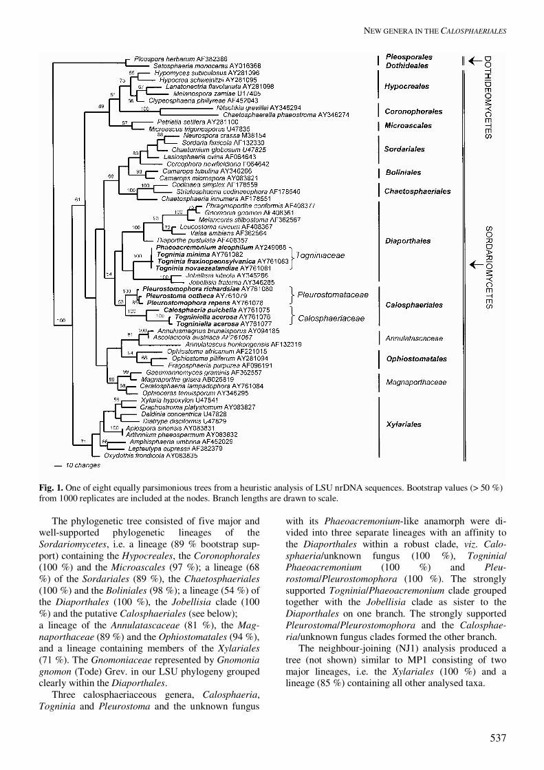
Fig. 1. One of eight equally parsimonious trees from a heuristic analysis of LSU nrDNA sequences. Bootstrap values (> 50 %) from 1000 replicates are included at the nodes. Branch lengths are drawn to scale. The phylogenetic tree consisted of five major and well-supported phylogenetic lineages of the Sordariomycetes, i.e. a lineage (89 % bootstrap sup-port) containing the Hypocreales, the Coronophorales (100 %) and the Microascales (97 %); a lineage (68 %) of the Sordariales (89 %), the Chaetosphaeriales (100 %) and the Boliniales (98 %); a lineage (54 %) of the Diaporthales (100 %), the Jobellisia clade (100 %) and the putative Calosphaeriales (see below); a lineage of the Annulatascaceae (81 %), the Mag-naporthaceae (89 %) and the Ophiostomatales (94 %), and a lineage containing members of the Xylariales (71 %). The Gnomoniaceae represented by Gnomonia gnomon (Tode) Grev. in our LSU phylogeny grouped clearly within the Diaporthales. Three calosphaeriaceous genera, Calosphaeria, Togninia and Pleurostoma and the unknown fungus
with its Phaeoacremonium-like anamorph were di-vided into three separate lineages with an affinity to the Diaporthales within a robust clade, viz. Calo-sphaeria/unknown fungus (100 %), Togninia/ Phaeoacremonium (100 %) and Pleu-rostoma/Pleurostomophora (100 %). The strongly supported Togninia/Phaeoacremonium clade grouped together with the Jobellisia clade as sister to the Diaporthales on one branch. The strongly supported Pleurostoma/Pleurostomophora and the Calosphae-ria/unknown fungus clades formed the other branch. The neighbour-joining (NJ1) analysis produced a tree (not shown) similar to MP1 consisting of two major lineages, i.e. the Xylariales (100 %) and a lineage (85 %) containing all other analysed taxa.

RÉBLOVÁ ET AL.
538
Fig. 2. One of three equally parsimonious trees from a heuristic analysis of SSU nrDNA sequences. Bootstrap values (> 50 %) from 1000 replicates are included at the nodes. Branch lengths are drawn to scale Within the second lineage a large clade with no support was generated with three separate branches discerned, i.e. the Diaporthales (100 %) was a sister to the monophyletic clade (83 %) containing the Pleu-rostoma/Pleurostomophora (100 %) and Calosphae-ria/unknown fungus subclades (100%), which are together a sister group to the Togninia/ Phaeoacremo-nium clade (100 %). The Jobellisia clade (100 %) is shown outside the Diaporthales on a basal branch to the Hypocreales/Microascales clade. The Mag-naporthaceae (99 %) grouped as a separate well-supported clade outside Diaporthales. Two constraint analyses (CA) were performed on LSU rDNA data set to assess the inclusion of Pleu-rostoma, Togninia and Jobellisia in the Calosphaeria-les and to test the monophyly of the Calosphaeriales. The Calosphaeriales is represented by a clade of Calosphaeria pulchella and the unknown fungus in our phylogenies. When Calosphaeria, the unknown fungus, and Togninia/Phaeoacremonium were treated
as monophyletic, 24 trees (not shown) were seven steps longer, and the Kishino-Hasegawa (KH) test did not reject them as significantly worse than the MPTs (P* ranged from 0.1938 to 0.3454). The CA forcing Calosphaeria, the unknown fungus, Togninia, and Pleurostoma with two Phialophora species to be monophyletic, 16 trees were one step longer than the MPTs and were considered acceptable hypotheses for the phylogeny by the KH test (P* = 0.7964−0.8619). Two other CA were run to assess the inclusion of the Calosphaeriales in a) the Magnaporthaceae, b) the Magnaporthaceae and Diaporthales without Jobel-lisia. The CA forcing the Calosphaeriales clade including Togninia, Pleurostoma and two related Phialophora species and the Magnaporthaceae to be monophyletic, generated 33 trees that were 15 steps longer than the MPTs and the KH test did not reject them as significantly worse than the MPTs (P* = 0.0588−0.1159). When the identical group of taxa from the latter CA was forced to be monophyletic
NEW GENERA IN THE CALOSPHAERIALES
539
with the Diaporthales 42 trees eight steps longer than the MPTs were obtained and were also accepted by the KH test (P* = 0.3392−0.3940).
Phylogenetic analysis of the SSU rDNA sequence data A maximum parsimony analysis (MP2) was per-formed using 316 phylogenetically informative char-acters in an alignment including 1724 nt from 57 taxa. Three MPTs were obtained (tree length 1128, CI = 0.508, RI = 0.733, HI = 0.492) (Fig. 2). The trees differed in the topology of branches within the Togninia/Phaeoacremonium clade. Two major lineages with branching order slightly different from MP1 were discerned in this analysis. A lineage (95 %) of the Hypocreales (53 %) and the Microascales (100 %) and a lineage (64 %) consisting of subgroupings of nine orders or families, i.e. the Sordariales (95 %), the Coniochaetales (98 %), and a group (63 %) of the Phyllachorales, the Chaetosphae-riales (100 %), and the Boliniales; a lineage (66 %) of the Diaporthales (99 %), and the calosphaeriaceous taxa; a lineage (74 %) of the Ophiostomatales (100 %) and the Magnaporthaceae (99 %), and the Xylariales lineage (86 %). The topology of branches of the three calosphaeriaceous genera and the unknown fungus within the Diaporthales clade was identical to that shown in MP1. These genera formed three strongly supported separate lineages, i.e. the Calosphae-ria/unknown fungus clade (100 %), the Pleu-rostoma/Pleurostomophora clade (100 %) and the Togninia/Phaeoacremonium clade (87 %). The NJ2 analysis produced a tree (not shown) with similar basic topology as MP2 but with a different branching order of i) the calosphaeriaceous genera, and ii) the Diaporthales and the Magnaporthaceae. In NJ2, two main lineages were the Hypocreales (82 %) and a poorly supported lineage (52 %) with other perithecial ascomycetes. A monophyletic clade with no branch support within the poorly supported lineage contained the three strongly supported calosphaeri-aceous lineages, i.e. Togninia (99 %) that was a sister to Calosphaeria/unknown fungus (100 %) and Pleu-rostoma/Pleurostomophora (100 %). The Dia-porthales (99 %) and the Magnaporthaceae (100 %) grouped within another large unsupported clade containing also the Ophiostomatales (100%) as a sister to the Diaporthales. A CA analysis was run on SSU nrDNA sequence data to test the monophyly of the Calosphaeriales by inclusion of Calosphaeria, the unknown fungus, Togninia/Phaeoacremonium, Pleurostoma and two related Phialophora species. Three trees were gener-ated that were one step longer than the MPTs and were accepted by the KH test (P* = 0.3175). Six trees that were 10 steps longer than the MPTs were ob-tained in CA, when the Calosphaeriales and Mag-naporthaceae were forced to be monophyletic, all of
which were rejected by the KH test (P* = 0.0330). The CA forcing the Calosphaeriales, Diaporthales and the Magnaporthaceae to be monophyletic resulted in 12 trees that were seven steps longer than the MPTs, all of which were rejected by the KH test (P* = 0.0348). Taxonomy Calosphaeria pulchella together with the unknown fungus appeared as a strongly supported monophyletic clade (100 %) in both parsimony and distance analy-ses. These two taxa share several similarities in teleo-morph morphology, i.e. dark, opaque perithecia with a globose venter and an elongate, cylindrical neck; true paraphyses; asci arranged in a palisade along the whole perithecial interior, long-stipitate asci, con-spicuously tapering below from the sporiferous por-tion, floating freely within the centrum, with thick-ened ascal apex without a visible discharge mecha-nism; hyaline, suballantoid to allantoid ascospores, arranged in a fascicle in the upper part of the ascus. However, both fungi can be distinguished in the arrangement of perithecia, asci and proliferation of ascogenous hyphae. The perithecia are aggregated in circinate groups with converging necks, but not united in a disc beneath the periderm in C. pulchella, while perithecia are separate, superficial to immersed in wood with separately protruding necks in the un-known fungus. Though in both fungi the asci are formed in acropetal succession, in C. pulchella the ascogenous hyphae produce terminal and lateral persistent cells, from each of which an ascus arises as an outgrowth, while in the unknown fungus the asco-genous hyphae elongate in the process of ascal forma-tion, producing short, persistent cells along a side, from which the asci then arise. Both fungi have phialidic conidiogenesis but differ in conidiophore structure and pigmentation. In the unknown fungus the conidiophores branch regularly, both basally and apically, and have prominent con-strictions at the septa in comparison with the mostly unbranched, predominantly subcylindrical-shaped conidiophores of C. pulchella. The unknown fungus has distinct tuberculate, brown hyphae and subhyaline phialides, that are hyaline towards the tip, with dis-tinct, shallow, flaring collarettes, contrasting with the mostly smooth and hyaline hyphae, perfectly hyaline phialides, with a finely pigmented apical region and deep, flaring collarettes of C. pulchella. These two taxa could also be distinguished on cultural characters. The unknown fungus produced brownish grey to olive-brown colonies, compared with the greyish red colonies of C. pulchella on 2 % MEA. Calosphaeria pulchella is a fast-growing fungus, reaching a colony radius of 18−20 mm after 8 d in the dark, contrasting with the slower-growing unknown fungus reaching 5−6 mm during the same period.

RÉBLOVÁ ET AL.
540
Based on the distinctions between the sequence data, perithecial arrangement, formation of asci on ascogenous hyphae, and conspicuous differences in the anamorphs obtained in vitro, the unknown fungus is described as a new genus, Togniniella, with a single new species, Togniniella acerosa. Two new mono-typic anamorph genera are also erected, namely Phaeocrella for the anamorph of Togniniella, and Calosphaeriophora for the anamorph of Calosphae-ria. The parsimony analyses show Calosphaeria, Togninia, Pleurostoma and Togniniella as three strongly supported lineages within a large clade con-taining the Diaporthales. Togninia with its Phaeoacremonium anamorph resides on the basal branch of the Diaporthales. The neighbor-joining analyses repeat the separation of the calosphaeri-aceous genera into three lineages but differ from MP analyses in the branching order for Togninia/Phaeoacremonium. The Togninia/Phae- oacremonium clade is shown either basal to the Di-aporthales and other calosphaeriaceous taxa in NJ1, or in NJ2 forms a monophyletic group with Calosphae-ria and Pleurostoma. Although the constraint analyses of LSU and SSU rDNA data sets do not preclude the monophyly of the three putative calosphaeriaceous lineages, their identi-cal grouping on separate strongly supported branches in all analyses and differences in their morphology, life history and ecology led us to introduce two new families, the Togniniaceae associated with the Di-aporthales and the Pleurostomataceae of the Calos-phaeriales. These families can then be distinguished from a refined diagnosis of the Calosphaeriaceae. The Calosphaeriaceae based on Calosphaeria, the type genus, accommodate fungi with perithecia solitary, superficial or basally immersed on wood, or in ellip-soidal to circinate groups on wood beneath the periderm, nonstromatic, globose to subglobose, dark, opaque, glabrous; necks central, elongate, separate or converging radially; perithecial wall leathery; ostiolar canal periphysate; ascogenous hyphae short-branched with several lateral and terminal cells; asci unituni-cate, octosporous, ascal apex thickened, without a discharge mechanism, long-stipitate; ascospores hyaline, allantoid, aseptate. The anamorphs of the Calosphaeriaceae are phialidic and have been linked to Calosphaeriophora and Phaeocrella (this study). Pleurostomataceae Réblová, L. Mostert, W. Gams & Crous, fam. nov. MycoBank MB500153. Perithecia superficialia vel basi submersa, non stromatica, globosa vel subglobosa, fusca, opaca, glabra, papillata. Paries peritheciorum coriaceus, bistratosus: stratum externum e cellulis pseudoparenchymatosis textura prismatica vel epidermoidea, stratum internum e cellulis
hyalinis applanatis compositum. Canalis ostiolaris periphysatus. Paraphyses haud visae. Hyphae ascogenae breviter proliferentes, hamis praeditae. Asci unitunicati, polyspori, apice inspissato, sporis haud vi expulsis, stipitati, stipite ascis liberatis ad hyphas ascogenas affixo. Ascosporae hyalinae, allantoideae, continuae. Anamorphe (Pleurostomophora) hyphomycetosa, hyalina, phialidica. Perithecia superficial or basally immersed, nonstromatic, globose to subglobose, dark, opaque, glabrous, papillate. Perithecial wall leathery, comprising two layers, the outer layer of thin-walled pseudoparenchymatous cells of textura prismatica to textura epidermoidea, the inner layer of non-pigmented flattened cells. Ostiolar canal periphysate. Paraphyses not observed. Ascogenous hyphae with short proliferations and formation of croziers. Asci unitunicate, polysporous, ascal apex thickened, without a discharge mechanism, stipitate, with stipe attached to the ascogenous hyphae after dehiscence. Ascospores hyaline, allantoid, aseptate. Anamorphs (Pleurostomophora) moniliaceous, hyphomycetous, with phialidic conidiogenesis. Typus: Pleurostoma Tul. & C. Tul., Selecta Fung. Carpol. 2: 247. 1863. Togniniaceae Réblová, L. Mostert, W. Gams & Crous, fam. nov. MycoBank MB500154. Perithecia superficialia vel immersa, non stromatica, globosa vel subglobosa, fusca, opaca, collo lungo, recto vel flexuoso praedita. Paries peritheciorum fragilis vel coriaceus, bistratosus: stratum externum e cellulis tenuitunicatis, brunneis, pseudoparenchymatosis textura prismatica vel angulari, stratum internum e cellulis hyalinis applanatis compositum. Canalis ostiolaris periphysatus. Paraphyses copiosae, septatae, latae, ad septa modice constricta, ramosae, sursum modice angustatae. Hyphae ascogenae ascis formatis elongascentes, hamis carentes; asci distincte spicati. Asci unitunicati, octospori, apice inspissato, sporis haud vi expulsis, haud stipitati, deorsum rotundati, parte basilari ascis liberatis ad hypham ascogenam affixa. Ascosporae hyalinae, continuae, allantoideae vel suballantoideae vel ellipsoideae. Anamorphe (Phaeoacremonium) hyphomycetosa dematiacea phialidica. Perithecia superficial to immersed, nonstromatic, globose to subglobose, dark, opaque, long-necked; neck straight or flexuous. Perithecial wall fragile to leathery, comprising two layers, the outer layer of thin-walled, brown, pseudoparenchymatous cells of textura prismatica to textura angularis, the inner layer of non-pigmented flattened cells. Ostiolar canal periphysate. Paraphyses abundant, broadly cellular, slightly constricted at the septa, branching, slightly tapering apically. Ascogenous hyphae elongating during ascus formation, without formation of croziers; asci in distinct spicate arrangement.
NEW GENERA IN THE CALOSPHAERIALES
541
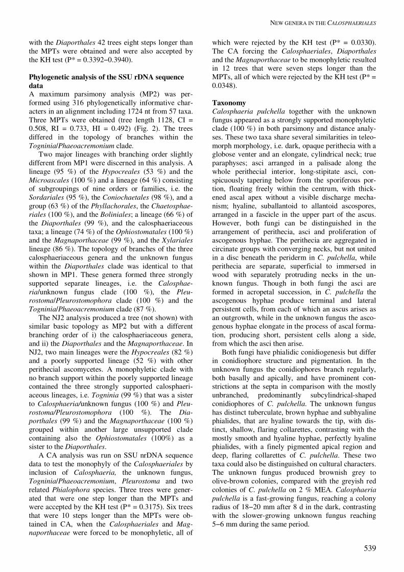
Figs 3–22. Calosphaeria pulchella. 3. Asci with ascogenous hyphae. 4–7. Asci. 8. Ascospores. 9–11, 15. Ascogenous hyphae. 12. Paraphyses. 13. Longitudinal section of perithecial wall. 14. Pallisade of asci in a hymenium attached to the inner layer of perithe-cial wall. Figs 16–22. Calosphaeriophora pulchella anamorph of Calosphaeria pulchella. 16. Germinating ascospore producing conidia. 17–21. Conidiophores, in culture. 22. Conidia, in culture. Figs 3–22 from PRM; 16–22 from CBS 115999 (holotype) ex PRM 901842 (PCA, 14 d old). DIC: 4–7, 13, 14, 18, 19. 21, 22; PC: 3, 9–12, 15–17, 20. Scale bars = 10 �m.

RÉBLOVÁ ET AL.
542
Asci unitunicate, 8-spored, ascal apex thickened without a discharge mechanism, without stipe, basally rounded, with remnants of basal parts attached to the ascogenous hyphae after dehiscence. Ascospores hyaline, aseptate, allantoid to suballantoid to ellipsoid. Anamorphs (Phaeoacremonium) dematiaceous hy-phomycetous, with phialidic conidiogenesis. Typus: Togninia Berl., Icon. Fung. 3: 9. 1900. Calosphaeria pulchella (Pers. : Fr.) J. Schröt., Pilze Schlesiens 2: 451. 1897. Figs 3–22, 23A–E. � Sphaeria pulchella Pers., Synop. Meth. Fung., p. 43. 1801 : Fries, Syst. Mycol. 2: 406. 1823. � Valsa pulchella (Pers. : Fr.) Fr., Summa Veg. Scand., p. 412. 1849. Anamorph: Calosphaeriophora pulchella Réblová, L. Mostert, W. Gams & Crous, sp. nov. Perithecia nonstromatic, densely aggregated in 2−3 levels in ellipsoidal to circinate groups of 20−40 individuals, 5−7.5 mm long and 3.5−4 mm wide, on wood beneath the periderm, dark brown to black, glabrous, venter globose to subglobose 400−500 �m diam, 400−520 �m high; necks central, elongate, up to 2000 �m long, 150−200 �m wide, straight or slightly flexuous, broadly rounded at the glabrous apex, tightly converging radially, at first decumbent to the substra-tum then upright, not united in a disc at the top and piercing separately the periderm in a narrow fissure; ostiolum periphysate. Perithecial wall leathery, two-layered, 67−80 �m thick, of pale brown to red-brown polyhedral cells of textura angularis. Ascogenous hyphae persistent, not proliferating, apparently termi-nated in growth, discrete, short-branched, each branch sequentially and simultaneously producing several lateral and terminal cells, 4−5 � 2.5−3 �m, from each of which an ascus arises as an outgrowth. Paraphyses persistent, abundant, unbranched, septate, hyaline, cylindrical, apically free, 3−4.5 �m wide near the base, tapering to 2−3 �m, longer than the asci. Asci unitunicate, clavate, (12−)18−24 � (4.5−)5−6 (mean ± se = 18.6 ± 1.4 � 5.4 ± 0.2) �m, L/W 3.5:1 in pars sporifera, stipe 27−39 �m long, truncate at the thick-ened apex, with no distinct discharge mechanism, tapering towards the base from the sporiferous por-tion, floating freely within the centrum at maturity, 8-spored. Ascospores suballantoid, 4.5−5 (mean ± se = 4.8 ± 0.1) � 1 �m, hyaline, aseptate, smooth, arranged in a fascicle in the upper part of the ascus. Notes: The type or any other authenticated material of C. pulchella could not be located in Persoon’s herbar-ium (L). Calosphaeriophora Réblová, L. Mostert, W. Gams & Crous, gen. nov. MycoBank MB500155. Etymology: Pointing to the teleomorph Calosphaeria with suffix from the morphologically similar genus Phialophora.
Anamorphe Calosphaeriae. Hyphae leves, hyalinae, Acre-monii similes, sed genus conidiophoris subcylindricis, rarissime ramosis, phialidibus hyalinis, regione apicali indistincte pigmentata et collari profundo expandente distinctum. Mycelium smooth, hyaline, similar to that of Acremo-nium, but distinct in having subcylindrical, mostly unbranched conidiophores, with hyaline phialides with a finely pigmented apical region and deep, flaring collarettes. Typus: Calosphaeriophora pulchella Réblová, L. Mostert, W. Gams & Crous, sp. nov. Calosphaeriophora pulchella Réblová, L. Mostert, W. Gams & Crous, sp. nov. MycoBank MB500156. Figs 17–22, 23C–E. Etymology: pulchellus (L), small and beautiful, refer-ring to the appearance of perithecial “nests” on natural substratum, chosen to match the epithet of the teleo-morph. Anamorphe Calosphaeriae pulchellae. Hyphae ramosae, septatae, singulae vel fasciculatae, plerumque hyalinae, nonnullae dilute brunneae, leves, 2–4(–7) µm latae. Conidiophora micronematosa, ex hyphis aeriis vel submersis oriunda, erecta, simplicia vel prope basim ramosa, plerumque hyalina, recta vel flexuosa, 1–2-septata, longitudine variabilia, (12–)14–29(–31) � 2–3(–4) µm, nonnumquam prope basim angustiora. Phialides terminales vel laterales, saepe ad hyphas fasciculatas dense aggregatae, plerumque monophialidicae, leves, hyaline, elongato-ampulliformes, saepe ad basim angustatae, in parte apicali sub collari pigmentatae, (6–)7–14 � 2–3(–4) µm; adelophialides frequentes, 2–6 � 1–2(–3) µm; collare apicale, infundibuliforme, 1.5–2 µm longum, 1.5–2 µm diam. Conidia in capitulis mucidis aggregata, hyalina, oblonge ellipsoidea vel cylindrica, ad basim angustata, 3–5(–6) � 1.5–2 µm. Mycelium consisting of branched, septate hyphae that occur singly or in bundles of up to 11; mostly hyaline, with some pale brown hyphae, smooth, 2–4(–7) µm wide. Conidiophores micronematous, arising from aerial or submerged hyphae, erect, simple or branched in the basal region, mostly hyaline, straight or flex-uous, 1–2-septate, variable in length, (12–)14–29(–31) µm long, 2–3(–4) µm wide, occasionally narrower at the base. Phialides terminal or lateral, often aggre-gated in dense clusters on strands of hyphae, mostly monophialidic, smooth, hyaline. Phialides elongate-ampulliform, often attenuated at the base or subcylin-drical, frequently pigmented in the apical region below the collarette, (6–)7–14 � 2–3(–4) µm; adelo-phialides occurring often, cylindrical or ampulliform, 2–6 � 1–2(–3) µm. Phialides developing a terminal, funnel-shaped collarette, 1.5–2 µm long, 1.5–2 µm wide. Conidia aggregated in round, slimy heads at the phialide tips, hyaline, oblong–ellipsoidal or cylindri-
NEW GENERA IN THE CALOSPHAERIALES
543
cal, with a tapered base, 3–5(–6) � 1.5–2 (mean � se = 4.1 � 0.8 � 1.7 � 0.2) µm. Cultural characteristics: Colonies on MEA flat, felty in texture, with entire margins; aerial mycelium me-dium to sparse; colony surface old rose in the centre (10C5), white (10A1) towards the margin, reverse greyish red (10D5) in the centre, becoming reddish white (10A2) towards the margin. Colony surface on OA hyaline with uneven patches of reddish white (10A2) and grey (10B1). Minimum temperature for growth 15 °C; optimum 30 °C and maximum 37 °C. Colonies reaching a radius of 18�20 mm after 8 d at 25 °C.
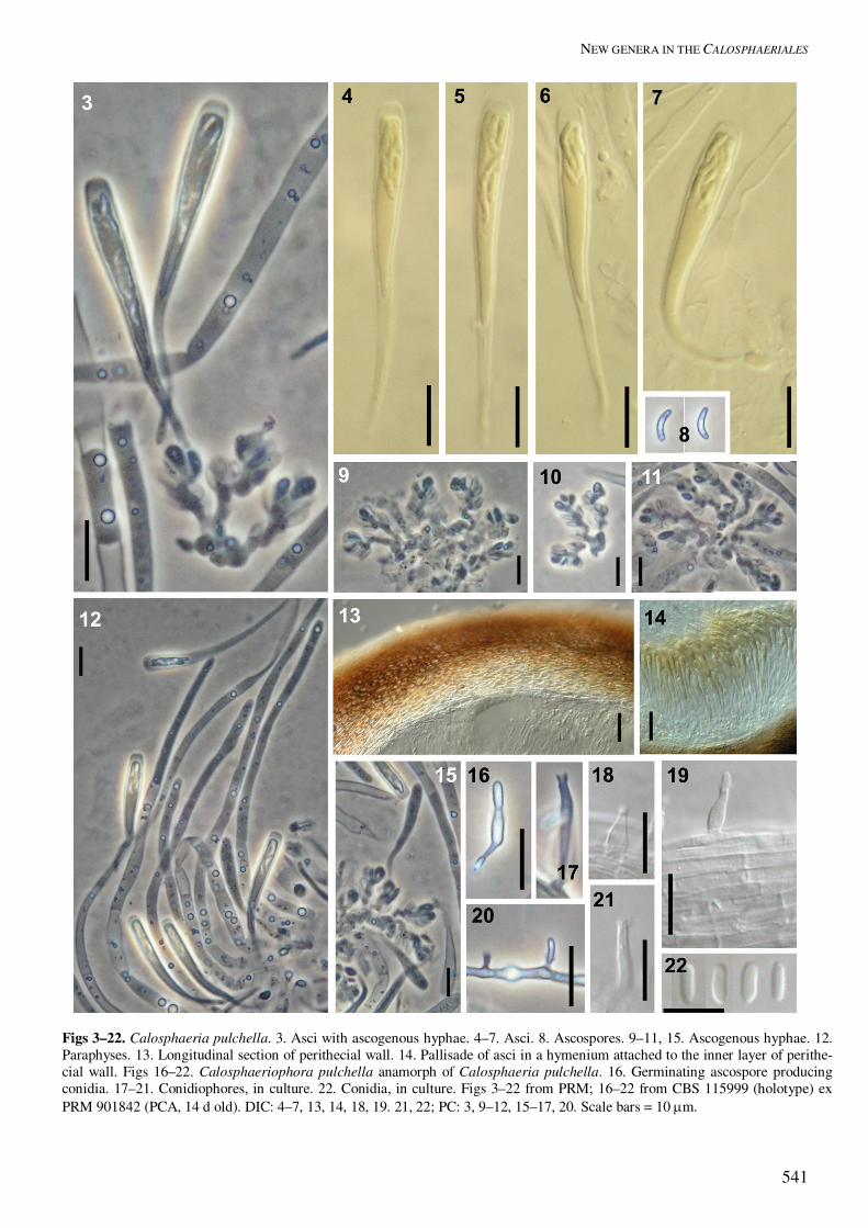
Fig. 23. Calosphaeria (A–E) and Togniniella (F–I). A–E. Calosphaeria pulchella. A. Asci with ascospores, ascoge-nous hyphae and paraphyses. B. Ascospores. C–E. Calosphaeriophora pulchella anamorph of Calosphaeria pulchella. C. Mycelial ropes of cohering hyphae of aerial mycelium with phialides, from culture. D. Single hypha with phialidic openings, from culture. E. Conidia, from culture. F–I. Togniniella acerosa. F. Asci with ascospores, ascogenous hyphae and paraphyses. G. Ascospores. H, I. Phaeocrella acerosa anamorph of Togniniella acerosa. H. Conidiophores, in culture. I. Conidia, in culture. A–B from PRM, C–E from CBS 115999 (holotype) ex PRM 901842; F–I from PDD 81431 (holotype); H, I from CBS 113648 ex PDD 81431 (holotype) (PCA, 14 d old). Scale bars = 10 �m. Habitat: Saprobic on decayed wood.
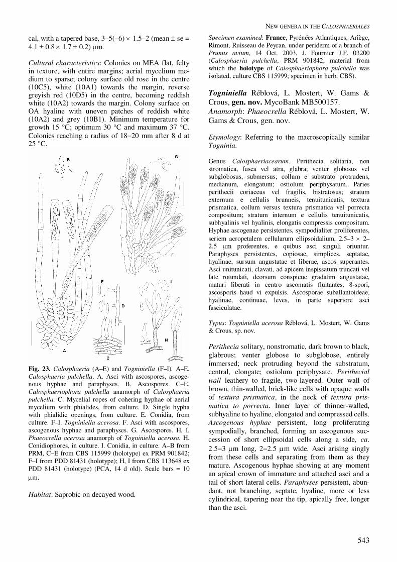
Specimen examined: France, Pyrénées Atlantiques, Ariège, Rimont, Ruisseau de Peyran, under periderm of a branch of Prunus avium, 14 Oct. 2003, J. Fournier J.F. 03200 (Calosphaeria pulchella, PRM 901842, material from which the holotype of Calosphaeriophora pulchella was isolated, culture CBS 115999; specimen in herb. CBS). Togniniella Réblová, L. Mostert, W. Gams & Crous, gen. nov. MycoBank MB500157. Anamorph: Phaeocrella Réblová, L. Mostert, W. Gams & Crous, gen. nov. Etymology: Referring to the macroscopically similar Togninia. Genus Calosphaeriacearum. Perithecia solitaria, non stromatica, fusca vel atra, glabra; venter globosus vel subglobosus, submersus; collum e substrato protrudens, medianum, elongatum; ostiolum periphysatum. Paries perithecii coriaceus vel fragilis, bistratosus; stratum externum e cellulis brunneis, tenuitunicatis, textura prismatica, collum versus textura prismatica vel porrecta compositum; stratum internum e cellulis tenuitunicatis, subhyalinis vel hyalinis, elongatis compressis compositum. Hyphae ascogenae persistentes, sympodialiter proliferentes, seriem acropetalem cellularum ellipsoidalium, 2.5–3 � 2–2.5 µm proferentes, e quibus asci singuli oriuntur. Paraphyses persistentes, copiosae, simplices, septatae, hyalinae, sursum angustatae et liberae, ascos superantes. Asci unitunicati, clavati, ad apicem inspissatum truncati vel late rotundati, deorsum conspicue gradatim angustatae, maturi liberati in centro ascomatis fluitantes, 8-spori, ascosporis haud vi expulsis. Ascosporae suballantoideae, hyalinae, continuae, leves, in parte superiore asci fasciculatae. Typus: Togniniella acerosa Réblová, L. Mostert, W. Gams & Crous, sp. nov. Perithecia solitary, nonstromatic, dark brown to black, glabrous; venter globose to subglobose, entirely immersed; neck protruding beyond the substratum, central, elongate; ostiolum periphysate. Perithecial wall leathery to fragile, two-layered. Outer wall of brown, thin-walled, brick-like cells with opaque walls of textura prismatica, in the neck of textura pris-matica to porrecta. Inner layer of thinner-walled, subhyaline to hyaline, elongated and compressed cells. Ascogenous hyphae persistent, long proliferating sympodially, branched, forming an ascogenous suc-cession of short ellipsoidal cells along a side, ca. 2.5−3 �m long, 2−2.5 �m wide. Asci arising singly from these cells and separating from them as they mature. Ascogenous hyphae showing at any moment an apical crown of immature and attached asci and a tail of short lateral cells. Paraphyses persistent, abun-dant, not branching, septate, hyaline, more or less cylindrical, tapering near the tip, apically free, longer than the asci.
RÉBLOVÁ ET AL.
544
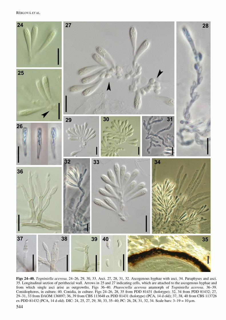
Figs 24–40. Togniniella acerosa. 24–26, 29, 30, 33. Asci. 27, 28, 31, 32. Ascogenous hyphae with asci. 34. Paraphyses and asci. 35. Longitudinal section of perithecial wall. Arrows in 25 and 27 indicating cells, which are attached to the ascogenous hyphae and from which single asci arise as outgrowths. Figs 36–40. Phaeocrella acerosa anamorph of Togniniella acerosa. 36–39. Conidiophores, in culture. 40. Conidia, in culture. Figs 24–26, 28, 35 from PDD 81431 (holotype); 32, 34 from PDD 81432; 27, 29–31, 33 from DAOM 136897; 36, 39 from CBS 113648 ex PDD 81431 (holotype) (PCA, 14 d old); 37, 38, 40 from CBS 113726 ex PDD 81432 (PCA, 14 d old). DIC: 24, 25, 27, 29, 30, 33, 35–40; PC: 26, 28, 31, 32, 34. Scale bars: 3–19 = 10 �m.
NEW GENERA IN THE CALOSPHAERIALES
545
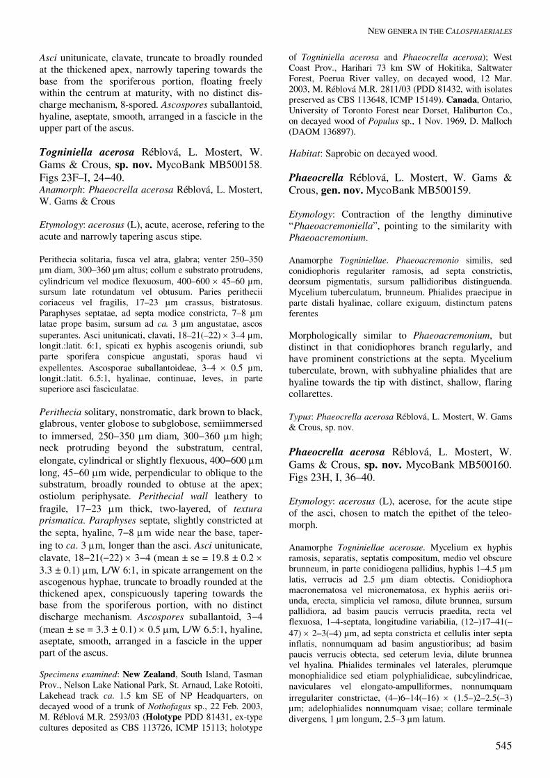
Asci unitunicate, clavate, truncate to broadly rounded at the thickened apex, narrowly tapering towards the base from the sporiferous portion, floating freely within the centrum at maturity, with no distinct dis-charge mechanism, 8-spored. Ascospores suballantoid, hyaline, aseptate, smooth, arranged in a fascicle in the upper part of the ascus. Togniniella acerosa Réblová, L. Mostert, W. Gams & Crous, sp. nov. MycoBank MB500158. Figs 23F–I, 24−40. Anamorph: Phaeocrella acerosa Réblová, L. Mostert, W. Gams & Crous Etymology: acerosus (L), acute, acerose, refering to the acute and narrowly tapering ascus stipe. Perithecia solitaria, fusca vel atra, glabra; venter 250–350 µm diam, 300–360 µm altus; collum e substrato protrudens, cylindricum vel modice flexuosum, 400–600 � 45–60 µm, sursum late rotundatum vel obtusum. Paries perithecii coriaceus vel fragilis, 17–23 µm crassus, bistratosus. Paraphyses septatae, ad septa modice constricta, 7–8 µm latae prope basim, sursum ad ca. 3 µm angustatae, ascos superantes. Asci unitunicati, clavati, 18–21(–22) � 3–4 µm, longit.:latit. 6:1, spicati ex hyphis ascogenis oriundi, sub parte sporifera conspicue angustati, sporas haud vi expellentes. Ascosporae suballantoideae, 3–4 � 0.5 µm, longit.:latit. 6.5:1, hyalinae, continuae, leves, in parte superiore asci fasciculatae. Perithecia solitary, nonstromatic, dark brown to black, glabrous, venter globose to subglobose, semiimmersed to immersed, 250−350 �m diam, 300−360 �m high; neck protruding beyond the substratum, central, elongate, cylindrical or slightly flexuous, 400−600 �m long, 45−60 �m wide, perpendicular to oblique to the substratum, broadly rounded to obtuse at the apex; ostiolum periphysate. Perithecial wall leathery to fragile, 17−23 �m thick, two-layered, of textura prismatica. Paraphyses septate, slightly constricted at the septa, hyaline, 7−8 �m wide near the base, taper-ing to ca. 3 �m, longer than the asci. Asci unitunicate, clavate, 18−21(−22) � 3−4 (mean ± se = 19.8 ± 0.2 � 3.3 ± 0.1) �m, L/W 6:1, in spicate arrangement on the ascogenous hyphae, truncate to broadly rounded at the thickened apex, conspicuously tapering towards the base from the sporiferous portion, with no distinct discharge mechanism. Ascospores suballantoid, 3−4 (mean ± se = 3.3 ± 0.1) � 0.5 �m, L/W 6.5:1, hyaline, aseptate, smooth, arranged in a fascicle in the upper part of the ascus. Specimens examined: New Zealand, South Island, Tasman Prov., Nelson Lake National Park, St. Arnaud, Lake Rotoiti, Lakehead track ca. 1.5 km SE of NP Headquarters, on decayed wood of a trunk of Nothofagus sp., 22 Feb. 2003, M. Réblová M.R. 2593/03 (Holotype PDD 81431, ex-type cultures deposited as CBS 113726, ICMP 15113; holotype
of Togniniella acerosa and Phaeocrella acerosa); West Coast Prov., Harihari 73 km SW of Hokitika, Saltwater Forest, Poerua River valley, on decayed wood, 12 Mar. 2003, M. Réblová M.R. 2811/03 (PDD 81432, with isolates preserved as CBS 113648, ICMP 15149). Canada, Ontario, University of Toronto Forest near Dorset, Haliburton Co., on decayed wood of Populus sp., 1 Nov. 1969, D. Malloch (DAOM 136897). Habitat: Saprobic on decayed wood. Phaeocrella Réblová, L. Mostert, W. Gams & Crous, gen. nov. MycoBank MB500159. Etymology: Contraction of the lengthy diminutive “Phaeoacremoniella”, pointing to the similarity with Phaeoacremonium. Anamorphe Togniniellae. Phaeoacremonio similis, sed conidiophoris regulariter ramosis, ad septa constrictis, deorsum pigmentatis, sursum pallidioribus distinguenda. Mycelium tuberculatum, brunneum. Phialides praecipue in parte distali hyalinae, collare exiguum, distinctum patens ferentes Morphologically similar to Phaeoacremonium, but distinct in that conidiophores branch regularly, and have prominent constrictions at the septa. Mycelium tuberculate, brown, with subhyaline phialides that are hyaline towards the tip with distinct, shallow, flaring collarettes. Typus: Phaeocrella acerosa Réblová, L. Mostert, W. Gams & Crous, sp. nov. Phaeocrella acerosa Réblová, L. Mostert, W. Gams & Crous, sp. nov. MycoBank MB500160. Figs 23H, I, 36–40. Etymology: acerosus (L), acerose, for the acute stipe of the asci, chosen to match the epithet of the teleo-morph. Anamorphe Togniniellae acerosae. Mycelium ex hyphis ramosis, separatis, septatis compositum, medio vel obscure brunneum, in parte conidiogena pallidius, hyphis 1–4.5 µm latis, verrucis ad 2.5 µm diam obtectis. Conidiophora macronematosa vel micronematosa, ex hyphis aeriis ori-unda, erecta, simplicia vel ramosa, dilute brunnea, sursum pallidiora, ad basim paucis verrucis praedita, recta vel flexuosa, 1–4-septata, longitudine variabilia, (12–)17–41(–47) � 2–3(–4) µm, ad septa constricta et cellulis inter septa inflatis, nonnumquam ad basim angustioribus; ad basim paucis verrucis obtecta, sed ceterum levia, dilute brunnea vel hyalina. Phialides terminales vel laterales, plerumque monophialidice sed etiam polyphialidicae, subcylindricae, naviculares vel elongato-ampulliformes, nonnumquam irregulariter constrictae, (4–)6–14(–16) � (1.5–)2–2.5(–3) µm; adelophialides nonnumquam visae; collare terminale divergens, 1 µm longum, 2.5–3 µm latum.

RÉBLOVÁ ET AL.
546
NEW GENERA IN THE CALOSPHAERIALES
547
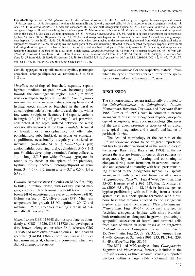
Figs 41–60. Species of the Calosphaeriaceae. 41, 42. Jattaea microtheca. 41, 42. Asci and ascogenous hyphae (arrows explained below). 43–46. Jattaea sp. 43, 46 Ascogenous hyphae with terminally and laterally attached cells. 44. Asci, ascospores and ascogenous hyphae. 45. Asci. 47–49. Romellia vibratilis. 47. Asci and ascospore. 48, 49. Asci with ascogenous hyphae. 50–54. Pleurostoma ootheca. 50. Asci and ascogenous hypha. 51. Asci detaching from the basal part. 52. Mature ascus with a stipe. 53. Mature ascus after dehiscence with an append-age at the base. 54. Old ascus without appendage. 55–57. Togninia novaezelandiae. 55, 56. Asci in a spicate arrangement on ascogenous hyphae. 57. Asci. 58, 59. Wegelina discreta. 58, 59. Asci and ascogenous hyphae. 60. Calosphaeria parasitica. Asci and branching ascoge-nous hyphae. Arrows in 42, 44, 46, 58, 59 indicating cells, which are attached to the ascogenous hyphae and from which single asci arise as outgrowths; arrows in 49, 56 indicating hyaline basal parts of the asci remaining attached to the elongated ascogenous hyphae; arrow in 50 indicating short ascogenous hyphae with a crozier system and attached basal parts of the asci; arrow in 51 indicating a thin appendage remaining attached to the base of the ascus after its dehiscence. Jattaea microtheca: 41, 42 from NY (isotype); Jattaea sp.: 43–46 from J.F. 02068; R. vibratilis: 47–49 from K. & L. Holm 3848a (NY); P. ootheca: 50–53 from K122385, 54 from K 122386 (isotype); T. novaezelan-diae: 55–57 from M.R. 2886/03; W. discreta: 58, 59 from DAOM 35410; C. parasitica: 60 from M.R. 2891/04. DIC: 42, 44, 45, 47, 54, 57, 59; PC: 41, 43, 45, 46, 48–53, 55, 56, 58, 60. Scale bars = 10 �m.
Conidia aggregata in capitulis mucidis, hyalina, plerumque obovoidea, oblongo-ellipsoidea vel reniformia, 3–4(–5) � 1–2 µm. Mycelium consisting of branched, separate, septate hyphae; medium- to pale brown, becoming paler towards the conidiogenous region, 1–4.5 µm wide; warts on hyphae up to 2.5 µm diam. Conidiophores macronematous or micronematous, arising from aerial hyphae, erect, simple or branched in the basal or apical region, pale brown, paler towards the tip, with a few warts, straight or flexuous, 1–4-septate, variable in length, (12–)17–41(–47) µm long, 2–3(4) µm wide, constricted at the septa, inflated between the septa; occasionally narrower at the base. Phialides terminal or lateral, mostly monophialidic, but often also polyphialidic, subcylindrical, navicular or elongate–ampulliform, occasionally irregulary constricted or indented, (4–)6–14(–16) � (1.5–)2–2.5(–3) µm; adelophialides occurring rarely, cylindrical, 5–6 � 1–2 µm. Phialides developing a terminal, flaring collarette, 1 µm long, 2.5–3 µm wide. Conidia aggregated in round, slimy heads at the apices of the phialides, hyaline, mostly obovoid, oblong–ellipsoid or reni-form, 3–4(–5) � 1–2 (mean � se = 3.7 � 0.5 � 1.4 � 0.3) µm. Cultural characteristics: Colonies on MEA flat, felty to fluffy in texture, dense, with radially striated mar-gins; colony surface brownish grey (4D2) with olive-brown (4F6) undertones, in reverse olive-brown (4F8). Colony surface on OA olive-brown (4F8). Minimum temperature for growth 15 °C; optimum 20 °C and maximum 25 °C. Colonies reaching a radius of 5�6 mm after 8 days at 25 °C. Notes: Isolate CBS 113648 did not sporulate as abun-dantly as CBS 113726. CBS 113726 also developed a dark brown colony colour after 22 d, whereas CBS 113648 had more olive-brown colonies. The Canadian specimen (DAOM 136897) of T. acerosa is an older herbarium material, chemically conserved, which we did not attempt to sequence.
Specimen examined: For the respective material, from which the type culture was derived, refer to the speci-mens examined in the teleomorph T. acerosa. DISCUSSION The six nonstromatic genera traditionally attributed to the Calosphaeriaceae, i.e. Calosphaeria, Jattaea, Pleurostoma, Romellia, Togninia, and Wegelina (Barr 1985, Barr et al. 1993) have in common a narrow arrangement of asci on ascogenous hyphae; morphol-ogy of ascospores; ascal apex morphology (thickness of the ascal apex, presence or absence of an apical ring, apical invagination and a canal), and habitat of perithecia in vivo. The detailed morphology of the centrum of the Calosphaeriaceae seems to be of great importance, but has been rather overlooked in the main studies of the group (Barr 1985, Barr et al. 1993). In the six genera the asci are formed in one of three patterns: a) ascogenous hyphae proliferating and continuing to elongate during ascus formation, in acropetal succes-sion and separated at maturity with basal parts remain-ing attached to the ascogenous hyphae, i.e. spicate arrangement with or without formation of croziers [Togniniaceae: Romellia: Figs 47−49; Togninia: Figs 55−57, Hausner et al. (1992: 727, Fig. 1), Mostert et al. (2003: 651, Figs 1−4, 12, 13)]; b) short ascogenous hyphae proliferating with asci arising from a crozier system; asci in a short spicate formation with a bul-bous base that remains attached to the ascogenous hyphae after ascal dehiscence (Pleurostomataceae: Pleurostoma: Figs 50–54), or c) asci arranged in fascicles; ascogenous hyphae with short branches, both terminated or elongated in growth, producing a sympodial succession of lateral and terminal cells from each of which an ascus arises as an outgrowth [Calosphaeriaceae: Calosphaeria s. str.: Figs 3, 9–11, 15; Togniniella: Figs 23, 27, 28, 32, 33; Jattaea: Figs 41−46, Romero & Samuels (1991: 233, Pl. 2J−L; 239, Pl. 4R); Wegelina: Figs 58, 59]. The MP1 and MP2 analyses show Calosphaeria, Togninia and Pleurostoma, formerly included in the Calosphaeriales, as three separate, strongly supported lineages within a large clade containing the Di-
RÉBLOVÁ ET AL.
548
aporthales. The constraint analyses that were run on the LSU rDNA sequence data set forcing the mono-phyly of the Calosphaeriales s. l. and testing the inclusion of the Calosphaeriales, Diaporthales and Magnaporthaceae into a monophyletic clade yielded trees that were all recognized by the KH test as ac-ceptable hypotheses for the phylogeny. However, these results were contradicted in two constraint analyses run on the SSU rDNA sequence data set, with the branching order of the three calosphaeri-aceous genera identical to those shown in MP1. Trees generated in these two CAs forcing members of the a) Calosphaeriales and Magnaporthaceae, or b) Calo-sphaeriales, Magnaporthaceae and Diaporthales, respectively, to be monophyletic, were all rejected as significantly worse than the MPTs. Although CAs of the LSU rDNA data set do not preclude a relatedness between the Calosphaeriales and the Magnaportha-ceae, and similarities in dark long- or short-beaked perithecia, hyaline ascospores and phialidic conidio-genesis might serve as other arguments for their relationship, we treat them as two distinct, phyloge-netic lineages that have evolved similar morphological characteristics in their holomorphs. The unique cen-trum, asci and ascospore morphology of the Calo-sphaeriales warrant their delimitation from the Mag-naporthaceae. Togninia seems to occupy a family on its own, the newly described Togniniaceae, which together with Jobellisia may be included in the Diaporthales. The phenotypically similar Gnomoniaceae represented by G. gnomon in MP1 and Gnomonia setacea (Pers. : Fr.) Ces. & De Not. and Gnomoniella fraxini Redlin & Stack in MP2 appears on the top branch of the Dia-porthales clade. Togninia of the Togniniaceae shares with the Diaporthales, particularly the Gnomoniaceae, dark, globose, long-beaked and nonstromatic perithe-cia; hyaline suballantoid to ellipsoid, smooth asco-spores; asci with rounded base, floating freely within the centrum, and a phialidic anamorph with phytopa-thogenic life style. The Togniniaceae occupy an isolated position in the Diaporthales and differ from the core taxa of the order by elongating, sympodially proliferating ascogenous hyphae with asci in a distinct spicate arrangement, presence of true paraphyses growing from the tissue at the bottom of the perithe-cial cavity and being apically freely from the begin-ning, and absence of any discharge mechanism in the ascal apex. The centrum in the Diaporthales is apara-physate or with paraphysoid tissue in the form of broad elongate cellular strands, soon deliquescent; the asci contain refractive, chitinoid, nonamyloid apical annulus and are not formed on proliferating ascoge-nous hyphae (Barr 1978). Togninia of the Togniniaceae and Pleurostoma of the Pleurostomataceae form two separate and well-supported clades in our phylogenies. Both genera are also well-distinguished, Togninia having octosporous,
basally rounded asci, ellipsoid to suballantoid to oblong ascospores, long-beaked perithecia and Phaeoacremonium anamorphs, and Pleurostoma having polysporous, stipitate asci with croziers, strictly allantoid ascospores, short-papillate perithecia and the Pleurostomophora Vijaykrishna et al. ana-morphs (Vijaykrishna et al. 2004, this volume). The arrangement of asci on the ascogenous hyphae also shows differences between the two genera. After ascus dehiscence in Pleurostoma, the ascal base contains a thin appendage (apparently a remnant of the inner ascus layer of the functionally unitunicate ascus wall) that disappears with age (Figs 52−54), while in Togninia the ascus base is smooth without any ap-pendage after dehiscence. The Calosphaeriales s. str. comprise the Calo-sphaeriaceae and the new family Pleurostomataceae. The thus characterised Calosphaeriales are sister to the Diaporthales. The Diaporthales and the Calos-phaeriales share the most recent common ancestry and form two closely related groups among the perithecial ascomycetes. The fungi attributed to the Calosphaeriales can be distinguished from the Di-aporthales by absence of stromatic tissue surrounding the perithecia; presence of true paraphyses; long-stipitate asci with thickened ascal apex without any discharge mechanism; ramifying and proliferating ascogenous hyphae; allantoid to suballantoid asco-spores and hyphomycetous phialidic anamorphs. Based on morphological characters, the new genus Togniniella is closely related to Calosphaeria pul-chella, with which it formed a monophyletic, strongly supported unit. Togniniella also resembles Togninia in many aspects, i.e. minute, nonstromatic, dark, long-beaked perithecia; spicate arrangement of asci; hyaline ascospores; true paraphyses many times longer than the asci, and phialidic conidiogenesis. However, the details in the shape and arrangement of asci, shape and organization of ascospores within the ascus, and presence of short ellipsoidal cells along the ascoge-nous hyphae, seem crucial for distinguishing the two genera. The anamorphs of the Calosphaeriales in the broad sense are reported as being either phialidic or ho-loblastic-denticulate. Phialidic anamorphs have been proven experimentally only for Togninia (Phaeo-acremonium anamorph; Hausner et al. 1992, Mostert et al. 2003) and Pachytrype, viz. P. princeps (Penz. & Sacc.) M.E. Barr et al. and P. graphidioides (Syd. & P. Syd.) M.E. Barr et al. (Cytospora anamorphs; Barr et al. 1993). The anamorph of P. rimosa F.A. Fern. et al., the third species in the genus, remains unknown (Fernández et al. 2004). The stromatic habitat of Pachytrype, perithecia with elongate, protruding beaks; short-stipitate asci with a round base floating free within the centrum, formed on a crozier system; diaporthaceous apical annulus; ellipsoid to oblong, hyaline ascospores, and the Cytospora anamorph,
NEW GENERA IN THE CALOSPHAERIALES
549
suggest affinities to the Diaporthales. Cytospora anamorphs have already been linked to Eutypella (Nitschke) Sacc., Cryptosphaeria Ces. & De Not., Leucostoma (Nitschke) Höhn., or Valsa Fr. of the Diaporthales (Grove 1935, Wehmeyer 1941, Shaw 1973, Glawe & Rogers 1986, Farr et al. 1989). The anamorph of Calosphaeria barbirostris (Dufour : Fr.) Ellis & Everh. is mentioned twice in the literature based on observations in vivo (Munk 1957, Barr 1985), but the descriptions of the putative anamorphs differ significantly from each other. The Ramichloridium-like and Sporothrix-like synanamorphs of Calosphaeria fagi Samuels & Cand. yielded in vitro are the only anamorphs within the heterogeneous Calosphaeriales representing the holoblastic, denticulate pattern of conidiogenesis. Graphostroma Piroz., in the Graphostromataceae of the Xylariales, formerly included in the Calosphaeria-les, and some other Calosphaeria species, e.g. C. fagi, C. dryina (Curr.) Nitschke (Samuels & Candoussau 1996), or C. parasitica Fuckel (Fig. 60) possess an ascogenous system typical of members of the Diatry-paceae; ascogenous hyphae branching, producing croziers and ultimately asci at successively higher levels on each branch. Unfortunately, the type culture of C. fagi is no longer viable (Gary J. Samuels, per-sonal comm.) and living cultures of C. dryina and C. parasitica were not available for this study. The perithecia of these three Calosphaeria species arise in sparse circinate groups beneath the periderm with radially converging beaks united in a disc, and asci possessing a distinct, non-amyloid apical ring with an apical invagination and canal. Samuels & Candoussau (1996) noted that C. fagi and C. dryina should be included in the Xylariales representing derivatives from the Diatrypaceae. The presence of phialidic and holoblastic (syn)anamorphs of C. pulchella and C. fagi, respectively, and conspicuous differences in arrangement of perithecia on the natural substratum, organization of the asci on ascogenous hyphae and morphology of ascal apex of C. fagi, C. dryina and C. parasitica and C. pulchella, suggest that the three former species are unrelated to Calosphaeria s. str. ACKNOWLEDGEMENTS This project was supported by the Grant Agency of Acad-emy of Sciences (KSK6005114) and the Research Project of the Institute of Botany, Academy of Sciences of the Czech Republic (AV0Z6005908). Dr Peter Johnston (Land-care Research, Auckland) is thanked for his help to obtain collecting permits for New Zealand and advice on collec-ting sites.
REFERENCES Barr ME (1978). The Diaporthales in North America with
emphasis on Gnomonia and its segregates. Mycological Memoirs 7: 1−232.
Barr ME (1983). The ascomycete connection. Mycologia 75: 1−13.
Barr ME (1985). Notes on the Calosphaeriales. Mycologia 77: 509−565.
Barr ME (1990). Prodromus to nonlichenized, pyrenomyce-tous members of class Hymenoascomycetes. Mycotaxon 39: 43−184.
Barr ME (1998). Wegelina, a reinstated genus in the Calos-phaeriales. Cryptogamie Mycologie 19: 169−173.
Barr ME, Rogers JD, Yu YM (1993). Revisionary studies in the Calosphaeriales. Mycotaxon 48: 529−535.
Berlese AN (1900). Icones Fungorum ad usum Sylloges Saccardianae adcommodatae. 3. Sphaeriaceae allanto-sporae. Padova.
Cannon PF (1994). The newly recognized family Mag-naporthaceae and its relationships. Systema Ascomy-cetum 13: 25�42.
Castlebury LA, Rossman AY, Jaklitsch W, Vasilyeva LN (2002). A preliminary overview of the Diaporthales based on large subunit nuclear ribosomal DNA se-quences. Mycologia 94: 1017�1031.
Crous PW, Gams W, Wingfield MJ, van Wyk PS (1996). Phaeoacremonium gen. nov., associated with wilt and decline diseases of woody hosts and human infections. Mycologia 88: 786–796.
Vijaykrishna D, Mostert L, Jeewon R, Gams W, Hyde KD, Crous PW (2004). Pleurostomophora, an anamorph of Pleurostoma (Calosphaeriales), a new anamorph genus morphologically similar to Phialophora. Studies in My-cology 50: 387–395.
Dupont J, Laloui W, Magnin S, Larignon P, Roquebert MF (2000). Phaeoacremonium viticola, a new species asso-ciated with Esca disease of grapevine in France. My-cologia 92: 499–504.
Eriksson OE, Baral HO, Currah RS, Hanser K, Kurtzman CP, Rambold G, Laessøe T. (eds) (2003). Outline of the Ascomycota � 2003. Myconet 9: 1�89.
Farr DF, Bills GF, Chamuris GP, Rossman AY. (1989). Fungi on plant and plant products in the United States. APS Press, St. Paul, Minnesota.
Fernández FA, Rogers JD, Ju Y-M, Huhndorf SM, Umana L (2004). Paramphisphaeria costaricensis gen. et sp. nov. and Pachytrype rimosa sp. nov. from Costa Rica. Mycologia 96: 175–179.
Gams W, Hoekstra ES, Aptroot A (eds) (1998) CBS course of mycology, 4th ed. Centraalbureau voor Schim-melcultures, Baarn, Delft.
Gargas A, DePriest PT, Taylor JW (1995). Positions of multiple insertions in SSU rDNA of lichen-forming fungi. Molecular Biology and Evolution 12: 208�218.
Gargas A, Taylor JW (1992). Polymerase chain reaction (PCR) primers for amplifying and sequencing nuclear 18S rDNA from lichenized fungi. Mycologia 84: 589–592.
Glawe DA, Rogers JD (1986). Conidial states of some species of Diatrypaceae and Xylariaceae. Canadian Journal of Botany 64: 1493–1498.
Groenewald M, Kang JC, Crous PW, Gams W (2001). ITS and beta-tubulin phylogeny of Phaeoacremonium,
RÉBLOVÁ ET AL.
550
Phaeomoniella spp. Mycological Research 105: 651�657.
Grove WB (1935). British stem- and leaf- fungi (Coelomy-cetes). I. Cambridge University Press.
Gutell RR (1993). Collection of small subunit (16S- and 16S-like) ribosomal RNA structures. Nucleic Acids Re-search 21: 3051–3054.
Gutell RR, Gray MW, Schnare MN (1993). A compilation of large subunit (23S and 23S-like ribosomal RNA structures: Nucleic Acids Research 21: 3055�3074.
Hall TA (1999). BioEdit 5.0.9: a user-friendly biological sequence alignment editor and analysis program for Windows 95/98/NT. Nucleic Acids Symposium Series 41: 95–98.
Hausner G, Eyjólfsdóttir GG, Reid J, Klassen GR (1992). Two additional species of the genus Togninia. Canadian Journal of Botany 70: 724�734.
Kirk PM, Cannon PF, David JC, Stalpers JA eds (2001). Ainsworth and Bisby's dictionary of the fungi, 9th ed. CABI Publishing: Wallingford, U.K.
Kornerup, A, Wanscher JH (1978). Methuen handbook of colour, 3rd ed. Eyre Methuen, London.
Lee S, Groenewald JZ, Crous PW (2004). Phylogenetic reassessment of the coelomycete genus Harknessia and its teleomorph Wuestneia (Diaporthales), and the intro-duction of Apoharknessia gen. nov. Studies in Mycology 50: 235–252.
Monod M (1983). Monographie taxonomique des Gnomo-niaceae (Ascomycètes de l´ordre des Diaporthales I.). Beihefte Sydowia 9: 1�120.
Mostert L, Crous PW, Groenewald JZ, Gams W, Summer-bell RC (2003). Togninia (Calosphaeriales) is con-firmed as teleomorph of Phaeoacremonium by means of morphology, sexual compatibility and DNA phylogeny. Mycologia 95: 646�659.
Mostert L, Groenewald JZ, Summerbell RC, Robert V, Sutton DA, Padhye AA, Crous PW (2004). Species of Phaeoacremonium associated with human infections and environmental reservoirs in infected woody plants. Journal of Clinical Microbiology (in press).
Munk A (1957). Danish Pyrenomycetes. Dansk Botanisk Arkiv 17(1): 1�491.
Pirozynski KA (1974). Xenotypa Petrak and Graphostroma gen. nov., segregates from Diatrypaceae. Canadian Journal of Botany 52: 2129–2135.
Petrak F, Sydow H (1936). Originaluntersuchungen über die Pyrenomyzeten, Sphaeropsideen und Melanconieen. Annales Mycologici 36: 11–52.
Rehner SA, Samuels GJ (1994). Taxonomy and phylogeny of Gliocladium analysed from nuclear large subunit ri-bosomal DNA sequences. Mycological Research 98: 625–634
Romero A, Samuels GJ (1991). Studies on xylophilous fungi from Argentina. VI. Ascomycotina on Eucalyptus viminalis (Myrtaceae). Sydowia 43: 228–248.
Samuels GJ, Candoussau F (1996). Heterogeneity in the Calosphaeriales: a new Calosphaeria with Ramichlo-ridium- and Sporothrix-like synanamorphs. Nova Hed-wigia 62: 47�60.
Shaw CG (1973). Host index for the Pacific Northwest II. Fungi. Washington Agricultural Experiment Station Bulletin 766 : 1–162.
Swofford DL (2002). PAUP*. Phylogenetic Analysis Using Parsimony (*and other methods). Version 4. Sunder-land, MA: Sinauer Associates.
Vilgalys R, Hester M (1990). Rapid genetic identification and mapping of enzymatically amplified ribosomal DNA from several Cryptococcus species. Journal of Bacteriology 172: 4238–4246.
Wehmeyer LE (1941). A revision of Melanconis, Pseu-dovalsa, Prosthecium and Titania. University of Michi-gan Studies 14: 1–161.
White TJ, Bruns T, Lee S, Taylor J (1990). Amplification and direct sequencing of fungal ribosomal RNA genes for phylogenetics. In: PCR protocols: a guide to meth-ods and applications (Innis MA, Gelfand DA, Sninsky JJ, White TJ, eds). Academic Press, San Diego, CA, U.S.A.: 315–322.
Zhang N, Blackwell M (2001). Molecular phylogeny of dogwood anthracnose fungus (Discula destructiva) and the Diaporthales. Mycologia 93: 355�365.