Embed Size (px)
Citation preview

New Insights into the Function and Global Distribution ofPolyethylene Terephthalate (PET)-Degrading Bacteria andEnzymes in Marine and Terrestrial Metagenomes
Dominik Danso,a Christel Schmeisser,a Jennifer Chow,a Wolfgang Zimmermann,b Ren Wei,b Christian Leggewie,c
Xiangzhen Li,d Terry Hazen,e Wolfgang R. Streita
aDepartment of Microbiology and Biotechnology, Biocenter Klein Flottbek, University of Hamburg, Hamburg,Germany
bInstitute of Biochemistry, Department of Microbiology and Bioprocess Technology, Leipzig University,Leipzig, Germany
cevoxx technologies GmbH, Monheim am Rhein, GermanydChengdu Institute of Biology, Chengdu, ChinaeThe University of Tennessee, Knoxville, Tennessee, USA
ABSTRACT Polyethylene terephthalate (PET) is one of the most important syntheticpolymers used today. Unfortunately, the polymers accumulate in nature and to dateno highly active enzymes are known that can degrade it at high velocity. Enzymesinvolved in PET degradation are mainly �- and �-hydrolases, like cutinases and re-lated enzymes (EC 3.1.1). Currently, only a small number of such enzymes are wellcharacterized. In this work, a search algorithm was developed that identified 504possible PET hydrolase candidate genes from various databases. A further globalsearch that comprised more than 16 Gb of sequence information within 108 marineand 25 terrestrial metagenomes obtained from the Integrated Microbial Genome(IMG) database detected 349 putative PET hydrolases. Heterologous expression offour such candidate enzymes verified the function of these enzymes and confirmedthe usefulness of the developed search algorithm. In this way, two novel and thermo-stable enzymes with high potential for downstream application were partially character-ized. Clustering of 504 novel enzyme candidates based on amino acid similarities indi-cated that PET hydrolases mainly occur in the phyla of Actinobacteria, Proteobacteria,and Bacteroidetes. Within the Proteobacteria, the Betaproteobacteria, Deltaproteobacte-ria, and Gammaproteobacteria were the main hosts. Remarkably enough, in the ma-rine environment, bacteria affiliated with the phylum Bacteroidetes appear to be themain hosts of PET hydrolase genes, rather than Actinobacteria or Proteobacteria, asobserved for the terrestrial metagenomes. Our data further imply that PET hydro-lases are truly rare enzymes. The highest occurrence of 1.5 hits/Mb was observed insequences from a sample site containing crude oil.
IMPORTANCE Polyethylene terephthalate (PET) accumulates in our environment with-out significant microbial conversion. Although a few PET hydrolases are already known,it is still unknown how frequently they appear and with which main bacterial phyla theyare affiliated. In this study, deep sequence mining of protein databases and meta-genomes demonstrated that PET hydrolases indeed occur at very low frequencies in theenvironment. Furthermore, it was possible to link them to phyla that were previouslynot known to harbor such enzymes. This work contributes novel knowledge on the phy-logenetic relationships, the recent evolution, and the global distribution of PET hydro-lases. Finally, we describe the biochemical traits of four novel PET hydrolases.
KEYWORDS HMM, hydrolases, metagenome, metagenomic screening, PETdegradation, polyethylene terephthalate (PET), BHET, TPA, metagenomes
Received 14 December 2017 Accepted 29January 2018
Accepted manuscript posted online 2February 2018
Citation Danso D, Schmeisser C, Chow J,Zimmermann W, Wei R, Leggewie C, Li X,Hazen T, Streit WR. 2018. New insights into thefunction and global distribution ofpolyethylene terephthalate �PET�-degradingbacteria and enzymes in marine and terrestrialmetagenomes. Appl Environ Microbiol84:e02773-17. https://doi.org/10.1128/AEM.02773-17.
Editor Rebecca E. Parales, University ofCalifornia, Davis
Copyright © 2018 Danso et al. This is an open-access article distributed under the terms ofthe Creative Commons Attribution 4.0International license.
Address correspondence to Wolfgang R. Streit,[email protected].
BIODEGRADATION
crossm
April 2018 Volume 84 Issue 8 e02773-17 aem.asm.org 1Applied and Environmental Microbiology
on May 28, 2020 by guest
http://aem.asm
.org/D
ownloaded from

Since its discovery, its first synthesis, and its patenting in 1941, polyethylene tereph-thalate (PET) has become a widely used material in several industrial branches (1).
The worldwide PET resin production amounted to 27.8 million tons in 2015 (https://www.plasticsinsight.com/global-pet-resin-production-capacity).
Due to its massive use, PET is highly enriched in nature. Microplastics and biggerfragments of plastic are found worldwide in oceans and terrestrial environments. Themost prominent example is the so-called Pacific garbage patch. PET debris is ofteneaten by fish and other marine creatures (2, 3). In this way, PET degradation productsand additives (i.e., solubilizers) are introduced into the food chain, where they havenegative impact on human and animal health (4). Until now, only a few species ofbacteria and fungi have been described as capable of partially degrading PET tooligomers or even monomers (5). Within this framework, however, it is noteworthy thatall known PET hydrolases have relatively low turnover rates, which makes their use forefficient bioremediation almost impossible (Table 1).
Intriguingly, the trait for PET degradation appears to be limited to a few bacterialphyla, and most bacterial isolates with potential for PET degradation are members ofthe Gram-positive phylum Actinobacteria (12). The best-characterized examples origi-nate from the genera Thermobifida and Thermomonospora (8, 10–12, 15, 16) (Table 1).The enzymes involved in the degradation (e.g., PET hydrolase and tannase) are typicalserine hydrolases, e.g., cutinases (EC 3.1.1.74), lipases (EC 3.1.1.3), and carboxylesterases(EC 3.1.1.1). These enzymes possess a typical �/�-hydrolase fold, and the catalytic triadis composed of a serine, a histidine, and an aspartate residue (17, 18).
More recently, polyethylene (PE)-degrading bacteria were reported in insect guts. Inthis recent study, Enterobacteria and Bacillus strains had been isolated and werecapable of degrading polyesters (19, 20).
Furthermore, a complete degradation of amorphous PET materials was described forthe Gram-negative bacterium Ideonella sakaiensis strain 201-F6, which is able to use PETas a major energy and carbon source (13). In addition to the hydrolase, the I. sakaiensisgenome encodes a second enzyme that appears to be unique, and which is designatedas a tannase capable of degrading mono(2-hydroxyethyl) terephthalic acid. In this way,the secreted PET hydrolase produces the intermediate mono(2-hydroxyethyl) tere-phthalic acid (MHET). MHET is presumably internalized by the cell and hydrolyzed bythe MHETase. The resulting monomers are then degraded in a downstream process andused for the bacterial metabolism. I. sakaiensis is affiliated with the phylum Betapro-teobacteria and belongs to the order Burkholderiales.
In this work, our intention was to mine metagenomes for the detection of novelgenes involved in PET degradation and to establish an overview on their taxonomicdistribution within the different bacterial phyla. Therefore, we have developed a hiddenMarkov model (HMM) to search existing genome and metagenome databases for thepresence of potential PET hydrolases (Fig. 1; see also Fig. 3B). Using this approach, weidentified �500 potential PET hydrolases in the UniProtKB database. In addition, 349sequence homologs were obtained from several public metagenome data sets depos-ited on the IMG server, and four of the identified candidate genes were functionallyverified. Together, these results imply that PET hydrolase genes are globally distributedin marine and terrestrial metagenomes. Furthermore, we provide evidence that inmarine environments, the PET hydrolases originate mainly from the phylum Bacte-roidetes and in the terrestrial metagenomes from Actinobacteria.
RESULTSConstruction of a hidden Markov model for PET hydrolases. Only a few well-
characterized PET hydrolases are currently known. The most prominent examples arePET hydrolases from T. fusca and I. sakaiensis (see references in Table 1 and Fig. 2). Inthis study, we set out to increase knowledge of the diversity of this intriguing group ofhydrolases. To identify potential novel PET hydrolases, an amino acid sequence align-ment of nine already-known examples was constructed using the T-Coffee multiplesequence alignment server. The enzyme sequences used for the model all have verified
Danso et al. Applied and Environmental Microbiology
April 2018 Volume 84 Issue 8 e02773-17 aem.asm.org 2
on May 28, 2020 by guest
http://aem.asm
.org/D
ownloaded from
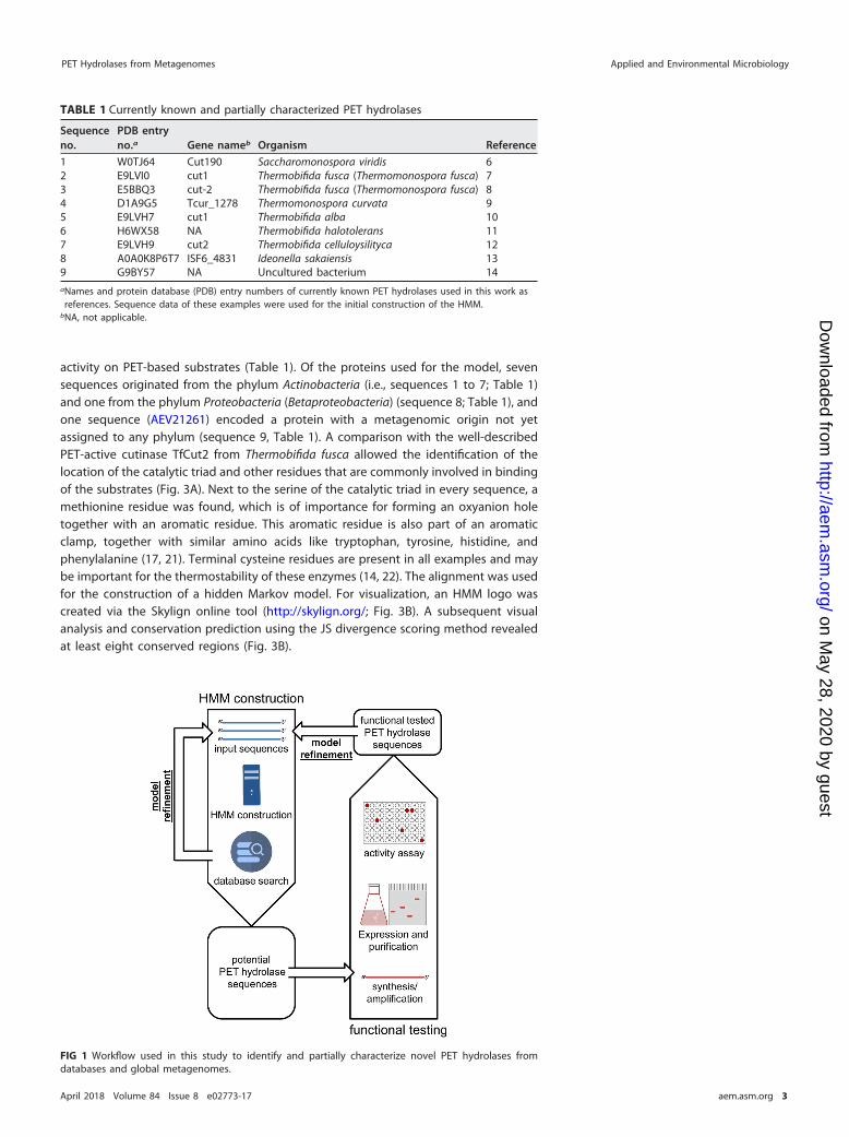
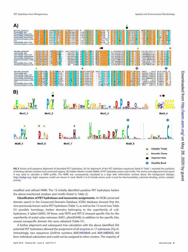
activity on PET-based substrates (Table 1). Of the proteins used for the model, sevensequences originated from the phylum Actinobacteria (i.e., sequences 1 to 7; Table 1)and one from the phylum Proteobacteria (Betaproteobacteria) (sequence 8; Table 1), andone sequence (AEV21261) encoded a protein with a metagenomic origin not yetassigned to any phylum (sequence 9, Table 1). A comparison with the well-describedPET-active cutinase TfCut2 from Thermobifida fusca allowed the identification of thelocation of the catalytic triad and other residues that are commonly involved in bindingof the substrates (Fig. 3A). Next to the serine of the catalytic triad in every sequence, amethionine residue was found, which is of importance for forming an oxyanion holetogether with an aromatic residue. This aromatic residue is also part of an aromaticclamp, together with similar amino acids like tryptophan, tyrosine, histidine, andphenylalanine (17, 21). Terminal cysteine residues are present in all examples and maybe important for the thermostability of these enzymes (14, 22). The alignment was usedfor the construction of a hidden Markov model. For visualization, an HMM logo wascreated via the Skylign online tool (http://skylign.org/; Fig. 3B). A subsequent visualanalysis and conservation prediction using the JS divergence scoring method revealedat least eight conserved regions (Fig. 3B).
TABLE 1 Currently known and partially characterized PET hydrolases
Sequenceno.
PDB entryno.a Gene nameb Organism Reference
1 W0TJ64 Cut190 Saccharomonospora viridis 62 E9LVI0 cut1 Thermobifida fusca (Thermomonospora fusca) 73 E5BBQ3 cut-2 Thermobifida fusca (Thermomonospora fusca) 84 D1A9G5 Tcur_1278 Thermomonospora curvata 95 E9LVH7 cut1 Thermobifida alba 106 H6WX58 NA Thermobifida halotolerans 117 E9LVH9 cut2 Thermobifida celluloysilityca 128 A0A0K8P6T7 ISF6_4831 Ideonella sakaiensis 139 G9BY57 NA Uncultured bacterium 14aNames and protein database (PDB) entry numbers of currently known PET hydrolases used in this work asreferences. Sequence data of these examples were used for the initial construction of the HMM.
bNA, not applicable.
FIG 1 Workflow used in this study to identify and partially characterize novel PET hydrolases fromdatabases and global metagenomes.
PET Hydrolases from Metagenomes Applied and Environmental Microbiology
April 2018 Volume 84 Issue 8 e02773-17 aem.asm.org 3
on May 28, 2020 by guest
http://aem.asm
.org/D
ownloaded from

An initial HMMER online tool database search with the model against the UniProtKBdatabase revealed a total of 10,854 significant query matches, with a highest bit scorevalue of 441.6. Of these, a subset was chosen that showed a bit score value of �180.In addition, a BLAST search was performed using the newly discovered potential PEThydrolases from the HMM search as initial query sequences against the nonredundantand the metagenomic datasets available at the NCBI database in May 2017. Thisresulted in the detection of 504 potential PET hydrolase candidate genes. From theobtained homologous sequences, 13 potential PET hydrolase homologs (Fig. 2) weremanually chosen due to their sequence similarity to known PET hydrolases (PET1 toPET13). These were used for initial verification and further in silico and/or biochemicalcharacterization. These novel predicted PET hydrolases are summarized in Table S1 inthe supplemental material, together with their UniProt entries and pfam domainsimilarities. It was of interest to select mainly nonactinobacterial proteins, in order todiversify the HMM. These 13 sequences were added to the alignment and used for a
FIG 2 Neighbor-joining tree of manually chosen potential PET hydrolase sequences found in this work. Sequences were obtained froma HMM search in the UniProtKB database and named PET1 to PET13. The tree was calculated using MEGA6. Besides the 13 newly foundPET hydrolase sequences (Table S1), 9 already-known PET hydrolases (Table 1) were added to the tree in order to visualize thephylogenetic distribution and similarity of the PET hydrolase sequence homologs.
Danso et al. Applied and Environmental Microbiology
April 2018 Volume 84 Issue 8 e02773-17 aem.asm.org 4
on May 28, 2020 by guest
http://aem.asm
.org/D
ownloaded from
modified and refined HMM. The 13 initially identified putative PET hydrolases harborthe above-mentioned residues and motifs (listed in Table 2).
Classification of PET hydrolases and taxonomic assignments. An NCBI conserveddomain search in the Conserved Domains Database (CDD) database showed that thenine previously known active PET hydrolases (Table 1), as well as the 13 novel (see TableS1) possible homologs, harbor domains belonging to the superfamily of �/�-hydrolases_5 (pfam12695). Of these, only PET9 and PET12 showed specific hits for thesuperfamily of acetyl xylan esterases (AXE1, pfam05448). In addition to the specific hits,several nonspecific domain hits were obtained (Table S1).
A further alignment and subsequent tree calculation with the above identified 504potential PET hydrolases allowed the assignment of all enzymes in 17 subclasses (Fig 4).Interestingly, two sequences (UniProt numbers A0A1N6SMU6 and A0A168EN35) didform individual subclusters and could not be assigned to other clusters. The majority of
FIG 3 Amino acid sequence alignment of described PET hydrolases. (A) An alignment of the PET hydrolase sequences listed in Table 1 revealed the positionsof binding relevant residues and conserved regions. (B) Hidden Markov model (HMM) of PET hydrolase amino acid motifs. The amino acid alignment from panelA was used to calculate a HMM profile. The HMM was consequently visualized as a logo with information content above the background (Skylign;http://skylign.org). Eight sequence motifs are shown in total. Motifs 2 to 8 include amino acids crucial for thermostability, substrate binding, and/or catalyticactivity.
PET Hydrolases from Metagenomes Applied and Environmental Microbiology
April 2018 Volume 84 Issue 8 e02773-17 aem.asm.org 5
on May 28, 2020 by guest
http://aem.asm
.org/D
ownloaded from

the subclasses were mainly affiliated with Actinobacteria, and only one subclass (XII)was associated with Proteobacteria. For the subclass XVII, no clear assignment waspossible. The Thermobifida PET hydrolase sequences are clustered within subcluster XV,together with the PET hydrolase from Saccharomonospora. The leaf compost cutinase(LCC) sequence was found in subcluster XI and the Thermomonospora curvata se-quence is located in group XII. The PET hydrolase from Ideonella sakaiensis islocated in subcluster VI.
TABLE 2 Determined search criteria for the identification of PET hydrolase candidategenes in databases
Sequenceno.
Search criterion(criteria)a Function
1 GxSMGGGG Serine of catalytic triade and methionine for oxyanion holeformation
2 F,Y62 Amino acids for oxyanion hole formation and aromatic clamp3 W,Y1574 I,V1805 F,W211 Optional aromatic amino acid for aromatic clamp formation6 C255 C262 C-terminal cysteine residues for thermostability supporting disulfide
bond formation7 DxDxR(Y)xxF(L)C Conserved sequence prior to first thermostability giving cysteineaThe letter x indicates a nonconserved position within the sequence pattern. Brackets indicate a lessconserved position within the sequence pattern. Numbering of amino acids is according to the HMM (seeFig. 3A).
FIG 4 Classification and phylogenetic tree of 504 novel and potential PET hydrolases obtained by HMM searches.Sequences were obtained from the UniProtKB database. A total of 504 sequences identified with the constructed HMM andhaving a bit score of �180 were visualized, of which the sequences of PET1 to PET13 (Table S1), as well as 9 alreadydescribed PET hydrolases (Table 1), represent a subset of the newly found potential enzymes.
Danso et al. Applied and Environmental Microbiology
April 2018 Volume 84 Issue 8 e02773-17 aem.asm.org 6
on May 28, 2020 by guest
http://aem.asm
.org/D
ownloaded from

Experimental verification of the HMM and characterization of selected novelPET hydrolases. Since the bioinformatic approach only delivered potential PET hydro-lases enzymes, we initiated work to verify a small number of the identified candidategenes with respect to their function. Therefore, we chose the enzymes PET 2, 5, 6, and12 (Table S1). The respective genes were either synthesized or amplified from genomicDNA using vectors and primers (as outlined in Tables 3 and 4) and were cloned into theexpression vectors. Initial tests indicated that all genes coded for active enzymes. Onagar plates containing PET nanoparticles or polycaprolactone (PCL) (9, 24), all activeclones produced halos after overnight incubation. They were compared to the PEThydrolase from Thermobifida fusca as a positive control (see Fig. S1 in the supplementalmaterial). PCL was used as a model substrate, as hydrolysis of this compound indicatespossible activities on the more complex PET. From these active enzymes, we chose twoenzymes for more detailed biochemical characterization. These were the two enzymesPET 2 and PET 6. PET 2 was derived from a marine metagenomics data set (25), and PET6 was derived from Vibrio gazogenes strain DSM-21264 (26). After successful expressionand purification of the two enzymes in sufficient amounts, the obtained enzymes werefurther characterized using para-nitrophenyl esters (pNP esters) (Fig. 5; see also Fig. S2and S3 in the supplemental material). Both enzymes showed best activity against pNPesters (C2 to C4), but were able to convert long-chain (�C10) substrates as well. Theirtemperature optimums were 55°C and 70°C for PET6 and PET2, respectively. Remark-ably, PET2 retained 80% of its relative activity at 90°C after incubation for �5 h. Bothenzymes preferred alkaline pH values of 8 to 9. Rubidium at a concentration of 1mmol/liter had a strong effect in the case of PET2 by increasing the activity by 50%. Asimilar but significantly smaller effect was observed in the case of PET6. Both enzymes
TABLE 3 Bacterial strains and plasmids used in this work
Strain or plasmid Property(ies)a Reference or source
StrainsE. coli DH5� supE44 ΔlacU169 (�80 lacZ ΔM15) hsdR17 recA1 endA1 gyrA96 thi-1 relA1 23E. coli BL21(DE3) F� ompT hsdS B (rB
� mB�) gal dcm �DE3 Novagen/Merck (Darmstadt, Germany)
E. coli T7SHuffle Express fhuA2 lacZ::T7 gene1 [lon] ompT ahpC gal �att::pNEB3-r1-cDsbC (SpecR,lacIq) �trxB sulA11 R(mcr-73::miniTn10-TetS)2 [dcm] R(zgb-210::Tn10-TetS) endA1 �gor �(mcrC-mrr)114::IS10
NEB (Frankfurt am Main, Germany)
Deinococcus maricopensisDSM-21211
Type strain DSMZ (Braunschweig, Germany)
Vibrio gazogenes DSM-21264 Type strain DSMZ (Braunschweig, Germany)Polyangium brachysporum
DSM-7029Type strain DSMZ (Braunschweig, Germany)
PlasmidspET21a(�) Expression vector, lacI, Ampr, T7 lac promoter, C-terminal His6-tag coding
sequenceNovagen/Merck (Darmstadt, Germany
pET28a(�) Expression vector, lacI, Ampr, T7 lac promoter, C-terminal His6-tag andN-terminal coding sequence
Novagen/Merck (Darmstadt, Germany
pEX-A2 Cloning vector, Ampr, PlaclacZ, pUC ori Eurofins MWG Operon (Ebersberg,Germany)
aAmpr, ampicillin resistance.
TABLE 4 Primers used in this work
Primer Sequence (5= ¡ 3=) Length (bp) Tm (°C)a Source
T7 promoter TAATACGACTCACTATAGGG 20 53.2 Eurofins MWG (Ebersberg, Germany)T7 terminator CTAGTTATTGCTCAGCGGT 19 54.5 Eurofins MWG (Ebersberg, Germany)PET5_for CGCCGCCATATGAATAAATCTATTCTAAAAAAACTCTC 38 68 This workPET5_rev CGATTCGGCGGCCGCGTAATTACATGTGTCACGG 34 77 This workPET6_for CGTAGTCATATGGTACCGTGTTCGGACTG 29 69 This workPET6_rev CAGCGGCCGCCTAATAGTAACTACAGTTGTCTCG 34 73 This workPET12_for CGCCATATGCAGACCAACCCCTACCAGCGAGGCCC 35 80 This workPET12_rev CTTGCGGCCGCTCAGTACGGGCAGCTCTCGCGGTACTCC 39 84 This workaTm, melting temperature.
PET Hydrolases from Metagenomes Applied and Environmental Microbiology
April 2018 Volume 84 Issue 8 e02773-17 aem.asm.org 7
on May 28, 2020 by guest
http://aem.asm
.org/D
ownloaded from

showed reduced hydrolytic activity in the presence of 5% SDS, 10 mM phenylmethyl-sulfonyl fluoride (PMSF), and 30% acetonitrile. Additional high-performance liquidchromatography (HPLC) analyses confirmed the above findings for PET2. In tests using14 mg of amorphous PET foil as the substrate, 100 �g of PET 2 was able to release 900�M terephthalic acid after 24 h of incubation (see Fig. S4 in the supplemental material).
Altogether, the data presented above indicate that the developed search algorithmis useful for the identification of novel and functionally active PET hydrolases fromsingle genomes and metagenomes.
Global distribution of PET hydrolases and their significance in marine andterrestrial environments. After the successful construction of a reliable HMM and theidentification, as well as partial characterization, of new PET hydrolases, we asked ifthese enzymes could be identified on a global level, and if so, to what extent. Toevaluate the environmental distribution of sequences encoding PET hydrolases, thedata from 108 marine and 25 terrestrial metagenomes were taken into account anddownloaded from the IMG database (27) (see Table S2 in the supplemental material).Criteria for the selection of marine data included sample depth (maximum 2 m),assembly status, global distribution of sample locations, and size and availability of thedata set. The same criteria, except for the sample depth, were chosen for terrestrialmetagenomes. The size of the assembled metagenome data in the case of marinemetagenomes ranged from 10.85 Mb up to 7.99 Gb. In the case of terrestrial meta-genomes, the number of assembled bases ranged from 58 Mb to 9.2 Gb. The modifiedHMM was used to find PET hydrolase homologs in the sequence data of thosemetagenomes on a global scale. The searches identified possible PET hydrolase ho-mologs in 31 marine and 11 terrestrial metagenomes. A total of 349 hits was observedfor these 42 samples. The number of hits per sample was normalized, calculated as hitsper Mb, and visualized on a global map representing the geographical location as well
FIG 5 Biochemical characterization of PET2 and PET6 with different pNP substrates. Data obtained with a pNP assay are shown in net diagrams for PET2 andPET6. Substrate preferences, temperature optimum, and pH optimum were tested. All tests besides substrate preferences were carried out with pNP octanoate.
Danso et al. Applied and Environmental Microbiology
April 2018 Volume 84 Issue 8 e02773-17 aem.asm.org 8
on May 28, 2020 by guest
http://aem.asm
.org/D
ownloaded from

as the frequency of PET hydrolase homologs (Fig. 6). Within the marine and terrestrialmetagenomes, PET hydrolase frequencies ranged from 0.004 to 0.92 hits/Mb and0.0001 to 1.513 hits/Mb, respectively.
The combined genome sizes of terrestrial metagenomes are nearly 2.5-fold higherthan those of the marine metagenomes, and they harbor 157 PET hydrolase homologsin average. In contrast, the marine metagenomes harbor an average of 42 PET hydro-lases. The terrestrial metagenome with the highest abundance of potential PET hydro-lases contains 135 sequence hits and was derived from the sediment core of a heavyoil reservoir in Canada (IMG genome number 3300001197). In the case of the marinemetagenomes, the maximum was 31 hits, found within the metagenome data of asample from the Delaware coast in the United States (Fig. 6).
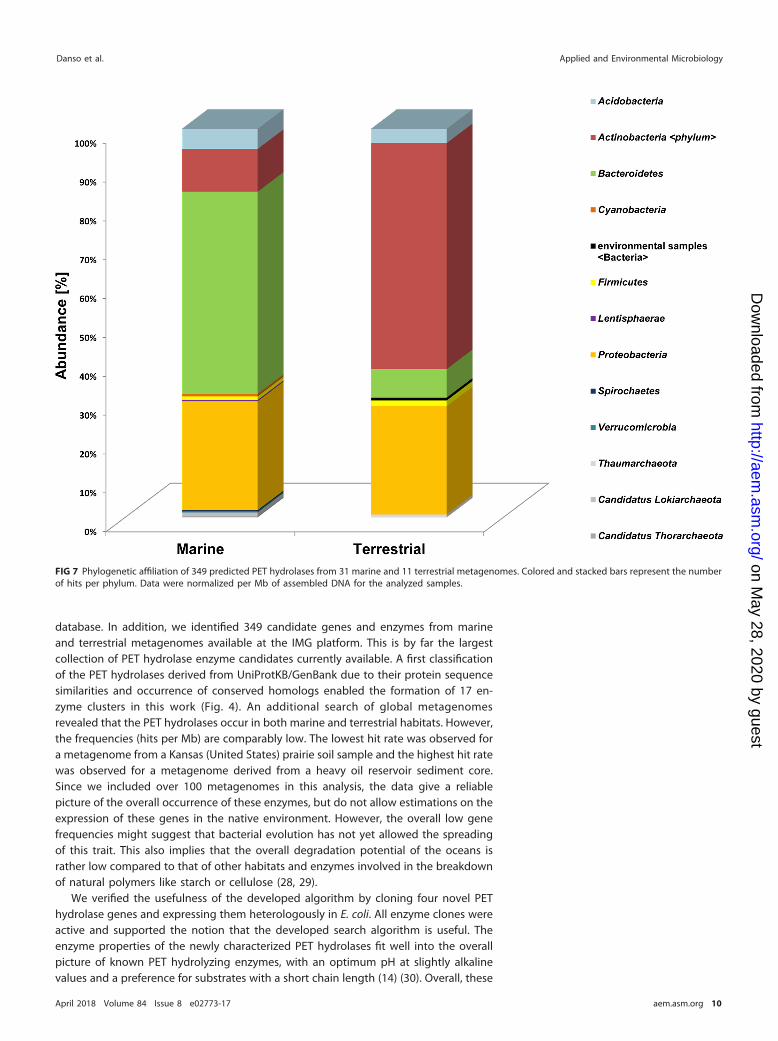
We further observed that within the terrestrial habitats, the Actinobacteria were themain hosts for the terrestrial-derived enzymes. However, in the marine samples, mostpredicted PET hydrolases originated from the phylum of Bacteroidetes (Fig. 7). Bacte-roidetes sequences in the marine samples were affiliated with 43% of all hits. Thephylum Proteobacteria was the second-most abundant in both data sets, with 23% ofhits in marine and 20% in terrestrial data (Fig. 7).
DISCUSSION
In this work, we developed a search algorithm that allows the in silico identificationof PET hydrolase gene candidates from genomes and metagenomes. Altogether, wewere able to identify 504 novel possible enzyme candidates in the UniProtKB andnonredundant RefSeq databases and the metagenomic database available in the NCBI
FIG 6 Global distribution of PET hydrolases in available metagenomes. Potential PET hydrolase containing metagenomes were visualized on a world map, usingcircles for marine and triangles for terrestrial metagenomes. Blue and red color shading indicates the frequency of PET hydrolase genes in hits/Mb for marineand terrestrial metagenomes, respectively. Red and green boxes magnify regions with overlapping spots (sample sites). (Map constructed using qGIS Desktop2.18.5 [http://www.qgis.org].)
PET Hydrolases from Metagenomes Applied and Environmental Microbiology
April 2018 Volume 84 Issue 8 e02773-17 aem.asm.org 9
on May 28, 2020 by guest
http://aem.asm
.org/D
ownloaded from
database. In addition, we identified 349 candidate genes and enzymes from marineand terrestrial metagenomes available at the IMG platform. This is by far the largestcollection of PET hydrolase enzyme candidates currently available. A first classificationof the PET hydrolases derived from UniProtKB/GenBank due to their protein sequencesimilarities and occurrence of conserved homologs enabled the formation of 17 en-zyme clusters in this work (Fig. 4). An additional search of global metagenomesrevealed that the PET hydrolases occur in both marine and terrestrial habitats. However,the frequencies (hits per Mb) are comparably low. The lowest hit rate was observed fora metagenome from a Kansas (United States) prairie soil sample and the highest hit ratewas observed for a metagenome derived from a heavy oil reservoir sediment core.Since we included over 100 metagenomes in this analysis, the data give a reliablepicture of the overall occurrence of these enzymes, but do not allow estimations on theexpression of these genes in the native environment. However, the overall low genefrequencies might suggest that bacterial evolution has not yet allowed the spreadingof this trait. This also implies that the overall degradation potential of the oceans israther low compared to that of other habitats and enzymes involved in the breakdownof natural polymers like starch or cellulose (28, 29).
We verified the usefulness of the developed algorithm by cloning four novel PEThydrolase genes and expressing them heterologously in E. coli. All enzyme clones wereactive and supported the notion that the developed search algorithm is useful. Theenzyme properties of the newly characterized PET hydrolases fit well into the overallpicture of known PET hydrolyzing enzymes, with an optimum pH at slightly alkalinevalues and a preference for substrates with a short chain length (14) (30). Overall, these
FIG 7 Phylogenetic affiliation of 349 predicted PET hydrolases from 31 marine and 11 terrestrial metagenomes. Colored and stacked bars represent the numberof hits per phylum. Data were normalized per Mb of assembled DNA for the analyzed samples.
Danso et al. Applied and Environmental Microbiology
April 2018 Volume 84 Issue 8 e02773-17 aem.asm.org 10
on May 28, 2020 by guest
http://aem.asm
.org/D
ownloaded from

novel enzymes revealed comparable activities to those already previously characterized(see Table 1 and references therein).
To our surprise, both enzymes characterized in more detail (PET2 and PET6) showedtraits of thermostability (Fig. 5) (25). PET2 was stable up to 90°C, with measurableresidual activity of more than 50%. This is more stable than the LCC derived from acompost metagenome (14).
All of the newly identified PET hydrolases originated mainly from three bacterialphyla, Proteobacteria, Actinobacteria, and Bacteroidetes. Within this framework, it isnotable that the Bacteroidetes have so far not been associated with PET degradation,but Bacteroidetes species have been described as very potent degraders of otherpolymers, and they harbor a multitude of hydrolases and binding modules (31–33).
The restriction of PET hydrolases to a few bacterial phyla could indicate that thismetabolic capability has only rather recently been evolved and is thus limited to a veryfew phylogenetic groups. The observation here that in the marine habitat the phylumBacteroidetes is the main host of PET hydrolases is new and intriguing for severalreasons. First, using classical searches and biochemical characterization, the Actinobac-teria and Proteobacteria were considered to be the main hosts for these enzymes (Table1). Second, the searches in UNIProtKB and other databases implemented on the NCBIwebsite underlined the presence of PET hydrolases in the phyla Actinobacteria andProteobacteria. Only when we extended our search for metagenomes of mainly non-cultivated bacterial phyla did we identify the Bacteroidetes as the main hosts for theseenzymes in the marine environment.
The recent findings on PET hydrolases described in this publication will significantlyextend the knowledge of these enzymes and provide promising candidates for bio-technological applications. In summary, the over 800 enzyme candidates identified inthis work will build the basis for a global repository and database of this urgentlyneeded enzyme class.
MATERIALS AND METHODSBacterial strains, plasmids, and primers. Bacterial strains, plasmids, and primers used in this study
are listed in Tables 3 and 4. If not otherwise mentioned, Escherichia coli clones were grown in LB medium(1% tryptone/peptone, 0.5% yeast extract, and 1% NaCl) supplemented with appropriate antibiotics (25�g/ml kanamycin or 100 �g/ml ampicillin) at 37°C for 18 h.
Databases used in this study and bioinformatic analysis. Nucleotide and amino acid sequencesof putative PET hydrolases were acquired from databases integrated into the NCBI (https://www.ncbi.nlm.nih.gov/), UniProt (http://www.uniprot.org/) and the Joint Genome Institute (JGI) IMG (https://img.jgi.doe.gov/) websites (34), (27, 35). Sequences were compared to others deposited in the NCBIdatabases using BLAST alignment tools (36). Amino acid sequence HMM search was carried out using theHMMER (http://hmmer.org/) webpage or a local version of the software (v3.1b2) with downloaded datasets. Structural information on the enzymes was retrieved from the RCSB-PDB (37) database.
Sequence data were processed using BioEdit and the Clone Manager suite version 9 (Sci-Ed Software,Denver, USA). Neighbor-joining phylogenetic trees based on amino acid sequence alignments wereconstructed using MEGA6 (38). Nine known and activity-confirmed bacterial PET hydrolase sequenceswere obtained from NCBI, aligned with T-Coffee (39) and manually revised. Afterwards, the alignmentwas used to construct a profile HMM with the “hmmbuild” function of the HMMER package (http://hmmer.org/). After the identification of PET hydrolase homologs, the obtained sequences were includedin the above-mentioned alignment and the HMM was refined. An HMM logo was visualized using theSkylign online tool (40). Metagenomic data were downloaded from the IMG database using a Globusendpoint and were further analyzed using “hmmsearch” from the HMMER package. Phylogeneticassignment was done via a local diamond-blast search (41) against the nonredundant protein database(36) and subsequent analysis with MEGAN6 (42). The map representing the frequency and geographicaldistribution of PET hydrolases in metagenomes (Fig. 6) was constructed using qGIS Desktop 2.18.5(http://www.qgis.org/).
Cloning and heterologous expression of PET2, PET5, PET6, and PET12 in Escherichia coliT7-SHuffle. Cloning of PET hydrolase genes into the expression vectors pET21a(�) and pET28a(�) wasaccomplished after amplification of genomic DNA using specific primer pairs with underlined homologregions to the vector or restriction sites. The sequence of PET2 was obtained from NCBI (GenBankaccession number ACC95208) and synthesized after codon usage optimization for E. coli (MWG Eurofins,Germany). Obtained DNAs were cloned into expression vectors, and the constructs were transformedinto E. coli T7-SHuffle cells. The cultures were grown aerobically in autoinduction medium (ZYM-5052)(43) containing 100 �g/ml ampicillin and 25 �g/ml kanamycin for pET21a(�) and pET28a(�), respec-tively, at 37°C until they reached an optical density at 600 nm (OD600) of 1.0. The proteins harboring aC- or N-terminal histidine tag were expressed afterwards at 17°C for 16 to 20 h. The cells were harvested
PET Hydrolases from Metagenomes Applied and Environmental Microbiology
April 2018 Volume 84 Issue 8 e02773-17 aem.asm.org 11
on May 28, 2020 by guest
http://aem.asm
.org/D
ownloaded from

and lysed with pressure using a French press. Afterwards, the proteins were purified with nickel-ionaffinity chromatography using nickel-nitrilotriacetic acid (Ni-NTA) agarose (Qiagen, Hilden, Germany) andanalyzed by SDS-PAGE. The elution buffer was exchanged against 0.1 mM potassium phosphate bufferpH 8.0 in a 10 kDa Amicon tube (GE Health Care, Solingen, Germany).
Biochemical characterization of PET2 and PET6. For activity tests, both enzymes were assayedusing purified recombinant protein. Unless otherwise indicated, the enzymes were added to a substratesolution containing 190 �l of either 0.2 M sodium phosphate buffer or 0.1 M Tris-HCl, with a defined pHbetween 7 and 8 and 0.5 mM pNP substrate dissolved in isopropanol. Incubation time ranged from 15to 30 min. As substrates, we tested pNP esters with chain lengths of C2, C4, C6, C8, C10, C12, C14, C16, andC18. After incubation at a defined temperature, the color change from colorless to yellow was measuredat 405 nm in a plate reader (Biotek, Winooski, USA). All samples were measured in triplicate. Fordetermination of the optimal temperature, samples were incubated between 17°C and 90°C for 15 min.The impact of pH conditions on the activity of each enzyme was measured in citrate phosphate (pH 3.0,4.0, and 5.0), potassium phosphate (pH 6.0, 7.0, and 8.0), and carbonate bicarbonate buffer (pH 9.2 and10.2). The influence of possible cofactors, solvents, detergents, and inhibitors was assayed at differentconcentration levels. After 1 h of incubation in the presence of the substances described below, theresidual activity was determined after 15 min incubation at optimal temperature with pNP-octanoate andoptimal pH. The possible cofactors Ca2�, Co2�, Cu2�, Fe3�, Mg2�, Mn2�, Rb2�, and Zn2�, with finalconcentrations of 1 and 10 mM, were used. To determine the solvent stability, dimethyl sulfoxide(DMSO), isopropanol, methanol, dimethylformamide (DMF), acetone, acetonitrile, and ethanol, with finalconcentrations of 10% and 30% (vol/vol) were added to the reaction. Detergent stability was assayedwith SDS, Triton X-100, and Tween 80 at 1% and 5% (wt/vol, vol/vol) concentration. The inhibitory effectsof EDTA, dithiothreitol (DTT), and PMSF were tested at 1 and 10 mM concentration. Substrate analysesusing the HPLC LaChrom Elite system from Hitachi (Tokyo, Japan) with a Lichrospher 100 RP-18e column(VWR International GmbH, Darmstadt, Germany), consisting of 5-�m diameter particles, were done aspreviously published (14). A 14-mg low-crystallinity PET film (Goodfellow GmbH, Bad Nauheim, Germany)was used as the substrate. For enzymatic hydrolysis, up to 50 �g of protein was incubated at 60°C withcontinuous shaking at 500 rpm. As a mobile phase, acetonitrile (A) and water with 0.1% trifluoroaceticacid (TFA) (B) were used in a isocratic method with 20% acetonitrile (A). The reaction buffer was 0.1 MTris-HCl (pH 7.5), with an injection volume of 99 �l. Detection was performed at 241 nm.
SUPPLEMENTAL MATERIAL
Supplemental material for this article may be found at https://doi.org/10.1128/AEM.02773-17.
SUPPLEMENTAL FILE 1, PDF file, 0.5 MB.
ACKNOWLEDGMENTSThis work was supported in part by the BMBF via the programs FuPol and MetaCat
and by the European Union project INMARE at the University of Hamburg.We declare that we have no conflict of interest.
REFERENCES1. Rex WJ, Tennant DJ. March 1949. Polymeric linear terephthalic esters. US
patent 2,465,319.2. Gregory MR. 2009. Environmental implications of plastic debris in marine
settings– entanglement, ingestion, smothering, hangers-on, hitch-hikingand alien invasions. Philos Trans R Soc Lond B Biol Sci 364:2013–2025.https://doi.org/10.1098/rstb.2008.0265.
3. Webb HK, Arnott J, Crawford RJ, Ivanova EP. 2013. Plastic degradation andits environmental implications with special reference to poly(ethylene te-rephthalate). Polymers 5:1–18. https://doi.org/10.3390/polym5010001.
4. Sax L. 2010. Polyethylene terephthalate may yield endocrine disruptors.Environ Health Perspect 118:445–448. https://doi.org/10.1289/ehp.0901253.
5. Wei R, Zimmermann W. 2017. Microbial enzymes for the recycling ofrecalcitrant petroleum-based plastics: how far are we? Microb Biotech-nol 10:1308 –1322. https://doi.org/10.1111/1751-7915.12710.
6. Kawai F, Oda M, Tamashiro T, Waku T, Tanaka N, Yamamoto M,Mizushima H, Miyakawa T, Tanokura M. 2014. A novel Ca2�-activated,thermostabilized polyesterase capable of hydrolyzing polyethyleneterephthalate from Saccharomonospora viridis AHK190. Appl Micro-biol Biotechnol 98:10053–10064. https://doi.org/10.1007/s00253-014-5860-y.
7. Dresler K, van den Heuvel J, Muller RJ, Deckwer WD. 2006. Production ofa recombinant polyester-cleaving hydrolase from Thermobifida fusca inEscherichia coli. Bioprocess Biosyst Eng 29:169 –183. https://doi.org/10.1007/s00449-006-0069-9.
8. Chen S, Tong X, Woodard RW, Du GC, Wu J, Chen J. 2008. Identification
and characterization of bacterial cutinase. J Biol Chem 283:25854 –25862. https://doi.org/10.1074/jbc.M800848200.
9. Wei R, Oeser T, Then J, Kuhn N, Barth M, Schmidt J, Zimmermann W.2014. Functional characterization and structural modeling of syntheticpolyester-degrading hydrolases from Thermomonospora curvata. AMBExpress 4:44. https://doi.org/10.1186/s13568-014-0044-9.
10. Hu X, Thumarat U, Zhang X, Tang M, Kawai F. 2010. Diversity ofpolyester-degrading bacteria in compost and molecular analysis of athermoactive esterase from Thermobifida alba AHK119. Appl MicrobiolBiotechnol 87:771–779. https://doi.org/10.1007/s00253-010-2555-x.
11. Ribitsch D, Acero EH, Greimel K, Dellacher A, Zitzenbacher S, Marold A,Rodriguez RD, Steinkellner G, Gruber K, Schwab H, Guebitz GM. 2012. Anew esterase from Thermobifida halotolerans hydrolyses polyethyleneterephthalate (PET) and polylactic acid (PLA). Polymers 4:617– 629.https://doi.org/10.3390/polym4010617.
12. Acero EH, Ribitsch D, Steinkellner G, Gruber K, Greimel K, Eiteljoerg I,Trotscha E, Wei R, Zimmermann W, Zinn M, Cavaco-Paulo A, Freddi G,Schwab H, Guebitz G. 2011. Enzymatic surface hydrolysis of PET:Effect of structural diversity on kinetic properties of cutinases fromThermobifida. Macromolecules 44:4632– 4640. https://doi.org/10.1021/ma200949p.
13. Yoshida S, Hiraga K, Takehana T, Taniguchi I, Yamaji H, Maeda Y, Toyo-hara K, Miyamoto K, Kimura Y, Oda K. 2016. A bacterium that degradesand assimilates poly(ethylene terephthalate). Science 351:1196 –1199.https://doi.org/10.1126/science.aad6359.
Danso et al. Applied and Environmental Microbiology
April 2018 Volume 84 Issue 8 e02773-17 aem.asm.org 12
on May 28, 2020 by guest
http://aem.asm
.org/D
ownloaded from

14. Sulaiman S, Yamato S, Kanaya E, Kim JJ, Koga Y, Takano K, Kanaya S.2012. Isolation of a novel cutinase homolog with polyethyleneterephthalate-degrading activity from leaf-branch compost by using ametagenomic approach. Appl Environ Microbiol 78:1556 –1562. https://doi.org/10.1128/AEM.06725-11.
15. Kleeberg I, Hetz C, Kroppenstedt RM, Muller RJ, Deckwer WD. 1998.Biodegradation of aliphatic-aromatic copolyesters by Thermomonosporafusca and other thermophilic compost isolates. Appl Environ Microbiol64:1731–1735.
16. Wei R, Oeser T, Zimmermann W. 2014. Synthetic polyester-hydrolyzingenzymes from thermophilic actinomycetes. Adv Appl Microbiol 89:267–305. https://doi.org/10.1016/B978-0-12-800259-9.00007-X.
17. Roth C, Wei R, Oeser T, Then J, Follner C, Zimmermann W, Strater N. 2014.Structural and functional studies on a thermostable polyethylene tereph-thalate degrading hydrolase from Thermobifida fusca. Appl Microbiol Bio-technol 98:7815–7823. https://doi.org/10.1007/s00253-014-5672-0.
18. Ollis DL, Cheah E, Cygler M, Dijkstra B, Frolow F, Franken SM, Harel M,Remington SJ, Silman I, Schrag J, et al. 1992. The alpha/beta hydrolasefold. Protein Eng 5:197–211. https://doi.org/10.1093/protein/5.3.197.
19. Yang J, Yang Y, Wu WM, Zhao J, Jiang L. 2014. Evidence of polyethylenebiodegradation by bacterial strains from the guts of plastic-eating wax-worms. Environ Sci Technol 48:13776 –13784. https://doi.org/10.1021/es504038a.
20. Yang Y, Yang J, Wu WM, Zhao J, Song YL, Gao LC, Yang RF, Jiang L. 2015.Biodegradation and mineralization of polystyrene by plastic-eatingmealworms: Part 1. Chemical and physical characterization and isotopictests. Environ Sci Technol 49:12080 –12086. https://doi.org/10.1021/acs.est.5b02661.
21. Wei R, Oeser T, Schmidt J, Meier R, Barth M, Then J, Zimmermann W.2016. Engineered bacterial polyester hydrolases efficiently degrade poly-ethylene terephthalate due to relieved product inhibition. BiotechnolBioeng 113:1658 –1665. https://doi.org/10.1002/bit.25941.
22. Kitadokoro K, Thumarat U, Nakamura R, Nishimura K, Karatani H, SuzukiH, Kawai F. 2012. Crystal structure of cutinase Est119 from Thermobifidaalba AHK119 that can degrade modified polyethylene terephthalate at1.76 angstrom resolution. Polym Degradation Stab 97:771–775. https://doi.org/10.1016/j.polymdegradstab.2012.02.003.
23. Hanahan D. 1983. Studies on transformation of Escherichia coli withplasmids. J Mol Biol 166:557–580. https://doi.org/10.1016/S0022-2836(83)80284-8.
24. Wei R, Oeser T, Barth M, Weigl N, Luebs A, Schulz-Siegmund M, HackerMC, Zimmermann W. 2014. Turbidimetric analysis of the enzymatichydrolysis of polyethylene terephthalate nanoparticles. J Mol CatalB-Enzym 103:72–78. https://doi.org/10.1016/j.molcatb.2013.08.010.
25. Meilleur C, Hupe JF, Juteau P, Shareck F. 2009. Isolation and character-ization of a new alkali-thermostable lipase cloned from a metagenomiclibrary. J Ind Microbiol Biotechnol 36:853– 861. https://doi.org/10.1007/s10295-009-0562-7.
26. Baumann P, Baumann L, Bang SS, Woolkalis MJ. 1980. Reevaluation ofthe taxonomy of Vibrio, Beneckea, and Photobacterium: abolition of thegenus Beneckea. Curr Microbiol 4:127–132. https://doi.org/10.1007/BF02602814.
27. Markowitz VM, Chen IM, Palaniappan K, Chu K, Szeto E, Grechkin Y,Ratner A, Jacob B, Huang J, Williams P, Huntemann M, Anderson I,Mavromatis K, Ivanova NN, Kyrpides NC. 2012. IMG: the IntegratedMicrobial Genomes database and comparative analysis system. NucleicAcids Res 40:D115–D122. https://doi.org/10.1093/nar/gkr1044.
28. Morrison M, Pope PB, Denman SE, McSweeney CS. 2009. Plant biomassdegradation by gut microbiomes: more of the same or something new?Curr Opin Biotechnol 20:358 –363. https://doi.org/10.1016/j.copbio.2009.05.004.
29. Kanokratana P, Uengwetwanit T, Rattanachomsri U, Bunterngsook B,Nimchua T, Tangphatsornruang S, Plengvidhya V, Champreda V, Eur-wilaichitr L. 2011. Insights into the phylogeny and metabolic potential ofa primary tropical peat swamp forest microbial community by meta-genomic analysis. Microb Ecol 61:518 –528. https://doi.org/10.1007/s00248-010-9766-7.
30. Thumarat U, Nakamura R, Kawabata T, Suzuki H, Kawai F. 2012. Biochem-ical and genetic analysis of a cutinase-type polyesterase from a thermo-philic Thermobifida alba AHK119. Appl Microbiol Biotechnol 95:419 – 430.https://doi.org/10.1007/s00253-011-3781-6.
31. Dodd D, Mackie RI, Cann IKO. 2011. Xylan degradation, a metabolicproperty shared by rumen and human colonic Bacteroidetes. Mol Mi-crobiol 79:292–304. https://doi.org/10.1111/j.1365-2958.2010.07473.x.
32. Thomas F, Hehemann J-H, Rebuffet E, Czjzek M, Michel G. 2011. Envi-ronmental and gut Bacteroidetes: the food connection. Front Microbiol2:16. https://doi.org/10.3389/fmicb.2011.00093.
33. Foley MH, Cockburn DW, Koropatkin NM. 2016. The Sus operon: a modelsystem for starch uptake by the human gut Bacteroidetes. Cell Mol LifeSci 73:2603–2617. https://doi.org/10.1007/s00018-016-2242-x.
34. Coordinators NR. 2017. Database resources of the National Center forBiotechnology Information. Nucleic Acids Res 45:D12–D17. https://doi.org/10.1093/nar/gkw1071.
35. The UniProt Consortium. 2017. UniProt: the universal protein knowl-edgebase. Nucleic Acids Res 45:D158 –D169. https://doi.org/10.1093/nar/gkw1099.
36. Agarwala R, Barrett T, Beck J, Benson DA, Bollin C, Bolton E, Bourexis D,Brister JR, Bryant SH, Lanese K, Charowhas C, Clark K, DiCuccio M,Dondoshansky I, Federhen S, Feolo M, Funk K, Geer LY, Gorelenkov V,Hoeppner M, Holmes B, Johnson M, Khotomlianski V, Kimchi A, Kimel-man M, Kitts P, Klimke W, Krasnov S, Kuznetsov A, Landrum MJ, Lands-man D, Lee JM, Lipman DJ, Lu ZY, Madden TL, Madcj T, Marchler-BauerA, Karsch-Mizrachi I, Murphy T, Orris R, Ostell J, O’Sullivan C, PanchenkoA, Phan L, Preuss D, Pruitt KD, Rodarmer K, Rubinstein W, Sayers EW,Schneider V, et al. 2016. Database resources of the National Center forBiotechnology Information. Nucleic Acids Res 44:D7–D19. https://doi.org/10.1093/nar/gkv1290.
37. Berman HM, Westbrook J, Feng Z, Gilliland G, Bhat TN, Weissig H,Shindyalov IN, Bourne PE. 2000. The Protein Data Bank. Nucleic Acids Res28:235–242. https://doi.org/10.1093/nar/28.1.235.
38. Tamura K, Stecher G, Peterson D, Filipski A, Kumar S. 2013. MEGA6:molecular evolutionary genetics analysis version 6.0. Mol Biol Evol 30:2725–2729. https://doi.org/10.1093/molbev/mst197.
39. Notredame C, Higgins DG, Heringa J. 2000. T-Coffee: a novel method forfast and accurate multiple sequence alignment. J Mol Biol 302:205–217.https://doi.org/10.1006/jmbi.2000.4042.
40. Wheeler TJ, Clements J, Finn RD. 2014. Skylign: a tool for creatinginformative, interactive logos representing sequence alignments andprofile hidden Markov models. BMC Bioinformatics 15:7. https://doi.org/10.1186/1471-2105-15-7.
41. Buchfink B, Xie C, Huson DH. 2015. Fast and sensitive protein alignmentusing DIAMOND. Nat Methods 12:59 – 60. https://doi.org/10.1038/nmeth.3176.
42. Huson DH, Beier S, Flade I, Górska A, El-Hadidi M, Mitra S, RuscheweyhH-J, Tappu R. 2016. MEGAN Community Edition - interactive explorationand analysis of large-scale microbiome sequencing data. PLoS CompBiol 12:e1004957. https://doi.org/10.1371/journal.pcbi.1004957.
43. Studier FW. 2005. Protein production by auto-induction in high-densityshaking cultures. Protein Expression Purif 41:207–234. https://doi.org/10.1016/j.pep.2005.01.016.
PET Hydrolases from Metagenomes Applied and Environmental Microbiology
April 2018 Volume 84 Issue 8 e02773-17 aem.asm.org 13
on May 28, 2020 by guest
http://aem.asm
.org/D
ownloaded from