Embed Size (px)
Citation preview
New Insights into the Respiratory Chain of PlantMitochondria. Supercomplexes and a UniqueComposition of Complex II1
Holger Eubel, Lothar Jansch, and Hans-Peter Braun*
Institut fur Angewandte Genetik, Universitat Hannover, Herrenhauser Strasse 2, D–30419 Hannover,Germany (H.E., H.-P.B.); and Gesellschaft fur Biotechnologische Forschung, Mascheroder Weg 1, 38124Braunschweig, Germany (L.J.)
A project to systematically investigate respiratory supercomplexes in plant mitochondria was initiated. Mitochondrialfractions from Arabidopsis, potato (Solanum tuberosum), bean (Phaseolus vulgaris), and barley (Hordeum vulgare) werecarefully treated with various concentrations of the nonionic detergents dodecylmaltoside, Triton X-100, or digitonin, andproteins were subsequently separated by (a) Blue-native polyacrylamide gel electrophoresis (PAGE), (b) two-dimensionalBlue-native/sodium dodecyl sulfate-PAGE, and (c) two-dimensional Blue-native/Blue-native PAGE. Three high molecularmass complexes of 1,100, 1,500, and 3,000 kD are visible on one-dimensional Blue native gels, which were identified byseparations on second gel dimensions and protein analyses by mass spectrometry. The 1,100-kD complex represents dimericATP synthase and is only stable under very low concentrations of detergents. In contrast, the 1,500-kD complex is stable atmedium and even high concentrations of detergents and includes the complexes I and III2. Depending on the investigatedorganism, 50% to 90% of complex I forms part of this supercomplex if solubilized with digitonin. The 3,000-kD complex,which also includes the complexes I and III, is of low abundance and most likely has a III4I2 structure. The complexes IV,II, and the alternative oxidase were not part of supercomplexes under all conditions applied. Digitonin proved to be the idealdetergent for supercomplex stabilization and also allows optimal visualization of the complexes II and IV on Blue-nativegels. Complex II unexpectedly was found to be composed of seven subunits, and complex IV is present in two differentforms on the Blue-native gels, the larger of which comprises additional subunits including a 32-kD protein resembling COXVIb from other organisms. We speculate that supercomplex formation between the complexes I and III limits access ofalternative oxidase to its substrate ubiquinol and possibly regulates alternative respiration. The data of this investigation areavailable at http://www.gartenbau.uni-hannover.de/genetik/braun/AMPP.
Structural basis for oxidative phosphorylation inmitochondria are five protein complexes termedNADH dehydrogenase (complex I), succinat dehy-drogenase (complex II), cytochrome c reductase(complex III, which is a functional dimer), cyto-chrome c oxidase (complex IV), and ATP synthase(complex V). They were first characterized about 40years ago by solubilizations of mitochondrial mem-brane proteins using detergents and differential pre-cipitations or chromatographic separations. Accord-ing to the popular “liquid state” model, the proteincomplexes of the respiratory chain are randomly ar-ranged in the membrane and freely diffuse in lateraldirection within the inner mitochondrial membrane(for review, see Rich, 1984). However, other resultsrather indicate an ordered association of these pro-tein complexes forming larger structures. These so-called “supercomplexes” were first described for bac-teria (Berry and Trumpower, 1985; Sone et al., 1987;Iwasaki et al., 1995; Niebisch and Bott, 2003). Later
the existence of respiratory supercomplexes was alsoreported for yeast and mammalian mitochondria(Schagger and Pfeiffer, 2000).
In Brewer’s yeast (Saccharomyces cerevisiae), whichdoes not comprise complex I, three large mitochon-drial complexes were identified by Blue-native gelelectrophoresis after gentle protein solubilization us-ing nonionic detergents: (a) dimeric ATP synthase,(b) a supercomplex containing dimeric complex III �one copy of complex IV, and (c) a supercomplexcontaining dimeric complex III � two copies of com-plex IV (Arnold et al., 1998; 1999; Cruciat et al., 2000;Schagger and Pfeiffer, 2000; Schagger, 2001a, 2002;Zhang et al., 2002). Dimeric ATP synthase from yeastincludes three dimer-specific subunits, two of whichare directly involved in dimer formation. Supercom-plexes containing complexes III and IV were not onlyprepared by Blue-native gel electrophoresis but alsoby gel filtrations and co-immunoprecipitations (Cru-ciat et al., 2000). Their formation depends on thecardiolipin content of the inner mitochondrial mem-brane and also is influenced by growth conditions.Functional implications of complex III-complex IVassociations were shown by ubiquinol-oxidase activ-ity measurements in the presence of mild detergents(Schagger and Pfeiffer, 2000).
1 This work was supported by the Fonds der ChemischenIndustrie.
* Corresponding author; e-mail [email protected];fax 49511–7623608.
Article, publication date, and citation information can be foundat www.plantphysiol.org/cgi/doi/10.1104/pp.103.024620.
274 Plant Physiology, September 2003, Vol. 133, pp. 274–286, www.plantphysiol.org © 2003 American Society of Plant Biologists
Dow
nloaded from https://academ
ic.oup.com/plphys/article/133/1/274/6111890 by guest on 01 M
arch 2022
In mammalian mitochondria, five large complexeswere found: (a) dimeric ATP synthase, (b) a supercom-plex containing dimeric complex III � one copy ofcomplex I, and (c–e) supercomplexes containingdimeric complex III � one copy of complex I � one tothree copies of complex IV (Schagger and Pfeiffer,2000, 2001; Schagger, 2001a, 2002). All of these super-complexes can be visualized on Blue-native gels aftersolubilization of mitochondrial proteins using digito-nin. Solubilization using Triton X-100 additionally al-low detection of a supercomplex consisting of dimericcomplex III � monomeric complex I � four copies ofcomplex IV. A high percentage of complex I formspart of supercomplexes, whereas dimeric complex IIIand monomeric complex IV also exist in singularform, because abundance of these protein complexesis significantly higher in comparison with complex I.NADH-cytochrome c activity measurements in de-pendence of various mild detergents have revealedfunctional importance of supercomplex formation be-tween complexes III2 � I. The term “respirasome” wassuggested for supercomplexes containing the com-plexes I, III2, and IV, which autonomously can carryout respiration in the presence of cytochrome c andubiquinone (Schagger and Pfeiffer, 2000).
The supramolecular structure of the respiratorychain of plant mitochondria is unknown. The fiveprotein complexes of oxidative phosphorylation arewell characterized and structurally resemble theircounterparts in fungi and mammals (Jansch et al.,1996; Vedel et al., 1999; Heazlewood et al., 2003b;Sabar et al., 2003). Some plant-specific subunits ofrespiratory chain complexes were described, e.g. thesubunits of the mitochondrial processing peptidase,which form an integral part of complex III in plantmitochondria (Braun et al., 1992a; Eriksson et al., 1994).Additionally, the electron transfer chain of plant mito-chondria is very much branched due to the presence ofseveral alternative oxidoreductases like a cyanide-insensitve terminal oxidase and rotenone-insensitiveNAD(P)H dehydrogenases (for review, see Siedow andUmbach, 1995; Vanlerberghe and McIntosh, 1997;Mackenzie and McIntosh, 1999; Rasmusson et al., 1999).
Here, we describe a systematic investigation ofsupercomplexes in plant mitochondria. Using gentleprotein solubilizations with nonionic detergents andBlue-native gel electrophoresis, three supercom-plexes could be visualized: (a) dimeric ATP synthase,(b) a supercomplex formed by dimeric complex IIIand complex I, and (c) a supercomplex containingtwo copies of dimeric complex III and two copies ofcomplex I. The complexes II and IV as well as thealternative oxidase (AOX) do not form part of super-complexes under all conditions applied. Further-more, a larger and a smaller form of cytochrome coxidase were found, which differ by at least twoprotein subunits, and a complex II is described,which has a very unusual subunit composition.
RESULTS
Identification of Respiratory Supercomplexes inMitochondria from Arabidopsis
Blue-native gel electrophoresis was previously em-ployed for the characterization of the respiratorychain of plant mitochondria (Jansch et al., 1995, 1996;Brumme et al., 1998; Kugler et al., 1998; Karpowa andNewton, 1999; Ducos et al., 2001; Kruft et al., 2001;Mihr et al., 2001; Rasmusson and Agius, 2001; Wer-hahn and Braun, 2002; Bykova and Moller, 2003;Heazlewood et al., 2003a, 2003b, 2003c; Sabar et al.,2003). Nevertheless, respiratory supercomplexeswere not described, most likely because very similarconditions for protein solubilization were chosen,which seem to have destabilizing effects on labileprotein-protein interactions. In an attempt to system-atically search for the occurrence of respiratory su-percomplexes in plants, mitochondria from Arabi-dopsis were solubilized using varying concentrationsof the nonionic detergents dodecylmaltoside, TritonX-100, and digitonin and analyzed by Blue-nativePAGE (Fig. 1, A–C). Protein complexes were identi-fied by their known subunit compositions upon anal-yses on second gel dimensions and by partial se-quence analysis of selected proteins using massspectrometry (Figs. 1, D and E, and 2; Table I).
Solubilization of Arabidopsis mitochondria with 1 gdodecylmaltoside g�1 protein allows resolution ofknown singular complexes of the oxidative phosphor-ylation system (Fig. 1, A and D): complex I (1,000 kD),F0F1 ATP synthase (580 kD), complex III (480 kD),which always is dimeric for functional reasons, andthe F1 part of ATP synthase (390 kD). Furthermore, thesoluble HSP60 (750 kD) and formate dehydrogenasecomplexes (200 kD) are visible on the gel. Addition-ally, some low amount of dimeric ATP synthase canbe seen at about 1,100 kD, which was overlooked onthe Blue-native gels shown before by Kruft et al.(2001). In contrast to yeast and mammals, the amountof dimeric ATP synthase does not increase if mito-chondrial proteins are solubilized with lower dodecyl-maltoside concentrations (Fig. 1A). Usage of dodecyl-maltoside to protein ratios �1 g per g allowsvisualization of a supercomplex of about 1,500 kD,which is composed of the complexes I and III andprobably has the structure III2I. However, only a smallproportion of total complex I forms part of this super-complex and an even smaller proportion of complexIII, which is more abundant than complex I.
Solubilization of Arabidopsis mitochondria withTriton X-100 allows visualization of the same proteincomplexes and supercomplexes on Blue-native gels(Fig. 1, B and E). The amount of dimeric ATP syn-thase is highest between 0.25 and 0.5 g Triton X-100g�1 protein, which is in line with observations re-ported for yeast (Arnold et al., 1998). The ratio ofdimeric to monomeric ATP synthase is about 1 uponsolubilization using 0.25 g Triton g�1 protein but
Respiratory Supercomplexes in Plants
Plant Physiol. Vol. 133, 2003 275
Dow
nloaded from https://academ
ic.oup.com/plphys/article/133/1/274/6111890 by guest on 01 M
arch 2022

decreases sharply upon solubilizations using higheramounts of detergent (Fig. 1B). About 50% of com-plex I forms part of the III2I supercomplex on Blue-native gels after protein solubilizations using 0.5 to1.0 g per g Triton X-100 per g protein (Fig. 1B).
In general, higher detergent to protein ratios arenecessary for protein solubilizations using digitonin,which is in accordance with results found for yeastand mammals. However, starting with a digitonin toprotein ratio of 2.5 g per g, this detergent proved tobe very suitable for supercomplex stabilization (Figs.1C and 2). Under these conditions, about 80% ofcomplex I forms part of the III2I supercomplex. Fur-
thermore, a supercomplex of about 3,000 kD can beseen on Blue-native gels (Fig. 1C), which also is com-posed of subunits of the complexes III and I as foundby two-dimensional Blue-native/SDS gel electro-phoresis and silver staining (data not shown). Thissupercomplex most likely has a III4I2 structure, be-cause the ratio of single complex I and complex IIIsubunits is unchanged if compared with their ratio inthe 1,500-kD III2I complex. Dimeric ATP synthaseonly is visible at very low digitonin to protein ratios(data not shown). The F1 part of the ATP synthasecomplex is not detectable on the Blue-native gels,indicating a stabilizing effect of the detergent on
Figure 1. Resolution of mitochondrial protein complexes and supercomplexes by Blue-native PAGE. A through C, Solubi-lization of mitochondrial protein complexes from Arabidopsis using different detergents. Isolated mitochondria were treatedwith varying concentrations of dodecylmaltoside (A), Triton X-100 (B), or digitonin (C), and protein complexes weresubsequently resolved by one-dimensional Blue-native PAGE. Detergent to protein ratios are given above the gels (in gramsof detergent per gram protein), and the identity of protein complexes is given to the right of the gels. D and E,Two-dimensional resolution of mitochondrial protein complexes from Arabidopsis by Blue-native/SDS PAGE after solubi-lization with dodecylmaltoside (1.5 g per g protein) (D) and Triton X-100 (0.5 g per g protein) (E). Designations of the proteincomplexes are given above the gels. FDH, Formate dehydrogenase; F1, F1-part of the ATP synthase complex; III2, dimericcytochrome c reductase; V, ATP synthase; H, HSP60 complex; I, NADH dehydrogenase; I�III2, supercomplex formed bycomplex I and dimeric complex III; IVa and VIb, large and small form of cytochrome c oxidase; V2, dimeric ATP synthase.
Eubel et al.
276 Plant Physiol. Vol. 133, 2003
Dow
nloaded from https://academ
ic.oup.com/plphys/article/133/1/274/6111890 by guest on 01 M
arch 2022
complex V (Fig. 1C). Furthermore, in contrast to do-decylmaltoside and Triton X-100, digitonin allowssolubilization of three novel protein complexes ofabout 150, 220, and 300 kD. These protein complexesdo not form visible bands on one-dimensional Blue-native gels, probably because the background on thegels is too high in this molecular mass range. How-ever, these multisubunit complexes nicely are re-solved on corresponding two-dimensional gels (Fig.2). The subunit compositions of the 220- and 300-kDcomplexes very much resemble the one of cytochromec oxidase from potato (Solanum tuberosum; Jansch etal., 1996). In contrast, identity of the 150-kD complexwas unclear on the basis of subunit composition.
Protein identifications by mass spectrometry al-lowed unambiguous identification of subunits ofcomplexes I, III, and IV (Fig. 2; Table I). Furthermorethe 1,000-kD prohibitin complex was identified, aswas the preprotein translocase of the outer mitochon-drial membrane, the so-called TOM complex, at 390kD (Werhahn et al., 2003).
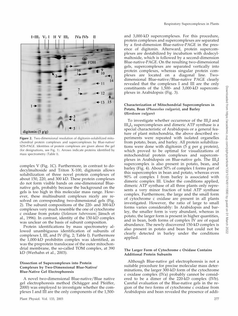
Dissection of Supercomplexes into ProteinComplexes by Two-Dimensional Blue-Native/Blue-Native Gel Electrophoresis
A novel two-dimensional Blue-native/Blue nativegel electrophoresis method (Schagger and Pfeiffer,2000) was employed to investigate whether the com-plexes I and III are the only components of the 1,500-
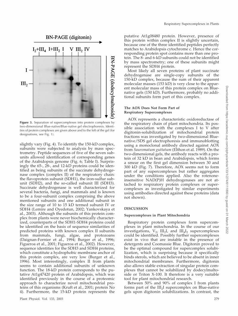
and 3,000-kD supercomplexes. For this procedure,protein complexes and supercomplexes are separatedby a first-dimension Blue-native-PAGE in the pres-ence of digitonin. Afterward, protein supercom-plexes are destabilized by incubation with dodecyl-maltoside, which is followed by a second-dimensionBlue-native-PAGE. On the resulting two-dimensionalgels, supercomplexes are separated vertically intoprotein complexes, whereas singular protein com-plexes are located on a diagonal line. Two-dimensional Blue-native/Blue-native PAGE clearlyrevealed that the complexes I and III are the onlyconstituents of the 1,500- and 3,000-kD supercom-plexes in Arabidopsis (Fig. 3).
Characterization of Mitochondrial Supercomplexes inPotato, Bean (Phaseolus vulgaris), and Barley(Hordeum vulgare)
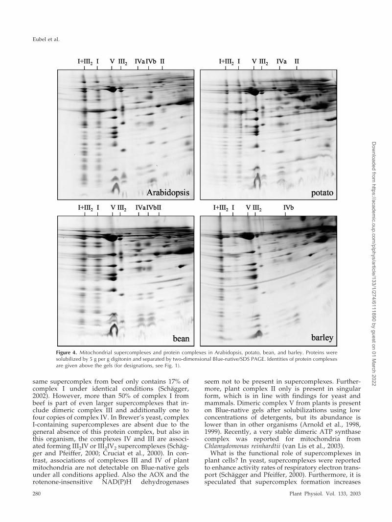
To investigate whether occurrence of the III2I andIII4I2 supercomplexes and dimeric ATP synthase is aspecial characteristic of Arabidopsis or a general fea-ture of plant mitochondria, the above described ex-periments were repeated with isolated organellesfrom potato, bean, and barley. All protein solubiliza-tions were done with digitonin (5 g per g protein),which proved to be optimal for visualizations ofmitochondrial protein complexes and supercom-plexes in Arabidopsis on Blue-native gels. The III2Isupercomplex is also present in potato, bean, andbarley (Fig. 4). About 50% of complex I forms part ofthis supercomplex in bean and potato, whereas even90% of complex I from barley is associated withdimeric complex III. Under the conditions applied,dimeric ATP synthase of all three plants only repre-sents a very minor fraction of total ATP synthasecomplex. Furthermore, the large and the small formof cytochrome c oxidase are present in all plantsinvestigated. However, the ratio of large to smallforms varies considerably: In Arabidopsis and bar-ley, the smaller form is very abundant, whereas inpotato, the larger form is present in higher quantities,and in bean, both forms of complex IV are of equalabundance. The newly discovered 150-kD complex isalso present in potato and bean but could not beclearly detected in barley under the conditionsapplied.
The Larger Form of Cytochrome c Oxidase ContainsAdditional Protein Subunits
Although Blue-native gel electrophoresis is not asuitable procedure for precise molecular mass deter-minations, the larger 300-kD form of the cytochromec oxidase complex (IVa) probably cannot be consid-ered to be a dimer of the 220-kD complex (IVb).Careful evaluation of the Blue-native gels in the re-gion of the two forms of cytochrome c oxidase fromArabidopsis and bean revealed the presence of addi-
Figure 2. Two-dimensional resolution of digitonin-solubilized mito-chondrial protein complexes and supercomplexes by Blue-native/SDS-PAGE. Identities of protein complexes are given above the gel(for designations, see Fig. 1). Arrows indicate proteins identified bymass spectrometry (Table I).
Respiratory Supercomplexes in Plants
Plant Physiol. Vol. 133, 2003 277
Dow
nloaded from https://academ
ic.oup.com/plphys/article/133/1/274/6111890 by guest on 01 M
arch 2022
tional subunits in the larger form, which might ex-plain the size difference between the two forms ofthis complex (Fig. 5). Data are especially clear forbean, because both forms of complex IV are equallyabundant. A 32-kD protein and at least one verysmall subunit of �6 kD are unique to complex IVa.Proteins of comparable size are also present in Ara-bidopsis (Fig. 5) but are difficult to detect in potatoand barley under the conditions applied. Accordingto our interpretation of the Blue-native gels, the sub-unit composition of cytochrome c oxidase is as fol-lows (see scheme in Fig. 5): Complex IVa includes 12separable proteins, and complex IVb is composed of10 proteins. Furthermore, complex IVb of Arabidop-sis can be further subdivided into two complexes ofvery similar molecular masses that differ with re-spect to the presence of a 10-kD subunit.
The 32-kD subunit of complex IVa is homologousto the 10-kD COX VIb protein of heterotrophic eu-karyotes (see Table I, protein 17), which is known tobe easily detached from cytochrome c oxidase inyeast and mammals and which was shown to haveregulatory functions on cytochrome c oxidase activ-ity (LaMarche et al., 1992; Weishaupt and Kaden-bach, 1992).
Complex II from Plant Mitochondria ContainsSeven Subunits
The newly discovered 150-kD complex of Arabi-dopsis comprises seven subunits of 65, 28, 18, 15, 12,8, and 6 kD. In bean and potato, this complex has avery comparable subunit composition, except thatthe molecular masses of the three smallest subunits
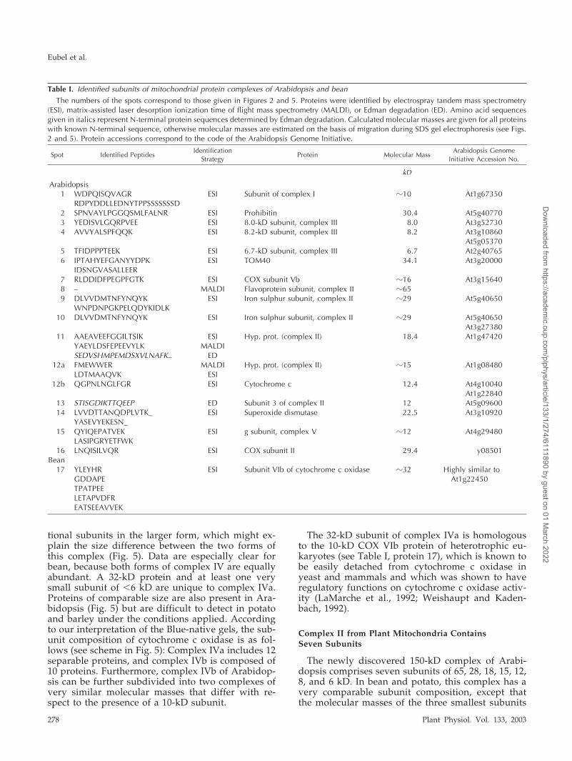
Table I. Identified subunits of mitochondrial protein complexes of Arabidopsis and bean
The numbers of the spots correspond to those given in Figures 2 and 5. Proteins were identified by electrospray tandem mass spectrometry(ESI), matrix-assisted laser desorption ionization time of flight mass spectrometry (MALDI), or Edman degradation (ED). Amino acid sequencesgiven in italics represent N-terminal protein sequences determined by Edman degradation. Calculated molecular masses are given for all proteinswith known N-terminal sequence, otherwise molecular masses are estimated on the basis of migration during SDS gel electrophoresis (see Figs.2 and 5). Protein accessions correspond to the code of the Arabidopsis Genome Initiative.
Spot Identified PeptidesIdentification
StrategyProtein Molecular Mass
Arabidopsis GenomeInitiative Accession No.
kD
Arabidopsis1 WDPQISQVAGR ESI Subunit of complex I �10 At1g67350
RDPYDDLLEDNYTPPSSSSSSSD2 SPNVAYLPGGQSMLFALNR ESI Prohibitin 30.4 At5g407703 YEDISVLGQRPVEE ESI 8.0-kD subunit, complex III 8.0 At3g527304 AVVYALSPFQQK ESI 8.2-kD subunit, complex III 8.2 At3g10860
At5g053705 TFIDPPPTEEK ESI 6.7-kD subunit, complex III 6.7 At2g407656 IPTAHYEFGANYYDPK ESI TOM40 34.1 At3g20000
IDSNGVASALLEER7 RLDDIDFPEGPFGTK ESI COX subunit Vb �16 At3g156408 – MALDI Flavoprotein subunit, complex II �659 DLVVDMTNFYNQYK ESI Iron sulphur subunit, complex II �29 At5g40650
WNPDNPGKPELQDYKIDLK10 DLVVDMTNFYNQYK ESI Iron sulphur subunit, complex II �29 At5g40650
At3g2738011 AAEAVEEFGGILTSIK ESI Hyp. prot. (complex II) 18.4 At1g47420
YAEYLDSFEPEEVYLK MALDISEDVSHMPEMDSXVLNAFK� ED
12a FMEWWER MALDI Hyp. prot. (complex II) �15 At1g08480LDTMAAQVK ESI
12b QGPNLNGLFGR ESI Cytochrome c 12.4 At4g10040At1g22840
13 STISGDIKTTQEEP ED Subunit 3 of complex II 12 At5g0960014 LVVDTTANQDPLVTK_ ESI Superoxide dismutase 22.5 At3g10920
YASEVYEKESN_15 QYIQEPATVEK ESI g subunit, complex V �12 At4g29480
LASIPGRYETFWK16 LNQISILVQR ESI COX subunit II 29.4 y08501
Bean17 YLEYHR ESI Subunit VIb of cytochrome c oxidase �32 Highly similar to
GDDAPE At1g22450TPATPEELETAPVDFREATSEEAVVEK
Eubel et al.
278 Plant Physiol. Vol. 133, 2003
Dow
nloaded from https://academ
ic.oup.com/plphys/article/133/1/274/6111890 by guest on 01 M
arch 2022
slightly vary (Fig. 4). To identify the 150-kD complex,subunits were subjected to analysis by mass spec-trometry. Peptide sequences of five of the seven sub-units allowed identification of corresponding genesof the Arabidopsis genome (Fig. 6; Table I). Surpris-ingly the 65-, 28-, and 12-kD proteins could be iden-tified as being subunits of the succinate dehydroge-nase complex (complex II) of the respiratory chain:the flavoprotein subunit (SDH1), the iron-sulfur sub-unit (SDH2), and the so-called subunit III (SDH3).Succinate dehydrogenase is well characterized forseveral bacteria, fungi, and mammals and is knownto be a four-subunit complex comprising the abovementioned subunits and one additional subunit inthe size range of 10 to 15 kD termed subunit IV orSDH4 (Lemire and Oyedotun, 2002; Yankovskaya etal., 2003). Although the subunits of this protein com-plex from plants were never biochemically character-ized, counterparts of the SDH1-SDH4 proteins couldbe identified on the basis of sequence similarities ofpredicted proteins with known complex II subunitsfrom mammals, fungi, algae, and protozoans(Daignan-Fornier et al., 1994; Burger et al., 1996;Figueroa et al., 2001; Figueroa et al., 2002). However,sequence identities for the SDH3 and SDH4 proteins,which constitute a hydrophobic membrane anchor ofthis protein complex, are very low (Burger et al.,1996). Most interestingly, complex II from plantsseems to contain additional subunits of unknownfunction. The 18-kD protein corresponds to the pu-tative At1g47420 protein of Arabidopsis, which wasidentified previously in the course of a proteomicapproach to characterize novel mitochondrial pro-teins of this organisms (Kruft et al., 2001; protein No4). Furthermore, the 15-kD protein represents the
putative At1g08480 protein. However, presence ofthis protein within complex II is slightly uncertain,because one of the three identified peptides perfectlymatches to Arabidopsis cytochrome c. Hence the cor-responding protein spot contains more than one pro-tein. The 8- and 6-kD subunits could not be identifiedby mass spectrometry; one of these subunits mightrepresent the SDH4 protein.
Most likely all seven proteins of plant succinatedehydrogenase are single-copy subunits of the150-kD complex, because the sum of their apparentmolecular masses (153 kD) is very close to the appar-ent molecular mass of this protein complex on Blue-native gels (150 kD). Furthermore, probably no addi-tional subunits form part of this complex.
The AOX Does Not Form Part ofRespiratory Supercomplexes
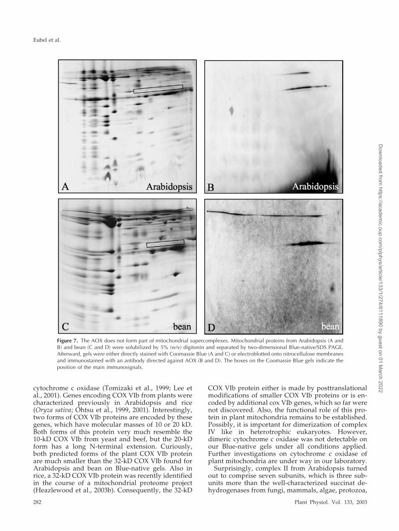
AOX represents a characteristic oxidoreductase ofthe respiratory chain of plant mitochondria. Its pos-sible association with the complexes I to V afterdigitonin-solubilization of mitochondrial proteinfractions was investigated by two-dimensional Blue-native/SDS gel electrophoresis and immunoblottingusing a monoclonal antibody directed against AOXfrom Sauromatum guttatum (Elthon et al. 1989). On thetwo-dimensional gels, the antibody reacts with a pro-tein of 32 kD in bean and Arabidopsis, which formsa smear on the first gel dimension between 30 and300 kD (Fig. 7). Therefore, AOX seems not to formpart of any supercomplexes but rather aggregatesunder the conditions applied. Also the rotenone-insensitive NAD(P)H dehydrogenases are not at-tached to respiratory protein complexes or super-complexes as investigated by similar experimentsusing antibodies directed against these proteins (datanot shown).
DISCUSSION
Supercomplexes in Plant Mitochondria
Respiratory protein complexes form supercom-plexes in plant mitochondria. In the course of ourinvestigations, V2, III2I, and III4I2 supercomplexescould be identified. Possibly further supercomplexesexist in vivo that are instable in the presence ofdetergents and Coomassie Blue. Digitonin proved tobe the optimal compound for supercomplex solubi-lization, which is surprising because it specificallybinds sterols, which are believed to be absent in innermitochondrial membranes. Furthermore, digitoninalso allows stable extraction of singular protein com-plexes that cannot be solubilized by dodecylmalto-side or Triton X-100. It therefore is a very suitabletool for plant mitochondrial research.
Between 50% and 90% of complex I from plantsforms part of the III2I supercomplex on Blue-nativegels upon digitonin solubilizations. In contrast, the
Figure 3. Separation of supercomplexes into protein complexes bytwo-dimensional Blue-native/Blue-native gel electrophoresis. Identi-ties of protein complexes are given above and to the left of the gel (fordesignations, see Fig. 1).
Respiratory Supercomplexes in Plants
Plant Physiol. Vol. 133, 2003 279
Dow
nloaded from https://academ
ic.oup.com/plphys/article/133/1/274/6111890 by guest on 01 M
arch 2022
same supercomplex from beef only contains 17% ofcomplex I under identical conditions (Schagger,2002). However, more than 50% of complex I frombeef is part of even larger supercomplexes that in-clude dimeric complex III and additionally one tofour copies of complex IV. In Brewer’s yeast, complexI-containing supercomplexes are absent due to thegeneral absence of this protein complex, but also inthis organism, the complexes IV and III are associ-ated forming III2IV or III2IV2 supercomplexes (Schag-ger and Pfeiffer, 2000; Cruciat et al., 2000). In con-trast, associations of complexes III and IV of plantmitochondria are not detectable on Blue-native gelsunder all conditions applied. Also the AOX and therotenone-insensitive NAD(P)H dehydrogenases
seem not to be present in supercomplexes. Further-more, plant complex II only is present in singularform, which is in line with findings for yeast andmammals. Dimeric complex V from plants is presenton Blue-native gels after solubilizations using lowconcentrations of detergents, but its abundance islower than in other organisms (Arnold et al., 1998,1999). Recently, a very stable dimeric ATP synthasecomplex was reported for mitochondria fromChlamydomonas reinhardtii (van Lis et al., 2003).
What is the functional role of supercomplexes inplant cells? In yeast, supercomplexes were reportedto enhance activity rates of respiratory electron trans-port (Schagger and Pfeiffer, 2000). Furthermore, it isspeculated that supercomplex formation increases
Figure 4. Mitochondrial supercomplexes and protein complexes in Arabidopsis, potato, bean, and barley. Proteins weresolubilized by 5 g per g digitonin and separated by two-dimensional Blue-native/SDS PAGE. Identities of protein complexesare given above the gels (for designations, see Fig. 1).
Eubel et al.
280 Plant Physiol. Vol. 133, 2003
Dow
nloaded from https://academ
ic.oup.com/plphys/article/133/1/274/6111890 by guest on 01 M
arch 2022

the capacity of the inner mitochondrial membrane forprotein insertion (Arnold et al., 1998). The proteincontent of this mitochondrial membrane, which isestimated to lie at about 70%, can only be realized ifproteins are very efficiently packed. In plant mito-chondria, the III2I supercomplex possibly has impor-tant consequences for the regulation of alternativerespiration, because it might reduce access of AOX toits substrate ubiquinol. Because alternative respira-tion is known to increase under various stress con-ditions (Vanlerberghe and McIntosh, 1997), theoccurrence of respiratory supercomplexes in Arabi-dopsis was investigated in mitochondria isolatedfrom suspension cell cultures that were treated withantimycin A, a known inhibitor of complex III. How-ever, our initial data reveal only small differencesconcerning respiratory supercomplexes in antimycin-treated and untreated cells, which are at the border-line of significance (data not shown). Therefore therole of supercomplexes in plant mitochondria has tobe further investigated.
Respiratory Protein Complexes in Plant Mitochondria
Recently, the subunit compositions of protein com-plexes of the oxidative phosphorylation system ofArabidopsis were studied intensively. Complex I fromplants can be resolved into 27 to 30 different subunitson two-dimensional Blue-native/SDS gels (Fig. 8) butpossibly comprises more than 40 proteins (Rasmussonet al., 1998). Heazlewood et al. (2003a) identified 30subunits of Arabidopsis complex I after separation on
two-dimensional gels by mass spectrometry. Severalof the identified proteins have counterparts in fungiand mammals, but others seem to be unique to plants.Using a similar approach, Heazlewood et al. (2003c)identified 10 subunits of Arabidopsis complex V.Some further subunits remain to be characterized, be-cause up to 13 proteins can be resolved on two-dimensional gels (Fig. 8). All 10 subunits of potatocomplex III were biochemically characterized (for re-view, see Braun and Schmitz, 1995) and counterpartsfor all 10 subunits are present in Arabidopsis proteindatabases at The Institute for Genomic Research or theMunich Information Center for Protein Sequences (�-MPP subunit, At3g02090; �-MPP subunit, At1g51980and At3g16480; cytochrome b, Y08501; cytochrome c1,At5g40810 and At3g27240; ‘Rieske FeS’ protein,At5g13440 and At5g13430; counterpart to 14-kD sub-unit from potato, At4g32470 and At5g25450; counter-part to 7.8-kD subunit from potato, At2g01090 andAt1g15120; counterpart to potato 8.0-kD subunit,At5g05370 and At3g10860; counterpart to potato8.2-kD subunit, At3g52730; counterpart to potato6.7-kD subunit, At2g40765).
The least characterized respiratory protein com-plexes of plants are the complexes IV and II. Arabi-dopsis complex IV can be resolved in two differentforms on Blue-native gels, which comprise 10 to 12subunits (Figs. 5 and 8). The identity of the fivelargest subunits is known, whereas the identity ofmost smaller subunits remains to be established. Thelarger form of cytochrome c oxidase includes an ad-ditional 32-kD protein, which resembles the 10-kDsubunit COX VIb of yeast and beef. This subunit isvery hydrophilic, lacks membrane spanning helices,and is localized on the intermembrane-space side ofcytochrome c oxidase (Tomizaki et al., 1999). Re-moval of this protein from complex IV was shown toactivate cytochrome c oxidase of beef (Weishauptand Kadenbach, 1992). Furthermore, COX VIb frombeef was shown to be important for dimerization of
Figure 5. Large and small forms of cytochrome c oxidase in bean andArabidopsis after two-dimensional resolution by Blue-native/SDSPAGE. Proteins only forming part of the larger form of this proteincomplex are indicated by arrows. Protein number 17 was identifiedby mass spectrometry (Table I). A scheme of the two-dimensionalBlue-native/SDS gel of cytochrome c oxidase from Arabidopsis isgiven to the right.
Figure 6. Subunit composition of complex II in Arabidopsis. Appar-ent molecular masses of the subunits and the accession numbers ofthe corresponding genes are given to the right of the gel.
Respiratory Supercomplexes in Plants
Plant Physiol. Vol. 133, 2003 281
Dow
nloaded from https://academ
ic.oup.com/plphys/article/133/1/274/6111890 by guest on 01 M
arch 2022
cytochrome c oxidase (Tomizaki et al., 1999; Lee etal., 2001). Genes encoding COX VIb from plants werecharacterized previously in Arabidopsis and rice(Oryza sativa; Ohtsu et al., 1999, 2001). Interestingly,two forms of COX VIb proteins are encoded by thesegenes, which have molecular masses of 10 or 20 kD.Both forms of this protein very much resemble the10-kD COX VIb from yeast and beef, but the 20-kDform has a long N-terminal extension. Curiously,both predicted forms of the plant COX VIb proteinare much smaller than the 32-kD COX VIb found forArabidopsis and bean on Blue-native gels. Also inrice, a 32-kD COX VIb protein was recently identifiedin the course of a mitochondrial proteome project(Heazlewood et al., 2003b). Consequently, the 32-kD
COX VIb protein either is made by posttranslationalmodifications of smaller COX VIb proteins or is en-coded by additional cox VIb genes, which so far werenot discovered. Also, the functional role of this pro-tein in plant mitochondria remains to be established.Possibly, it is important for dimerization of complexIV like in heterotrophic eukaryotes. However,dimeric cytochrome c oxidase was not detectable onour Blue-native gels under all conditions applied.Further investigations on cytochrome c oxidase ofplant mitochondria are under way in our laboratory.
Surprisingly, complex II from Arabidopsis turnedout to comprise seven subunits, which is three sub-units more than the well-characterized succinat de-hydrogenases from fungi, mammals, algae, protozoa,
Figure 7. The AOX does not form part of mitochondrial supercomplexes. Mitochondrial proteins from Arabidopsis (A andB) and bean (C and D) were solubilized by 5% (w/v) digitonin and separated by two-dimensional Blue-native/SDS PAGE.Afterward, gels were either directly stained with Coomassie Blue (A and C) or electroblotted onto nitrocellulose membranesand immunostained with an antibody directed against AOX (B and D). The boxes on the Coomassie Blue gels indicate theposition of the main immunosignals.
Eubel et al.
282 Plant Physiol. Vol. 133, 2003
Dow
nloaded from https://academ
ic.oup.com/plphys/article/133/1/274/6111890 by guest on 01 M
arch 2022
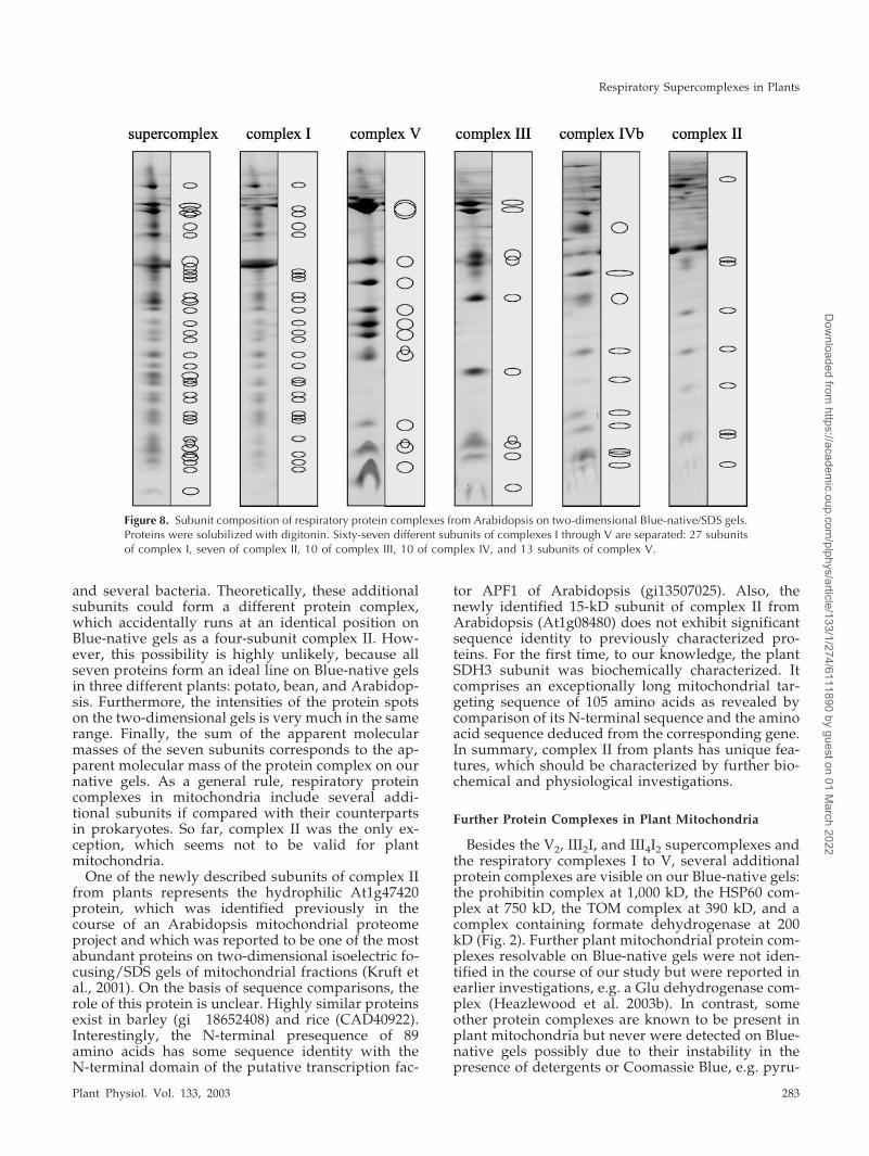
and several bacteria. Theoretically, these additionalsubunits could form a different protein complex,which accidentally runs at an identical position onBlue-native gels as a four-subunit complex II. How-ever, this possibility is highly unlikely, because allseven proteins form an ideal line on Blue-native gelsin three different plants: potato, bean, and Arabidop-sis. Furthermore, the intensities of the protein spotson the two-dimensional gels is very much in the samerange. Finally, the sum of the apparent molecularmasses of the seven subunits corresponds to the ap-parent molecular mass of the protein complex on ournative gels. As a general rule, respiratory proteincomplexes in mitochondria include several addi-tional subunits if compared with their counterpartsin prokaryotes. So far, complex II was the only ex-ception, which seems not to be valid for plantmitochondria.
One of the newly described subunits of complex IIfrom plants represents the hydrophilic At1g47420protein, which was identified previously in thecourse of an Arabidopsis mitochondrial proteomeproject and which was reported to be one of the mostabundant proteins on two-dimensional isoelectric fo-cusing/SDS gels of mitochondrial fractions (Kruft etal., 2001). On the basis of sequence comparisons, therole of this protein is unclear. Highly similar proteinsexist in barley (gi 18652408) and rice (CAD40922).Interestingly, the N-terminal presequence of 89amino acids has some sequence identity with theN-terminal domain of the putative transcription fac-
tor APF1 of Arabidopsis (gi13507025). Also, thenewly identified 15-kD subunit of complex II fromArabidopsis (At1g08480) does not exhibit significantsequence identity to previously characterized pro-teins. For the first time, to our knowledge, the plantSDH3 subunit was biochemically characterized. Itcomprises an exceptionally long mitochondrial tar-geting sequence of 105 amino acids as revealed bycomparison of its N-terminal sequence and the aminoacid sequence deduced from the corresponding gene.In summary, complex II from plants has unique fea-tures, which should be characterized by further bio-chemical and physiological investigations.
Further Protein Complexes in Plant Mitochondria
Besides the V2, III2I, and III4I2 supercomplexes andthe respiratory complexes I to V, several additionalprotein complexes are visible on our Blue-native gels:the prohibitin complex at 1,000 kD, the HSP60 com-plex at 750 kD, the TOM complex at 390 kD, and acomplex containing formate dehydrogenase at 200kD (Fig. 2). Further plant mitochondrial protein com-plexes resolvable on Blue-native gels were not iden-tified in the course of our study but were reported inearlier investigations, e.g. a Glu dehydrogenase com-plex (Heazlewood et al. 2003b). In contrast, someother protein complexes are known to be present inplant mitochondria but never were detected on Blue-native gels possibly due to their instability in thepresence of detergents or Coomassie Blue, e.g. pyru-
Figure 8. Subunit composition of respiratory protein complexes from Arabidopsis on two-dimensional Blue-native/SDS gels.Proteins were solubilized with digitonin. Sixty-seven different subunits of complexes I through V are separated: 27 subunitsof complex I, seven of complex II, 10 of complex III, 10 of complex IV, and 13 subunits of complex V.
Respiratory Supercomplexes in Plants
Plant Physiol. Vol. 133, 2003 283
Dow
nloaded from https://academ
ic.oup.com/plphys/article/133/1/274/6111890 by guest on 01 M
arch 2022
vate dehydrogenase or the so-called AAA complexes.Recently, the occurrence of protein complexes com-prising mitochondrial dehydrogenases of the citricacid cycle was reported on the basis of diffusion ratemeasurements of individual enzymes of this meta-bolic pathway (Haggie and Verkman, 2002). Mostlikely, these protein complexes are too unstable forbiochemical preparations. Protein complexes and su-percomplexes offer several physiological advantagesin comparison with singular proteins, including sub-strate channeling, metabolic pathway regulation, andthe realization of complicated biochemical reactionswith reactive intermediates. Therefore the majority ofmitochondrial proteins probably form part of proteincomplexes, and possibly most protein complexes areinvolved in the formation of even larger supermolec-ular structures, which remain to be discovered.
MATERIALS AND METHODS
Isolation of Mitochondria from Arabidopsis, Bean(Phaseolus vulgaris), Potato (Solanum tuberosum), andBarley (Hordeum vulgare)
Starting material for plant mitochondrial preparations were non-greenArabidopsis suspension cell cultures, potato tubers, 6-d-old etiolated barleyseedlings, and 18-d-old etiolated bean seedlings. Arabidopsis cell lines werecultivated in the dark at 24°C to 26°C, 30% humidity, and gentle shaking (90rpm) as described previously (Werhahn et al., 2001), and etiolated seedlingswere grown at 24°C. All organelle preparations were carried out on the basisof filtration, differential centrifugation, and Percoll density centrifugation asoutlined by Werhahn et al. (2001) for Arabidopsis; Focke et al. (2003) forbarley; and Braun et al. (1992b) for potato and bean. Purified organelleswere finally resuspended in a buffer containing 0.4 m mannitol, 1 mm EGTA,0.2 mm phenylmethylsulfonyl fluoride (PMSF), and 10 mm Tricine/KOH,pH 7.2, at a protein concentration of 10 mg mL�1, divided into aliquots of100 �L, and directly used for investigations (the amount of some supercom-plexes was significantly reduced if mitochondrial fractions were frozen andstored before analyses).
Solubilization of Mitochondrial Proteins
Mitochondrial aliquots were centrifuged for 10 min at 14,300g, andsedimented organelles were resuspended in one of the following buffers(conditions adopted from Arnold et al., 1998; Schagger, 2001): (a) 100 �L ofdigitonin solubilization buffer (30 mm HEPES pH 7.4, 150 mm potassiumacetate, 10% [v/v] glycerol, 2 mm PMSF, and [1–10 g per g protein] digitonin[Fluka, Buchs, Switzerland]); (b) 100 �L of dodecylmaltosid solubilizationbuffer (750 mm aminocaproic acid, 50 mm BisTris, pH 7.0, 0.5 mm EDTA, 1mm PMSF, and docedylmaltoside [0.1–2 g per g protein; Roche, Mannheim,Germany]; and (c) 100 �L of Triton solubilization buffer (50 mm NaCl, 2 mmaminocaproic acid, 1 mm EDTA, 50 mm imidazole-HCl, pH 7.4, 10% glyc-erol, 5 mm PMSF, and Triton X-100 [0.1–2 g per g protein; Amersham-Pharmacia-Biotech Uppsala].
After incubation for 20 min on ice, samples were centrifuged at 18,000gfor 30 min to remove insoluble material and were subsequently supple-mented with 5 �L of Coomassie Blue solution (5% [w/v] Coomassie Blue in750 mm aminocaproic acid). Dodecylmaltoside-solubilized samples werecentrifuged immediately after resuspension of organelles in solubilizationbuffer and afterward were supplemented with 20 �L of Coomassie Bluesolution. Coomassie Blue-treated protein samples were directly loaded ontoBlue-native gels.
Two-Dimensional Blue-Native/SDS PAGE
One-dimensional Blue-native PAGE and two-dimensional Blue-native/SDS PAGE were carried out as described by Schagger (2001b). Gradient gels
(4.5%–16% [w/v] acrylamide) were used for the Blue-native gel dimensionsand two-step Tricine-SDS gels (10% and 16% [w/v] acrylamide) for secondgel dimensions. The cathode buffer of Blue-native gel dimensions did notinclude detergent; only for electrophoresis of dodecylmaltoside-solubilizedsamples 0.03% of the detergent was added. Gels were either stained withCoomassie-colloidal (Neuhoff et al., 1985, 1990) or with silver (Heukeshovenand Dernick, 1986)
Two-Dimensional Blue-Native/Blue NativeGel Electrophoresis
Two-dimensional Blue-native/Blue-native PAGE was carried out as pub-lished by Schagger and Pfeiffer (2000). It proved to be important to stopfirst-dimension electrophoresis runs after 50% completion to avoid proteincomplexes and supercomplexes getting stuck in the gels. In contrast, it wasimportant to extend the electrophoresis runs of second gel dimensions byfactor two, because protein complexes stuck in gels were best resolved.
Protein Preparations for Mass Spectrometry
For mass spectrometry, gels were colloidal stained with Coomassie Blue(Neuhoff et al., 1990) and single proteins were cut out, transferred into anEppendorf tube, and incubated with Milli-Q water for 10 min. Rebufferingwas carried out by incubating the gel pieces for 15 min in acetonitrile and 0.1m NH4HCO3, respectively. Subsequently, the proteins were dehydrated byacetonitrile and incubated with 20 �L of digestion solution (0.5 �g of trypsin[Promega, Madison, WI] in 20 �L of 50 mm NH4HCO3) overnight at 37°C.Peptide extraction was performed at 37°C as follows: Samples were supple-mented with 20 �L of 50 mm NH4HCO3 and shaken for 15 min, andafterward, supernatants were taken and stored. Gel pieces were then shakenfor 15 min in the presence of 20 �L of 5% (v/v) formic acid. Subsequently,the same volume of acetonitrile was added, and samples were shaken foranother 15 min. Afterward, all supernatants were pooled and dried down toa volume of about 10 �L. Purification of the generated peptides wasachieved using ZipTips (Millipore, Bedford, MA) according to the manu-facturer’s instructions.
Matrix-Assisted Laser Desorption Ionization/Time ofFlight Mass Spectrometry
Determination of the molecular masses of Zip-Tip purified peptides wascarried out by positive-ion matrix assisted laser desorption ionization/timeof flight mass spectrometry using an Ultraflex instrument (Bruker, Newark,DE) equipped with delayed-extraction and a N2 laser (337 nm). For eachsample, 1 �L of matrix solution (10 mg of �-cyano-4-hydroxycinnamic acidin 1 mL of 60% [v/v] acetonitrile/0.1% [v/v] formic acid) was placed on theScout ion source and crystallized as a thin layer. One microliter of samplewas given directly on the top of the thin matrix layer, and cocrystallizationwas carried out at room temperature. Spectra were recorded in reflectionmode with an acceleration voltage of 25 kV and a reflection voltage of 26.3kV. Monoisotopic masses from spectra were selected automatically andwere used for protein identification with the help of MASCOT (MatrixScience, London).
Electrospray Ionization Tandem Mass Spectrometry
For peptide sequencing, 3 �L of Zip-Tip purified sample was filled intoAu/Pd-coated nanospray glass capillaries (Protana, Odense, Denmark). Thetip of the capillary was placed orthogonally in front of the entrance hole ofa quadropole time-of-flight mass spectrometry instrument (Q-TOF II, Mi-cromass, Watres, Milford, MA) equipped with a nanospray ion source. Acapillary voltage between 750 and 1,000 V and a cone voltage of 30 V wasapplied. Two-fold charged peptides were chosen for collision-induced dis-sociation experiments, and the corresponding parent ions were selectivelytransmitted from the quadropole mass analyzer into the collision cell. Argonwas used as collision gas, and the kinetic energy was set between 20 and 40eV. The resulting daughter ions were separated by an orthogonal time-of-flight mass analyzer. Peptide sequencing and protein identification werecarried out with the programs PeptideSequencing of the BioLynx software
Eubel et al.
284 Plant Physiol. Vol. 133, 2003
Dow
nloaded from https://academ
ic.oup.com/plphys/article/133/1/274/6111890 by guest on 01 M
arch 2022
package (v3.5, Mircomass), Sonar of the Knexus software package (Proteo-metrics, Manitoba, Canada), and MASCOT (Matrix Science).
ACKNOWLEDGMENTS
We are very grateful to Tom Elton for providing antibodies directedagainst the AOX and to Jean-Michel Grienenberger and Sergei Kushnir forencouraging the presented work. Furthermore, we thank Dagmar Lewejo-hann for the cultivation of Arabidopsis suspension cell cultures and forexpert technical assistance.
Received April 1, 2003; returned for revision April 22, 2003; accepted May27, 2003.
LITERATURE CITED
Arnold I, Pfeiffer K, Neupert W, Stuart RA, Schagger H (1998) Yeastmitochondrial F1F0-ATP synthase exists as a dimer: identification of threedimer-specific subunits. EMBO J 17: 7170–7178
Arnold I, Pfeiffer K, Neupert W, Stuart RA, Schagger H (1999) ATPsynthase of yeast mitochondria: isolation of subunit j and disruption ofthe ATP18 gene. J Biol Chem 274: 36–40
Berry EA, Trumpower BL (1985) Isolation of ubiquinol oxidase from Para-coccus denitrificans and resolution into cytochrome bc1 and cytochromec-aa3 complexes. J Biol Chem 260: 2458–2467
Braun HP, Emmermann M, Kruft V, Schmitz UK (1992a) The generalmitochondrial processing peptidase from potato is an integral part ofcytochrome c reductase of the respiratory chain. EMBO J 11: 3219–3227
Braun HP, Emmermann M, Kruft V, Schmitz UK (1992b) Cytochrome c1
from potato: a protein with a presequence for targeting to the mitochon-drial intermembrane space. Mol Gen Genet 231: 217–225
Braun HP, Schmitz UK (1995) The bifunctional cytochrome c reductase/processing peptidase complex from plant mitochondria. J Bioenerg Bio-membr 27: 423–436
Brumme S, Kruft V, Schmitz UK, Braun HP (1998) New insights into theco-evolution of cytochrome c reductase and the mitochondrial processingpeptidase. J Biol Chem 273: 13143–13149
Burger G, Lang F, Reith M, Gray MW (1996) Genes encoding the same threesubunits of respiratory complex II are present in the mitochondrial DNAof two phylogenetically distinct eukaryotes. Proc Natl Acad Sci USA 93:2328–2332
Bykova NV, Moller IM (2003) Identification of 14 new phosphoproteinesinvolved in important plant mitochondrial functions. FEBS Lett 540:141–146
Cruciat CM, Brunner S, Baumann F, Neupert W, Stuart RA (2000) Thecytochrome bc1 and cytochrome c oxidase complexes associate to form asingle supracomplex in yeast mitochondria. J Biol Chem 275: 18093–18098
Daignan-Fornier B, Valens M, Lemire BD, Bolotin-Fukuhara M (1994)Structure and regulation of SDH3, the yeast gene encoding the cyto-chrome b560 subunit of respiratory complex II. J Biol Chem 269:15469–15472
Ducos E, Touzet P, Boutry M (2001) The male sterile G cytoplasm of wildbeet displays modified mitochondrial respiratory complexes. Plant J 26:171–180
Elthon TE, Nickels RL, McIntosh L (1989) Monoclonal antibodies to thealternative oxidase of higher plant mitochondria. Plant Physiol 89:1311–1317
Eriksson AC, Sjoling S, Glaser E (1994) The ubiquinol cytochrome c oxi-doreductase complex of spinach leaf mitochondria is involved in bothrespiration and protein processing. Biochim Biophys Acta 1186: 221–231
Figueroa P, Leon G, Elorza A, Holuigue L, Jordana X (2001) Three differentgenes encode the iron-sulfur subunit of succinate dehydrogenase inArabidopsis thaliana. Plant Mol Biol 46: 241–250
Figueroa P, Leon G, Elorza A, Holuigue L, Araya A, Jordana X (2002) Thefour subunits of the mitochondrial respiratory complex II are encoded bythe multiple nuclear genes and targeted to mitochondria in Arabidopsisthaliana. Plant Mol Biol 50: 725–734
Focke M, Gieringer E, Schwan S, Jänsch L, Binder S, Braun HP (2003) Fattyacid biosynthesis in mitochondrial from grasses: Malouyl-CoA is gener-ated by a mitochondrial-localized acetyl-CoA carboxylase. Plant Physiol(in press)
Haggie PM, Verkman AS (2002) Diffusion of tricarboxylic acid cycle en-zymes in the mitochondrial matrix: evidence for restricted mobility of amultienzyme complex. J Biol Chem 277: 40782–40788
Heazlewood JA, Howell KA, Millar AH (2003a) Mitochondrial complex Ifrom Arabidopsis and rice: orthologs of mammalian and yeast compo-nents coupled to plant-specific subunits. Biochim Biophys Acta 1604:159–169
Heazlewood JL, Howell KA, Whelan J, Millar AH (2003b) Towards ananalysis of the rice mitochondrial proteome. Plant Physiol 132: 230–242
Heazlewood JL, Whelan J, Millar AH (2003c) The products of the mito-chondrial ORF25 and ORFB genes are FO components of the plant F1FO
ATP synthase. FEBS Lett 540: 201–205Heukeshoven J, Dernick R (1986) Silver staining of proteins. In Electro-
phoresis Forum ’86, B.J. Radula, ed. Elektrophoresis Forum ’86. In B.J.Radola, ed, Technische Universitat Munchen. pp 22–27
Iwasaki T, Matsuura K, Oshima T (1995) Resolution of the aerobic respi-ratory system of the thermoacidophilic archaeon, Sulfolobus sp. strain 7: I.The archael terminal oxidase supercomplex is a functional fusion ofrespiratory complexes III and IV with no c-type cytochromes. J Biol Chem270: 30881–30892
Jansch L, Kruft V, Schmitz UK, Braun HP (1995) Cytochrome c reductasefrom potato does not comprise three core proteins but contains an addi-tional low molecular weight subunit. Eur J Biochem 228: 878–885
Jansch L, Kruft V, Schmitz UK, Braun HP (1996) New insights into thecomposition, molecular mass and stoichiometry of the protein complexesof plant mitochondria. Plant J 9: 357–368
Karpowa OV, Newton KJ (1999) A partially assembled complex I in NAD4-deficient mitochondria of maize. Plant J 17: 511–521
Kruft V, Eubel H, Werhahn W, Jansch L, Braun HP (2001) Proteomicapproach to identify novel mitochondrial functions in Arabidopsis thali-ana. Plant Physiol 127: 1694–1710
Kugler M, Brumme S, Jansch L, Werhahn W, Schmitz UK, Braun HP (1998)Characterization of plant mitochondria by blue native polyacrylamidegel electrophoresis (BN-PAGE). In IM Moller, P Gardestrom, K Glimelius,E Glaser, eds, Plant Mitochondria: From Gene to Function. BlackhuysPublishers, Leiden, The Netherlands, pp 273–276
LaMarche AE, Abata MI, Chan SH, Trumpower BL (1992) Isolation andcharacterization of COX12, the nuclear gene for a previously unrecog-nised subunit of Saccharomyces cerevisiae cytochrome c oxidase. J BiolChem 267: 22473–22480
Lee SJ, Yamashita E, Abe T, Fukumoto Y, Tsukihara T, Shinzawa IK, UedaH, Yoshikawa S (2001) Intermonomer interactions in dimer of bovineheart cytochrome c oxidase. Acta Crystallogr D Biol Crystallogr 57:941–947
Lemire BL, Oyedotun KS (2002) The Saccharomyces cerevisiae mitochondrialsuccinate:ubiquinone oxidoreductase. Biochim Biophys Acta 1553:102–116
Mackenzie S, McIntosh L (1999) Higher plant mitochondria. Plant Cell 11:571–585
Mihr C, Baumgartner M, Dieterich JH, Schmitz UK, Braun HP (2001)Proteomic approach for investigation of cytoplasmic male sterility (CMS)in Brassica. J Plant Physiol 158: 787–794
Neuhoff V, Stamm R, Eibl H (1985) Clear background and highly sensitiveprotein staining with Coomassie Blue dyes in polyacrylamide gels: asystematic analysis. Electrophoresis 6: 427–448
Neuhoff V, Stamm R, Pardowitz I, Arold N, Ehrhardt W, Taube D (1990)Essential problems in quantification of proteins following colloidal stain-ing with Coomassie Brilliant Blue dyes in polyacrylamide gels, and theirsolution. Electrophoresis 11: 101–117
Niebisch A, Bott M (2003) Purification of a cytochrome bc1-aa3 supercom-plex with quinol oxidase activity from Corynebacterium glutamicum: iden-tification of a fourth subunit of cytochrome aa3 oxidase and mutationalanalysis of diheme cytochrome c1. J Biol Chem 278: 4339–4346
Ohtsu K, Hamanaka S, Yamazaki K, Nakazono M, Hirai A (1999) Charac-terization of a cDNA encoding a novel subunit for cytochrome c oxidase(COX6b) from rice. Breed Sci 49: 211–215
Ohtsu K, Nakazono M, Tsutsumi N, Hirai A (2001) Characterization andexpression of the genes for cytochrome c oxidase subunit VIb (COX6b)from rice and Arabidopsis thaliana. Gene 264: 233–239
Rasmusson AG, Agius SC (2001) Rotenone-insensitive NAD(P)H dehydro-genases in plants: immunodetection and distribution of native proteins inmitochondria. Plant Physiol Biochem 39: 1057–1066
Respiratory Supercomplexes in Plants
Plant Physiol. Vol. 133, 2003 285
Dow
nloaded from https://academ
ic.oup.com/plphys/article/133/1/274/6111890 by guest on 01 M
arch 2022
Rasmusson AG, Heiser VV, Zabaleta E, Brennicke A, Grohmann L (1998)Physiological, biochemical and molecular aspects of mitochondrial com-plex I in plants. Biochim Biophys Acta 1364: 101–111
Rasmusson AG, Svensson AS, Knoop V, Grohmann L, Brennicke A (1999)Homologues of yeast and bacterial rotenone-insensitive NADH dehydro-genases in higher eukaryotes: two enzymes are present in potato mito-chondria. Plant J 20: 79–87
Rich PR (1984) Electron and proton transfer through quinones and cyto-chrome bc complexes. Biochim Biophys Acta 768: 53–79
Sabar M, Gagliardi D, Balk J, Leaver CJ (2003) ORFB is a subunit ofF(1)F(O)-ATP synthase: insight into the basis of cytoplasmic male sterilityin sunflower. EMBO Rep 4: 1–6
Schagger H (2001a) Respiratory chain supercomplexes. International Unionof Biochemistry and Molecular Biology (IUBMB) Life 52: 119–128
Schagger H (2001b) Blue-native gels to isolate protein complexes frommitochondria. Methods Cell Biol 65: 231–244
Schagger H (2002) Respiratory supercomplexes of mitochondria and bacte-ria. Biochim Biophys Acta 1555: 154–159
Schagger H, Pfeiffer K (2000) Supercomplexes in the respiratory chains ofyeast and mammalian mitochondria. EMBO J 19: 1777–1783
Schagger H, Pfeiffer K (2001) The ratio of oxidative phosphorylation com-plexes I-V in bovine heart mitochondria and the composition of respira-tory chain supercomplexes. J Biol Chem 276: 37861–37867
Siedow JN, Umbach AL (1995) Plant mitochondrial electron transfer andmolecular biology. Plant Cell 7: 821–831
Sone N, Sekimachi M, Kutoh E (1987) Identification and properties of aquinol oxidase supercomplex composed of a bc1 complex and cyto-chrome oxidase in the thermophilic bacterium PS3. J Biol Chem 262:15386–15391
Tomizaki T, Yamashita E, Yamaguchi H, Aoyama H, Tsukihara T,Shinzawa-Itoh K, Nakashima R, Yaono R, Yoshikawa S (1999) Structure
analysis of bovine heart cytochrome c oxidase at 2.8 Å resolution. ActaCrystallogr 55: 31–45
Vanlerberghe GC, McIntosh L (1997) Alternative oxidase: from gene tofunction. Annu Rev Plant Physiol Plant Mol Biol 48: 703–734
van Lis R, Atteia A, Mendoza-Hernandez G, Gonzalez-Halphen D (2003)Identification of novel mitochondrial protein components of Chlamydo-monas reinhardtii: a proteomic approach. Plant Physiol 132: 318–330
Vedel F, Lalanne E, Sabar M, Chetrit P, de Paepe R (1999) The mitochon-drial respiratory chain and ATP synthase complexes: composition, struc-ture and mutational studies. Plant Physiol Biochem 37: 629–643
Weishaupt A, Kadenbach B (1992) Selective removal of subunit VIb in-creases the activity of cytochrome c oxidase. Biochemistry 46:11477–11481
Werhahn W, Braun HP (2002) Biochemical dissection of the mitochondrialproteome from Arabidopsis thaliana by three-dimensional gel electro-phoresis. Electrophoresis 23: 640–646
Werhahn W, Jansch L, Braun HP (2003) Identification of novel subunits ofthe TOM complex from Arabidopsis thaliana. Plant Physiol Biochem 41:407–416
Werhahn W, Niemeyer A, Jansch L, Kruft V, Schmitz UK, Braun HP (2001)Purification and characterization of the preprotein translocase of theouter mitochondrial membrane from Arabidopsis thaliana: identification ofmultiple forms of TOM20. Plant Physiol 125: 943–954
Yankovskaya V, Horsefield R, Tornroth S, Luna-Chavez C, Miyoshi H,Leger C, Byrne B, Cecchini G, Iwata S (2003) Architecture of succinatedehydrogenase and reactive oxygen species generation. Science 299:700–704
Zhang M, Mileykovskaya E, Dowhan W (2002) Gluing the respiratorychain together: Cardiolipin is required for supercomplex formation in theinner mitochondrial membrane. J Biol Chem 277: 43553–43556
Eubel et al.
286 Plant Physiol. Vol. 133, 2003
Dow
nloaded from https://academ
ic.oup.com/plphys/article/133/1/274/6111890 by guest on 01 M
arch 2022


![Structure and function of mitochondrial supercomplexes...newest edition of the Lehninger textbook is the first general textbook that presents a section on OXPHOS supercomplexes [17]](https://img.pdfslide.net/doc/110x75/5f7f11723a4eb942540eb7d2/structure-and-function-of-mitochondrial-supercomplexes-newest-edition-of-the.jpg)