Embed Size (px)
Citation preview
34
Received:August 25, 2017, Revised:September 28, 2017, Accepted:October 16, 2017
Corresponding to:Kwang Nam Kim, Department of Pediatrics, Hallym University Sacred Heart Hospital, 22 Gwanpyeong-ro 170beon-gil, Dongan-gu, Anyang 14068, Korea. E-mail:[email protected]
Copyright ⓒ 2018 by The Korean College of Rheumatology. All rights reserved.This is a Open Access article, which permits unrestricted non-commerical use, distribution, and reproduction in any medium, provided the original work is properly cited.
Original ArticlepISSN: 2093-940X, eISSN: 2233-4718Journal of Rheumatic Diseases Vol. 25, No. 1, January, 2018https://doi.org/10.4078/jrd.2018.25.1.34
New Provisional Classification of Juvenile Idiopathic Arthritis Applying Rheumatoid Factor and Antinuclear Antibody
Hyuck Jin Kwon, Myung Hoon Bang, Kwang Nam Kim Department of Pediatrics, Hallym University Sacred Heart Hospital, Anyang, Korea
Objective. Previous classification systems for juvenile idiopathic arthritis (JIA) were based on the number of joints involved and did not categorize homogenous disease entities. Therefore, JIA patients were reclassified retrospectively by applying rheuma-toid factor (RF) and antinuclear antibody (ANA), which have been proven to constitute a homogenous disease entity. Methods. The medical records of JIA patients were investigated retrospectively and reclassified into six categories using the new provi-sional classification. The nomenclature was based on Dr. Martini’s proposal in the 23rd European Paediatric Rheumatology Congress (2016) at Genoa, Italy. New categories included systemic JIA (sJIA), RF-positive JIA (RF-JIA), early-onset ANA-positive JIA (eoANA-JIA), enthesitis/spondylitis-related JIA (ESR-JIA), “other JIA”, and “unclassified JIA”. Results. Of a total of 262 JIA pa-tients, 71 (27.1%) were reclassified as sJIA, 31 (11.8%) as RF-JIA, 22 (8.4%) as eoANA-JIA, 63 (24.0%) as ESR-JIA, 65 (24.8%) as “other JIA”, and 10 (3.8%) as “unclassified JIA”. A comparison of RF-JIA, eoANA-JIA, and ESR-JIA revealed significant differ-ences in the gender ratio, age of disease onset, and the cumulative number and type of joints involved among the three groups. “Other JIA” comprised a significant proportion (24.8%) and warrants the need for further classification. The characteristics of the RF-positive patients were comparable to those of the anti-cyclic citrullinated peptide antibody-positive patients. The ANA positivity was lower (28.2%) than that in Western studies but showed similar clinical features. Conclusion. This is the first study applying RF and ANA to classify JIA without considering the joint counts. The six new categories include sJIA, RF-JIA, eoANA-JIA, ESR-JIA, “other JIA,” and “unclassified JIA”. (J Rheum Dis 2018;25:34-46)
Key Words. Juvenile idiopathic arthritis, Classification, Rheumatoid factor, Antinuclear antibody
INTRODUCTION
The term “juvenile idiopathic arthritis (JIA)” was first proposed by the International League of Associations for Rheumatology (ILAR) in Santiago (1994) and was re-vised twice in Durban (1997) and in Edmonton (2001) [1-3]. It encompasses all forms of arthritis that begin be-fore the age of 16, persist for more than 6 weeks, and are of unknown origins [4]. Before the ILAR classification, the American College of Rheumatology (ACR) had devel-oped classification criteria (1972) for chronic childhood arthritis and revised it (1977) [5,6], and the European League against Rheumatism (EULAR) also proposed their classification system in Basel (1977) [7]. The ACR
and EULAR classifications used the terms juvenile rheu-matoid arthritis (JRA) and juvenile chronic arthritis (JCA), respectively, and there were some differences in contents as well. ILAR classification resolved this dis-parity of terminology between the European and North American versions and aimed at identifying more homo-genous and mutually exclusive disease groups [8].However, ILAR classification for JIA had a limitation of
being an incomplete system as it was based on the con-sensus of experts rather than being data-driven [8]. In other words, the ILAR classification was part of “work in progress” to create more precise classification criteria in the future. As a result, numerous suggestions for revising the ILAR classification have been proposed until now
New Provisional Classification of JIA Applying RF and ANA
www.jrd.or.kr 35
[8-19]. First, patients with a distinct set of features, including
antinuclear antibody (ANA) positivity, young age at dis-ease onset, female predilection, asymmetric arthritis, and high risk for chronic uveitis, represent a homogenous group [8,10]. Second, the ILAR classification includes less well-characterized categories, such as rheumatoid factor (RF)-negative polyarthritis and psoriatic arthritis [20]. Multiple studies have reported that these subtypes comprise heterogeneous disease entities [11,16]. Third, the number of joints involved or presence of psoriasis no longer represent useful markers for defining a homoge-nous disease group [9,11,17]. In addition, these findings were supported by various genetic studies [16,19,21]. With the accumulation of these diverse evidences, ex-perts in pediatric rheumatology became more enthusi-astic to revise the JIA classification system. As part of this process, Dr. Martini proposed and issued a prospective re-search plan on establishing a new provisional classi-fication system for JIA in the 23rd European Paediatric Rheumatology Congress (2016) at Genoa, Italy [12]. This newly proposed classification for JIA applied RF
and ANA, which are the most frequently tested and use-ful autoantibodies for analyzing JIA [22]. Conversely, the number of joints involved and the presence of psoriasis, which were included criteria in previous classification systems, were excluded. As a result, the concepts of oli-goarthritis, polyarthritis, and psoriatic arthritis based on the old criteria were replaced by new categories that con-sidered the presence of RF and ANA. Conversely, sys-temic arthritis remained untouched because it has prom-inent extra-articular manifestations, such as quotidian fe-ver and evanescent rash. As mentioned above, autoantibodies have a pivotal role
in the new provisional classification for JIA [22]. In par-ticular, RF has been extensively studied in connection with anti-cyclic citrullinated peptide antibody (anti-CCP Ab). In adult rheumatoid arthritis (RA), RF and anti-CCP Ab have high specificity for RA and are a part of the diag-nostic criteria [23]. Anti-CCP Ab is also considered a strong predictive factor of RA [24]. In the pediatric pop-ulation, anti-CCP Ab is associated with RF-positive poly-arthritis, and more erosive disease [25,26]. It was also found to be increased in human leukocyte antigen (HLA)-DR4-positive polyarthritis in a Western study [27]. In this study, we included anti-CCP Ab in the diag-nostic criteria of RF-positive JIA as recommended in Dr. Martini’s proposal.
To establish a more precise classification for JIA, further large-scaled, multi-center studies should be conducted. The aim of this study is to reclassify JIA patients in a rela-tively simple way based on tests for RF and ANA.
MATERIALS AND METHODS
Patients We conducted a retrospective, single-center study of 262
patients with JIA by reviewing their medical records be-tween February 2002 and April 2016. The inclusion crite-rion was a diagnosis of JIA based on the 2nd revision of ILAR classification for JIA in Edmonton (2001) [3]. Patients were categorized into seven disease subtypes on the basis of features presented in the first 6 months of ill-ness: systemic arthritis, oligoarthritis (persistent type and extended type), RF-negative polyarthritis, RF-pos-itive polyarthritis, psoriatic arthritis (PsA), enthesi-tis-related arthritis (ERA), and undifferentiated arthritis. Then, we reclassified the patients based on the new pro-
visional classification that considers RF and ANA modi-fied from the proposal by Dr. Martini in the 23rd Euro-pean Paediatric Rheumatology Congress (2016) at Genoa, Italy. The subtypes of the new classification in-cluded systemic JIA (sJIA), RF-positive JIA (RF-JIA), ear-ly-onset ANA-positive JIA (eoANA-JIA), enthesitis/spon-dylitis-related JIA (ESR-JIA), “other JIA” and “unclassified JIA.” The inclusion criteria for each subtype are as follows.1. sJIA remained unchanged from the ILAR classi-
fication, because it is a well-defined disease category by its prominent systemic, extra-articular features.
2. RF-JIA comprised patients with positive RF, irre-spective of the number of joints involved. It also in-cluded patients with positive Anti-CCP Ab.
3. ESR-JIA comprised patients presenting with the fea-tures of spondyloarthritis (SpA). In this study, we simply combined ERA patients and PsA patients (with ANA-negative) in the ILAR classification.
4. eoANA-JIA comprised ANA-positive patients (aged ≤6 years) who are not included in the above three groups.
5. “Other JIA” comprised patients not included in the above four groups.
6. “Unclassified JIA” comprised patients having fea-tures of more than two of the first four subtypes in this list.
A comparison of subtypes between the ILAR classi-fication and the new provisional classification for JIA is
Hyuck Jin Kwon et al.
36 J Rheum Dis Vol. 25, No. 1, January, 2018
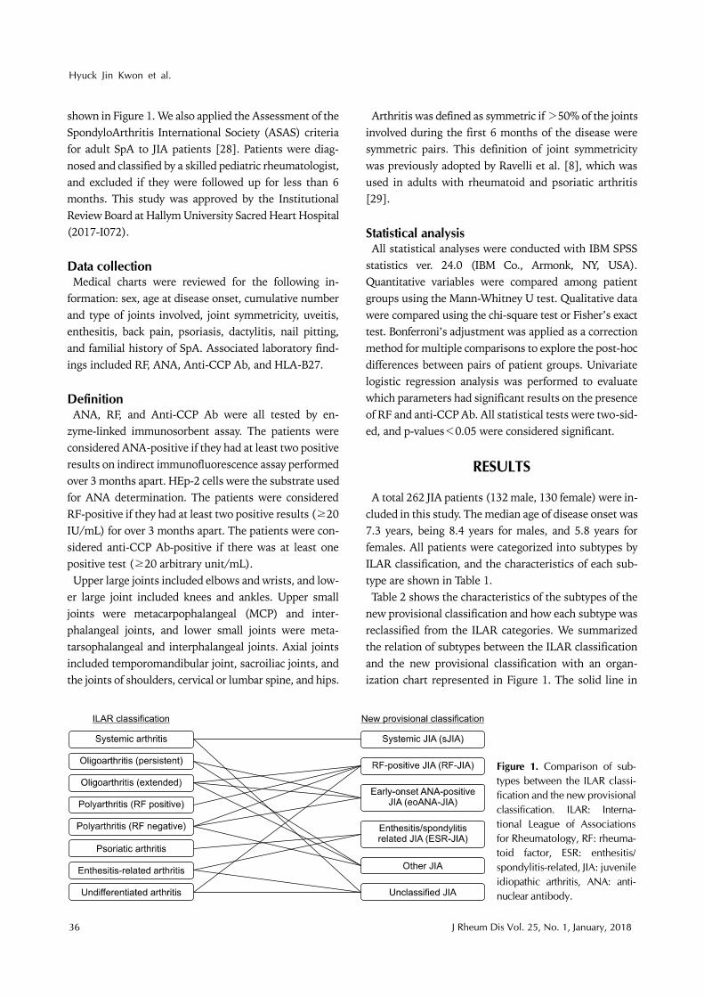
Figure 1. Comparison of sub-types between the ILAR classi-fication and the new provisionalclassification. ILAR: Interna-tional League of Associations for Rheumatology, RF: rheuma-toid factor, ESR: enthesitis/ spondylitis-related, JIA: juvenileidiopathic arthritis, ANA: anti-nuclear antibody.
shown in Figure 1. We also applied the Assessment of the SpondyloArthritis International Society (ASAS) criteria for adult SpA to JIA patients [28]. Patients were diag-nosed and classified by a skilled pediatric rheumatologist, and excluded if they were followed up for less than 6 months. This study was approved by the Institutional Review Board at Hallym University Sacred Heart Hospital (2017-I072).
Data collectionMedical charts were reviewed for the following in-
formation: sex, age at disease onset, cumulative number and type of joints involved, joint symmetricity, uveitis, enthesitis, back pain, psoriasis, dactylitis, nail pitting, and familial history of SpA. Associated laboratory find-ings included RF, ANA, Anti-CCP Ab, and HLA-B27.
DefinitionANA, RF, and Anti-CCP Ab were all tested by en-
zyme-linked immunosorbent assay. The patients were considered ANA-positive if they had at least two positive results on indirect immunofluorescence assay performed over 3 months apart. HEp-2 cells were the substrate used for ANA determination. The patients were considered RF-positive if they had at least two positive results (≥20 IU/mL) for over 3 months apart. The patients were con-sidered anti-CCP Ab-positive if there was at least one positive test (≥20 arbitrary unit/mL). Upper large joints included elbows and wrists, and low-
er large joint included knees and ankles. Upper small joints were metacarpophalangeal (MCP) and inter-phalangeal joints, and lower small joints were meta-tarsophalangeal and interphalangeal joints. Axial joints included temporomandibular joint, sacroiliac joints, and the joints of shoulders, cervical or lumbar spine, and hips.
Arthritis was defined as symmetric if >50% of the joints involved during the first 6 months of the disease were symmetric pairs. This definition of joint symmetricity was previously adopted by Ravelli et al. [8], which was used in adults with rheumatoid and psoriatic arthritis [29].
Statistical analysisAll statistical analyses were conducted with IBM SPSS
statistics ver. 24.0 (IBM Co., Armonk, NY, USA). Quantitative variables were compared among patient groups using the Mann-Whitney U test. Qualitative data were compared using the chi-square test or Fisher’s exact test. Bonferroni’s adjustment was applied as a correction method for multiple comparisons to explore the post-hoc differences between pairs of patient groups. Univariate logistic regression analysis was performed to evaluate which parameters had significant results on the presence of RF and anti-CCP Ab. All statistical tests were two-sid-ed, and p-values<0.05 were considered significant.
RESULTS
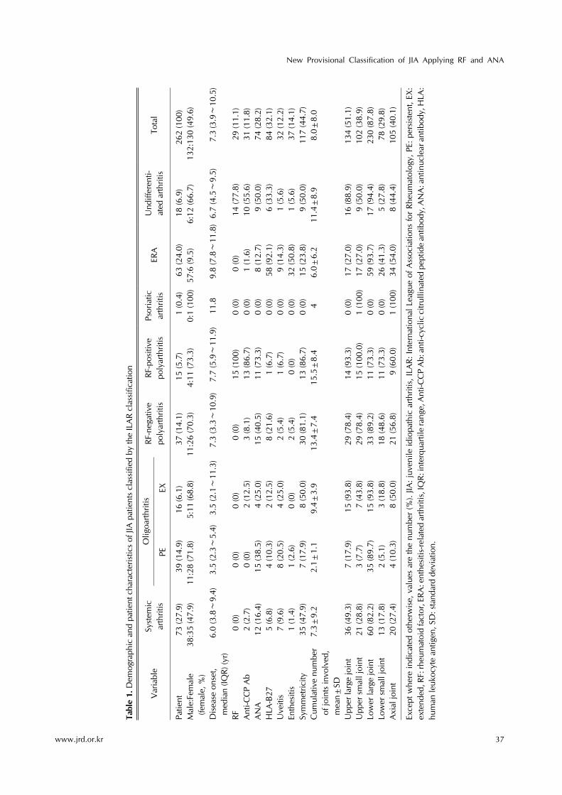
A total 262 JIA patients (132 male, 130 female) were in-cluded in this study. The median age of disease onset was 7.3 years, being 8.4 years for males, and 5.8 years for females. All patients were categorized into subtypes by ILAR classification, and the characteristics of each sub-type are shown in Table 1. Table 2 shows the characteristics of the subtypes of the
new provisional classification and how each subtype was reclassified from the ILAR categories. We summarized the relation of subtypes between the ILAR classification and the new provisional classification with an organ-ization chart represented in Figure 1. The solid line in
New Provisional Classification of JIA Applying RF and ANA
www.jrd.or.kr 37
Tabl
e 1.
Dem
ogra
phic
and
pat
ient
cha
ract
eris
tics
of JI
A p
atie
nts
clas
sifie
d by
the
ILA
R cl
assi
ficat
ion
Var
iabl
eSy
stem
ic
arth
ritis
Olig
oarth
ritis
RF-n
egat
ive
poly
arth
ritis
RF
-pos
itive
poly
arth
ritis
Psor
iatic
ar
thrit
isER
AU
ndiff
eren
ti-at
ed a
rthrit
isTo
tal
PEEX
Patie
nt
73 (2
7.9)
39 (1
4.9)
16 (6
.1)
37 (1
4.1)
15 (5
.7)
1 (0
.4)
63 (2
4.0)
18 (6
.9)
262
(100
)M
ale:
Fem
ale
(fem
ale,
%)
38:3
5 (4
7.9)
11:2
8 (7
1.8)
5:11
(68.
8)11
:26
(70.
3)4:
11 (7
3.3)
0:1
(100
)57
:6 (9
.5)
6:12
(66.
7)13
2:13
0 (4
9.6)
Dis
ease
ons
et,
med
ian
(IQR)
(yr)
6.
0 (3
.8∼
9.4)
3.5
(2.3∼
5.4)
3.5
(2.1∼
11.3
)
7.3
(3.3∼
10.9
)
7.7
(5.9∼
11.9
)11
.8
9.8
(7.8∼
11.8
)6.
7 (4
.5∼
9.5)
7.
3 (3
.9∼
10.5
)
RF0
(0)
0 (0
)0
(0)
0 (0
)15
(100
)0
(0)
0 (0
)14
(77.
8) 2
9 (1
1.1)
Ant
i-CC
P A
b2
(2.7
)0
(0)
2 (1
2.5)
3 (8
.1)
13 (8
6.7)
0 (0
)1
(1.6
)10
(55.
6) 3
1 (1
1.8)
AN
A12
(16.
4)15
(38.
5)4
(25.
0)15
(40.
5)11
(73.
3)0
(0)
8 (1
2.7)
9 (5
0.0)
74
(28.
2)H
LA-B
275
(6.8
)4
(10.
3)2
(12.
5)8
(21.
6)1
(6.7
)0
(0)
58 (9
2.1)
6 (3
3.3)
84
(32.
1)U
veiti
s7
(9.6
)8
(20.
5)4
(25.
0)2
(5.4
)1
(6.7
)0
(0)
9 (1
4.3)
1 (5
.6)
32
(12.
2)En
thes
itis
1 (1
.4)
1 (2
.6)
0 (0
)2
(5.4
)0
(0)
0 (0
)32
(50.
8)1
(5.6
) 3
7 (1
4.1)
Sym
met
ricity
35 (4
7.9)
7 (1
7.9)
8 (5
0.0)
30 (8
1.1)
13 (8
6.7)
0 (0
)15
(23.
8)9
(50.
0)11
7 (4
4.7)
Cum
ulat
ive
num
ber
of jo
ints
invo
lved
, m
ean±
SD
7.3±
9.2
2.1±
1.1
9.4±
3.9
13.4
±7.
415
.5±
8.4
46.
0±6.
211
.4±
8.9
8.0±
8.0
Upp
er la
rge
join
t 36
(49.
3)7
(17.
9)15
(93.
8)29
(78.
4)14
(93.
3)0
(0)
17 (2
7.0)
16 (8
8.9)
134
(51.
1)U
pper
sm
all j
oint
21 (2
8.8)
3 (7
.7)
7 (4
3.8)
29 (7
8.4)
15 (1
00.0
)
1 (1
00)
17 (2
7.0)
9 (5
0.0)
102
(38.
9)Lo
wer
larg
e jo
int
60 (8
2.2)
35 (8
9.7)
15 (9
3.8)
33 (8
9.2)
11 (7
3.3)
0 (0
)59
(93.
7)17
(94.
4)23
0 (8
7.8)
Low
er s
mal
l joi
nt13
(17.
8)2
(5.1
)3
(18.
8)18
(48.
6)11
(73.
3)0
(0)
26 (4
1.3)
5 (2
7.8)
78
(29.
8)A
xial
join
t20
(27.
4)4
(10.
3)8
(50.
0)21
(56.
8)9
(60.
0)
1 (1
00)
34 (5
4.0)
8 (4
4.4)
105
(40.
1)
Exce
pt w
here
indi
cate
d ot
herw
ise,
val
ues
are
the
num
ber
(%).
JIA: j
uven
ile id
iopa
thic
arth
ritis
, ILA
R: In
tern
atio
nal L
eagu
e of
Ass
ocia
tions
for
Rheu
mat
olog
y, P
E: p
ersi
sten
t, EX
: ex
tend
ed, R
F: rh
eum
atoi
d fa
ctor
, ERA
: ent
hesi
tis-re
late
d ar
thrit
is, I
QR:
inte
rqua
rtile
rang
e, A
nti-C
CP
Ab:
ant
i-cyc
lic c
itrul
linat
ed p
eptid
e an
tibod
y, A
NA
: ant
inuc
lear
ant
ibod
y, H
LA:
hum
an le
ukoc
yte
antig
en, S
D: s
tand
ard
devi
atio
n.

Hyuck Jin Kwon et al.
38 J Rheum Dis Vol. 25, No. 1, January, 2018
Tabl
e 2.
Dem
ogra
phic
and
pat
ient
cha
ract
eris
tics
of s
ubty
pes
in n
ew p
rovi
sion
al c
lass
ifica
tion
Var
iabl
esJ
IARF
-JIA
eoA
NA
-JIA
ESR-
JIAO
ther
JIA
Unc
lass
ified
JIATo
tal
p-va
lue*
Com
paris
ons
sig
nific
ant o
n po
st-h
oc te
sts†
Patie
nt71
(27.
1)31
(11.
8)22
(8.4
)63
(24.
0)65
(24.
8)10
(3.8
)26
2 (1
00)
ILA
R su
btyp
es (n
)
Sys
tem
ic a
rthrit
is71
0 0
0 0
273
O
ligoa
rthrit
is P
E 0
012
027
039
O
ligoa
rthrit
is E
X 0
2 3
011
016
P
olya
rthrit
is R
F (−
) 0
3 7
027
037
P
olya
rthrit
is R
F (+
) 0
15 0
0 0
015
P
soria
tic a
rthrit
is 0
0 0
1 0
0 1
E
RA 0
0 0
62 0
163
U
ndiff
eren
tiate
d 0
11 0
0 0
718
Mal
e:Fe
mal
e (fe
mal
e, %
)38
:33
(46.
5)7:
24 (7
7.4)
5:17
(77.
3)56
:7 (1
1.1)
20:4
5 (6
9.2)
6:4
(40.
0)13
2:13
0 (4
9.6)
<0.
0000
1RF
vs.
ESR
AN
A v
s. E
SRD
isea
se o
nset
, m
edia
n (IQ
R) (y
r) 6
.0 (
3.8~
9.3)
7.0
(4.0
~9.
6)2.
4 (2
.1~
4.2)
9.8
(7.8
~11
.8)
5
.6 (3
.0~
10.9
) 9
.0 (5
.2~
12.2
)
7
.3 (3
.9~
10.5
)<
0.00
001
AN
A v
s. R
FRF
vs.
ESR
AN
A v
s. E
SRRF
0 (0
)26
(83.
9)0
(0)
0 (0
)0
(0)
3 (3
0.0)
29 (1
1.1)
Ant
i-CC
P A
b 0
(0)
27 (8
7.1)
0 (0
) 0
(0)
0 (0
)4
(40.
0)31
(12.
7)A
NA
1
2 (1
6.9)
19 (6
1.3)
22 (1
00)
8 (1
2.7)
11 (1
6.9)
2 (2
0.0)
74 (2
8.2)
HLA
-B27
5
(7.4
)5
(17.
2)4
(20.
0)57
(90.
5)7
(10.
8)6
(60.
0)84
(32.
9)<
0.00
001
RF v
s. E
SRA
NA
vs.
ESR
Uve
itis
7
(9.9
)2
(6.5
)7
(31.
8)9
(14.
3)7
(10.
8)0
(0)
32 (1
2.2)
0.03
9En
thes
itis
1
(1.4
)0
(0)
0 (0
)31
(49.
2)3
(4.6
)2
(20.
0)37
(14.
1)<
0.00
001
RF v
s. E
SRA
NA
vs.
ERA
Sym
met
ricity
3
4 (4
7.9)
19 (6
1.3)
5 (2
2.7)
14 (2
2.2)
38 (5
8.5)
7 (7
0.0)
117
(44.
7) 0
.000
4RF
vs.
ESR
AN
A v
s. R
FC
umul
ativ
e no
. of j
oint
s in
volv
ed, m
ean±
SD7.
4±9.
314
.2±
7.9
6.0±
6.3
6.1±
6.2
8.0±
7.2
8.8±
9.4
8.0±
8.0
<0.
0000
1A
NA
vs.
RF
RF v
s. E
RAU
pper
larg
e jo
int
35 (4
9.3)
29 (9
3.5)
12 (5
4.5)
17 (2
7.0)
35 (5
3.8)
6 (6
0.0)
134
(51.
1)<
0.00
001
RF v
s. E
SRA
NA
vs.
RF
Upp
er s
mal
l joi
nt20
(28.
2)25
(80.
6)9
(40.
9)18
(28.
6)26
(40.
0)4
(40.
0)10
2 (3
8.9)
0.
0000
1RF
vs.
ESR
AN
A v
s. R
FLo
wer
larg
e jo
int
59 (8
3.1)
27 (8
7.1)
19 (8
6.4)
58 (9
2.1)
59 (9
0.8)
8 (8
0.0)
230
(87.
8)0.
647
Low
er s
mal
l joi
nt12
(16.
9)16
(51.
6)9
(27.
3)26
(41.
3)15
(23.
1)3
(30.
0)78
(29.
8)0.
208
Axi
al jo
int
20 (2
8.2)
16 (5
1.6)
3 (1
3.6)
35 (5
5.6)
27 (4
1.5)
4 (4
0.0)
105
(40.
1)0.
003
AN
A v
s. E
SRA
NA
vs.
RF
New Provisional Classification of JIA Applying RF and ANA
www.jrd.or.kr 39
Figure 1 indicates how patients were actually reclassified within this study. In the new provisional classification, classification re-
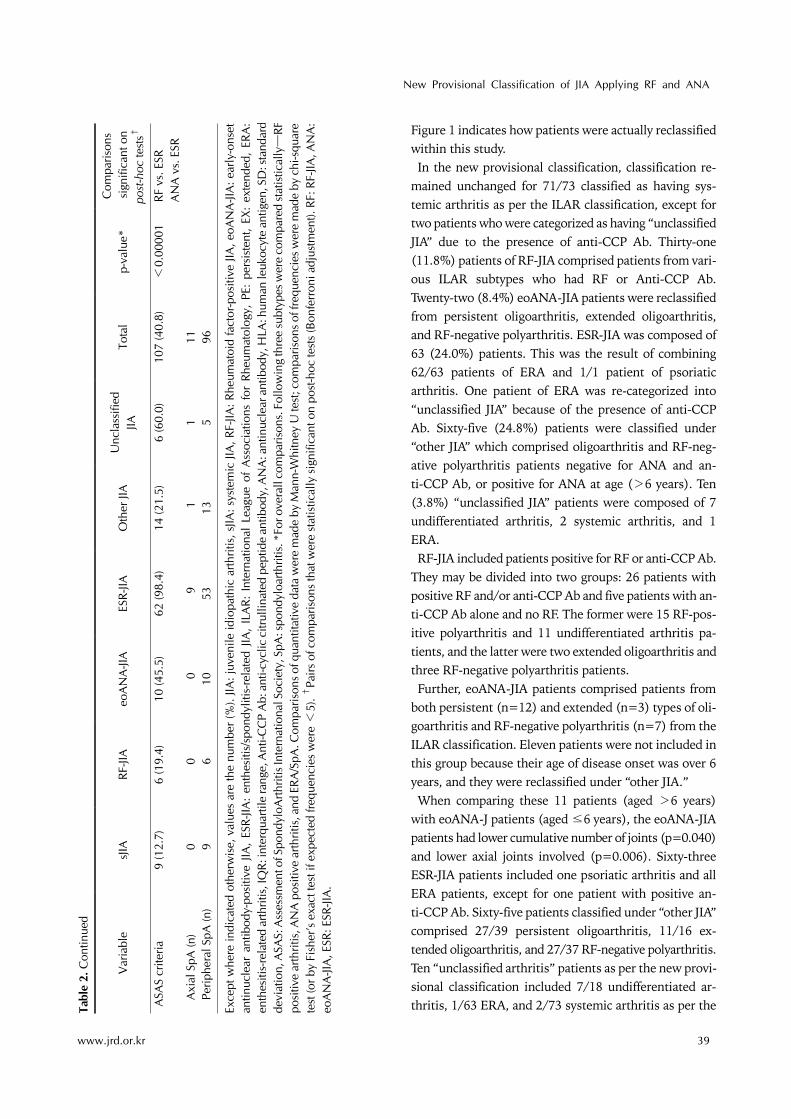
mained unchanged for 71/73 classified as having sys-temic arthritis as per the ILAR classification, except for two patients who were categorized as having “unclassified JIA” due to the presence of anti-CCP Ab. Thirty-one (11.8%) patients of RF-JIA comprised patients from vari-ous ILAR subtypes who had RF or Anti-CCP Ab. Twenty-two (8.4%) eoANA-JIA patients were reclassified from persistent oligoarthritis, extended oligoarthritis, and RF-negative polyarthritis. ESR-JIA was composed of 63 (24.0%) patients. This was the result of combining 62/63 patients of ERA and 1/1 patient of psoriatic arthritis. One patient of ERA was re-categorized into “unclassified JIA” because of the presence of anti-CCP Ab. Sixty-five (24.8%) patients were classified under “other JIA” which comprised oligoarthritis and RF-neg-ative polyarthritis patients negative for ANA and an-ti-CCP Ab, or positive for ANA at age (>6 years). Ten (3.8%) “unclassified JIA” patients were composed of 7 undifferentiated arthritis, 2 systemic arthritis, and 1 ERA.RF-JIA included patients positive for RF or anti-CCP Ab.
They may be divided into two groups: 26 patients with positive RF and/or anti-CCP Ab and five patients with an-ti-CCP Ab alone and no RF. The former were 15 RF-pos-itive polyarthritis and 11 undifferentiated arthritis pa-tients, and the latter were two extended oligoarthritis and three RF-negative polyarthritis patients. Further, eoANA-JIA patients comprised patients from
both persistent (n=12) and extended (n=3) types of oli-goarthritis and RF-negative polyarthritis (n=7) from the ILAR classification. Eleven patients were not included in this group because their age of disease onset was over 6 years, and they were reclassified under “other JIA.” When comparing these 11 patients (aged >6 years)
with eoANA-J patients (aged ≤6 years), the eoANA-JIA patients had lower cumulative number of joints (p=0.040) and lower axial joints involved (p=0.006). Sixty-three ESR-JIA patients included one psoriatic arthritis and all ERA patients, except for one patient with positive an-ti-CCP Ab. Sixty-five patients classified under “other JIA” comprised 27/39 persistent oligoarthritis, 11/16 ex-tended oligoarthritis, and 27/37 RF-negative polyarthritis. Ten “unclassified arthritis” patients as per the new provi-sional classification included 7/18 undifferentiated ar-thritis, 1/63 ERA, and 2/73 systemic arthritis as per the Ta
ble
2. C
ontin
ued
Var
iabl
esJ
IARF
-JIA
eoA
NA
-JIA
ESR-
JIAO
ther
JIA
Unc
lass
ified
JIATo
tal
p-va
lue*
Com
paris
ons
sig
nific
ant o
n po
st-h
oc te
sts†
ASA
S cr
iteria
9 (1
2.7)
6 (1
9.4)
10 (4
5.5)
62 (9
8.4)
14 (2
1.5)
6 (6
0.0)
107
(40.
8)<
0.00
001
RF v
s. E
SRA
NA
vs.
ESR
Axi
al S
pA (n
) 0
0 0
9 1
111
Per
iphe
ral S
pA (n
) 9
610
5313
596
Exce
pt w
here
indi
cate
d ot
herw
ise,
val
ues
are
the
num
ber
(%).
JIA: j
uven
ile id
iopa
thic
arth
ritis
, sJIA
: sys
tem
ic JI
A, R
F-JIA
: Rhe
umat
oid
fact
or-p
ositi
ve JI
A, e
oAN
A-JI
A: e
arly
-ons
et
antin
ucle
ar a
ntib
ody-
posi
tive
JIA,
ESR-
JIA:
enth
esiti
s/sp
ondy
litis
-rela
ted
JIA,
ILA
R: I
nter
natio
nal
Leag
ue o
f A
ssoc
iatio
ns f
or R
heum
atol
ogy,
PE:
per
sist
ent,
EX:
exte
nded
, ER
A:
enth
esiti
s-re
late
d ar
thrit
is, I
QR:
inte
rqua
rtile
rang
e, A
nti-C
CP
Ab:
ant
i-cyc
lic c
itrul
linat
ed p
eptid
e an
tibod
y, A
NA
: ant
inuc
lear
ant
ibod
y, H
LA: h
uman
leuk
ocyt
e an
tigen
, SD
: sta
ndar
dde
viat
ion,
ASA
S: A
sses
smen
t of S
pond
yloA
rthrit
is In
tern
atio
nal S
ocie
ty, S
pA: s
pond
yloa
rthrit
is. *
For o
vera
ll co
mpa
rison
s. F
ollo
win
g th
ree
subt
ypes
wer
e co
mpa
red
stat
istic
ally―
RFpo
sitiv
e ar
thrit
is, A
NA
pos
itive
arth
ritis
, and
ERA
/SpA
. Com
paris
ons o
f qua
ntita
tive
data
wer
e m
ade
by M
ann-
Whi
tney
U te
st; c
ompa
rison
s of
freq
uenc
ies w
ere
mad
e by
chi
-squ
are
test
(or b
y Fi
sher
’s e
xact
test
if e
xpec
ted
frequ
enci
es w
ere <
5). †
Pairs
of c
ompa
rison
s th
at w
ere
stat
istic
ally
sig
nific
ant o
n po
st-h
oc te
sts
(Bon
ferr
oni a
djus
tmen
t). R
F: R
F-JIA
, AN
A:
eoA
NA
-JIA
, ESR
: ESR
-JIA
.
Hyuck Jin Kwon et al.
40 J Rheum Dis Vol. 25, No. 1, January, 2018
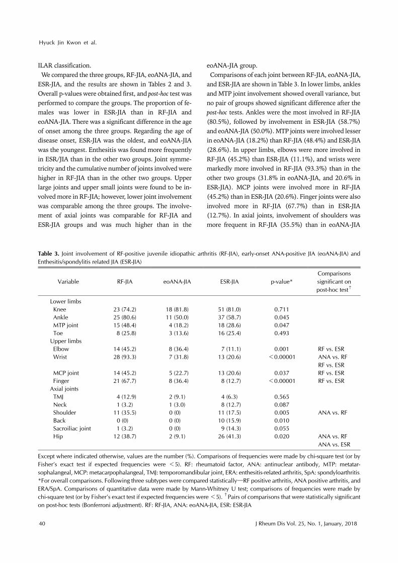
Table 3. Joint involvement of RF-positive juvenile idiopathic arthritis (RF-JIA), early-onset ANA-positive JIA (eoANA-JIA) and Enthesitis/spondylitis related JIA (ESR-JIA)
Variable RF-JIA eoANA-JIA ESR-JIA p-value*Comparisons significant on post-hoc test†
Lower limbs Knee 23 (74.2) 18 (81.8) 51 (81.0) 0.711 Ankle 25 (80.6) 11 (50.0) 37 (58.7) 0.045 MTP joint 15 (48.4) 4 (18.2) 18 (28.6) 0.047 Toe 8 (25.8) 3 (13.6) 16 (25.4) 0.493Upper limbs Elbow 14 (45.2) 8 (36.4) 7 (11.1) 0.001 RF vs. ESR Wrist 28 (93.3) 7 (31.8) 13 (20.6) <0.00001 ANA vs. RF
RF vs. ESR MCP joint 14 (45.2) 5 (22.7) 13 (20.6) 0.037 RF vs. ESR Finger 21 (67.7) 8 (36.4) 8 (12.7) <0.00001 RF vs. ESRAxial joints TMJ 4 (12.9) 2 (9.1) 4 (6.3) 0.565 Neck 1 (3.2) 1 (3.0) 8 (12.7) 0.087 Shoulder 11 (35.5) 0 (0) 11 (17.5) 0.005 ANA vs. RF Back 0 (0) 0 (0) 10 (15.9) 0.010 Sacroiliac joint 1 (3.2) 0 (0) 9 (14.3) 0.055 Hip 12 (38.7) 2 (9.1) 26 (41.3) 0.020 ANA vs. RF
ANA vs. ESR
Except where indicated otherwise, values are the number (%). Comparisons of frequencies were made by chi-square test (or by Fisher’s exact test if expected frequencies were <5). RF: rheumatoid factor, ANA: antinuclear antibody, MTP: metatar-sophalangeal, MCP: metacarpophalangeal, TMJ: temporomandibular joint, ERA: enthesitis-related arthritis, SpA: spondyloarthritis.*For overall comparisons. Following three subtypes were compared statistically―RF positive arthritis, ANA positive arthritis, and ERA/SpA. Comparisons of quantitative data were made by Mann-Whitney U test; comparisons of frequencies were made by chi-square test (or by Fisher’s exact test if expected frequencies were <5). †Pairs of comparisons that were statistically significanton post-hoc tests (Bonferroni adjustment). RF: RF-JIA, ANA: eoANA-JIA, ESR: ESR-JIA
ILAR classification.We compared the three groups, RF-JIA, eoANA-JIA, and
ESR-JIA, and the results are shown in Tables 2 and 3. Overall p-values were obtained first, and post-hoc test was performed to compare the groups. The proportion of fe-males was lower in ESR-JIA than in RF-JIA and eoANA-JIA. There was a significant difference in the age of onset among the three groups. Regarding the age of disease onset, ESR-JIA was the oldest, and eoANA-JIA was the youngest. Enthesitis was found more frequently in ESR/JIA than in the other two groups. Joint symme-tricity and the cumulative number of joints involved were higher in RF-JIA than in the other two groups. Upper large joints and upper small joints were found to be in-volved more in RF-JIA; however, lower joint involvement was comparable among the three groups. The involve-ment of axial joints was comparable for RF-JIA and ESR-JIA groups and was much higher than in the
eoANA-JIA group. Comparisons of each joint between RF-JIA, eoANA-JIA,
and ESR-JIA are shown in Table 3. In lower limbs, ankles and MTP joint involvement showed overall variance, but no pair of groups showed significant difference after the post-hoc tests. Ankles were the most involved in RF-JIA (80.5%), followed by involvement in ESR-JIA (58.7%) and eoANA-JIA (50.0%). MTP joints were involved lesser in eoANA-JIA (18.2%) than RF-JIA (48.4%) and ESR-JIA (28.6%). In upper limbs, elbows were more involved in RF-JIA (45.2%) than ESR-JIA (11.1%), and wrists were markedly more involved in RF-JIA (93.3%) than in the other two groups (31.8% in eoANA-JIA, and 20.6% in ESR-JIA). MCP joints were involved more in RF-JIA (45.2%) than in ESR-JIA (20.6%). Finger joints were also involved more in RF-JIA (67.7%) than in ESR-JIA (12.7%). In axial joints, involvement of shoulders was more frequent in RF-JIA (35.5%) than in eoANA-JIA

New Provisional Classification of JIA Applying RF and ANA
www.jrd.or.kr 41
Table 4. Univariate logistic regression analysis on rheumatoid factor (RF) and anti-cyclic citrullinated peptide antibody (anti-CCPAb)
VariableRF Anti-CCP Ab
p-value Exp(β) (95% CI) p-value Exp(β) (95% CI)
Female 0.002 4.514 (1.773∼11.495) 0.005 3.332 (1.431∼7.757)Age (>6 yr) 0.743 0.879 (0.406∼1.903) 0.662 1.183 (0.557∼2.512)Uveitis 0.363 0.501 (0.113∼2.216) 0.137 0.215 (0.028∼1.634)Enthesitis 0.998 0.000 (not available) 0.104 0.187 (0.025∼1.414)ANA 0.000 7.366 (3.169∼17.123) 0.000 4.327 (1.995∼9.385)HLA-B27 0.018 0.227 (0.066∼0.776) 0.238 0.585 (0.240∼1.424)Number of Joints involved (≥5) 0.001 5.885 (1.986∼17.440) 0.001 6.464 (2.192∼19.059)Symmetricity 0.020 2.617 (1.166∼5.876) 0.051 2.149 (0.997∼4.633)
CI: confidence interval, ANA: antinuclear antibody, HLA: human leukocyte antigen.
(0%). Involvement of hip joints was higher in RF-JIA (38.7%) and ESR-JIA (41.3%) than in eoANA-JIA (9.1%).The ASAS criteria for adult SpA were applied to JIA pa-
tients, and the results were also shown in Table 2. Overall, SpA was diagnosed in 40.8% (107/262) of total JIA pa-tients by the ASAS criteria (axial SpA, 11; peripheral SpA, 96). They included as many as 98.4% (62/63) of ESR-JIA, 60% (6/10) of “unclassified JIA,” and 45.5% (10/22) of eoANA-JIA.We performed univariate logistic regression analysis by
setting the positivity of RF and anti-CCP Ab as in-dependent variables (Table 4). Each variable showed al-most consistent results between RF-positive patients and anti-CCP Ab-positive patients. In both groups, female gender, ANA positivity, and the number of joints involved (≥5) were statistically significant, while age of disease onset (>6 years), uveitis, and enthesitis were not. Joint symmetricity was not statistically significant in Anti-CCP Ab-positive patients (p=0.051). We analyzed whether there were significant differences
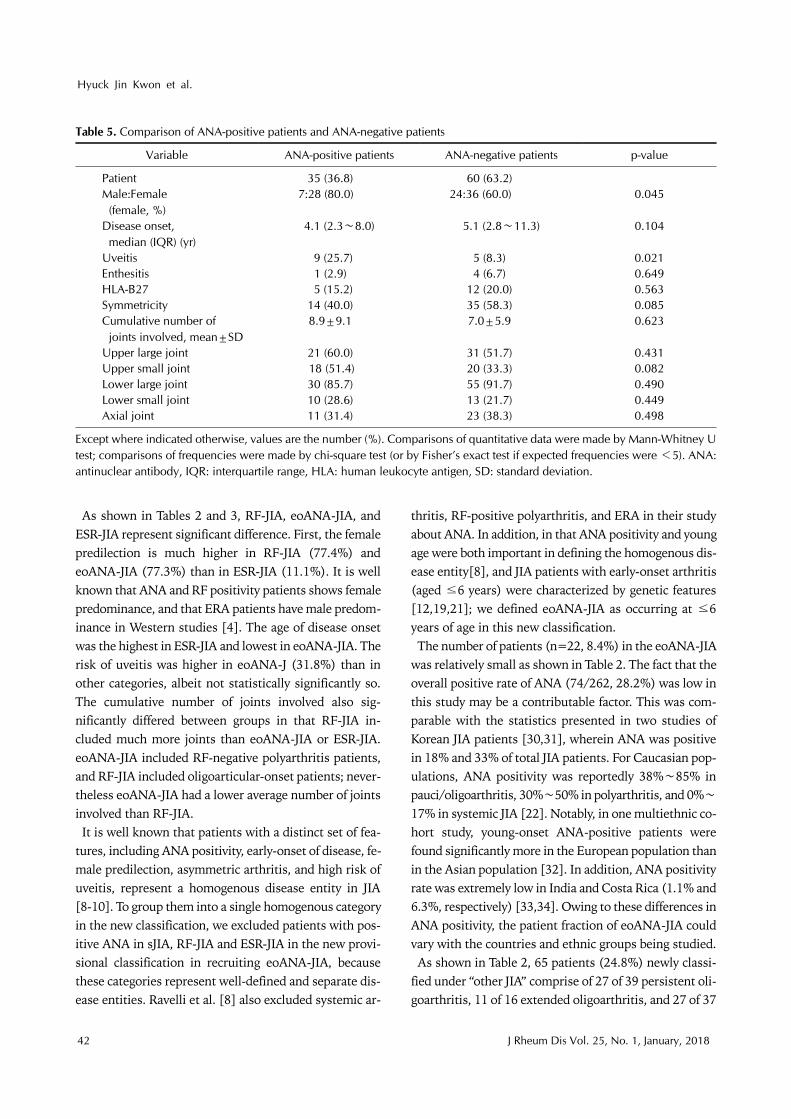
between ANA-positive patients and ANA-negative pa-tients (Table 5). sJIA, RF-JIA, and ESR-JIA were excluded from this comparison because these subtypes were pre-sumed to have prominent features regardless of ANA-po-sitivity [8]. Thus, the remaining 95 patients were inves-tigated. The female proportion and the risk of uveitis were both significantly higher in ANA-positive patients (p=0.045 and p=0.021, respectively). The ANA-positive group showed younger age of disease onset and lower joint symmetricity, although these were not statistically significant. Enthesitis, HLA-B27, and the cumulative number and types of joints involved did not significantly differ between the two groups.
We divided 35 ANA-positive patients in Table 5 by age of disease onset (6 years) and compared each other (not shown in table). Patients with disease onset at an age of ≤6 years had fewer occurrences of enthesitis (p=0.046), frequent involvement of lower large joints (p=0.045), and lesser involvement of axial joints (p=0.022). Although not statistically significant, there was some tendency of higher female predilection, higher risk for uveitis, lesser risk for HLA-B27 positivity, lower joint symmetricity, and small cumulative number of joints involved in patients with disease onset at an age of ≤6 years.
DISCUSSION
The most important point in the new provisional classi-fication for JIA is that the previous diagnostic criteria based on the number of joints were substituted by the positivity for RF and ANA. Thus, in the new provisional classification, we discontinued the use of the term pau-ci/oligoarthritis and polyarthritis which have been used in the pediatric field for over 4 decades. The category of PsA excluded as well. Instead, patients who once be-longed to these groups were re-categorized to RF-JIA, eoANA-JIA, ESR-JIA, “other JIA,” and “unclassified JIA.” sJIA remained unchanged from the ILAR classification.As mentioned earlier, this nomenclature was originated
from the new provisional classification for JIA proposed by Dr. Martini in 2016. The Paediatric Rheumatology European society (PReS) is currently conducting a large-scale prospective study in Europe with this new provisional classification. The aim of our current study was to retrospectively apply the concept of the new classi-fication to JIA patients.
Hyuck Jin Kwon et al.
42 J Rheum Dis Vol. 25, No. 1, January, 2018
Table 5. Comparison of ANA-positive patients and ANA-negative patients
Variable ANA-positive patients ANA-negative patients p-value
Patient 35 (36.8) 60 (63.2)Male:Female
(female, %)7:28 (80.0) 24:36 (60.0) 0.045
Disease onset, median (IQR) (yr)
4.1 (2.3∼8.0) 5.1 (2.8∼11.3) 0.104
Uveitis 9 (25.7) 5 (8.3) 0.021Enthesitis 1 (2.9) 4 (6.7) 0.649HLA-B27 5 (15.2) 12 (20.0) 0.563Symmetricity 14 (40.0) 35 (58.3) 0.085Cumulative number of
joints involved, mean±SD8.9±9.1 7.0±5.9 0.623
Upper large joint 21 (60.0) 31 (51.7) 0.431Upper small joint 18 (51.4) 20 (33.3) 0.082Lower large joint 30 (85.7) 55 (91.7) 0.490Lower small joint 10 (28.6) 13 (21.7) 0.449Axial joint 11 (31.4) 23 (38.3) 0.498
Except where indicated otherwise, values are the number (%). Comparisons of quantitative data were made by Mann-Whitney Utest; comparisons of frequencies were made by chi-square test (or by Fisher’s exact test if expected frequencies were <5). ANA:antinuclear antibody, IQR: interquartile range, HLA: human leukocyte antigen, SD: standard deviation.
As shown in Tables 2 and 3, RF-JIA, eoANA-JIA, and ESR-JIA represent significant difference. First, the female predilection is much higher in RF-JIA (77.4%) and eoANA-JIA (77.3%) than in ESR-JIA (11.1%). It is well known that ANA and RF positivity patients shows female predominance, and that ERA patients have male predom-inance in Western studies [4]. The age of disease onset was the highest in ESR-JIA and lowest in eoANA-JIA. The risk of uveitis was higher in eoANA-J (31.8%) than in other categories, albeit not statistically significantly so. The cumulative number of joints involved also sig-nificantly differed between groups in that RF-JIA in-cluded much more joints than eoANA-JIA or ESR-JIA. eoANA-JIA included RF-negative polyarthritis patients, and RF-JIA included oligoarticular-onset patients; never-theless eoANA-JIA had a lower average number of joints involved than RF-JIA. It is well known that patients with a distinct set of fea-
tures, including ANA positivity, early-onset of disease, fe-male predilection, asymmetric arthritis, and high risk of uveitis, represent a homogenous disease entity in JIA [8-10]. To group them into a single homogenous category in the new classification, we excluded patients with pos-itive ANA in sJIA, RF-JIA and ESR-JIA in the new provi-sional classification in recruiting eoANA-JIA, because these categories represent well-defined and separate dis-ease entities. Ravelli et al. [8] also excluded systemic ar-
thritis, RF-positive polyarthritis, and ERA in their study about ANA. In addition, in that ANA positivity and young age were both important in defining the homogenous dis-ease entity[8], and JIA patients with early-onset arthritis (aged ≤6 years) were characterized by genetic features [12,19,21]; we defined eoANA-JIA as occurring at ≤6 years of age in this new classification.The number of patients (n=22, 8.4%) in the eoANA-JIA
was relatively small as shown in Table 2. The fact that the overall positive rate of ANA (74/262, 28.2%) was low in this study may be a contributable factor. This was com-parable with the statistics presented in two studies of Korean JIA patients [30,31], wherein ANA was positive in 18% and 33% of total JIA patients. For Caucasian pop-ulations, ANA positivity was reportedly 38%∼85% in pauci/oligoarthritis, 30%∼50% in polyarthritis, and 0%∼
17% in systemic JIA [22]. Notably, in one multiethnic co-hort study, young-onset ANA-positive patients were found significantly more in the European population than in the Asian population [32]. In addition, ANA positivity rate was extremely low in India and Costa Rica (1.1% and 6.3%, respectively) [33,34]. Owing to these differences in ANA positivity, the patient fraction of eoANA-JIA could vary with the countries and ethnic groups being studied.As shown in Table 2, 65 patients (24.8%) newly classi-
fied under “other JIA” comprise of 27 of 39 persistent oli-goarthritis, 11 of 16 extended oligoarthritis, and 27 of 37

New Provisional Classification of JIA Applying RF and ANA
www.jrd.or.kr 43
RF-negative polyarthritis from the ILAR classification. In other words, 70.7% (65/92) of these three ILAR subtypes were recruited to “other JIA,” while the remaining 29.3% (27/92) were reclassified into eoANA-JIA and RF-JIA be-cause of the presence of Anti-CCP Ab. Although “other JIA” does not represent a homogenous disease group by definition, it accounts for as many as one fourth of total JIA patients. One of the reasons for this significant pro-portion of “other JIA” is thought to be relatively low pos-itive rate of ANA than in Western studies as mentioned above. Patients of oligoarthritis and RF-negative poly-arthritis without ANA positivity are most likely to be re-classified as “other JIA” in the new provisional classification. That is, lower positive rate of ANA might result in higher proportion of “other JIA.” This high proportion of “other JIA” is far from the original intent to specifically classify most types and identify homogenous disease groups; thus there is a need to reform this group in the future. As part of this attempt, we used the ASAS criteria to identify patients who have features of spondyloarthritis among those classified under “other JIA.” Table 2 shows the results of applying the ASAS criteria
to JIA patients. A total 107 patients (40.8%) were diag-nosed as SpA (axial SpA: 11 patients, peripheral SpA: 96 patients). In “other JIA,” a total 14 patients were diag-nosed as SpA (axial SpA: 1 patient, peripheral SpA: 13 pa-tients). However, we should be careful when applying the ASAS criteria to JIA patients. In the ASAS criteria, periph-eral SpA can be diagnosed if the patients have both arthri-tis and uveitis. However, uveitis is a common manifes-tation in early-onset ANA-positive JIA group, which is known not to be relevant with SpA. For example, 10 of 22 patients (45.5%) of eoANA-JIA were diagnosed as SpA by the ASAS criteria (Table 2). They were all peripheral SpA, with eight of them being “arthritis+uveitis” and two be-ing “arthritis+HLA-B27.” It is unlikely that all these eoANA-JIA patients are at high risk of SpA in the future. Therefore, it is necessary to modify the ASAS criteria to resolve this controversy before applying it to JIA patients. Thus, Dr. Martini proposed to combine the existing ERA and ASAS criteria in the new provisional classification.In Table 2, the overall positive rate of HLA-B27 was
32.9% (84/262) which was similar to the multiethnic co-hort study on JIA [32]. This significant rate of HLA-B27 positivity in JIA might be due to the considerable in-clusion of juvenile SpA patients in some JIA subtypes. Especially, ESR-JIA is intended to categorize juvenile SpA patients, and it is the subtype most associated with
HLA-B27 that 57 of 63 ESR-JIA patients (90.5%) are HLA-B27 positive. In “unclassified JIA”, HLA-B27 pos-itive rate was as high as 60% (6/10), but those of the oth-er subtypes were relatively low (7.4%∼20.0%). “Unclas-sified JIA” included patients who had features belonging to more than 2 subtypes except for “other JIA,” and 6 of 10 “unclassified JIA” patients had the features of ESR-JIA with the positivity of HLA-B27. In addition, peripheral SpA by the ASAS criteria can be diagnosed when patients both have arthritis and HLA-B27. So JIA patients, who al-ready have arthritis by definition, can be diagnosed as pe-ripheral SpA if they are positive for HLA-B27. Thus, when applying the ASAS criteria to JIA patients, it is highly cor-related with HLA-B27 positivity. The presentation of SpA differs in children and adults;
most notably, spinal involvement is uncommon and hip arthritis and enthesitis are frequently seen in juvenile SpA [35]. Although most juvenile SpA is classified as ERA by the ILAR classification, the ILAR system does not specifically acknowledge the presence of axial disease in juvenile SpA [35]. In addition, unlike adult SpA classi-fication, psoriatic features comprise a separate category in JIA [35]. Further, it is now known that psoriatic arthri-tis in the ILAR classification is another heterogeneous category that has two identifiable populations: a) one that belongs to ERA and thus represents a form of undifferen-tiated SpA and b) another that has the same features as those of early-onset ANA-positive JIA patients [9,12,18]. These differences are making difficult to communicate between pediatric and adult rheumatologists, when JIA patients transit to adult clinic. It is important to under-stand that all the different forms of adult SpA can be found in children and that there is much higher pro-portion of undifferentiated SpA in childhood [12]. Currently, these pediatric patients with juvenile SpA or SpA features are sorted into ERA, PsA, or undifferentiated arthritis as per the ILAR classification, or diagnosed as ju-venile ankylosing spondylitis (JAS) or juvenile SpA. Thus, by eliminating the concept of the subtype psoriatic arthri-tis and by reforming the ERA criteria to better recruit SpA patients, we could more reliably classify juvenile SpA in the new provisional classification.RF-positive polyarthritis in the ILAR classification is
considered the same as adult RF-positive RA, and it is the only category wherein anti-CCP Abs are found [4,36]. However, there has been controversy over applying RF to only polyarthritis, and not oligoarthritis in the proposal of the ILAR classification [13,14]. Patients with oli-
Hyuck Jin Kwon et al.
44 J Rheum Dis Vol. 25, No. 1, January, 2018
goarticular onset and positive RF have been classified un-der “undifferentiated arthritis” in the ILAR classification. It is now considered that the number of joints involved simply reflects a more rapid spread of arthritis within the same disease and thus may not represent a suitable mark-er defining a homogeneous JIA subgroup [8,9]. Moreover, imaging techniques, such as ultrasonography, which are useful in detecting subclinical synovitis in JIA make phys-ical examination seem unreliable in assessing the number of joints involved [12,17,37]. In the new classification, therefore, the number of joints is no longer used as a cri-terion in defining the subtype.Anti-CCP Ab, together with RF, has higher specificity in
diagnosing RA, and is involved in diagnostic criteria of adult RA [23]. RF and anti-CCP are known to precede the RA symptoms and present at the early process of RA [24,38]. In addition, a study reported the development of more severe radiological damage in anti-CCP Ab-positive patients than in anti-CCP Ab-negative patients [39]. In pediatric population, numerous studies have been con-ducted on the Anti-CCP Ab recently [25,26,36]. Overall, RF is approximately found between 2% and 12% of JIA patients and was 11.1% in this study [22]. Anti-CCP Ab is known to be detected almost exclusively in RF-positive polyarthritis (57%∼73%) and only seldom in other sub-types (2%∼3%) [22,36]. Although anti-CCP Ab is not yet used as a diagnostic cri-
terion in JIA, there is an ongoing attempt by Dr. Martini to apply it as a criterion for RF-positive JIA. We also re-cruited anti-CCP Ab-positive patients to RF-JIA in this study. In Table 2, positivity of RF and Anti-CCP Ab are not 100% in RF-JIA because RF-JIA included patients with only RF-positive or Anti-CCP Ab-positive. As shown in Table 4, most of the variables in RF-positive patients and anti-CCP Ab-positive patients show consistent results in univariate logistic regression analysis. These results pro-vide some evidence that these two factors represent the same disease entity―RF-positive JIA―in the new provi-sional classification.We did not make any changes in the sJIA classification in
this study except for the two patients who were re-catego-rized to “unclassified arthritis” due to the presence of an-ti-CCP Ab. sJIA is characterized by prominent systemic features, marked activation of the innate immune system, and important pathogenic roles played by interleukin 1 (IL-1), IL-6, and interferon-γ [12,40]. Adult-onset Still’s disease is considered similar to sJIA, but the former is dif-ferent in terms of arthritis not being an essential diag-
nostic criterion [12]. Among the pediatric patients, there are cases that cannot be diagnosed as sJIA because arthri-tis does not develop during the extra-articular manifes-tation period. Thus, Martini [12] suggested that these pa-tients with systemic features and no arthritis should be included in the sJIA category. However, given the absence of arthritis, Dr. Martini also suggested that the term sJIA should also be revised to a new name, such as Still’s dis-ease, owing to analogy with the adult counterpart―adult-onset Still’s disease.We acknowledge the limitations that this is a retro-
spective study with a relatively small number of patients in a single center. Furthermore, we used a relatively sim-ple classification system that is modified from the pro-posal of Dr. Martini from 2016 PReS annual meeting. However, it is meaningful that this is the first study im-
plementing the new JIA classification system that uses laboratory factors such as RF and ANA rather than the number of joints involved or the presence of psoriatic fea-ture as key identifiers. In the new provisional classi-fication, eoANA JIA, RF-JIA, and ESR-JIA showed sig-nificantly distinct features such as age of disease onset, male-to-female ratio, risk of uveitis, and cumulative number and type of joints involved. In other words, JIA patients who were previously classified into subtypes with heterogeneous nature were re-categorized into ho-mogenous categories based on the new evidence-based classification. RF-positive patients were comparable to anti-CCP Ab-positive patients, and they could be catego-rized together. The number of ANA-positive patients was lower (28.2%) than that reported in Western studies (30%∼50%), but ANA positivity showed clear difference in terms of lower age of onset and higher risk of uveitis. Ethnic differences such as rate of ANA positivity should be taken into consideration when developing a new clas-sification system for JIA in the future.
CONCLUSION
Previous classification systems for JIA including the ILAR classification do not categorize homogenous dis-ease entities. The number of joints involved and the psoriatic features are no longer valid criteria for dividing subtypes in the new classification and the concepts of oli-goarthritis, polyarthritis, and psoriatic arthritis should be excluded. Herein, we reclassified the JIA patients by new provisional classification applying RF and ANA which have been proven to constitute homogenous disease
New Provisional Classification of JIA Applying RF and ANA
www.jrd.or.kr 45
entities.The new provisional classification for JIA includes six
subtypes; sJIA, RF-JIA, early-onset eoANA-JIA, ESR-JIA, “other JIA,” and “unclassified JIA”. RF-JIA, eoANA-JIA, and ESR-JIA were well distinguished by female ratio, age of disease onset, HLA-B27, enthesitis, joint symmetricity, and cumulative number and type of joints involved. Criteria for RF-JIA included anti-CCP Ab positivity in this study, which was proven to have almost same features as RF. ANA positivity ratio differed among ethnicities, but the features of ANA positivity seemed to be shared, such as lower age of onset and higher risk of uveitis. A sig-nificant proportion (24.8%) of the “other JIA” in this study warrant the need for further reforming the classi-fication in the future. sJIA was not changed from the ILAR classification in this study. To establish a more precise and globally accepted classi-
fication system for JIA, further large-scale, prospective, and multiethnic studies are needed.
CONFLICT OF INTEREST
No potential conflict of interest relevant to this article was reported.
REFERENCES
1. Fink CW. Proposal for the development of classification cri-teria for idiopathic arthritides of childhood. J Rheumatol 1995;22:1566-9.
2. Petty RE, Southwood TR, Baum J, Bhettay E, Glass DN, Manners P, et al. Revision of the proposed classification cri-teria for juvenile idiopathic arthritis: Durban, 1997. J Rheumatol 1998;25:1991-4.
3. Petty RE, Southwood TR, Manners P, Baum J, Glass DN, Goldenberg J, et al. International League of Associations for Rheumatology classification of juvenile idiopathic arthritis: second revision, Edmonton, 2001. J Rheumatol 2004;31: 390-2.
4. Ravelli A, Martini A. Juvenile idiopathic arthritis. Lancet 2007;369:767-78.
5. Criteria for the classification of juvenile rheumatoid arthritis. Bull Rheum Dis 1972;23:712-9.
6. Brewer EJ Jr, Bass J, Baum J, Cassidy JT, Fink C, Jacobs J, et al. Current proposed revision of JRA Criteria. JRA Criteria subcommittee of the diagnostic and therapeutic criteria committee of the American rheumatism section of the ar-thritis foundation. Arthritis Rheum 1977;20(2 Suppl):195-9.
7. European League Against Rheumatism. EULAR Bulletin No. 4: Nomenclature and classification of arthritis in children. Basel, National Zeitung AG, 1977.
8. Ravelli A, Varnier GC, Oliveira S, Castell E, Arguedas O, Magnani A, et al. Antinuclear antibody-positive patients
should be grouped as a separate category in the classi-fication of juvenile idiopathic arthritis. Arthritis Rheum 2011;63:267-75.
9. Martini A. Are the number of joints involved or the presence of psoriasis still useful tools to identify homogeneous dis-ease entities in juvenile idiopathic arthritis? J Rheumatol 2003;30:1900-3.
10. Ravelli A, Felici E, Magni-Manzoni S, Pistorio A, Novarini C, Bozzola E, et al. Patients with antinuclear antibody- pos-itive juvenile idiopathic arthritis constitute a homogeneous subgroup irrespective of the course of joint disease. Arthritis Rheum 2005;52:826-32.
11. Stoll ML, Zurakowski D, Nigrovic LE, Nichols DP, Sundel RP, Nigrovic PA. Patients with juvenile psoriatic arthritis comprise two distinct populations. Arthritis Rheum 2006;54:3564-72.
12. Martini A. It is time to rethink juvenile idiopathic arthritis classification and nomenclature. Ann Rheum Dis 2012;71: 1437-9.
13. Hofer MF, Mouy R, Prieur AM. Juvenile idiopathic arthri-tides evaluated prospectively in a single center according to the Durban criteria. J Rheumatol 2001;28:1083-90.
14. Petty RE. Growing pains: the ILAR classification of juvenile idiopathic arthritis. J Rheumatol 2001;28:927-8.
15. Tsitsami E, Bozzola E, Magni-Manzoni S, Viola S, Pistorio A, Ruperto N, et al. Positive family history of psoriasis does not affect the clinical expression and course of juvenile idio-pathic arthritis patients with oligoarthritis. Arthritis Rheum 2003;49:488-93.
16. Griffin TA, Barnes MG, Ilowite NT, Olson JC, Sherry DD, Gottlieb BS, et al. Gene expression signatures in poly-articular juvenile idiopathic arthritis demonstrate disease heterogeneity and offer a molecular classification of disease subsets. Arthritis Rheum 2009;60:2113-23.
17. Magni-Manzoni S, Epis O, Ravelli A, Klersy C, Veisconti C, Lanni S, et al. Comparison of clinical versus ultrasound- de-termined synovitis in juvenile idiopathic arthritis. Arthritis Rheum 2009;61:1497-504.
18. Stoll ML, Lio P, Sundel RP, Nigrovic PA. Comparison of Vancouver and international league of associations for rheumatology classification criteria for juvenile psoriatic arthritis. Arthritis Rheum 2008;59:51-8.
19. Barnes MG, Thompson SD, Griffin TA, Grom AA, Glass DN, Colbert RA. B-cell signature in patients with JIA is asso-ciated with age of onset suggesting biologically relevant classification criteria. Arthritis Rheum 2009;60 Suppl 10: 620.
20. Prakken B, Albani S, Martini A. Juvenile idiopathic arthritis. Lancet 2011;377:2138-49.
21. Barnes MG, Grom AA, Thompson SD, Griffin TA, Luyrink LK, Colbert RA, et al. Biologic similarities based on age at onset in oligoarticular and polyarticular subtypes of juvenile idiopathic arthritis. Arthritis Rheum 2010;62:3249-58.
22. Borchers AT, Selmi C, Cheema G, Keen CL, Shoenfeld Y, Gershwin ME. Juvenile idiopathic arthritis. Autoimmun Rev 2006;5:279-98.
23. Aletaha D, Neogi T, Silman AJ, Funovits J, Felson DT, Bingham CO 3rd, et al. 2010 Rheumatoid arthritis classi-fication criteria: an American College of Rheumatology/ European League Against Rheumatism collaborative initiative. Arthritis Rheum 2010;62:2569-81.
Hyuck Jin Kwon et al.
46 J Rheum Dis Vol. 25, No. 1, January, 2018
24. Rantapää-Dahlqvist S, de Jong BA, Berglin E, Hallmans G, Wadell G, Stenlund H, et al. Antibodies against cyclic cit-rullinated peptide and IgA rheumatoid factor predict the de-velopment of rheumatoid arthritis. Arthritis Rheum 2003; 48:2741-9.
25. Kang M, Sohn TY, Kim SH, Lee HR, Kang HJ, Kim KN. The clinical significance of anti-cyclic citrullinated peptide anti-bodies in juvenile rheumatoid arthritis. J Rheum Dis 2014;21:236-40.
26. Omar A, Abo-Elyoun I, Hussein H, Nabih M, Atwa H, Gad S, et al. Anti-cyclic citrullinated peptide (anti-CCP) anti-body in juvenile idiopathic arthritis (JIA): correlations with disease activity and severity of joint damage (a multicenter trial). Joint Bone Spine 2013;80:38-43.
27. Ferucci ED, Majka DS, Parrish LA, Moroldo MB, Ryan M, Passo M, et al. Antibodies against cyclic citrullinated pep-tide are associated with HLA-DR4 in simplex and multiplex polyarticular-onset juvenile rheumatoid arthritis. Arthritis Rheum 2005;52:239-46.
28. Burgos-Vargas R. The assessment of the spondyloarthritis international society concept and criteria for the classi-fication of axial spondyloarthritis and peripheral spondy-loarthritis: A critical appraisal for the pediatric rheumatologist. Pediatr Rheumatol Online J 2012;10:14.
29. Helliwell PS, Hetthen J, Sokoll K, Green M, Marchesoni A, Lubrano E, et al. Joint symmetry in early and late rheuma-toid and psoriatic arthritis: comparison with a mathematical model. Arthritis Rheum 2000;43:865-71.
30. Shin JI, Kim KH, Chun JK, Lee TJ, Kim KJ, Kim HS, et al. Prevalence and patterns of anti-nuclear antibodies in Korean children with juvenile idiopathic arthritis according to ILAR criteria. Scand J Rheumatol 2008;37:348-51.
31. Lee JH, Ryu JM, Park YS. Clinical observations of juvenile rheumatoid arthritis. Korean J Pediatr 2006;49:424-30.
32. Saurenmann RK, Rose JB, Tyrrell P, Feldman BM, Laxer
RM, Schneider R, et al. Epidemiology of juvenile idiopathic arthritis in a multiethnic cohort: ethnicity as a risk factor. Arthritis Rheum 2007;56:1974-84.
33. Arguedas O, Fasth A, Andersson-Gäre B, Porras O. Juvenile chronic arthritis in urban San José, Costa Rica: a 2 year pro-spective study. J Rheumatol 1998;25:1844-50.
34. Aggarwal A, Misra R. Juvenile chronic arthritis in India: is it different from that seen in Western countries? Rheumatol Int 1994;14:53-6.
35. Colbert RA. Classification of juvenile spondyloarthritis: Enthesitis-related arthritis and beyond. Nat Rev Rheumatol 2010;6:477-85.
36. van Rossum M, van Soesbergen R, de Kort S, ten Cate R, Zwinderman AH, de Jong B, et al. Anti-cyclic citrullinated peptide (anti-CCP) antibodies in children with juvenile idi-opathic arthritis. J Rheumatol 2003;30:825-8.
37. Guzmán J, Burgos-Vargas R, Duarte-Salazar C, Gómez- Mora P. Reliability of the articular examination in children with juvenile rheumatoid arthritis: interobserver agreement and sources of disagreement. J Rheumatol 1995;22:2331-6.
38. Nielen MM, van Schaardenburg D, Reesink HW, van de Stadt RJ, van der Horst-Bruinsma IE, de Koning MH, et al. Specific autoantibodies precede the symptoms of rheuma-toid arthritis: a study of serial measurements in blood donors. Arthritis Rheum 2004;50:380-6.
39. Kroot EJ, de Jong BA, van Leeuwen MA, Swinkels H, van den Hoogen FH, van't Hof M, et al. The prognostic value of an-ti-cyclic citrullinated peptide antibody in patients with re-cent-onset rheumatoid arthritis. Arthritis Rheum 2000;43: 1831-5.
40. Bracaglia C, de Graaf K, Pires Marafon D, et al. Elevated cir-culating levels of interferon-γ and interferon-γ-induced chemokines characterise patients with macrophage activa-tion syndrome complicating systemic juvenile idiopathic arthritis. Ann Rheum Dis 2017;76:166-72.