Embed Size (px)
Citation preview
Uni et al. Parasites & Vectors (2015) 8:59 DOI 10.1186/s13071-015-655-2
RESEARCH Open Access
New zoonotic cases of Onchocerca dewitteijaponica (Nematoda: Onchocercidae) in Honshu,JapanShigehiko Uni1,2*, Masako Fukuda3, Yasushi Otsuka4, Nobuo Hiramatsu5, Kenichi Yokobayashi6, Hiroshi Takahashi6,Susumu Murata7, Kenji Kusatake7, Eishin Morita7, Haruhiko Maruyama8, Hideo Hasegawa9, Kuninori Shiwaku10,Rosli Ramli1, Mohd Sofian Azirun1 and Hiroyuki Takaoka1
Abstract
Background: Zoonotic infections with Onchocerca species are uncommon, and to date only 25 clinical cases havebeen reported worldwide. In Japan, five previous zoonotic infections were concentrated in Oita, Kyushu (thesouthern island), with one previous case in Hiroshima in the western part of Honshu (the main island). Thecausative agent in Japan was identified as Onchocerca dewittei japonica Uni, Bain & Takaoka, 2001 from Japanesewild boars (Sus scrofa leucomystax Temminck, 1842). Here we report two infections caused by a female and maleO. dewittei japonica, respectively, among residents of Hiroshima and Shimane Prefectures in the western part ofHonshu.
Methods: In both cases, nodules were surgically removed. The parasites in nodules were identified on the basis oftheir histopathological characteristics. Identification was confirmed by sequencing the mitochondrial cytochrome coxidase subunit 1 (cox1) gene from worms in the tissues used in the histological preparations.
Results: Case 1 was a 61-year-old woman from Hiroshima Prefecture who complained of a painful subcutaneousnodule on the back of her right hand. The causative agent was identified as a female O. dewittei japonica owing totransverse ridges on the cuticle and molecular analysis. Case 2 was a 78-year-old woman from Shimane Prefecturewho had a painful nodule in the left temporal region. Histopathological characteristics and cox1 sequencing of theworm indicated that the causative agent was a male O. dewittei japonica.
Conclusions: For Cases 1 and 2, we diagnosed the causative agents as a female and male O. dewittei japonica,respectively. These findings indicate the spread of a zoonosis caused by O. dewittei japonica in the western part ofHonshu, where wild boars have recently extended their habitats because of decreased annual snowfall, unused ricefields and a decline in the number of hunters in Japan. The O. dewittei japonica infection rate among wild boarswas reported as 78% in Shimane Prefecture, in the western part of Honshu. Therefore, in the near future, zoonoticonchocercosis is likely to occur in Honshu as well as Kyushu, where wild boars, blackfly vectors and humans sharethe same habitat.
Keywords: Filarioid, Global warming, Japanese wild boar, Onchocerca dewittei japonica, Vector-borne disease, Zoonosis
* Correspondence: [email protected] of Biological Sciences, Faculty of Science, University of Malaya,50603 Kuala Lumpur, Malaysia2Department of Parasitology, Graduate School of Medicine, Osaka CityUniversity, Osaka 545-8585, JapanFull list of author information is available at the end of the article
© 2015 Uni et al.; licensee BioMed Central. This is an Open Access article distributed under the terms of the CreativeCommons Attribution License (http://creativecommons.org/licenses/by/4.0), which permits unrestricted use, distribution, andreproduction in any medium, provided the original work is properly credited. The Creative Commons Public DomainDedication waiver (http://creativecommons.org/publicdomain/zero/1.0/) applies to the data made available in this article,unless otherwise stated.
Uni et al. Parasites & Vectors (2015) 8:59 Page 2 of 10
BackgroundZoonotic filariosis is a human infection caused by animalfilarioids, which are transmitted by blood-sucking vectors.The reported incidence of vector-borne parasitic zoonoseshas recently increased throughout the world. The alter-ations in climate (particularly global warming), defor-estation, urbanisation and human demographics haveaffected the transmission of parasites among vectors,host animals and humans. These factors have led tothe occurrence of vector-borne parasitic zoonoses inareas where such infections have not been previouslyreported in humans [1-4].Twenty-five clinical cases caused by Onchocerca spp.
transmitted from animals have been reported worldwide:eight in North America, six in Japan, five in Europe,three in Turkey, one in Kuwait, one in Tunisia and onein Iran. Among these, five ocular infections and one cer-vical spinal mass caused by Onchocerca lupi Rodonaja,1967 were recently reported in Turkey, Tunisia, USAand Iran [5-14]. Five suspected or identified causativeagents were O. gutturosa Neumann, 1910 from cattle,O. cervicalis Railliet & Henry, 1910 from horses, O.dewittei japonica from Japanese wild boars, O. jakutensis(Gubanov, 1964) from European deer and O. lupi fromcarnivores (e.g. dogs) [7,15-18].In Japan, seven Onchocerca species (O. gutturosa, O.
lienalis Stiles, 1892, O. cervicalis, O. suzukii Yagi, Bain &Shoho, 1994, O. dewittei japonica, O. skrjabini Rukhlyadev,1964 and O. eberhardi Uni & Bain, 2007) have been identi-fied in domestic and wild animals [4,19-21]. Two unnamedOnchocerca species (Onchocerca sp. from wild boars andOnchocerca sp. Type A from blackflies) were recently differ-entiated from other congeneric species by molecular ana-lyses [22-24]. Six clinical cases of zoonotic onchocercosishave been reported from Japan, where five infections wereconcentrated in Oita, Kyushu (the southern island), andanother case occurred in Hiroshima in the western part ofHonshu (the main island) [5]. The blackfly Simuliumbidentatum (Shiraki, 1935), anthropophilic and zoophilic,was verified as a natural vector of O. dewittei japonica inOita, Kyushu [22-24] and the blackfly vectors have beenfound in Honshu [4].In the current study, we present two cases of O. dewittei
japonica infections in the western part of Honshu thatwere identified on the basis of their histopathologicaland molecular characteristics. These findings indicatethat zoonotic infections caused by O. dewittei japonicahave occurred in the western part of Honshu, owingto the increase in numbers and the habitat expansionof wild boars in Honshu as well as Kyushu [25]. Thenomenclature of parasitic diseases follows the guide-line proposed by the Executive Committee of theWorld Association for the Advancement of VeterinaryParasitology [26].
MethodsClinical historyCase 1 was a 61-year-old woman from the HigashihiroshimaCity, Hiroshima Prefecture, Japan. The patient was ahousewife who lived in a rural area near mountainsinhabited by wild boars. She often observed blackfliesand had a dog as a pet. She had not travelled outsideJapan during the previous 10 years. In November 2010,she developed a painful nodule on the back of her righthand. On 25 November, the nodule was surgically re-moved at a hospital in Hiroshima. After the surgery, shewas examined for the parasitic infections at HiroshimaUniversity Hospital until February 2011 and no signswere found.Case 2 was a 78-year-old woman from Izumo City,
Shimane Prefecture, Japan. The patient lived in a ruralarea near mountains inhabited by wild boars. She occa-sionally worked outside as a farmer. She reported thatshe had been bitten by blackflies and mosquitoes. Shehad never travelled outside Japan and had not visitedKyushu for 10 years. She had a cat as a pet. In January2011, she developed a nodule in the left temporal regionof her head. She reported that the nodule caused painfor 2 weeks and was increasing in size. On 1 April 2011,the nodule was surgically removed at Shimane MedicalUniversity Hospital. After this, she was treated with anti-biotics and analgesics for 3 days. For 4 months after thesurgery, her head, thoracic, abdominal and pelvic areaswere continuously examined by computed tomographyscan for any parasitic infections. The results showed thatthere were no lesions. Eosinophilia in the patient decreasedfrom 10.2% after the surgery on 19 April 2011 to 4.2% (thenormal value) on 29 June 2011. Laboratory examinationsdetected no immunological deficiencies.
Histopathological and molecular analysesFor Case 1, the excised mass (1 × 3 cm, Figure 1A) wasfixed in 10% buffered formalin for several hours and em-bedded in paraffin. Sections (3 μm thick) were stainedwith haematoxylin and eosin (HE). Microscopic exami-nations revealed five longitudinal sections, two obliquesections and five transverse sections of a worm. Five histo-logical sections stained with HE were used for the molecu-lar analysis. These sections were immersed in xylene for2-3 days to remove the cover glass. The worm tissues(0.6 mm2) were scraped off with a sterile scalpel under astereomicroscope for use in the molecular analysis. Thetissues were used to determine the nucleotide sequence ofthe mitochondrial cytochrome c oxidase subunit 1 (cox1)gene. The tissues were incubated with 0.5 mL of DEXPAT(Takara Bio Inc., Otsu, Japan) for 10 min at 100°C andcentrifuged for 10 min at 12,000 rpm at 4°C. The superna-tants containing the extracted DNA were mixed and con-centrated by ethanol precipitation. Approximately 30 ng
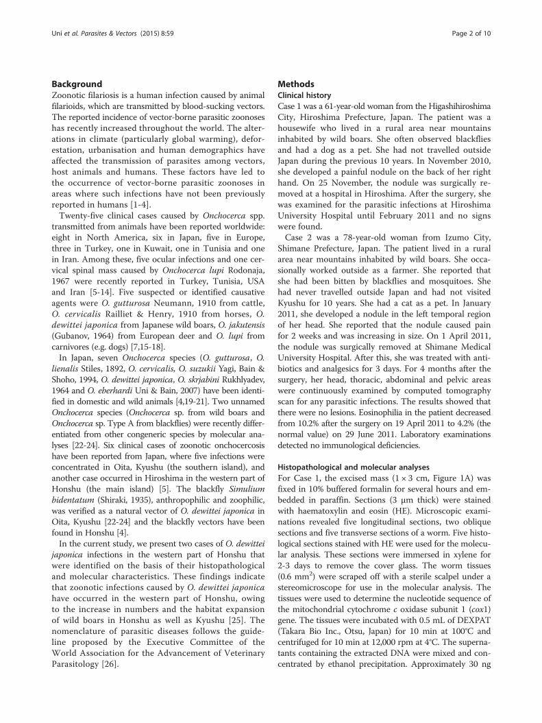
Figure 1 Histopathological characteristics of Onchocerca dewittei japonica in nodules excised from Cases 1 in Hiroshima (A–C) and 2 inShimane (D–F). A) Mass (arrow, 1 × 3 cm) excised from the back of the right hand. B) Longitudinal section of the female O. dewittei japonica.Arrows, triangular transverse ridges; arrowhead, middle layer of the cuticle without the inner striae; white vertical line, cuticle; M, muscle layer.HE staining. C) Slightly oblique transverse section of the female worm showing the thick cuticle. Arrow, elevation of the cuticle, indicating thetransverse ridge; asterisks, lateral chords; M, muscle layer. HE staining. D) Transverse section of the male O. dewittei japonica. I, intestine; M, musclelayer; SV, seminal vesicle with spermatozoids. HE staining. E) Enlarged transverse section. Arrows, small ridges on the cuticle; M, muscle layer;SV, seminal vesicle. HE staining. F) Slightly oblique transverse section. Arrows, small longitudinal ridges; M, muscle layer; SV, seminal vesicle.PAS staining. Unit of bars, μm.
Uni et al. Parasites & Vectors (2015) 8:59 Page 3 of 10
of DNA was used as a template for PCR. PCR amplifi-cation was performed using the primers CO1fF-CO1fR(expected size: 239 bp) as described previously [6].However, the primers failed to amplify cox1 from ourspecimens; therefore, we designed a new reverse primercalled CO1f1R (5′- AAAATAATAACATAAACCTCAGGATG-3′) and used a new primer set CO1fF-CO1f1Rfor amplification. The position of the primer in thecomplete mitochondrial genome of O. volvulus [GenBank:AF015193] is 3013–3038. The thermal conditions were asfollows: initial denaturation at 94°C for 2 min, followed by40 cycles at 98°C for 10 s, 47°C for 30 s and 68°C for 30 s.The PCR product of the expected size (155 bp) was
excised from the agarose gel, purified with a QIAEX II GelExtraction Kit (Qiagen, Hilden, Germany) and cloned intothe HincII site of the pUC118 plasmid vector with a MightyCloning Reagent Set < Blunt End > (Takara Bio Inc.). Theinserted fragments from six colonies were sequenced usingM13F (-20) and M13R primers, a BigDye Terminator v3.1Cycle Sequencing Kit (Applied Biosystems, Foster City, CA,USA) and an Applied Biosystems 3130 Genetic Analyzer(Applied Biosystems).The nucleotide sequence was aligned with the published
sequences of 11 Onchocerca species using CLUSTAL Wwith the default settings in BioEdit ver. 7.0.5.3. [27,28].Two unnamed species (Onchocerca sp. wild boar and
Uni et al. Parasites & Vectors (2015) 8:59 Page 4 of 10
Onchocerca sp. Type A) were included in the comparison.Two other filarial species (Loxodontofilaria caprini Uni& Bain, 2006 and Cercopithifilaria longa Uni, Bain &Takaoka, 2002) were used as outgroups. The Kimuratwo-parameter method [29] was used to estimate evolu-tionary distances from the alignments. The phylogenetictree was constructed using the neighbour-joining method[30], and the bootstrap probabilities were estimated. Theanalyses were performed based on 110 sites of the cox1using MEGA 5.05 [31].For Case 2, the excised mass (1 × 1.5 cm) was fixed in
10% buffered formalin for 6 h and embedded in paraffin.To obtain morphological observations, four sections onone glass slide were stained with HE, and four sectionson another glass slide underwent periodic acid–Schiff(PAS) staining. In the molecular analysis, 30 sections(5 μm thick) of the worm tissues were obtained from theparaffin block, and the unstained worm tissues (0.11 mm2
in 10 sections) were used. The scraped worm tissues weretransferred into nine micro-centrifuge tubes. The DNAextraction procedure and PCR amplification using the pri-mer set CO1fF-CO1f1R were performed as describedabove. Template DNA (0.8 μg) was amplified under thefollowing thermal conditions: initial denaturation at 94°Cfor 2 min, followed by 45 cycles at 98°C for 10 s, 42°C for30 s and 68°C for 30 s. The procedures used to sequencethe PCR products and analyse the data were performed asdescribed above. The sequences determined in Cases 1and 2 were deposited in DDBJ/EMBL/GenBank.
ResultsIn the longitudinal sections of Case 1, the worm exhib-ited transverse cuticular ridges, which are typical offemale Onchocerca species (Table 1 and Figure 1B). Eachridge formed a triangular projection in the longitudinalsections, and the distance between two adjacent ridgeswas 150–250 μm (arrows in Figure 1B). There were noinner striae in the middle layer of the cuticle (arrowheadin Figure 1B). The cuticle was thick and consisted offour layers. The slightly oblique transverse sections wereoval and measured 125–153 × 278–280 μm (Figure 1C).An elevated portion of the cuticle indicated the presenceof a ridge (arrow in Figure 1C). The cuticle, hypodermis,lateral chords and polymyarian muscle layer wereobserved; however, the uterus and intestine were lostduring the preparation of the sections. There was noinner cuticular projection on the lateral chord, but irregu-lar waves were observed in the hypodermis. In terms ofthe host–worm interaction, the worm was surrounded byfibrous tissue, containing macrophages, neutrophils, eo-sinophils and lymphocytes. In conclusion, the morpho-logical characteristics of the worm were identical or verysimilar to those of adult females of O. dewittei japonicacollected from wild boars (Table 1).
The cox1 sequences (excluding primers) of our speci-mens were compared with those of 11 species that con-sisted of Onchocerca spp., Onchocerca sp. wild boarand Onchocerca sp. Type A from GenBank (Table 2).The nucleotide sequence of cox1 from the causativeagent of Case 1 was identical to that of O. dewittei japon-ica from wild boars (Table 2 and Figure 2). Furthermore,in the phylogenetic tree, the causative agent of Case 1 andO. dewittei japonica were separated from other congenericspecies, with a high bootstrap value (Figure 2). On thebasis of the molecular analysis, we confirmed the causativeagent of Case 1 to be O. dewittei japonica. In Case 2, thetransverse sections of the midbody contained the seminalvesicle, intestine and the polymyarian muscle layer(Figure 1D). The seminal vesicle was filled with sperma-tozoids. We observed small longitudinal ridges (138–152ridges) on the outer cuticle of the midbody, and the height/width of the ridges was 1/1 μm (arrows in Figure 1E).In the slightly oblique sections, small longitudinalridges were observed (arrows in Figure 1F). The sizeand number of the ridges were identical or very similar tothose of a male O. dewittei japonica (Table 1). Therefore,we identified the causative agent as a male O. dewitteijaponica.The nucleotide sequence of cox1 was compared with
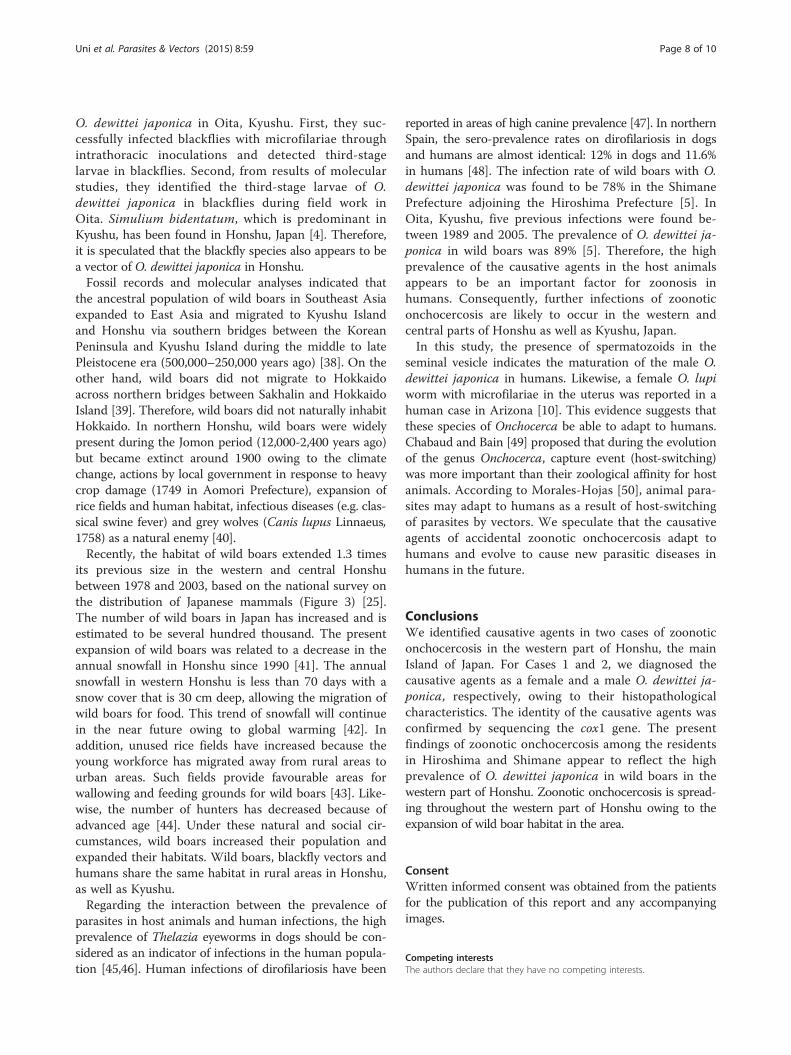
that of congeneric species from the GenBank (Table 2).With the exception of one of the 110 sequences, thenucleotide sequences of our specimen from Case 2 wereidentical to those of O. dewittei japonica, confirmingthat the causative agent was O. dewittei japonica. Theareas where Cases 1 and 2 were found are indicated onthe map of Honshu, Japan (asterisks in Figure 3).
DiscussionWe compared the morphological findings of causativeagents obtained in the present study with those of congen-eric species in Japan. For Case 1, the distance between theadjacent ridges and their triangular shape were very simi-lar to that found in females of O. dewittei japonica, whilediffering from those of other species (Table 1). Further-more, this specimen did not exhibit the inner striae in themiddle layer of the cuticle that are typical for females ofO. gutturosa, O. lienalis, O. skrjabini, O. eberhardi and O.cervicalis (Table 1) [5,21,32-36]. Females of O. suzukii lackthe transverse ridges and inner striae in the cuticle [19].We therefore diagnosed the causative agent of Case 1 as afemale O. dewittei japonica.To the best of our knowledge, most male Onchocerca
spp. have no specific characteristics on their cuticle[21,35]. In contrast, males of O. dewittei japonica fromJapanese wild boars and of O. dewittei dewittei Bain,Ramachandran, Petter & Mak, 1977 from Malaysian wildboars (Sus scrofa jubatus Miller, 1906) have small longitu-dinal ridges on the cuticle [20]. In Case 2, the transverse
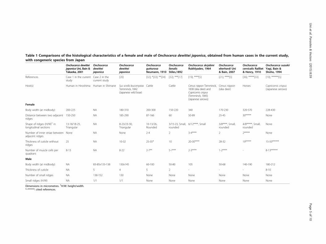
Table 1 Comparisons of the histological characteristics of a female and male of Onchocerca dewittei japonica, obtained from human cases in the current study,with congeneric species from Japan
Onchocerca dewitteijaponica Uni, Bain &Takaoka, 2001
Onchocercadewitteijaponica
Onchocercadewitteijaponica
OnchocercagutturosaNeumann, 1910
OnchocercalienalisStiles,1892
Onchocerca skrjabiniRukhlyadev, 1964
Onchocercaeberhardi Uni& Bain, 2007
Onchocercacervicalis Railliet& Henry, 1910
Onchocerca suzukiiYagi, Bain &Shoho, 1994
References Case 1 in the currentstudy
Case 2 in thecurrent study
[20] [32]; *[33]; **[34] [32]; ***[17] [19]; ****[5] [21]; ****[5] [36]; *****[33] [19]; ******[5]
Host(s) Human in Hiroshima Human in Shimane Sus scrofa leucomystaxTemminck, 1842(Japanese wild boar)
Cattle Cattle Cervus nippon Temminck,1838 (sika deer) andCapricornis crispus(Temminck, 1845)(Japanese serows)
Cervus nippon(sika deer)
Horses Capricornis crispus(Japanese serows)
Female
Body width (at midbody) 200-225 NA 180-310 200-300 150-220 340 170-230 320-570 228-430
Distance between two adjacentridges
150-250 NA 185-290 87-166 60 50-89 25-45 30***** None
Shape of ridges (H/W)† inlongitudinal sections
13-18/18-25,Triangular
NA 8-23/23-30,Triangular
10-13/26,Rounded
3/15-23, Small,rounded
6/12****, Small 3/8****, Small,rounded
4/8*****, Small,rounded
None
Number of inner striae betweenadjacent ridges
None NA None 2-4 2 3-4**** 2 2***** None
Thickness of cuticle withoutridges
25 NA 10-32 25-35* 10 20-30**** 28-32 19***** 15-50******
Number of muscle cells perquadrant
8-13 NA 8-22 2-7** 5-7*** 2-3**** 1-2**** - 8-13******
Male
Body width (at midbody) NA 83-85x133-138 130x145 60-100 50-80 105 50-68 140-190 180-212
Thickness of cuticle NA 5 4 5 2 - - - 8-10
Number of small ridges NA 138-152 130 None None None None None None
Small ridges (H/W) NA 1/1 1/1 None None None None None None
Dimensions in micrometres. †H/W: height/width.*-******: cited references.
Uniet
al.Parasites&Vectors
(2015) 8:59 Page
5of
10
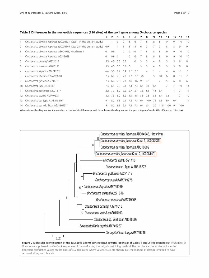
Table 2 Differences in the nucleotide sequences (110 sites) of the cox1 gene among Onchocerca species
1 2 3 4 5 6 7 8 9 10 11 12 13 14
1 Onchocerca dewittei japonica (LC008531, Case 1 in the present study) 1 0 0 6 6 7 8 8 8 9 9 10 10
2 Onchocerca dewittei japonica (LC008149, Case 2 in the present study) 0.9 1 1 5 5 6 7 7 7 8 8 9 9
3 Onchocerca dewittei japonica AB604943, Hiroshima 1 0 0.9 0 6 6 7 8 8 8 9 9 10 10
4 Onchocerca dewittei japonica AB518689 0 0.9 0 6 6 7 8 8 8 9 9 10 10
5 Onchocerca ochengi AJ271618 5.5 4.5 5.5 5.5 0 3 3 4 8 3 5 8 8
6 Onchocerca volvulus AF015193 5.5 4.5 5.5 5.5 0 3 3 4 8 3 5 8 8
7 Onchocerca skrjabini AM749269 6.4 5.5 6.4 6.4 2.7 2.7 4 1 7 4 6 7 7
8 Onchocerca eberhardi AM749268 7.3 6.4 7.3 7.3 2.7 2.7 3.6 5 10 6 8 11 7
9 Onchocerca gibsoni AJ271616 7.3 6.4 7.3 7.3 3.6 3.6 9.1 4.5 7 5 6 8 6
10 Onchocerca lupi EF521410 7.3 6.4 7.3 7.3 7.3 7.3 6.4 9.1 6.4 7 7 10 13
11 Onchocerca gutturosa AJ271617 8.2 7.3 8.2 8.2 2.7 2.7 3.6 5.5 4.5 6.4 4 7 11
12 Onchocerca suzukii AM749275 8.2 7.3 8.2 8.2 4.5 4.5 5.5 7.3 5.5 6.4 3.6 7 10
13 Onchocerca sp. Type A AB518876* 9.1 8.2 9.1 9.1 7.3 7.3 6.4 10.0 7.3 9.1 6.4 6.4 11
14 Onchocerca sp. wild boar AB518693* 9.1 8.2 9.1 9.1 7.3 7.3 6.4 6.4 5.5 11.8 10.0 9.1 10.0
Values above the diagonal are the numbers of nucleotide differences, and those below the diagonal are the percentages of nucleotide differences. *See text.
Figure 2 Molecular identification of the causative agents (Onchocerca dewittei japonica) of Cases 1 and 2 (red rectangles). Phylogeny ofOnchocerca spp. based on GenBank sequences of the cox1 using the neighbour-joining method. The numbers at the nodes indicate thebootstrap confidence values on the basis of 500 replicates, where values >50% are shown. Bar, the number of changes inferred to haveoccurred along each branch.
Uni et al. Parasites & Vectors (2015) 8:59 Page 6 of 10
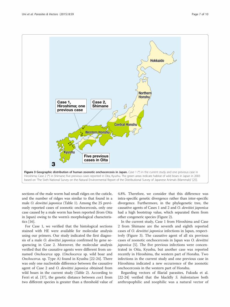
Figure 3 Geographic distribution of human zoonotic onchocercosis in Japan. Case 1 (*) in the current study and one previous case inHiroshima; Case 2 (*) in Shimane; five previous cases reported in Oita, Kyushu. The green areas indicate habitat of wild boars in Japan in 2003based on ‘The Sixth National Survey on the Natural Environmental Report of the Distributional Survey of Japanese Animals (Mammals)’ [25].
Uni et al. Parasites & Vectors (2015) 8:59 Page 7 of 10
sections of the male worm had small ridges on the cuticle,and the number of ridges was similar to that found in amale O. dewittei japonica (Table 1). Among the 25 previ-ously reported cases of zoonotic onchocercosis, only onecase caused by a male worm has been reported (from Oitain Japan) owing to the worm’s morphological characteris-tics [16].For Case 1, we verified that the histological sections
stained with HE were available for molecular analysisusing our primers. Our study indicated the first diagno-sis of a male O. dewittei japonica confirmed by gene se-quencing in Case 2. Moreover, the molecular analysisverified that the causative agents were different from un-named Onchocerca spp. (Onchocerca sp. wild boar andOnchocerca. sp. Type A) found in Kyushu [22-24]. Therewas only one nucleotide difference between the causativeagent of Case 2 and O. dewittei japonica obtained fromwild boars in the current study (Table 2). According toFerri et al. [37], the genetic distance between cox1 fromtwo different species is greater than a threshold value of
4.8%. Therefore, we consider that this difference wasintra-specific genetic divergence rather than inter-specificdivergence. Furthermore, in the phylogenetic tree, thecausative agents of Cases 1 and 2 and O. dewittei japonicahad a high bootstrap value, which separated them fromother congeneric species (Figure 2).In the current study, Case 1 from Hiroshima and Case
2 from Shimane are the seventh and eighth reportedcases of O. dewittei japonica infections in Japan, respect-ively (Figure 3). The causative agent of all six previouscases of zoonotic onchocercosis in Japan was O. dewitteijaponica [5]. The five previous infections were concen-trated in Oita, Kyushu, but another case was reportedrecently in Hiroshima, the western part of Honshu. Twoinfections in the current study and one previous case inHiroshima indicated a new occurrence of the zoonoticonchocercosis in the western part of Honshu.Regarding vectors of filarial parasites, Fukuda et al.
[22-24] verified that the blackfly S. bidentatum bothanthropophilic and zoophilic was a natural vector of
Uni et al. Parasites & Vectors (2015) 8:59 Page 8 of 10
O. dewittei japonica in Oita, Kyushu. First, they suc-cessfully infected blackflies with microfilariae throughintrathoracic inoculations and detected third-stagelarvae in blackflies. Second, from results of molecularstudies, they identified the third-stage larvae of O.dewittei japonica in blackflies during field work inOita. Simulium bidentatum, which is predominant inKyushu, has been found in Honshu, Japan [4]. Therefore,it is speculated that the blackfly species also appears to bea vector of O. dewittei japonica in Honshu.Fossil records and molecular analyses indicated that
the ancestral population of wild boars in Southeast Asiaexpanded to East Asia and migrated to Kyushu Islandand Honshu via southern bridges between the KoreanPeninsula and Kyushu Island during the middle to latePleistocene era (500,000–250,000 years ago) [38]. On theother hand, wild boars did not migrate to Hokkaidoacross northern bridges between Sakhalin and HokkaidoIsland [39]. Therefore, wild boars did not naturally inhabitHokkaido. In northern Honshu, wild boars were widelypresent during the Jomon period (12,000-2,400 years ago)but became extinct around 1900 owing to the climatechange, actions by local government in response to heavycrop damage (1749 in Aomori Prefecture), expansion ofrice fields and human habitat, infectious diseases (e.g. clas-sical swine fever) and grey wolves (Canis lupus Linnaeus,1758) as a natural enemy [40].Recently, the habitat of wild boars extended 1.3 times
its previous size in the western and central Honshubetween 1978 and 2003, based on the national survey onthe distribution of Japanese mammals (Figure 3) [25].The number of wild boars in Japan has increased and isestimated to be several hundred thousand. The presentexpansion of wild boars was related to a decrease in theannual snowfall in Honshu since 1990 [41]. The annualsnowfall in western Honshu is less than 70 days with asnow cover that is 30 cm deep, allowing the migration ofwild boars for food. This trend of snowfall will continuein the near future owing to global warming [42]. Inaddition, unused rice fields have increased because theyoung workforce has migrated away from rural areas tourban areas. Such fields provide favourable areas forwallowing and feeding grounds for wild boars [43]. Like-wise, the number of hunters has decreased because ofadvanced age [44]. Under these natural and social cir-cumstances, wild boars increased their population andexpanded their habitats. Wild boars, blackfly vectors andhumans share the same habitat in rural areas in Honshu,as well as Kyushu.Regarding the interaction between the prevalence of
parasites in host animals and human infections, the highprevalence of Thelazia eyeworms in dogs should be con-sidered as an indicator of infections in the human popula-tion [45,46]. Human infections of dirofilariosis have been
reported in areas of high canine prevalence [47]. In northernSpain, the sero-prevalence rates on dirofilariosis in dogsand humans are almost identical: 12% in dogs and 11.6%in humans [48]. The infection rate of wild boars with O.dewittei japonica was found to be 78% in the ShimanePrefecture adjoining the Hiroshima Prefecture [5]. InOita, Kyushu, five previous infections were found be-tween 1989 and 2005. The prevalence of O. dewittei ja-ponica in wild boars was 89% [5]. Therefore, the highprevalence of the causative agents in the host animalsappears to be an important factor for zoonosis inhumans. Consequently, further infections of zoonoticonchocercosis are likely to occur in the western andcentral parts of Honshu as well as Kyushu, Japan.In this study, the presence of spermatozoids in the
seminal vesicle indicates the maturation of the male O.dewittei japonica in humans. Likewise, a female O. lupiworm with microfilariae in the uterus was reported in ahuman case in Arizona [10]. This evidence suggests thatthese species of Onchocerca be able to adapt to humans.Chabaud and Bain [49] proposed that during the evolutionof the genus Onchocerca, capture event (host-switching)was more important than their zoological affinity for hostanimals. According to Morales-Hojas [50], animal para-sites may adapt to humans as a result of host-switchingof parasites by vectors. We speculate that the causativeagents of accidental zoonotic onchocercosis adapt tohumans and evolve to cause new parasitic diseases inhumans in the future.
ConclusionsWe identified causative agents in two cases of zoonoticonchocercosis in the western part of Honshu, the mainIsland of Japan. For Cases 1 and 2, we diagnosed thecausative agents as a female and a male O. dewittei ja-ponica, respectively, owing to their histopathologicalcharacteristics. The identity of the causative agents wasconfirmed by sequencing the cox1 gene. The presentfindings of zoonotic onchocercosis among the residentsin Hiroshima and Shimane appear to reflect the highprevalence of O. dewittei japonica in wild boars in thewestern part of Honshu. Zoonotic onchocercosis is spread-ing throughout the western part of Honshu owing to theexpansion of wild boar habitat in the area.
ConsentWritten informed consent was obtained from the patientsfor the publication of this report and any accompanyingimages.
Competing interestsThe authors declare that they have no competing interests.
Uni et al. Parasites & Vectors (2015) 8:59 Page 9 of 10
Authors’ contributionsSU and HT conceived the research and wrote the first draft. SU, RR and MSAperformed the morphological study and contributed to the data analysisand interpretation. MF and YO contributed to the molecular analysis ofsamples. NH, KY, HT, SM, KK, EM, HM, HH and KS collected samples and wereresponsible for the medical aspects. All authors read and approved the finalversion of the manuscript.
AcknowledgementsWe sincerely thank Professor Dr. Rosli Hashim, Deputy Dean of the Faculty ofScience, University of Malaya, Malaysia for his academic support andMr. Takao Kenko, the Central Laboratory, the Graduate School of Medicine,Osaka City University, Japan, for preparing the histological specimens. Thisresearch was partly supported by the Ministry of Higher Education of Malaysia(FRGS FP020-2012).
Author details1Institute of Biological Sciences, Faculty of Science, University of Malaya,50603 Kuala Lumpur, Malaysia. 2Department of Parasitology, Graduate Schoolof Medicine, Osaka City University, Osaka 545-8585, Japan. 3ResearchPromotion Institute, Oita University, Oita 879-5593, Japan. 4Research Centerfor the Pacific Islands, Kagoshima University, Kagoshima 890-8580, Japan.5Hiramatsu Orthopedic Clinic, Hiroshima 730-0016, Japan. 6HiroshimaUniversity Hospital, Hiroshima 734-8551, Japan. 7Department of Dermatology,Faculty of Medicine, Shimane University, Shimane 693-8501, Japan.8Department of Infectious Diseases, Division of Parasitology, Faculty ofMedicine, University of Miyazaki, Miyazaki 889-1692, Japan. 9Department ofBiology, Faculty of Medicine, Oita University, Oita 879-5593, Japan.10Department of Environmental and Preventive Medicine, Faculty ofMedicine, Shimane University, Shimane 693-8501, Japan.
Received: 10 October 2014 Accepted: 12 January 2015
References1. Orihel TC, Eberhard ML. Zoonotic filariasis. Clin Microbiol Rev.
1998;11:366–81.2. Otranto D, Eberhard ML. Zoonotic helminths affecting the human eye.
Parasit Vectors. 2011;4:41.3. Colwell DD, Dantas-Torres F, Otranto D. Vector-borne parasitic zoonoses:
emerging scenarios and new perspectives. Vet Parasitol. 2011;182:14–21.4. Takaoka H, Fukuda M, Otsuka Y, Aoki C, Uni S, Bain O. Blackfly vectors of
zoonotic onchocerciasis in Japan. Med Vet Entomol. 2012;26:372–8.5. Uni S, Boda T, Daisaku K, Ikura Y, Maruyama H, Hasegawa H, et al. Zoonotic
filariasis caused by Onchocerca dewittei japonica in a resident of HiroshimaPrefecture, Honshu, Japan. Parasitol Int. 2010;59:477–80.
6. Fukuda M, Otsuka Y, Uni S, Boda T, Daisaku H, Hasegawa H, et al. Zoonoticonchocerciasis in Hiroshima, Japan, and molecular analysis of a paraffinsection of the agent for a reliable identification. Parasite. 2011;18:185–8.
7. Otranto D, Sakru N, Testini G, Gürlü VP, Yakar K, Lia RP, et al. Case report:first evidence of human zoonotic infection by Onchocerca lupi (Spirurida,Onchocercidae). Am J Trop Med Hyg. 2011;84:55–8.
8. Otranto D, Dantas-Torres F, Cebeci Z, Yeniad B, Buyukbabani N, Boral OB,et al. Human ocular filariasis: further evidence on the zoonotic role ofOnchocerca lupi. Parasit Vectors. 2012;5:84.
9. Eberhard ML, Sims AC, Bishop HS, Mathison BA, Hoffman RS. Ocularzoonotic Onchocerca infection in a resident of Oregon. Am J Trop Med Hyg.2012;87:1073–5.
10. Eberhard ML, Ostovar GA, Chundu K, Hobohm D, Feiz-Erfan I, Mathison BA,et al. Zoonotic Onchocerca lupi infection in a 22-month-old child in Arizona:first report in the United States and a review of the literature. Am J TropMed Hyg. 2013;88:601–5.
11. Ilhan HD, Yaman A, Morishima Y, Sugiyama H, Muto M, Yamasaki H, et al.Onchocerca lupi infection in Turkey: a unique case of a rare human parasite.Acta Parasitol. 2013;58:384–8.
12. Biswas A, Yassin MH: An unexpected cause of eye irritation: a case of zoonoticocular onchocerciasis. Case Rep Infect Dis 2013, doi:10.1155/2013/504749.
13. Lai JH, Walsh NMG, Pritt BS, Sloan L, Gibson LE, Desormeau L, et al.Cutaneous manifestations of a zoonotic Onchocerca species in an adultmale acquired in Nova Scotia: report of a case and review of the literature.J Clin Microbiol. 2014;52:1968–70.
14. Mowlavi G, Farzbod F, Kheirkhah A, Mobedi I, Bowman DD, Naddaf SR. Humanocular onchocerciasis caused by Onchocerca lupi (Spirurida, Onchocercidae) inIran. J Helminthol. 2014;88:250–5.
15. Beaver PC, Horner GS, Bilos JZ. Zoonotic onchocercosis in a resident of Illioisand observations on the identification of Onchocerca species. Am J TropMed Hyg. 1974;23:595–607.
16. Takaoka H, Bain O, Uni S, Korenaga M, Tada K, Ichikawa H, et al. Humaninfection with Onchocerca dewittei japonica, a parasite from wild boar inOita, Japan. Parasite. 2001;8:261–3.
17. Takaoka H, Bain O, Uni S, Korenaga M, Kozek WJ, Shirasaka C, et al. Zoonoticonchocerciasis caused by a parasite from wild boar in Oita, Japan. Acomprehensive analysis of morphological characteristics of the worms forits diagnosis. Parasite. 2004;11:285–92.
18. Koehsler M, Soleiman A, Aspöck H, Auer H, Walochnik J. Onchocercajakutensis filariasis in humans. Emerg Infect Dis. 2007;13:1749–52.
19. Yagi K, Bain O, Shoho C. Onchocerca suzukii n. sp. and O. skrjabini (= O. tarsicola)from a relict bovid, Capricornis crispus, in Japan. Parasite. 1994;1:349–56.
20. Uni S, Bain O, Takaoka H, Miyashita M, Suzuki Y. Onchocerca dewitteijaponica n. subsp., a common parasite from wild boar in Kyushu Island,Japan. Parasite. 2001;8:215–22.
21. Uni S, Bain O, Agatsuma T, Harada M, Torii H, Fukuda M, et al. Onchocercaeberhardi n. sp. (Nematoda: Filarioidea) from sika deer in Japan; relationshipsbetween species parasitic in cervids and bovids in the Holarctic region.Parasite. 2007;14:199–211.
22. Fukuda M, Takaoka H, Uni S, Bain O. Infective larvae of five Onchocercaspecies from experimentally infected Simulium species in an area ofzoonotic onchocerciasis in Japan. Parasite. 2008;15:111–9.
23. Fukuda M, Otsuka Y, Uni S, Bain O, Takaoka H. Genetic evidence for thepresence of two species of Onchocerca from the wild boar in Japan.Parasite. 2010;17:33–7.
24. Fukuda M, Otsuka Y, Uni S, Bain O, Takaoka H. Molecular identification ofinfective larvae of three species of Onchocerca found in wild-caught femalesof Simulium bidentatum in Japan. Parasite. 2010;17:39–45.
25. Japan Wildlife Research Center. The Sixth National Survey on the NaturalEnvironmental Report of the Distributional Survey of Japanese Animals(Mammals), Biodiversity Center of Japan, Nature Conservation Bureau,Ministry of the Environment, Japan. 2004. p. 60–5. in Japanese.
26. Kassai T, Cordero del Campillo M, Euzeby J, Gaafar S, Hiepe T, Himonas CA.Standardized nomenclature of animal parasitic diseases (SNOAPAD). VetParasitol. 1988;29:299–326.
27. Thompson JD, Higgins DG, Gibson TJ. CLUSTAL W: improving the sensitivityof progressive multiple sequence alignment through sequence weighting,position-specific gap penalties and weight matrix choice. Nucleic Acids Res.1994;22:4673–80.
28. Hall TA. BioEdit: a user-friendly biological sequence alignment editor andanalysis program for Windows 95/98/NT. Nucleic Acids Symp Ser.1999;41:95–8.
29. Kimura M. A simple method for estimating evolutionary rates of basesubstitutions through comparative studies of nucleotide sequences. J MolEvol. 1980;16:111–20.
30. Saitou N, Nei M. The neighbor-joining method: a new method forreconstructing phylogenetic trees. Mol Biol Evol. 1987;4:406–25.
31. Tamura K, Peterson D, Peterson N, Stecher G, Nei M, Kumar S. MEGA5:molecular evolutionary genetics analysis using maximum likelihood,evolutionary distance, and maximum parsimony methods. Mol Biol Evol.2011;28:2731–9.
32. Eberhard ML. Studies on the Onchocerca (Nematoda: Filarioidea) found incattle in the United States. I. Systematics of O. gutturosa and O. lienalis witha description of O. stilesi sp. n. J Parasitol. 1979;65:379–88.
33. Bain O. Redescription de cinq espèces d’onchocerques. Ann Parasitol HumComp. 1975;50:763–88.
34. Bain O, Petit G, Poulain B. Validité des deux espèces Onchocerca lienalis et O.gutturosa, chez les bovins. Ann Parasitol Hum Comp. 1978;53:421–30.
35. Bain O. Le genre Onchocerca: hypothèses sur son évolution et clédichotomique des espèces. Ann Parasitol Hum Comp. 1981;56:503–26.
36. Mellor PS. Studies on Onchocerca cervicalis Railliet and Henry 1910: 3.Morphological and taxonomic studies on Onchocerca cervicalis from Britishhorses. J Helminthol. 1974;48:145–53.
37. Ferri E, Barbuto M, Bain O, Galimberti A, Uni S, Guerrero RA, et al. Integratedtaxonomy: traditional approach and DNA barcoding for the identification offilarioid worms and related parasites (Nematoda). Front Zool. 2009;6:1.
Uni et al. Parasites & Vectors (2015) 8:59 Page 10 of 10
38. Kawamura Y. Immigration of mammals into the Japanese Islands during theQuaternary. Quat Res. 1998;37:251–7.
39. Watanobe T, Ishiguro N, Nakano M. Phylogeography and populationstructure of the Japanese wild boar Sus scrofa leucomystax: mitochondrialDNA variation. Zool Sci. 2003;20:1477–89.
40. Tsujino R, Ishimaru E, Yumoto T. Distribution patterns of five mammals inthe Jomon period, middle Edo period, and the present, in the JapaneseArchipelago. Mam Stud. 2010;35:179–89.
41. Tokida K, Maruyama N: Factors affecting the geographical distribution ofJapanese wild boars. In The Second National Survey on the NaturalEnvironmental Report of the Distributional Survey of Japanese Animals(Mammals). Japan Wildlife Research Center, 1980, (in Japanese with Englishsummary). www.biodic.go.jp/reports/2-6/ad097.html
42. Japan Meteorological Agency: Climate change and its impact in Japan,2012. Page 25 (in Japanese), 2013. http://www.env.go.jp/earth/ondanka/rep130412/report_full.pdf
43. Honda T. Environmental factors affecting the distribution of the wild boar,sika deer, Asiatic black bear and Japanese macaque in central Japan, withimplications for human-wildlife conflict. Mam Stud. 2009;34:107–16.
44. Ministry of the Environment, Japan. Statistics on capture of wildlife, Page 85,(in Japanese), 2014. https://www.env.go.jp/nature/choju/capture/pdf/d1.pdf
45. Otranto D, Traversa D. Thelazia eyeworm: an original endo- and ecto-parasiticnematode. Trends Parasitol. 2005;21:1–4.
46. Otranto D, Dantas-Torres F, Brianti E, Traversa D, Petrić D, Genchi C, et al.Vector-borne helminths of dogs and humans in Europe. Parasit Vectors.2013;6:16.
47. Lee AC, Montgomery SP, Theis JH, Blagburn BL, Eberhard ML. Public healthissues concerning the widespread distribution of canine heartworm disease.Trends Parasitol. 2010;26:168–73.
48. Morchón R, Moya I, González-Miguel J, Montoya MN, Simón F. ZoonoticDirofilaria immitis infections in a province of Northern Spain. EpidemiolInfect. 2010;138:380–3.
49. Chabaud AG, Bain O. The evolutionary expansion of the Spirurida. Int JParasitol. 1994;24:1179–201.
50. Morales-Hojas R. Molecular systematics of filarial parasites, with an emphasison groups of medical and veterinary importance, and its relevance forepidemiology. Infect Genet Evol. 2009;9:748–59.
Submit your next manuscript to BioMed Centraland take full advantage of:
• Convenient online submission
• Thorough peer review
• No space constraints or color figure charges
• Immediate publication on acceptance
• Inclusion in PubMed, CAS, Scopus and Google Scholar
• Research which is freely available for redistribution
Submit your manuscript at www.biomedcentral.com/submit