Embed Size (px)
Citation preview

Newborn Screening for Sickle Cell Disease and other Haemoglobinopathies
Stephan Lobitz, Jacques Elion, Raffaella Colombatti and Elena Cela
www.mdpi.com/journal/ijns
Edited by
Printed Edition of the Special Issue Published in
International Journal of Neonatal Screening

Newborn Screening for Sickle CellDisease and other Haemoglobinopathies


Newborn Screening for Sickle CellDisease and other Haemoglobinopathies
Special Issue Editors
Stephan Lobitz
Jacques Elion
Raffaella Colombatti
Elena Cela
MDPI • Basel • Beijing • Wuhan • Barcelona • Belgrade

Jacques Elion
Universite Paris Diderot-USPC France
Elena CelaUniversidad Complutense de Madrid Spain
Special Issue Editors
Stephan Lobitz
Gemeinschaftsklinikum Mittelrhein gGmbH
Germany
Raffaella ColombattiUniversita di Padova
Italy
Editorial Office
MDPISt. Alban-Anlage 66
4052 Basel, Switzerland
This is a reprint of articles from the Special Issue published online in the open access journal
International Journal of Neonatal Screening (ISSN 2409-515X) from 2018 to 2019 (available at: https:
//www.mdpi.com/journal/IJNS/special issues/hemoglobinopathies)
For citation purposes, cite each article independently as indicated on the article page online and as
indicated below:
LastName, A.A.; LastName, B.B.; LastName, C.C. Article Title. Journal Name Year, Article Number,
Page Range.
ISBN 978-3-03921-614-7 (Pbk)
ISBN 978-3-03921-615-4 (PDF)
c© 2019 by the authors. Articles in this book are Open Access and distributed under the Creative
Commons Attribution (CC BY) license, which allows users to download, copy and build upon
published articles, as long as the author and publisher are properly credited, which ensures maximum
dissemination and a wider impact of our publications.
The book as a whole is distributed by MDPI under the terms and conditions of the Creative Commons
license CC BY-NC-ND.
Contents
About the Special Issue Editors . . . . . . . . . . . . . . . . . . . . . . . . . . . . . . . . . . . . . vii
Raffaella Colombatti, Elena Cela, Jacques Elion and Stephan Lobitz
Editorial for Special Issue “Newborn Screening for Sickle Cell Disease andother Haemoglobinopathies”Reprinted from: Int. J. Neonatal Screen. 2019, 5, 36, doi:10.3390/ijns5040036 . . . . . . . . . . . . . 1
J. Gerard Loeber
European Union Should Actively Stimulate and Harmonise Neonatal Screening InitiativesReprinted from: Int. J. Neonatal Screen. 2018, 4, 32, doi:10.3390/ijns4040032 . . . . . . . . . . . . . 3
Baba P.D. Inusa, Lewis L. Hsu, Neeraj Kohli, Anissa Patel, Kilali Ominu-Evbota, Kofi A. Anie and Wale Atoyebi
Sickle Cell Disease—Genetics, Pathophysiology, Clinical Presentation and TreatmentReprinted from: Int. J. Neonatal Screen. 2019, 5, 20, doi:10.3390/ijns5020020 . . . . . . . . . . . . . 8
Michael Angastiniotis and Stephan Lobitz
Thalassemias: An OverviewReprinted from: Int. J. Neonatal Screen. 2019, 5, 16, doi:10.3390/ijns5010016 . . . . . . . . . . . . . 23
Claudia Frommel
Newborn Screening for Sickle Cell Disease and Other Hemoglobinopathies: A Short Review onClassical Laboratory Methods—Isoelectric Focusing, HPLC, and Capillary ElectrophoresisReprinted from: Int. J. Neonatal Screen. 2018, 4, 39, doi:10.3390/ijns4040039 . . . . . . . . . . . . . 34
Yvonne Daniel and Charles Turner
Newborn Sickle Cell Disease Screening Using Electrospray Tandem Mass SpectrometryReprinted from: Int. J. Neonatal Screen. 2018, 4, 35, doi:10.3390/ijns4040035 . . . . . . . . . . . . . 44
Pierre Naubourg, Marven El Osta, David Rageot, Olivier Grunewald, Gilles Renom, Patrick Ducoroy and Jean-Marc Perini
A Multicentre Pilot Study of a Two-Tier Newborn Sickle Cell Disease Screening Procedure with a First Tier Based on a Fully Automated MALDI-TOF MS PlatformReprinted from: Int. J. Neonatal Screen. 2019, 5, 10, doi:10.3390/ijns5010010 . . . . . . . . . . . . . 49
Maddalena Martella, Giampietro Viola, Silvia Azzena, Sara Schiavon, Andrea Biondi,
Giuseppe Basso, Paola Corti, Raffaella Colombatti, Nicoletta Masera and Laura Sainati
Evaluation of Technical Issues in a Pilot Multicenter Newborn Screening Program for SickleCell DiseaseReprinted from: Int. J. Neonatal Screen. 2019, 5, 2, doi:10.3390/ijns5010002 . . . . . . . . . . . . . . 62
Yvonne Daniel, Jacques Elion, Bichr Allaf, Catherine Badens, Marelle J. Bouva, Ian Brincat, Elena Cela, Cathy Coppinger, Mariane de Montalembert, Beatrice Gulbis, Joan Henthorn, Olivier Ketelslegers, Corrina McMahon, Allison Streetly, Raffaella Colombatti and Stephan Lobitz
Newborn Screening for Sickle Cell Disease in EuropeReprinted from: Int. J. Neonatal Screen. 2019, 5, 15, doi:10.3390/ijns5010015 . . . . . . . . . . . . . 70
v

Beatrice Gulbis, Phu-Quoc Le, Olivier Ketelslegers, Marie-Francoise Dresse, Anne-Sophie
Adam, Frederic Cotton, Francois Boemer, Vincent Bours, Jean-Marc Minon and Alina Ferster
Neonatal Screening for Sickle Cell Disease in Belgium for More than 20 Years: An Experiencefor Comprehensive Care ImprovementReprinted from: Int. J. Neonatal Screen. 2018, 4, 37, doi:10.3390/ijns4040037 . . . . . . . . . . . . . 82
Nura El-Haj and Carolyn C. Hoppe
Newborn Screening for SCD in the USA and CanadaReprinted from: Int. J. Neonatal Screen. 2018, 4, 36, doi:10.3390/ijns4040036 . . . . . . . . . . . . . 90
Jennifer Knight-Madden, Ketty Lee, Gisele Elana, Narcisse Elenga, Beatriz Marcheco-Teruel,
Ngozi Keshi, Maryse Etienne-Julan, Lesley King, Monika Asnani, Marc Romana and
Marie-Dominique Hardy-Dessources
Newborn Screening for Sickle Cell Disease in the Caribbean: An Update of the Present Situationand of the Disease PrevalenceReprinted from: Int. J. Neonatal Screen. 2019, 5, 5, doi:10.3390/ijns5010005 . . . . . . . . . . . . . . 100
Ana C. Silva-Pinto, Maria Candida Alencar de Queiroz, Paula Juliana Antoniazzo Zamaro,
Miranete Arruda and Helena Pimentel dos Santos
The Neonatal Screening Program in Brazil, Focus on Sickle Cell Disease (SCD)Reprinted from: Int. J. Neonatal Screen. 2019, 5, 11, doi:10.3390/ijns5010011 . . . . . . . . . . . . . 109
Roshan B. Colah, Pallavi Mehta and Malay B. Mukherjee
Newborn Screening for Sickle Cell Disease: Indian ExperienceReprinted from: Int. J. Neonatal Screen. 2018, 4, 31, doi:10.3390/ijns4040031 . . . . . . . . . . . . . 116
Athena Anderle, Germana Bancone, Gonzalo J. Domingo, Emily Gerth-Guyette, Sampa Pal
and Ari W. Satyagraha
Point-of-Care Testing for G6PD Deficiency: Opportunities for ScreeningReprinted from: Int. J. Neonatal Screen. 2018, 4, 34, doi:10.3390/ijns4040034 . . . . . . . . . . . . . 124
John James and Elizabeth Dormandy
Improving Screening Programmes for Sickle Cell Disorders and Other Haemoglobinopathies inEurope: The Role of Patient OrganisationsReprinted from: Int. J. Neonatal Screen. 2019, 5, 12, doi:10.3390/ijns5010012 . . . . . . . . . . . . . 137
Baba P.D. Inusa, Kofi A. Anie, Andrea Lamont, Livingstone G. Dogara, Bola Ojo, Ifeoma Ijei,
Wale Atoyebi, Larai Gwani, Esther Gani and Lewis Hsu
Utilising the ‘Getting to Outcomes R©’ Framework in Community Engagement for Developmentand Implementation of Sickle Cell Disease Newborn Screening in Kaduna State, NigeriaReprinted from: Int. J. Neonatal Screen. 2018, 4, 33, doi:10.3390/ijns4040033 . . . . . . . . . . . . . 141
vi

About the Special Issue Editors
Stephan Lobitz is the director of the Department of Pediatric Hematology and Oncology
at Gemeinschaftsklinikum Mittelrhein in Koblenz, Germany. He studied Medicine and
Hemoglobinopathies in Dusseldorf, Berlin, and London, and trained at Charite University Hospital
in Berlin. He is the spokesperson of the Sickle Cell Disease Management Program of the German
Society of Pediatric Hematology and Oncology and the coordinator of the German Sickle Cell Disease
Treatment Guideline. Dr. Lobitz has a special interest in newborn screening and coordinated the
recent European consensus statement on newborn screening for sickle cell disease.
Jacques Elion received his MD from Paris Descartes and a PhD from Paris Diderot Universities.
He was a Research Assistant at the Mayo Graduate School of Medicine, University of Minnesota
and a Fogarty Scientist at the US National Institutes of Health. Dr Elion is Professor of Molecular
Genetics at the Universite de Paris and Visiting Professor at the Universidade de Sao Paulo. He is the
former Director of the Dept of Medical Genetics at the Robert Debre University Hospital. Dr Elion’s
research is focused on the pathophysiology, prevention, and global care of SCD. It is conducted at
Unit 1134 of the French National Institute of Health and Medical Research (Inserm) sheltered by the
National Institute of Blood Transfusion in Paris and at the University Hospital in Guadeloupe. The
Unit is part of the French Laboratory of Excellence on the Red Cell (GR-Ex). Dr Elion has developed
extensive international collaborations, notably in sub-Saharan Africa, India, the Caribbean, and
Brazil. Dr Elion has organized and chaired several international meetings, including the scientific
session at the inaugural ceremony for the 1st World SCD Day, 19 June 2009, UN Headquarters, NYC.
Raffaella Colombatti is a pediatric hematologist and oncologist at Padova University in Italy. She
is the coordinator of the Red Cell Disorder Working Group of the Italian Association of Pediatric
Hematology Oncology (AIEOP) and the Vice Chair of the Veneto Region Reference Center for the
Diagnosis and Treatment of Sickle Cell Disease in Childhood and the Pilot Universal Newborn
Screening Program for Sickle Cell Disease. Her main interests are in hemoglobinopathies and child
global health care.
Elena Cela graduated at Complutense University in Madrid, Spain and specialized in pediatric
hematology and oncology. She is the coordinator of the hemoglobinopathy group of the
Spanish Society of Pediatric Hematology and Oncology (SEHOP) and the Spanish Registry of
Hemoglobinopathies (REHem). Prof. Cela is the head of the Department of Pediatric Hematology and
Oncology at Hospital Gregorio Maranon in Madrid, which is a referral center for erythropathology.
She contributed to the implementation of the Spanish newborn screening for sickle cell disease in
Madrid in 2003 and coordinated the edition of the Spanish guidelines on sickle cell disease (2010
and 2019). Her main interests are red cell disorders and newborn screening.
vii


International Journal of
Neonatal Screening
Editorial
Editorial for Special Issue “Newborn Screening forSickle Cell Disease and other Haemoglobinopathies”
Raffaella Colombatti 1,*, Elena Cela 2, Jacques Elion 3 and Stephan Lobitz 4
1 Department of Child and Maternal Health, Clinic of Pediatric Hematology/Oncology, AziendaOspedaliera-Università di Padova, 35129 Padova, Italy
2 Department of Pediatric Oncology/Hematology, Hospital Universitario General Gregorio Marañón,Facultad de Medicina, Universidad Complutense Madrid, 28007 Madrid, Spain;[email protected]
3 Laboratoire d’Excellence GR-Ex, UMR_S1134, Inserm, Université Paris Diderot, Sorbonne Paris Cité,Institut National de la Transfusion Sanguine, 75015 Paris, France; [email protected]
4 Department of Pediatric Hematology and Oncology, Gemeinschaftsklinikum Mittelrhein gGmbH, 56073Koblenz, Germany; [email protected]
* Correspondence: [email protected]
Received: 17 September 2019; Accepted: 17 September 2019; Published: 20 September 2019
Sickle cell disease (SCD) is among the most common genetic disorders in the world, affecting over300,000 newborns annually, with estimates for further increases to over 400,000 annual births withinthe next generation and with a wider geographical distribution of affected individuals due to globalmigration [1,2]. Both the World Health Organization (WHO) and the United Nations have identifiedSCD as a current global health burden [3,4].
The optimal care for children with SCD starts with newborn screening (NBS), which can establisha diagnosis before the onset of symptoms and allow early interventions such as prophylactic penicillin,pneumococcal immunization, screening with Transcranial Doppler ultrasound, caregiver education,and comprehensive care [5]. NBS followed by adequate comprehensive care reduce morbidity, mortality,and healthcare costs while improving the quality of life for patients.
Universal NBS is now recommended in the United States, Europe, and Brazil, although widespreadimplementation still needs to be achieved and many challenges remain to ensure that every child withSCD is diagnosed through NBS [6,7].
In this Special Issue on Newborn Screening for Sickle Cell Disease and Other Hemoglobinopathies(https://www.mdpi.com/journal/IJNS/special_issues/hemoglobinopathies), we have assembled acollection of review and original articles.
We have tried to cover the most widely faced challenges in the field of newborn screening forSCD: unmet needs in Europe and healthcare policy implementation as well as patient involvementand development of new diagnostic techniques.
We would like to commend the authors for the excellent reviews on the pathophysiology of SCDand thalassemia, the state of the art of NBS at a global level, and the technologies available for NBS.We would also like to praise the authors who provided original articles on specific technical topicswhich understanding is essential for more reliable, technically sound, and faster diagnosis.
Global diseases can be tackled only with global and coordinated efforts of different experts rangingfrom clinicians to technicians and basic scientists, as well as healthcare planners and the patientsthemselves across different countries. This Special Issue brings together a multidisciplinary globalteam presenting the actual situation and proposals for future developments.
Conflicts of Interest: The authors declare no conflict of interest.
Int. J. Neonatal Screen. 2019, 5, 36; doi:10.3390/ijns5040036 www.mdpi.com/journal/ijns1

Int. J. Neonatal Screen. 2019, 5, 36
References
1. Piel, F.B.; Tatem, A.J.; Huang, Z.; Gupta, S.; Williams, T.N.; Weatherall, D.J. Global migration and the changingdistribution of sickle haemoglobin: A quantitative study of temporal trends between 1960 and 2000. LancetGlob. Health 2014, 2, e80–e89. [CrossRef]
2. Piel, F.B.; Hay, S.I.; Gupta, S.; Weatherall, D.J.; Williams, T.N. Global burden of sickle cell anaemia in childrenunder five, 2010–2050: Modelling based on demographics, excess mortality, and interventions. PLoS Med.2013, 10, e1001484. [CrossRef] [PubMed]
3. WHO Report A59/9 on Sickle Cell Anemia. 2006. Available online: www.who.int/gb/ebwha/pdf_files/WHA59-REC1/e/WHA59_2006_REC1-en.pdf (accessed on 18 September 2019).
4. UN Resolution A/63/L.63 “Recognition of Sickle-Cell Anaemia as a Public Health Problem”. 2008.Available online: www.un.org/News/Press/docs/2008/ga10803.doc.htm (accessed on 18 Sepember 2019).
5. Yawn, B.P.; Buchanan, G.R.; Afenyi-Annan, A.N.; Ballas, S.K.; Hassell, K.L.; James, A.H.; Jordan, L.;Lanzkron, S.M.; Lottenberg, R.; Savage, W.J.; et al. Management of sickle cell disease: Summary of the 2014evidence-based report by expert panel members. JAMA 2014, 312, 1033–1048. [CrossRef] [PubMed]
6. Shook, L.M.; Ware, R.E. Sickle cell screening in Europe: The time has come. Br. J. Haematol. 2018, 183,534–535. [CrossRef] [PubMed]
7. Lobitz, S.; Telfer, P.; Cela, E.; Allaf, B.; Angastiniotis, M.; Backman Johansson, C.; Badens, C.; Bento, C.;Bouva, M.J.; Canatan, D.; et al. Newborn screening for sickle cell disease in Europe: recommendations froma Pan-European Consensus Conference. Br. J. Haematol. 2018, 183, 648–660. [CrossRef] [PubMed]
© 2019 by the authors. Licensee MDPI, Basel, Switzerland. This article is an open accessarticle distributed under the terms and conditions of the Creative Commons Attribution(CC BY) license (http://creativecommons.org/licenses/by/4.0/).
2

International Journal of
Neonatal Screening
Editorial
European Union Should Actively Stimulate andHarmonise Neonatal Screening Initiatives
J. Gerard Loeber
International Society for Neonatal Screening Office, Burgemeester Fabiuspark 55, 3721CK Bilthoven,The Netherlands; [email protected]; Tel.: +31-6-4616-3922
Received: 27 September 2018; Accepted: 8 November 2018; Published: 14 November 2018
Abstract: Neonatal screening programmes have been introduced in almost all European countries.In practice there are large differences, especially in the panel of conditions that are screened for,often without clear reasons. Policy making on a European level is lacking in contrast to the situation inthe USA. Professionals have the knowledge to expand the panels but are dependent on policy-makersfor the necessary funds. This paper is a call on the EU Commission to take up a role in providingequal access to neonatal screening for all children within the EU.
Keywords: (recommended) screening panel; policy making; harmonisation; patient advocacy
1. Introduction
Neonatal screening, in some countries called newborn screening (NBS), has been recognised as avaluable public health tool in many countries around the world. Based on the work by, e.g., Følling,Penrose and Centerwall in the 20th-century interbellum, Guthrie developed the first relatively easyand cheap assay for the identification of newborn children suffering from phenylketonuria [1]. In thefollowing decade, the development of (radio)immunoassay systems opened the gate for the detectionof blood components indicative of congenital hypothyroidism (CH) [2]. These two conditions werejust the first of many more. Especially the development of the tandem mass spectrometry in the 1990sled to the possibility of high throughput screening of many newborn children for up to 40–50 differentconditions [3,4].
It is self-evident that the implementation of an NBS programme based on modern technologiescomes with a certain cost; the apparatus and the necessary manpower are relatively expensive, althoughthis must also be seen in relation to the annual workload of each laboratory. In addition, so-called“multiplexing” methods as tandem mass spectrometry, facilitate the detection of more conditionswithout a substantial increase in running costs. On the other hand, most of the conditions in thescreening panel, if undetected, very often lead to serious health problems, such as mental disabilitieswith concomitant high health care costs.
NBS is a clear example of a prevention programme with “cost before benefit”. This notion shouldappeal to politicians and policy-makers in any country or jurisdiction. Everywhere money is tight andchoices have to be made. An often-used framework for decision making are the criteria by Wilson andJungner [5], which, even though they were published 50 years ago, are still valid today.
It would be ideal if policy-makers from different countries who are using these same criteria whenjudging how to structure their NBS programmes, could come to more or less the same conclusionswhether or not to include a certain condition in their screening panel. In practice, this is not the case.Policy-makers give different “weights” to the various factors involved, such as who is the primarybeneficiary of the screening system (baby/parent/society), how is the scientific evidence evaluated,and how is the system financed [6,7]. As a consequence, within Europe, the number of conditionsscreened for ranges from 1 (in Montenegro) to 35 (in Italy) [8–10].
Int. J. Neonatal Screen. 2018, 4, 32; doi:10.3390/ijns4040032 www.mdpi.com/journal/neonatalscreening3

Int. J. Neonatal Screen. 2018, 4, 32
Of course, it is understandable that poorer countries have less room for manoeuvring than richercountries. Moreover, the prevalence of a condition may vary, making it a good candidate in one countryand an unlikely candidate in another; a well-known and often cited example is phenylketonuria (PKU),which hardly occurs in the Finnish population. Furthermore, the prevalence of certain conditions mayincrease over time, such as haemoglobinopathies, which have been part of the panels in Mediterraneancountries since the 1990s. However, because of recent migration this prevalence is now so high that itwas deemed appropriate to be included in the screening panels in the Netherlands, the U. and partsof Belgium.
Unfortunately, there is little interaction and discussion among policy-makers in various countries.Everyone is fixed on their own national situation. There is little willingness to accept the scientificdata from other countries that indicate the net benefit of the inclusion of a condition in the screeningpanel. On the contrary, often the policy-makers require repetition of data collection by the medicalprofessionals within their own country to satisfy the politicians before they decide.
Viewed from a distance, one could imagine that this situation would not apply to the memberstates of the European Union. After all, the EU, or at least its predecessors European Steel andCoal Community (ECSC) and European Economic (EEC), was established to facilitate interactionand collaboration between and among member states in many fields. The European Treaties haveoutlined how such collaboration should take place. Yet, health care has been a contentious topic. In theMaastricht and Amsterdam Treaties, Article 129.1 clearly states that Community action, which shallcomplement national policies, shall be directed towards improving public health, preventing human illness anddiseases, and obviating sources of danger to human health. However, in the following paragraph, this hasbeen limited immediately to an encouraging role only while leaving the initiative to the member statesthemselves (principle of subsidiarity): The Community shall encourage cooperation between the MemberStates in the areas referred to in this Article and, if necessary, lend support to their action. Member States shall,in liaison with the Commission, coordinate among themselves their policies and programmes in the areas referredto in paragraph 1. The Commission may, in close contact with the Member States, take any useful initiative topromote such coordination. (Art 129.2) [11,12]. This balancing act has been a major theme also in laterEU documents on health care. See, for example, the European Committee of Experts in Rare Diseases(EUCERD) Opinion 2013 [13].
Therefore, for the neonatal screening community, the EU Council recommendations concerningrare diseases in 2009 came as a surprise [14]. Although it is focussed on rare diseases in general,it is very applicable to NBS since virtually all conditions in the NBS panels fall in the category ofrare diseases. Subsequently, a call for tender was issued [15] that has led to the financing of anExecutive Agency for Health and Concumers (EAHC)-project concerning the practices of newbornscreening in European countries, within and outside the EU. The results have been published ina series of publications [16–18]. The Expert Opinion Document [18] has been submitted to the EUCommission that referred it to EUCERD. The conclusions of this committee have been documented [13]:Any subsequent action has been left to the member states.
Surprisingly, in other aspects of the fight against rare diseases, European wide collaborationappeared to be possible, such as the development of European Reference Networks (ERNs) in recentyears. ERN’s are centres of expertise with knowledge of only one or a limited number of rare diseasesfocussed on the treatment of patients. The intention is that within the EU, patients can be referred toan ERN even cross-border.
The question is why such collaboration among member states is possible at the back end ofthe process, i.e., treatment of identified individuals and not at the front end, i.e., the neonatalscreening phase.
2. Achievements in the USA
As mentioned above neonatal screening started in the USA in the 1960s. To better judge thedeveloping situation in Europe, and in particular in the European Union, it is informative to look
4

Int. J. Neonatal Screen. 2018, 4, 32
at how NBS further developed and evolved in the USA. The USA has a federal government that inmany aspects, including health, can give recommendations to the individual states that, in turn, canadopt them or not. Concerning NBS, until the 21st century, the States followed their own policy, basedon their own appreciation of scientific evidence as well as learning from practical experience fromother states. This led to large variations in the NBS systems between and among states. Some panelscontained three conditions, others, more than 40 [19]. In 2002 the American College of Medical Geneticswas commissioned to come up with “recommendations focussed on newborn screening, including but notlimited to the development of a uniform condition panel”. The study, completed in 2005, led to a panelof 29 primary conditions, i.e., those that every state should screen for, and 25 secondary conditions,i.e., those that do not meet screening criteria but are identified anyway because the same markersare abnormal as in the primary conditions. The 29 were called the “recommended uniform screeningpanel” or RUSP.
The federal Secretary of Health adopted these recommendations. In the following years, the statesharmonised their programmes such that every state now works towards full implementation ofthe RUSP.
New technical possibilities enabled further conditions to be picked up. To judge the value of suchnew developments a Secretary’s Advisory Committee was created, issuing reports, based on whichthe RUSP was increased step by step, now comprising 35 conditions [20]. A protocol was developed toascertain the scientific basis when considering further expansions of the RUSP [20].
3. Differences between the USA and Europe
Americans regard themselves as being an inhabitant of the country called the United States ofAmerica and can move around in the whole country. A recent estimate is that annually 10–25% of thepopulation moves its household to another state [21]. Moreover, Americans have only a few commonlanguages, English being the most prominent.
Europeans regard themselves as inhabitants of their own country and consider “Europe” to be faraway, in the present time even more so than say 20 years ago. Moving around is much more restricted,although in principle there is free movement within the EU. Nevertheless, it is estimated that only 0.4%of European citizens move to another EU country [22]. In Europe, there is a multitude of languages,at least 40 or so.
These factors make Americans aware of what is going on elsewhere in their country, whereasEuropeans, in general, have little or no idea of what is happening in neighbouring countries.
It has been mentioned above that in the USA the States receive recommendations from the federalgovernment. In principle, they could ignore theses, but certainly, as regards to neonatal screening,this does not happen often. Parents- and advocacy-groups, such as March of Dimes and Save Babiesthrough Screening, are very active and with a strong voice. They feel that it is unjustified if a newborninfant in State A is screened for a large number of conditions and in another state B for just a handfulof conditions. They exert pressure via social networks and newspapers.
In Europe, there are also such advocacy groups, but they often work only within one country.On a European scale, there are umbrella organisations for professionals but not so many for advocacygroups. Thus, this diminishes the possibility of successful lobbying on a larger scale with politiciansand policy-makers for harmonisation of neonatal screening panels.
4. The Way Forward in Europe and Especially within the EU
It is probably wishful thinking that the policy making concerning neonatal screening systemsand the panel of screened conditions within Europe could be structured in a similar way as in theUSA, at least within the next couple of years. EU Member States can always invoke the principle ofsubsidiarity if they are not inclined to collaborate. That should not prevent the EU Commission fromstrongly issuing recommendations, even if these are not adopted by all member states [23].
5

Int. J. Neonatal Screen. 2018, 4, 32
It could be argued that the European screening professionals should develop a “European RUSP”before calling upon action from the EU Commission. However, there seems to be no point in repeatingwhat has been done elsewhere. NBS professionals already exchange views on who is doing whatand share positive and negative experiences. The International Society for Neonatal Screening (ISNS),with members in almost all European countries as well as in many countries on other continents,facilitates these exchanges by frequently (co-)organising conferences, sending out monthly newslettersand providing information on its website. ISNS was very much involved in the above mentionedEAHC project [15]. ISNS recently teamed up with the International Patient Organisation for PrimaryImmunodeficiencies (IPOPI) to approach Commission officials as well as Members of the EuropeanParliament to ask for attention for this topic.
It would be of value if patient and advocacy groups in the various countries would also join forcesto convince policy-makers and politicians that collaboration saves time and money and that preventionthrough NBS is cheaper than treating unscreened patients who have developed clinical symptoms.
Finally, all European countries have ratified the UN Convention of the Rights of the Child [24].Article 24 concerns the right to have optimal health care. Neonatal screening cannot solve all possiblehealth problems, but it can certainly indicate which children in whatever country will need extraattention of the healthcare professionals.
It is high time that the European Commission instruct the Steering Group on health promotion,disease prevention and management of non-communicable diseases [25] to start work on this topicforthwith. The NBS professionals are ready to help!
Funding: This research received no external funding.
Acknowledgments: The author want to thanks Rodney R. Howell, Miami, USA, for helpful comments in thepreparation of this manuscript.
Conflicts of Interest: The author declares no conflict of interest.
References
1. Guthrie, R.; Susi, A. A simple phenylalanine method for detecting phenylketonuria in large populations ofnewborn infants. Pediatrics 1963, 32, 338–343. [PubMed]
2. Dussault, J.H.; Laberge, C. Thyroxine (T4) determination by radioimmunological method in dried bloodeluate: New diagnostic method of neonatal hypothyroidism? Union Med. Can. 1973, 102, 2062–2064.[PubMed]
3. Chace, D.H.; Millington, D.S.; Terada, N.; Kahler, S.G.; Roe, C.R.; Hofman, L.H. Rapid diagnosis ofphenylketonuria by quantitative analysis of phenylalanine and tyrosine in neonatal blood spots usingtandem mass spectrometry. Clin. Chem. 1993, 39, 66–71. [PubMed]
4. Rashed, M.S.; Rahbeeni, Z.; Ozand, P.T. Application of electrospray tandem mass spectrometry to neonatalscreening. Seminars Perinatol. 1999, 23, 183–193. [CrossRef]
5. Wilson, J.M.; Jungner, Y.G. Principles and practice of mass screening for disease. Bull. WHO 1968, 65,281–393.
6. Jansen, M.E.; Metternick-Jones, S.C.; Lister, K.J. International differences in the evaluation of conditions fornewborn blood sot screening; a review of scientific literature and policy documents. Eur. J. Hum. Genet. 2016,25, 10–16. [CrossRef] [PubMed]
7. Jansen, M.E.; Lister, K.J.; van Kranen, H.J.; Cornel, M.C. Policy making in newborn screening needs astructured and transparent approach. Front. Public Health 2017, 5, 53. [CrossRef] [PubMed]
8. Loeber, J.G. Neonatal screening in Europe; situation in 2004. J. Inher. Metab. Dis. 2007, 30, 430–438. [CrossRef][PubMed]
9. Therrell, B.L.; Padilla, C.D.; Loeber, J.G.; Khneisser, I.; Saadallah, A.; Borrajo, G.J.C.; Adams, J. Current statusof newborn screening worldwide 2015. Seminars Perinatol. 2015, 39, 171–187. [CrossRef] [PubMed]
10. Loeber, J.G. Status of neonatal screening in Europe revisited. in preparation.
6

Int. J. Neonatal Screen. 2018, 4, 32
11. Treaty on European Union, Signed at Maastricht on 7 February 1992. Off J Europ Commun 35: C191.Available online: http://eur-lex.europa.eu/legal-content/EN/TXT/PDF/?uri=OJ:C:1992:191:FULL&from=EN (accessed on 17 September 2018).
12. European Union. Treaty of Amsterdam, Off J Europ Commun 1997, ISBN 82-828-1652-46986. Available online:http://www.europarl.europa.eu/topics/treaty/pdf/amst-en.pdf (accessed on 17 September 2018).
13. EUCERD Opinion 2013 Newborn Screening in Europe; Opinion of the EUCERD on Potential Areasfor European Collaboration. Available online: http://www.eucerd.eu/wp-content/uploads/2013/07/EUCERD_NBS_Opinion_Adopted.pdf (accessed on 17 September 2018).
14. EU Council Recommendation of 8 June 2009 on an action in the field of rare diseases (2009/C 151/02).Available online: http://ec.europa.eu/chafea/documents/health/prague-rd-council-recommendation_en.pdf (accessed on 17 September 2018).
15. EU-Executive Agency for Health and Consumers. Call for Tender “Evaluation of Population Newborn ScreeningPractices for Rare Disorders in Member States of the European Union” EHC/2009/Health/09. Available online:http://old.iss.it/binary/cnmr4/cont/TechnicalSpecifications.pdf (accessed on 17 September 2018).
16. Loeber, J.G.; Burgard, P.; Cornel, M.C.; Rigter, T.; Weinreich, S.S.; Rupp, K.; Hoffmann, G.F.; Vittozzi, L.Newborn screening programmes in Europe; arguments and efforts regarding harmonization. Part 1–Fromblood spot to screening result. J. Inher. Metab. Dis. 2012, 35, 603–611. [CrossRef] [PubMed]
17. Burgard, P.; Rupp, K.; Lindner, M.; Haege, G.; Rigter, T.; Weinreich, S.S.; Loeber, J.G.; Taruscio, D.;Vittozzi, L.; Cornel, M.C.; et al. Newborn screening programmes in Europe; arguments and efforts regardingharmonization. Part 2–From screening laboratory results to treatment, follow-up and quality assurance.J. Inher. Metab. Dis. 2012, 35, 613–625. [CrossRef] [PubMed]
18. Cornel, M.C.; Rigter, T.; Weinreich, S.S.; Burgard, P.; Hoffmann, G.F.; Lindner, M.; Loeber, J.G.; Rupp, K.;Taruscio, D.; Vittozzi, L. Expert Opinion document Newborn screening in Europe. Based on the EU Tender“Evaluation of population newborn screening practices for rare disorders in Member States of the EuropeanUnion”. Eur. J. Hum. Genet. 2014, 22, 12–17.
19. Watson, M.S.; Mann, M.Y.; Lloyd-Puryear, M.A.; Rinaldo, P.; Howell, R.R. Newborn screening: Towarda uniform screening panel and system—Executive summary. Pediatrics 2006, 117, S296–S307. [CrossRef][PubMed]
20. Advisory Committee on Heritable Disorders in Newborns and Children. Available online: https://www.hrsa.gov/advisory-committees/heritable-disorders/rusp/index.html (accessed on 13 September 2018).
21. Avric. Available online: http://avrickdirect.com/homedata/?p=31 (accessed on 17 September 2018).22. Eurostat. Available online: https://ec.europa.eu/eurostat/statistics-explained/index.php/Migration_and_
migrant_population_statistics/nl (accessed on 17 September 2018).23. Anonymous. Addressing the high burden and significant unmet needs in phenylketonuria (PKU).
In Proceedings of the European Parliament Policy Roundtable, Brussels, Belgium, 11 July 2018; p. 6.24. UN Convention of the Rights of the Child. Available online: https://www.ohchr.org/en/
professionalinterest/pages/crc.aspx (accessed on 17 September 2018).25. EU Steering Group on Health Promotion. Disease Prevention and Management of Non-Communicable
Diseases. Available online: https://ec.europa.eu/health/non_communicable_diseases/steeringgroup_promotionprevention_en (accessed on 18 September 2018).
© 2018 by the author. Licensee MDPI, Basel, Switzerland. This article is an open accessarticle distributed under the terms and conditions of the Creative Commons Attribution(CC BY) license (http://creativecommons.org/licenses/by/4.0/).
7

International Journal of
Neonatal Screening
Review
Sickle Cell Disease—Genetics, Pathophysiology,Clinical Presentation and Treatment
Baba P. D. Inusa 1,*, Lewis L. Hsu 2, Neeraj Kohli 3, Anissa Patel 4, Kilali Ominu-Evbota 5,
Kofi A. Anie 6 and Wale Atoyebi 7
1 Paediatric Haematology, Evelina London Children’s Hospital, Guy’s and St Thomas NHS Trust,London SE1 7EH, UK
2 Pediatric Hematology-Oncology, University of Illinois at Chicago, Chicago, IL 60612, USA; [email protected] Haematology, Guy’s and St Thomas NHS Trust, London SE1 7EH, UK; [email protected] Holborn Medical Centre, GP, London WC1N 3NA, UK; [email protected] Paediatrics Department, Basildon and Thurrock University Hospitals, NHS Foundation Trust,
Basildon SS16 5NL, UK; [email protected] Haematology and Sickle Cell Centre, London North West University Healthcare NHS Trust,
London NW10 7NS, UK; [email protected] Department of Clinical Haematology, Cancer and Haematology Centre, Oxford University Hospitals NHS
Foundation Trust, Churchill Hospital, Oxford OX3 9DU, UK; [email protected]* Correspondence: [email protected]
Received: 16 February 2019; Accepted: 24 April 2019; Published: 7 May 2019
Abstract: Sickle cell disease (SCD) is a monogenetic disorder due to a single base-pair point mutationin the β-globin gene resulting in the substitution of the amino acid valine for glutamic acid inthe β-globin chain. Phenotypic variation in the clinical presentation and disease outcome is acharacteristic feature of the disorder. Understanding the pathogenesis and pathophysiology of thedisorder is central to the choice of therapeutic development and intervention. In this special editionfor newborn screening for haemoglobin disorders, it is pertinent to describe the genetic, pathologicand clinical presentation of sickle cell disease as a prelude to the justification for screening. Througha systematic review of the literature using search terms relating to SCD up till 2019, we identifiedrelevant descriptive publications for inclusion. The scope of this review is mainly an overview ofthe clinical features of pain, the cardinal symptom in SCD, which present following the drop infoetal haemoglobin as young as five to six months after birth. The relative impact of haemolysis andsmall-vessel occlusive pathology remains controversial, a combination of features probably contributeto the different pathologies. We also provide an overview of emerging therapies in SCD.
Keywords: sickle cell disease (SCD); pathophysiology; hydroxyurea/hydroxycarbamide; haemolysis;vaso-occlusive crisis; acute chest syndrome; end-organ damage; bone marrow transplant; anaemia;foetal haemoglobin; gene therapy for haemoglobinopathies
1. Introduction
Sickle cell disease (SCD) was first reported by Herrick in 1910 even though reports suggest priordescription of the disorder [1]; it is the result of homozygous and compound heterozygote inheritanceof a mutation in the β-globin gene. A single base-pair point mutation (GAG to GTG) results in thesubstitution of the amino acid glutamic acid (hydrophilic) to Valine (hydrophobic) in the 6th positionof the β-chain of haemoglobin referred to as haemoglobin S (HbS) [2]. Phenotypic variation in clinicalpresentation is a unique feature of SCD despite a well-defined Mendelian inheritance, the first to bemolecularly characterised as described by Pauling [3] and confirmed to be due to a single amino acidsubstitution by Ingram [3] almost 70 years ago. SCD is a multi-organ, multi-system disorder with both
Int. J. Neonatal Screen. 2019, 5, 20; doi:10.3390/ijns5020020 www.mdpi.com/journal/ijns8

Int. J. Neonatal Screen. 2019, 5, 20
acute and chronic complications presenting when foetal haemoglobin (HbF) drops towards the adultlevel by five to six months of age [4].
2. Classification
The inheritance of homozygous HbS otherwise referred to as sickle cell anaemia (SCA) is the mostpredominant form of SCD, the proportion varies according the country of origin [5–7]. The next mostcommon form of SCD is the co-inheritance of HbS and HbC—referred to as HbSC, this is most prevalentin Western Africa, particularly Burkina Fasso and Mali and the coastal countries including Ghana, Beninand Western Nigeria [5,7,8]. The co-inheritance with β thalassaemia results in a sickle β thalassaemiagenotype (HbS/βo or HbS/β+), depending on the genetic lesion on the thalassaemia component, theclinical presentation may be mild or equally as severe as homozygous SCD (HbS/HbS) [9]. Those withHbS/βo-thalassaemia have a more severe course of disease similar to homozygous SS patients, whileoffspring with HbS/β+-thalassaemia depending on the β-globin mutation is associated with variablephenotype from mild to severe phenotypes SCD [3,10].
3. Epidemiology
SCD is one of the most common inherited life-threatening disorders in human, it predominantlyaffect people of African, Indiana and Arab ancestry [5,11,12]. It is estimated that over 80% ofover 300,000 annual births occur in sub-Saharan Africa (SSA), the largest burden from Nigeria andDemocratic Republic of Congo [13]. The gene frequency is highest in West African countries with1 in 4 to 3 (25–30%) being carriers of HbS compared to 1/400 African Americans and is variable inEuropean populations [14–16]. The prevalence of SCD in developed countries is increasing partlydue to migration from high prevalent countries [17–20]. It is estimated that over 14,000 people livewith SCD in the UK, similar to France, while countries like Italy, Germany have seen increasingnumbers from Africa [21–24]. With increasing survival, the age distribution of SCD is changing from achildhood disorder pattern that patients now survive into adulthood and old age. It is now reportedthat over 94% of those born with SCD now survive into adulthood in the US, France and UK in contrastto the high mortality in SSA where 50–90% may die in the first five years of life [12,25,26]. In lowresource settings and countries where newborn screening is not yet standard care, patients may dieyoung even before diagnosis is confirmed [27]. Among the common causes of death in the absence ofearly diagnosis followed by education and preventive therapies such as penicillin prophylaxis andregular surveillance include infections, severe anaemia (acute splenic sequestration, aplastic anaemia)and multi-organ failure [28]. It is essential therefore that Newborn and Early Infant diagnosis isgiven the priority it deserves by those countries where SCD is a public health problem [28,29].Theimplementation of early infant diagnosis remains out of reach for the majority of countries in SSAdespite multiple declarations by international organisations and public statements by politicians tohonour such commitments. The benefits for screening can only become meaningful when such practiceis embraced by policy-makers across the continent and India where the majority of SCD are bornand live. Comprehensive care includes penicillin V prophylaxis, Hydroxycarbamide therapy andpreventive therapies such as antimalarials and health promotion where relevant will improve outcomesand health related quality of life [30].
4. Pathophysiology
The schematic representation in Figure 1 highlights the pathophysiology of SCD [31]. Redblood cells (RBCs) that contain HbS or HbS in combination with other abnormal β alleles, whenexposed to deoxygenated environment undergo polymerisation and become rigid. The rigid RBC’sare liable to haemolysis, and due to increased density may affect blood flow and endothelial vesselwall integrity. The dense rigid RBC’s lead to vaso-occlusion, tissue ischaemia, infarction as well ashaemolysis [32]. The consequence of haemolysis is a complex cascade of events including nitric oxideconsumption; haemolysis linked nitric oxide dysregulation and endothelial dysfunction which underlie
9

Int. J. Neonatal Screen. 2019, 5, 20
complications such as leg ulceration, stroke, pulmonary hypertension and priapism [33]. Unlikenormal RBC’s with half-life of approximately 120 days, sickle RBC’s (sRBC) may survive just 10–20days due to increased haemolysis [34]. During deoxygenation; healthy haemoglobin rearranges itselfinto a different conformation, enabling binding with carbon dioxide molecules which reverts to normalwhen released [32]. In contrast, HbS tends to polymerise into rigid insoluble strands and tactoids,which are gel-like substances containing Hb crystals. During acute sickling, intravascular haemolysisresults in free haemoglobin in the serum, while RBC’s gaining Na+, Ca2+ with corresponding loss ofK+ [31]. The increase in the concentration of Ca2+ leads to dysfunction in the calcium pump. Thecalcium depends on ATPase but it is unclear what role calcium plays in membrane rigidity attributedto cytoskeletal membrane interactions. [35]. Furthermore, hypoxia also inhibits the production ofnitric oxide, thereby causing the adhesion of sickle cells to the vascular endothelium [33]. The lysis oferythrocytes leads to increase in extracellular haemoglobin, thus increasing affinity and binding toavailable nitric oxide or precursors of nitric oxide; thereby reducing its levels and further contributingto vasoconstriction [32].
Figure 1. Schematic representation of the pathophysiology (in part) of sickle cell anemia. A singlegene mutation (GAG→GTG and CTC→CAC) results in a defective haemoglobin that when exposed tode-oxygenation (depicted in the right half of the diagram) polymerizes (upper right of the diagram),resulting in the formation of sickle cells. Vaso-occlusion can then occur. The disorder is also characterizedby abnormal adhesive properties of sickle cells; peripheral blood mononuclear cells (depicted in lightblue; shown as the large cells under the sickle cells) and platelets (depicted in dark blue; shown as thedark circular shapes on the mononuclear cells) adhere to the sickled erythrocytes. This aggregate islabelled 1. The mononuclear cells have receptors (e.g., CD44 (labeled 3 and depicted in dark greenon the cell surface)) that bind to ligands, such as P-selectin (labeled 2 and shown on the endothelialsurface), that are unregulated. The sickle erythrocytes can also adhere directly to the endothelium.Abnormal movement or rolling and slowing of cells in the blood also can occur. These changes resultin endothelial damage. The sickled red cells also become dehydrated as a result of abnormalities in theGardos channel. Hemolysis contributes to oxidative stress and dysregulation of arginine metabolism,both of which lead to a decrease in nitric oxide (NO) that, in turn, contributes to the vasculopathy thatcharacterizes SCD.
10

Int. J. Neonatal Screen. 2019, 5, 20
5. Disease Modifiers
The sickleβ-globin mutation renders the sickle gene pleiotropic in nature, with variable phenotypicexpression associated with complex genetic and environmental interactions, as well as diseasemodifiers that are increasingly being recognised. Sickle RBC (sRBC) polymerisation in deoxygenatedenvironments is influenced by a number of factors including the co-inheritance of alpha thalassaemia,foetal haemoglobin (HbF) level which is determined by a number of genetic factors including geneticvariations of BLC11A, HBS1L-MYB and HBB loci and hydroxycarbamide therapy amongst others [35].The BCL11A and ZBTB7A genes (LRF protein) are responsible for the suppression of γ chains, andHbF production. HbF reduces sickle cell polymerisation due to reduction in HbS concentration andthe fact that it is excluded from the sickle cell polymers. HbF also has high oxygen retention therebyameliorating both the vaso-occlusive and haemolytic pathology in SCD. Among the four main sicklehaplotypes, the level of HbF is highest in the Indian/Arab haplotype [8] predominantly found inthe Arab Peninsula and India followed by the Senegal, and Benin haplotypes most predominant inSub-Saharan Africa. The Bantu haplotype with the lowest HbF is predominantly found in CentralAfrican countries [8,36]. Alpha thalassaemia also modulates the expression of SCD. People with one ortwo α genes deleted have less haemolysis and fewer vasculopathy complications [8].
6. Clinical Manifestations
SCD is characterised by protean manifestations ranging from acute generalised pain to early onsetstroke, leg ulcers and the risk of premature deaths from multi-organ failure [8]. As a result of the effectof HbF, clinical features do not begin until the middle to second part of the first year of post-natal lifewhen this has predominantly switched to adult haemoglobin [36–40].
6.1. Vaso-Occlusive Crisis (Pain)
Patients with SCD may experience intense pain early in infancy, childhood and adulthood. Painusually accounts for the majority of hospitalisations and overall negative impact in patients’ healthrelated quality of life. Pain is the cardinal feature of SCD and it is characteristically unpredictable,episodic in nature, described as one of the most excruciating forms of pain that affects human beings.Pain occurs due to stimulation of nociceptive nerve fibres caused by microvascular occlusion. Themicrocirculation is obstructed by sRBCs, thereby restricting the flow of blood to the organ and thisresults in (i) ischaemia, (ii) oedema, (iii) pain, (iv) necrosis, and (v) organ damage [10]. In the first yearof life one of the cardinal features is the ’hand-foot syndrome’ due to vaso-occlusion of post-capillaryvasculature resulting in tissue oedema and pain of the extremities [41]. Infants display their painnonverbally with irritability and apparent ‘regression’ tendencies such as inability to weight bear, walkor crawl. In older children and adults, vaso-occlusive pain can affect any part of the body. The onset ofpain is spontaneous, usually no precipitating factors; well-known triggers include infections, fever,dehydration, acidosis, sudden change in weather including wind speed, cold, rain and air pollution.Resolution of pain is unpredictable. Acute pain might lead to chronic pain [42].
6.2. Anaemia
Symptomatic anaemia is the commonest symptom in SCD generally more common in SCA(Homozygous S), which usually runs the lowest haemoglobin level common to double heterozygousstates. The steady state haemoglobin for asymptomatic patients varies according to the phenotype,ranging from levels as low as 60–80 g/L for homozygous S and /Sβo to 100–110 g/L in doubleheterozygous SC and Sβ+ forms. However, the rate of fall from individual steady state haemoglobinlevel may trigger symptoms of hypoxia (aplastic crises) or a shock-like state (e.g., acute splenicsequestration) [4,43].
11

Int. J. Neonatal Screen. 2019, 5, 20
6.3. Acute Aplastic Crisis
The most common cause of acquired bone marrow failure in SCD and other haemolytic disordersis caused by Parvovirus B19 [44]. This virus causes the fifth disease and normally in healthy children isquite mild, associated with malaise, fever and sometimes a mild rash; the virus affects erythropoiesisby invading progenitors of RBCs in the bone marrow and destroying them, thus preventing new RBCsfrom being made. There’s a slight drop in haematocrit in children with HbAA who are generallyunaffected, however in SCD as the lifespan of RBC’s is reduced to about 10–20 days, there is a significantdrop in haemoglobin concentration. Parvovirus B19 infection usually takes about four days to oneweek to resolve and patients with SCD usually require a blood transfusion [39,44].
6.4. Infection
SCD increases susceptibility to infections, notably bacterial sepsis and malaria in children underfive years [45]. Respiratory infections can trigger the sickle-cell acute chest syndrome, with a high riskof death. Risk factors for infections include: (i) functional asplenia/hyposplenia which present withreduced splenic immune response at a very young age, (ii) impaired fixation of complement, (iii) reducedoxidative burst capacity of chronically activated neutrophils, dysfunctional IgM and IgG antibodyresponses and defective opsonisation. The main pathogen of concern is Streptococcus pneumoniae, thoughsevere and systemic infections arise with Haemophilus influenzae, Neisseria meningitides, and Salmonellaeleads to osteomyelitis especially Salmonella due to bowel ischaemia and gut flora dissemination [46,47].
6.5. Splenic Sequestration Crisis
The main function of the spleen is the removal of defective red blood cells including sickledRBCs (sRBC) resulting in further haemolysis [48]. Blood flow through the spleen is slow reducingoxygen tension and increased polymerisation in HbS. As a result of the narrow capillaries in thesplenic vascular bed, further hypoxia occurs with RBC polymerisation and entrapment of affectedblood cells. This leads to a cycle of hypoxia, RBC polymerisation, and reduced blood flow causingthe spleen to enlarge, for unexplained reasons, this may occur suddenly with pooling of blood withinthe vascular bed resulting in shock and circulatory failure. The rapidly increasing spleen size maylead to abdominal distension, sudden weakness, increased thirst, tachycardia and tachypnoea. Splenicsequestration crisis is an emergency because if left untreated, it can lead to death in 1–2 h due tocirculatory failure [49].
6.6. Other Complications
The complications listed above are highlighted as those affecting babies and young children,because these are immediately relevant after newborn screening. Older children, adolescents, andyoung adults develop many chronic complications: stroke, cognitive dysfunction, priapism, leg ulcers,avascular necrosis (of the femoral head or humeral head), chronic pain, retinopathy, pulmonaryhypertension, acute kidney injury, chronic kidney disease, thromboembolic events, and hepaticsequestration [50,51], cholelithiasis (gallstones) and cholecystitis as a result of excessive productionand precipitation of bilirubin due to haemolysis. SCD also increases susceptibility to complications inpregnancy [52].
6.7. Psychosocial Impact
SCD has a significant psychosocial impact on patients and families [53]. This mainly results fromthe effect of pain and symptoms on their daily lives, and society’s attitudes towards them. Culturalfactors are particularly important to these problems because of beliefs and practices [54]. Furthermore,the ability of people with SCD to cope with their condition varies greatly because severity, generalhealth, and quality of life varies greatly among individuals [55,56].
12
Int. J. Neonatal Screen. 2019, 5, 20
7. Treatment and Management
SCD causes a range of acute and long-term complications, requiring a multi-disciplinary approach,involving various medical specialists. In the United Kingdom, comprehensive SCD care is coordinatedby specialist haemoglobinopathy teams [57]. Such teams play a key role in education about SCD forpatients and their families, as well as guiding treatment with disease-modifying therapies, accessto psychology, social and welfare support. Additionally, they coordinate screening services suchas Transcranial Doppler (TCD) ultrasound monitoring in children, detection of iron overload orallo-antibody formation in individuals on transfusion programmes, and referral to specialists for majororgan complications with an interest in SCD.
8. Management of Acute Vaso-Occlusive Crises (Pain)
Pain is the commonest acute complication of SCD, and significantly impact health-related qualityof life [58]. Pain management may vary from patient to patient depending on family dynamics andindividual patient thresholds or access to health care, however mild to moderate painful episodes maybe treated in the home without the need of attending a health facility. Self-help psychological strategiesincluding distraction techniques such as guided imagery can be a useful evidence-based adjunctto managing pain, and patients who utilise complementary coping strategies tend to require fewerhospitalisations [53,59]. The management strategy for pain includes 4 stages: which are assessment,treatment, reassessment, and adjustment [60]. It is also important to take into consideration (1) theseverity of pain and (2) the patient’s past response to different analgesics and to follow their regularprotocols to alleviate the patient’s pain.
Supportive Care
Given the fact that pain is often triggered by infection, exposure to cold or dehydration, supportivecare during these episodes involves providing hydration, warmth and treating any treating theunderlying infection. Simple devices such as incentive spirometry can be critical in preventingcomplications such as acute chest syndrome. Longer-term infection prevention varies regionally butcan involve vaccination programs and penicillin prophylaxis [61].
9. Disease Modifying and Curative Treatments
Currently the only available disease-modifying medications for SCD are hydroxycarbamide andl-glutamine. Both are given daily to reduce the rate of acute complications, but results vary from personto person. Another effective disease modifying therapy is blood transfusion to raise the haemoglobinfor improved oxygenation in severe anaemia and also to reduce the proportion of sickle haemoglobin(HbS%) may be give as a simple top-up blood transfusion or as exchange transfusion (manual orautomated). The main curative therapy is stem cell transplantation while gene therapy is in the horizonin clinical Trials.
9.1. Hydroxycarbamide
Hydroxycarbamide has gained widely accepted use globally [62]. Although it was originallyused as a cytoreductive agent by inhibiting ribonucleotide reductase, the main mechanism throughwhich hydroxycarbamide works in SCD is through increasing total haemoglobin concentration andHbF production [63]. Hydroxycarbamide also reduces the number of leucocytes in blood, and reducesexpression of surface adhesion molecules on neutrophils, red cells and vascular endothelium resultingin improved blood flow and reducing vaso-occlusion [62]. A number of trials in adults and childrenhave shown beneficial effects of long-term hydroxycarbamide use, including reducing the severityand frequency of crisis in children with SCD [64]. The Multi-Centre Study of Hydroxycarbamide(MSH) showed that over a two-year follow-up period, adults on hydroxycarbamide had a significantlylower frequency of painful crises compared with placebo (median 2.5 versus 4.5 respectively, p = 0.001),
13

Int. J. Neonatal Screen. 2019, 5, 20
as well as lower incidence of acute chest syndrome (25 versus 51, p = 0.001) and lower need forblood transfusion (48 patients versus 73, p = 0.001). A subsequent observational study followingup on participants of the MSH study over a nine-year period showed a 40% reduction in mortalityamongst patients on hydroxycarbamide. Patients with SCD who have increased HbF levels sufferless pain and live longer [62]. A meta-analysis in 2007 looked at effectiveness, efficacy and toxicity ofhydroxycarbamide in children with SCD and they found that HbF levels increased by about 10% andalso found a significant increase in haemoglobin concentration by approximately 1%; and on averagethere was a decrease in hospitalisation rates by 71% as well as a decrease in the frequency of paincrisis [62,65]. Indications for Hydroxycarbamide vary according to the phenotype, age and individualpractice [65]. The US National Institute of Health (NIH) evidence-based guidelines and British Society ofHaematology recommend offering Hydroxycarbamide to all HbSS and HbS/β0 thalassaemia genotypechildren from age of 1 year even though actual practice varies widely between continents.
In adults, indications for hydroxycarbamide may include [66–68]:
1. Frequent painful episodes (>3 per annum) or chronic debilitating pain not controlled byusual protocols.
2. History of stroke or a high risk for stroke or other severe vaso-occlusive events.3. Severe symptomatic anaemia.4. History of acute chest syndrome.
Patients on hydroxycarbamide undergo regular monitoring for the development of leucopeniaand/or thrombocytopenia. Hydroxycarbamide can cause birth defects in animal models, hence thecaution about its use during pregnancy, but hydroxycarbamide has not yet been linked to birthdefects in humans. Short term research has shown only minor side effects and the benefits of usinghydroxycarbamide outweigh any short-term adverse effects [62,65].
9.2. l-Glutamine
Glutamine is a conditionally essential amino acid, meaning that although the body normally makessufficient amounts, at times of stress the body’s need for glutamine increases, and in such instances, italso relies on dietary glutamine to meet this demand. The U.S. Food and Drug Administration (FDA)approved use of pharmaceutical-grade l-glutamine for sickle patients aged five years or older in July2017 [69]. Formal clinical trials showed that this purified version of glutamine significantly reducedthe frequency of acute complications of SCD. Side effects appear to be minor and do not require labmonitoring [31,69].
FDA approval was based on the results of two double-blind randomized placebo-controlled trialsstudying the effect of l-glutamine on clinical end-points in adults and children over five years oldwith HbSS or HbS/βo thalassaemia. A phase III double-blind placebo-controlled trial randomising twohundred and thirty patients aged 5 to 58 years in a 2:1 ratio to either 0.3 g/kg oral l-glutamine twicedaily, rounded to 5 g doses to a maximum of 30 g, or placebo. Concerns around the study results aredue to the high dropout rate, with 97 (63.8%) participants in the intervention group and 59 (75.6%) inthe placebo group completing the eleven-month study. The researchers showed a 17.9% reductionin the mean frequency of sickle crises in the l-glutamine group (3.2 versus 3.9 in the l-glutamineand placebo groups respectively (p = 0.0152)) and significantly fewer pain crises in the l-glutaminegroup (median 3.0 in the l-glutamine group and 4.0 in the placebo group, p = 0.005). There wassignificantly fewer hospitalisations in the l-glutamine group (median 2.0 in the l-glutamine group and3.0 in the placebo group, p = 0.005) [69]. There were also significant reductions in the frequency ofacute chest crises (8.6% on the l-glutamine group versus 23.1% in the placebo group, p = 0.003), andduration of hospital admissions (median 6.5 in the l-glutamine group versus 11 in the placebo group,p = 0.02) [69]. Potential concerns around use include the lack of long-term follow up data, financial costcompared with hydroxycarbamide, and theoretical concern around reducing treatment concordancewith hydroxycarbamide therapy amongst patients seeking more naturalistic medication [70].
14

Int. J. Neonatal Screen. 2019, 5, 20
9.3. Blood Transfusion
Individuals with SCD have a baseline level of anaemia due to their chronic haemolysis. Bloodtransfusions are not given to correct this baseline anaemia or for acute pain episodes. Instead,transfusions are given to correct acute severe anaemia where the haemoglobin falls significantly belowthat individual’s baseline, and the resulting impairment in oxygen delivery to body tissues wouldotherwise propagate further sickling of deoxygenated Hb. Examples include red cell aplasia causedby Parvovirus B19 infection, acute splenic sequestration or hyperhaemolysis crises [71]. In an acutesetting, transfusion is also used to bridge periods of severe physiologic stress like major surgery orcritical illness including acute chest crises. In this setting, blood transfusion with HbS-negative bloodreduces the proportion of circulating haemoglobin that is able to sickle, and hence reducing vesselocclusion and haemolysis from abnormal sickle RBCs. Long-term transfusions are instituted as adisease-modifying treatment in specific situations, such as to prevent stroke. The issues that arise as aresult of long term transfusion complications include: (i) Allo-immunization, where after receiving ablood transfusion an individual develops antibodies to an antigen on the transfused red cells, whichcan increase the risk of having haemolytic reactions to blood that they are transfused in future; (ii) Ironoverload, although treatment of iron overload is becoming more tolerable with the new oral chelatorsand, (iii) Risk of transfusion-transmitted infections, especially in countries where only limited screeningof donated blood is available [39,72].
9.4. Bone Marrow Transplantation (BMT)
BMT is the only current cure for SCD and is one of the newer methods of treatments available.Results indicate an event-free survival rate of approximately 91% and a mortality rate of less than5% [51]. BMT carries significant risks, such as the new bone marrow producing leucocytes attackinghosts tissue cells which is known as Graft-versus-host-disease (GVHD) [73]. Tissues affected includeskin, liver, gastrointestinal tract and eyes, symptoms include nausea, weight loss and jaundice. Therisk of developing GVHD is low when the donor and the recipient are related and matched for HLAtype. When the donor and recipient aren’t related or there is a mismatch in HLA types, there is greaterthe likelihood of developing GVHD; strategies for careful immunosuppression after transplant canreduce the risk of GVHD [74]. Other risks from undergoing BMT include strokes, fatal infection, organdamage, and fits. Thus, BMT requires specialist centres with highly experienced teams and advancedtechnological resources. Due to the current levels of risk, a bone marrow transplant is only usuallyrecommended if the symptoms and complications of SCD are severe enough to warrant the risks ofBMT [75].
10. New and Emerging Therapies for Sickle Cell Disease
Researchers are studying several novel and existing medicines for SCD, in order to address differentpathophysiological mechanisms. Candidates in the “pipeline” of clinical studies include adhesionblockers, HbS polymerization blockers, antioxidants, regulators of inflammation and activation, andpromoters for nitric oxide. Developing a range of mechanistic targets may allow combination therapiesto be developed, both to prevent and to treat acute sickle cell complications. For this reason, manyphase II and III studies have included patients already established on hydroxycarbamide, to see ifcombination treatments can provide additional benefit. Examples of some of the key agents underinvestigation are discussed below.
10.1. Treatments that Reduce HbS Polymerisation
GBT440 (Voxelotor) is an oral small molecule designed to increase the oxygen affinity of HbS,shifting the oxygen dissociation curve of oxy-HbS to the left [76]. It does this by reversibly bindingwith the N-terminal valine of alpha (α) chain of Haemoglobin, changing its conformational structureand stabilizing the oxygenation form of the molecule. This reduces the concentration of deoxygenated
15

Int. J. Neonatal Screen. 2019, 5, 20
HbS, which is the form of the molecule that polymerises to give the sickle phenotype. An initialphase I/II study showed Voxelotor to be well tolerated, with predictable pharmacodynamics andpharmacokinetics [76].
10.2. Nutritional Supplements
Omega-3 fatty acids have been purified from fish oil and tested for benefits as antioxidant,antithrombotic, and anti-inflammatory benefit. The clinical trials have used products with differentpurity, different proportions of types of omega-3 fatty acids, and different dosages. A trial in Georgiashowed significantly decreased pain and decreased platelet activation in adults with sickle cell anemiaon large daily quantities of fish oil capsules compared to adults on large daily quantities of olive oilcapsules as placebo control [31,77,78]. A large trial in Sudan showed school absences were significantlydecreased in children with sickle cell anemia taking fish oil compared to those taking placebo [77].
Folic acid is widely prescribed for SCD with the rationale that increased erythropoiesiscauses increased risk of folate deficiency. A Cochrane Review evaluated the one double-blindedplacebo-controlled clinical trial that was conducted in the 1980’s and concluded that the trial presentsmixed evidence of benefit in children and no trials were found in adults [79]. Although the Cochranereviewers recommend further investigation, they also state that no further trials of folic acid in SCDare expected [79].
EvenFlo, an herbal mixture marketed online by Healing Blends, is the only one of several herbalsupplements to begin clinical trials. An open-label observational study was published online bythe company in 2017 [78] and https://healingblendsglobal.com/2017/02/clinical-study-evenflo/. Arandomized controlled trial was recently completed and submitted for peer reviewed publication, stillpending at the time of this writing.
A traditional herbal product used to treat SCD in Nigeria, Niprisan, showed promising pre-clinicaldata although it is likely to have drug interactions from its significant inhibition of cytochrome CYP3A4activity. Niprisan was awarded “Orphan Drug Status” by the FDA [32,80]. However, financial barriershalted production and Niprisan has not progressed to clinical trials [81].
10.3. Agents that Reduce Cell Adhesion to Activated Microvascular Endothelium: Targeted Selectin Inhibitors(Crizanlizumab, Rivipansel, Heparins and Heparin-Derived Molecules)
Selectins are transmembrane glycoproteins that are important for cell trafficking for the innateimmune system, lymphocytes and platelets. Different families of selectins are expressed on endothelialcells, leucocytes and platelets. Leucocyte rolling and tethering by P and E-selectin, expressed onthe surface of activated microvascular endothelium may contribute to reduced blood flow velocityand increased sickling and vaso-occlusion. Therefore, targeted P-selectin inhibitors (Crizanlizumab(SEG101, previously SELG1)), pan-selectin inhibitors (Rivipansel/GMI-1070) have undergone phaseII trials. Crizanlizumab was evaluated in a double-blind, randomized, placebo-controlled phase IItrial which assigned participants aged 16 to 65 years to either low-dose intravenous Crizanlizumab(2.5 mg/kg), high-dose intravenous Crizanlizumab (5.0 mg/kg), or placebo, administered over 30 min,14 times throughout the course of one year. Results showed that a 43 percent relative risk reduction inannual acute pain episodes (1.63 vs. 2.98) occurred when comparing the high-dose Crizanlizumaband placebo treatment groups (p = 0.01). Not only did the therapy decrease the annual acute painepisode rate, but the high dose of Crizanlizumab also delayed the first and second acute pain episodeswhen compared with placebo (first episode, 4.07 months vs. 1.38 months, p = 0.001; second episode,10.32 months vs. 5.09 months, p = 0.02; respectively [31,82].
Heparins are able to bind selectins and it has been posited that this allows heparins to reduce sicklecell adhesion to activated endothelium. A double-blind placebo-controlled randomised trial reported astatistically significant reduction in duration of painful crisis and duration of hospital admission withuse of tinzaparin, a low molecular weight heparin, compared with supportive care only, amongst 253patients with SCD admitted with acute painful crisis. Sevuparin is a novel heparin-derived compound
16

Int. J. Neonatal Screen. 2019, 5, 20
in which the anticoagulant effect has been removed, but which retains the selectin-binding effect ofheparins [83,84].
10.4. Agents that Improve Blood Flow through Anticoagulant Effect: Antiplatelet and Anticoagulant Agents
10.4.1. Prasugrel
Prasugrel inhibits ADP-mediated platelet aggregation. Previous research suggested that activatedplatelets adhere to endothelium during vaso-occlusive episodes and recruit leucocytes. A phase IIIstudy of 341 children with SCD did not show a significant reduction in vaso-occlusive events perperson-year in children taking Prasugrel compared with those taking placebo. It also did not show asignificant reduction in diary-reported pain events [85,86].
10.4.2. Apixaban
Apixaban is an oral direct Factor Xa inhibitor, which therefore prevents the activation ofprothrombin to thrombin. A phase III randomised placebo-controlled trial is underway investigatingthe effectiveness of prophylactic dose Apixaban at reducing mean daily pain scores in adults withSCD [87].
10.5. Agents that Restore Depleted Nitric Oxide within the Microvasculature: Statins, l-Arginine, PDE9
Nitric oxide released from endothelium promotes vessel smooth muscle relaxation, resulting invasodilatation and improved blood flow. It also suppresses platelet aggregation, as well as reducingexpression of cell adhesion molecules on endothelium and reducing release of procoagulant factors.Intravascular haemolysis results in release of free haemoglobin into the patient’s plasma [63]. This actsas a nitric oxide scavenger. In addition, arginase released from lysed red cells breaks down arginine,which is a substrate used to make endogenous nitric oxide. Both these events result in depletion ofnitric oxide levels. Two drug groups that have been investigated for their effect on improving nitricoxide reserves in SCD are statins and l-arginine. Statins inhibit Rho kinase resulting in endothelialnitric oxide synthase activation [35,88].
10.6. Gene Therapy
Gene therapy is in early studies as a possible cure for sickle cell anaemia. The approach is basedon stem cells and gene therapy; instead of using embryonic stem cells, host stem cells are derived bymanipulating and reprogramming cells from patient’s own blood cells with genetic engineering usedto correct the inborn genetic error. Because the cells are provided by the patient, there is no need tofind another person to serve as a donor of stem cells and there should be no risk of GVHD. The aimis to transform a patient’s blood cells into pluripotent stem cells and replace the defective portion ofthe gene. These cells will then be coaxed into becoming hematopoietic cells which can specificallyregenerate the entire range of red blood cells. At the time of this writing, a handful of people haveapparently been cured of SCD in three gene therapy clinical studies with different lentiviral vectors [89].
A number of new sickle cell therapeutic options are on the horizon; the promise of combinationtherapy is no longer a far-fetched aspiration. This calls for an urgent debate with regards to the correctcombinations, the right patient phenotype and access for the majority of patients. It is therefore timelyto commission such a review on newborn sickle cell screening not just for European countries which ofcourse face the migration challenge, but also Africa and India [90].
Author Contributions: B.P.D.I. designed the manuscript which was reviewed by L.L.H., W.A., N.K., K.A.A.offering substantial modification and further development of sections. A.P. had written an essay of the role ofthrombosis in SCD, some of the concepts were included. K.O.-E. reviewed the manuscript. All authors approvedthe manuscript.
Funding: This research received no external funding.
17

Int. J. Neonatal Screen. 2019, 5, 20
Acknowledgments: Temiladeoluwa Bademosi who wrote an essay on a related topic in sickle cell disease as amedical student.
Conflicts of Interest: B.P.D.I. receives educational grant from Global Therapeutics, Pfizer, Novartis plc Cyclerionand honorarium from Novartis plc. L.L.H. is a consultant for Hilton Publishing, Pfizer, AstraZeneca, Emmaus,Emmi Solutions, and University of Cincinnati.
References
1. Herrick, J.B. Peculiar Elongated and Sickle-shaped Red Blood Corpuscles in a Case of Severe Anemiaa. Yale J.Biol. Med. 2001, 74, 543–548.
2. Hoban, M.D.; Orkin, S.H.; Bauer, D.E. Genetic treatment of a molecular disorder: Gene therapy approachesto sickle cell disease. Blood 2016, 127, 839–848. [CrossRef] [PubMed]
3. Ingram, V.M. Anecdotal, Historical and Critical Commentaries on Genetics Sickle-Cell Anemia Hemoglobin:The Molecular Biology of the First “Molecular Disease”—The Crucial Importance of Serendipity. Genetics2004, 167, 1–7. [CrossRef] [PubMed]
4. Ballas, S.K.; Kesen, M.R.; Goldberg, M.F.; Lutty, G.A.; Dampier, C.; Osunkwo, I.; Wang, W.C.; Hoppe, C.;Hagar, W.; Darbari, D.S.; et al. Beyond the Definitions of the Phenotypic Complications of Sickle Cell Disease:An Update on Management. Sci. World J. 2012, 2012, 949535. [CrossRef]
5. Weatherall, D.J. The inherited diseases of hemoglobin are an emerging global health burden. Blood 2010, 115,4331–4336. [CrossRef] [PubMed]
6. American Society of Haematology. Sickle Cell Disease Family Fact Sheet 2015. Available online: http://www.michigan.gov/documents/mdch/Sickle_Cell_Fact_Sheet_465285_7.pdf (accessed on 10 February 2019).
7. Ansong, D.; Akoto, A.O.; Ocloo, D.; Ohene-frempong, K. Sickle Cell Disease: Management Options andChallenges in Developing Countries. Mediterr. J. Hematol. Infect. Dis. 2013, 5, e2013062. [CrossRef] [PubMed]
8. Piel, F.B.; Steinberg, M.H.; Rees, D.C. Sickle Cell Disease. N. Engl. J. Med. 2017, 376, 1561–1573. [CrossRef]9. Steinberg, M.H. Genetic etiologies for phenotypic diversity in sickle cell anemia. Sci. World J. 2009, 9, 46–67.
[CrossRef]10. De Montalembert, M. Management of children with sickle cell anemia: A collaborative work. Arch. Pediatr.
2002, 9, 1195–1201. [PubMed]11. Weatherall, D.J. The challenge of haemoglobinopathies in resource-poor countries. Br. J. Haematol. 2011, 154,
736–744. [CrossRef]12. Grosse, S.D.; Odame, I.; Atrash, H.K.; Amendah, D.D.; Piel, F.B.; Williams, T.N. Sickle cell disease in Africa:
A neglected cause of early childhood mortality. Am. J. Prev. Med. 2011, 41, S398–S405. [CrossRef] [PubMed]13. Piel, F.B.; Patil, A.P.; Howes, R.E.; Nyangiri, O.A.; Gething, P.W.; Dewi, M.; Temperley, W.H.; Williams, T.N.;
Weatherall, D.J.; Hay, S.I. Global epidemiology of sickle haemoglobin in neonates: A contemporarygeostatistical model-based map and population estimates. Lancet 2013, 381, 142–151. [CrossRef]
14. Cronin, E.K.; Normand, C.; Henthorn, J.S.; Hickman, M.; Davies, S.C. Costing model for neonatal screeningand diagnosis of haemoglobinopathies. Arch. Dis. Child. Fetal Neonatal Ed. 1998, 79, F161–F167. [CrossRef][PubMed]
15. Martinez, P.A.; Angastiniotis, M.; Eleftheriou, A.; Gulbis, B.; Pereira, M.D.; Petrova-Benedict, R.; Corrons, J.L.Haemoglobinopathies in Europe: Health & migration policy perspectives. Orphanet J. Rare. Dis. 2014, 9, 97.
16. Modell, B.; Petrou, M.; Layton, M.; Slater, C.; Ward, R.H.; Rodeck, C.; Varnavides, L.; Nicolaides, K.;Gibbons, S.; Fitches, A.; et al. Audit of prenatal diagnosis for haemoglobin disorders in the United Kingdom:The first 20 years. BMJ 1997, 315, 779–784. [CrossRef]
17. Cela, E.; Bellón, J.M.; de la Cruz, M.; Beléndez, C.; Berrueco, R.; Ruiz, A.; Elorza, I.; Díaz de Heredia, C.;Cervera, A.; Vallés, G.; et al. National registry of hemoglobinopathies in Spain (REPHem). Pediatr. BloodCancer 2017, 64, e26322. [CrossRef] [PubMed]
18. Huntsman, R.G. Hemoglobinopathies: How big a problem? Can. Med. Assoc. J. 1978, 119, 675. [PubMed]19. Inusa, B.P.; Colombatti, R. European migration crises: The role of national hemoglobinopathy registries in
improving patient access to care. Pediatr. Blood Cancer 2017, 64, e26515. [CrossRef] [PubMed]20. Lindenau, J.D.; Wagner, S.C.; Castro, S.M.; Hutz, M.H. The effects of old and recent migration waves in the
distribution of HBB* S globin gene haplotypes. Genet. Mol. Biol. 2016, 39, 515–523. [CrossRef]
18

Int. J. Neonatal Screen. 2019, 5, 20
21. Dormandy, E.; James, J.; Inusa, B.; Rees, D. How many people have sickle cell disease in the UK? J. Public Health2017, 40, e291–e295. [CrossRef]
22. Lobitz, S.; Telfer, P.; Cela, E.; Allaf, B.; Angastiniotis, M.; Backman Johansson, C.; Badens, C.; Bento, C.;Bouva, M.J.; Canatan, D.; et al. Newborn screening for sickle cell disease in Europe: Recommendations froma Pan-European Consensus Conference. Br. J. Haematol. 2018, 183, 648–660. [CrossRef] [PubMed]
23. Grosse, R.; Lukacs, Z.; Cobos, P.N.; Oyen, F.; Ehmen, C.; Muntau, B.; Timmann, C.; Noack, B. The prevalenceof sickle cell disease and its implication for newborn screening in Germany (Hamburg metropolitan area).Pediatr. Blood Cancer 2016, 63, 168–170. [CrossRef] [PubMed]
24. Colombatti, R.; Martella, M.; Cattaneo, L.; Viola, G.; Cappellari, A.; Bergamo, C.; Azzena, S.; Schiavon, S.;Baraldi, E.; Dalla Barba, B.; et al. Results of a multicenter universal newborn screening program for sicklecell disease in Italy: A call to action. Pediatr. Blood Cancer 2019, 66, e27657. [CrossRef] [PubMed]
25. Telfer, P.; Coen, P.; Chakravorty, S.; Wilkey, O.; Evans, J.; Newell, H.; Smalling, B.; Amos, R.; Stephens, A.;Rogers, D.; et al. Clinical outcomes in children with sickle cell disease living in England: A neonatal cohortin East London. haematologica 2007, 92, 905–912. [CrossRef] [PubMed]
26. Quinn, C.T.; Rogers, Z.R.; McCavit, T.L.; Buchanan, G.R. Improved survival of children and adolescents withsickle cell disease. Blood 2010, 115, 3447–3452. [CrossRef]
27. Makani, J.; Cox, S.E.; Soka, D.; Komba, A.N.; Oruo, J.; Mwamtemi, H.; Magesa, P.; Rwezaula, S.; Meda, E.;Mgaya, J.; et al. Mortality in sickle cell anemia in Africa: A prospective cohort study in Tanzania. PLoS ONE2011, 6, e14699. [CrossRef]
28. Chakravorty, S.; Williams, T.N. Sickle cell disease: A neglected chronic disease of increasing global healthimportance. Arch. Dis. Child. 2015, 100, 48–53. [CrossRef]
29. Kuznik, A.; Habib, A.G.; Munube, D.; Lamorde, M. Newborn screening and prophylactic interventions forsickle cell disease in 47 countries in sub-Saharan Africa: A cost-effectiveness analysis. BMC Health Serv. Res.2016, 16, 304. [CrossRef]
30. Makani, J.; Soka, D.; Rwezaula, S.; Krag, M.; Mghamba, J.; Ramaiya, K.; Cox, S.E.; Grosse, S.D. Health policyfor sickle cell disease in Africa: Experience from Tanzania on interventions to reduce under-five mortality.Trop. Med. Int. Health 2015, 20, 184–187. [CrossRef] [PubMed]
31. Gardner, R.V. Sickle Cell Disease: Advances in Treatment. Ochsner J. 2018, 18, 377–389. [CrossRef]32. Manwani, D.; Frenette, P.S. Vaso-occlusion in sickle cell disease: Pathophysiology and novel targeted
therapies. Blood 2013, 122, 3892–3898. [CrossRef] [PubMed]33. Hebbel, R.P. Ischemia-reperfusion injury in sickle cell anemia: Relationship to acute chest syndrome,
endothelial dysfunction, arterial vasculopathy, and inflammatory pain. Hematol. Oncol. Clin. North Am.2014, 28, 181–198. [CrossRef]
34. Sebastiani, P.; Nolan, V.G.; Baldwin, C.T.; Abad-Grau, M.M.; Wang, L.; Adewoye, A.H.; McMahon, L.C.;Farrer, L.A.; Taylor, J.G.; Kato, G.J.; et al. A network model to predict the risk of death in sickle cell disease.Blood 2007, 110, 2727–2735. [CrossRef]
35. Frenette, P.S.; Atweh, G.F. Sickle cell disease: Old discoveries, new concepts, and future promise.J. Clin. Investig. 2007, 117, 850–858. [CrossRef]
36. Akinsheye, I.; Alsultan, A.; Solovieff, N.; Ngo, D.; Baldwin, C.T.; Sebastiani, P.; Chui, D.H.; Steinberg, M.H.Fetal hemoglobin in sickle cell anemia. Blood 2011, 118, 19–27. [CrossRef]
37. Bhatnagar, P.; Purvis, S.; Barron-Casella, E.; DeBaun, M.R.; Casella, J.F.; Arking, D.E.; Keefer, J.R. Genome-wideassociation study identifies genetic variants influencing F-cell levels in sickle-cell patients. Eur. J. Hum. Genet.2011, 56, 316. [CrossRef]
38. Watson, J. A study of sickling of young erythrocytes in sickle cell anemia. Blood 1948, 3, 465–469.39. Inati, A. Recent advances in improving the management of sickle cell disease. Blood Rev. 2009, 23, S9–S13.
[CrossRef]40. Bonds, D.R. Three decades of innovation in the management of sickle cell disease: The road to understanding
the sickle cell disease clinical phenotype. Blood Rev. 2005, 19, 99–110. [CrossRef]41. Watson, R.J.; Burko, H.; Megas, H.; Robinson, M. The hand-foot syndrome in sickle-cell disease in young
children. Pediatrics 1963, 31, 975–982.42. Piel, F.B.; Tewari, S.; Brousse, V.; Analitis, A.; Font, A.; Menzel, S.; Chakravorty, S.; Thein, S.L.; Inusa, B.;
Telfer, P.; et al. Associations between environmental factors and hospital admissions for sickle cell disease.Haematologica 2017, 102, 666–675. [CrossRef]
19

Int. J. Neonatal Screen. 2019, 5, 20
43. Brousse, V.; Buffet, P.; Rees, D. The spleen and sickle cell disease: The sick (led) spleen. Br. J. Haematol. 2014,166, 165–176. [CrossRef]
44. Minhas, P.S.; KVirdi, J.; Patel, R. Double whammy-acute splenic sequestration crisis in patient with aplasticcrisis due to acute parvovirus infection. J. Commun. Hosp. Int. Med. Perspect. 2017, 7, 194–195. [CrossRef][PubMed]
45. Tubman, V.N.; Makani, J. Turf wars: Exploring splenomegaly in sickle cell disease in malaria-endemicregions. Br. J. Haematol. 2017, 177, 938–946. [CrossRef] [PubMed]
46. Booth, C.; Inusa, B.; Obaro, S.K. Infection in sickle cell disease: A review. Int. J. Infect. Dis. 2010, 14, e2–e12.[CrossRef] [PubMed]
47. Morrissey, B.J.; Bycroft, T.P.; Almossawi, O.; Wilkey, O.B.; Daniels, J.G. Incidence and predictors of bacterialinfection in febrile children with sickle cell disease. Hemoglobin 2015, 39, 316–319. [PubMed]
48. Rezende, P.V.; Viana, M.B.; Murao, M.; Chaves, A.C.; Ribeiro, A.C. Acute splenic sequestration in a cohort ofchildren with sickle cell anemia. J. Pediatr. (Rio J.) 2009, 85, 163–169. [CrossRef]
49. Araujo, A.N. Acute splenic sequestration in children with sickle cell anemia. J. Pediatr. (Rio J.) 2009, 85,373–374. [CrossRef]
50. Habara, A.; Steinberg, M.H. Minireview: Genetic basis of heterogeneity and severity in sickle cell disease.Exp. Biol. Med. 2016, 241, 689–696. [CrossRef]
51. National Heart Lung and Blood Institute. Evidence-Based Management of Sickle Cell Disease: Expert PanelReport. 2014. Available online: https://www.nhlbi.nih.gov/health-topics/evidence-based-management-sickle-cell-disease (accessed on 10 February 2019).
52. Oteng-Ntim, E.; Meeks, D.; Seed, P.T.; Webster, L.; Howard, J.; Doyle, P.; Chappell, L.C. Adverse maternaland perinatal outcomes in pregnant women with sickle cell disease: Systematic review and meta-analysis.Blood 2015, 125, 3316–3325. [CrossRef]
53. Anie, K.A.; Green, J. Psychological therapies for sickle cell disease and pain. Cochrane Database Syst. Rev.2015. [CrossRef]
54. Mulder, N.; Nembaware, V.; Adekile, A.; Anie, K.A.; Inusa, B.; Brown, B.; Campbell, A.; Chinenere, F.;Chunda-Liyoka, C.; Derebail, V.K.; et al. Proceedings of a Sickle Cell Disease Ontology workshop—Towardsthe first comprehensive ontology for sickle cell disease. Appl. Transl. Genom. 2016, 9, 23–29. [CrossRef]
55. Hsu, L.L.; Green, N.S.; Ivy, E.D.; Neunert, C.E.; Smaldone, A.; Johnson, S.; Castillo, S.; Castillo, A.;Thompson, T.; Hampton, K.; et al. Community health workers as support for sickle cell care. Am. J. Prev. Med.2016, 51, S87–S98. [CrossRef]
56. Ohaeri, J.U.; Shokunbi, W.A. Psychosocial burden of sickle cell disease on caregivers in a Nigerian setting.J. Natl. Med. Assoc. 2002, 94, 1058. [PubMed]
57. Sickle Cell Society. Standards for the Clinical Care of Adults with Sickle Cell Disease in the UK—2018.Available online: https://www.sicklecellsociety.org/sicklecellstandards/ (accessed on 10 Fbruary 2019).
58. Dampier, C.; LeBeau, P.; Rhee, S.; Lieff, S.; Kesler, K.; Ballas, S.; Rogers, Z.; Wang, W.; ComprehensiveSickle Cell Centers (CSCC) Clinical Trial Consortium (CTC) Site Investigators. Health-related quality of lifein adults with sickle cell disease (SCD): A report from the comprehensive sickle cell centers clinical trialconsortium. Am. J. Hematol. 2011, 86, 203–205. [CrossRef] [PubMed]
59. Lebensburger, J.D.; Miller, S.T.; Howard, T.H.; Casella, J.F.; Brown, R.C.; Lu, M.; Iyer, R.V.; Sarnaik, S.;Rogers, Z.R.; Wang, W.C.; et al. Influence of severity of anemia on clinical findings in infants with sickle cellanemia: Analyses from the BABY HUG study. Pediatr. Blood Cancer 2012, 59, 675–678. [CrossRef] [PubMed]
60. Vichinsky, E.P. Comprehensive care in sickle cell disease: Its impact on morbidity and mortality.Semin. Hematol. 1991, 28, 220–226.
61. Matthews, C.; Walton, E.K.; Inusa, B. Sickle cell disease in childhood. Stud. BMJ 2014, 22. [CrossRef]62. Murad, M.H.; Hazem, A.; Prokop, L. Hydroxyurea for Sickle Cell Disease: A Systematic Review of Benefits,
Harms, and Barriers of Utilization, 2012 Prepared for the National Heart, Lung, and Blood Institute (NHLBI)Prepared by the Knowledge and Encounter Research Unit. Mayo Clin. 2012, 2012, 1–116.
63. Ware, R.E. Optimizing hydroxyurea therapy for sickle cell anemia. ASH Educ. Program Book 2015, 2015,436–443. [CrossRef]
64. Steinberg, M.H.; Barton, F.; Castro, O.; Pegelow, C.H.; Ballas, S.K.; Kutlar, A.; Orringer, E.; Bellevue, R.;Olivieri, N.; Eckman, J.; et al. Effect of hydroxyurea on mortality and morbidity in adult sickle cell anemia:Risks and benefits up to 9 years of treatment. JAMA 2003, 289, 1645–1651. [CrossRef] [PubMed]
20

Int. J. Neonatal Screen. 2019, 5, 20
65. Strouse, J.J.; Lanzkron, S.; Beach, M.C.; Haywood, C.; Park, H.; Witkop, C.; Wilson, R.F.; Bass, E.B.; Segal, J.B.Hydroxyurea for sickle cell disease: A systematic review for efficacy and toxicity in children. Pediatrics 2008,122, 1332–1342. [CrossRef] [PubMed]
66. Inusa, B.P.D.; Atoyebi Wale, A.A.; Idhate, T.; Dogara, L.; Ijei, I.; Qin, Y.; Anie, K.; Lawson, J.O.; Hsu, L.Low-dose hydroxycarbamide therapy may offer similar benefit as maximum tolerated dose for children andyoung adults with sickle cell disease in low-middle-income settings. F1000Research 2018, 7, F1000 FacultyRev-1407. [CrossRef]
67. Qureshi, A.; Kaya, B.; Pancham, S.; Keenan, R.; Anderson, J.; Akanni, M.; Howard, J.; British Society forHaematology. Guidelines for the use of hydroxycarbamide in children and adults with sickle cell disease: ABritish Society for Haematology Guideline. Br. J. Haematol. 2018, 181, 460–475. [CrossRef]
68. Hassan, A.; Awwalu, S.; Okpetu, L.; Waziri, A.D. Effect of hydroxyurea on clinical and laboratory parametersof sickle cell anaemia patients in North–West Nigeria. Egypt J. Haematol. 2017, 42, 70. [CrossRef]
69. Niihara, Y.; Miller, S.T.; Kanter, J.; Lanzkron, S.; Smith, W.R.; Hsu, L.L.; Gordeuk, V.R.; Viswanathan, K.;Sarnaik, S.; Osunkwo, I.; Guillaume, E. A phase 3 trial of l-glutamine in sickle cell disease. N. Engl. J. Med.2018, 379, 226–235. [CrossRef]
70. Quinn, C.T. l-glutamine for sickle cell anemia: More questions than answers. Blood 2018, 132, 689–693.[CrossRef]
71. Danaee, A.; Inusa, B.; Howard, J.; Kesse-Adu, R.; Robinson, S. hyperhaemolysis in patients withhaemoglobinopathies: A single centre experience: 229. Br. J. Haematol. 2014, 165, 96.
72. Adewoyin, A.S.; Obieche, J.C. Hypertransfusion therapy in sickle cell disease in Nigeria. Adv. Hematol. 2014,2014, 923593. [CrossRef]
73. Kassim, A.A.; Sharma, D. Hematopoietic stem cell transplantation for sickle cell disease: The changinglandscape. Hematol. Oncol. Stem Cell Ther. 2017, 10, 259–266. [CrossRef]
74. Wiebking, V.; Hütker, S.; Schmid, I.; Immler, S.; Feuchtinger, T.; Albert, M.H. Reduced toxicity, myeloablativeHLA-haploidentical hematopoietic stem cell transplantation with post-transplantation cyclophosphamidefor sickle cell disease. Ann. Hematol. 2017, 96, 1373–1377. [CrossRef] [PubMed]
75. Hashmi, S.K.; Srivastava, A.; Rasheed, W.; Adil, S.; Wu, T.; Jagasia, M.; Nassar, A.; Hwang, W.Y.;Hamidieh, A.A.; Greinix, H.T.; et al. Cost and quality issues in establishing hematopoietic cell transplantprogram in developing countries. Hematol. Oncol. Stem Cell Ther. 2017, 10, 167–172. [CrossRef]
76. Metcalf, B.; Chuang, C.; Dufu, K.; Patel, M.P.; Silva-Garcia, A.; Johnson, C.; Lu, Q.; Partridge, J.R.;Patskovska, L.; Patskovsky, Y.; et al. Discovery of GBT440, an orally bioavailable R-state stabilizer of sicklecell hemoglobin. ACS Med. Chem. Lett. 2017, 8, 321–326. [CrossRef] [PubMed]
77. Daak, A.A.; Ghebremeskel, K.; Hassan, Z.; Attallah, B.; Azan, H.H.; Elbashir, M.I.; Crawford, M. Effect ofomega-3 (n−3) fatty acid supplementation in patients with sickle cell anemia: Randomized, double-blind,placebo-controlled trial. Am. J. Clin. Nutr. 2012, 97, 37–44. [CrossRef] [PubMed]
78. Tomer, A.; Kasey, S.; Connor, W.E.; Clark, S.; Harker, L.A.; Eckman, J.R. Reduction of pain episodes andprothrombotic activity in sickle cell disease by dietary n-3 fatty acids. Thromb. Haemost. 2001, 85, 966–974.[CrossRef]
79. Dixit, R.; Nettem, S.; Madan, S.S.; Soe, H.H.; Abas, A.B.; Vance, L.D.; Stover, P.J. Folate supplementation inpeople with sickle cell disease. Cochrane. Database Syst. Rev. 2018. [CrossRef]
80. Adzu, B.; Masimirembwa, C.; Mustapha, K.B.; Thelingwani, R.; Kirim, R.A.; Gamaniel, K.S. Effect ofNIPRISAN® on CYP3A4 activity in vitro. Eur. J. Drug Metab. Pharmacokinet. 2015, 40, 115–118. [CrossRef]
81. Cordeiro, N.J.; Oniyangi, O. Phytomedicines (medicines derived from plants) for sickle cell disease.Cochrane Database Syst. Rev. 2004. [CrossRef]
82. Ataga, K.I.; Kutlar, A.; Kanter, J.; Liles, D.; Cancado, R.; Friedrisch, J.; Guthrie, T.H.; Knight-Madden, J.;Alvarez, O.A.; Gordeuk, V.R.; et al. Crizanlizumab for the prevention of pain crises in sickle cell disease.N. Engl. J. Med. 2017, 376, 429–439. [CrossRef]
83. Kelley, D.; Jones, L.T.; Wu, J.; Bohm, N. Evaluating the safety and effectiveness of venous thromboembolismprophylaxis in patients with sickle cell disease. J. Thromb. Thrombolysis 2017, 43, 463–468. [CrossRef]
84. Van Zuuren, E.J.; Fedorowicz, Z. Low-molecular-weight heparins for managing vasoocclusive crises inpeople with sickle cell disease: A summary of a cochrane systematic review. Hemoglobin 2014, 38, 221–223.[CrossRef]
21

Int. J. Neonatal Screen. 2019, 5, 20
85. Heeney, M.M.; Hoppe, C.C.; Abboud, M.R.; Inusa, B.; Kanter, J.; Ogutu, B.; Brown, P.B.; Heath, L.E.;Jakubowski, J.A.; Zhou, C.; et al. A multinational trial of prasugrel for sickle cell vaso-occlusive events.N. Engl. J. Med. 2016, 374, 625–635. [CrossRef]
86. Hoppe, C.C.; Styles, L.; Heath, L.E.; Zhou, C.; Jakubowski, J.A.; Winters, K.J.; Brown, P.B.; Rees, D.C.;Heeney, M.M. Design of the DOVE (Determining Effects of Platelet Inhibition on Vaso-Occlusive Events) trial:A global Phase 3 double-blind, randomized, placebo-controlled, multicenter study of the efficacy and safetyof prasugrel in pediatric patients with sickle cell anemia utilizing a dose titration strategy. Pediatr. BloodCancer 2016, 63, 299–305.
87. Telen, M.J. Beyond hydroxyurea: New and old drugs in the pipeline for sickle cell disease. Blood 2016, 127,810–819. [CrossRef] [PubMed]
88. Zhang, D.; Xu, C.; Manwani, D.; Frenette, P.S. Neutrophils, platelets, and inflammatory pathways at thenexus of sickle cell disease pathophysiology. Blood 2016, 127, 801–809. [CrossRef] [PubMed]
89. Ribeil, J.A.; Hacein-Bey-Abina, S.; Payen, E.; Magnani, A.; Semeraro, M.; Magrin, E.; Caccavelli, L.; Neven, B.;Bourget, P.; El Nemer, W.; et al. Gene therapy in a patient with sickle cell disease. N. Engl. J. Med. 2017, 376,848–855. [CrossRef]
90. Inusa, B.P.; Anie, K.A.; Lamont, A.; Dogara, L.G.; Ojo, B.; Ijei, I.; Atoyebi, W.; Gwani, L.; Gani, E.;Hsu, L. Utilising the ‘Getting to Outcomes®’ Framework in Community Engagement for Development andImplementation of Sickle Cell Disease Newborn Screening in Kaduna State, Nigeria. Int. J. Neonatal Screen.2018, 4, 33. [CrossRef]
© 2019 by the authors. Licensee MDPI, Basel, Switzerland. This article is an open accessarticle distributed under the terms and conditions of the Creative Commons Attribution(CC BY) license (http://creativecommons.org/licenses/by/4.0/).
22
International Journal of
Neonatal Screening
Review
Thalassemias: An Overview
Michael Angastiniotis 1,* and Stephan Lobitz 2
1 Thalassemia International Federation, Strovolos 2083, Nicosia, Cyprus2 Department of Pediatric Oncology/Hematology, Kinderkrankenhaus Amsterdamer Straße,
50735 Cologne, Germany; [email protected]* Correspondence: [email protected]; Tel.: +357-22-319129
Received: 2 November 2018; Accepted: 18 March 2019; Published: 20 March 2019
Abstract: Thalassemia syndromes are among the most serious and common genetic conditions.They are indigenous in a wide but specific geographical area. However, through migration theyare spreading across regions not previously affected. Thalassemias are caused by mutations in the α
(HBA1/HBA2) and β globin (HBB) genes and are usually inherited in an autosomal recessive manner.The corresponding proteins form the adult hemoglobin molecule (HbA) which is a heterotetramerof two α and two β globin chains. Thalassemia-causing mutations lead to an imbalanced globinchain production and consecutively to impaired erythropoiesis. The severity of the disease is largelydetermined by the degree of chain imbalance. In the worst case, survival is dependent on regular bloodtransfusions, which in turn cause transfusional iron overload and secondary multi-organ damagedue toiron toxicity. A vigorous monitoring and treatment regime is required, even for the mildersyndromes. Thalassemias are a major public health issue in many populations which many healthauthorities fail to address. Even though comprehensive care has resulted in long-term survivaland good quality of life, poor access to essential components of management results in complicationswhich increase the cost of treatment and lead to poor outcomes. These requirements are not recognizedby measures such as the Global Burden of Disease project, which ranks thalassemia very low interms of disability-adjusted life years (DALYs), and fails to consider that it ranks highly in the one tofour-year-old age group, making it an important contributor to under-5 mortality. Thalassemia does notfulfil the criteria to be accepted as a target disease for neonatal screening. Nevertheless, depending onthe screening methodology, severe cases of thalassemia will be detected in most neonatal screeningprograms for sickle cell disease. This is very valuable because: (1) it helps to prepare the affectedfamilies for having a sick child and (2) it is an important measure of secondary prevention.
Keywords: thalassemia; burden of disease; newborn screening; hemoglobinopathies
1. Introduction
The hereditary disorders of the hemoglobin molecule are among the commonest of clinicallyserious genetic conditions [1]. They are of two general types: those in which a mutation interfereswith the amount of protein produced (thalassemias), and those that result in a structural change ofthe hemoglobin molecule, leading to the production of a variant protein (hemoglobinopathies).
In this article we will review the pathophysiology and the clinical and public health consequencesof thalassemias. These include two categories, the α- and β-thalassemias, according to which the globinchain of the hemoglobin molecule is inadequately produced. The clinically most serious conditions arethe β-thalassemias in the homozygous state, while the α-thalassemia homozygotes are usually lethalin utero.
The numbers of affected patients are not known. Very few countries maintain a patientregistry and in many others, children die from the more severe transfusion-dependent syndromesbefore they are even diagnosed. Rough estimates of expected global annual births are around
Int. J. Neonatal Screen. 2019, 5, 16; doi:10.3390/ijns5010016 www.mdpi.com/journal/ijns23

Int. J. Neonatal Screen. 2019, 5, 16
60,000 [1]. The distribution of the thalassemia genes stretches from the Mediterranean basinand Sub-Saharan Africa through the Middle East to the Far East including South China and the PacificIslands. In northern regions, these genes are rare in the indigenous populations, but populationmovements, both for economic reasons and due topolitical instability, are contributing to a changingepidemiology [2,3]. The necessity for lifelong treatment, the prevention of serious complicationsthrough regular monitoring, and premature deaths in many patients make these disorders a significanthealth burden requiring public health planning and policy making [4]. This is a process which countrieswith few resources are often unable to follow. Even in the well-resourced countries of the West,the rarity of the condition does not always allow for expertise to develop, and optimum care is alsolacking here.
2. Pathophysiology of Thalassemias
In the physiological state, the hemoglobin molecule is a heterotetramer consisting of two α
and two non-α globin chains, each carrying a heme molecule with a central iron. In this state,the oxygen-carrying capacity of the molecule is maximal. The non-α globin chains can be β chainswhich coupled with α chains form adult hemoglobin (HbA), while α chains and δ chains form a minorfraction of adult hemoglobin (HbA2). Finally, α and γ chains form the fetal hemoglobin (HbF).The production of the globin chains is regulated by the α globin cluster on chromosome 16 withthe two α globin genes HBA1 and HBA2, and the β globin cluster on chromosome 11 with the genesfor the γ, δ, and β globin chains. The physiological situation is characterized by a balanced productionof the α and the non-α globin chains that ensures a reciprocal pairing into the normal tetramers.In the thalassemias, this equilibrium is disrupted by the defective production of one of the globinchains. Any reduced production of one of the globin chains within the developing red cell will causean accumulation of the normally produced chain that can no longer find the equivalent amount of itsheterologous partner to assemble to the normal heterotetramer. If α globin chains are not produced inadequate amounts there will be an accumulation of β globin chains (α-thalassemia); if β globin chainsare inadequately produced then α globin chains will accumulate (β-thalassemia). These observationswere made possible by the introduction of methods to separate and quantify these globin chains [5,6].These studies enabled the understanding of the pathophysiology of these conditions as being the resultof the chain imbalance [7].
The excess unpaired and insoluble α globin chains in β-thalassemia cause apoptosis of red cellprecursors, resulting in ineffective erythropoiesis. The excess non-α globin chains in α-thalassemiaassemble as γ4 tetramers (Hb Bart’s) in intrauterine life and β4 tetramers (HbH) after birth. Both ofthese abnormal homotetramers are poor carriers of oxygen (too high affinity for oxygen). The excesschains have further devastating effects on the function of erythrocytes and their ability to deliveroxygen [8,9].
• The production of hemoglobin starts in the proerythroblast and increases during erythroidmaturation through the basophilic, polychromatophilic and orthochromatic phases of red cellmaturation. In erythroblasts, the excess α globin chains in β-thalassemia precipitate at the cellmembrane and cause oxidative membrane damage and premature cell death by apoptosis.This happens within the erythropoietic tissue and so results in ineffective erythropoiesis [10].
• Some of the immature red cells pass into the circulation. Because of their membrane defect,they are fragile and prone to hemolysis. They also exhibit an altered deformability and aretrapped by the spleen where they are destroyed by macrophages. This leads to an enlargement ofthe spleen which can become massive, leading to the development of functional hypersplenismwith removal of platelets and white cells as well as red cells.
• Ineffective erythropoiesis, removal of abnormal cells by the spleen, and hemolysis all contributeto an anemia of variable severity.
The response to anemia is twofold:
24

Int. J. Neonatal Screen. 2019, 5, 16
• The kidneys increase secretion of erythropoietin (EPO). EPO is a cytokine that targets redcell precursors in response to the oxygen requirement of tissues. EPO secretion results inan increased red cell production, but because of the defect of erythroblast maturation this willmake the ineffective erythropoiesis worse. This is a vicious cycle that results in expansionof hematopoietic tissue within the bone marrow and the destruction of bone architecture,thus contributing to bone disease and fragility. In some patients, extramedullary hematopoieticmasses develop within the liver, the spleen, and the reticuloendothelial system.
• Hepcidin is a regulator of iron absorption [11] and produced by liver cells. It regulatesthe expression of ferroportin, a protein which directly facilitates enterocytic iron absorptionin the gut. Independently of the cause, in severe anemia, hepcidin production is suppressedwhich results in increased iron absorption [12]. This contributes to iron overload, especially inpatients who are not regularly transfused.
The degree of anemia is variable and depends on the mutation or combination of mutations ineach individual patient. There are about 200 known mutations on the β gene cluster. Some mutationsdo not allow any β globin chain production. These are known as β0 mutations while other mutationsallow some β globin chain production and are referred to as β+ and β++ mutations, respectively [13].Likewise, in α-thalassemia more than 100 varieties have been described [14]. The degree of anemiaand the severity of the clinical effect can be modified by other mitigating factors. The most common ofthese is the co-inheritance of factors that reduce globin chain imbalance such as when α-thalassemia isco-inherited in β-thalassemia homozygotes, resulting in a milder β-thalassemia syndrome.
The treatment of severe anemia is blood transfusion. In the serious transfusion dependentforms, regular transfusions from early childhood lead to severe iron overload. In the physiologicalstate 1–2 mg of iron are absorbed from food sources daily and the same amount is excreted fecally.Increased gastrointestinal absorption of iron in thalassemia aggravates transfusional iron burdenand results in the excess iron being taken up by proteins produced in the liver, including transferrinand ferritin. Protein bound iron is stored mainly in the liver and is not toxic. However, since eachunit of transfused blood contains 100–200 mg of iron (0.47 mg/mL), in regularly transfused patients,the capacity of these proteins to bind iron is saturated soon and non-transferrin bound iron (NTBI)is released into the plasma [15,16]. This free iron, particularly a species known as labile plasma iron(LPI), generates reactive-oxygen species resulting in organelle damage and cell death, especially ofhepatocytes, cardiomyocytes and the cells of endocrine glands [17]. Vital organ function is disturbed inthis way, leading to serious complications which may be lethal. This necessitates the daily consumptionof iron chelating agents to prevent complications and ensure survival. The degree to which theseeffects of iron overload occur is related to transfusion dependency. In non-transfusion dependentforms of thalassemia (NTDT), such as β-thalassemia intermedia and α-thalassemia, there is also ironoverload secondary to increased absorption from the gut. However, this develops at a much slowerrate than in transfusion-dependent thalassemia (TDT) [18]. Complications in these NTDT appear laterin life, mostly in the second and third decades (the clinical effects of NTDT are summarized below).
3. Clinical Considerations
According to the causative genetic defect, the thalassemia syndromes are usually classified as β-or α-thalassemias. Here, in Table 1, we attempt to use a classification according to clinical severity,which may include several genetic types in one category.
25

Int. J. Neonatal Screen. 2019, 5, 16
Table 1. Thalassemia groupings according to clinical severity.
α-Thalassemia hydrops fetalis Leads to death in utero in most cases
Transfusion-dependent (β) thalassemia Leads to death in early infancy unless treated
Non transfusion-dependent thalassemia Occasional blood transfusions required (may becometransfusion-dependent in later life)
Thalassemia minorMostly heterozygotes for thalassemia genes (carriers), but mayinclude some homozygotes/compound heterozygotes for verymild β-thalassemia mutations and HbE
Alpha thalassemia hydrops fetalis is caused by deletion or inactivation of all four α globinalleles. The result is that excess gamma globin chains form tetramers (γ4 = Hb Bart’s) in uterine life,which because of their high oxygen affinity cannot effectively deliver oxygen to tissues. This leads tosevere hypoxia [19]. Intrauterine anemia leads to heart failure although the underlying mechanismsare still to be fully understood [20]. There are signs of pronounced fetal edema, hepatosplenomegalyand hydramnios. Maternal pre-eclampsia and the need for caesarean section endanger mother’s healthand life. These possible outcomes have made it a rule that prenatal diagnosis is offered to at-riskpregnancies with termination of pregnancy before maternal health is affected. However, intra-uterineblood transfusions have led some pregnancies to a successful outcome and the babies to surviveas transfusion dependent patients [21]. The molecular defects that can cause hydrops includedeletional mutations found in the Mediterranean countries like –MED/–MED, but more commonlyin Asia like –SEA/–SEA; in addition, in South East Asia severe non-deletional mutations are morecommon, and so hydrops is more commonly encountered in that region. Some of these non-deletionalmutations correspond to a mixed defect in which the thalassemic determinant is associated withan unstable abnormal hemoglobin. Examples are Constant Spring (common), Quong Sze, Suan Dok,Pakse, and Adana (rare) hemoglobin [22].
TDTs are the most serious clinical entities which become clinically apparent in infancy and resultfrom β0 or severe β+ homozygosity. Triplication or quadruplication of α genes aggravate β-thalassemiaand can even transform a classically asymptomatic β-thalassemia heterozygosity into a clinicallyrelevant condition.
The hallmark of TDT is a steadily progressive anemia which makes the child transfusion-dependentfrom the first few months of life. The onset of clinical symptoms coincides with the fetal to adulthemoglobin switch in which HbF production decreases and is normally replaced by the production ofHbA. However, because of the thalassemic defect, the switch is either abolished (β0/β0 homozygosity)or the production of HbA is grossly insufficient to compensate for the HbF decrease (β0/β+ or severeβ+/β+). The continuous fall of the hemoglobin level and all the consequences described above leadto the need for repeated blood transfusions. The therapeutic aim is to keep a level of hemoglobinthat will not only ensure good oxygenation, but also reduce the stimulus for EPO secretion and thusreduce endogenous erythropoiesis [23]. Keeping the pre-transfusion hemoglobin above 9–10 g/dLachieves this aim and allows for physical development with reduced or no bony changes and deformities.Regular lifelong transfusions have several possible adverse effects which include immunological reactionsand transmission of infectious agents of which the hepatitis C virus is currently the most common.In many countries, inadequate supplies of donors result in low hemoglobin levels, with the consequencesof anemia described before. The most important side effect, however, is the accumulation of iron,the pathophysiological consequences of which have been described above. The life endangering effects ofiron toxicity necessitate close monitoring and quantification of the iron load in the tissues and removal ofthe iron by iron chelating agents [24]. The thalassemia patient is therefore subjected to a series of testsaiming at prevention or at least early recognition of tissue toxicity.
26
Int. J. Neonatal Screen. 2019, 5, 16
Regular monitoring of regularly transfused patients includes
• Regular blood tests: hematology, biochemistry and serology• Imaging: MRI (to measure heart and liver iron load), abdominal ultrasound, bone density• Echocardiography to assess cardiac function and pulmonary hypertension• Ophthalmological examinations and audiometry• Organ biopsies as required (largely replaced by MRI)
On a global level, effective iron chelation is hampered by:
• Poor availability of drugs in many countries and catastrophic out of pocket expenses [25].• Patient non-adherence to prescribed treatment [26]. In many clinics, non-adherence is regarded
as a major cause of treatment failure. This is not surprising since chelation treatment is a dailyroutine, and any short or long interruption leads to the exposure of cells to free iron radicals withconsecutive tissue damage. Various interventions have been suggested to reduce this phenomenonmainly relying on psychosocial support and the patient partaking in management decisionswhich concern them. Understanding patient concerns is still an open subject [27,28] and effectiveinterventions are still a problem in everyday life of thalassemia clinics across the world.
• Inexperience and inadequate adherence of physicians to evidence based guidelines. This isa phenomenon which is not well documented in scientific publications but is a common experiencewhere rare conditions are concerned [29]. Due to the rarity of the condition in many localities,thalassemia suffers from all the weaknesses reported by EURORDIS and other rare diseaseorganizations, such as delayed diagnosis and recognizing life-threatening complications too late [30].
The long survival experienced by the latest birth cohorts of patients with the most severethalassemia syndromes, mainly treated in centers of expertise, is due toa combination of safeand effective blood transfusion, adequate iron chelation and early recognition of complications witheffective interventions by a multidisciplinary team of experts. The excellent outcomes in terms ofsurvival and quality of life were not achieved by the introduction of new additional therapies [31],but by adherence to evidence-based guidelines. New treatments are expected in the near future [32]which will probably further improve quality of life. Curative treatments, apart from haemopoieticstem cell transplantation (HSCT) which has long been available [33], are also in the pipeline, utilizinggenetic therapies.
Milder, non-transfusion dependent forms (NTDT), which can survive without regular bloodtransfusions may require occasional transfusions during intercurrent illnesses or pregnancies.However, regular transfusions may be required in later life due tocomplications. NTDT syndromes arecaused by mild β++ mutations (allowing some β globin chain production) and/or the co-inheritanceof mitigating factors such as α-thalassemia or persistence of fetal hemoglobin which reducethe globin chain imbalance. Another mechanism is the co-inheritance of another hemoglobinvariant, the most common being HbE which is commonly encountered in South East Asia [34].HbE is a “thalassemic variant”, i.e., a hemoglobin variant that is produced insufficiently and thuspromotes anemia.
Since erythropoietic tissue expansion is not suppressed early in life by transfusions, its effectson bone marrow expansion and extramedullary erythropoiesis continues and so damage tobone structure, leading to deformities and relatively early onset of osteopenia/osteoporosisand pressure from hematopoietic masses, characterize this syndrome. Chronic anemia and ineffectiveerythropoiesis persist despite a relatively steady hemoglobin level with the result of increasingiron absorption from the gut through hepcidin suppression. This causes iron toxicity mainly tothe liver while the heart remains relatively free of iron-related damage [35–37]. Iron overloadwith the increased circulation of NTBI leads to hepatocyte damage which progresses to fibrosis,presumably due tolonger duration of exposure [38] and to hepatocellular carcinoma, which ismore frequent in NTDT. Erythropoietic expansion as evidenced by the respective markers like
27

Int. J. Neonatal Screen. 2019, 5, 16
soluble transferrin receptors (sTfR), increased nucleated red cells (NRBCs) and growth differentiationfactor 15 (GDF-15) [39], will also contribute to splenomegaly leading many clinicians to recommendsplenectomy. The pathophysiological effects of splenectomy and hypercoagulability are more oftenencountered in NTDT compared to TDT and so stroke, pulmonary hypertension and vasculardisease are more frequent in NTDT compared to regularly transfused patients. It is because ofthese complications that splenectomy is avoided and that regular transfusions are initiated inNTDT patients when complications arise such as symptomatic extramedullary hematopoietic masses,pulmonary hypertension, thrombotic events, and leg ulcers [40]. Interestingly, causative factors ofNTDT are variable, including mostly β-thalassemia intermedia and HbE/β-thalassemia but also HbHdisease (which belongs to the α-thalassemia group). There are differences in both the biomarkersof iron metabolism and erythropoiesis as well as in the clinical manifestations among the variouscausative factors [39]. For instance, HbH disease in the Mediterranean has the mildest effects but canbe severe in South East Asia. Similarly, for unknown reasons, HbE/β-thalassemia presentation variesfrom relatively moderate to very severe.
4. Management of the Thalassemia Syndromes: The Global Perspective
It is not possible here to go over the details of all treatment modalities and their possibleeffectiveness or side effects. The impression in many high prevalence areas is that providing adequatesupplies of ‘clean’ blood and a choice of iron chelating agents is the basis of managing these syndromeseffectively. This is particularly true if the thalassemia population consists of children. From adolescenceand even earlier, a monitoring schedule should be in place, aiming to recognize early complicationswhich should be dealt with. Centers, mainly in the economically developed world, which are able tofully follow internationally accepted guidelines [41] are serving a minority of the global communityof patients [1,3,4]. Such privileged patients are now surviving to their fifties with a good quality oflife. Even in locations with few resources, essential components of care cannot be ignored or putaside because of “other priorities”. The reason is that any reductions will increase the chance ofcomplications and so increase the cost of care and/or result in premature death. This is a waste ofresources that is often not recognized. The burden of disease, in the case of congenital disorders,cannot be simply assessed by the numbers of patients affected. In the reports of the Global Burdenof Disease (GBD), thalassemia was ranked 68th in 2010 in terms of DALYs, yet it ranked 24th whenthe one-to-four-year age group was considered, indicating its important contribution to under-5mortality [42]. It is doubtful whether this ranking is based on accurate data since many children incountries with high prevalence of thalassemia and with less privileged populations may die withouteven a diagnosis. National policies are usually formulated by public health officials who have noclinical experience and use DALYs and GBD data to rank their country’s priorities. The hemoglobindisorders are therefore not regarded as a priority. Late-onset diseases such as cardiovascular disordersand diabetes are given high ranking and are prioritized, even by WHO in its non-communicabledisease (NCD) program, leaving congenital and hereditary disorders with no plan either for patientsurvival or even for prevention. The results are disastrous, and early death often makes the probleminvisible [4]. Any improvements in thalassemia management will benefit health services for manyneeds in the community:
• Adequacy of blood supplies—Regularly transfused patients require more blood than the generalpopulation and so blood collection drives, donor education, and good practices in donormanagement are organized. These efforts which aim to have adequate supplies will benefitthe whole community and patients who require blood transfusion circumstantially for whateverreason will also benefit
• Safe blood—Regularly transfused patients are at higher risk from contaminated blood from bothbacteria and viruses, and in some locations malaria is also a threat. Having strict screeningprocedures to screen donors will make blood safer for all the community.
28

Int. J. Neonatal Screen. 2019, 5, 16
• Reactions to blood transfusion are more common in regularly transfused patients,especially alloimmunization. Having procedures and technology for leukodepletion and extendedantigen typing (including molecular typing) in place, will help many patients in the community(N.B., regularly transfused patients are not only those with hemoglobin disorders but includeother congenital anemias, myelodysplasias, and bleeding disorders).
• Having availability of quality medication, so that effectiveness and safety of drugs is guaranteed,is a universal requirement. As generic drugs are increasingly becoming available and affordable,their quality should be more strictly controlled. This will help all patients, especially those withlife-long dependency due tochronic disease.
• Centers of expertise are healthcare facilities where standards of care for chronic and rare diseasescan be guaranteed. Coordinated multidisciplinary teams have been shown to improve patientoutcomes where multi-organ disorders are concerned [43]. Centers of expertise can supportother centers with fewer patients and less experience in an organized and officially recognizednetworking system. This is a universal recommendation supported by a system of accreditationof centers. This concept has been recognized by the European Commission through projectslike EURORDIS and ENERCA which has resulted in criteria for centers of expertise [44,45]and the creation of European Reference Networks (ERNs) for rare diseases including rare anemias.The Thalassemia International Federation (TIF) is now developing disease-specific standardsaiming at the accreditation of centers as a means for quality improvement.
• Following evidence-based guidelines is another universal recommendation.
• Universal health coverage will ensure that families are not bankrupted by the demandsof a chronic lifelong condition. Out of pocket expenses are the major reason whyin some countries optimum care is not accessible for all patients—with all the knownconsequences. “Health is a human right. No one should get sick and die justbecause they are poor, or because they cannot access the health services they need”(Dr. Tedros Adhanom Ghebreyesus, Director General WHO, World Health Day 2018 AdvocacyToolkit; 7th April 2018; https://www.who.int/campaigns/world-health-day/2018/World-Health-Day-2018-Policy-Advocacy-Toolkit-Final.pdf?ua=1; last access: 20 March 2019)
5. Prevention and Screening
Prevention programs have reduced the birth prevalence of thalassemia in some countriesand possibly saved resources for patient care. Such programs require planning and investment in orderto include public awareness, screening to identify carriers, genetic counselling aiming to assist couplesin making informed choices, and finally making available solutions such as prenatal diagnosis [46].There are considerable differences in the attitude of people towards screening as well as for prenataldiagnosis and termination of pregnancy. Cultural, religious, ethical, and legal considerations must beconsidered in each country, but also, in this era of increasing population mix, different attitudes withincommunities in any country have to be considered in planning services [47]. Even though preventionhas been shown to be cost-effective [48] very few countries have adopted nationally planned programs.
Neonatal screening to identify thalassemia syndromes early is not of great benefit especially inhigh prevalence areas where full prevention programs are in effect, since the clinical manifestationsand transfusion dependency appear early in life. Where neonatal screening for sickle cell disease isestablished, some thalassemia homozygote cases and hemoglobin variants can be identified usingthe same laboratory techniques (mainly high-pressure liquid chromatography [48] and/or capillaryelectrophoresis and even isoelectric focusing). However, in many countries’ patients are not identifiedand/or there is no patient registry on a national level and so the numbers are not known. In thesesituations, neonatal screening (when universal) can be useful in collecting more accurate data thansurveys which often include small cohorts of a population:
29

Int. J. Neonatal Screen. 2019, 5, 16
• Hemoglobin variants can be accurately identified and so result in the well-known benefits ofscreening for sickle cell disease and epidemiological data on other variants can be obtained.
• Epidemiology of α-thalassemia can also be obtained through the detection of Hb Bart’s [49].• Thalassemia major can be identified in neonatal blood by using a cut off value of 1.5% HbA [50,51].
These tests are useful for secondary prevention and epidemiological studies, especially ifsupplemented by molecular studies. Other forms of technology such as tandem mass spectrometrymay even be sensitive enough to identify β-thalassemia heterozygotes [52].
6. Conclusions
The thalassemia syndromes are hereditary disorders with a complex pathophysiology and seriousmulti-organ involvement. Current treatment may lead to long survival and a good quality of life.This includes benefitting from a full education, marriage, and parenthood, as well as contributingto the society as ordinary citizens do. In contrast, for the majority of patients, access to qualityand holistic care is not possible. For these patients, thalassemia is a tragic disease with life-threateningcomplications which imply death in adolescence or early adulthood and result in a life of disability.Even in well-organized and well-resourced health services the provision of adequate supplies of safeblood and iron chelation are thought to meet patient needs, often ignoring the role of endocrine, cardiac,and liver monitoring by specialized teams which can deal with emerging vital organ dysfunction.The need for at least one expert reference center supporting secondary centers within each country inan organized network must be part of a policy directed and supported at the central level. This is inaccordance with the concept of European Reference Networks (ERNs) for rare disorders; thalassemiafalls into this category of disease in most countries.
Emerging new therapies, such as genetic interventions aiming to reduce globin chain imbalance,are likely to benefit those able to afford current management modalities leaving the “silent majority”to struggle with what basic treatments that they can afford. The solution is for health authorities tomeet their obligations to those born with these conditions and persuade society and economists thatinvestment in their health is meeting an obligation to human rights.
In this picture the question is whether neonatal screening programs can effectively contributeto achieve the desired outcomes. In areas where effective pre-conceptual or premarital preventionprograms are fully applied, few cases will be picked up postnatally. In such a setting, new-born affectedinfants are often in families which have been informed and have chosen to give birth to an affectedchild. Even where there is no prevention policy, infants generally present clinically at a very youngage and require immediate intervention due tosevere anemia. To develop a policy solely for the earlydetection of thalassemia does not seem necessary. However, where there is a program for the detectionof sickle cell disease, some thalassemia syndromes and most variants may be identified. This can bebeneficial for secondary prevention but also in some settings for the early detection of new cases.
Author Contributions: Conceptualization, M.A.; Writing—Original Draft Preparation, M.A.; Writing—Review & Editing,M.A., S.L.
Funding: This research received no external funding.
Conflicts of Interest: The authors declare no conflict of interest.
References
1. Modell, B.; Darlison, M. Global epidemiology of haemoglobin disorders and derived service indicators.Bull. World Health Organ. 2008, 86, 480–487. [CrossRef]
2. Angastiniotis, M.; Vives Corrons, J.L.; Soteriades, E.S.; Eleftheriou, A. The impact of migrations on the healthservices of Europe: The example of haemoglobin disorders. Sci. World J. 2013, 2013, 727905. [CrossRef][PubMed]
3. Weatherall, D.J. The inherited diseases of haemoglobin are an emerging global health burden. Blood 2010,115, 4331–4333. [CrossRef] [PubMed]
30
Int. J. Neonatal Screen. 2019, 5, 16
4. Weatherall, D.J. The challenge of haemoglobinopathies in poor resource countries. Br. J. Haematol. 2011,154, 736–744. [CrossRef] [PubMed]
5. Clegg, J.B.; Naughton, M.A.; Weatherall, D.J. An improved method for the characterisation of humanhaemoglobin mutants: Identification of alpha-2-beta-95 GLU haemoglobin N (Baltimore). Nature 1965,207, 944. [CrossRef]
6. Weatherall, D.J.; Clegg, J.B.; Naughton, M.A. Globin synthesis in thalassaemia: An in vitro study. Nature 1965,208, 1061–1065. [CrossRef] [PubMed]
7. Nathan, D.G.; Gunn, R.B. Thalassemia: The consequence of unbalanced haemoglobin synthesis. Am. J. Med.1966, 41, 815–830. [CrossRef]
8. Nathan, D.G.; Strossel, T.B.; Gunn, R.B.; Zarkowsky, H.S.; Laforet, M.T. Influence of haemoglobinprecipitation on erythrocyte metabolism in alpha and beta thalassaemia. J. Clin. Investig. 1969, 48, 33–41.[CrossRef] [PubMed]
9. Nienhuis, A.W.; Nathan, D.G. Pathophysiology and clinical manifestations of the β-thalassemias. Cod. SpringHarb. Perspect. Med. 2012, 2, a011726. [CrossRef]
10. Rivella, S. Ineffective erythropoiesis and thalassemias. Curr. Opin. Hematol. 2009, 16, 187–194. [CrossRef]11. Ganz, T. Hepcidin, a key regulator of iron metabolism and mediator of anemia of inflammation. Blood 2003,
102, 783–788. [CrossRef]12. Papanikolaou, G.; Tzilianos, M.; Christakis, J.I.; Bagdanos, D.; Tsimirika, K.; MacFarlane, J.; Goldberg, Y.P.;
Sakellaropoulos, N.; Ganz, T.; Nemeth, E. Hepcidin in iron overload disorders. Blood 2005, 105, 4103–4105.[CrossRef]
13. Higgs, D.R.; Engel, J.D.; Stamatoyannopoulos, G. Thalassaemia. Lancet 2012, 379, 373–383. [CrossRef]14. Piel, F.; Weatherall, D.J. The alpha thalassaemias. N. Engl. J. Med. 2014, 371, 1908–1916. [CrossRef]15. Porter, J.B. Practical management of iron overload. Br. J. Haematol. 2001, 115, 239–252. [CrossRef]16. Porter, J.B.; Garbowski, M. The pathophysiology of transfusion iron overload. Hematol. Oncol. Clin. N. Am.
2014, 28, 683. [CrossRef]17. Fibach, E.; Rachmilevitz, E. The role of anti-oxidants and iron chelators in the treatment of oxidative stress in
thalassaemia. Ann. N. Y. Acad. Sci. 2010, 1202, 10–16. [CrossRef]18. Taher, A.; Weatherall, D.J.; Cappellini, M.D. Thalassaemia. Lancet 2018, 391, 115–167. [CrossRef]19. Origa, R.; Moi, P. Alpha thalassaemia. In GeneReviews®[Internet]; Adam, M.P., Ardinger, H.H., Pagon, R.A.,
Wallace, S.E., Bean, L.J.H., Stephens, K., Amemiya, A., Eds.; University of Washington, Seattle: Seattle, WA,USA, 2005.
20. Jatavan, P.; Chattipakorn, N.; Tongsong, T. Fetal haemoglobin Bart’s hydrops fetalis: Pathophysiology,prenatal diagnosis and possibility of intrauterine treatment. J. Mater. Fetal. Neonatal. Med. 2018, 31, 946–957.[CrossRef]
21. Songdej, D.; Babbs, C.; Higgs, D.R.; BHFS Consortium. An international registry of survivors withhaemoglobin Bart’s hydrops fetalis syndrome. Blood 2017, 129, 1251–1259. [CrossRef]
22. Farashi, S.; Harteveld, C.L. Molecular basis of α-thalassaemia. Blood Cells Mol. Dis. 2018, 70, 43–53. [CrossRef][PubMed]
23. Cazzola, M.; De Stefano, P.; Ponchio, L.; Locatelli, F.; Beguin, Y.; Dessi, C.; Barella, S.; Cao, A.; Galanello, R.Relationship between transfusion regimen and suppression of erythropoiesis in beta-thalassaemia major.Br. J. Haematol. 1995, 89, 473–478. [CrossRef] [PubMed]
24. Porter, J.B.; Garbowski, M.W. Interaction of transfusion and iron chelation in thalassaemia. Hematol. Oncol.Clin. N. Am. 2018, 32, 247–259. [CrossRef]
25. Hisam, A.; Sadiq Khan, N.; Tariq, N.A.; Irfan, H.; Arif, B.; Noor, M. Perceived stress and monetary burdenamong thalassaemia patients and their caregivers. Pak. J. Med. Sci. 2018, 34, 901–906. [CrossRef]
26. Fortin, P.M.; Fisher, S.A.; Madgwick, K.V.; Trivella, M.; Hopewell, S.; Doree, C.; Estcourt, L.J. Strategies to increaseadherence to iron chelation therapy in people with sickle cell disease or thalassaemia. Cochrane Database Syst. Rev.2018. [CrossRef] [PubMed]
27. Trachtenberg, F.L.; Mednick, L.; Kwiatkowski, J.L.; Neufeld, E.J.; Haines, D.; Pakbaz, Z.; Thompson, A.A.;Quinn, C.T.; Grady, R.; Sobota, A.; et al. Beliefs about chelation among thalassemia patients. Health Qual.Life Outcomes 2012, 10, 148. [CrossRef] [PubMed]
28. Vosper, J.; Evangeli, M.; Porter, J.B.; Shah, F. Psychological factors associated with episodic chelationadherence in thalassaemia. Hemoglobin 2018, 42, 30–36. [CrossRef] [PubMed]
31

Int. J. Neonatal Screen. 2019, 5, 16
29. Budych, K.; Helms, T.M.; Schultz, C. How do patients with rare diseases experience the medical encounter?Exploring role behaviour and the impact on patient physician interaction. Health Policy 2012, 105, 154–164.[CrossRef]
30. EURORDIS. Rare Disease: Understanding the Public Health Policy. 2005. Available online: www.eurordis.org(accessed on 20 March 2019).
31. Taher, A.T.; Cappellini, M.D. How I manage medical complications of beta thalassemia in adults. Blood 2018,132, 1781–1791. [CrossRef] [PubMed]
32. Cappellini, M.D.; Porter, J.B.; Viprakasit, V.; Taher, A.T. A paradigm shift on beta thalassaemia treatment:How will we manage this old disease with new therapies? Blood Rev. 2018, 32, 300–311. [CrossRef]
33. Shenoy, S.; Angelucci, E.; Arnold, S.D.; Baker, K.S.; Bhatia, M.; Bresters, D.; Dietz, A.C.; De La Fuente, J.;Duncan, C.; Gaziev, J.; et al. Current Results and Future Research Priorities in Late Effects after HematopoieticStem Cell Transplantation for Children with Sickle Cell Disease and Thalassemia: A Consensus Statementfrom the Second Pediatric Blood and Marrow Transplant Consortium International Conference on LateEffects after Pediatric Hematopoietic Stem Cell Transplantation. Biol. Blood Marrow. Transplant. 2017,23, 552–561. [PubMed]
34. Sleiman, J.; Tarhini, A.; Bou-Fakhredin, R.; Saliba, A.N.; Cappellini, M.D.; Taher, A.T.Non-Transfusion-Dependent Thalassemia: An Update on Complications and Management. Int. J.Mol. Sci. 2018, 19, 182. [CrossRef] [PubMed]
35. Bou-Fakhredin, R.; Bazarbachi, A.H.; Chaya, B.; Sleiman, J.; Cappellini, M.D.; Taher, A.T. Iron Overloadand Chelation Therapy in Non-Transfusion Dependent Thalassemia. Int. J. Mol. Sci. 2017, 18, 2778.[CrossRef] [PubMed]
36. Mavrogeni, S.; Gotsis, E.; Ladis, V.; Berdousis, E.; Verganelakis, D.; Toulas, P.; Cokkinos, D.V.Magnetic resonance evaluation of liver and myocardial iron deposition in thalassemia intermediaand b-thalassemia major. Int. J. Cardiovasc. Imaging 2008, 24, 849–854. [CrossRef] [PubMed]
37. Origa, R.; Barella, S.; Argiolas, G.M.; Bina, P.; Agus, A.; Galanello, R. No evidence of cardiac iron in 20 never-or minimally-transfused patients with thalassemia intermedia. Haematologica 2008, 93, 1095–1096. [CrossRef]
38. Olynyk, J.K.; St Pierre, T.G.; Britton, R.S.; Brunt, E.M.; Bacon, B.R. Duration of hepatic iron exposure increasesthe risk of significant fibrosis in hereditary hemochromatosis: A new role for magnetic resonance imaging.Am. J. Gastroenterol. 2005, 100, 837–841. [CrossRef]
39. Porter, J.B.; Cappellini, M.D.; Kattamis, A.; Viprakasit, V.; Mussalam, K.M.; Zhu, Z.; Taher, A.T. Iron overloadacross the spectrum of non-transfusion dependent thalassaemias: Role of erythropoiesis, splenectomyand transfusions. Br. J. Haematol. 2017, 176, 288–299. [CrossRef] [PubMed]
40. Taher, A.; Vichinsky, E.; Mussalam, K.M.; Cappellini, M.D.; Viprakasit, V. Guidelines for the Managementof Non-Transfusion Dependent Thalassaemia (NTDT), 2nd ed.; Thalassaemia International Federation:Nicosia, Cyprus, 2017.
41. Cappellini, M.D.; Cohen, A.; Porter, J.; Taher, A.; Viprakasit, V. Guidelines for the Management of TransfusionDependent Thalassaemia (TDT), 3rd ed.; Thalassaemia International Federation: Nicosia, Cyprus, 2014.
42. Piel, F. The present and future Global Burden of the inherited disorders of haemoglobin. Hematol. Oncol.Clin. N. Am. 2016, 30, 327–341. [CrossRef]
43. Kattamis, C.; Sofocleous, C.; Ladis, V.; Kattamis, A. Athens University thalassemia expertise unit: Evolution,structure, perspectives and patients’ expectations. Georgian Med. News 2013, 222, 94–98.
44. Vives-Corrons, J.L.; Manu Pereira, M.M.; Romeo-Casabona, C.; Ncolas, P.; Gulbis, B.; Eleftheriou, A.;Angastiniotis, M.; Aguilar-Martinez, P.; Bianchi, P.; Van Wijk, R.; et al. Recommendations for centres ofexpertise in rare anaemias. The ENERCA White book. Thalass. Rep. 2014, 4, 4878.
45. EUCERD Recommendations for Centres of Expertise for Rare Diseases 2013. Available online: www.eucerd.eu (accessed on 20 March 2019).
46. Angastiniotis, M.; Eleftheriou, A.; Galanello, R.; Harteveld, C.L.; Petrou, M.; Traeger-Synodinos, J.; Giordano, P.;Jauniaux, E.; Modell, B.; Serour, G.; et al. Prevention of Thalassaemias and Other Haemoglobin Disorders: Volume 1:Principles [Internet], 2nd ed.; Thalassaemia International Federation: Nicosia, Cyprus, 2013.
47. Cousens, N.E.; Gaff, C.L.; Metcalf, S.A.; Delatycki, M.B. Carrier screening for beta thalassaemia: A review ofinternational practice. Eur. J. Hum. Genet. 2010, 18, 1077–1083. [CrossRef]
32
Int. J. Neonatal Screen. 2019, 5, 16
48. Koren, A.; Profeta, L.; Zalman, L.; Palmor, H.; Levin, C.; Zamir, R.B.; Shalev, S.; Blondheim, O. Prevention ofβ-thalassemia in Northern Israel—A Cost-Benefit Analysis. Med. J. Hematol. Infect. Dis. 2014, 6, e2014012.[CrossRef]
49. Allaf, B.; Patin, F.; Elion, J.; Couque, N. New approaches to accurate interpretation of sickle cell diseasenewborn screening by applying multiple of median cut offs and ratios. Pediatr. Blood Cancer 2018, 65, e27230.[CrossRef]
50. Rugless, M.J.; Fisher, C.A.; Stephens, A.D.; Amos, R.J.; Mohammed, T.; Old, J.M. Hb Bart’s in cord blood:An accurate indicator of alpha-thalassemia. Hemoglobin 2006, 30, 57–62. [CrossRef]
51. Streely, A.; Latinovic, R.; Henthorn, J.; Daniel, Y.; Dormandy, E.; Darbyshire, P.; Mantio, D.; Fraser, L.;Farrar, L.; Will, A.; et al. Newborn blood spots results: Predictive value of screen positive test for thalassaemiamajor. J. Med. Screen 2013, 20, 183–187. [CrossRef]
52. Yu, C.; Huang, S.; Wang, M.; Zhang, J.; Liu, H.; Yuan, Z.; Wang, X.; He, X.; Wang, J.; Zou, L. A novel tandemmass spectrometry method for first line screening of mainly beta-thalassaemia from dried blood spots.J. Proteom. 2017, 154, 78–84. [CrossRef]
© 2019 by the authors. Licensee MDPI, Basel, Switzerland. This article is an open accessarticle distributed under the terms and conditions of the Creative Commons Attribution(CC BY) license (http://creativecommons.org/licenses/by/4.0/).
33

International Journal of
Neonatal Screening
Review
Newborn Screening for Sickle Cell Disease and OtherHemoglobinopathies: A Short Review on ClassicalLaboratory Methods—Isoelectric Focusing, HPLC,and Capillary Electrophoresis
Claudia Frömmel 1,2
1 MVZ Alexianer Labor GmbH, Groβe Hamburger Str. 5–11, 10115 Berlin, Germany;[email protected]; Tel.: +49-30-2311-2820
2 Newborn Screening Laboratory, Charité-Universitätsmedizin Berlin, Augustenburger Platz 1,13353 Berlin, Germany
Received: 30 September 2018; Accepted: 1 November 2018; Published: 5 December 2018
Abstract: Sickle cell disease (SCD) and other hemoglobinopathies are a major health concern with ahigh burden of disease worldwide. Since the implementation of newborn screening (NBS) for SCDand other hemoglobinopathies in several regions of the world, technical progress of laboratorymethods was achieved. This short review aims to summarize the current practice of classicallaboratory methods for the detection of SCD and other hemoglobinopathies. This includes thenewborn screening technologies of high-performance liquid chromatography (HPLC), capillaryelectrophoresis (CE), and isoelectric focusing (IEF).
Keywords: newborn screening; sickle cell disease; hemoglobinopathy; laboratory methods; neonatalscreening; hemoglobin pattern; HPLC; IEF; capillary electrophoresis
1. Introduction
Newborn screening (NBS) for sickle cell disease (SCD) as one element of prevention programs forhemoglobinopathies has been in place in several regions in the world for more than 40 years now. Firstattempts were made in the United States of America (USA) and Jamaica, as well as in Great Britainin the 1970s [1,2]. Starting with alkaline electrophoresis, the development of faster and more precisemethods permitted the development of extended and more sophisticated prevention programs ofhemoglobinopathies. The aim of NBS for SCD is to prevent major health complications of SCD fromearly childhood onward. SCD in this context comprises all forms of relevant sickling disorders,originating from different genetic constellations (e.g., homozygous HbS (HbSS) and compoundheterozygous forms as HbS/β0 thalassemia, HbS/β+ thalassemia, HbS/HbC, HbS/HbDPunjab,HbS/HbE, HbS/HbOArab, HbS/Hb Lepore, and HbS/δβ thalassemia). As life-threatening events canoccur in children with SCD from three months of age onward, early diagnosis is desirable to establishpreventive measures. Patients with thalassemia and other major hemoglobin disorders do not benefitin the same way from early diagnosis. However, these disorders have important implications forfamily health, and the burden of disease is very high in several regions of the world, thus demandingpreventive action. In this context, many newborn screening programs make use of the possibility todetect carriers of hemoglobin variants as a by-product to provide families with reproductive knowledgeand informed decision-making regarding hemoglobinopathies.
Laboratory methods to detect disease states and carrier states (if included in the program) shouldbe very sensitive and highly specific. To prevent harm, they should not miss patients and should notfalsely identify patients. Psychosocial and logistic burdens to families from screening or diagnosticevaluation, an increased risk of unnecessary medical treatment, or a delayed diagnosis from false
Int. J. Neonatal Screen. 2018, 4, 39; doi:10.3390/ijns4040039 www.mdpi.com/journal/neonatalscreening34

Int. J. Neonatal Screen. 2018, 4, 39
negative results, as well as psychosocial harm from false positive results and uncertainty of clinicaldiagnosis, should be minimized and be outweighed by the benefits [3].
While laboratories for newborn screening for endocrine and metabolic disorders mainlyuse tandem mass spectrometry (MS/MS) or photometric assays, technologies, which arecommonly established for NBS for hemoglobinopathies include HPLC, isoelectric focusing (IEF),and capillary electrophoresis (CE). They were developed for larger sample sizes from routinehemoglobinopathy testing and permit the clear separation of hemoglobin variants of interest inan automated or semi-automated way. Recent developments of screening methods like tandemmass spectrometry (MS/MS) and Matrix-assisted laser desorption/ionization time-of-flight massspectrometry (MALDI-TOF MS) or molecular genetic testing are explained elsewhere.
2. Technologies
Technologies are based on the separation of hemoglobin species (Hb) and the quantification ofrespective hemoglobin fractions from dried blood spot (DBS) or fresh cord blood samples. They shouldbe able to detect HbS and HbC, and separate HbF and HbA, as well as being able to detect otherhemoglobin variants of clinical relevance, such as HbDPunjab, HbE, Hb Lepore, and HbOArab. Thescreening methods reviewed here provide provisional results and identify putative abnormal fractionsof hemoglobin and have to be confirmed to give a definite diagnosis.
In contrast to the diagnosis of carriers and patients with hemoglobinopathies in clinical routine orcarrier screening programs, where a complete blood count with erythrocyte indices and parametersof iron metabolism are included, newborn screening is only based on the separation of hemoglobinfrom DBS or cord blood. Additional tests such as the solubility test for HbS are not applicable. Assymptoms usually do not occur before two months of age, results of screening should be providedwithin this period. An algorithm designed to clearly state when parents should be informed and adefined follow-up help avoid reduced effectiveness of the whole process.
2.1. HPLC
Automated cation-exchange high-performance liquid chromatography (HPLC) is a widely usedmethod to screen for hemoglobinopathies with a clear separation and quantification of hemoglobinfractions [4–11]. The method is based on the elution of hemoglobin bound to a solid phase overtime by buffers with a pH gradient. The time from the injection of the sample into the column tothe elution of the hemoglobin fraction is called the retention time. Eluted hemoglobin is detectedby a dual-wavelength detector and quantified by integrating the area under curve of the producedchromatogram, expressed as a percentage of the total area (Figure 1). A characteristic retention time(window of detection), together with the relative quantification, permits presumptive identificationof all relevant hemoglobin species: HbF, HbA, HbA2, HbS, HbC, HbDPunjab, HbE, Hb Lepore, andHbOArab. Hb Bart’s is also detected. HPLC analysis is very precise and reproducible with an inter-runcoefficient of variation (CV) of <5%. The detection limit for HbA and HbS was shown to be around1% of total hemoglobin [4,6,12]. Calibrators and material for internal quality assurance containing HbFAES and FADC (where letters represent different Hb species) are available.
35

Int. J. Neonatal Screen. 2018, 4, 39
Figure 1. Chromatogram of HPLC (Variant™ newborn screening (nbs), Bio-Rad laboratories, Europe):the x-axis represents the time in minutes, and the y-axis represents the response in volts; retentiontimes of integrated peaks are shown above the peaks, and peaks of Hb variants included in thepattern are named and indicated with an arrow: (a) pattern of HbF/HbA/HbS (FAS); (b) pattern ofHbF/HbS/HbC (FSC).
2.2. CE
Capillary electrophoresis (CE) combines two principles of separation of hemoglobins, theelectrophoretic mobility in alkaline buffer and the electro-osmotic flow resulting in excellent separation.High voltage applied to an in silica glass capillary prompt hemoglobin molecules to migrate towarda detector of 415-nm wavelength. Detected hemoglobin fractions can be relatively quantified andproduce a pherogram. HbF is centered in the neonatal system (note: it is HbA in the adult system),followed by the automated integration of peaks and defined migration zones (N1 to N13—specific tothe neonatal system) which allow the easy interpretation of pherograms and help identify hemoglobinpatterns (Figure 2).
Figure 2. Pherogram of capillary electrophoresis (CE; Capillarys™ neonat fast, Sebia, France); zonefrom left to right: N13–N1. Peaks of Hb variants included in the pattern are named: (a) pattern of FAS;(b) pattern of FSC.
CE is able to detect and relatively quantify HbF, HbA, HbA2, HbS, HbC, HbDPunjab, HbOArab,HbE, and Hb Lepore. Hb Bart’s is also detected. Many other Hb variants may be detected includingγ- and α-chain abnormalities with an overlap in the zones of the more common hemoglobins, e.g.,
36

Int. J. Neonatal Screen. 2018, 4, 39
HbDPunjab and Hb Korle Bu in zone N5 [13]. Interpretation has to be diligent with regards to reportingnamed hemoglobin variants following the presumptive identification of the system. Controls forinternal quality assurance are provided containing HbF, HbA, HbS and HbC. The detection limit forHbA and HbS was found to be around 1% of total hemoglobin.
2.3. IEF
IEF is a very sensitive method and is widely used at relatively low costs. IEF separates hemoglobinspecies according to their isoelectric point on a gel medium with very high resolution. Hb variantsmigrate in a pH gradient to the point where their net charge becomes zero. Bands are narrow comparedto classical electrophoresis and give a precise picture (Figure 3). HbF and HbA, as well as relevanthemoglobins HbS, HbC, HbDPunjab, HbE, and HbOArab, can be separated. Hb Bart’s can also bedetected. IEF separates post-translationally modified variants, which sometimes makes interpretationdifficult; thus, an experienced staff is needed to read results.
Figure 3. Isoelectric focusing gel picture (RESOLVE™, Perkin Elmer, Finland); from left to right:patterns of FAC, FAS, FS, and FA, adopted from Reference [14].
IEF is a semi-automated system, which allows the parallel run of several gels entailing somelabor-intensive steps. Easier work-up and documentation of gels are achieved using a scanningsoftware. Controls for internal quality assurance contain HbF, HbA, HbS and HbC.
3. Interpretation of Newborn Screening Results and Hemoglobin Patterns
Normal newborn samples mainly contain HbF (α2/γ2) and a smaller amount of HbA (α2/β2).HbA levels range from 6 to 40% with an average of 19%, showing inter-individual variations. HbAlevels in newborns are dependent on gestational age and date of sampling, reflecting the stage ofhemoglobin switch [6,12]. In Table 1, patterns of the detected hemoglobins are listed in the orderof highest percentage to the lowest. One has to be aware that relevant Hb variants may present anabnormal pattern similar to those consistent with a target disease. Today, there are more than 1000variants of hemoglobin known. Software solutions for HPLC and CE suggest a pattern, which hasto be verified by the technician reviewing the chromatogram or pherogram. In IEF, an experiencedstaff reviews the gel, and densitometry helps quantify hemoglobin variants to determine the pattern.Guidelines and lab handbooks can be used to interpret hemoglobin patterns, specifying the significanceof a found pattern and the possible pitfalls according to the methods applied. An abnormal result withthe screening method (first line) is repeated from the sample using a secondary method (second line),or using the same method if another is not available.
37

Int. J. Neonatal Screen. 2018, 4, 39
Table 1. Hemoglobinopathies relevant in newborn screening (NBS) and their respective patterns.
Target Disease Pattern
Sickle cell disease Primary target
HbSS, FS 1
HbS/β0 thalassemia,HbS/δβ thalassemia,HbS/HPFHHbS/C FSCHbS/β+ thalassemia FSAHbS/DPunjab FSD 2
HbS/E FSEHbS/other variant 3 FSX
Thalassemia syndromes and other hemoglobinopathies Secondary target
β0 thalassemia F onlyβ+ thalassemia FA 4
HbEEHbE/β0 thalassemia FE
HbE/β+ thalassemia FEAHbCC orHbC/β0 thalassemia FC
HbC/β+ thalassemia FCAHbDDPunjab orHbDPunjab/β0 thalassemia
FD
HbDPunjab/β+ thalassemia FDAHb variant not specified homozygousHb variant/β0 or β+ thalassemia
FXFXA
Severe α thalassemia,HbH disease FABart’s 5
Severe α thalassemia,HbH disease with other variants 6
FE Bart’s (e.g)FAX Bart’s
Traits Secondary target
HbS FASHbDPunjab FADHbC FACHbE FAEOther Hb variant 7 FAX
Normal FA1 Hb variants migrating similarly to HbS may be hidden; 2 HbD has to be verified as HbDPunjab, as many othervariants run like HbDPunjab; 3 Hb variants not specified during screening, or specified with second-line methods,e.g., Hb Lepore, or HbOArab if included in the program; 4 HbA <5% or <1.5% (overlap of premature newborns andnormal newborns) depend on cut-off used; 5 detected Hb Bart’s (over cut-off or qualitative); 6 includes detected HbBart’s with combined α- or β-variants and, e.g., HbE with Hb Bart’s; 7 Hb variants not specified during screening orspecified with second-line methods, e.g., Hb Lepore, or HbOArab if included in the program.
3.1. Target Diseases
Patients with sickle cell disease and non-sickling disorders, as well as carriers of abnormalhemoglobin variants, can be detected by NBS. Choosing primary and secondary target diseasesinfluences false negative and false positive results of the overall screening program and, therefore, areimportant to the evaluation of the screening process. NBS for hemoglobinopathies has sensitivity andspecificity close to 100% for SCD with regards to the different genetic forms defined as target diseases(see below); however, it loses sensitivity and specificity when non-sickling disorders are included.Furthermore, pre-analytic factors like prematurity and blood transfusions prior to sampling influencethe sensitivity. The positive and negative predictive values vary with prevalence of the abnormalvariants and thalassemia syndromes.
38

Int. J. Neonatal Screen. 2018, 4, 39
3.2. Sickle Cell Disease
Sickle cell disease is a severe hematological disorder with acute and chronic manifestations causedby a complex pathomechanism of hemolysis and recurrent vaso-occlusive events. Life-threateningearly complications can be largely avoided by initiating simple and effective prophylactic measuressuch as penicillin, vaccination, and parent education. Primary target diseases are all common and aregenerally severe sickling disorders (see Table 1).
HbS/hereditary persistence of fetal hemoglobin (HPFH) is included as a very mild disorder, asit cannot be distinguished from homozygous HbS and β0 thalassemia/HbS. This is accepted, as thedisorder seems relatively rare. The definite diagnosis should be ruled out by confirmatory diagnosticsfor appropriate counseling and treatment [15].
For the distinction between the disease states of HbS/β+ thalassemia or HbS/β++ thalassemiaand a simple carrier state for HbS, the relative quantification of HbA and HbS and the ratio betweenHbA/HbS is used (mainly a cut-off of <1). This approach bears the risk of false positive and falsenegative results, as there is an overlap between HbS/β+ thalassemia or HbS/β++ thalassemia anda simple carrier state for HbS, especially in premature babies. In these cases, HbA is present, but atdifferent levels depending on gestational age. Additionally, precise quantification is a problem closeto the detection limit of a variant for HPLC and CE [5,7]. To standardize quantitative interpretationof screening results, cut-offs and ratios can be expressed as multiples of median (MoM) [12]. IEF isa method with a very high resolution, but it sometimes hampers precise quantification and correctclassification of bands, and there is a slight tendency to over-detect variants of no significance, e.g.,modified HbA or γ-variants [5,16].
For all described methods, there are difficulties due to the fact that, with over 1000 knownabnormal Hb variants, some of them will migrate, differently from HbA, but together with HbS orother common variants (HbC, HbD, and HbE). Genetic constellations like Hb Hope and HbS can leadto false positive screening results for SCD, which can then be excluded by a second-line method or byconfirmatory testing [17]. Unusual analytical pictures may occur, depending on whether these rareabnormal variants with migration properties similar to HbS (or other common variants) derive fromγ-, α-, or β-globin chain mutations. Heterozygous α-globin chain variants produce additional peaks orbands and comprise about one-fourth of HbF and HbA (or HbS if an additional β-mutation is present)if all four α-genes are functional, or about one-third if one α-gene is missing, or one-half if two α-genesare missing, provided the α-variant is stable. From all possible genotypes for SCD, the suitable targetdiseases should be defined according to the whole screening process, in both the screening laboratoryand the confirmatory laboratory. With regards to this definition, cases of SCD caused by very raregenotypes, such as dominant HbS with a double mutation, are missed by classical NBS [17]; moreover,milder disorders such as HbS/β++ thalassemia or HbS/HPFH might be detected.
3.3. Non-Sickling Disorders
Other hemoglobinopathies with clinical relevance, including severe disorders such asβ-thalassemia major and severe α-thalassemia (HbH disease), are often also included in NBS forSCD as secondary target diseases.
3.3.1. β-Thalassemia Major and Intermedia
β-thalassemia major or intermedia results from homozygous or compound heterozygous β0 or β+
mutations. Patients suffer from severe anemia, which requires regular or irregular blood transfusionand chelation therapy to prevent iron overload. Early detection is wanted to prevent babies frombecoming severely anemic and to support family health in social and medical aspects by givingthe possibility of informed choice for future pregnancies and by including the affected child in acomprehensive care program for thalassemia. β-thalassemia major can be expected in samples with a
39

Int. J. Neonatal Screen. 2018, 4, 39
hemoglobin pattern of HbF only, or HbF and HbA below a certain cut-off from 0–5% (<1.5% in theUnited Kingdom (UK)) [18,19].
3.3.2. Severe α-Thalassemia, HbH Disease
HbH disease has a variable phenotype ranging from mild microcytic anemia to severetransfusion-dependent anemia. HbH disease is caused by deletion or inactivation of three α-globingenes with an imbalance of chain synthesis, leading to the association of β-chains in adults (β2/β2),known as HbH, and of γ-chains in newborns, known as Hb Bart’s (γ2/γ2). Clinically significanthemolytic anemia, which possibly necessitates blood transfusion, can occur during fever, infections,and pregnancy. Severe aplastic crises after infection with parvovirus B19 or growth retardation duringchildhood, as well as iron overload in adults with or without previous transfusion, are possiblecomplications in a minority of patients. In regions with high prevalence and also in some lowprevalence areas for α-thalassemia, HbH disease is included in the newborn screening program as atarget disease. Technologies such as IEF, HPLC, and CE are able to detect Hb Bart’s [20–24]. Severalcut-offs were evaluated for HPLC or a qualitative detection of Hb Bart’s in the fraction of fast-elutinghemoglobins (≥15% or ≥20% or ≥25% for the FAST fraction on the Bio-Rad™ nbs System, evaluatedon different integration principles). IEF and CE represent a qualitative analysis of Hb Bart’s. Detectionof Hb Bart’s shows considerable overlap between HbH disease and heterozygous α0 thalassemia.Literature about the sensitivity and the specificity for Hb Bart’s is scarce. Specificity seems lower thanthat for β-thalassemia and positive predictive values in a low-prevalence region will be unsatisfactory.Screening policy should decide on whether false negative results outweigh false positive results orvice versa. As Hb Bart’s is unstable, sensitivity also depends on material (DBS vs. fresh-cord blood),the day of sampling, and the storage of samples until measurement.
Including HbH disease in a neonatal screening setting is still controversial, as over-diagnosisis probable [20]. It seems more acceptable in high-prevalence areas and if the definite diagnosisby verifying deletional and/or non-deletional α-thalassemia through DNA analysis is done in thescreening center [10,23]. Asian publications show a higher detection rate using DNA analysis in thefirst line [25].
3.4. Carrier Detection
With IEF, CE, and HPLC, carriers of relevant Hb variants can be incidentally detected. HPLC andCE provide presumptive identification of a variant. Variant peaks occur in their respective windowsor zones and, together with the percentages of HbA and the variant peak, a presumptive result canbe reported. All screening programs which include carrier detection recommend two independentmethods as first- and second-line laboratory methods (e.g., IEF and HPLC or HPLC and CE). Whereascarriers of heterozygous β-thalassemia are not detectable in newborns by measurement of HbA2, themeasurement of HbA and a cut-off <15% may be indicative for the β-thalassemia trait in newborns [19].Confirmatory diagnostic follow-ups using a new sample with blood count, hemoglobin separationanalysis, and, if necessary, DNA analysis then provide a definite result. These inexpensive methodscan be used for carrier detection to improve family health, as well as in states where no antenatalscreening program is in place; however, they can also lead to stigmatization and counterproductiveeffects [26–28].
4. Pre-Analytics
Limitations of the aforementioned methods lie often in the pre-analytical phase. The used materialcan be fresh cord blood or dried blood spot from a heel prick. Storage of cord blood should not exceedseven days at 4 ◦C and that of dried blood spots should not exceed 1 month. Older samples showlower elution of samples from the DBS and, often, the degradation of hemoglobin, resulting in a highbaseline, increased noise, and unclear peaks with difficulties in zone adjustment and quantification,especially in Hb species present at low percentage.
40

Int. J. Neonatal Screen. 2018, 4, 39
Blood from premature babies contains lower percentages of HbA or other β-chain variants, whichmay lead to false negative results in screening for SCD. Although an exact gestational age for requestinga second sample is not defined, premature babies under 33 weeks of gestational age should receiverepeated testing.
Babies who received blood transfusions show increased percentages of adult hemoglobin HbAand HbA2 and the presence of a severe hemoglobinopathy may be masked. Therefore, it seemsessential that transfused babies can be identified using either HbA levels above the 2.5 multiples ofmedian (MoM) dependent on gestational age or ratios of HbF/HbA [12].
5. Conclusions
HPLC, CE, and IEF have proven to be suitable methods for NBS for sickle cell disease and otherhemoglobinopathies in several different states and regions with specific legal and economic conditions.They have very high sensitivity and high specificity regarding SCD with patterns of FS, FSC, andFSE, and just slightly lower for patterns of FSA and FSD. Other non-sickling hemoglobinopathieswere successfully included in newborn screening programs, and future developments must match theachievements of this recent era of hemoglobin analysis systems and will add new flexibility.
Funding: This research did not receive any specific funding in the public, commercial, or non-profit sectors.
Acknowledgments: I would like to thank Stephan Lobitz and Jeanette Klein for their constant support, CorneliusFrömmel, Bryony Fox, and Daniel Wiesenfeld for their help with the manuscript, Barbara Wild for providing thegel picture and Thomas Burmeister for assistance refining the figures. Cornelis Harteveld and Bichr Allaf forhelpful discussions, and Annemarie Brose for technical advice.
Conflicts of Interest: The author declares no conflicts of interest.
References
1. Serjeant, B.E.; Forbes, M.; Williams, L.L.; Serjeant, G.R. Screening cord bloods for detection of sickle celldisease in Jamaica. Clin. Chem. 1974, 20, 666–669. [PubMed]
2. Pearson, H.A.; O’Brien, R.T.; McLntosh, S.; Aspnes, G.T.; Yang, M. Routine screening of umbilical cord bloodfor sickle cell diseases. JAMA 1974, 227, 420–421. [CrossRef] [PubMed]
3. Goldenberg, A.J.; Comeau, A.M.; Grosse, S.D.; Tanksley, S.; Prosser, L.A.; Ojodu, J.; Botkin, J.R.; Kemper, A.R.;Green, N.S. Evaluating Harms in the Assessment of Net Benefit: A Framework for Newborn ScreeningCondition Review. Matern. Child Health J. 2016, 20, 693–700. [CrossRef] [PubMed]
4. Eastman, J.W.; Wong, R.; Liao, C.L.; Morales, D.R. Automated HPLC screening of newborns for sickle cellanemia and other hemoglobinopathies. Clin. Chem. 1996, 42, 704–710. [CrossRef]
5. Campbell, M.; Henthorn, J.S.; Davies, S.C. Evaluation of cation-exchange HPLC compared with isoelectricfocusing for neonatal hemoglobinopathy screening. Clin. Chem. 1999, 45, 969–975.
6. Bouva, M.J.; Mohrmann, K.; Brinkman, H.B.; Kemper-Proper, E.A.; Elvers, B.; Loeber, J.G.; Verheul, F.E.;Giordano, P.C. Implementing neonatal screening for haemoglobinopathies in the Netherlands. J. Med. Screen.2010, 17, 58–65. [CrossRef] [PubMed]
7. Frommel, C.; Brose, A.; Klein, J.; Blankenstein, O.; Lobitz, S. Newborn screening for sickle cell disease:Technical and legal aspects of a German pilot study with 38,220 participants. BioMed Res. Int. 2014, 2014,695828–695838. [CrossRef] [PubMed]
8. Greene, D.N.; Pyle, A.L.; Chang, J.S.; Hoke, C.; Lorey, T. Comparison of Sebia Capillarys Flex capillaryelectrophoresis with the BioRad Variant II high pressure liquid chromatography in the evaluation ofhemoglobinopathies. Clin. Chim. Acta 2012, 413, 1232–1238. [CrossRef] [PubMed]
9. Lorey, F.; Cunningham, G.; Shafer, F.; Lubin, B.; Vichinsky, E. Universal screening for hemoglobinopathiesusing high-performance liquid chromatography: Clinical results of 2.2 million screens. Eur. J. Hum. Genet.1994, 2, 262–271. [CrossRef] [PubMed]
41

Int. J. Neonatal Screen. 2018, 4, 39
10. Hoppe, C.C. Newborn screening for hemoglobin disorders. Hemoglobin 2011, 35, 556–564. [CrossRef][PubMed]
11. Upadhye, D.S.; Jain, D.L.; Trivedi, Y.L.; Nadkarni, A.H.; Ghosh, K.; Colah, R.B. Newborn screening forhaemoglobinopathies by high performance liquid chromatography (HPLC): Diagnostic utility of differentapproaches in resource-poor settings. Clin. Chem. Lab. Med. 2014, 52, 1791–1796. [CrossRef] [PubMed]
12. Allaf, B.; Patin, F.; Elion, J.; Couque, N. New approach to accurate interpretation of sickle cell diseasenewborn screening by applying multiple of median cutoffs and ratios. Pediatr. Blood Cancer 2018, 65, e27230.[CrossRef] [PubMed]
13. Renom, G.; Mereau, C.; Maboudou, P.; Perini, J.M. Potential of the Sebia Capillarys neonat fast automatedsystem for neonatal screening of sickle cell disease. Clin. Chem. Lab. Med. 2009, 47, 1423–1432. [CrossRef][PubMed]
14. Wild, B. The Haemoglobinopathies: Learning from EQA, UK NEQAS. Available online: https://www.ukneqash.org/documents.php (accessed on 29 September 2018).
15. Serjeant, G.R.; Serjeant, B.E.; Hambleton, I.R.; Oakley, M.; Thein, S.L.; Clark, B. A Plea for the NewbornDiagnosis of Hb S-Hereditary Persistence of Fetal Hemoglobin. Hemoglobin 2017, 41, 216–217. [CrossRef][PubMed]
16. Hustace, T.; Fleisher, J.M.; Sanchez Varela, A.M.; Podda, A.; Alvarez, O. Increased prevalence of false positivehemoglobinopathy newborn screening in premature infants. Pediatr. Blood Cancer 2011, 57, 1039–1043.[CrossRef] [PubMed]
17. Moradkhani, K.; Riou, J.; Wajcman, H. Pitfalls in the genetic diagnosis of Hb S. Clin. Biochem. 2013, 46,291–299. [CrossRef] [PubMed]
18. Ryan, K.; Bain, B.J.; Worthington, D.; James, J.; Plews, D.; Mason, A.; Roper, D.; Rees, D.C.; de la Salle, B.;Streetly, A. Significant haemoglobinopathies: Guidelines for screening and diagnosis. Br. J. Haematol. 2010,149, 35–49. [CrossRef]
19. Mantikou, E.; Arkesteijn, S.G.; Beckhoven van, J.M.; Kerkhoffs, J.L.; Harteveld, C.L.; Giordano, P.C. Abrief review on newborn screening methods for hemoglobinopathies and preliminary results selectingbeta thalassemia carriers at birth by quantitative estimation of the HbA fraction. Clin. Biochem. 2009, 42,1780–1785. [CrossRef] [PubMed]
20. Bouva, M.J.; Sollaino, C.; Perseu, L.; Galanello, R.; Giordano, P.C.; Harteveld, C.L.; Cnossen, M.H.;Schielen, P.C.; Elvers, L.H.; Peters, M. Relationship between neonatal screening results by HPLC andthe number of alpha-thalassaemia gene mutations; consequences for the cut-off value. J. Med. Screen. 2011,18, 182–186. [CrossRef] [PubMed]
21. Jindatanmanusan, P.; Riolueang, S.; Glomglao, W.; Sukontharangsri, Y.; Chamnanvanakij, S.; Torcharus, K.;Viprakasit, V. Diagnostic applications of newborn screening for alpha-thalassaemias, haemoglobins E and Hdisorders using isoelectric focusing on dry blood spots. Ann. Clin. Biochem. 2014, 51, 237–247. [CrossRef]
22. Liao, C.; Zhou, J.Y.; Xie, X.M.; Tang, H.S.; Li, R.; Li, D.Z. Newborn screening for Hb H disease bydetermination of Hb Bart’s using the Sebia capillary electrophoresis system in southern China. Hemoglobin2014, 38, 73–75. [CrossRef] [PubMed]
23. Uaprasert, N.; Settapiboon, R.; Amornsiriwat, S.; Sarnthammakul, P.; Thanapat, T.; Rojnuckarin, P.;Sutcharitchan, P. Diagnostic utility of isoelectric focusing and high performance liquid chromatography inneonatal cord blood screening for thalassemia and non-sickling hemoglobinopathies. Clin. Chim. Acta 2014,427, 23–26. [CrossRef] [PubMed]
24. Rugless, M.J.; Fisher, C.A.; Stephens, A.D.; Amos, R.J.; Mohammed, T.; Old, J.M. Hb Bart’s in cord blood: Anaccurate indicator of alpha-thalassemia. Hemoglobin 2006, 30, 57–62. [CrossRef]
25. Wu, M.Y.; Xie, X.M.; Li, J.; Li, D.Z. Neonatal screening for alpha-thalassemia by cord hemoglobin Barts: Howeffective is it? Int. J. Lab. Hematol. 2015, 37, 649–653. [CrossRef] [PubMed]
26. Bain, B.J. Haemoglobinopathy diagnosis: Algorithms, lessons and pitfalls. Blood Rev. 2011, 25, 205–213.[CrossRef] [PubMed]
42

Int. J. Neonatal Screen. 2018, 4, 39
27. Bombard, Y.; Miller, F.A.; Hayeems, R.Z.; Wilson, B.J.; Carroll, J.C.; Paynter, M.; Little, J.; Allanson, J.;Bytautas, J.P.; Chakraborty, P. Health-care providers’ views on pursuing reproductive benefit throughnewborn screening: The case of sickle cell disorders. Eur. J. Hum. Genet. 2012, 20, 498–504. [CrossRef][PubMed]
28. Ross, L.F. Newborn screening for sickle cell disease: Whose reproductive benefit? Eur. J. Hum. Genet. 2012,20, 484–485. [CrossRef]
© 2018 by the author. Licensee MDPI, Basel, Switzerland. This article is an open accessarticle distributed under the terms and conditions of the Creative Commons Attribution(CC BY) license (http://creativecommons.org/licenses/by/4.0/).
43

International Journal of
Neonatal Screening
Review
Newborn Sickle Cell Disease Screening UsingElectrospray Tandem Mass Spectrometry
Yvonne Daniel 1,* and Charles Turner 2
1 Viapath, Guy’s & St Thomas Hospital, London SE17EH, UK2 WellChild Laboratory, Evelina London Children’s Hospital, London SE17EH, UK;
[email protected]* Correspondence: [email protected]
Received: 7 September 2018; Accepted: 22 November 2018; Published: 24 November 2018
Abstract: There is a growing demand for newborn sickle cell disease screening globally. Historicallytechniques have relied on the separation of intact haemoglobin tetramers using electrophoreticor liquid chromatography techniques. These techniques also identify haemoglobin variants ofno clinical significance. Specific electrospray ionization-mass spectrometry-mass spectrometrytechniques to analyse targeted peptides formed after digestion of the haemoglobin with trypsinwere reported in 2005. Since this time the method has been further developed and adopted in severalEuropean countries. It is estimated that more than one million babies have been screened with nofalse-negative cases reported. This review reports on the current use of the technique and reviews therelated publications.
Keywords: screening; sickle cell disease; newborn; mass spectrometry
1. Introduction
The first report of newborn screening for sickle cell disease (SCD) by mass spectrometry (MS)utilised matrix-assisted laser desorption-time of flight (MALDI-TOF) [1]. Intact haemoglobin chainswere initially analysed followed by mass mapping of trypsin-active MS targets to localise the mutationsite. This proof of concept had not translated into large-scale trials or prospective screening untilrecently. Newborn screening using MALDI-TOF is covered in a separate article in this issue and itwill not be reviewed further here. The other ionisation mode for MS in common use is electrosprayionisation (ESI). ESI of undigested haemoglobin produces a large number of multiply charged ions,formed in the source when charged droplets are dried in the presence of an inert gas such as nitrogen.The ions formed can then be analysed.
Wild et al. [2] reported the use of ESI-MS to analyse intact globin chains in 147 newborn bloodspots. The protocol utilised software to produce a deconvoluted profile spectrum displaying the α, β,and γ globin peaks along with peaks of variant haemoglobins with a mass shift sufficiently differentfrom normal to allow resolution. This allowed detection of sickle globin and other variants with amass difference of –30 daltons (Da) from normal β chains but did not clearly resolve those clinicallysignificant variants with a mass difference of 1 Da. The method was not specific for Hb S, as thereare 5 amino acid substitutions which result in a mass difference of 30 Da. The authors concludedthat the technique allowed for detection of sickle cell disease, but samples found to have a positivesignal for Hb S would require confirmation by an alternate technique. No large-scale trial of thisESI-MS approach has been published. In 2005, Daniel et al. [3] published the first report of a targetedESI-MSMS approach, based on tryptic digestion. Developments of this method have been widelyadopted into active newborn screening programmes and are the main subject of this review.
Int. J. Neonatal Screen. 2018, 4, 35; doi:10.3390/ijns4040035 www.mdpi.com/journal/neonatalscreening44

Int. J. Neonatal Screen. 2018, 4, 35
2. The MSMS (Tandem Mass Spectrometry) Technique
ESI coupled to tandem mass spectrometry (MSMS), in which ions formed are selected in a firstquadrupole (Q1), product ions are produced via collision in Q2 and detected in Q3, is in common usein newborn screening for small molecules such as amino acids and acylcarnitiines. The 2005 paper ofDaniel et al. [3] achieved targeted detection of haemoglobin variants including clinically significantvariants with 1 Da mass difference from normal using ESI-MSMS with a series of experiments runsimultaneously on whole blood samples digested with trypsin. Trypsin cleaves peptide bonds adjacentto arginine and lysine residues producing a predictable and reproduceable series of peptide fragments.As the clinically significant haemoglobin variants have been well characterised, the amino acidsequences are known along with the expected mass differences from normal. The target beta chainhaemoglobin variants were Hb S and Hb C, Hb DPunjab and Hb OArab, and Hb E which occur in trypticpeptides (T) 1, 13, and 3 respectively. Selected reaction monitoring experiments were designed toselect the required tryptic peptide in Q1 and a specific fragmentation ion in Q3 generated followingcollision induced fragmentation in Q2. Experiments were designed for wildtype and the designatedhaemoglobin variants. Unlike the previous method using intact chains, this method was targetedand highly specific. Additionally, the use of ESI-MSMS allowed rationalisation of equipment andbetter use of existing resources. The tryptic digestion had been simplified by the demonstrationthat informative peptides were produced within 30–45 min and that chemical denaturation of theprotein or purification of haemoglobin prior to the addition of trypsin was unnecessary. A patentwas awarded to the final method in 2006. The method of Daniel et al. [3] was commercialised bySpOtOn Clinical Diagnostics from 2013, and CE-marked reagent kits were made available in 2015.The kits include stable isotope-labeled Hb S peptide and trypsin, and peptide standards for instrumentoptimization are also available from the company. Dried blood spot samples are punched (3 mm spot),stable isotope and trypsin are added, and the samples incubated at 37 ◦C for 30 min. Following theaddition of mobile phase to stop the reaction and act as diluent, samples are introduced into the MSMSusing flow injection (without chromatography). The method utilizes simple acetonitrile/water/formicacid mobile phases as used for newborn screening for amino acids and acyl carnitines. The stableisotope-labeled Hb S allows sample-by-sample assurance of trypsin activity and MSMS performance.
3. Review of Published Results
The initial method was validated in a project funded by the NHS Sickle Cell & ThalassaemiaScreening programme, which compared newborn blood spots analysed by the existing isoelectricfocussing technique (IEF) in Leeds with the ESI-MSMS technique [4]. Over 40,000 blood spots weretested between August 2007 and August 2008. The results were analysed by reviewing the abundanceratio of the variant peptide to the corresponding wild-type peptide with no discrepant results observed,shown in Table 1.
Subsequently Boemer et al. [5] reported the results of 2082 newborn samples screened using aslightly modified MSMS method with the same principle. Results were compared with IEF and highperformance liquid chromatography (HPLC). Only haemoglobins S, C, E, and wild-type β and γ weretargeted with multiple experiments for each haemoglobin of interest. No discrepancies were reported.In 2011, the group reported a review of three years’ experience presenting the results of 43,736 newbornsamples [6]. First-line MSMS analysis identified 444 samples as screen-positive. All were confirmedusing molecular techniques. The paper also reported ranges obtained for the amalgamated ratio dataof the variant to corresponding wild-type peptides as well as the β to γ peptides used to screen forβ thalassaemia disease. These showed clear discrimination between unaffected and affected caseswithin each of the investigated disease categories, as well as carriers for the haemoglobin variants.The group noted the lack of an experiment to detect Hb Bart’s and therefore possible Hb H diseaseand noted the possibility that Hb S co-inherited with other clinically significant variants not targetedin the experimental protocol would have a result pattern identical to that of a sickle carrier.
45

Int. J. Neonatal Screen. 2018, 4, 35
Table 1. Results of comparison of blood spots and the isoelectric focussing technique (IEF).
Haemoglobins Detected Number (n)
Total samples tested 40,054No abnormality detected 39,710
FS 9FSC 3FC 1
FAS 187FAC 38
FADPunjab 49FAE 47
FAOArab 0Wild-type β absent # 4
F = fetal; A = adult/wild type; # all subsequently confirmed as β thalassaemia disease.
Moat et al. [7] used the SpOtOn kit method in a study to inform the introduction of newbornscreening for sickle cell disease in Wales. The published protocol built on the use of ratios and utilisedlocked software algorithms to screen for sickle cell and β thalassaemia disease, the latter as a clinicallysignificant by-product. The only parameters available and routinely reviewed by the operator werethose required to check for appropriate tryptic sample digestion, prematurity, and transfusion status.The developed algorithms prevented the identification of most sickle cell carriers and all carriersand homozygotes of C, DPunjab, OArab, and E, as the operator was not alerted to the results of theseexperiments unless Hb S was detected at levels above the designated ratio action values. Ratio actionvalues were set using the residual blood spots of 2154 normal and 675 known positive cases and weresubsequently evaluated using 13,249 blood spots run in parallel with HPLC. The protocol identifiedsome Hb S carriers as ratio action values were set sufficiently low to ensure that all possible casesof coinheritance of Hb S and β plus thalassaemia were detected. Unblinding of the data revealed afurther 328 cases of infants who were carriers of either Hb S, C, DPunjab, or E, which had not beenidentified to the operator using the locked protocol. As the protocol is designed not to identify casesthat are carriers of Hb S, it does not permit the detection of rare Hb variants that interact with HbS. As these rare mutations are not targeted in the protocol, only the sickle mutation will be detected,giving screening results that mimic that of a sickle carrier. Examples include Hb Maputo and HbNorth Shore.
An update and three-year review of the Welsh screening programme was published in 2017 [8].At the time of writing, 100,456 babies had been screened using the protocol. Findings were similarto those of previous studies (10 true-positive sickle cell disease cases and 6 false positives) with nofalse negatives reported. The latter six cases were sickle cell carriers with results that fell above theaction value in the Welsh Programme, which aims to detect only sickle cell disease. Such cases areconsidered to be false positives. The protocol had been transferred successfully to a second instrumentmaintaining the set action values, which correlated well with values established by the Public HealthEngland (PHE) Sickle Cell & Thalassaemia screening programme [9]. Work had also been carried outto correlate observed ratios of the γ and β chain ratios to gestational age in premature samples and toage after birth. These ratios were used to guide interpretation of results obtained and to reduce thenumber of samples referred unnecessarily for second testing.
This work was overlapped by a multi-centre pilot study carried out by the PHE SickleCell & Thalassaemia screening programme during 2012 and 2013 [10,11]. The aim of the studywas to investigate integration of the SpOtOn Clinical Diagnostics method into routine screeningservices, determine if common action values could be established for all manufacturers andlaboratories, and assess consistency with existing methods. Four laboratories participated in thestudy; 23,878 samples were analysed using either ABI Sciex AP4000 (2 laboratories) or WatersMicromass (Xevo TQMS (1 laboratory) and Premier (1 laboratory) instruments. The study wasunable to recommend the use of the latter instrument in this context, as false-positive rates were
46

Int. J. Neonatal Screen. 2018, 4, 35
unacceptably high due to variable ratios. The need to replicate existing practice and ensure consistencywith existing methods (HPLC, capillary electrophoresis (CE), and IEF) required the detection of carriersof the targeted haemoglobin variants as well as beta thalassaemia disease. Common action valueswere set for all experiments with the exception of Hb C, which has manufacturer-specific values [9].
The programme operates a two-test protocol such that results are only reported following secondtesting, so conservative action values were set to minimise the likelihood of false-negative resultsfrom the first line test. The action values are available online [9] and subject to ongoing review andoptimisation with laboratories who have implemented the method. At the time of writing, three Englishnewborn screening laboratories have implemented the protocol with a further three actively assessingthe method. Over 150 positive cases have been identified in a cohort of approximately 250,000 babies.
The protocol has also been investigated for use in a German setting with 29,079 newborn samplesscreened as part of an evaluation project carried out between November 2015 and September 2016 inBerlin [12]. Samples were analysed in parallel with CE, with 100% concordance reported. Samplespositive for sickle cell disease (n = 7) were also confirmed by molecular techniques. The authors haveconcluded that MSMS is a suitable technique for newborn screening in Germany, citing the benefitsof the existing expertise in MSMS techniques as well as the ability to use software algorithms to onlyfind sickle cell disease cases. This is in keeping with the requirements of their genetic testing act,which prohibits the testing of minors for heterozygous states considered to be not relevant in childhoodor adolescence [12].
4. Summary
The development of ESI-MSMS for newborn sickle cell disease screening was driven by theconcept that rationalization of resource within newborn screening laboratories would be of benefit in ahealth care environment where there is increasing pressure on equipment, cost, and workforce skillmix. The protocol uses the same equipment as that used for newborn metabolic screening. Initial set upis more time-consuming when compared to HPLC, and is similar to IEF, however analysis and resultinterpretation time is significantly reduced, particularly if software algorithms are used to scrutinizethe data. This also has the advantage of removing operator-dependent variability. The method istargeted and more specific than existing procedures, whilst the inherent flexibility has enabled usersto develop protocols that fit with local practice and requirements. Reported sensitivity for Hb S is100%, where this data is presented [4,11,12]. Costs vary according to laboratory arrangements but arecomparable with other available techniques.
Since the first report of the procedure in 2005 [3], the literature shows that the protocol hasbeen adopted in a number of different settings, and it is estimated that more than one million babieshave been screened with no false-negative cases reported. The ability to prevent the operator fromidentifying the majority of cases of carriers fulfils ethical requirements and is seen as advantageousby some users, although use of the protocol in this way does mean that some rare cases of sickle celldisease will be missed. The rationalization of equipment and skills, along with reduced interpretativerequirements, is also advantageous in the current environment. Where screening programmes alreadyexist, it is important to ensure standardization with existing practice, and this has been demonstrated tobe possible in the English setting [10]. The current lack of an experiment to detect Hb Bart’s may limituptake in areas where this currently falls into newborn screening requirements. However, strategies totarget Hb Bart’s are being investigated by the manufacturer. Other areas under development includequality control material.
In conclusion, ESI-MSMS has been shown to be a specific, sensitive, and practical technique fornewborn screening for sickle cell disease.
5. Patents
WO2006082389A1, Screening method. WO2008/0135756A1, Peptide Standards.
47

Int. J. Neonatal Screen. 2018, 4, 35
Conflicts of Interest: Charles Turner is a director and shareholder of SpOtOn Clinical Diagnostics. Yvonne Danielis named as co-inventor on the screening method patent.
References
1. Kiernan, U.A.; Black, J.A.; Williams, P.; Nelson, R.W. High-Throughput analysis of haemoglobin fromneonates using matrix-assisted laser desorption/ionization time-of-flight mass spectrometry. Clin. Chem.2002, 48, 946–949.
2. Wild, B.J.; Green, B.N.; Stephens, A.D. The potential of electrospray ionization mass spectrometry for thediagnosis of hemoglobin variants found in newborn screening. Blood Cells Mol. Dis. 2004, 33, 308–317.[CrossRef] [PubMed]
3. Daniel, Y.A.; Turner, C.; Haynes, R.M.; Hunt, B.J.; Dalton, R.N. Rapid and specific detection of clinicallysignificant haemoglobinopathies using electrospray mass spectrometry-mass spectrometry. Br. J. Haem. 2005,130, 635–643. [CrossRef] [PubMed]
4. Daniel, Y.; Turner, C.; Farrar, L.; Dalton, R.N. A comparison of IEF and MSMS for clinical hemoglobinopathyscreening in 40,000 newborns. Blood 2008, 112, 2387.
5. Boemer, F.; Ketelslegers, O.; Minon, J.-M.; Bours, V.; Schoos, R. Newborn screening for sickle cell diseaseusing tandem mass spectrometry. Clin. Chem. 2008, 54, 2036–2041. [CrossRef] [PubMed]
6. Boemer, F.; Cornet, Y.; Libioulle, C.; Segers, K.; Bours, V.; Schoos, R. 3-years experience review of neonatalscreening for hemoglobin disorders using tandem mass spectrometry. Clin. Chim. Act. 2011, 412, 1476–1479.[CrossRef] [PubMed]
7. Moat, S.J.; Rees, D.; King, L.; Ifederu, A.; Harvey, K.; Hall, K.; Lloyd, G.; Morell, G.; Hillier, S. Newborn bloodspot screening for sickle cell disease by using tandem mass spectrometry: implementation of a protocol toidentify only the disease states of sickle cell disease. Clin. Chem. 2014, 60, 373–380. [CrossRef] [PubMed]
8. Moat, S.J.; Rees, D.; George, R.S.; King, L.; Dodd, A.; Ifederu, A.; Ramgoolam, T.; Hillier, S. Newbornscreening for sickle cell disorders using tandem mass spectrometry: Three years’ experience of using aprotocol to detect only the disease states. Ann. Clin. Biochem. 2017, 54, 601–611. [PubMed]
9. Daniel, Y.; Henthorn, J. Public Health England. Available online: https://assets.publishing.service.gov.uk/government/uploads/system/uploads/attachment_data/file/664932/Sickle_cell_and_thalassaemia_screening_action_values_for_tandem_mass_spectrometry_screening.pdf (accessed on 23 November 2018).
10. Daniel, Y.; Henthorn, J. Public Health England. Available online: https://assets.publishing.service.gov.uk/government/uploads/system/uploads/attachment_data/file/488858/Tandem_Mass_Spectrometry_for_Sickle_Cell_and_Thalassaemia_Newborn_Screening_Pilot_Study_2015.pdf (accessed on 23 November 2018).
11. Daniel, Y.A.; Henthorn, J. Newborn screening for sickling and other haemoglobin disorders using tandemmass spectrometry: A pilot study of methodology in laboratories in England. J. Med. Screen. 2016, 23,175–178. [CrossRef] [PubMed]
12. Lobitz, S.; Klein, J.; Brose, A.; Blankenstein, O.; Frömmel, C. Newborn screening by tandem mass spectrometyconfirms the high prevalence of sickle cell disease among German newborns. Ann. Hem. 2018. [CrossRef]
© 2018 by the authors. Licensee MDPI, Basel, Switzerland. This article is an open accessarticle distributed under the terms and conditions of the Creative Commons Attribution(CC BY) license (http://creativecommons.org/licenses/by/4.0/).
48

International Journal of
Neonatal Screening
Article
A Multicentre Pilot Study of a Two-Tier NewbornSickle Cell Disease Screening Procedure with a FirstTier Based on a Fully Automated MALDI-TOFMS Platform
Pierre Naubourg 1, Marven El Osta 1, David Rageot 2, Olivier Grunewald 3, Gilles Renom 3,
Patrick Ducoroy 1 and Jean-Marc Périni 3,*
1 Biomaneo, 22B boulevard Winston Churchill, F-21000 Dijon, France; [email protected] (P.N.);[email protected] (M.E.O.); [email protected] (P.D.)
2 CLIPP, Clinical Innovation Proteomic Platform, Université de Bourgogne Franche Comté,F-21000 Dijon, France; [email protected]
3 Newborn Screening Laboratory, Biology and Pathology Center, Lille University Medical Centre,F-59000 Lille, France; [email protected] (O.G.); [email protected] (G.R.)
* Correspondence: [email protected]
Received: 4 December 2018; Accepted: 21 January 2019; Published: 23 January 2019
Abstract: The reference methods used for sickle cell disease (SCD) screening usually include twoanalytical steps: a first tier for differentiating haemoglobin S (HbS) heterozygotes, HbS homozygotesand β-thalassemia from other samples, and a confirmatory second tier. Here, we evaluated a first-tierapproach based on a fully automated matrix-assisted laser desorption/ionization time-of-flightmass spectrometry (MALDI-TOF MS) platform with automated sample processing, a laboratoryinformation management system and NeoSickle® software for automatic data interpretation. A totalof 6701 samples (with high proportions of phenotypes homozygous (FS) or heterozygous (FAS) forthe inherited genes for sickle haemoglobin and samples from premature newborns) were screened.The NeoSickle® software correctly classified 98.8% of the samples. This specific blood samplecollection was enriched in qualified difficult samples (premature newborns, FAS samples, late andvery late samples, etc.). In this study, the sensitivity of FS sample detection was found to be 100% onthe Lille MS facility and 99% on the Dijon MS facility, and the specificity of FS sample detection wasfound to be 100% on both MS facilities. The MALDI-MS platform appears to be a robust solution forfirst-tier use to detect the HbS variant: it is reproducible and sensitive, it has the power to analyze600–1000 samples per day and it can reduce the unit cost of testing thanks to maximal automation,minimal intervention by the medical team and good overall practicability. The MALDI-MS approachmeets today’s criteria for the large-scale, cost-effective screening of newborns, children and adults.
Keywords: newborn screening; sickle cell disease; MALDI-TOF; mass spectrometry;thalassemia; prevention
1. Introduction
The use of mass spectrometry (MS) to screen newborns for haemoglobinopathies differsfrom the MS-based diagnosis of a broad range of clinically significant haemoglobin (Hb) variants.Newborn screening requires a robust, high-throughput and cost-effective MS method that solelydetects the biomarkers of sickle cell disease (SCD) and β-thalassemia major. In contrast, the broaderdiagnostic procedure involves the detection of as many changes in globin proteins as possible in asingle step. Newborn screening programs are typically organised as single-tier or two-tier procedures.In the two-tier procedure, the first tier is typically a routine MS method (electrospray ionization
Int. J. Neonatal Screen. 2019, 5, 10; doi:10.3390/ijns5010010 www.mdpi.com/journal/ijns49

Int. J. Neonatal Screen. 2019, 5, 10
(ESI)-MS [1–3] or matrix-assisted laser desorption/ionization (MALDI)-MS [4,5] capable of analysingintact globin chains. Pathological samples with a single mutation in the Hb β-chain can thus beunambiguously classified into three groups: heterozygotes without HbS variants, heterozygotes withHbS variants and HbS homozygotes [5]. Furthermore, β-globin production defects (β-thalassemia)can be detected [5]. The second tier is based on standard methods (such as HPLC, isoelectric focusing,and capillary electrophoresis (CE)) in order to distinguish between heterozygotes Hb F/AS (FAS) andcompound heterozygotes (F/SC, F/SD, F/SE and F/S-OArab). In a single-tier procedure, only themost common variants (e.g., HbS, HbC, DPunjab, OArab and HbE) are detected in a tryptic peptideanalysis of haemoglobin using ESI-MS/MS [4,6–8] or the direct surface sampling of dried blood spotscoupled to high-resolution MS [9–11].
In a multicentre pilot study, we evaluated a two-tier newborn SCD screening procedure in whichthe first tier is based on the fully automated MALDI-TOF MS classification of Hb profiles as FA, FS orFAS. To achieve this “plug-and-play” first tier, we maximised the performance levels of our previouslydescribed preanalytical and analytical procedures [5]. Likewise, we improved the software used forautomatic data interpretation. Lastly, a laboratory information management system was set up toensure data traceability. We investigated the usability of this MALDI-TOF MS newborn SCD screeningplatform by assessing its analytical throughput, user-friendliness, ease of implementation, potentialinterfering factors, MS spectral quality and percentage of correctly classified samples. We establishedquality criteria for standard and nonstandard MS profiles as a function of the newborn’s phenotype,determined our algorithm’s ability to correctly classify MS profiles and identified interfering factorsand their causes. Lastly, we performed a preliminary analysis of the impact of β chain variants otherthan HbS on the MALDI-TOF profile. Newborns having received one or more blood transfusionswere excluded from the present analysis, and are being assessed in a separate, dedicated study.Our results demonstrated the potential value of a MALDI-TOF MS approach for high-throughputnewborn SCD screening.
2. Materials and Methods
2.1. Sample Collection
Residual blood spots from standard Guthrie cards (used in our laboratory’s routine screeningactivity) were investigated with MALDI-TOF MS. All samples were collected with the parents’ consentfor use in the French national newborn screening programme. No additional consent was requiredbecause the specimens were not used for purposes other than for which the blood sample was initiallycollected. The source of the samples has been described previously [5]. The study was registered withthe French National Consultative Committee on Information Processing in Medical Research (Comitéconsultatif sur le traitement de l’Information en matière de recherche dans le domaine de la santé; reference:14.818 12/23/2014). After collection, the samples were anonymised and stored at +4 ◦C.
Sets of samples from our routine activity were selected each week, according to the followingcriteria: (i) samples presenting a β chain variant in CE and HPLC assays; (ii) samples from prematurenewborns (gestational age at delivery: 22–32 weeks); (iii) samples corresponding to late and very latescreening; and (iv) and randomly selected samples of daily screening activity that had an interpretableCE profile.
We analyzed 6701 samples with full datasets (i.e., with clinical data as follows: N◦ of sample, sex,term, weight, name of maternity, transfusion, maternity location and date of screening, birth date plusvalidated results using CE in the first line and HPLC in the second line; Table 1). The percentage ofFAS and FS phenotypes was higher for the premature newborns than for a standard population ofnewborns. The sample collection date was optimal (3–5 days after delivery) for the great majorityof newborns.
50

Int. J. Neonatal Screen. 2019, 5, 10
Table 1. Characteristics of the newborns and samples.
Characteristics of the Neonates and Samples
Phenotype NumberTerm Sample Collection Date
23–32 weeks 33–36 weeks >37 weeks 3–5 day 6–10 day 11–30 day >30 day
FS 71 0 5 66 67 3 1 0F/A 2834 203 809 1822 2651 90 77 16
F/AC 576 13 50 513 547 20 2 7F/AE 141 2 9 130 138 2 0 1
F/AO-Arab 21 0 2 19 19 2 0 0F/AD 17 0 0 17 17 0 0 0
F/AKorle-Bu 10 0 0 10 10 0 0 0F/AX 86 4 3 79 81 3 0 2F/C 7 0 1 6 6 0 0 1F/E 3 0 0 3 2 1 0 0
F/O-Arab 1 0 0 1 1 0 0 0Corrected FA 3696 222 874 2600 3472 118 79
F/AS 2894 57 247 2590 2763 89 23 19F/SC 23 0 1 22 22 1 0 0F/SE 1 0 0 1 1 0 0 0
F/SO-Arab 1 0 1 0 1 0 0 0Corrected FAS 2919 57 249 2613 2787 90 23 19S-β+-thalassaemia 15 0 1 14 15 0 0 0
The number of corrected FA phenotype corresponding to the sum of all samples (FA, FAC, FAE, FAO-Arab, FAD,FAKorle-Bu, FAX, FC, FE, FO Arab where either βA gene was duplicated [βAβA] or associated with other β chainvariants (βAβC, βAβE, βAβO-Arab, βAβD, βAβC, βAβX) or where duplicate ß chain variants were found (βCβC,βEβE, βO ArabβO-Arab) that are detected FA by MALDI. The number of corrected FAS phenotype correspondingto the sum of all heterozygous (FAS) and composite heterozygous (FSC, FSE, FSO-Arab) samples where βS genewas associated respectively to βA gene or to other ß chain variants (βSβC, βSβE, βSβO-Arab) that are detected FASby MALDI.
2.2. Definition of Corrected Phenotypes
Subjects who were heterozygous or homozygous for HbC, HbD-Punjab, HbE, HbO-Arab,HbKorle-Bu and some unidentified variants (HbX) according to conventional methods were detectedas HbA by linear MALDI-TOF due to the technique’s lack of mass resolution. Hence, these newbornswere classified as having a corrected FA phenotype. Likewise, corrected FAS phenotypes included thecomposite heterozygous FSC, FSE and FS-OArab phenotypes.
2.3. Sample Processing and Analysis
In the present pilot study, all samples were measured with MALDI-MS at two analytical facilities (theUniversity of Burgundy’s CLIPP facility (Dijon, France) and Lille University Hospital’s newborn screeninglaboratory (Lille, France)) after two chips (diameter: 3.5 mm) had been isolated from each blood spot.
2.4. Sample Preparation for MS Measurements
At both facilities, samples were prepared for MS measurements using a research version of theNeoSickle® kit (Biomaneo, Dijon, France) for an EVO 200 automated system in Dijon and an EVO100 automated system in Lille (Tecan, Lyon, France). The NeoSickle® kit was used according to themanufacturer’s instructions, which included a first step of solubilization of proteins by a specificsolution, and a second step of mixing with the matrix that was specially adapted for the MALDI-TOFMS analysis of blood proteins. The sample–matrix mixture was then deposited in quadruplicate on a384-spot polished steel MALDI target (Bruker Daltonik GmbH, Bremen, Germany).
2.5. Mass Spectrometry Measurements
At both facilities, MS was performed with a MALDI-TOF system (an AutoFlex™ Speed with a2000 Hz Smartbeam™ II laser (Bruker Daltonik GmbH) in Dijon, and an AutoFlex™ III with a 200 HzSmartbeam™ laser (Bruker Daltonik GmbH) in Lille.
51
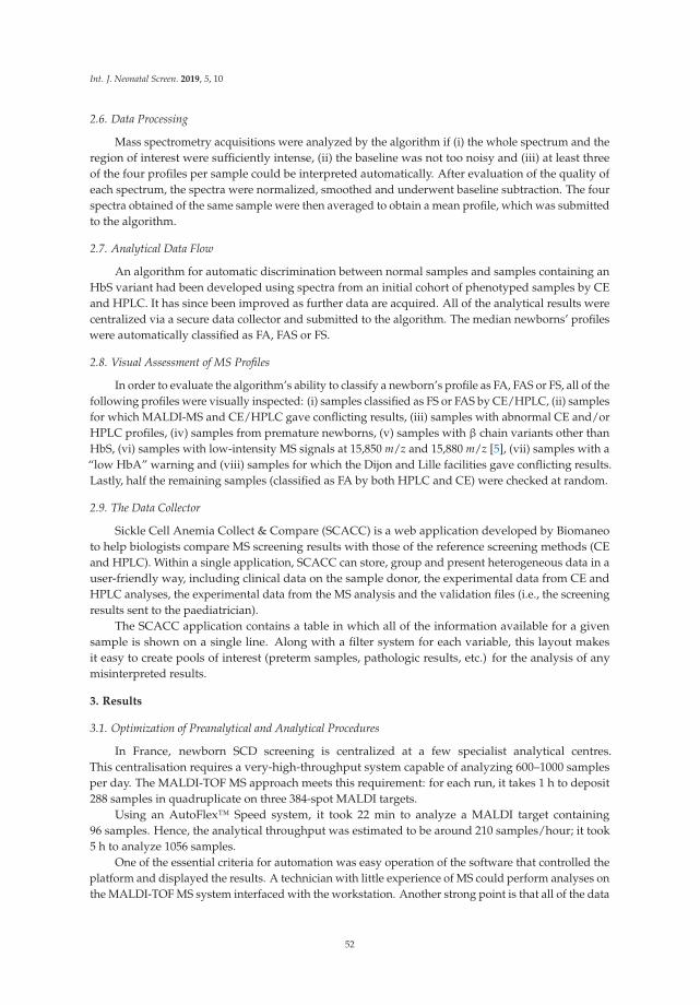
Int. J. Neonatal Screen. 2019, 5, 10
2.6. Data Processing
Mass spectrometry acquisitions were analyzed by the algorithm if (i) the whole spectrum and theregion of interest were sufficiently intense, (ii) the baseline was not too noisy and (iii) at least threeof the four profiles per sample could be interpreted automatically. After evaluation of the quality ofeach spectrum, the spectra were normalized, smoothed and underwent baseline subtraction. The fourspectra obtained of the same sample were then averaged to obtain a mean profile, which was submittedto the algorithm.
2.7. Analytical Data Flow
An algorithm for automatic discrimination between normal samples and samples containing anHbS variant had been developed using spectra from an initial cohort of phenotyped samples by CEand HPLC. It has since been improved as further data are acquired. All of the analytical results werecentralized via a secure data collector and submitted to the algorithm. The median newborns’ profileswere automatically classified as FA, FAS or FS.
2.8. Visual Assessment of MS Profiles
In order to evaluate the algorithm’s ability to classify a newborn’s profile as FA, FAS or FS, all of thefollowing profiles were visually inspected: (i) samples classified as FS or FAS by CE/HPLC, (ii) samplesfor which MALDI-MS and CE/HPLC gave conflicting results, (iii) samples with abnormal CE and/orHPLC profiles, (iv) samples from premature newborns, (v) samples with β chain variants other thanHbS, (vi) samples with low-intensity MS signals at 15,850 m/z and 15,880 m/z [5], (vii) samples with a“low HbA” warning and (viii) samples for which the Dijon and Lille facilities gave conflicting results.Lastly, half the remaining samples (classified as FA by both HPLC and CE) were checked at random.
2.9. The Data Collector
Sickle Cell Anemia Collect & Compare (SCACC) is a web application developed by Biomaneoto help biologists compare MS screening results with those of the reference screening methods (CEand HPLC). Within a single application, SCACC can store, group and present heterogeneous data in auser-friendly way, including clinical data on the sample donor, the experimental data from CE andHPLC analyses, the experimental data from the MS analysis and the validation files (i.e., the screeningresults sent to the paediatrician).
The SCACC application contains a table in which all of the information available for a givensample is shown on a single line. Along with a filter system for each variable, this layout makesit easy to create pools of interest (preterm samples, pathologic results, etc.) for the analysis of anymisinterpreted results.
3. Results
3.1. Optimization of Preanalytical and Analytical Procedures
In France, newborn SCD screening is centralized at a few specialist analytical centres.This centralisation requires a very-high-throughput system capable of analyzing 600–1000 samplesper day. The MALDI-TOF MS approach meets this requirement: for each run, it takes 1 h to deposit288 samples in quadruplicate on three 384-spot MALDI targets.
Using an AutoFlex™ Speed system, it took 22 min to analyze a MALDI target containing96 samples. Hence, the analytical throughput was estimated to be around 210 samples/hour; it took5 h to analyze 1056 samples.
One of the essential criteria for automation was easy operation of the software that controlled theplatform and displayed the results. A technician with little experience of MS could perform analyses onthe MALDI-TOF MS system interfaced with the workstation. Another strong point is that all of the data
52
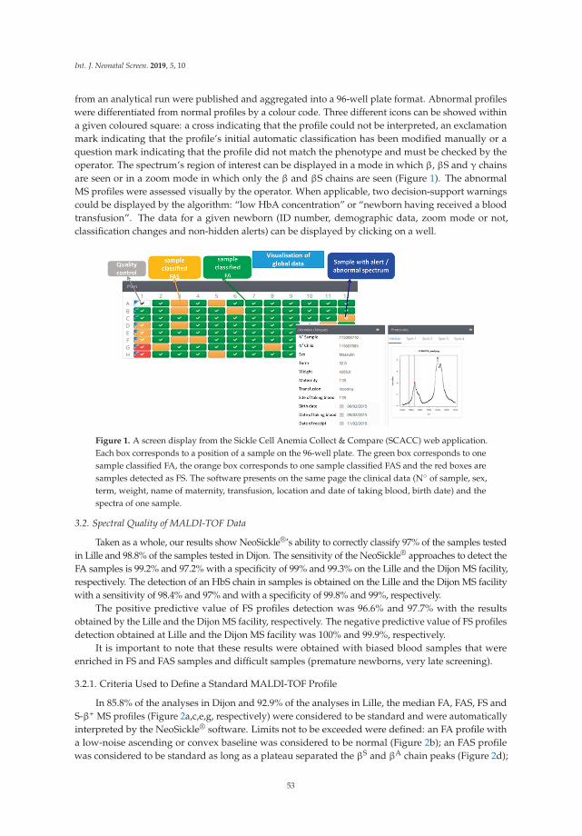
Int. J. Neonatal Screen. 2019, 5, 10
from an analytical run were published and aggregated into a 96-well plate format. Abnormal profileswere differentiated from normal profiles by a colour code. Three different icons can be showed withina given coloured square: a cross indicating that the profile could not be interpreted, an exclamationmark indicating that the profile’s initial automatic classification has been modified manually or aquestion mark indicating that the profile did not match the phenotype and must be checked by theoperator. The spectrum’s region of interest can be displayed in a mode in which β, βS and γ chainsare seen or in a zoom mode in which only the β and βS chains are seen (Figure 1). The abnormalMS profiles were assessed visually by the operator. When applicable, two decision-support warningscould be displayed by the algorithm: “low HbA concentration” or “newborn having received a bloodtransfusion”. The data for a given newborn (ID number, demographic data, zoom mode or not,classification changes and non-hidden alerts) can be displayed by clicking on a well.
Figure 1. A screen display from the Sickle Cell Anemia Collect & Compare (SCACC) web application.Each box corresponds to a position of a sample on the 96-well plate. The green box corresponds to onesample classified FA, the orange box corresponds to one sample classified FAS and the red boxes aresamples detected as FS. The software presents on the same page the clinical data (N◦ of sample, sex,term, weight, name of maternity, transfusion, location and date of taking blood, birth date) and thespectra of one sample.
3.2. Spectral Quality of MALDI-TOF Data
Taken as a whole, our results show NeoSickle®’s ability to correctly classify 97% of the samples testedin Lille and 98.8% of the samples tested in Dijon. The sensitivity of the NeoSickle® approaches to detect theFA samples is 99.2% and 97.2% with a specificity of 99% and 99.3% on the Lille and the Dijon MS facility,respectively. The detection of an HbS chain in samples is obtained on the Lille and the Dijon MS facilitywith a sensitivity of 98.4% and 97% and with a specificity of 99.8% and 99%, respectively.
The positive predictive value of FS profiles detection was 96.6% and 97.7% with the resultsobtained by the Lille and the Dijon MS facility, respectively. The negative predictive value of FS profilesdetection obtained at Lille and the Dijon MS facility was 100% and 99.9%, respectively.
It is important to note that these results were obtained with biased blood samples that wereenriched in FS and FAS samples and difficult samples (premature newborns, very late screening).
3.2.1. Criteria Used to Define a Standard MALDI-TOF Profile
In 85.8% of the analyses in Dijon and 92.9% of the analyses in Lille, the median FA, FAS, FS andS-β+ MS profiles (Figure 2a,c,e,g, respectively) were considered to be standard and were automaticallyinterpreted by the NeoSickle® software. Limits not to be exceeded were defined: an FA profile witha low-noise ascending or convex baseline was considered to be normal (Figure 2b); an FAS profilewas considered to be standard as long as a plateau separated the βS and βA chain peaks (Figure 2d);
53

Int. J. Neonatal Screen. 2019, 5, 10
and, lastly, an FS profile with a low-noise ascending or convex baseline to the right of the βS chainpeak was considered to be interpretable (Figure 2f). The profile of a newborn with S-β+-thalassemiawas characterized by a much more intense βS peak, relative to the βA peak (Figure 2g,h).
Figure 2. Examples of standard MS profiles correctly classified by the NeoSickle® software, fromnewborns with the following phenotypes: FA (a,b), FAS (c,d), FS (e,f) and S-β + (g,h).
3.2.2. Classification by the Algorithm of “Standard” Profiles, as a Function of the Newborn’sCorrected Phenotype
There were no classification errors among the corrected FA and FS samples (Table 2).Concerning the corrected FAS samples, only 2 of the 2753 standard profiles from Lille were misclassified(as FS) by MS, and 17 of the 2507 standard profiles from Dijon were misclassified (16 cases classified asFA by MS, and 1 case classified as FS by MS). Although the spectra of the FAS samples misclassifiedas FA showed a shoulder with a plateau at 15,850 m/z ± 10 m/z, the signal intensity was too low.This characteristic should be introduced in the criteria for a nonstandard MS profile.
The FAS samples misclassified as FS by MALDI-MS came from composite heterozygous newbornsclassified as FS-O Arab and FSE by CE and HPLC. Regular peaks at 18,850 and 1880 m/z wereobserved; however, the error was induced by the low intensity of the peak at 15,880 m/z. A profilewith asymmetry for the βA and βS chains (a more intense βS peak) prompted its classification as FS bythe e-NeoSickle software.
54
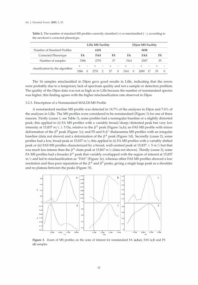
Int. J. Neonatal Screen. 2019, 5, 10
Table 2. The number of standard MS profiles correctly classified (+) or misclassified (−), according tothe newborn’s corrected phenotype.
Lille MS Facility Dijon MS Facility
Number of Standard Profiles 6181 5698
Corrected Phenotype FA FAS FS FA FAS FS
Number of samples 3386 2753 37 3161 2507 35
classification by the algorithm+ − + − + − + − + − + −
3386 0 2751 2 37 0 3161 0 2490 17 35 0
The 16 samples misclassified in Dijon gave good results in Lille, indicating that the errorswere probably due to a temporary lack of spectrum quality and not a sample or detection problem.The quality of the Dijon data was not as high as in Lille because the number of nonstandard spectrawas higher; this finding agrees with the higher misclassification rate observed in Dijon.
3.2.3. Description of a Nonstandard MALDI-MS Profile
A nonstandard median MS profile was detected in 14.7% of the analyses in Dijon and 7.6% ofthe analyses in Lille. The MS profiles were considered to be nonstandard (Figure 3) for one of threereasons. Firstly (cause 1, see Table 3), some profiles had a nonregular baseline or a slightly distortedpeak; this applied to (i) FA MS profiles with a variably broad/sharp/distorted peak but very lowintensity at 15,837 m/z ± 5 Da, relative to the βA peak (Figure 3a,b); an FAS MS profile with minordeformation of the βS peak (Figure 3c); and FS and S-β+-thalassemia MS profiles with an irregularbaseline (data not shown) and a deformation of the βA peak (Figure 3d). Secondly (cause 2), someprofiles had a low, broad peak at 15,837 m/z; this applied to (i) FA MS profiles with a variably shiftedpeak or (ii) FAS MS profiles characterized by a broad, well-centred peak at 15,837 ± 5 m/z but thatwas much less intense than the βA chain peak at 15,867 m/z (data not shown). Thirdly (cause 3), someFA MS profiles had a broader βA peak that variably overlapped with the region of interest at 15,837m/z and led to misclassification as “FAS” (Figure 3e), whereas other FAS MS profiles showed a lowresolution and thus poor separation of the βA and βS peaks, giving a single large peak as a shoulderand no plateau between the peaks (Figure 3f).
Figure 3. Zoom of MS profiles on the zone of interest for nonstandard FA (a,b,e), FAS (c,f) and FS(d) samples.
55

Int. J. Neonatal Screen. 2019, 5, 10
3.2.4. Classification by the Algorithm of Profiles Considered to be Nonstandard in a VisualAssessment, as a Function of the Newborn’s Corrected Phenotype
The data in Table 3 show that the great majority of nonstandard profiles (84.5% in Lille and 81.2%in Dijon) were nevertheless correctly classified by the e-NeoSickle® algorithm. Furthermore, 7.5% of thenonstandard profiles in Lille and 4.6% of the nonstandard profiles Dijon were misclassified, and 8% ofthe nonstandard profiles in Lille and 14.2% of the nonstandard profiles in Dijon were uninterpretable.
It is important to note that the nonstandard profiles accounted for less than 14% of the analysesin Dijon and 7% of the analyses in Lille. Overall, only 2% of the profiles in Dijon and 0.6% of theprofiles in Lille were uninterpretable, and 0.9% of the profiles in Dijon and 0.6% of the profiles in Lillewere misclassified.
Table 3. Classification by the algorithm of profiles considered to be nonstandard in a visual assessment.Some spectra could not be interpreted by the algorithm (IN). The percentage is based on the number ofprofiles considered to be nonstandard (780 profiles at Lille and 985 at Dijon).
Corrected Phenotypes
FA FAS FS
MALDI-TOF MS classification ofnon-standard profiles
Lille
FA 282 (54.7%) 30 (5.9%) 0 (0%)
FAS 7 (1.4%) 118 (23.2%) 0 (0%)
FS 0 (0%) 1 (0.2%) 34 (6.7%)
IN 21 (4.5%) 17 (3.4%) 0 (0%)
Dijon
FA 446 (45%) 22 (2.2%) 0 (0%)
FAS 21 (2.2%) 323 (32.7%) 1 (0.2%)
FS 0 (0%) 1 (0.2%) 36 (3.7%)
IN 68 (7.2%) 67 (6.8%) 0 (0%)
The frequencies of “nonregular baseline” (cause 1) and “low/broad peak” (cause 2) features weresimilar at the two MS facilities. Our analysis showed that most of the nonstandard spectra in Dijon(54%) were due to a lack of resolution (cause 3), whereas this was the case for only 13% in Lille.Proportionally, the corrected phenotype FS (n = 71) was more likely to be labelled as “low resolution”,with respectively 31 and 36 abnormal spectra in Lille and Dijon. However, this low resolution had verylittle impact on the automatic classification, since less than 1% of all spectra labelled as “low resolution”were misclassified.
Sixteen percent of the spectra with a low, broad peak at 15,837 m/z were misclassified by the algorithm(Table 4). Of the 21 spectra of this type misclassified in Lille, 20 (95%) came from newborns with a correctedFAS phenotype. Likewise, of the 20 spectra of this type misclassified in Dijon (Table 4), 18 (90%) camefrom newborns with a corrected FAS phenotype. These spectra were very difficult to classify becausethe ratio between the βS and βA peaks was abnormal. A very low βS peak (relative to the βA peak) canbe interpreted as a sample with high background noise or as a sample from an S homozygote or an ASheterozygote having received a blood transfusion (which induces a strong imbalance). Given that thesespectra have low peak intensities, we recommend repeating the analysis or depositing a new sample.
56

Int. J. Neonatal Screen. 2019, 5, 10
Table 4. (A) Numbers and proportions of nonstandard spectra by corrected phenotype and by cause.(B) Numbers and proportions of correctly classified and misclassified spectra by cause. Cause 1 resultedin spectra with an irregular baseline, cause 2 resulted in spectra with a low, broad peak and cause3 resulted in spectra with a low resolution. The percentage is based on the number of profiles consideredto be nonstandard (780 profiles at Lille and 985 at Dijon).
ACorrected Phenoztype
B Correctly Classified MisclassifiedFA FAS FS
Lille
Cause 1 179 (37%) 90 (19%) 3 (0%)
Lille
Cause 1 252 (94%) 17 (6%)
Cause 2 98 (21%) 28 (5%) 0 (0%) Cause 2 105 (84%) 21 (16%)
Cause 3 12 (3%) 31 (7%) 31 (7%) Cause 3 77 (100%) 0 (0%)
Dijon
Cause 1 193 (23%) 69 (8%) 0 (0%)
Dijon
Cause 1 248 (95%) 14 (5%)
Cause 2 103 (12%) 24 (3%) 0 (0%) Cause 2 107 (84%) 20 (16%)
Cause 3 171 (20%) 253 (30%) 36 (4%) Cause 3 449 (98%) 11 (2%)
3.2.5. Causes of Abnormal Spectral Features in Nonstandard MS Profiles
A spectral analysis showed that nonstandard profiles mainly resulted from the compilation of differingspectra within a quadruplicate. Figure 4 shows the variability in the raw data and its impact on dataprocessing. The spectra in Figure 4a,b,e,f correspond to the raw data generated for the first (Figure 4a,b)and third (Figure 4e,f) of the quadruplicate sample depositions. The two depositions varied markedly interms of the raw spectral intensity (less than 40 ua for deposition 1, and 200 ua for deposition 3). The dataprocessing steps (including peak alignment and baseline normalisation and subtraction) normalise thespectrum intensity but also remove differences (see Figure 4c,g, corresponding to the normalized spectra,and a blow-up view of the zone of interest Figure 4d,h). The compilation of normalized data introduces abias, and might lead to misinterpretation of the results by the software. Figure 4i shows a median spectrumwith an irregular baseline; this “pseudoβS peak” can be confused with a trueβS peak. However, the relativeintensity of the pseudo βS peak differed from that of a true βA peak.
The analysis of each individual spectrum enabled us to affirm that at least one good-quality,high-resolution spectrum was obtained for each biological sample. The sample deposition methodfor MALDI-MS is known not to be highly reproducible, and thus makes it difficult to obtain veryhomogeneous results. Even though our study highlighted this difficulty, our analysis also showed thata high-quality result can be obtained systematically.
57
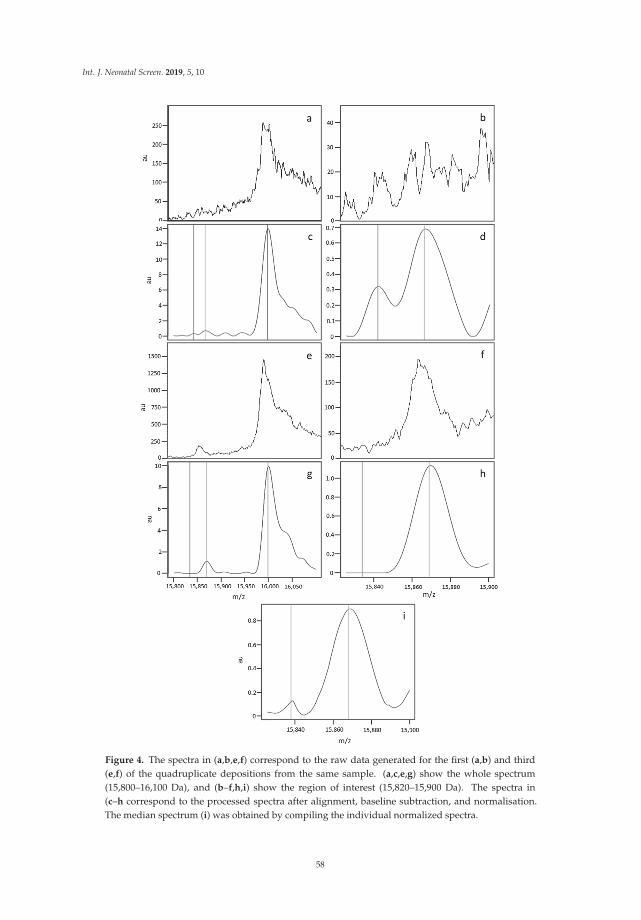
Int. J. Neonatal Screen. 2019, 5, 10
Figure 4. The spectra in (a,b,e,f) correspond to the raw data generated for the first (a,b) and third(e,f) of the quadruplicate depositions from the same sample. (a,c,e,g) show the whole spectrum(15,800–16,100 Da), and (b–f,h,i) show the region of interest (15,820–15,900 Da). The spectra in(c–h correspond to the processed spectra after alignment, baseline subtraction, and normalisation.The median spectrum (i) was obtained by compiling the individual normalized spectra.
58

Int. J. Neonatal Screen. 2019, 5, 10
4. Occasional Misclassifications
The analysis of samples that were classified differently by the two analytical facilities confirmed thatMALDI-MS can provide an interpretable result for each sample. In fact, the data in Table 5 showed that the68 samples with a corrected FA phenotype that could not be interpreted in Dijon were correctly classifiedin Lille (67 standard spectra and 1 low-resolution spectrum). Likewise, the 21 samples with a corrected FAphenotype and that could not be interpreted in Lille were correctly classified at Dijon. Identical resultswere obtained with the corrected FAS samples.
Table 5. Paired classification of the samples by the two analytical facilities. Relationship with thespectral quality. This table summarizes the number of profiles correctly classified, misclassified anduninterpretable for each category of profile quality and for the two analytical laboratories.
Profile Abnormalities at 15,850 ± 10 m/z
Neonates with a Corrected FA Phenotype
Automatic Classification of MatchedProfiles
Irregular Base Line Low, Broad Peak Low Resolution Regular Base Line Uninterpretable
Lille Dijon Lille Dijon Lille Dijon Lille Dijon Lille Dijon Lille Dijon
correctly classified uninterpretable 1 0 0 0 0 0 67 0 0 68
correctly classified Misclassified 4 10 7 0 1 5 17 0 0 0
uninterpretable correctly classified 0 0 0 0 0 0 0 21 0 0
Misclassified correctly classified 4 0 3 0 0 0 0 7 0 0
Neonates with a Corrected FAS Phenotype
Automatic Classification of MatchedProfiles
Irregular Base Line Low, Broad Peak Low Resolution Regular Base Line Uninterpretable
Lille Dijon Lille Dijon Lille Dijon Lille Dijon Lille Dijon Lille Dijon
correctly classified uninterpretable 0 0 0 0 0 0 68 0 0 68
correctly classified Misclassified 0 4 0 14 1 5 23 0 0 0
uninterpretable correctly classified 0 0 0 0 0 1 0 16 17 0
Misclassified correctly classified 12 0 12 0 0 4 6 26 0 0
When a profile was misclassified or uninterpretable at one MS facility, the corresponding profile at theother MS facility was correctly classified and was less likely to be a nonstandard profile. Only one samplewith a nonstandard profile was misclassified at both MS facilities (data not shown). A good classificationwith a standard spectrum was obtained for each sample on at least one of the MS facilities, which indicatesthat a low HbA concentration was not the main cause of misclassifications or uninterpretable results.Misclassifications or uninterpretable results were mainly due to the absence of a high-quality spectruminduced by technical incidents (tip blockage and non-deposition of the sample). Consequently, repeatingthe analysis of a sample with a nonstandard profile was the best way of obtaining a standard profile and acorrect classification. Different data, such as peak intensities or the ratio between different peaks of βA andβS chains, allow for us to detect the nonstandard profile and to add an alert to the results.
5. Discussion
Newborn disease screening programmes are front-line public health measures, and as such mustbe based on robust analytical methods and data-processing software. Cost effectiveness is a furtherrequirement, prompting the implementation of high-throughput screening units that reduce unitcosts. Lastly, the maximal use of automation enables the analytical results to be validated with aslittle intervention as possible by the medical team. Our MALDI-MS platform and the associateddata-processing and interpretation software were designed to address these challenges.
In newborn SCD screening with a MALDI MS system, the user-friendliness of the softwareinterface and a high-throughput analysis coupled to automatic sample classification and traceabilityare directly related to practicability. As mentioned above, newborn SCD screening in France iscentralised at a few specialist analytical centres. This centralisation requires a throughput of up to600–1000 samples per day. The MALDI-TOF MS approach meets this requirement.
The next performance criterion of note concerns the method’s robustness: in other words, its abilityto generate good-quality standard MS profiles that enable correct classification in agreement with the
59

Int. J. Neonatal Screen. 2019, 5, 10
sample’s validated phenotype, whatever the newborn’s clinical status. In order to test our MALDI-MSunder extreme conditions, we increased the proportion of difficult samples in the presently analyzed cohort;this corresponds to pathological specimens and specimens collected and/or stored under non-optimalconditions (samples from premature newborns, samples collected long after delivery, samples collectedfrom France’s overseas regions and dependencies that may have been stored for a long time in a tropicalatmosphere before delivery to the screening laboratory in continental France, etc.). In the present study,only samples from newborns having received one or more blood transfusions were discarded. The impactof blood transfusion will be described in a specific study of the effect of high HbA levels on the method’ssensitivity, specificity and resolution as a function of the βs chain concentration. Moreover, we evaluatedthe MALDI-TOF method’s robustness by visually checking the spectral quality as well as by consideringits ability to correctly classify the newborns as FA, FAS or FS. Benchmark MALDI-TOF MS spectra werethus established on the basis of the classification results and the visual quality of the spectra. The referencespectra correspond to all of the spectra defined as standard for which the information obtained is consideredto be sufficient and of good quality. Indeed, visual spectral validation has proved to be most effective forrevealing shot-to-shot variations.
Using this strategy, we determined the percentages of newborns with a standard profile and who werecorrectly classified (relative to their validated phenotype). The highest level of efficiency was obtained in theset of newborns with a corrected FA phenotype; around 85% of these analyses could be validated directly.A similar percentage was obtained for newborns with a corrected FAS phenotype. Moreover, nonstandardMS spectra were not systematically misclassified, and most were correctly classified. We consider theseresults to be very promising because it should be possible to further improve the procedures for sampledeposition, raw data analysis and selection of the mass spectra to which the classification algorithmis applied.
None of the samples from FS newborns were misclassified, and a sample with an FAS-correctedphenotype was misclassified as an FA sample. This type of misclassification can lead to false negativesfor newborns with an FSC phenotype. This major error required us to set up a strategy for correcting theautomatic classification. The development and characteristics of tracking alerts for the nonstandard MSprofiles will be described elsewhere. This approach should enable an occasional lack of reproducibility,sensitivity or resolution in Hb profiling to be detected.
There were two main reasons for the incorrect automatic classification of newborn phenotypes.Firstly, a low signal intensity at 15,850 and/or 15,880 m/z (i.e., similar to background noise) wasintensified by the data processing (normalisation and alignment). Secondly, poor resolution of the ßand/or βS chains generated signal overlap. The quadruplicate sample analysis improved the results,but was not always able to counterbalance shot-to-shot variability.
In a previous article [5], very poor spectral quality prevented the analysis of 20 of 844 samples (2.5%),even after several repeat analyses. The relatively high frequency of this incident was probably due tothe inclusion of a high proportion of samples from premature newborns and/or HbS carriers. We havenow resolved this problem. In most cases, a single repeat analysis was enough to obtain a standardprofile. Some problems arose from time to time but were mainly related to robotic preparation of the bloodspot samples. Further optimization of sample processing and the MS acquisition protocol should furtherimprove the standardization of the MS profiles and reduce the frequency of repeat analysis.
Taken as a whole, our results for pooled standard and nonstandard spectra demonstrated NeoSickle®’sability to classify correctly 97% of the samples tested in Lille and 98.8% of the samples tested in Dijon.Furthermore, only 2% of the nonstandard spectra in Dijon were uninterpretable, and less than 0.9% weremisclassified. In Lille, 0.6% of the nonstandard spectra were uninterpretable and 0.6% were misclassified.It is important to note that these percentages were obtained with “difficult” blood samples (prematurenewborns, very late screening).
Our pilot study showed that the NeoSickle® approach can differentiate between heterozygous FSE,FSO-Arab and S-β + samples on one hand and heterozygous FAS and homozygous FS samples on theother. Indeed, the signal intensity of the βE, βO-Arab variants was much weaker than that of the β chain.
60

Int. J. Neonatal Screen. 2019, 5, 10
In addition to classifying samples as FA, FAS or FS, studies of a larger cohort of patients might make itpossible to clearly differentiate a fourth class (corresponding to heterozygous SX samples in which the βX
chain is E, O-Arab, a).In conclusion, our new MALDI-TOF MS approach already meets today’s requirements [12]
for large-scale, cost-effective newborn SCD screening, and is well-positioned to address futurerequirements for even greater throughputs and total automation to detect the HbS variants.
Author Contributions: D.R. and M.E.O.: MALDI-TOF methodology; P.N.: software development; G.R. and O.G.:validation; P.D. and J.-M.P.: conceptualization, data curation, acquisition of funding and drafting of the manuscript.
Funding: The present research was funded by Lille University Medical Center and the Conseil Régionalde Bourgogne.
Conflicts of Interest: Patrick Ducoroy was an employee of the University of Burgundy at the time of the researchdescribed here. He then founded the company Biomaneo, and currently serves as its CEO.
References
1. Rai, D.K.; Griffiths, W.J.; Landin, B.; Wild, B.J.; Alvelius, G.; Green, B.N. Accurate mass measurement byelectrospray ionization quadrupole mass spectrometry: Detection of variants differing by <6 Da from normalin human hemoglobin heterozygotes. Anal. Chem. 2003, 75, 1978–1982. [CrossRef] [PubMed]
2. Wild, B.J.; Green, B.N.; Cooper, E.K.; Lalloz, M.R.; Erten, S.; Stephens, A.D.; Layton, D.M. Rapid identificationof hemoglobin variants by electrospray ionization mass spectrometry. Blood Cells Mol. Dis. 2001, 27, 691–704.[CrossRef] [PubMed]
3. Wild, B.J.; Green, B.N.; Stephens, A.D. The potential of electrospray ionization mass spectrometry for the diagnosisof hemoglobin variants found in newborn screening. Blood Cells Mol. Dis. 2004, 33, 308–317. [CrossRef] [PubMed]
4. Kiernan, U.A.; Black, J.A.; Williams, P.; Nelson, R.W. High-throughput analysis of hemoglobin from neonatesusing matrix-assisted laser desorption/ionization time-of-flight mass spectrometry. Clin. Chem. 2002, 48,947–949. [PubMed]
5. Hachani, J.; Duban-Deweer, S.; Pottiez, G.; Renom, G.; Flahaut, C.; Périni, J.M. MALDI-TOF MSprofiling as the first-tier screen for sickle cell disease in neonates: Matching throughput to objectives.Proteomics Clin. Appl. 2011, 5, 405–414. [CrossRef] [PubMed]
6. Daniel, Y.A.; Turner, C.; Haynes, R.M.; Hunt, B.J.; Dalton, R.N. Rapid and specific detection of clinicallysignificant haemoglobinopathies using electrospray mass spectrometry-mass spectrometry. Br. J. Haematol.2005, 130, 635–643. [CrossRef] [PubMed]
7. Boemer, F.; Ketelslegers, O.; Minon, J.M.; Bours, V.; Schoos, R. Newborn screening for sickle cell diseaseusing tandem mass spectrometry. Clin. Chem. 2008, 54, 2036–2041. [CrossRef] [PubMed]
8. Moat, S.J.; Rees, D.; King, L.; Ifederu, A.; Harvey, K.; Hall, K.; Lloyd, G.; Morrell, C.; Hillier, S. Newborn bloodspot screening for sickle cell disease by using tandem mass spectrometry: Implementation of a protocol toidentify only the disease states of sickle cell disease. Clin. Chem. 2014, 60, 373–380. [CrossRef] [PubMed]
9. Edwards, R.L.; Griffiths, P.; Bunch, J.; Cooper, H.J. Compound heterozygotes and beta-thalassemia: Top-downmass spectrometry for detection of hemoglobinopathies. Proteomics 2014, 14, 1232–1238. [CrossRef] [PubMed]
10. Edwards, R.L.; Griffiths, P.; Bunch, J.; Cooper, H.J. Top-down proteomics and direct surface sampling ofneonatal dried blood spots: Diagnosis of unknown hemoglobin variants. J. Am. Soc. Mass Spectrom. 2012, 23,1921–1930. [CrossRef] [PubMed]
11. Edwards, R.L.; Creese, A.J.; Baumert, M.; Griffiths, P.; Bunch, J.; Cooper, H.J. Hemoglobin variant analysis viadirect surface sampling of dried blood spots coupled with high-resolution mass spectrometry. Anal. Chem.2011, 15, 2265–2270. [CrossRef] [PubMed]
12. Lobitz, S.; Telfer, P.; Cela, E.; Allaf, B.; Angastiniotis, M.; Backman Johansson, C.; Badens, C.; Bento, C.;Bouva, M.J.; Canatan, D.; et al. Newborn screening for sickle cell disease in Europe: Recommendations froma Pan-European consensus conference. Br. J. Haematol. 2018. [CrossRef] [PubMed]
© 2019 by the authors. Licensee MDPI, Basel, Switzerland. This article is an open accessarticle distributed under the terms and conditions of the Creative Commons Attribution(CC BY) license (http://creativecommons.org/licenses/by/4.0/).
61

International Journal of
Neonatal Screening
Article
Evaluation of Technical Issues in a Pilot MulticenterNewborn Screening Program for Sickle Cell Disease
Maddalena Martella 1,*, Giampietro Viola 1, Silvia Azzena 1, Sara Schiavon 1, Andrea Biondi 2,
Giuseppe Basso 1, Paola Corti 2, Raffaella Colombatti 1, Nicoletta Masera 2 and Laura Sainati 1
1 Dipartimento di Salute della Donna e del Bambino, Università di Padova, 35128 Padova, Italy;[email protected] (G.V.); [email protected] (S.A.); [email protected] (S.S.);[email protected] (G.B.); [email protected] (R.C.); [email protected] (L.S.)
2 Dipartimento di Pediatria, Università di Milano-Bicocca-Fondazione MBBM, San Gerardo Hospital,20900 Monza, Italy; [email protected] (A.B.); [email protected] (P.C.);[email protected] (N.M.)
* Correspondence: [email protected]; Tel.: +39-49-8211451
Received: 30 October 2018; Accepted: 19 December 2018; Published: 21 December 2018
Abstract: A multicenter pilot program for universal newborn screening of Sickle cell disease (SCD)was conducted in two centres of Northern Italy (Padova and Monza). High Performance LiquidChromatography (HPLC) was performed as the first test on samples collected on Guthrie cardsand molecular analysis of the β-globin gene (HBB) was the confirmatory test performed on theHPLC-positive or indeterminate samples. 5466 samples of newborns were evaluated. Of these,5439/5466 were submitted to HPLC analysis and the molecular analysis always confirmed in allthe alteration detected in HPLC (62/5439 newborns); 4/5439 (0.07%) were SCD affected, 37/5439(0.68%) were HbAS carriers and 21/5439 (0.40%) showed other hemoglobinopathies. Stored driedblood spots were adequate for HPLC and β-globin gene molecular analysis. Samples were suitablefor analysis until sixteen months old. A cut-off of A1 percentage, in order to avoid false negative orunnecessary confirmation tests, was identified. Our experience showed that several technical issuesneed to be addressed and resolved while developing a multicenter NBS program for SCD in a countrywhere there is no national neonatal screening (NBS) program for SCD and NBS programs occur ona regional basis.
Keywords: sickle cell disease; high performance liquid chromatography (HPLC); β-globin gene
1. Introduction
Neonatal screening (NBS) for Sickle cell disease (SCD) is an effective tool for the early detectionof affected individuals, to direct them at the clinical programs to prevent complications and finallyto offer genetic counseling to a family and is therefore highly recommended as the first step ofcomprehensive care [1–7]. In detail, early identification of affected SCD patients through a NBSprogram allows the introduction of penicillin prophylaxis from two months of age and performing ofan adequate vaccination schedule with the reduction of mortality from infection, prompt enrolmentin comprehensive care programs with timely parent health education, Trans Cranial Doppler (TCD)screening and prevention of acute events [1,2].
In Italy a national NBS program is not available and NBS programs are organized on a regionalbasis [8]. Haemoglobinopathies are included in the regional NBS program of only 1 out of the20 regions [9], but some pilot programs have been conducted in the past years [10–12].
Padova and Monza, two towns located in the North of Italy, in the Veneto and LombardiaRegions respectively, have a high percentage of an immigrant population and an average of more
Int. J. Neonatal Screen. 2019, 5, 2; doi:10.3390/ijns5010002 www.mdpi.com/journal/neonatalscreening62

Int. J. Neonatal Screen. 2019, 5, 2
than 3500 births every year, with 25% of them from immigrant parents [13]. We developed a pilotmulticenter, multiregional universal NBS program for SCD [14].
The methods used and recommended for the SCD neonatal screening by International Guidelinescan be qualitative, as IsoElectric Focusing (IEF), and quantitative, as Capillary Electrophoresis (CE),High Performance Liquid Chromatography (HPLC) and recently the Tandem Mass Spectrometry(MS/MS) [15], but the abnormal chromatogram must always have a confirmatory test with the differentmethodologies. Our protocol is an innovative way for SCD screening with the combination of HPLCanalysis and molecular analysis. This type of approach has been used in very few studies [16,17].
The purpose of this manuscript is to highlight some of the technical challenges that we had to facein the development of our pilot screening. The objectives of this study were: (i) to define the sensitivityof our methods in order to establish a cut-off percentage for normality in the presence of low levelsof β-globin chains, (ii) to maintain high-quality results to ensure the accurate interpretation of theanalysis for immediate initiation of supportive care for affected newborns, (iii) to check the stability ofthe sample over time to ensure reliable measurements, a very important issue in a multicenter setting,(iv) to verify the organization and feasibility of a multiregional screening program. Finally, the flowchart for newborn screening for SCD is shown in Figure 1.
Figure 1. Flow chart for newborn screening for Sickle cell disease.
2. Materials and Methods
2.1. Study Design and Population
In Padova the NBS pilot program enrolled newborns from 2 May 2016 until 30 November 2017while in Monza from 1 September 2016 until 31 August 2017. The screening was performed on allnewborns from the 36th hour to the third day after birth. Blood collection was performed by prickingthe heel of the newborn and collection on a Guthrie card (903 Whatman® in Padova and AHLSTROM®
226 in Monza) after informed consent from the parents was obtained.Dried blood spots from Monza were sent to Padova through courier once a week and stored
at room temperature and in a dark dry place in the NBS laboratory of Padova’s Clinic of PediatricHematology-Oncology. The samples were analyzed after 7 days and the first test was performed withthe VARIANT NBS Newborn Screening System (Bio-Rad Laboratories, Munich, Germany) and theconfirmatory test, on the same sample, with molecular analysis of β-globin gene.
63
Int. J. Neonatal Screen. 2019, 5, 2
2.2. High Performance Liquid Chromatography (HPLC)
All specimens were first examined by High Performance Liquid Chromatography (HPLC)performed on a VARIANT NBS Newborn Screening System (Bio-Rad Laboratories) following themanufacturer’s recommendations, within 7 days after blood spots had been taken. All chromatogramswere automatically analyzed and visually inspected for absent Hemoglobin A and variant hemoglobins.The columns and all reagents such as buffers, primers, and hemoglobin standards were purchasedfrom the manufacturer. Two or three disks (diameter of 3.2 mm) were punched out of the driedblood spot (DBS) and placed in a well of 96-well plate round bottom. In each well 280μL of distilledwater was added and mixed with the pipette several times at room temperature. Two mixtures ofhemoglobin standards FAES and FADC, respectively were analyzed in duplicate even if the runincluded more plates.
HPLC uses an ion exchange column with gradient elution. The presence of different hemoglobinsis revealed through a UV-VIS detector settled at 415 nm. The time that passes from the injection of thesample to the output of the peak of hemoglobin type, is known as the retention time of that particularhemoglobin and represents a reproducible value for that column, at that gradient elution and atthat temperature. For the different hemoglobins we have different retention times and characteristicchromatographic profiles, with the exception of HbE and HbA2 that elute in the same peak, thereforemaking them indistinguishable from each other.
In addition, it is possible to quantify in a relative way the percentage of the different hemoglobins;the HPLC analysis helps to identify the differences between carrier individuals (HbS/HbA) andhomozygous affected individuals (HbS/HbS) and also to differentiate heterozygous compounds suchas HbS/HbC.
Moreover, the sensibility of the protocol has been verified with experiments of dilution on theSCD sample (HbSS) until 1:10.
2.3. Molecular Analysis of the β-globin Gene
Molecular analysis of the β-globin gene (HBB; MIM # 603903; NM_000518) represented theconfirmatory test and was performed on all the following samples: those with an altered hemoglobinprofile at HPLC, due to the presence of HbS or another hemoglobin variant; the specimens withHbA<5% or with HbA>30%, excluded transfusion anamnesis, were sent for molecular study.The samples with a red blood cell transfusion and HbA>30% were submitted for HPLC analysisthree months later.
Genomic DNA, extracted using the Qiagen kit (QIAamp®DNA Mini and Blood), fromblood spots allowed to air dry, were amplified by chain reaction of polymerase (PCR) atthe exon level and at the intron-exon junctions with the primers below reported: HBB1Fw5′-AAAAGTCAGGGCAGAGCCAT-3′, HBB1 Rw5′-CCCAGTTTCTATTGGTCTCCTTAA-3′, HBB2Fw5′-GGGTTTCTGATAGGCACTGACTC-3′, HBB2 Rw5′-AAAAGAAGGGGAAAGAAAACATCA-3′,HBB3 Fw5′-TAGCAGCTACAATCCAGCTACCA-3′, HBB3 Rw5′-GGACTTAGGGAACAAAGGACCT-3′.
Alterations of the coding sequence were analyzed and characterized by sequencing of thehemoglobin β chain coding HBB gene using an ABI Prism® 310 Genetic Analyzer Applied Biosystems.Sanger sequencing is the most comprehensive method of mutation detection and determines the exactsequence spanning the area of the primers used.
2.4. Management of Results
HPLC negative samples for abnormal hemoglobins didn’t require further analysis. While, HPLCpositive samples for abnormal chains underwent a confirmatory test with molecular analysis of theβ-globin gene on the same sample collected at the nursery. Families with SCD children were calledfor a visit in the clinic within two months, while parents of HbS carriers were called for a visit and
64

Int. J. Neonatal Screen. 2019, 5, 2
counselling within six months. Carriers of other hemoglobin variants received a letter, informing themof the results.
3. Results
In this study, 5466 samples of newborns from both birth centers were enrolled. Of these, 5439/5466(2821/2826 of Padova, 99.8% and 2618/2640 of Monza 99.1%) were submitted for HPLC analysis.None of the samples collected were excluded from the analysis. Each sample was analyzed within7 days after the blood had been spotted on the paper.
The results are summarized in Table 1. Molecular analysis always confirmed the abnormalitydetected in HPLC in 62/5466 newborns; other hemoglobin variants were also detected (HbC, HbD,HbE) in 0.5% of the cases in Monza and in 0.21% of those in Padova.
Table 1. Hemoglobin patterns observed in the pilot newborn screening.
Hb Pattern (HPLC) HBB Genotype Newborns (n.)
FAS HBB: c.20A>T/wt 37 (0.68%)
FS HBB: c.20A>T/c.20A>T 3 (0.055%)
FSC HBB: c.20A>T/c.19A>T 1 (0.02%)
FAC HBB: c.19A>T/wt 9 (0.16%)
FAD HBB: c.364G>C/wt 8 (0.15%)
FAE HBB: c.79C>T/wt 4 (0.07%)
Total 62 (1.14%)
n. = number of newborns.
28 samples (0.86%) of preterm infants, showed a value of HbA<5% at HPLC. Molecular analysisconfirmed that 26/28 had genotype HbAA, while 2 samples with HbA values of 2.2% and 3.8%,respectively, had a genotype HbAD and a FAST value >10% (potential HbBart) and had been reportedto the referring center. In an attempt to determine normal values for HbA and total HbF, the peakpercentages of samples with a normal hemoglobin pattern were plotted against the gestational week.The results (see Figure 2) showed a correct Gaussian distribution for the HbA and HbF with a mean of16.6% and 77.1% respectively as expected for at term newborns.
All dried blood spot cards collected and the storage methods turned out adequate both for HPLCanalysis and β globin gene molecular analysis.
The experiments of dilution on the SCD sample (HbSS) detected low levels of HbS until 1:10.In our experience we verified storage conditions and the reliability of the DBS a few months
later [16] to understand if it was possible to identify the different hemoglobins according to ourstandard settings of the instrument. We repeated the HPLC analysis of different DBS series stored forsixteen months. In each series, the values of HbA peak area percentage identified in HPLC analysis,although reduced, allowed for the detection of the hemoglobin peaks; retention time was not modifiedby the samples aging: the HbA value was never <7% and never <800,000 μV*s, in terms of area underthe curve (AUC) according to manufacturer instructions. HbS, when present was easily identified byHPLC analysis and similarly to HbA it was not influenced by sample aging [18]. The reduction ofthe HbA percentage should be attributed to physiological degradation of HbA and to the presence ofcellulose particles removed from the Guthrie card together with the blood. In this study, the DBS up tosixteen months old provided satisfactory results (see Figure 3). The quality and the efficiency of theanalysis was not affected by different types of Guthrie cards and the two different types of Guthriecards gave the same performance.
65

Int. J. Neonatal Screen. 2019, 5, 2
Figure 2. Percentages of HbF (red circles) and HbA (gray circles) according to the gestational age(weeks from conception to blood withdrawal) found in the cohort of Padova (A) and Monza (B).Distribution frequency of HbA and HbF in the cohort of Padova (C,D) and Monza (E,F).
Furthermore, we have noted in our experience that the needle restrains randomly, probably byelectrostatic reasons, some blood spots that could cause not only the obstruction of the needle, but alsothe contamination of other wells. Thus, we recommend that the run must be always monitored and ifnecessary the run should be interrupted. At the end of every analysis session the instrument must becarefully cleaned.
66

Int. J. Neonatal Screen. 2019, 5, 2
Figure 3. Representative chromatograms of a dried blood spot analyzed within 7 days from bloodwithdrawal (A,C,E) and after four (B), eight (D) and sixteen months (F). Percentages of HbA analyzedafter different months from the blood withdrawal (G). At least five samples for group were analyzed.
4. Discussion
Our study represents the first multicenter pilot project of universal NBS for SCD in two differentregions of Northern Italy.
The methods of investigation were adequate and highly specific, the confirmatory test alwaysconfirmed the abnormal hemoglobinopathies detected in HPLC. The two different types of Guthriecards gave the same performances. The quality, the storage of the sample and the volume of thecollected blood are very important factors that may affect the results in particular the retention time inHPLC of the different hemoglobins [19]. Our setting proved to be effective.
Moreover, in our experience we noted that it is very important for the setting of the 96-well platefor HPLC analysis to ensure the acquisition of the total area >800,000 μV*s, as recommended by themanufacture: The number of the spots, the volume of the water for Hb elution, mixed with the pipetteseveral times at room temperature. The 96-well plate can be store at room temperature. This protocolensures a homogeneous solution to detect the different hemoglobins. The sensitivity of the system canbe considered optimal for all different hemoglobins in agreement with the hemoglobin standards FAESand FADC provided by the manufacturer. It is worthwhile to note that our data showed a normaldistribution both for HbA and HbF in well agreement to that reported by Bouva et al. [20].
Finally, the sensibility of the protocol verified with experiments of dilution on the SCD sample(HbSS) until 1:10 guaranteed the possibility ofdetecting low levels of HbS.
Newborns born prematurely may yield misleading results. Premature infants may have verylow levels of A1 hemoglobin at birth; in the absence of a significant amount of A1, β chains mutations
67

Int. J. Neonatal Screen. 2019, 5, 2
can be undetectable. The cut-off of HbA<5% is efficient also for preterm newborns. The percentagesof HbA and HbF were in agreement with previous reports [21,22], which found a great variabilityaccording to the maternal ethnic origin and the gestational age.
The VARIANT NBS Newborn Screening System for our experience has critical issues: the spots canremain attached to the needle causing obstruction or even the random fall of the spot. Therefore, cautionis necessary and it is better to check the run and to wash frequently the needle and the instrument.
5. Conclusions
Our data demonstrate the feasibility of a multicentric SCD screening and indicate the robustnessand reliability of the screening system. The data obtained from HPLC analysis were in excellentagreement with the large amount of data present in the literature. The sensitivity of the system can beconsidered optimal in conjunction with the molecular analysis of the β globin gene. A scaling up ofthe project to other areas of the two regions is now planned.
Author Contributions: Conceptualization, data curation, original draft preparation M.M. and G.V.; investigation,S.A. and S.S.; resources, G.B. and A.B.; project administration, N.M. and P.C.; L.S. and R.C. supervision, projectadministration and writing—review and editing.
Funding: The research was supported through a grant from Team for Children Onlus and one from the FondazioneMBBM (Monza e Brianza per il Bambino e la sua Mamma).
Conflicts of Interest: The authors declare no conflicts of interest.
References
1. Ware, R.E.; de Montalembert, M.; Tshilolo, L.; Abboud, M.R. Sickle cell disease. Lancet 2017, 390, 311–323.[CrossRef]
2. Piel, F.B.; Steinberg, M.H.; Rees, D.C. Sickle Cell Disease. N. Engl. J. Med. 2017, 376, 1561–1573. [CrossRef][PubMed]
3. National Health System (NHS). Sicke Cell Disease in Childhood. Standard and Guidelines for clinical Care.2nd Edition 2010. Available online: https://assets.publishing.service.gov.uk/government/uploads/system/uploads/attachment_data/file/408961/1332-SC-Clinical-Standards-WEB.pdf (accessed on 20 December 2018).
4. De Montalembert, M.; Ferster, A.; Colombatti, R.; Rees, D.C.; Gulbis, B. European Network for Rare andCongenital Anemias. ENERCA clinical recommendations for disease management and prevention ofcomplications of sickle cell disease in children. Am J. Hematol. 2011, 86, 72–75. [CrossRef] [PubMed]
5. Colombatti, R.; Perrotta, S.; Samperi, P.; Casale, M.; Masera, N.; Palazzi, G.; Sainati, L.; Russo, G.; ItalianAssociation of Pediatric Hematology-Oncology (AIEOP) Sickle Cell Disease Working Group. Organizingnational responses for rare blood disorders: The Italian experience with sickle cell disease in childhood.Orphanet J. Rare Dis. 2013, 8, 169. [CrossRef] [PubMed]
6. De Franceschi, L.R.G.; Sainati, L.; Venturelli, D. SITE-AIEOP Recommendations for Sickle Cell DiseaseNeonatal Screening. Collana Scientifica Site n◦5. Available online: http://www.site-italia.org/collana_scientifica.php (accessed on 20 December 2018).
7. Lobitz, S.; Telfer, P.; Cela, E.; Allaf, B.; Angastiniotis, M.; Backman, J.C.; Badens, C.; Bento, C.; Bouva, M.J.;Canatan, D.; et al. Newborn screening for sickle cell disease in Europe: Recommendations from a Pan-EuropeanConsensus Conference. Br. J. Haematol. 2018, 183, 648–660. [CrossRef] [PubMed]
8. Available online: http://www.aismme.org (accessed on 20 December 2018).9. De Zen, L.; Dall’Amico, R.; Sainati, L.; Colombatti, R.; Testa, E.R.; Catapano, R.; Zanolli, F. Screening
neonatale per le emoglobinopatie su Dried Blood Spot. In Proceedings of the XXXVI Congresso NazionaleAssociazione Italiana Ematologia Oncologia Pediatrica (AIEOP), Pisa, Italy, 6–8 June 2010.
10. Rolla, R.; Castagno, M.; Zaffaroni, M.; Grigollo, B.; Colombo, S.; Piccotti, S.; Dellora, C.; Bona, G.; Bellomo, G.Neonatal screening for sickle cell disease and other hemoglobinopathies in “the changing Europe”. Clin. Lab.2014, 60, 2089–2093. [CrossRef] [PubMed]
11. Venturelli, D.; Lodi, M.; Palazzi, G.; Bergonzini, G.; Doretto, G.; Zini, A.; Monica, C.; Cano, M.C.; Ilaria, M.;Montagnani, G.; et al. Sickle cell disease in areas of immigration of high-risk populations: A low cost andreproducible method of screening in northern Italy. Blood Transfus. 2014, 12, 346–351. [PubMed]
68
Int. J. Neonatal Screen. 2019, 5, 2
12. Ballardini, E.; Tarocco, A.; Marsella, M.; Bernardoni, R.; Carandina, G.; Melandri, C.; Guerra, G.;Patella, A.; Zucchelli, M.; Ferlini, A.; et al. Universal neonatal screening for sickle cell disease and otherhaemoglobinopathies in Ferrara, Italy. Blood Transfus. 2013, 11, 245–249. [PubMed]
13. RapportoAnnuale ISTAT. 2017. Available online: https://www.istat.it/it/files//2017/05/RapportoAnnuale2017.pdf (accessed on 20 December 2018).
14. Martella, M.; Cattaneo, L.; Viola, G.; Azzena, S.; Cappellari, A.; Baraldi, E.; Zorloni, C.; Masera, N.; Biondi, A.;Basso, G.; et al. Universal Newborn Screening for Sickle Cell Disease: Preliminary Results of the First Year ofa Multicentric Italian Project. In Proceedings of the 22nd Annual Congress of the European HematologyAssociation, Madrid, Spain, 22–25 June 2017.
15. Lobitz, S.; Klein, J.; Brose, A.; Blankenstein, O.; Frömmel, C. Newborn screening by tandem massspectrometry confirms the high prevalence of sickle cell disease among German newborns. Ann. Hematol.2017, 23, 1–6. [CrossRef] [PubMed]
16. Detemmerman, L.; Olivier, S.; Bours, V.; Boemer, F. Innovative PCR without DNA extraction for Africansickle cell disease diagnosis. Hematology 2017, 23, 181–186. [CrossRef] [PubMed]
17. Kunz, J.B.; Awad, S.; Happich, M.; Muckenthaler, L.; Lindner, M.; Gramer, G.; Okun, J.G.; Hoffmann, G.F.;Bruckner, T.; Muckenthaler, M.U.; et al. Significant prevalence of sickle cell disease in Southwest Germany:Results from a birth cohort study indicate the necessity for newborn screening. Ann. Hematol. 2016,95, 397–402. [CrossRef] [PubMed]
18. Martella, M.; Viola, G. Università di Padova, Padova, Italy. Chromatograms derived from HPLC analysis.Unpublish work, 2018.
19. Frömmel, C.; Brose, A.; Klein, J.; Blankenstein, O.; Lobitz, S. Newborn Screening for Sickle Cell Disease:Technical and Legal Aspects of a German Pilot Study with 38,220 Participants. BioMed Res. Int. 2014,2014, 695828. [CrossRef] [PubMed]
20. Bouva, M.J.; Mohrmann, K.; Brinkman, H.B.J.M.; Kemper-Proper, E.A.; Elvers, B.; Loeber, J.G.;Verheul, F.E.A.M.; Giordano, P.C. Implementing neonatal screening for haemoglobinopathies in theNetherlands. J. Med. Screen. 2010, 17, 58–65. [CrossRef] [PubMed]
21. CorteÂs-Castell, E.; PalazoÂn-Bru, A.; Pla, C.; Goicoechea, M.; Rizo-Baeza, M.M.; Juste, M.; Gil-Guillen, V.F.Impact of prematurity and immigration on neonatal screening for sickle cell disease. PLoS ONE 2017,12, e0171604. [CrossRef] [PubMed]
22. Allaf, B.; Patin, F.; Elion, J.; Couque, N. New approach to accurate interpretation of sickle cell diseasenewborn screening by applying multiple of median cutoffs and ratios. Pediatr. Blood Cancer 2018, 65, e27230.[CrossRef] [PubMed]
© 2018 by the authors. Licensee MDPI, Basel, Switzerland. This article is an open accessarticle distributed under the terms and conditions of the Creative Commons Attribution(CC BY) license (http://creativecommons.org/licenses/by/4.0/).
69

International Journal of
Neonatal Screening
Review
Newborn Screening for Sickle Cell Disease in Europe
Yvonne Daniel 1,†,*, Jacques Elion 2,†, Bichr Allaf 3, Catherine Badens 4, Marelle J. Bouva 5,
Ian Brincat 6, Elena Cela 7, Cathy Coppinger 1, Mariane de Montalembert 8, Béatrice Gulbis 9,
Joan Henthorn 1, Olivier Ketelslegers 10, Corrina McMahon 11, Allison Streetly 12,13,
Raffaella Colombatti 14,† and Stephan Lobitz 15,†
1 Public Health England, NHS Sickle Cell and Thalassemia Screening Programme, London SE16LH, UK;[email protected] (C.C.); [email protected] (J.H.)
2 Laboratoire d’Excellence GR-Ex, UMR_S1134, Inserm, Université Paris Diderot, Sorbonne Paris Cité, InstitutNational de la Transfusion Sanguine, 75015 Paris, France; [email protected]
3 NBS Laboratory for Haemoglobinopathies, Hôpital Universitaire Robert-Debré, 75019 Paris, France;[email protected]
4 Département de génétique médicale, Aix-Marseille Université, Hôpital de la Timone, 13385 Marseille,France; [email protected]
5 National Institute for Public Health and the Environment, Centre for Health Protection,3720 Bilthoven, The Netherlands; [email protected]
6 Pediatric Medicine Laboratory, Department of Pathology, Mater Dei Hospital, Triq Tal-Qroqq,MSD2090 Msida, Malta; [email protected]
7 Department of Pediatric Oncology/Hematology, Hospital Universitario General Gregorio Marañón,Facultad de Medicina, Universidad Complutense Madrid, 28007 Madrid, Spain;[email protected]
8 Department of Pediatrics, Reference Center for Sickle Cell Disease, AP-HP Hôpital UniversitaireNecker-Enfants Malades, 75743 Paris, France; [email protected]
9 Department of Clinical Chemistry, Cliniques Universitaires de Bruxelles, Hôpital Erasme—ULB,1070 Bruxelles, Belgium; [email protected]
10 Laboratoire—Biologie Clinique, Centre Hospitalier Régional de la Citadelle, 4000 Liège, Belgium;[email protected]
11 Our Lady’s Children’s Hospital, Crumlin, D12V004 Dublin, Ireland; [email protected] School of Population Health and Environmental Sciences, Faculty of Life Sciences & Medicine, King’s
College London, London WC2R2LS, UK; [email protected] Division of Healthcare Public Health, Health Protection and Medical Directorate, Public Health England,
London SE18UG, UK14 Department of Child and Maternal Health, Clinic of Pediatric Hematology/Oncology, Azienda
Ospedaliera-Università di Padova, 35129 Padova, Italy; [email protected] Department of Pediatric Oncology/Hematology, Kinderkrankenhaus Amsterdamer Straße, 50735 Cologne,
Germany; [email protected]* Correspondence: [email protected]† These authors contributed equally to the manuscript.
Received: 30 December 2018; Accepted: 6 February 2019; Published: 12 February 2019
Abstract: The history of newborn screening (NBS) for sickle cell disease (SCD) in Europe goes backalmost 40 years. However, most European countries have not established it to date. The Europeanscreening map is surprisingly heterogenous. The first countries to introduce sickle cell screening on anational scale were France and England. The French West Indies started to screen their newbornsfor SCD as early as 1983/84. To this day, all countries of the United Kingdom of Great Britain andNorthern Ireland have added SCD as a target disease to their NBS programs. The Netherlands,Spain and Malta also have national programs. Belgium screens regionally in the Brussels and Liègeregions, Ireland has been running a pilot for many years that has become quasi-official. However,the Belgian and Irish programs are not publicly funded. Italy and Germany have completed severalpilot studies but are still in the preparatory phase of national NBS programs for SCD, although bothcountries have well-established concepts for metabolic and endocrine disorders. This article will give
Int. J. Neonatal Screen. 2019, 5, 15; doi:10.3390/ijns5010015 www.mdpi.com/journal/ijns70

Int. J. Neonatal Screen. 2019, 5, 15
a brief overview of the situation in Europe and put a focus on the programs of the two pioneers ofthe continent, England and France.
Keywords: screening; sickle cell disease; newborn
1. England
1.1. Historical Background
Newborn screening (NBS) for sickle cell disease (SCD) was implemented in England between 2002and 2005. Migration to the United Kingdom (UK) after 1945 had led to an increase in the populationat risk from SCD, whilst the 1972 USA National Sickle Cell Anemia Control Act [1] had resulted inincreased recognition and politicization of the disorder. The 1986 randomized trial of penicillin in SCDchildren aged less than 5 by Gaston et al. [2] demonstrated improved outcomes and provided a clearjustification for NBS for SCD. Pockets of NBS had been implemented locally in some high prevalenceareas in the 1970s but these were not systematic and resulted in inequitable policies throughout thecountry. These factors led to a major review by a Health Technology Assessment [3] working partywhich formed the basis for subsequent policy decisions. Political commitment was achieved via theNational Health Service (NHS) plan in 2000 [4] and this led to the formation of a small team to planand drive implementation of a linked newborn and antenatal screening program.
At this time, despite political commitment there were no policies and no financial resourceshad been allocated. Funding was only able to be sourced once an implementation plan had beendeveloped. The working group had to resolve the controversy and sensitivities associated withimplementing a genetic test in a minority group along with resistance from some who questioned thevalue of the program. There was also conflict between the desires of stakeholders and what couldrealistically be achieved. Initially overseas practices were reviewed, and a survey of local practiceswas carried out to inform decisions. A steering group was established in 2001, comprised of keyhealth professionals, users and representatives from patient groups to endorse the policy decisionsput forward by the expert working sub groups. Sub groups were developed to work on specificissues such as training, laboratory practices, education and information for users and professionals.The UK National Screening Committee makes recommendations to ministers about all decisionsregarding screening policy in the UK and in 2002 approval was given for universal screening of SCDin children. The techniques in use also detected other hemoglobinopathies including beta thalassemiadisease (defined clinically as transfusion dependent beta thalassemia or non-transfusion dependentbeta thalassemia or HbE/beta thalassemia) thus it was also agreed that these cases along with all otherhemoglobin variants detected would be reported. Policy was subsequently updated following reviewin 2011 such that the only other haemoglobins now reported are beta thalassemia disease, hemoglobinsS, C, D, E and OArab, along with any variant co-inherited with Hb S. The four countries which form theUK operate semi-autonomously and screening in Scotland, Northern Ireland and Wales was adoptedlater. Scotland and Northern Ireland broadly follow the English policy whilst Wales implementedscreening much later and has its own strategy [5].
1.2. Implementation
Along with stakeholder engagement, initial work was related to the development of policies forimplementation. These covered areas such as sample type, technology to be used, testing protocols,reporting, delivery of results and referral into care. It was agreed that the blood spot sample alreadycollected for metabolic screening would be used and that the program would be incorporated intothe newborn blood spot screening pathway. Following reorganization there were 13 laboratoriesperforming NBS in England, and thus implementation required policies which ensured consistency
71

Int. J. Neonatal Screen. 2019, 5, 15
as well as a significant training investment. A two-stage testing protocol was implemented toincrease the robustness of data with only high-performance liquid chromatography (HPLC) andiso-electric focusing (IEF) recognized as acceptable techniques (https://www.gov.uk/government/publications/sickle-cell-and-thalassemia-screening-handbook-for-laboratories). Counselling wasaligned with genetics and different models were developed to fit with local prevalence of theconditions, resources and practices. Areas with high incidence had dedicated counsellors. Clinicalpolicies and standards were developed, and these were subsequently published (https://www.gov.uk/government/publications/sickle-cell-disease-in-children-standards-for-clinical-care). Somefunds were provided towards the development of initiatives such as transcranial Doppler scanning.Standards and the key performance indicators to measure performance against these standardswere introduced (https://www.gov.uk/government/publications/standards-for-sickle-cell-and-thalassaemia-screening). The standards have evolved over time and are now reviewed annuallyto ensure they remain fit for purpose. A communication strategy is an important aspect toensure engagement in the process. Educational materials such as booklets and leaflets forprofessionals, parents and the public (available in different languages) were developed. Morerecently blogs and other online resources have been used. Information and educational resourceswere developed for professionals and a variety of education and training for NHS screening staffis available (https://www.gov.uk/guidance/sickle-cell-and-thalassaemia-screening-education-and-training#haemoglobinopathies-sct-screening-programme-update).
Annual data collection and reporting has been developed and the information is used to informdecisions and to monitor trends, performance and the effect of changes in practice. Quality assurance ofthe whole pathway via peer review and inspection has been an important factor in ensuring consistencyand maintaining standards. The ongoing evaluation present since the program’s inception has ledto the development of initiatives such as a failsafe to ensure samples are received on all babies andprovision for the testing of babies who have been transfused. An initiative to measure outcomes fromearly detection of SCD in England was commenced in 2010 and has subsequently reported [6].
1.3. Results
More than 8 million babies have now been screened in the England; 3686 screen positive babieswere identified between 2005 and 31 March 2017 [6], a summary of results is shown in Table 1.
Table 1. Screen positive babies between 2005 and 31 March 2017.
Screening Result Number Percentage
FS 2580 70.0%
FSC 927 25.1%
FS + other hemoglobin variant * 179 4.9%
* Refers to Hb D, E, OArab and any other rare variants detected.
The recently published newborn outcomes project which assessed timeliness of entry into careand treatment, antenatal screening history, and mortality reported on 1313 screen positive infants [6].The paper concludes that the screening program accurately identifies babies with SCD, enrolment intocare is timely but does not meet program standards, and despite mortality being low deaths still occurfrom invasive pneumococcal disease with adherence to antibiotics remaining important.
1.4. Summary
The long road from the first pockets of screening to full implementation of universal NBS for SCDin the whole of the UK took almost 30 years. Initially implementation was in England followed byScotland, Northern Ireland and eventually Wales in 2014. Implementation was driven by increasingimmigration and public pressure for inclusion in health care strategy. Testing for SCD is now firmly
72

Int. J. Neonatal Screen. 2019, 5, 15
embedded in newborn blood spot screening which is assessed as a complete pathway rather thanindividual components. Additionally as part of linked antenatal and newborn screening, the programacts as a quality measure for antenatal screening with any apparent discrepancies being investigated.A key element in the success of the program has been the input of the small team who have driven theprocess, and ensured and maintained stakeholder engagement and development and implementationof policies. Ongoing evaluation and assessment is an essential aspect to ensure the program is fit forpurpose. The program has proven itself to be adaptable, for example by adopting new technologiessuch as mass spectrometry testing [7,8]. The use of the new technology to withhold detection of carrierconditions has led to differing strategies in the UK. Within England, consistency has been maintaineddespite some adoption of this technique. Balancing all stakeholder expectations and requirementscontinues to be a challenge in the program.
The newborn outcomes project has shown that the program accurately identifies babies withSCD but there are some issues including acceptance of penicillin and delays between babies’ positivescreening results and enrolment into care and mortality associated with invasive pneumococcal disease.A detailed review into all aspects of the process and stakeholder requirements (a discovery process),identified a lack of a centralized view of the baby’s progress along the pathway, lack of feedback todata providers, and duplication of data entry. A new improved system has been developed which will:
1. Automate the process of referring screen-positive babies into treatment centres and record theirhealth outcomes;
2. Help ensure affected babies are treated as early as possible;3. Provide oversight of the progress along the pathway to prevent babies getting ‘lost’ in the system;4. Improve the process of collecting and sharing data;5. Remove the need for clinicians to enter the same data twice;6. Integrate with the National Hemoglobinopathy Register and National Congenital Anomalies and
Rare Disorders Registration Service.
Beta thalassemia continues to be reported when found in NBS for SCD, and this has been due tolack of available evidence on the efficacy of detection and the clinical justification for early detection.There is now improved evidence on the sensitivity and specificity of the test [9,10]. However publishedclinical evidence remains scarce.
In conclusion, over 10 years’ experience in England has shown this to be a robust program, whichbenefits from technical expertise along with strong stakeholder engagement. A key aspect of theprogram’s success has been the development of clinical care pathways to ensure screen positive babiesreceive appropriate care in a timely manner.
2. France
2.1. Historical Background and Implementation
The first pilot study on NBS for SCD in France was conducted on the Caribbean island ofGuadeloupe in 1983 and led to a universal screening program in Guadeloupe within one year [11].The program was rapidly extended to Martinique, and in 1986 the first pilot studies in MetropolitanFrance (Paris and Marseille) were started.
In 1995, SCD became an additional target disease of the national NBS program in France (togetherwith phenylketonuria, congenital hypothyroidism, congenital adrenal hyperplasia and cystic fibrosis).However, although the French NBS is universal in nature, screening for SCD in Metropolitan France istargeted at couples at risk to date, while in French overseas territories SCD screening is universal [12].
NBS for SCD is centralized in France and performed in five reference laboratories. Three of theseare located in Metropolitan France (Paris, Marseille, Lille) and two are run on the Caribbean islands ofGuadeloupe and Martinique. Since the year 2000, all French maternity clinics (public and private) arecovered by the program which receives funding from the French social security system.
73

Int. J. Neonatal Screen. 2019, 5, 15
2.2. Methods
NBS for SCD in France is done in a two-tier strategy from heel prick dried blood spots. Initially,IEF was the method of choice. Today, the first-tier methods are HPLC, capillary electrophoresis(CE) and very recently MALDI-TOF mass spectrometry found its way into NBS in one laboratory.Second-tier testing is done with an established method different from the first one (e.g., first-tier: HPLC;second-tier: CE). The United Kingdom National External Quality Assessment Service (UK NEQAS)provides the external quality control assessment for the French NBS program. Absolute numbersof new cases, incidence, coverage and genotypes are collected and published annually globally andfor each of the French regions (http://www.afdphe.org/sites/default/files/bilan_afdphe_2016.pdf).Screening results consistent with SCD are communicated to the families by a trained pediatrician of thenational care program. Parents of carriers are informed by mail and offered information by a physicianor a genetic counsellor. Testing of the father is recommended to identify couples at risk of havinga significant disorder of hemoglobin. In this latter context, genetic counselling including extendedinformation on SCD is provided and the opportunities of having prenatal or preimplantation geneticdiagnosis are mentioned. A psychologist will participate in the process whenever necessary.
2.3. Results
Between the beginning of the national program in 1995 and 2016, 5.4 million newborns have beenscreened and 7,644 of tested newborns were diagnosed with SCD. A summary of results is shown inTable 2.
Table 2. Screen positive babies between 1995 and 2016.
Screening Result Number Percentage
FS 6015 78.7%
FSC 1600 20.9%
FS + other hemoglobin variant * 29 0.4%
* Refers to Hb D, E, OArab and any other rare variants detected.
In 2016, 431 cases of SCD were identified by NBS which corresponded with a birth prevalence of1 in 1836, i.e., the highest among all target diseases tested in the French national NBS program. 356 ofthese 431 cases were diagnosed in Metropolitan France, and 218 of the 356 cases in Metropolitan Francewere diagnosed in the Ile de France, i.e., in the Parisian area. This translated into a birth prevalence of1 in 824 in the Ile de France. In the French Overseas, 75 babies were affected by SCD. This was a meanbirth prevalence of 1 in 525, varying from 1 in 1968 on the Island of Réunion and 1 in 206 in FrenchGuiana (http://www.afdphe.org/sites/default/files/bilan_afdphe_2016.pdf).
The birth coverage by universal NBS in the French Overseas is over 98%. In the targeted programof Metropolitan France, a mean of 38.9% of all newborns was screened in 2016 (minimum: 9.1% inthe Brittany region; maximum: 73.6% in the Ile de France). Only 277 parents (0.035%) refused toparticipate to the French NBS program in 2016. Between 2006 and 2016, there was a significant increaseof couples at risk that led to a subsequent increase of the newborns screened in the targeted programin Metropolitan France from 27 to 38.9% (see above). In the same period, the prevalence of affectednewborns rose by 24%.
The targeted SCD NBS in Metropolitan France is probably more cost-effective than a universalapproach. The present cost per test is EUR 3.06. This equals to total costs of EUR 1.02 million per yearfor the whole program or EUR 2,359 per newborn identified. However, there are several drawbacks:most importantly, about 2% of affected newborns are missed, because they are not screened for variousreasons [13]. Hence, patients’ associations and most of the concerned medical health care professionalsadvocate a universal screening approach. Beyond missed cases, arguments in favor of universalscreening include putative discrimination and inequality to the access to care. Still, in its December
74

Int. J. Neonatal Screen. 2019, 5, 15
2013 orientation report the Haute Autorité de Santé concluded that there is currently no evidence tojustify universal NBS for SCD in Metropolitan France.
2.4. Follow-Up and Organization of Care
The first French Sickle Cell Disease Comprehensive Care Center was established in Guadeloupein 1990 [11]. In 2005, a pioneering national program on rare diseases was started in France.Its objectives include the definition of pathways to access to care, the prevention of diagnosticwavering, the establishment of reference centers, the organization of specialized training for healthcare professionals, the development of information platforms for patients, relatives, health careprofessionals and the community, and the promotion of research.
During the first phase of this program (2005–2008), two accredited reference centers for SCD (Ilede France and French Antilles), one reference center for thalassemia (Marseille) and a network ofreference laboratories were founded. The mission of these institutions was to establish clear healthcarechannels for the patients through the formation of a national network of qualified centers. In 2005,national treatment guidelines for SCD were published online.
In the second phase of the national program for rare diseases (2011–2014), 13 constitutive referencecenters and 46 competence centers in addition to the coordination reference centers were establishedto ensure a dense and comprehensive coverage of the whole French national territory.
A national registry for thalassemias has been established that included 692 patients (betathalassemia: 612; alpha thalassemia: 80) in 2016, but a national registry for SCD is only nowbeing established. (Definitions: beta thalassemia = transfusion dependent beta thalassemia ornon-transfusion dependent beta thalassemia or HbE/beta thalassemia; alpha thalassemia = alphathalassemia hydrops fetalis syndrome, HbH disease and HbH-Constant Spring disease.)
The whole network was certified in 2017 for five years. The program is evaluated regularly for itsefficiency by health authorities and through published studies [14,15] and the next turn of certificationis to occur in 2022.
In addition, local networks have been established at the initiative of some dedicated physicians,like RoFSED (Réseau Francilien de Soin des Enfants Drépanocytaires) in the Parisian area, and supportedby public funds to connect the national health care system with proximity stakeholders, e.g., generalpractitioners, local health care and social professionals and school personnel. They are essentialactors in terms of therapeutic education of the patients and parents and information of the generalpublic (e.g., brochures, educational sessions and tools, games, films etc.; for examples, please see:http://www.rofsed.fr).
3. The Netherlands
The Netherlands has had a national NBS since 1974. Participation is voluntary, but virtually all ofthe newborns (approximately 175,000 annually) are tested. Since 2015 also newborns from the DutchCaribbean, Bonaire, St Eustatius and Saba (200–250 children), are screened. For this, the specimens aresent to one of the five designated NBS laboratories in The Netherlands. Specimens are heel prick driedblood spots that are currently examined for 19 conditions. 16 of which are endocrine and metabolicdisorders. The other three are blood disorders. NBS for SCD was introduced in 2007 [16,17] and in2017 the program was extended to include beta thalassemia major and hemoglobin H disease [18,19].
An inquiry performed in 2002 resulted in the estimation of 50 sickle cell patients born annuallyand at least 800 SCD patients in total [20]. The total number of SCD patients, adults and children,in The Netherlands is now estimated to be in an order of 1500–2000. This number is based on expertopinion only, but clinicians and researchers are working together to set up a national registry.
NBS for hemoglobinopathies is universal, i.e., without any preselection on the basis of the putativeethnic origin of the baby. The method of choice to analyze dried blood spots is HPLC. A distinctivefeature of the Dutch program is that there is no second-tier method in place. Children with positivescreening tests are referred to pediatric hematologists for confirmation. In spite of this practice, only five
75

Int. J. Neonatal Screen. 2019, 5, 15
false positives were reported in the first 10 years of screening. Almost all of these false positives werecarriers of HbS or had another hemoglobinopathy. The first year of screening resulted in 40 diagnosesof SCD [21]. The 2016 annual bulletin reported 30 referrals for SCD and 30 referrals for alpha andbeta thalassemia combined (https://www.rivm.nl/sites/default/files/2018-11/HielprikMon2016-Engels-def.pdf). In addition 800–850 carriers are detected every year [22]. The carrier state is reportedroutinely to the general practitioner to facilitate family testing if parents do not object to knowing thecarrier status of their child.
4. Spain
A national universal newborn screening program has been available in Spain since 1978. It ison a voluntary basis, but has a nearly 100% coverage. Universal screening for hemoglobinopathiesstarted in the Madrid region in 2003, in the Basque country in 2011, in Valencia in 2012, and wasextended nationwide in 2015. More than 400,000 neonates were screened in 2016 with 48 cases of SCDidentified [23]. HPLC is the first test used in most of the regions. Confirmatory testing is done withHPLC on the same dried blood spot and on a new capillary sample before the age of three months.
5. Belgium
Belgium does not screen for hemoglobinopathies on a national level. However, there are currentlythree regional programs in place [24]. The Cliniques Universitaires de Bruxelles, hôpital Erasme (currentlythe LHUB-ULB) are responsible for the Brussels region. They were the first institutions to introduceNBS for SCD in Belgium in 1994 [25–28] and the only institutions to receive public funding until 2015.
Both of the other two screening projects are located in Liège at the Centre Hospitalier Régional dela Citadelle (CHR Citadelle), which started NBS for SCD solely in its own institution in 2002 [27] andthe Centre Hospitalier Universitaire de Liège (CHU de Liège) that covers 14 maternity wards in Walloniasince 2007 [29,30]. SCD screening in the Liège region has a quasi-official status, but is not reimbursedby the insurance companies. It is financed through hospital funding.
Since 2008, Belgium has been running a registry for SCD patients. From the academicpoint-of-view it is interesting that all patients diagnosed with SCD in Belgium are reported to thisregistry. While some of them have been identified through NBS, others have been identified later inlife on the basis of clinical symptoms. By comparing the two populations, the Belgian group was ableto demonstrate that NBS for SCD improves clinical outcome by a significant reduction of bacteremiaand hospitalization [31,32].
To complement the screening data from the well-established programs in Brussels and Liège,two thirds (n = 39,599) of all Belgian newborns were screened in a six-month pilot study [24]. Thehighest incidences were found in urban areas. However, in total, SCD was more common than anyother target disease in the Belgian NBS program. 17 babies (1:2329) were diagnosed with sicklingdisorders. 1 in 77 was demonstrated to be heterozygous for hemoglobin S.
In terms of the methodology, the Belgian programs are heterogenous. While LHUB-ULB andCHR Citadelle used to screen by CE from liquid cord blood, CHU de Liège applied tandem massspectrometry to dried blood spots on heel prick filter paper. HPLC and molecular genetics are used asconfirmatory methods. In the pilot study, dried blood spots were examined by capillary electrophoresis.Positive results were confirmed by either HPLC or IEF. All Belgian projects have in common that allnewborns were screened without any preselection on the basis of their putative ethnic origin (i.e.,universal screening).
6. Italy
A National Newborn Screening Program (NBS) has been present in Italy since the ninetiesand around 500,000 babies are born every year. The National Health System is organized on aregional basis and, therefore, every region can organize its own NBS. Therefore, there are significantdifferences in the regional legislations regulating NBS and in the development of the regional NBS
76

Int. J. Neonatal Screen. 2019, 5, 15
programs in terms of the diseases to be included, funding allocated and program organization (http://www.aismme.org/images/Linee_guida_05.08.pdf). The screening is mandatory but does notinclude hemoglobinopathies. Nevertheless, since the end of 2000, due to the increase of patientswith SCD as a result of immigration, several scientific societies have started to raise the issue of NBSfor SCD: the Italian Association of Pediatric Hematology Oncology (AIEOP) highly recommendedNBS for SCD in its guidelines published in 2012 in Italian and 2013 in English [33] and in 2017 theAIEOP together with the Italian Society of Thalassemia and Hemoglobinopathies (SITE) issued ajoint document for the recommendation of NBS for SCD in Italy (http://www.site-italia.org/file/collana_scientifica/libretto_5_2017/index.php). These efforts have led to the development of sixNBS pilot programs [34–39], three of which are currently active [35,38,39] and one is being scaledup [37]. Two programs are universal [37,39] and the remaining are targeted [35]; one is antenatal.Methodologies and techniques vary: some use cord blood, others dried blood spot, HPLC is usuallythe first test whilst the confirmatory test can be molecular biology or IEF. Overall, the results of theuniversal programs are in line with those of other European countries with high immigration, with1.16% incidence of positive tests (0.07% with SCD, 0.68% with sickle cell trait) [37].
7. Germany
NBS in Germany started in 1969 with nationwide testing of all newborns for phenylketonuria.Over the years, the number of targeted diseases rose to 16 with cystic fibrosis and tyrosinemia beingthe last implemented in 2016 and 2017, respectively. All target diseases are endocrine or metabolicdisorders. NBS in Germany is offered to every family, but participation is voluntary. However,coverage is very close to 100%. In 2015, 737,575 babies were born in Germany and 539 confirmeddiagnoses of NBS target diseases were made (1:1368).
SCD is not a target disease of the German NBS program. Principally, this is owing to the fact thatthe disease has not been endemic in Germany in the past. It has thus been neglected over decades,although a significant and steady increase of diagnoses was observed by Kohne and Kleihauer as earlyas in the late 1960s [40,41]. The first systematic attempt to identify newborns suffering from SCD wasmade by Genzel who started a targeted pilot project in her Department of Obstetrics in Munich [42].One affected baby was diagnosed in a targeted screening approach of 306 children born to Sub-SaharanAfrican mothers.
With the formation of the GPOH-Konsortium Sichelzellkrankheit and its official assignment by theGerman Society for Pediatric Oncology and Hematology (GPOH) to develop a management programfor children and adolescents with SCD, awareness and care improved dramatically. To date, membersof this consortium have completed four regional pilot studies on NBS for SCD [43–47]. Two havebeen done in the Berlin NBS laboratory and one in the Hamburg and the Heidelberg NBS laboratory,respectively. Another study in Berlin is currently recruiting patients and a joint German–Polish pilotproject has recently been started.
Altogether, dried blood spot samples of 118,019 newborns have been investigated in the fourpublished studies and 32 affected babies (1:3688) were identified as having SCD. These results led theauthors to the conclusion that NBS for SCD is clearly indicated in Germany.
The formal application to introduce NBS for SCD was assessed by the Gemeinsamer Bundesausschuss(GBA) in May 2018 for the first time. The GBA is the political body that decides what the public healthinsurance companies have to pay for. It consists of representatives of the different interest groupsin the German healthcare system (e.g., patients, doctors, insurance companies etc.). The GBA hasadmitted the application for the highly formal decision-making process. A result is not expected beforeMay 2021.
8. Ireland
Ireland has had a national NBS program since 1966, for metabolic and endocrine diseases usingheel prick and dried blood spot analysis. Participation is mandatory but opting out is possible.
77

Int. J. Neonatal Screen. 2019, 5, 15
Currently the program captures 99.9% of all newborn infants. Targeted and voluntary nationalhemoglobinopathy screening has been available since 2003. Currently, it is performed on cord bloodusing CE as first line testing and HPLC/IEF as a confirmatory test [48]. The number of newbornsdiagnosed by NBS with SCD or beta thalassemia major is 10–15/year and all are referred to a singlepediatric hemoglobinopathy center.
9. Malta
In Malta, all newborn are screened for SCD (universal NBS program). In 2017, 4370 babies weretested, none of which suffered from the disorder. However, eight carriers were detected. As in manyother countries, a two-tier strategy is applied. IEF serves as the first tier method. Any sample containingHb S (i.e., heterozygous, homozygous or compound heterozygous, respectively) is re-examined byHPLC to confirm the first-tier result. In case of a positive result, the screening laboratory notifies thethalassemia and sickle cell clinic who in turn will notify the parents and an appointment is given for afollow up. If requested, parents and siblings are screened (personal communication).
10. Closing Remarks
Despite all these varieties, there is broad consensus among European NBS and SCD experts on thefundamental standards that a state-of-the-art NBS program for SCD should fulfil. These standards haverecently been defined in a two-day consensus conference of more than 50 experts from 13 Europeancountries and published elsewhere [49,50]. Future intensification of this collaboration will hopefullylead to a harmonization of NBS programs for SCD in Europe.
Funding: This research received no external funding.
Conflicts of Interest: The authors declare no conflict of interest.
References
1. National Sickle Cell Anemia Control Act. Available online: https://www.govinfo.gov/content/pkg/STATUTE-86/pdf/STATUTE-86-Pg136-2.pdf (accessed on 9 February 2019).
2. Gaston, M.H.; Verter, J.I.; Woods, G.; Pegelow, C.; Kelleher, J.; Presbury, G.; Zarkowsky, H.; Vichinsky, E.;Iyer, R.; Lobel, J.S.; et al. Prophylaxis with oral penicillin in children with sickle cell anemia. A randomizedtrial. N. Engl. J. Med. 1986, 314, 1593–1599. [CrossRef] [PubMed]
3. Zeuner, D.; Ades, A.E.; Karnon, J.; Brown, J.; Dezateux, C.; Anionwu, E.N. Antenatal and neonatalhaemoglobinopathy screening in the UK: Review and economic analysis. Health Technol. Assess. 1999,3, 1–186.
4. The NHS Plan: A Plan for Investment, A Plan for Reform; Department of Health: London, UK, 2000.5. Moat, S.J.; Rees, D.; King, L.; Ifederu, A.; Harvey, K.; Hall, K.; Lloyd, G.; Morrell, C.; Hillier, S. Newborn blood
spot screening for sickle cell disease by using tandem mass spectrometry: Implementation of a protocol toidentify only the disease states of sickle cell disease. Clin. Chem. 2014, 60, 373–380. [CrossRef] [PubMed]
6. Streetly, A.; Sisodia, R.; Dick, M.; Latinovic, R.; Hounsell, K.; Dormandy, E. Evaluation of newborn sickle cellscreening programme in England: 2010-2016. Arch. Dis Child. 2018, 103, 648–653. [CrossRef] [PubMed]
7. Daniel, Y.; Henthorn, J. Tandem Mass Spectrometry for Sickle Cell and Thalassaemia Newborn Screening Pilot Study;Public Health England: London, UK, 2015.
8. Daniel, Y.A.; Henthorn, J. Newborn screening for sickling and other haemoglobin disorders using tandemmass spectrometry: A pilot study of methodology in laboratories in England. J. Med. Screen. 2016, 23,175–178. [CrossRef] [PubMed]
9. Daniel, Y.; Henthorn, J. Reliability of the current newborn screening action value for beta thalassaemiadisease detection in England: A prospective study. J. Med. Screen. 2018. [CrossRef] [PubMed]
10. Streetly, A.; Latinovic, R.; Henthorn, J.; Daniel, Y.; Dormandy, E.; Darbyshire, P.; Mantio, D.; Fraser, L.;Farrar, L.; Will, A.; et al. Newborn bloodspot results: Predictive value of screen positive test for thalassaemiamajor. J. Med. Screen 2013, 20, 183–187. [CrossRef] [PubMed]
78

Int. J. Neonatal Screen. 2019, 5, 15
11. Saint-Martin, C.; Romana, M.; Bibrac, A.; Brudey, K.; Tarer, V.; Divialle-Doumdo, L.; Petras, M.;Keclard-Christophe, L.; Lamothe, S.; Broquere, C.; et al. Universal newborn screening forhaemoglobinopathies in Guadeloupe (French West Indies): A 27-year experience. J. Med. Screen. 2013,20, 177–182. [CrossRef] [PubMed]
12. Bardakdjian-Michau, J.; Bahuau, M.; Hurtrel, D.; Godart, C.; Riou, J.; Mathis, M.; Goossens, M.; Badens, C.;Ducrocq, R.; Elion, J.; et al. Neonatal screening for sickle cell disease in France. J. Clin. Pathol. 2009, 62, 31–33.[CrossRef] [PubMed]
13. Thuret, I.; Sarles, J.; Merono, F.; Suzineau, E.; Collomb, J.; Lena-Russo, D.; Levy, N.; Bardakdjian, J.; Badens, C.Neonatal screening for sickle cell disease in France: Evaluation of the selective process. J. Clin. Pathol. 2010,63, 548–551. [CrossRef] [PubMed]
14. Bardakdjian-Michau, J.; Guilloud-Batailie, M.; Maier-Redelsperger, M.; Elion, J.; Girot, R.; Feingold, J.;Galacteros, F.; de Montalembert, M. Decreased morbidity in homozygous sickle cell disease detected at birth.Hemoglobin 2002, 26, 211–217. [CrossRef] [PubMed]
15. Couque, N.; Girard, D.; Ducrocq, R.; Boizeau, P.; Haouari, Z.; Missud, F.; Holvoet, L.; Ithier, G.; Belloy, M.;Odievre, M.H.; et al. Improvement of medical care in a cohort of newborns with sickle-cell disease in NorthParis: Impact of national guidelines. Br. J. Haematol. 2016, 173, 927–937. [CrossRef] [PubMed]
16. Bouva, M.J.; Mohrmann, K.; Brinkman, H.B.; Kemper-Proper, E.A.; Elvers, B.; Loeber, J.G.; Verheul, F.E.;Giordano, P.C. Implementing neonatal screening for haemoglobinopathies in the Netherlands. J. Med. Screen.2010, 17, 58–65. [CrossRef] [PubMed]
17. Neonatal Screening; Health Council of the Netherlands: The Hague, The Netherlands, 2005.18. Bouva, M.J.; Sollaino, C.; Perseu, L.; Galanello, R.; Giordano, P.C.; Harteveld, C.L.; Cnossen, M.H.;
Schielen, P.C.; Elvers, L.H.; Peters, M. Relationship between neonatal screening results by HPLC andthe number of alpha-thalassaemia gene mutations; consequences for the cut-off value. J. Med. Screen. 2011,18, 182–186. [CrossRef] [PubMed]
19. Neonatal Screening: New Recommendations; Health Council of the Netherlands: The Hague, The Netherlands,2015.
20. Giordano, P.C.; Bouva, M.J.; Harteveld, C.L. A confidential inquiry estimating the number of patients affectedwith sickle cell disease and thalassemia major confirms the need for a prevention strategy in the Netherlands.Hemoglobin 2004, 28, 287–296. [CrossRef] [PubMed]
21. Peter, M.; Appel, I.M.; Cnossen, M.H.; Breuning-Boers, J.M.; Heijboer, H. Sickle cell disease in heel prickscreening. I. Ned. Tijdschr. Geneeskd. 2009, 153, 854–857.
22. Vansenne, F.; de Borgie, C.; Bouva, M.J.; Legdeur, M.A.; van Zwieten, R.; Petrij, F.; Peters, M. Sickle celldisease in heel prick screening. II. Ned. Tijdschr. Geneeskd. 2009, 153, 858–861. [PubMed]
23. Cela, E.; Bellon, J.M.; de la Cruz, M.; Belendez, C.; Berrueco, R.; Ruiz, A.; Elorza, I.; Diaz de Heredia, C.;Cervera, A.; Valles, G.; et al. National registry of hemoglobinopathies in Spain (REPHem). Pediatr. BloodCancer 2017, 64. [CrossRef] [PubMed]
24. Ketelslegers, O.; Eyskens, F.; Boemer, F.; Bours, V.; Minon, J.-M.; Gulbis, B. Epidemiological data on sicklecell disease in Belgium. Belg. J. Hematol. 2015, 6, 135–141.
25. Gulbis, B.; Tshilolo, L.; Cotton, F.; Lin, C.; Vertongen, F. Newborn screening for haemoglobinopathies:The Brussels experience. J. Med. Screen. 1999, 6, 11–15. [CrossRef] [PubMed]
26. Gulbis, B.; Ferster, A.; Cotton, F.; Lebouchard, M.P.; Cochaux, P.; Vertongen, F. Neonatal haemoglobinopathyscreening: Review of a 10-year programme in Brussels. J. Med. Screen. 2006, 13, 76–78. [CrossRef] [PubMed]
27. Gulbis, B.; Cotton, F.; Ferster, A.; Ketelslegers, O.; Dresse, M.F.; Ronge-Collard, E.; Minon, J.M.; Le, P.Q.;Vertongen, F. Neonatal haemoglobinopathy screening in Belgium. J. Clin. Pathol. 2009, 62, 49–52. [CrossRef][PubMed]
28. Wolff, F.; Cotton, F.; Gulbis, B. Screening for haemoglobinopathies on cord blood: Laboratory and clinicalexperience. J. Med. Screen. 2012, 19, 116–122. [CrossRef] [PubMed]
29. Boemer, F.; Cornet, Y.; Libioulle, C.; Segers, K.; Bours, V.; Schoos, R. 3-years experience review of neonatalscreening for hemoglobin disorders using tandem mass spectrometry. Clin. Chim. Acta 2011, 412, 1476–1479.[CrossRef] [PubMed]
30. Boemer, F.; Ketelslegers, O.; Minon, J.M.; Bours, V.; Schoos, R. Newborn screening for sickle cell diseaseusing tandem mass spectrometry. Clin. Chem. 2008, 54, 2036–2041. [CrossRef] [PubMed]
79

Int. J. Neonatal Screen. 2019, 5, 15
31. Le, P.Q.; Ferster, A.; Cotton, F.; Vertongen, F.; Vermylen, C.; Vanderfaeillie, A.; Dedeken, L.; Heijmans, C.;Ketelslegers, O.; Dresse, M.F.; et al. Sickle cell disease from Africa to Belgium, from neonatal screening toclinical management. Med. Trop. 2010, 70, 467–470.
32. Le, P.Q.; Ferster, A.; Dedeken, L.; Vermylen, C.; Vanderfaeillie, A.; Rozen, L.; Heijmans, C.; Huybrechts, S.;Devalck, C.; Cotton, F.; et al. Neonatal screening improves sickle cell disease clinical outcome in Belgium.J. Med. Screen. 2017, 25, 57–63. [CrossRef] [PubMed]
33. Colombatti, R.; Perrotta, S.; Samperi, P.; Casale, M.; Masera, N.; Palazzi, G.; Sainati, L.; Russo, G.; ItalianAssociation of Pediatric Hematology-Oncology Sickle Cell Disease Working Group. Organizing nationalresponses for rare blood disorders: The Italian experience with sickle cell disease in childhood. Orphanet J.Rare Dis. 2013, 8, 169. [CrossRef] [PubMed]
34. Rolla, R.; Castagno, M.; Zaffaroni, M.; Grigollo, B.; Colombo, S.; Piccotti, S.; Dellora, C.; Bona, G.; Bellomo, G.Neonatal screening for sickle cell disease and other hemoglobinopathies in “the changing Europe”. Clin. Lab.2014, 60, 2089–2093. [CrossRef] [PubMed]
35. Venturelli, D.; Lodi, M.; Palazzi, G.; Bergonzini, G.; Doretto, G.; Zini, A.; Monica, C.; Cano, M.C.; Ilaria, M.;Montagnani, G.; et al. Sickle cell disease in areas of immigration of high-risk populations: A low cost andreproducible method of screening in northern Italy. Blood Transfus. 2014, 12, 346–351. [CrossRef] [PubMed]
36. Ballardini, E.; Tarocco, A.; Marsella, M.; Bernardoni, R.; Carandina, G.; Melandri, C.; Guerra, G.;Patella, A.; Zucchelli, M.; Ferlini, A.; et al. Universal neonatal screening for sickle cell disease and otherhaemoglobinopathies in Ferrara, Italy. Blood Transfus. 2013, 11, 245–249. [CrossRef] [PubMed]
37. Martella, M.; Cattaneo, L.; Viola, G.; Azzena, S.; Cappellari, A.; Baraldi, E.; Zorloni, C.; Masera, N.; Biondi, A.;Basso, G.; et al. Universal Newborn Screening for Sickle Cell Disease: Preliminary Results of the First Year ofa Multicentric Italian Project. In Proceedings of the 22nd Annual Congress of the European HematologyAssociation, Madrid, spain, June 2017; pp. 22–25.
38. De Zen, L.; Dall’Amico, R.; Sainati, L.; Colombatti, R.; Testa, E.R.; Catapano, R.; Zanolli, F. Screeningneonatale per le emoglobinopatie su Dried Blood Spot. In Proceedings of the XXXVI. Congresso NazionaleAssociazione Italiana Ematologia Oncologia Pediatrica (AIEOP), Pisa, Italy, June 2010; pp. 6–8.
39. Mandrile, G.; Cavazzuti, C.; Gaglioti, C.M.; Cociglio, S.; Lux, C.M.; Chiavilli, F.; Piga, A. 40 anni di screeningdelle emoglobinopatie: l’esperienza di Rovigo. In Proceedings of the X. Congresso Nazionale SITE, Rome,Italy, September 2018; pp. 27–29.
40. Kohne, E.; Kleihauer, E. Häufigkeit und Formen von anomalen Hämoglobinen und Thalassämie-Syndromenin der deutschen Bevölkerung. Klinische Wochenschrift 1974, 52, 1003–1010. [CrossRef]
41. Kohne, E.; Kleihauer, E. Hemoglobinopathies: A longitudinal study over four decades. Dtsch. Arztebl. Int.2010, 107, 65–71. [CrossRef] [PubMed]
42. Dickerhoff, R.; Genzel-Boroviczeny, O.; Kohne, E. Haemoglobinopathies and newborn haemoglobinopathyscreening in Germany. J. Clin. Pathol. 2009, 62, 34. [CrossRef] [PubMed]
43. Lobitz, S.; Klein, J.; Brose, A.; Blankenstein, O.; Frommel, C. Newborn screening by tandem massspectrometry confirms the high prevalence of sickle cell disease among German newborns. Ann. Hematol.2018. [CrossRef] [PubMed]
44. Lobitz, S.; Frommel, C.; Brose, A.; Klein, J.; Blankenstein, O. Incidence of sickle cell disease in an unselectedcohort of neonates born in Berlin, Germany. Eur. J. Hum. Genet. 2014, 22, 1051–1053. [CrossRef] [PubMed]
45. Frommel, C.; Brose, A.; Klein, J.; Blankenstein, O.; Lobitz, S. Newborn screening for sickle cell disease:Technical and legal aspects of a German pilot study with 38,220 participants. BioMed Res. Int. 2014, 2014,695828. [CrossRef] [PubMed]
80

Int. J. Neonatal Screen. 2019, 5, 15
46. Kunz, J.B.; Awad, S.; Happich, M.; Muckenthaler, L.; Lindner, M.; Gramer, G.; Okun, J.G.; Hoffmann, G.F.;Bruckner, T.; Muckenthaler, M.U.; et al. Significant prevalence of sickle cell disease in Southwest Germany:Results from a birth cohort study indicate the necessity for newborn screening. Ann. Hematol. 2016, 95,397–402. [CrossRef] [PubMed]
47. Grosse, R.; Lukacs, Z.; Cobos, P.N.; Oyen, F.; Ehmen, C.; Muntau, B.; Timmann, C.; Noack, B. The Prevalenceof Sickle Cell Disease and Its Implication for Newborn Screening in Germany (Hamburg Metropolitan Area).Pediatr. Blood Cancer 2015. [CrossRef] [PubMed]
48. Gibbons, C.; Geoghegan, R.; Conroy, H.; Lippacott, S.; O’Brien, D.; Lynam, P.; Langabeer, L.; Cotter, M.;Smith, O.; McMahon, C. Sickle cell disease: Time for a targeted neonatal screening programme. Ir. Med. J.2015, 108, 43–45. [PubMed]
49. Shook, L.M.; Ware, R.E. Sickle cell screening in Europe: The time has come. Br. J. Haematol. 2018, 183,534–535. [CrossRef] [PubMed]
50. Lobitz, S.; Telfer, P.; Cela, E.; Allaf, B.; Angastiniotis, M.; Backman Johansson, C.; Badens, C.; Bento, C.;Bouva, M.J.; Canatan, D.; et al. Newborn screening for sickle cell disease in Europe: Recommendations froma Pan-European Consensus Conference. Br. J. Haematol. 2018, 183, 648–660. [CrossRef] [PubMed]
© 2019 by the authors. Licensee MDPI, Basel, Switzerland. This article is an open accessarticle distributed under the terms and conditions of the Creative Commons Attribution(CC BY) license (http://creativecommons.org/licenses/by/4.0/).
81

International Journal of
Neonatal Screening
Article
Neonatal Screening for Sickle Cell Disease inBelgium for More than 20 Years: An Experience forComprehensive Care Improvement
Béatrice Gulbis 1,*, Phu-Quoc Lê 2, Olivier Ketelslegers 3, Marie-Françoise Dresse 4,
Anne-Sophie Adam 1, Frédéric Cotton 1, François Boemer 5, Vincent Bours 5, Jean-Marc Minon 3
and Alina Ferster 2
1 Department of Clinical Chemistry, LHUB-ULB, Université Libre de Bruxelles (ULB) 322, Rue Haute,1000 Brussels, Belgium; [email protected] (A.-S.A.); [email protected] (F.C.)
2 Department of Hemato-Oncology Hôpital Universitaire des Enfants Reine Fabiola, Université Libre deBruxelles (ULB) 15, av. J.J. Crocq, 1020 Brussels, Belgium; [email protected] (P.-Q.L.);[email protected] (A.F.)
3 Department of Laboratory Medicine CHR de la Citadelle, 1, Boulevard de la 12ème Ligne, 4000 Liège,Belgium; [email protected] (O.K.); [email protected] (J.-M.M.)
4 Department of Pediatric, University Hospital Liège, CHR de la Citadelle, 1, Boulevard de la 12ème Ligne,4000 Liège, Belgium; [email protected]
5 Department of Human Genetics CHU Sart Tilman, Université de Liège (ULg) Domaine Universitaire du SartTilmant Bâtiment 35-B, 4000 Liège, Belgium; [email protected] (F.B.); [email protected] (V.B.)
* Correspondence: [email protected]; Tel.: +32-2-555-34-27; Fax: +32-2-555-66-55
Received: 7 October 2018; Accepted: 20 November 2018; Published: 27 November 2018
Abstract: Our previous results reported that compared to sickle cell patients who were not screenedat birth, those who benefited from it had a lower incidence of a first bacteremia and a reduced numberand days of hospitalizations. In this context, this article reviews the Belgian experience on neonatalscreening for sickle cell disease (SCD). It gives an update on the two regional neonatal screeningprograms for SCD in Belgium and their impact on initiatives to improve clinical care for sickle cellpatients. Neonatal screening in Brussels and Liège Regions began in 1994 and 2002, respectively.Compiled results for the 2009 to 2017 period demonstrated a birth prevalence of sickle cell disorderabove 1:2000. In parallel, to improve clinical care, (1) a committee of health care providers dedicatedto non-malignant hematological diseases has been created within the Belgian Haematology Society;(2) a clinical registry was implemented in 2008 and has been updated in 2018; (3) a plan of action hasbeen proposed to the Belgian national health authority. To date, neonatal screening is not integratedinto the respective Belgian regional neonatal screening programs, the ongoing initiatives in Brusselsand Liège Regions are not any further funded and better management of the disease through theimplementation of specific actions is not yet perceived as a public health priority in Belgium.
Keywords: sickle cell disease; neonatal screening program; registry; birth prevalence
1. Introduction
At the World Health Organization, sickle cell disease has been recognized as a global publichealth problem [1]. The different migratory flows of recent decades have influenced the disease inEuropean countries and in particular in Belgium [2]. According to our survey in 2007, there wereapproximately 400 patients (0.0036% of the total population) with sickle cell disease (SCD) living inBelgium. The implementation of a Belgian registry for SCD in 2008 allowed us to demonstrate that atthe end of 2012, 469 SCD patients were regularly followed and registered by eight Belgian hospitals [3].
Int. J. Neonatal Screen. 2018, 4, 37; doi:10.3390/ijns4040037 www.mdpi.com/journal/neonatalscreening82

Int. J. Neonatal Screen. 2018, 4, 37
The benefit of neonatal screening for SCD has been established for many years [4]. Birth prevalenceof this disease is not the only criterion for choosing to include sickle cell disease in the neonatalscreening program, but in countries where birth prevalence of the condition is greater than 1:6000,this has been shown to be cost-effective [5]. However, in several European countries such as Belgium,neonatal screening for SCD is not part of the national neonatal screening program. Indeed, in Belgium,there are no national recommendations for sickle cell disease screening at birth and only local initiativesoffer the benefit of early diagnosis to a small number of families. Following a successful Belgianscreening in 2013 for two-thirds of neonates performed as part of a pilot study, it has been shown thatthe birth prevalence of SCD was 1:2329 [6].
In this context, this paper gives an update on the neonatal screening results for SCD and overallhemoglobinopathies in two different Belgian regions. In parallel with the increase in the number ofsickle cell neonates born in Belgium, it also highlights the initiatives that have been conducted toimprove the clinical care program.
2. Material and Methods
2.1. Neonatal Screening for Sickle Cell Disease
Neonatal screening for SCD began in five Brussels maternity wards in December 1994, but hasbeen offered to all neonates in all maternity wards of the Brussels Region since 2004 (2004–2017: 310,053neonates screened); screening began in one maternity ward in Liège Region in 2002 and was extendedto 15 maternity wards in Liège Region in 2009 (East of Belgium; 2008–2017: 186,829 neonates screened).It is realized in three screening centers i.e., Brussels University Laboratory (LHUB-ULB), CentreHospitalier Régional (CHR) de la Citadelle and Centre Hospitalier Universitaire (CHU) du Sart Tilman.The neonatal screening process is described in Table 1 and has been detailed previously [7–9]. Briefly,in LHUB-ULB and CHR de la Citadelle, liquid umbilical cord blood samples in EDTA were screenedinitially using an isoelectric focusing technique (IEF) (Perkin Elmer Life Sciences, Zaventem, Belgium)and since 2008, using a capillary zone electrophoresis (CZE) technique (Sebia Benelux, Vilvoorde,Belgium) [6]. In CHU du Sart Tilman, heel prick samples on filter paper were screened by tandemmass spectrometry (TMS) [7,8]. If a hemoglobin variant is detected, further analysis is performed onthe same sample using high performance liquid chromatography (BioRad, Hercules, CA, USA) orDNA analysis [7–9]. If confirmed, a new sample is requested to use as a control.
Table 1. Neonatal screening process (offered to all neonates). CHR = Centre Hospitalier Régional.IEF = isoelectric focusing. CZE = capillary zone electrophoresis. TMS = tandem mass spectrometry.
ScreeningCentre
SampleFirst Screening
TestConfirmation Test
(Same Sample)Report of Results Ref.
Brussels Region Umbilical cordblood liquid
IEF (<2008)CZE (≥2008) HPLC Any hemoglobinopathy
detected [7]
CHR Citadelle Umbilical cordblood liquid CZE HPLC Any hemoglobinopathy
detected [8]
Liège RegionHeel
prick/filterpaper
TMS DNA Any hemoglobinopathydetected [9]
For the three screening centers, reports of results concern all SCD and all minor and major forms ofhemoglobinopathy detected by the technique (Table 1). The screening of SCD in a neonate is reportedimmediately to the local coordinator and the medical staff of the maternity ward concerned. A newsample is requested for diagnosis.
For clarity, all results were also compiled in one period, i.e., 2009 to 2017, that allows us to reportall results obtained from the three screening centers.
83

Int. J. Neonatal Screen. 2018, 4, 37
After confirmation of sickle cell disease on a new sample and, if feasible, before leaving thematernity ward, the families of an affected neonate received counselling and were referred to aspecialized healthcare center where an initial visit was scheduled. The reference centers ensuredthe monitoring of affected children. The comprehensive care program includes the education of thepatient and parents on subjects such as the prevention of complications, lifestyle, and management offever and pain. The priority is to offer comprehensive and integrated care from neonatal screeningthroughout childhood and beyond to prevent acute complications, to delay the onset of chronic organdamage, and to treat acute complications for all neonates diagnosed with SCD. It is coordinated by apediatrician who has acquired special skills in the care of patients with SCD.
2.2. Belgian Network of Health Care Providers and Registry for Sickle Cell Patients
In 2006, a red blood cell (RBC) disorders committee within a scientific society (the BelgianHaematology Society (BHS)) called the BHS RBC Committee was created. To date, it consists of25 partners working in 14 different Belgian hospitals. The main objective of this network was toimprove health care for patients affected by a non-malignant hematological disease, and in particularSCD. It does not currently benefit from any operating subsidy. It was supported on a temporarybasis by grants. The main actors are pediatricians, adult hematologists, nurses and clinical biologists.To offer a unique tool to monitor the evolution of the population with SCD and to collect informationon the main SCD complications (in particular for neonates screened at birth), a registry has been setup in 2008 by this committee. The objectives and implementation of the Belgian registry has beendetailed previously [2]. Briefly, the BHS RBC Committee administers a centralized Belgian registryof patients with SCD. Eight centers participated with patient registration. Without national support,but thanks to a grant, in 2018, an updated registry has been launched and today, 12 centers participate.The items in the database were the subject of a consensus within the BHS RBC Committee. These dataare updated annually. They make it possible to evaluate the benefit of neonatal screening and thefollow-up of the patients screened at birth. For data privacy reasons, the information can only beaccessed by healthcare professionals.
2.3. Comprehensive Care Improvement Plan
In 2014, to improve health care at a national level, the BHS RBC Committee proposed to define aplan of action. As part of the Belgian plan for rare diseases, this was submitted to the health authorities.
3. Results
3.1. Neonatal Screening for Sickle Cell Disease
During 2009 to 2017, 396,894 neonates were screened. This represents approximately 33% ofbirths in Belgium. Screening coverage is almost 100%, except in two Brussels maternity wards whereit is 85%. Birth prevalence of SCD and likely heterozygosity for hemoglobin (Hb) S are reported inTable 2. One sickle cell child born in one of the maternity wards where screening is performed wasreported as not having been screened i.e., FSDPunjab. The maternity ward has a screening coverage ofaround 85%. Most of the patients are homozygous for Hb S or compound heterozygous for Hb S andbeta-thalassemia (210/246); 27/246 patients had a Hb SC disease.
84

Int. J. Neonatal Screen. 2018, 4, 37
Table 2. Distribution by year of neonates screened as having a sickle cell disease (SCD) i.e., homozygousfor Hb S, compound heterozygous for Hb S and β-thalassemia, Hb C or another Hb variant; or likelyheterozygous for hemoglobin S (FAS).
Year
NeonatesScreened
SCD SCD FAS FAS FAS FAS
All Regions Both Regions Both Regions Brussels Region Brussels Region Liège Region Liège Region
(n) (n) Birth Prevalence (n) Birth Prevalence (n) Birth Prevalence
2009 40,026 25 1:1601 421 1:54 159 1:1092010 40,579 25 1:1561 449 1:52 150 1:1152011 40,262 36 1:1088 458 1:50 112 1:1542012 40,675 24 1:1768 460 1:51 175 1:982013 40,241 22 1:1829 520 1:45 161 1:1042014 40,144 28 1:1487 447 1:52 192 1:872015 39,748 27 1:1529 449 1:52 174 1:942016 39,292 25 1:1572 504 1:46 200 1:822017 37,364 35 1:1068 503 1:42 193 1:83Total 358,331 251 1:1427 4211 1:49 1516 1:100
The highest birth prevalence of neonates likely heterozygous for Hb S were observed for the twomost recent years in both Brussels and Liège regions (Table 2). It is the most frequent hemoglobinvariant observed in both regions that offer screening for SCD, with the most prevalent sickle celldisease being homozygosity for Hb S; β-thalassemia was observed only for two and seven neonates inthe Brussels and Liège regions, respectively (Table 3).
Table 3. Neonatal screening for SCD by region: 2009–2017.
Type
Birth Prevalence Birth Prevalence
Brussels Region Liège Region
n = 206,984 n = 151,347
FS 1:1522 1:2481FSC 1:7666 1:16,816FSX 1:68,995 -FE - 1:151,347FC 1:34,497 1:21,621F- 1:103,492 1:21,621
FAS 1:49 1:100FAC 1:394 1:655FAE 1:2275 1:2259
FAD * 1:2587 1:7567FAO * 1:4600 1:75,674
* Variant not screened by the method used in Centre Hospitalier Universitaire de Liège before 2014. Neonateshomozygous for Hb S or compound heterozygous for Hb S and β-thalassemia (FS) or compound heterozygousfor Hb S and C (FSC) or another Hb variant (FSX). Neonates homozygous for Hb C or compound heterozygousfor Hb C and β-thalassemia (FC). Neonates homozygous for Hb E or compound heterozygous for Hb CE andβ-thalassemia (FE). Neonates with absence of Hb A (F-). Neonates likely heterozygous for a hemoglobin variant i.e.,Hb S (FAS), Hb C (FAC), Hb E (FAE), Hb D-Punjab (FAD), Hb O-Arab (FAO).
3.2. Belgian Network of Health Care Providers and Registry for Sickle Cell Patients
At the end of 2012, 469 patients were registered in the Belgian registry of patients with SCD.In September 2018, 538 patients had been registered in the updated database; 285 were born in Belgium,of which 64% (182/285) benefited from the neonatal screening program for SCD and 53 did not (i.e.,born in other regions than those covered by neonatal screening and at least one born in the Brusselsregion). Compared to data in 2012, this means a longer follow-up and an increase in the number ofaffected patients screened at birth (or not).
85

Int. J. Neonatal Screen. 2018, 4, 37
3.3. Comprehensive Care Improvement Plan
A plan of action to improve health care for SCD patients has been proposed and submitted to theNational Institute of Health in 2014 (Figure 1). To date, only the recognition of SCD as a rare diseasein Belgium (Step 1) through the recognition of reference centers for hemoglobinopathies (Step 4) atthe Belgian and European levels i.e., Cliniques Universitaires de Bruxelles Hôpital Erasme, and onerelated department i.e., Department of Hemato-Oncology, Immunology and Transplantation—HôpitalUniversitaire des Enfants Reine Fabiola, has been effective.
Step 1
•Sickle cell disease (hemoglobinopathies)•Recognition as a rare disease in Belgium•Integration into the Belgian Plan for Rare Diseases•Creation of an advocacy group (umbrella of patients associations)
Step 2
•Diagnosis•Implementing SCD in the Belgian neonatal screening program
Step 3
•Coordination of care•Recognition of reference centers•Recognition of nurses for coordination of care
Step 4
•Clinical care•Recognition of the SCD registry at the national level (sustainability)•Integration of the registry into the Belgian e-record project and the National Registry for Rare Diseases
Step 5
•Education and training•Support training courses for nurses
Figure 1. Plan of action to improve clinical care for SCD patients in Belgium.
The other steps of the plan (Figure 1) have not been debated or implemented. In particular,sickle cell disease has still not been included in the national neonatal screening program.
4. Discussion
Our study demonstrated that throughout the period of 2009 to 2017, the birth prevalence of sicklecell disease in two Belgian regions was higher than 1:2000 and heterozygosity for Hb S was the highestduring the years 2016 and 2017 (>1:50 and >1:90 for Brussels and Liège Regions, respectively). To date,neonatal screening for SCD is not yet integrated into the national neonatal screening program. It alsohas no funding. Neonatal screening has been and is an important lever for providing health care
86
Int. J. Neonatal Screen. 2018, 4, 37
improvements to sickle cell patients. In this respect, the creation of a Belgian network of health careprofessionals, particularly composed of many pediatricians, within a Belgian scientific society and theresulting initiatives (such as the implementation of a clinical registry of sickle cell patients) have beenimportant steps. An action plan has also been proposed to the National Institute of Health in 2014.
In recent decades, as a result of migratory flows, the disease has spread throughout the world,particularly in Western Europe. It is therefore becoming a major concern of public health policies [10,11].Belgium, with 20% of migrants including 12.5% of at-risk origin and its history of colonization inAfrica and more particularly in the Democratic Republic of Congo, is also concerned [12]. During apilot phase of neonatal screening extended over a period of six months and covering 2/3 of births inBelgium, the birth prevalence observed for SCD is 1/2329 [6]. This makes SCD one of the most commoninherited diseases in Belgium, far more prevalent than other commonly screened disorders such asphenylketonuria. Birth prevalence of SCD remains at >1:2500 in regions were neonatal screening isperformed. Despite the birth prevalence increases in 2016 and 2017 in the two regions where screeningwas performed, without national data, it is quite difficult to draw conclusions.
The advantage of neonatal screening in reducing early mortality is demonstrated in the US,various European countries and Jamaica. Vichinsky et al. showed that after a median follow-up ofseven years, the overall mortality rate of patients diagnosed at birth was 1.8%, compared to 8% inchildren diagnosed after the age of three months [13]. Similarly, in the Jamaican cohort, less than 1%of children died in the first two years of life when preventive strategies were available, compared to14% when early interventions could not be implemented [14]. In Brazil, however, the mortality rate ofchildren with SCD remains high, at 7.4%, despite an effective, ongoing and comprehensive screeningprogram. There are many reasons for this, including the low socio-economic and cultural status ofaffected families complicating regular clinical monitoring, long trips to health facilities, the shortinterval between onset of symptoms and death and inexperience of the health staff to recognize andmanage SCD acute events [15]. Quinn et al. have demonstrated that neonatal screening minimizesmorbidity and mortality through antibiotic prophylaxis and parental education [16,17]. Currently,with the addition of combined vaccination against pneumococcus and Haemophilus influenzae on the onehand, and integrated management that focuses on family education on the other hand, the mortalityof newborns screened decreased to less than 1%. Screening is not widespread in Belgium, and in2008, our BHS RBC Committee established a sickle cell registry. These two aspects allowed us tocompare the future of SCD children that were screened (or not) at birth [18]. If we were unable todemonstrate a significant reduction in mortality among children screened in the neonatal periodcompared to those that were not, our previous results showed a benefit of neonatal screening in termsof decreasing the incidence of a first bacteremia and reducing the number and days of hospitalizations(expressed per 100 patient-years). Our only two deaths (in 1996 and 2000) occurred in very youngpatients, during a period marked by poor compliance and when integrated management was stillimmature [18]. Couque et al. also emphasized the importance of optimal adherence and integratedcare on the prognosis of patients in 2016 [19]. The update of our cohort in the Belgian registry willallow us to monitor the effectiveness of the management of sickle cell patients and to provide anyadditional data regarding the benefit of screening.
The establishment of an integrated system for diagnosis and patient care is only beginning toemerge in Europe for several reasons. It concerns migrant minority communities of various origins,often isolated, disadvantaged and whose access to health care is not always easy; the perception ofsickle cell disease in the communities concerned was not or is no longer adequate; and by lack ofknowledge, the disease is often not recognized or trivialized by members of the medical community aswell as by health managers [1]. In Belgium, a group of doctors involved in the management of sicklecell disease began in the early 2000s to reflect on the feasibility of an integrated care system initially inBrussels. This group wanted to focus on prevention while promoting health within a multidisciplinarygroup, as recommended by the World Health Organization [1]. This network aims to bring together allhealth professionals and patient associations concerned in a search for the best evidence of prevention,
87

Int. J. Neonatal Screen. 2018, 4, 37
care and epidemiological data. It also aims to alert national health authorities to the real public healthproblems posed by SCD. This explains the approach that aims to propose an action plan for improvingthe management of SCD.
5. Conclusions
Neonatal screening for SCD performed in two Belgian regions demonstrated that its birthprevalence is higher than 1:2000. Those results pose the question of its integration into the regionalneonatal screening program. In order to appreciate the benefit of neonatal screening in our care setting,a sickle cell registry has been set up and has been updated in 2018 with an increasing number ofpatients screened for ongoing evaluation. These initiatives have been one of the driving forces behindthe creation of a Belgian network of healthcare professionals that aims to improve the management ofall sickle cell patients.
Author Contributions: Methodology, B.G., O.K., F.B.; validation, P.-Q.L., A.F., F.C., F.B. and M.-F.D.; formalanalysis, B.G., O.K., F.B., V.B., F.C., A.-S.A., P.-Q.L., A.F.; data curation, B.G., O.K., M.-F.D., J.-M.M., F.B., V.B., F.C.,A.-S.A., P.-Q.L., A.F.; writing—original draft preparation, B.G.; writing—review and editing, P.-Q.L., O.K., M.-F.D.,A.-S.A., F.C., F.B., V.B. and A.F.; funding acquisition, B.G., J.-M.M., V.B., A.F.
Funding: Neonatal screening for SCD was supported in the Brussels Region by a grant from “INAMI/RIZIV” from2004 to 2015. The SCD registry was supported by grants from Novartis Pharma S.A.S. and “Iris-Recherche—N◦2017-J1820690-206899”.
Acknowledgments: The authors would like to thank all caregivers who participated in the neonatalhemoglobinopathy screening program and the members of the Red Blood Cell Belgian Haematology Societycommittee for their fruitful collaboration.
Conflicts of Interest: The authors declare no conflict of interest.
References
1. Sickle cell anemia. Available online: http://apps.who.int/gb/archive/pdf_files/wha59/a59_9-en.pdf(accessed on 11 November 2018).
2. Aguilar Martinez, P.; Angastiniotis, M.; Eleftheriou, A.; Gulbis, B.; Mañú Pereira Mdel, M.;Petrova-Benedict, R.; Corrons, J.L. Haemoglobinopathies in Europe: Health & migration policy perspectives.Orphanet J. Rare Dis. 2014, 9, 97. [CrossRef] [PubMed]
3. Lê, P.Q.; Gulbis, B.; Dedeken, L.; Dupont, S.; Vanderfaeillie, A.; Heijmans, C.; Huybrechts, S.; Devalck, C.;Efira, A.; Dresse, M.F.; et al. Survival among children and adults with sickle cell disease in belgium: Benefitfrom hydroxyurea treatment. Pediatr. Blood Cancer 2015, 62, 1956–1961. [CrossRef] [PubMed]
4. Gaston, M.H.; Verter, J.I.; Woods, G.; Pegelow, C.; Kelleher, J.; Presbury, G.; Zarkowsky, H.; Vichinsky, E.;Iyer, R.; Lobel, J.S.; et al. For the Prophylactic Penicillin Study Group. Prophylaxis with oral penicillin inchildren with sickle cell anemia. A randomized trial. N. Engl. J. Med. 1986, 314, 1593–1599. [CrossRef][PubMed]
5. Castilla-Rodríguez, I.; Cela, E.; Vallejo-Torres, L.; Valcárcel-Nazco, C.; Dulín, E.; Espada, M.; Rausell, D.;Mar, J.; Serrano-Aguilar, P. Cost-effectiveness analysis of newborn screening for sickle-cell disease in Spain.Exp. Opin. Orphan Drugs 2016, 4, 567–575. [CrossRef]
6. Ketelslegers, O.; Eyskens, F.; Boemer, F.; Bours, V.; Minon, J.M.; Gulbis, B. Epidemiological data on sickle celldisease in Belgium. Belg. J. Hematol. 2015, 6, 135–141.
7. Wolff, F.; Cotton, F.; Gulbis, B. Screening for haemoglobinopathies on cord blood: Laboratory and clinicalexperience. J. Med. Screen. 2012, 19, 116–122. [CrossRef] [PubMed]
8. Gulbis, B.; Cotton, F.; Ferster, A.; Ketelslegers, O.; Dresse, M.F.; Rongé-Collard, E.; Minon, J.M.; Lê, P.Q.;Vertongen, F. Neonatal haemoglobinopathy screening in Belgium. J. Clin. Pathol. 2009, 62, 49–52. [CrossRef][PubMed]
9. Boemer, F.; Cornet, Y.; Libioulle, C.; Segers, A.; Bours, V.; Schoos, R. 3-Years experience review of neonatalscreening for hemoglobin disorders using tandem mass spectrometry. Clin. Chim. Acta 2011, 412, 1476–1479.[CrossRef] [PubMed]
88

Int. J. Neonatal Screen. 2018, 4, 37
10. Weatherall, D.J. The inherited diseases of hemoglobin are an emerging global health burden. Blood 2010, 115,4331–4436. [CrossRef] [PubMed]
11. Piel, F.B.; Tatem, A.J.; Huang, Z.; Gupta, S.; Williams, T.N.; Weatherall, D.J. Global migration and thechanging distribution of sickle haemoglobin: A quantitative study of temporal trends between 1960 and2000. Lancet Glob. Health 2014, 2, 80–89. [CrossRef]
12. Migrations en Belgique: Données statistiques. Available online: www.myria.be/files/Migration-rapport-2015-C2.pdf (accessed on 7 October 2018).
13. Vichinsky, E.; Hurst, D.; Earles, A.; Kleman, K.; Lubin, B. Newborn screening for sickle cell disease: Effect onmortality. Pediatrics 1988, 81, 749–755. [PubMed]
14. King, L.; Fraser, R.; Forbes, M.; Grindley, M.; Ali, S.; Reid, M. Newborn sickle cell disease screening:The Jamaican experience (1995–2006). J. Med. Screen. 2007, 14, 117–122. [CrossRef] [PubMed]
15. Sabarense, A.P.; Lima, G.O.; Silva, L.M.; Viana, M.B. Survival of children with sickle cell disease in thecomprehensive newborn screening programme in Minas Gerais, Brazil. Paediatr. Int. Child Health 2015, 35,329–332. [CrossRef] [PubMed]
16. Quinn, C.T.; Rogers, Z.R.; Buchanan, G.R. Survival of children with sickle cell disease. Blood 2004, 103,4023–4027. [CrossRef] [PubMed]
17. Quinn, C.T.; Rogers, Z.R.; McCavit, T.L.; Buchanan, G.R. Improved survival of children and adolescents withsickle cell disease. Blood 2010, 115, 3447–3452. [CrossRef] [PubMed]
18. Lê, P.Q.; Ferster, A.; Dedeken, L.; Vermylen, C.; Vanderfaeillie, A.; Rozen, L.; Heijmans, C.; Huybrechts, S.;Devalck, C.; Efira, A.; et al. Neonatal screening improves sickle cell disease clinical outcome in Belgium.J. Med. Screen 2018, 25, 57–63. [CrossRef] [PubMed]
19. Couque, N.; Girard, D.; Ducrocq, R.; Boizeau, P.; Haouari, Z.; Misud, F.; Holvoet, L.; Ithier, G.; Belloy, M.;Odièvre, M.H.; et al. Improvement of medical care in a cohort of newborns with sickle-cell disease in NorthParis: Impact of national guidelines. Br. J. Haematol. 2016, 173, 927–937. [CrossRef] [PubMed]
© 2018 by the authors. Licensee MDPI, Basel, Switzerland. This article is an open accessarticle distributed under the terms and conditions of the Creative Commons Attribution(CC BY) license (http://creativecommons.org/licenses/by/4.0/).
89

International Journal of
Neonatal Screening
Review
Newborn Screening for SCD in the USA and Canada
Nura El-Haj and Carolyn C. Hoppe *
Department of Hematology-Oncology, UCSF Benioff Children’s Hospital Oakland, Oakland, CA 94609, USA;[email protected]* Correspondence: [email protected]; Tel.: +(510)-428-3193
Received: 3 October 2018; Accepted: 20 November 2018; Published: 26 November 2018
Abstract: Sickle cell disease (SCD) encompasses a group of inherited red cell disorders characterizedby an abnormal hemoglobin, Hb S. The most common forms of SCD in the United States andCanada are identified through universal newborn screening (NBS) programs. Now carried outin all fifty U.S. states and 8 Canadian provinces, NBS for SCD represents one of the major publichealth advances in North America. The current status of NBS programs for hemoglobinopathiesand the screening techniques employed in many regions worldwide reflect in large part the U.S. andCanadian experiences. Although the structure, screening algorithms and laboratory procedures,as well as reporting and follow up, vary between NBS programs, the overall workflow is similar.The current review summarized the historical background, current approaches, and methods used toscreen newborns for SCD in the United States and Canada.
Keywords: hemoglobinopathies; newborn screening; methods; review
1. Introduction
Sickle cell disease (SCD) refers to a clinically heterogeneous group of disorders characterized by astructurally abnormal hemoglobin, hemoglobin S (Hb S), inherited in either a homozygous fashion(Hb SS) or in combination with other Hb variants (e.g., Hb SC, Hb SD, Hb S/O-Arab) or a betathalassemia mutation (Hb S/beta thalassemia).
Under hypoxic conditions, Hb S polymerizes causing red blood cells to become rigid anddeformable [1]. These sickled red blood cells adhere to the vascular endothelium, as well as circulatingblood cells, leading to vaso-occlusion and impaired tissue oxygenation [2]. Repeated sickling alsodamages the red blood cell membrane, resulting in a chronic hemolytic anemia.
Elevated levels of fetal hemoglobin (Hb F) normally present at birth prevent Hb S polymerizationand hemolysis and protect affected newborns from complications. As Hb F becomes replaced by Hb Sin the first few months of life infants with sickle cell disease (SCD) become at risk for life-threateningcomplications associated with sickling and hemolysis. These complications, including infection and acutesplenic sequestration, are associated with increased morbidity and mortality in the first five years of life [3].
In the USA and Canada, the rationale for newborn hemoglobinopathy screening is based on thebenefit provided by penicillin prophylaxis against life-threatening pneumococcal infection in infantswith SCD [4]. Since implementation of universal NBS for SCD in the United States, mortality hasdecreased by 50% in affected children ages 1 to 4 years, and the overall life expectancy has increasedfrom a median of 14.3 years to between 42 and 53 years in males, and between 46 to 58.5 years infemales [5]. Additional benefits of NBS include prompt clinical intervention for infection or splenicsequestration episodes, and early education of caretakers about the signs and symptoms of illnessin infancy and early childhood [6]. Early parent education on assessment of spleen size reducedmortality due to splenic sequestration by nearly 10-fold in an observational study [7]. Moreover, in a7-year follow up study, NBS was found to be most effective in reducing mortality when coupled withcomprehensive medical care and parent education [5,8].In addition to the public health impact on
Int. J. Neonatal Screen. 2018, 4, 36; doi:10.3390/ijns4040036 www.mdpi.com/journal/neonatalscreening90

Int. J. Neonatal Screen. 2018, 4, 36
affected infants, NBS carries the added benefit of identifying at-risk couples, providing the opportunityfor genetic counseling regarding options for future pregnancies.
2. Epidemiology
SCD affects over 300,000 newborns per year worldwide [9]. Although hemoglobin disordersare most prevalent in sub-Saharan Africa, throughout Asia, the Middle East, and around theMediterranean, population migration from these regions has changed the demographic landscape inNorth America, where the carrier rates for SCD and other hemoglobin disorders have increased inrecent years [10]. Contemporary national data regarding the overall prevalence of sickle cell disease inthe United States is lacking. However, data derived from the U.S. Census and NBS programs estimatesa sickle cell carrier frequency of 8–10% in African-Americans and 0.6% in Hispanics, respectively,and an overall disease prevalence of 100,000 Americans [11].
Using national birth cohort data spanning the 20-year period from 1991–2010, Therrell et al.reported an overall annual birth prevalence of 1:1941 across the USA [12]. Although the absolutenumber of affected births is lower in Canada, annual birth prevalence rates of SCD in provinces withlarge multi-ethnic populations, such as Ontario and Quebec, are similar to those in the USA [13]
In Canada, an estimated 3000 to 5000 individuals are living with SCD, the majority of whomreside in Ontario and Quebec. As in the USA, the estimated annual birth incidence of SCD variesgeographically with reported estimates of 1:17,721 births in British Columbia, 1:5650 births in Ontarioand 1:1852 births in Quebec with the highest rates of 1:2800 births observed in populations of African,Middle Eastern, and Mediterranean descent [14].
3. History of NBS for SCD
The evolution of NBS for SCD, from the initial discovery of a laboratory test to detect sicklehemoglobin to the development of a complex state-based public health program, has been previouslydescribed in detail [15]. An overview of the landmark events leading to NBS for SCD is shown inFigure 1. In the USA, population screening for SCD began in the late 1960′s in response to mountingpolitical pressure by African American advocacy groups. Only a few states performed testing for SCDin newborns and only on a selective basis.
Figure 1. SCD includes Hb SS, Hb S/beta thalassemia and Hb SC genotypes. Abbreviations: ACMG,American College of Medical Genetics; CSSCD, Cooperative Study of Sickle Cell Disease; HHS, Healthand Human Services; HRSA, Health Resources and Services Administration; NBS, newbornscreening; NHLBI, National Heart, Lung and Blood Institute; NIH, National Institutes of Health;RUSP, Recommended Uniform Screening Panel.
91

Int. J. Neonatal Screen. 2018, 4, 36
With increasing recognition of SCD as a significant public health issue, Congress passed theNational Sickle Cell Anemia Control Act in 1972, which gave authority to establish education, screening,testing, counseling, research, and treatment programs [16]. At the same time, dried blood spots wereintroduced as an effective method to collect and test blood samples from newborns [17]. By 1973,12 state public health laboratories had adopted some form of sickle cell screening program and thefirst statewide NBS program for SCD was established in New York in 1975 [18].
As the first federal program to support a genetic disease, The National Sickle Cell DiseaseProgram funded over 250 general screening programs, 41 sickle cell centers and clinics, as well as69 research grants and contracts with numerous locally supported screening, education, and counselingclinics [19]. A centralized Hemoglobinopathy Reference Laboratory was also created at the U.S. Centersfor Disease Control (CDC) serving as a national resource to assist states in testing for SCD and otherhemoglobinopathies, and to provide external proficiency testing and continuing education [20]. Afterthe CDC Hemoglobinopathy Reference Laboratory’s closure in 1993, screening and confirmation forall hemoglobin disorders was left to the individual state programs and the proficiency testing programwas subsequently transitioned to the Laboratory Quality Assurance Program.
Despite national recognition and federal funding for NBS, states outside of New York, were slowto follow in implementing universal NBS [15,21]. By 1983 only 3 states, New York, Colorado, Texas,had added hemoglobinopathies to the screening panel. The landmark Prophylactic Penicillin Study(PROPS) multicenter clinical trial demonstrating the lifesaving benefit of penicillin prophylaxis againstpneumococcal infection in infants and young children with SCD, provided the impetus and justificationfor universal NBS in the USA [4]. Following publication of this study, the National Institutes of Healthconvened a consensus conference that resulted in unanimous support for universal NBS for SCD,as mandated by state law [22]. Regionally-centered laboratory services were also recommended inorder to improve efficiency and minimize potential errors.
Automated dried blood spot (DBS) punching facilitated expanded screening by significantlyreducing the sample preparation time and effort. U.S. and Canadian programs were the first to applycomputerized data management to NBS in the 1980s. In 1993, the Agency for Health Care Policy andResearch (AHCPR) concluded that NBS for SCD combined with comprehensive health care couldsignificantly reduce infant morbidity and mortality rates, and the agency recommended universalscreening rather than targeted screening based on race [23].
By 1999, all but nine states in the USA had implemented universal NBS for SCD. However, therewas still no national process to ensure uniformity among states in quality of testing, interpretation ofresults, or collection of outcome data. In response, the Health Resources and Services Administrationcommissioned the American College of Medical Genetics to gather expert opinion for a consensusdocument outlining a core panel of conditions, minimum standards, policies, and procedures for NBSprograms nationally [24]. Three SCD genotypes (SS, S/beta thalassemia, and SC) were included in therecommended core panel of state-mandated screening conditions. All other hemoglobinopathies werelisted as recommended secondary targets for optional screening. By 2006, all 50 states, the District ofColumbia, and many U.S. territories had adopted universal NBS for SCD [15].
Although all NBS programs in the US currently screen for the most common forms of SCD (HbSS,Hb SC, and Hb S/β thalassemia), there is wide variability across states with respect to screening,reporting, and referral of other hemoglobinopathies identified in the course of screening for the corepanel conditions. Some states screen for a number of secondary conditions, such as Hb SD, that areconcomitantly detected by the screening test methods. A few states have expanded the core screeningpanel to include clinically relevant non-sickling hemoglobin disorders, such as beta thalassemia and HbH disease, but most states consider these as secondary screening targets and will refer newborns witha screening test showing a Hb F-only pattern or elevated Bart’s hemoglobin for definitive moleculartesting [25]. Hemoglobin H disease, an alpha thalassemia disorder prevalent in Southeast Asian,Middle Eastern, and Mediterranean populations, has been given consideration for inclusion as a core
92

Int. J. Neonatal Screen. 2018, 4, 36
condition on the Recommended Uniform Screening Panel (RUSP) RUSP, but lacks sufficient evidenceto meet selection criteria [26]
All states require newborn screening, and with the exception of two NBS programs, state statutesthat govern screening do not require parental consent. Another 13 states require that parents beinformed about NBS prior to testing. All states, except one, allow parents to refuse NBS on religious orpersonal grounds [27].
Unlike the U.S. where NBS for SCD has been a consideration since 1975, with complete coverageby 2006, screening in Canada was only formalized in one program in 2006, with others slowly comingon board. Targeted screening for SCD began in Quebec in 1988 [13]. The Committee for Developmentof Newborn Screening for Sickle Cell Disease in Ontario followed in 1989 by launching initiativesand lobbying for universal NBS in Canada. Universal NBS was first implemented in 2006 in Ontario,followed by British Columbia and Yukon in 2009. In 2013, Quebec piloted a universal screeningprogram based on the results from the earlier targeted screening program in Montreal and ultimatelyimplemented a province-wide universal NBS in 2016 [28]. Universal SCD screening is now carried outin 7 Canadian provinces (British Columbia, Ontario, Quebec, New Brunswick, Nova Scotia, Yukon,and Prince Edward Island). Inclusion of SCD on the core NBS panel in Alberta is under considerationpending available funds and service capacity [28].
With no national NBS policy in either the USA or Canada, individual NBS programs screen for avariable number of conditions beyond the recommended core panel, and employ different methodsand procedures to carry out screening. Government funding and political support for NBS also varyby region in both countries. Similar to the state governed NBS programs in the USA, universal NBS isunder provincial jurisdiction in Canada. Whereas NBS for a recommended core panel of conditions,including SCD, is mandated by individual states in the USA, NBS is considered standard of care inCanada [29]. Moreover, there is no formal province-wide mechanism to document consent, and healthcare providers are responsible for giving sufficient information to allow parents to make informeddecisions, and for documenting parental consent or refusal in the infant’s medical record and/or asigned form indicating parent refusal [27].
4. Components of Newborn Screening for SCD
Although logistical aspects, testing methods, and referral policies vary across NBS programs inthe United States and Canada, the overarching system for NBS is the same and is comprised of sixparts: (1) Education, (2) Screening, (3) Short-Term Follow-Up (STFU), (4) Diagnosis, (5) Management,and (6) Evaluation and Long-Term Follow-Up (LTFU) [30]. NBS programs in both countries follow asimilar workflow using newborn DBS specimens, which are tested in specialized screening laboratoriesand linked to clinical follow-up programs for confirmatory testing and referral to subspecialtycomprehensive care [29]. In keeping with national guidelines, state NBS programs in the United Stateshave developed policy and procedure manuals following uniform standards for the performance anddocumentation of all NBS testing [31].
5. Screening
5.1. Specimen Collection
NBS programs in the United States and Canada have incorporated SCD into the existing laboratoryalgorithms, and initial screening is now performed in conjunction with testing for other selectedcongenital disorders. Both countries perform screening using blood spots collected by heel-prickbetween 24 to 72 h of age, or prior to discharge from the hospital or birthing facility. The timing ofcollection for repeat blood specimens varies by state. Almost one-third of states require collection ofa second sample at age 1–2 weeks [32]. For infants transfused prior to newborn screening, a repeatspecimen is recommended between 90 to 120 days after transfusion, unless DNA testing is part of theNBS protocol.
93

Int. J. Neonatal Screen. 2018, 4, 36
5.2. Specimen Submission
Dried blood spot specimens are sent to a laboratory that is designated by the state or territorywithin 24–72 h after collection to avoid hemoglobin degradation from prolonged storage and exposureto heat and humidity [33]. Quality checks are performed and the data from the DBS card are enteredinto a database. Testing is performed within 72 h, typically on the same day the specimen is receivedin the laboratory.
5.3. Testing Methods
Current methodologies recommended by The Agency for Health Care Policy and Research(AHCPR) for initial and second-tier screening include isoelectric focusing (IEF), high performanceliquid chromatography (HPLC), and capillary electrophoresis, which are more sensitive and specificthan alkaline and acid gel electrophoresis [34]. Less common screening techniques that are continuingto evolve include tandem mass spectrometry (MS/MS) and various molecular methods [35].
Most NBS programs still use a two-tiered approach, wherein the initial test is followedby a complementary method, such as IEF, HPLC, CE, or dual citrate and cellulose acetateelectrophoresis [20]. Alternatively, some NBS laboratories use a modified protocol to improveresolution of the initial test method as the second-tier method. Most NBS laboratories in the USand Canada use HPLC and/or IEF to screen for SCD.
As of 2015, an equal number of NBS laboratories participating in the CDC newborn screen qualityassurance proficiency testing program used either HPLC or IEF as the primary screening method [20].Although HPLC and IEF are highly sensitive and specific, results and interpretation can be confoundedby extreme prematurity, previous blood transfusion, or degradation of hemoglobin on the DBS [31].
The increasing availability of automated, reliable, and relatively inexpensive moleculartechnologies has expanded the use of DNA-based methods for confirmation or definitive diagnosisof NBS results. Molecular techniques can be used either as a second-tier screening method or as aconfirmatory test. DNA analysis is performed for second-tier testing in three states (New York, Texas,and Washington). In California, DNA testing on a second specimen is included as part of the screeningprotocol. In Canada, molecular testing may be requested for NBS results indicating a presumptiveβ-thalassemia or other genetic variants. Samples are referred to a centralized specialty laboratory fortesting at no cost [36].
6. Short Term Follow up
6.1. Primary Screening Results Reporting
As with the provision of NBS laboratory services, follow up and education are also definedby individual NBS programs in both the USA and Canada. Short-term follow up begins withcommunication of the presumptive positive screening results to the submitting hospital, physician ofrecord, and/or designated NBS follow-up program.
In Canada, all screen-positive test results are reported to the primary care provider, as well as tothe provincial Regional Treatment Centers responsible for tracking all presumptive positive cases [36].The primary care provider (PCP) is given recommendations for the necessary follow up, includinginitiation of penicillin prophylaxis and is responsible for arranging confirmatory testing on a secondliquid blood sample “recall specimen.”
A few state NBS programs have incorporated a web-based screening information system allowingfollow-up staff at regional coordinating centers and health care providers to directly access the NBSresults [37]. This system has been shown to facilitate tracking of positive results and timely enrollmentof infants into a comprehensive treatment program.
94

Int. J. Neonatal Screen. 2018, 4, 36
6.2. Confirmatory Testing
All abnormal or unusual screening results require follow-up of the presumptive screen-positiveresult through a confirmatory testing protocol to verify the screening result. Policies and practices forconfirmatory testing of hemoglobinopathies vary by state. Even in cases where molecular testing onthe initial NBS specimen has confirmed the likelihood of a disorder, additional testing is necessary toverify the specimen identification, determine the type of SCD, and to prove or disprove an initial resultindicating a presumptive hemoglobinopathy of potential clinical significance. A screening result reportof “Other” or “Unknown” hemoglobin variant(s) (often abbreviated as “Hb V”) usually indicates thepresence of a hemoglobin variant for which the proper comparison to a validated control has notbeen made. In Canada, confirmatory testing of SCD follows a similar pathway, performed on a liquidblood sample, in contracted reference laboratories, using similar laboratory techniques, either HPLCand/or IEF.
In both the US and Canada, abnormal screening results are reported to a dedicated follow-upagency, sickle cell treatment center or provider to ensure that the newborn and the family have accessto appropriate follow-up care. Each US state and Canadian province is responsible for directingits follow-up program state involving NBS program staff, primary care providers, hematologists,and genetic counselors [38]
In the USA, the processes and algorithms for notification and follow-up of newborns withconfirmed SCD often place the primary care physician as the initial “medical home,” which ideallyincludes specialists with expertise in SCD, as well as families and community-based organizationsworking in partnership to obtain the comprehensive services needed. Many states have long-standingcontracts with academic SCD and thalassemia centers to provide follow-up services [39]. However,the extent of the comprehensive services provided by the “medical home” is variable and often limitedby a lack of resources, competing priorities or incomplete understanding of child health qualitymeasures and care [39].
7. Long-term Follow-up
Long term follow-up is part of the quality assurance component of the NBS system, specificallythe evaluation of the effectiveness of the NBS follow-up program in ensuring access to treatmentand preventive care throughout the lifespan [40]. In both Canada and the US, treatment andfollow-up services are delivered through programs that are either government sponsored, private orpublic-private collaborations [29]. Currently, all states are required to follow established standardsfor laboratory practices [41]. All laboratories that perform testing for state NBS programs voluntarilyparticipate in the CDC’s Newborn Screening Quality Assurance Program (NSQAP) to verify theaccuracy of the screening tests performed.
However, many state programs fall short in tracking the long-term data from comprehensive carecenters and specialists, and in evaluating the outcomes. The absence of standard case definitions acrossstates has also been identified as a barrier to aggregate data for comparative analyses. To address thischallenge, a HRSA-funded data repository, the Newborn Screening Technical assistance and EvaluationProgram (NewSTEPs), was created to facilitate data collection using a uniform nomenclature [42].These case definitions have been piloted, but not yet validated. State NBS programs are encouraged toutilize the case definitions and to enter case data into the repository [42].
The Health Resources and Services Administration (HRSA)-sponsored Registry and SurveillanceSystem for Hemoglobinopathies (RuSH) is another example of a data resource for evaluating thequality of NBS follow-up by linking multiple data sources across seven states for population-levelsurveillance of newborns and individuals with SCD [43].
95

Int. J. Neonatal Screen. 2018, 4, 36
8. Challenges of NBS for SCD
Newborn screening has improved the prognosis for individuals with SCD through earlyintervention measures, such as penicillin prophylaxis and immunization and early education. However,there remain ongoing challenges with regards to the timely follow-up and implementation ofcomprehensive care [44]. Wide variation in reporting of screening results by states has led to delays inearly intervention [38]. Other states have reported gaps in compliance with early medical intervention,parental education, and the provision of comprehensive health services.
As state NBS programs have different systems for capturing data, the use of differentcase definitions to evaluate the burden and birth prevalence of SCD has been challenging.Federally-sponsored capacity building for long term follow-up data capture is now a focus across manystates, using resources to support coordinated data collection and tracking of cohorts of individualswith SCD to evaluate the long term outcomes [45]. While systems linking state NBS programs usingcommon data definitions are in development, there is currently no mechanism linking NBS systemswith clinical data systems, nor is there a standardized approach to assess the impact of NBS on healthoutcomes in children.
Patient registries using medical record linkage with NBS programs are being explored to facilitatethe transfer and exchange of information between the NBS program and clinical providers [46]. Healthinformation technology (e.g., electronic medical record exchanges and interoperability standards)provides a tool for consistent data collection, current care practices, and identifying gaps. The costsassociated with building this capacity for long term follow-up remains a major challenge. Moreover,the decline in reimbursement for services from public insurance programs has further limited theability to provide adequate clinical care and education to affected patients [23].
Only a few NBS programs have established activities focused on policy development and systemchange [47]. Despite the recently published NHLBI evidence-based clinical standards of care for SCD,the available preventative and therapeutic interventions are not reaching the affected individuals [48].A framework for widespread application of the NHLBI guidelines, using implementation sciencemethods, has been proposed to close the “quality gap” [47].
9. Conclusion
The history of NBS for SCD in the USA and Canada spans many years beginning with therecognition of SCD as a significant public health issue, and the identification of hemoglobinopathiesfrom the same dried blood spot used to screen for other congenital disorders. Many of the screeningtests that have become part of routine screening worldwide were initially developed in the U.S. andCanadian laboratories, including hemoglobinopathy testing. In both countries, the components of NBSinclude a screening process incorporating specimen collection, submission, and a two-tiered testingapproach using complementary methods to identify a hemoglobinopathy. However, NBS programsin both countries use different methods of testing and reporting results to public health programs,hospitals, and individual providers, with wide variation in the content and format. Although theimplementation of NBS for SCD has led to improved outcomes in children, few studies have evaluatedthe long-term health outcomes in the growing population of adults with SCD. Efforts to standardizenomenclature and collection of outcomes data through the development of linked registries are afirst step in achieving the long-term follow-up goal to ensure appropriate delivery of health care toindividuals with SCD identified by the NBS.
Author Contributions: For research articles with several authors, a short paragraph specifying their individualcontributions must be provided. The following statements should be used “Conceptualization, N.E.-H. andC.C.H.; Data Curation, N.E.-H.; Writing-Original Draft Preparation, N.E.-H.; Writing-Review & Editing, N.E.-H.and C.C.H.; Supervision, C.C.H.
Funding: This research received no external funding.
Conflicts of Interest: The authors declare no conflict of interest.
96

Int. J. Neonatal Screen. 2018, 4, 36
References
1. Brittenham, G.M.; Schechter, A.N.; Noguchi, C.T. Hemoglobin S polymerization: Primary determinant of thehemolytic and clinical severity of the sickling syndromes. Blood 1985, 65, 183–189. [PubMed]
2. Frenette, P.S. Sickle cell vaso-occlusion: Multistep and multicellular paradigm. Curr. Opin. Hematol. 2002, 9,101–106. [CrossRef] [PubMed]
3. Gill, F.M.; Sleeper, L.A.; Weiner, S.J.; Brown, A.K.; Bellevue, R.; Grover, R.; Pegelow, C.H.; Vichinsky, E.Clinical events in the first decade in a cohort of infants with sickle cell disease. Cooperative Study of SickleCell Disease. Blood 1995, 86, 776–783. [PubMed]
4. Gaston, M.H.; Verter, J.I.; Woods, G.; Pegelow, C.; Kelleher, J.; Presbury, G.; Zarkowsky, H.; Vichinsky, E.;Iyer, R.; Lobel, J.S.; et al. Prophylaxis with oral penicillin in children with sickle cell anemia. A randomizedtrial. N. Engl. J. Med. 1986, 314, 1593–1599. [CrossRef] [PubMed]
5. Vichinsky, E.; Hurst, D.; Earles, A.; Kleman, K.; Lubin, B. Newborn screening for sickle cell disease: Effect onmortality. Pediatrics 1988, 81, 749–755. [PubMed]
6. Frempong, T.; Pearson, H.A. Newborn screening coupled with comprehensive follow-up reduced earlymortality of sickle cell disease in Connecticut. Connect. Med. 2007, 71, 9–12.
7. Emond, A.M.; Collis, R.; Darvill, D.; Higgs, D.R.; Maude, G.H.; Serjeant, G.R. Acute splenic sequestration inhomozygous sickle cell disease: Natural history and management. J. Pediatr. 1985, 107, 201–206. [CrossRef]
8. Vichinsky, E.P. Comprehensive care in sickle cell disease: Its impact on morbidity and mortality.Semin. Hematol. 1991, 28, 220–226. [PubMed]
9. Piel, F.B. The Present and future global burden of the inherited disorders of hemoglobin. Hematol. Oncol.Clin. North. Am. 2016, 30, 327–341. [CrossRef] [PubMed]
10. Piel, F.B.; Tatem, A.J.; Huang, Z.; Gupta, S.; Williams, T.N.; Weatherall, D.J. Global migration and thechanging distribution of sickle haemoglobin: A quantitative study of temporal trends between 1960 and2000. Lancet Glob. Health 2014, 2, e80–e89. [CrossRef]
11. Hassell, K.L. Population estimates of sickle cell disease in the U.S. Am. J. Prev. Med. 2010, 38 (Suppl. 4),S512–S521. [CrossRef] [PubMed]
12. Therrell, B.L., Jr.; Lloyd-Puryear, M.A.; Eckman, J.R.; Mann, M.Y. Newborn screening for sickle cell diseasesin the United States: A review of data spanning 2 decades. Semin. Perinatol. 2015, 39, 238–251. [CrossRef][PubMed]
13. Robitaille, N.; Delvin, E.E.; Hume, H.A. Newborn screening for sickle cell disease: A 1988-2003 Quebecexperience. Paediatr. Child Health 2006, 11, 223–227. [CrossRef] [PubMed]
14. Davies, C.; Potter, B.K.; Khangura, M.; Hawken, S.; Hawken, J. Epidemiology and health system impact ofhemoglobinopathy: Results from Newborn Screening Ontario. 2015 Canadian Newborn and Child ScreeningSymposium, Ottawa, ON, Canada, 30 April–1 May 2015.
15. Benson, J.M.; Therrell, B.L., Jr. History and current status of newborn screening for hemoglobinopathies.Semin. Perinatol. 2010, 34, 134–144. [CrossRef] [PubMed]
16. National Sickle Cell Anemia Control Act; US Congress: Washington, DC, USA, 1972; Volume 86, p. 136.17. Garrick, M.D.; Dembure, P.; Guthrie, R. Sickle-cell anemia and other hemoglobinopathies. Procedures and
strategy for screening employing spots of blood on filter paper as specimens. N. Engl. J. Med. 1973, 288,1265–1268. [CrossRef] [PubMed]
18. Grover, R.; Shahidi, S.; Fisher, B.; Goldberg, D.; Wethers, D. Current sickle cell screening program fornewborns in New York City, 1979–1980. Am. J. Public Health 1983, 73, 249–252. [CrossRef] [PubMed]
19. Schmidt, R.M. Hemoglobinopathy screening: Approaches to diagnosis, education and counseling. Am. J.Public Health 1974, 64, 799–804. [CrossRef] [PubMed]
20. Laboratories, A.O.P.H. Hemoglobinopathies: Current Practices for Screening, Confirmation and Follow up; Centersfor Disease Control: Atlanta, GA, USA, 2015.
21. Selekman, J. Update: New guidelines for the treatment of infants with sickle cell disease. Agency for HealthCare Policy and Research. Pediatr. Nurs. 1993, 19, 600–605. [PubMed]
22. Consensus conference. Newborn screening for sickle cell disease and other hemoglobinopathies. JAMA1987, 258, 1205–1209. [CrossRef]
23. Smith, J.A.; Kinney, T.R. Sickle cell disease: Screening and management in newborns and infants. Agency forHealth Care Policy and Research. Am. Fam. Phys. 1993, 48, 95–102.
97

Int. J. Neonatal Screen. 2018, 4, 36
24. Newborn screening: Toward a uniform screening panel and system-executive summary. Pediatrics 2006, 117,S296–S307. [CrossRef] [PubMed]
25. Calonge, N.; Green, N.S.; Rinaldo, P.; Lloyd-Puryear, M.; Dougherty, D.; Boyle, C.; Watson, M.; Trotter, T.;Terry, S.F.; Howell, R.R.; et al. Committee report: Method for evaluating conditions nominated forpopulation-based screening of newborns and children. Genet. Med. 2010, 12, 153–159. [CrossRef] [PubMed]
26. Knapp, A.A.; Metterville, D.R.; Kemper, A.R.; Perrin, J.M. Newborn Screening for Hemoglobin H Disease:A Summary of the Evidence and Advisory Committee Decision; Health Resources and Services Administration:Rockville, MD, USA, 2010.
27. Hiller, E.H.; Landenburger, G.; Natowicz, M.R. Public participation in medical policy-making and thestatus of consumer autonomy: The example of newborn-screening programs in the United States. Am. J.Public Health 1997, 87, 1280–1288. [CrossRef] [PubMed]
28. Newborn Blood Spot Screening for Galactosemia, Tyrosinemia Type 1, Homocystinuria, Sickle Cell Anemia, SickleCell/Beta-Thallassemia, Sickle Cell/Hemoglobin C Disease and Severe Combined Immunodeficiency: Costs and CostAnalysis; 2016-03; Institute of Health Economics: Edmonton, AB, Canada, 2016.
29. Therrell, B.L.; Adams, J. Newborn screening in North America. J. Inherit. Metab. Dis. 2007, 30, 447–465.[CrossRef] [PubMed]
30. Therrell, B.L.; Hannon, W.H. National evaluation of US newborn screening system components. Ment. Retard.Dev. Disabil. Res. Rev. 2006, 12, 236–245. [CrossRef] [PubMed]
31. Campbell, M.; Henthorn, J.S.; Davies, S.C. Evaluation of cation-exchange HPLC compared with isoelectricfocusing for neonatal hemoglobinopathy screening. Clin. Chem. 1999, 45, 969–975. [PubMed]
32. CLSI Document NBS01-A6. Blood Collection on Filter Paper for Newborn Screening Programs, 6th ed.; ApprovedStandard; CLSI: Wayne, PA, USA, 2013.
33. Adam, B.W.; Haynes, C.A.; Chafin, D.L.; De Jesus, V.R. Stabilities of intact hemoglobin molecules andhemoglobin peptides in dried blood samples. Clin. Chim. Acta 2014, 429, 59–60. [CrossRef] [PubMed]
34. Huisman, T.H. Separation of hemoglobins and hemoglobin chains by high-performance liquidchromatography. J. Chromatogr. 1987, 418, 277–304. [CrossRef]
35. Boemer, F.; Ketelslegers, O.; Minon, J.M.; Bours, V.; Schoos, R. Newborn screening for sickle cell diseaseusing tandem mass spectrometry. Clin. Chem. 2008, 54, 2036–2041. [CrossRef] [PubMed]
36. Newborn Screening Ontario. Regional Treatment Centres. Available online: https://www.newbornscreening.on.ca/en/health-care-providers/regional-treatment-centres (accessed on 28 September 2018).
37. Feuchtbaum, L.; Dowray, S.; Lorey, F. The context and approach for the California newborn screening short-and long-term follow-up data system: Preliminary findings. Genet. Med. 2010, 12 (Suppl. 12), S242–S250.[CrossRef]
38. Kavanagh, P.L.; Wang, C.J.; Therrell, B.L.; Sprinz, P.G.; Bauchner, H. Communication of positive newbornscreening results for sickle cell disease and sickle cell trait: Variation across states. Am. J. Med. Genet. Part CSemin. Med. Genet. 2008, 148C, 15–22. [CrossRef] [PubMed]
39. Hinton, C.F.; Feuchtbaum, L.; Kus, C.A.; Kemper, A.R.; Berry, S.A.; Levy-Fisch, J.; Luedtke, J.; Kaye, C.;Boyle, C.A. What questions should newborn screening long-term follow-up be able to answer? A statement ofthe US Secretary for Health and Human Services’ Advisory Committee on Heritable Disorders in Newbornsand Children. Genet. Med. 2011, 13, 861–865. [CrossRef] [PubMed]
40. Kemper, A.R.; Boyle, C.A.; Aceves, J.; Dougherty, D.; Figge, J.; Fisch, J.L.; Hinman, A.R.; Greene, C.L.;Kus, C.A.; Miller, J.; et al. Long-term follow-up after diagnosis resulting from newborn screening: Statementof the US Secretary of Health and Human Services’ Advisory Committee on Heritable Disorders and GeneticDiseases in Newborns and Children. Genet. Med. Off. J. Am. Coll. Med. Genet. 2008, 10, 259–261. [CrossRef][PubMed]
41. Centers for Disease. Prevention, Good laboratory practices for biochemical genetic testing and newbornscreening for inherited metabolic disorders. MMWR. Recomm. Rep. Morb. Mortal. Wkly. Rep. Recomm. Rep.2012, 61, 1–44.
42. Sontag, M.K.; Sarkar, D.; Comeau, A.M.; Hassell, K.; Botto, L.D.; Parad, R.; Rose, S.R.; Wintergerst, K.A.;Smith-Whitley, K.; Singh, S.; et al. Case definitions for conditions identified by newborn screening publichealth surveillance. Int. J. Neonatal Screen. 2018, 4, 16. [CrossRef] [PubMed]
98

Int. J. Neonatal Screen. 2018, 4, 36
43. Paulukonis, S.T.; Harris, W.T.; Coates, T.D.; Neumayr, L.; Treadwell, M.; Vichinsky, E.; Feuchtbaum, L.B.Population based surveillance in sickle cell disease: Methods, findings and implications from the Californiaregistry and surveillance system in hemoglobinopathies project (RuSH). Pediatr. Blood Cancer 2014, 61,2271–2276. [CrossRef] [PubMed]
44. Sox, H.C. Resolving the tension between population health and individual health care. JAMA 2013, 310,1933–1934. [CrossRef] [PubMed]
45. Wharton, M.; Chorba, T.L.; Vogt, R.L.; Morse, D.L.; Buehler, J.W. Case Definitions for Public Health Surveillance;U.S. Department of Health and Human Services: Atlanta, GA, USA, 1990; pp. 1–43.
46. Wang, Y.; Caggana, M.; Sango-Jordan, M.; Sun, M.; Druschel, C.M. Long-term follow-up of children withconfirmed newborn screening disorders using record linkage. Genet. Med. 2011, 13, 881–886. [CrossRef][PubMed]
47. DiMartino, L.D.; Baumann, A.A.; Hsu, L.L.; Kanter, J.; Gordeuk, V.R.; Glassberg, J.; Treadwell, M.J.;Melvin, C.L.; Telfair, J.; Klesges, L.M.; et al. The sickle cell disease implementation consortium: Translatingevidence-based guidelines into practice for sickle cell disease. Am. J. Hematol. 2018. [CrossRef] [PubMed]
48. Hassell, T.; Hennis, A. Chronic Disease Challenges in the Caribbean. Glob. Heart 2016, 11, 437–438. [CrossRef][PubMed]
© 2018 by the authors. Licensee MDPI, Basel, Switzerland. This article is an open accessarticle distributed under the terms and conditions of the Creative Commons Attribution(CC BY) license (http://creativecommons.org/licenses/by/4.0/).
99
International Journal of
Neonatal Screening
Article
Newborn Screening for Sickle Cell Disease in theCaribbean: An Update of the Present Situation and ofthe Disease Prevalence
Jennifer Knight-Madden 1, Ketty Lee 2, Gisèle Elana 3, Narcisse Elenga 4,
Beatriz Marcheco-Teruel 5, Ngozi Keshi 6, Maryse Etienne-Julan 7, Lesley King 1,
Monika Asnani 1, Marc Romana 8,9,† and Marie-Dominique Hardy-Dessources 8,9,10,*,† on behalf
of the CAREST Network
1 Caribbean Institute for Health Research—Sickle Cell Unit, The University of the West Indies, Mona,Kingston 7, Jamaica; [email protected] (J.K.-M.); [email protected] (L.K.);[email protected] (M.A.)
2 Laboratory of Molecular Genetics, Academic Hospital of Guadeloupe, 97159 Pointe-à-Pitre, Guadeloupe;[email protected]
3 Referral Center for Sickle Cell Disease, Department of Pediatrics, Academic Hospital of Martinique,97261 Fort de France, Martinique, France; [email protected]
4 Referral Center for Sickle Cell Disease, Department of Pediatric Medicine and Surgery, Andrée RosemonGeneral Hospital, 97306 Cayenne, French Guiana, France; [email protected]
5 National Center of Medical Genetics, 11300 La Habana, Cuba; [email protected] Paediatric Department, Scarborough General Hospital, 00000 Scarborough, Tobago; [email protected] Referral Center for Sickle Cell Disease, Sickle Cell Unit, Academic Hospital of Guadeloupe,
97159 Pointe-à-Pitre, Guadeloupe, France; [email protected] UMR Inserm 1134 Biologie Intégrée du Globule Rouge, Inserm/Université Paris Diderot—Université
Sorbonne Paris Cité/INTS/Université des Antilles, Hôpital Ricou, Academic Hospital of Guadeloupe,97159 Pointe-à-Pitre, Guadeloupe; [email protected]
9 Laboratoire d’Excellence du Globule Rouge (Labex GR-Ex), PRES Sorbonne, 75015 Paris, France10 CAribbean Network of REsearchers on Sickle Cell Disease and Thalassemia, UMR Inserm 1134,
Hôpital Ricou, Academic Hospital of Guadeloupe, 97159 Pointe-à-Pitre, Guadeloupe* Correspondence: [email protected]; Tel.: (+590)-590-83-48-99† These authors contributed equally to this work.
Received: 13 November 2018; Accepted: 1 January 2019; Published: 8 January 2019
Abstract: The region surrounding the Caribbean Sea is predominantly composed of island nationsfor its Eastern part and the American continental coast on its Western part. A large proportion ofthe population, particularly in the Caribbean islands, traces its ancestry to Africa as a consequenceof the Atlantic slave trade during the XVI–XVIII centuries. As a result, sickle cell disease has beenlargely introduced in the region. Some Caribbean countries and/or territories, such as Jamaica andthe French territories, initiated newborn screening (NBS) programs for sickle cell disease more than20 years ago. They have demonstrated the major beneficial impact on mortality and morbidityresulting from early childhood care. However, similar programs have not been implemented in muchof the region. This paper presents an update of the existing NBS programs and the prevalence of sicklecell disease in the Caribbean. It demonstrates the impact of the Caribbean Network of Researchers onSickle Cell Disease and Thalassemia (CAREST) on the extension of these programs. The presenteddata illustrate the importance of advocacy in convincing policy makers of the feasibility and benefitof NBS for sickle cell disease when coupled to early care.
Keywords: sickle cell disease; newborn screening; Caribbean
Int. J. Neonatal Screen. 2019, 5, 5; doi:10.3390/ijns5010005 www.mdpi.com/journal/neonatalscreening100

Int. J. Neonatal Screen. 2019, 5, 5
1. Introduction
The Caribbean is defined as the geographical region including the Caribbean Sea, more than700 islands, and the surrounding coasts. The region is located southeast of the Gulf of Mexico andthe North American mainland, East of Central America, and North of South America. A widerdefinition includes Belize, the Caribbean region of Colombia, the Yucatán Peninsula, and the Guyanas(Guyana, Suriname, French Guiana, the Guyana region in Venezuela, and the state of Amapà inBrazil); these areas have political and cultural ties to the region. The Caribbean islands are organizedinto 13 sovereign states and 17 overseas territories/departments and dependencies, with 43 millioninhabitants and at least five official spoken languages.
The Caribbean countries and territories share some common historical features. Less than twocenturies after the arrival of Christopher Columbus in the New World, these territories were all underthe rules of the European colonial powers (France, United Kingdom, Spain, Portugal, The Netherlands,and Denmark). The introduction of new crops which needed intensive work, such as tobacco, cotton,and sugarcane, led to the development of the transatlantic slave trade. More than 12 million Africanswere deported to the New World [1]. One of the consequences of this massive forced migration was theintroduction of sickle cell disease (SCD) into the New World. The migration of Indians as indenturedlaborers when slavery was abolished led to the introduction of the Arabo-Indian haplotype of the βS
gene and thalassemia alleles.The Caribbean occupies a unique position in the history of SCD in the modern era. Indeed, the
first case ever described in the Western medical literature by Herricks in 1910 was that of a young fellowfrom Grenada studying dentistry in Chicago (USA) [2]. Additionally, the benefits of newborn screening(NBS) for SCD as a public health tool, including evidence of both the feasibility of the test and its majorimpact on mortality and morbidity, were first demonstrated in Jamaica [3]. Available data also suggestthat the prevalence of SCD in the Caribbean Region is second only to Sub-Saharan Africa [4].
However, prior to 2006, accurate SCD prevalence and epidemiological data were available foronly a limited number of these countries and territories. These data were provided from NBS programsimplemented in Jamaica [5] and in the French territories [6], as well as from a prenatal diagnosisprogram in Cuba [7,8]. Collaboration between medical and scientific Caribbean teams began toincrease in 2006, leading to the founding in 2011 of the Caribbean Network of Researchers on SickleCell Disease and Thalassemia (CAREST) as a not-for-profit organization [9]. Promotion of NBS forthe hemoglobinopathies and assistance for the establishment of sickle cell centers were the primarygoals of CAREST. CAREST has worked with all regional stakeholders with common goals, includingthe SickKids Caribbean Initiative. It has included outreach initiatives to clinicians and policy makersto share regional data regarding the status of SCD NBS and the implications for their own countries.As a result, we present an SCD NBS program initiated in Tobago since 2008 and pilot NBS programsconducted in Grenada and Saint Lucia in 2014–2015 and 2015–2017, respectively.
Beyond the presentation and discussion on the experience of CAREST in establishing NBSprograms, we also present an update on the prevalence of SCD in the Caribbean.
2. Materials and Methods
2.1. Neonatal Screening for Sickle Cell Disease
Blood samples obtained neonatally are collected on Guthrie cards. As shown in Table 1, cordblood samples are collected in Martinique, Jamaica, and St Lucia, whereas in Guadeloupe, Tobago,Grenada, and French Guiana, heel prick samples are obtained. Heel prick samples were also collectedfor the Saint Lucia’s pilot NBS project funded by the SickKids Caribbean Initiative (2015–2017) andthese samples were sent and processed in Jamaica. This project sought to test the feasibility of replacingcord blood sampling and hemoglobin electrophoresis which had been in place since 1992, by heelprick sampling and high-performance liquid chromatography (HPLC); during the pilot, both systems
101

Int. J. Neonatal Screen. 2019, 5, 5
ran concomitantly. Blood samples from Tobago and Grenada are sent by post-mail and analyzed inGuadeloupe (Table 1).
Table 1. Neonatal screening process.
Site Sample Screening Center First Test Confirmation Test
GuadeloupeTobago
GrenadaHeel prick/Guthrie cards Guadeloupe HPLC IEF
Martinique Cord blood/Guthrie cards Martinique IEF HPLC
French Guiana Heel prick/Guthrie cards France (Lille) CE HPLC
Jamaica 1995–2015Cord blood/Guthrie cards Jamaica
Citrate agar Cellulose acetateJamaica 2015–2018 HPLC IEF
Saint Lucia 1992–2018 † Cord blood/Guthrie cards Saint-Lucia Citrate agar Cellulose acetateSaint Lucia 2015–2017 †† Heel prick/Guthrie cards Jamaica HPLC IEF
† From Alexander et al. [10]; †† From pilot (results not previously published). HPLC: high-performance liquidchromatography; IEF: isoelectric focusing; CE: capillary electrophoresis.
As indicated in Table 1, the samples are primarily tested in reference laboratories based inGuadeloupe (University Hospital of Guadeloupe), Martinique (University Hospital of Martinique),and Jamaica (Sickle Cell Unit, Caribbean Institute for Health Research, Kingston and Southern RegionalHealth Authority, Manchester). French Guiana specimens are sent to a reference laboratory basedin mainland France (regional center for metabolic disease screening, Lille). Saint Lucian cord bloodsamples, except for those obtained during the pilot study, are tested locally.
NBS itself now primarily uses three laboratory-based methodologies for detecting Hb variants:isoelectric focusing (IEF), capillary electrophoresis (CE), and high-performance liquid chromatography(HPLC), one is used as the first-line screening method and a second as a confirmatory test. In SaintLucia, however, hemoglobin cellulose acetate and citrate agar electrophoresis are used locally forcord blood samples. In Guadeloupe, DNA analysis is also secondarily performed as a confirmatorydiagnosis of a new sample (peripheral blood on EDTA as anticoagulant) in the following cases: FSphenotype when the two parents cannot be tested in order to distinguish SS, S/beta-thalassemia, orS/HPFH; and the FSX or FCX phenotype in order to formally identify the abnormal hemoglobin (HbX),as well as for ambiguous primary screening results [11,12].
2.2. Prenatal Screening for SCD
In Cuba, the screening for SCD is a prenatal diagnosis based on fetal DNA analysis. This procedurehas been established since 1982 for mothers at risk and the screening program is performed on pregnantwomen at the provincial centers of genetics located all over the country [7,8].
2.3. Initiation of Early Childhood Care for SCD
Children confirmed to have a diagnosis of SCD are enrolled in the different sickle cell centersor clinical structures as soon as possible to initiate clinical management and to provide informationto the parents. To shorten delays and promote early medical management of the newly identifiedSCD children from Tobago, the results are sent by e-mail from the laboratory in Guadeloupe to theappropriate health care provider using secure file transfer systems. Similar communication procedureswere also used during the pilot projects in Grenada and Saint Lucia.
2.4. Data Analysis
Allele frequencies were estimated by gene-counting and the prevalence of SCD was calculatedfrom the newborn screening results.
102

Int. J. Neonatal Screen. 2019, 5, 5
3. Results
Table 2 summarizes the main results of the hemoglobinopathy NBS programs performed in theFrench territories [6], Jamaica [5], Grenada [13], Tobago [9], and Saint Lucia [10].
Table 2. Results from the newborn screening programs in the Caribbean.
Site PeriodNumber of
SamplesScreened
FAS FAC FS FSC FC Other
Jamaica
1995–2006 * 150,80314,688 5420 557 332 115 9729.74% 3.59% 0.37% 0.22% 0.08 0.64%
9.59–9.89% 3.50–3.69% 0.34–0.40% 0.20–0.25% 0.06–0.09% 0.61–0.69%
2016–2017 40,4444020 1481 165 95 35 63
9.94% 3.66% 0.41% 0.23% 0.09% 0.16%9.65–10.24 3.48–3.85 0.35–0.47 0.19–0.29 0.06–0.12 0.12–0.20
Guadeloupe 1984–2010 178,42814,126 4375 310 231 39 2487.92% 2.45% 0.17% 0.13% 0.02 0.14
7.79–8.04% 2.38–2.52% 0.16–0.19% 0.11–0.15% 0.02–0.03% 0.12–0.16%
Martinique 2009–2015 ** 30,1712134 910 44 29 11 88
7.07% 3.02% 0.15% 0.10% 0.04% 0.29%6.79–7.37% 2.83–3.22% 0.11–0.20% 0.07–0.14% 0.02–0.07% 0.29–0.36%
FrenchGuiana
1992–2013 115,2008824 2797 293 186
NA NA7.66% 2.43% 0.25% 0.16%7.51–7.81% 2.34–2.52% 0.23–0.29% 0.14–0.19%
Grenada 2014–2015 1914183 63 10 2 1 2
(9.56%) 3.29% 0.52% 0.10% 0.05% 0.10%8.32–10.96% 2.58–4.19% 0.28–0.96% 0.03–0.38% 0.01–0.3% 0.03–0.38%
Tobago 2008–2017 7389689 285 28 14 5 21
9.32% 3.86% 0.38% 0.19% 0.07% 0.28%8.68–10.01% 3.44–4.32% 0.26–0.55% 0.11–0.32% 0.03–0.16% 0.19–0.43%
St Lucia
1992–2010 † 36,2533146
NA180 59
NA NA8.68% 0.50% 0.16%8.39–8.97% 0.43–0.57% 0.13–0.21%
2015–2017 †† 2023238 42 3 5 2
NA11.76% 2.08% 0.15% 0.25% 0.10%10.43–13.24% 1.54–2.79% 0.05–0.44% 0.11–0.58% 0.04–3.6%
Number of samples screened with the FAS, FAC, FS, and FSC phenotypes are indicated (first line), as well as theprevalence (%) and (95% confidence interval). Other: samples presenting with an abnormal hemoglobin other thanHbS or HbC; NA: Not available; * samples collected under the South–East Regional Jamaican Health Authoritiesonly and tested at the Sickle Cell Unit, Caribbean Institute for Health Research (from King et al. [5]); ** universalscreening was initiated in Martinique in 1986 in two different laboratories and in 2009, it was centralized in onesingle center; reliable data are only available from 2009 onwards; † from Alexander et al. [10]; †† From pilot (resultsnot previously published).
The current coverage of the NBS programs is as follows: Guadeloupe (>98%), Martinique (>99%),Jamaica (>98%), and Tobago (96%). The coverage of the two pilot NBSs in Grenada and Saint Luciawere 79% and 45% respectively. In Cuba, since the beginning of the program until December 2016,7659 couples at risk have been identified.
The highest frequency of sickle cell trait carriers is observed in Jamaica (9.74–9.94%), Grenada(9.56%), and Tobago (9.32%). In Jamaica, less samples presenting abnormal hemoglobin other than HbSor HbC have been observed during the 2016–2017 period (0.16%) than during the previous screeningperiod (0.64%), probably due to an optimization of the Hb variant detection.
The low screening coverage rate (45%) in Saint-Lucia during the period 2015–2017 could explainthe differences in the frequency of phenotypes observed compared to the 1992–2010 period.
Table 3 summarizes the currently available data on βS and βC allele frequencies, carrier prevalence,and SCD prevalence in the Caribbean area.
103
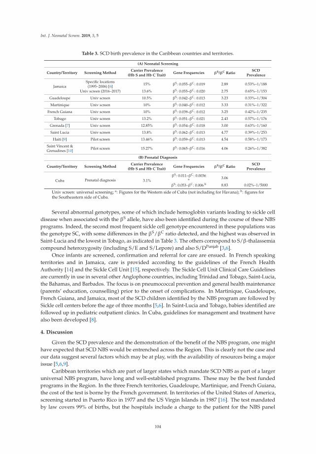
Int. J. Neonatal Screen. 2019, 5, 5
Table 3. SCD birth prevalence in the Caribbean countries and territories.
(A) Neonatal Screening
Country/Territory Screening MethodCarrier Prevalence
(Hb S and Hb C Trait)Gene Frequencies βS/βC Ratio
SCDPrevalence
JamaicaSpecific locations(1995–2006) [6] 15% βS: 0.055–βC: 0.019 2.89 0.53%–1/188
Univ screen (2016–2017) 13.6% βS: 0.055–βC: 0.020 2.75 0.65%–1/153
Guadeloupe Univ screen 10.5% βS: 0.042–βC: 0.013 3.23 0.33%–1/304
Martinique Univ screen 10% βS: 0.040–βC: 0.012 3.33 0.31%–1/322
French Guiana Univ screen 10% βS: 0.039–βC: 0.012 3.25 0.42%–1/235
Tobago Univ screen 13.2% βS: 0.051–βC: 0.021 2.43 0.57%–1/176
Grenada [7] Univ screen 12.85% βS: 0.054–βC: 0.018 3.00 0.63%–1/160
Saint Lucia Univ screen 13.8% βS: 0.062–βC: 0.013 4.77 0.39%–1/253
Haiti [9] Pilot screen 13.46% βS: 0.059–βC: 0.013 4.54 0.58%–1/173
Saint Vincent &Grenadines [10] Pilot screen 15.27% βS: 0.065–βC: 0.016 4.06 0.26%–1/382
(B) Prenatal Diagnosis
Country/Territory Screening MethodCarrier Prevalence
(Hb S and Hb C Trait)Gene Frequencies βS/βC Ratio
SCDPrevalence
Cuba Prenatal diagnosis 3.1%βS: 0.011–βC: 0.0036
a 3.06
βS: 0.053–βC: 0.006 b 8.83 0.02%–1/5000
Univ screen: universal screening; a: Figures for the Western side of Cuba (not including for Havana); b: figures forthe Southeastern side of Cuba.
Several abnormal genotypes, some of which include hemoglobin variants leading to sickle celldisease when associated with the βS allele, have also been identified during the course of these NBSprograms. Indeed, the second most frequent sickle cell genotype encountered in these populations wasthe genotype SC, with some differences in the βS/βC ratio detected, and the highest was observed inSaint-Lucia and the lowest in Tobago, as indicated in Table 3. The others correspond to S/β-thalassemiacompound heterozygosity (including S/E and S/Lepore) and also S/DPunjab [3,6].
Once infants are screened, confirmation and referral for care are ensued. In French speakingterritories and in Jamaica, care is provided according to the guidelines of the French HealthAuthority [14] and the Sickle Cell Unit [15], respectively. The Sickle Cell Unit Clinical Care Guidelinesare currently in use in several other Anglophone countries, including Trinidad and Tobago, Saint-Lucia,the Bahamas, and Barbados. The focus is on pneumococcal prevention and general health maintenance(parents’ education, counselling) prior to the onset of complications. In Martinique, Guadeloupe,French Guiana, and Jamaica, most of the SCD children identified by the NBS program are followed bySickle cell centers before the age of three months [5,6]. In Saint-Lucia and Tobago, babies identified arefollowed up in pediatric outpatient clinics. In Cuba, guidelines for management and treatment havealso been developed [8].
4. Discussion
Given the SCD prevalence and the demonstration of the benefit of the NBS program, one mighthave expected that SCD NBS would be entrenched across the Region. This is clearly not the case andour data suggest several factors which may be at play, with the availability of resources being a majorissue [5,6,9].
Caribbean territories which are part of larger states which mandate SCD NBS as part of a largeruniversal NBS program, have long and well-established programs. These may be the best fundedprograms in the Region. In the three French territories, Guadeloupe, Martinique, and French Guiana,the cost of the test is borne by the French government. In territories of the United States of America,screening started in Puerto Rico in 1977 and the US Virgin Islands in 1987 [16]. The test mandatedby law covers 99% of births, but the hospitals include a charge to the patient for the NBS panel
104

Int. J. Neonatal Screen. 2019, 5, 5
(http://bft.stage.bbox.ly/newborn-screening/states/puerto-rico). Overseas British territories haveseparate health systems and SCD NBS is not uniformly offered.
Among the independent nations, Cuba has an integrated public health program and SCD prenataltesting for couples at risk and carrier women was mandated by law in 1983 [7,8]. This program seeksto actively prevent the births of children with SCD, and is perhaps the most active in promoting thetermination of pregnancies; since termination of pregnancy was requested by 76.5% of at-risk couples.
In the other independent Caribbean nations, screening varies based on factors such as historicalcontext, current champions, and public health commitment. Jamaica has a unique historical context asthe site of the Jamaica Sickle Cell Cohort Study. Nevertheless, after the completion of recruitment forthe cohort study in 1981, SCD NBS ceased until 1995, when, through the advocacy of the Sickle CellSupport Club of Jamaica (now the Sickle Cell Support Foundation), it was restarted. For a decade, itwas limited to three hospitals in the South East Regional Health Authority, providing screening forapproximately 43% of all national births. The coverage gradually increased from 2008 to 2015 whenessentially universal coverage was achieved. While testing was mandated in the National StrategicPlan for the Prevention and Control of Non-communicable Disease in Jamaica 2012–2018, there is nolegislative mandate and the integration of the program into the fabric of the public health systemremains incomplete. Thus, the sustainability of the program depends on the support of incumbentpolicy makers.
The Saint Lucia Sickle Cell Association (SSCA), a local non-governmental organization, has beenstrong for many years. It was influential in the introduction of universal SCD NBS in 1992 and itsintegration into the Ministry of Health’s Community Child Health Service (CCHS) [10]. The programuses hemoglobin electrophoresis to test cord blood samples. A pilot program funded by the SickKidsCaribbean Initiative using HPLC testing of heel prick samples had a disappointing uptake and theinitial approach continues. The pilot in Tobago has also been successful; it has been continuous fora decade. It is funded by the Regional Health Authority of Tobago, which has made the diagnosisand treatment of SCD a priority. Screening of the immediate family of babies identified with thetrait or the disease is done by electrophoresis in the hospital laboratory, thus a greater percentageof the population now know their genotype and there is a greater awareness through education.The initiation of SCD NBS at a major obstetric hospital in Trinidad in 2018 is further evidence of theacceptance of this program.
Pilot screening projects in Grenada [13], St Vincent and the Grenadines [17], and Haiti [18], fundedby CAREST, the Medical University of South Carolina (Charleston, SC, USA), and University HospitalsMedical Center (Cleveland, OH, USA), respectively, were not sustained once project funding ended.Local policy makers were not able to identify funding and human resources for continued screening.A pilot posited in Barbados was not undertaken [19]. Instead, screening of pregnant women andtesting postnatally of at risk children was the approach chosen.
Currently, champions in Antigua and Guyana are pressing to start pilots and perhaps sustainableprograms. These outcomes again indicate the importance of advocacy in convincing policy makers ofthe feasibility and benefit of SCD NBS. In both Jamaica and Saint Lucia, advocacy groups have beencritical to convincing public health officials to initiate and maintain screening. CAREST has a rolein supporting their advocacy efforts to secure governmental support and sustainable funding, evenas screening begins. In this framework, CAREST has recently obtained funding from the EuropeanRegional Development Fund. This funding dedicated to the development of cooperation betweenthe French Departments of the Americas and the other countries/territories of the Caribbean Basinwill allow the screening of Grenada to be re-launched, initiate a pilot study for Antigua, and evaluatestrategies to ensure the sustainability of this NBS.
The Saint-Lucia and Tobago experience clearly demonstrate that the model of using a few regionallaboratories to increase efficiencies of scale, decreasing per cost tests, can be used. Actually, given thehigh cost of equipment and requisite disposables, and the relatively small populations in the Region,the use of two regional laboratories (Jamaica and Guadeloupe) proved to be a cost-effective approach,
105

Int. J. Neonatal Screen. 2019, 5, 5
once reasonably costed transportation of samples and secure data flows are available. However, it isworthwhile to notice that a significant proportion of the Caribbean populations do not have access toscreening program, such as Haiti, which accounts for more than 90% the Francophone inhabitants ofthe Caribbean, and the Dominican Republic, with approximately 40% of Spanish-speakers. Up to now,only a little more than 50% of the English and Spanish speaking infants are screened.
The cost of national SCD NBS programmes may decrease significantly if efficient, accurate, andinexpensive point-of-care (POC) devices become available; a number of such POC testing deviceshave recently been developed [20]. These low-cost devices, which must have high specificity todetect HbS and HbC in the presence of HbF and the capacity to distinguish the trait (HbAS) fromsamples with SCD, must also be easy to use. Two of them, relying on lateral flow immunoassays,the SickleSCAN [21] and the HemoTypeSC tests [22], could be viable screening tools for the earlydiagnosis of SCD conducted by health workers with little expertise. Preliminary data using theHemoTypeSC test on a small series of children and adults in Martinique, in comparison with a largerseries in Ghana and the USA, showed the good specificity and sensitivity of the test [23]. We plan toconduct larger studies to evaluate the performance and implementation feasibility of these POC testingdevices as screening tools in Caribbean territories where the reference “gold-standard” tests, IEF, andHPLC are not available. This approach may also reduce the number of samples to be screened by thereference laboratories and the delay between the blood sampling and the transmission of the result andultimately reduce the age of inclusion of the newly identified children. Mothers would get immediatefeedback if their children’s tests are normal, and be advised of the need to do confirmatory tests incases consistent with traits or SCD. This promising strategy is expected to promote the extension ofscreening programs, and lead to the clarification of the prevalence and to a better management ofSCD in the Caribbean. Audits of important outcomes, such as time to enrollment in clinic, initiationof splenic palpation, and Pneumococcal prophylaxis, as well as continued ongoing ascertainmentof survival in countries and territories with SCD NBS, will help to determine what implementationmodels are most successful and guide the subsequent initiation of programs in other settings.
Differences of SCD prevalence in the Caribbean islands could be observed, with the highest beingdetected in Jamaica and Grenada and the lowest in Cuba. Various factors may explain these variations,such as the selective introduction of crop production requiring a greater or lesser need for slaves, thesettlement policy of the colonial powers with France and the United Kingdom importing few of theirown population compared to Spain, for example, as well as the significance of migrations after the endof slavery (ranging from 1804 in Haiti to 1888 in Brazil). In addition, various βS/βC ratios have beendetected in the studied populations. Since the distribution of the βC allele is more restricted than thatof the βS allele in Africa, this data could be related to differences in the African origins of the deportedslaves in these territories. These differences in the βS/βC ratio could also result from sampling effects;a relatively small number of newborns were screened in some populations. Few epidemiologicaldata from continental countries of the Western coast of the Caribbean Sea coast have been producedso far. An NBS pilot study was conducted in Costa Rica with a total of 70,943 samples and led tothe identification of five SS and one SC children [24]. In addition, several clinical reports or geneticstudies indicating the presence of SCD in Panama [25], Colombia [26], and Venezuela [27] have beenpublished, but none of these countries have implemented an NBS program on SCD and no accuratedata of the prevalence of the disease are available so far, to the best of our knowledge.
In summary, SCD is a perfect example of a disease which fulfils all requirements for doing NBS.Parents are usually asymptomatic and may not know of their risk. Tests done on an asymptomatic babycan allow them to access interventions that decrease morbidity and preventable mortality. CAREST willcontinue to advocate and work towards universal SCD NBS across the Caribbean Region, regardlessof language, per capita income, or political system.
106

Int. J. Neonatal Screen. 2019, 5, 5
Author Contributions: Conceptualization: M.R., M.-D.H.-D., B.M.-T., and J.K.-M.; Methodology: M.R., L.K., K.L.,N.E., G.E., M.E.-J., B.M.-T., M.-D.H.-D., and J.K.-M.; Resources: L.K., K.L., N.E., G.E., M.E.-J., B.M.-T., M.A., andN.K.; Writing-Original Draft Preparation: M.R., J.K.-M., B.M.-T., and M.-D.H.-D.; Writing-Review & Editing: M.R.,M.-D.H.-D., L.K., K.L., J.K.-M., B.M.-T., M.A., N.E., G.E., and N.K.; Project administration: M.-D.H.-D., and J.K.-M.
Funding: Funding was provided by the regional councils of Guadeloupe and Martinique, and the general councilof Guadeloupe. Funding in Jamaica was provided by the Ministry of Health, the National Health Fund. Equipmentwas provided by the Brazilian Government and Sagicor. The SickKids Caribbean Initiative funded the SCD NBScoordinator in Jamaica and the St Lucia pilot.
Acknowledgments: The authors wish to acknowledge the contribution of the team of the diagnostic laboratoryof hemoglobinopathies of the University Hospital of Guadeloupe for its implication in the NBS of Tobagoand Grenada.
Conflicts of Interest: The authors declare no conflict of interest.
References
1. Curtin, P.D. Distribution in space: The colonies of the North Europeans. In The Atlantic Slave Trade: A Census;University of Wisconsin Press: Madison, WI, USA, 1969; pp. 51–94.
2. Herrick, J.B. Peculiar elongated and sickle-shaped red blood cell corpuscules in a case of severe anemia.Arch. Intern. Med. 1910, 6, 517–521. [CrossRef]
3. Serjeant, G.; Serjeant, B.E.; Forbes, M.; Hayes, R.J.; Higgs, D.R.; Lehmann, H. Haemoglobin gene frequenciesin the Jamaican population: A study in 100,000 newborns. Br. J. Haematol. 1986, 64, 253–262. [CrossRef][PubMed]
4. Kato, G.J.; Piel, F.B.; Reid, C.D.; Gaston, M.H.; Ohene-Frempong, K.; Krishnamurti, L.; Smith, W.R.;Panepinto, J.A.; Weatherall, D.J.; Costa, F.F.; et al. Sickle cell disease. Nat. Rev. Dis. Primers 2018, 4,18010. [CrossRef] [PubMed]
5. King, L.; Fraser, R.; Forbes, M.; Grindley, M.; Ali, S.; Reid, M. Newborn sickle cell disease screening:The Jamaican experience (1995–2006). J. Med. Screen. 2007, 14, 117–122. [CrossRef] [PubMed]
6. Saint-Martin, C.; Romana, M.; Bibrac, A.; Brudey, K.; Tarer, V.; Divialle-Doumdo, L.; Petras, M.;Keclard-Christophe, L.; Lamothe, S.; Broquere, C.; et al. Universal newborn screening forhaemoglobinopathies in Guadeloupe (French West Indies): A 27-year experience. J. Med. Screen. 2013,20, 177–182. [CrossRef]
7. Heredero-Baute, L. Community-based program for the diagnosis and prevention of genetic disorders inCuba. Twenty years of experience. Community Genet. 2004, 7, 130–136. [CrossRef]
8. Svarch, E.; Machín García, S.; Marcheco Teruel, B.; Triana, R.M.; González Otero, A.; Menéndez Veitía, A.Program for comprehensive sickle cell disease care in Cuba. Revista Cubana de Hematología, Inmunologíay Hemoterapia 2017, 33, 1–2.
9. Knight-Madden, J.; Romana, M.; Villaescusa, R.; Reid, M.; Etienne-Julan, M.; Boutin, L.; Elana, G.; Elenga, N.;Wheeler, G.; Lee, K.; et al. CAREST—Multilingual Regional Integration for Health Promotion and Researchon Sickle Cell Disease and Thalassemia. Am. J. Public Health 2016, 106, 851–853. [CrossRef]
10. Alexander, S.; Belmar-George, S.; Eugene, A.; Elias, V. Knowledge of an attitudes toward heel prick screeningfor sickle cell disease in Saint Lucia. Rev. Panam. Salud Publica 2017, 41, e70.
11. Kéclard, L.; Romana, M.; Lavocat, E.; Saint-Martin, C.; Berchel, C.; Mérault, G. Sickle cell disorder, beta-globingene cluster haplotypes and alpha-thalassemia in neonates and adults from Guadeloupe. Am. J. Hematol.1997, 55, 24–27. [CrossRef]
12. Romana, M.; Kéclard, L.; Froger, A.; Lavocat, E.; Saint-Martin, C.; Berchel, C.; Mérault, G. Diverse geneticmechanisms operate to generate atypical betaS haplotypes in the population of Guadeloupe. Hemoglobin2000, 24, 77–87. [CrossRef] [PubMed]
13. Antoine, M.; Lee, K.; Donald, T.; Belfon, Y.; Drigo, A.; Polson, S.; Martin, F.; Mitchell, G.; Etienne-Julan, M.;Hardy-Dessources, M.D. Prevalence of sickle cell disease among Grenadian newborns. J. Med. Screen. 2018,25, 49–50. [CrossRef] [PubMed]
14. Available online: https://www.has-sante.fr/portail/upload/.../ald_10_pnds_drepano_enfant_web.pdf(accessed on 2 April 2010).
107

Int. J. Neonatal Screen. 2019, 5, 5
15. Aldred, K.; Asnani, M.; Beckford, M.; Bhatt-Poulose, K.; Bortolusso Ali, S.; Chin, N.; Daley, C.; Grindley, M.;Hammond-Gabbadon, C.; Harris, J.; et al. Sickle Cell Disease: The Clinical Care Guidelines of the Sickle Cell Unit;Bortolusi-Ali, S., Ed.; Sickle Cell Unit, Tropical Medicine Research Institute, University of the West Indies:Kingston, Jamaica, 2016.
16. Morales, A.; Wierenga, A.; Cuthbert, C.; Sacharow, S.; Jayakar, P.; Velazquez, D.; Loring, J.; Barbouth, D.Expanded newborn screening in Puerto Rico and the US Virgin Islands: Education and barriers assessment.Genet. Med. 2009, 11, 169–175. [CrossRef] [PubMed]
17. Williams, S.A.; Browne-Ferdinand, B.; Smart, Y.; Morella, K.; Reed, S.G.; Kanter, J. Newborn Screeningfor Sickle Cell Disease in St. Vincent and the Grenadines: Results of a Pilot Newborn Screening Program.Glob. Pediatr. Health 2017, 4. [CrossRef]
18. Rotz, S.; Arty, G.; Dall’Amico, R.; De Zen, L.; Zanolli, F.; Bodas, P. Prevalence of sickle cell disease, hemoglobinS, and hemoglobin C among Haitian newborns. Am. J. Hematol. 2013, 88, 827–828. [CrossRef] [PubMed]
19. Quimby, K.R.; Moe, S.; Sealy, I.; Nicholls, C.; Hambleton, I.R.; Landis, R.C. Clinical findings associatedwith homozygous sickle cell disease in the Barbadian population–do we need a national SCD registry?BMC Res. Notes 2014, 7, 102. [CrossRef] [PubMed]
20. McGann, P.T.; Hoppe, C. The pressing need for point-of-care diagnostics for sickle cell disease: A review ofcurrent and future technologies. Blood Cells Mol. Dis. 2017, 67, 104–113. [CrossRef]
21. Nwegbu, M.M.; Isa, H.A.; Nwankwo, B.B.; Okeke, C.C.; Edet-Offong, U.J.; Akinola, N.O.; Adekile, A.D.;Aneke, J.C.; Okocha, E.C.; Ulasi, T.; et al. Preliminary Evaluation of a Point-of-Care Testing Device(SickleSCAN™) in Screening for Sickle Cell Disease. Hemoglobin 2017, 41, 77–82. [CrossRef]
22. Quinn, C.T.; Paniagua, M.C.; DiNello, R.K.; Panchal, A.; Geisberg, M. A rapid, inexpensive anddisposable point-of-care blood test for sickle cell disease using novel, highly specific monoclonal antibodies.Br. J. Haematol. 2016, 175, 724–732. [CrossRef]
23. Steele, C.; Sinski, A.; Asibey, J.; Hardy-Dessources, M.D.; Elana, G.; Brennan, C.; Odame, I.; Hoppe, C.;Geisberg, M.; Serrao, E.; et al. Point-of-care screening for sickle cell disease in low-resource settings:A multi-center evaluation of HemoTypeSC, a novel rapid test. Am. J. Hematol. 2018. [CrossRef]
24. Abarca, G.; Navarrete, M.; Trejos, R.; de Céspedes, C.; Saborío, M. Abnormal haemoglobins in the newbornhuman population of Costa Rica. Rev. Biol. Trop. 2008, 56, 995–1001. [PubMed]
25. Rusanova, I.; Cossio, G.; Moreno, B.; Javier Perea, F.; De Borace, R.G.; Perea, M.; Escames, G.;Acuña-Castroviejo, D. β-globin gene cluster haplotypes in sickle cell patients from Panamá. Am. J. Hum. Biol.2011, 23, 377–380. [CrossRef] [PubMed]
26. Fong, C.; Lizarralde-Iragorri, M.A.; Rojas-Gallardo, D.; Barreto, G. Frequency and origin of haplotypesassociated with the beta-globin gene cluster in individuals with trait and sickle cell anemia in the Atlanticand Pacific coastal regions of Colombia. Genet. Mol. Biol. 2013, 36, 494–497. [CrossRef] [PubMed]
27. Arends, A.; Alvarez, M.; Velázquez, D.; Bravo, M.; Salazar, R.; Guevara, J.M.; Castillo, O. Determination ofbeta-globin gene cluster haplotypes and prevalence of alpha-thalassemia in sickle cell anemia patients inVenezuela. Am. J. Hematol. 2000, 64, 87–90. [CrossRef]
© 2019 by the authors. Licensee MDPI, Basel, Switzerland. This article is an open accessarticle distributed under the terms and conditions of the Creative Commons Attribution(CC BY) license (http://creativecommons.org/licenses/by/4.0/).
108

International Journal of
Neonatal Screening
Review
The Neonatal Screening Program in Brazil, Focus onSickle Cell Disease (SCD)
Ana C. Silva-Pinto 1,2,*, Maria Cândida Alencar de Queiroz 2, Paula Juliana Antoniazzo Zamaro 3,
Miranete Arruda 1,4 and Helena Pimentel dos Santos 1,5
1 Policy of Integral Attention to People with Sickle Cell Disease (PIAPSCD), Technical Advisory Council forSickle Cell Disease (TAC-SCD), CGSH/DAET/SAS, Ministry of Health, Asa Norte Brasília 70719-040, Brazil;[email protected] (M.A.); [email protected] (H.P.d.S.)
2 Regional Blood Center of Ribeirão Preto, HC-FMRP, University of Sao Paulo (USP), Campus Universitário,Ribeirão Preto 14049-900, Brazil; [email protected]
3 National Newborn Screening Program Policy (NNSPP)/CGSH/SAS, Ministry of Health, Asa NorteBrasília 70719-040, Brazil; [email protected]
4 State Health Secretariat, State of Pernambuco, Recife 50751-530, Brazil5 Newborn Screening Program, APAE-Salvador, Salvador 41830-141, Brazil* Correspondence: [email protected]; Tel.: +55-162101-9300
Received: 28 September 2018; Accepted: 23 January 2019; Published: 26 January 2019
Abstract: Since 2001, the Brazilian Ministry of Health has been coordinating a National NeonatalScreening Program (NNSP) that now covers all the 26 states and the Federal District of the BrazilianRepublic and targets six diseases including sickle cell disease (SCD) and other hemoglobinopathies.In 2005, the program coverage reached 80% of the total live births. Since then, it has oscillatedbetween 80% and 84% globally with disparities from one state to another (>95% in São Paulo State).The Ministry of Health has also published several Guidelines for clinical follow-up and treatment forthe diseases comprised by the neonatal screening program. The main challenge was, and still is, toorganize the public health network (SUS), from diagnosis and basic care to reference centers in orderto provide comprehensive care for patients diagnosed by neonatal screening, especially for SCDpatients. Considerable gains have already been achieved, including the implementation of a networkwithin SUS and the addition of scientific and technological progress to treatment protocols. The goalsfor the care of SCD patients are the intensification of information provided to health care professionalsand patients, measures to prevent complications, and care and health promotion, considering thesepatients in a global and integrated way, to reduce mortality and enhance their quality of life.
Keywords: neonatal screening; sickle cell disease; hemoglobinopathies
1. Background
The Brazilian population is currently estimated at over 208 million people. For the most part,three distinct peoples form the ethnic backgrounds of this population: the Native Americans (Indians),the Portuguese, and the Africans. Population data from the 2010 Demographic Census showed that50.7% of the Brazilian population is made up of Afro-Americans, 47.7% Caucasians, and 0.7% Indians(0.9% unspecified). Since the population is broadly miscegenated, all national programs must beuniversalized and not directed at a specific portion of the population. Therefore, the neonatal screeningprogram is national and universal, aiming to reach 100% of live births in the country [1].
Int. J. Neonatal Screen. 2019, 5, 11; doi:10.3390/ijns5010011 www.mdpi.com/journal/ijns109

Int. J. Neonatal Screen. 2019, 5, 11
2. The Neonatal Screening Program
The history of neonatal screening (NS) in Brazil goes back to 1976, when APAE-São Paulobegan neonatal screening for phenylketonuria (PKU). Several pilot NS initiatives were then initiatedindependently without coordination nor standardization among Brazilian states. In 1992, the screeningfor PKU and congenital hypothyroidism (CH) was included in the public health system, known asSUS [2]. In 1998, the State of Minas Gerais was the first to introduce a universal State NS Program forsickle cell disease (SCD) in the already existing programs for PK and CH [3].
However, the recognition of NS as a specific public health program only happened in 2001 afterthe founding of the National Neonatal Screening Program (NNSP) coordinated by the Ministry ofHealth [4]. The program led to the definition of standards and protocols for the whole country, andplaced screening as a global health action aimed at preventing child health problems not only throughtesting to detect the disease but also the active search of suspected cases, diagnostic confirmation,treatment, and follow-up of patients [5]. The NNSP initially focused on PKU, CH, cystic fibrosis,SCD, and other hemoglobinopathies. In 2014, the program was extended to screening for biotinidasedeficiency and congenital adrenal hyperplasia.
The Ministry of Health defined priorities and divided the program into four phases, accreditingthe states according to each one’s capacity. From 2001 onward, reference services were certifiedsequentially in one of the four phases of the NNSP. Table 1 shows the years at which each of the phasesreached universal implementation in all the states and the Federal District.
Figure 1 illustrates the sequential expansion of the neonatal screening program in the variousstates of Brazil between 2010 and 2014.
Table 1. The implementation of neonatal screening divided into phases, and its year of universalization.
Phases Diseases Year of National Implementation
Phase I Phenylketonuria and congenital hypothyroidism 2006
Phase II Sickle cell disease and other hemoglobinopathies 2013
Phase III Cystic fibrosis 2013
Phase IV Congenital adrenal hyperplasia and biotinidase deficiency 2014
The coverage rate of the program (percentage of the screened live births) varied over the years(Figure 2). Since 2006, it has remained above 85% at the national level, with few variations. This figurerelates to variations in the health care network capacity from state to state and to variations in thepopulation’s access to it. In São Paulo State, for example, the percentage of coverage reaches over 95%.
110

Int. J. Neonatal Screen. 2019, 5, 11
Figure 1. Expansion of the neonatal screening program in Brazil from 2010 to 2014 regarding the fourphases of the National Neonatal Screening Program (NNSP).
111

Int. J. Neonatal Screen. 2019, 5, 11
Figure 2. The annual rate of the program coverage from 2004 to 2016.
3. Resolutions and Guidelines from the Brazilian Ministry of Health
In addition to the implementation of neonatal screening and with the goal of standardizingstandards of care, the Ministry of Health has published several guidelines for clinical follow-up andtreatment for each of the diseases included in the neonatal screening program (see, for example, theClinical Protocol and Therapeutic Guidelines for Sickle Cell Disease [6]). These include the descriptionof the general concepts for each disease, diagnostic criteria, inclusion and exclusion criteria, treatmentand mechanisms of regulation, and control and treatment evaluation. Altogether, they establish anational policy and should be used by the state and municipal Health Secretariats for the regulation ofcare access, authorization, registration, and reimbursement of the corresponding procedures.
4. The Sickle Cell Disease Network of Care Inside SUS: From Neonatal Screening to Follow-Upin Reference Centers
Health is inscribed in the national Constitution as a right for everyone and a duty of the BrazilianRepublic. This was the framework for the creation of the Unified Health System, known as SUS,which contemplates the guidelines for health promotion and provision of services and establishesthe community participation and transfer of federal resources to other jurisdictions [7,8]. Despite thegreat barriers that SUS has faced since its conception, it has managed to establish itself as a system thatproposes to be an institutional redistributive model, guaranteeing universal coverage. In this context,the inclusion of sickle cell disease (SCD) and other hemoglobinopathies in the National NeonatalScreening Program (NNSP) instituted by the SUS in 2001 shows recognition of this group of pathologiesas a national public health issue [9].
The proportion of live births with SCD varies widely among the states, being more frequent instates with a higher concentration of African descendants. For example, in Bahia for every 650 births, 1child has SCD, followed by Rio de Janeiro (1:1200) and then Pernambuco, Maranhão, and Minas Gerais,with 1:1400. Table 2 shows the incidence of newborns affected by SCD in 12 states representative ofthe four most populated regions of Brazil (the fifth region being the North that covers the Amazonianforest).
112

Int. J. Neonatal Screen. 2019, 5, 11
Table 2. Incidence of sickle cell disease and sickle cell trait in some Brazilian states.
Regions of Brazil State Incidence/Live Births Incidence of the S Mutation
Northeast
Maranhão 1:1400 1:23
Pernambuco 1:1400 1:23
Bahia 1:650 1:17
Center-westGoiás 1:1400 1:28
Mato Grosso do Sul 1:8300 1:70
Southeast
Minas Gerais 1:1400 1:30
Espírito Santo 1:1800 1:28
Rio de Janeiro 1:1200 1:21
São Paulo 1:4000 1:35
South
Paraná 1:13,000 1:65
Santa Catarina 1:13,000 1:65
Rio Grande do Sul 1:13,500 1:65
Building up the current network and system of care for the people with SCD in Brazil has been along process, in some ways reminiscent of the 1970s struggle for civil rights and equality for accessto care in the USA that led to the US National Sickle Cell Anemia Control Act in 1972 [10,11]. In the1980s, Brazilian representative entities for the the defense of the rights of family and people withSCD took the first steps to formulate claims, initially at the local level, and then expanded theircoordination with the formation in 2001 of the National Federation of Associations of People withSickle Diseases (FENAFAL). The march Zumbi dos Palmares against Racism, for Citizenship and forLife, held in Brasilia in 1995, was an important milestone in the struggle of black people against theBrazilian State for affirmative action and against racism in terms of health care. From this action,important policies emerged, including the Policy of Integral Attention to People with Sickle Cell Disease(PIAPSCD) [12]. The major goals were to change the natural history of SCD in Brazil, by reducingmorbidity and mortality, promoting greater survival, and improving the quality of life for peoplewith SCD. Commitments were made to provide genetic guidance, safeguarding reproductive rights ofpeople with the sickle cell trait, and to disseminate information about SCD to the general population.
In 2006, the Ministry of Health established a Technical Advisory Council for Sickle Cell Disease(TAC-SCD) to help states and municipalities organize their programs of care for people withSCD. TAC is composed of specialists, managers of state and municipal jurisdictions of SUS, andrepresentatives of educational, research, and assistance institutions involved in the care of people withSCD who are invited to act voluntarily. There is also a representation of the users chosen by FENAFAL.TAC is responsible for producing the guidelines for the management of SCD and its complications,genetic guidelines, family guidelines, guidelines for oral health and on the role of nursing, which areprinted and distributed in the reference centers and in the basic SUS network. These materials areavailable electronically in the SUS Virtual Library (VL). The members of TAC also conduct trainingcourses, workshops, and symposia on SCD in all regions of the country, contributing to the continuingeducation of health professionals.
Within SUS, the standardized attention to people with SCD encompasses all levels from earlydiagnosis to bone marrow transplantation. Assistance begins in basic care with the early diagnosisfree of charge through the NNSP that links the affected child to the basic health unit, responsible forthe first procedures of prevention such as prophylaxis with penicillin initiated in the first month oflife, immunobiological approaches, and then referral to the regional Specialized Care Service. In moststates, the reference center is the Blood Center, which is normally integrated with the local healthnetwork to meet other health needs, such as the use of hydroxyurea, transcranial Doppler, and allprocedures of greater complexity. The implementation of the SCD network in all the states is intended
113

Int. J. Neonatal Screen. 2019, 5, 11
to guarantee decentralized care beginning with diagnosis, with the assistance of a multi-professionaland multidisciplinary team, providing health education with a focus on self-management and accessto specialized and high complexity care in all the 26 states and the Federal District [13].
5. Challenges for the Full Implementation of the SCD Care Network
Despite major advances, the organization of a robust health care network for people with SCDis still a major challenge to SUS. Only a network with the potential for the collective construction ofsolutions is able to cope with the complexity of demands and to guarantee the promotion of autonomyand citizenship to people with SCD. The disease has not yet reached the level of visibility pursued thatis necessary to provide its target population with the right to health they deserve to extend their lifehorizon with personal and social well-being.
Notably, considerable gains have already been achieved, including the implementation of neonatalscreening at the national level together with scientific and technological progress with respect totreatment. Preliminary data on the effect of hydroxyurea treatment and mortality rates have beenpublished [14–16]. However, more precise epidemiological data covering the entire country are stillneeded. A national registry for SCD patients is being set up to collect more reliable informationto evaluate the system: percentage of new cases that actually enter follow-up at reference centers,immunization rate, percentage of patients that started pneumococcal prophylaxis, rate of childrenscreened with transcranial Doppler, use of hydroxyurea, and death rates at the country level. What issought to bring attention to people with SCD is the intensification of information provided, measures toprevent injuries, and care and health promotion, considering these patients in a global and integratedway, to reduce mortality and enhance their quality of life.
Author Contributions: All authors contributed equally to the manuscript: A.C.S.-P. was responsible for thedesign, writing, and review of the manuscript; M.C.A.d.Q. was responsible for the SCD network writing andreview; P.J.A.Z. was responsible for the newborn screening data and figures; M.A. was responsible for SCDepidemiological data and the review of the final version; and H.P.d.S. was responsible for the newborn screeningbackground and review of the manuscript.
Funding: This research received no external funding.
Conflicts of Interest: The authors declare no conflict of interest.
References
1. Brasil, IBGE. Demographic Census 2010. Available online: https://www.ibge.gov.br/ (accessed on17 September 2018).
2. Brasil, Ministério da Saúde. Portaria GM/MS nº22 de 15/01/1992—triagem para fenilcetonúria ehipotireoidismo Congênito. Diário Oficial da União, 15 January 1992.
3. Paixão, M.C.; Cunha Ferraz, M.H.; Januário, J.N.; Viana, M.B.; Lima, J.M. Reliability of isoelectrofocusing forthe detection of Hb S, Hb C, and HB D in a pioneering population-based program of newborn screening inBrazil. Hemoglobin 2001, 25, 297–303. [CrossRef] [PubMed]
4. Brasil, Ministério da saúde. Portaria GM/MS nº 822 de 06 de junho de 2001—Programa de triagem neonatalno Brasil. Diário Oficial da União, 6 June 2001.
5. Carvalho, T.M.; Santos, H.P.; Santos, I.C.G.P.; Vargas, P.R.; Pedrosa, J. Newborn screening: A national publichealth programme in Brazil. J. Inherit. Metab. Dis. 2007, 30, 615. [CrossRef] [PubMed]
6. Brasil, Ministério da saúde. Portaria SAS/MS Nº 55 de 29 de janeiro de 2010—Protocolo Clínico e DiretrizesTerapêuticas para Doença Falciforme”. Diário Oficial da União, 29 January 2010.
7. Brasil, Ministério da saúde. Lei nº 8.080, de 19 de setembro de 1990, criação do SUS—Sistema Único deSaúde. Diário Oficial da União, 19 September 1990.
8. Seta, M.H.; Oliveira, C.V.D.S.; Pepe, V.L.E. Health protection in Brazil: the National Sanitary SurveillanceSystem. Cien Saude Colet 2017, 22, 3225–3234. [CrossRef] [PubMed]
9. Ramalho, A.S.; Magna, L.A.; Paiva-Silva, R.B. A portaria nº 822/01 do Ministério da Saúde e as peculiaridadesdas hemoglobinopatias em saúde pública no Brasil. Cad. Saúde Pública 2003, 19, 1195–1199. [CrossRef][PubMed]
114

Int. J. Neonatal Screen. 2019, 5, 11
10. Vichinsky, E. Sickle cell disease and thalassemia: disorders of globin production. (2008) pages 3-5, inSpecial anniversary brochure, American Society of Hematology: 50 Years in Hematology: Research ThatRevolutionized Patient Care. Available online: http://www.hematology.org/About/History/50-Years/1533.aspx (accessed on 13 January 2019).
11. Bassett, M.T. Beyond Berets: The Black Panthers as Health Activists. Am. J. Public Health 2016, 106, 1741–1743.[CrossRef] [PubMed]
12. Brasil, Ministério da saúde. Portaria de nº 1391 de 16 de agosto de 2005—Diretrizes para a Política Nacionalde Atenção Integral às Pessoas com Doença Falciforme e outras Hemoglobinopatias. Diário Oficial da União,16 August 2005.
13. Cançado, R.D.; Jesus, J.A. A doença falciforme no Brasil. Rev. Bras. Hematol. Hemoter. 2007, 29, 204–206.[CrossRef]
14. Lobo, C.L.; Pinto, J.F.; Nascimento, E.M.; Moura, P.G.; Cardoso, G.P.; Hankins, J.S. The effect ofhydroxcarbamide therapy on survival of children with sickle cell disease. Br. J. Haematol. 2013, 161,852–860. [CrossRef] [PubMed]
15. Fernandes, A.P.; Januário, J.N.; Cangussu, C.B.; Macedo, D.L.; Viana, M.B. Mortality of children with sicklecell disease: A population study. J. Pediatr. (Rio J). 2010, 86, 279–284. [CrossRef]
16. Lobo, C.L.C.; Nascimento, E.M.D.; Jesus, L.J.C.; Freitas, T.G.; Lugon, J.R.; Ballas, S.K. Mortality in children,adolescents and adults with sickle cell anemia in Rio de Janeiro, Brazil. Rev. Bras. Hematol. Hemoter. 2018, 40,37–42. [CrossRef] [PubMed]
© 2019 by the authors. Licensee MDPI, Basel, Switzerland. This article is an open accessarticle distributed under the terms and conditions of the Creative Commons Attribution(CC BY) license (http://creativecommons.org/licenses/by/4.0/).
115

International Journal of
Neonatal Screening
Review
Newborn Screening for Sickle Cell Disease:Indian Experience
Roshan B. Colah, Pallavi Mehta and Malay B. Mukherjee *
ICMR-National Institute of Immunohaematology, KEM Hospital Campus, Mumbai 400012, India;[email protected] (R.B.C.); [email protected] (P.M.)* Correspondence: [email protected]
Received: 12 September 2018; Accepted: 7 November 2018; Published: 13 November 2018
Abstract: Sickle cell disease (SCD) is a major public health problem in India with the highestprevalence amongst the tribal and some non-tribal ethnic groups. The clinical manifestations areextremely variable ranging from a severe to mild or asymptomatic condition. Early diagnosis andproviding care is critical in SCD because of the possibility of lethal complications in early infancy inpre-symptomatic children. Since 2010, neonatal screening programs for SCD have been initiated in afew states of India. A total of 18,003 babies have been screened by automated HPLC using either cordblood samples or heel prick dried blood spots and 2944 and 300 babies were diagnosed as sickle cellcarriers and SCD respectively. A follow up of the SCD babies showed considerable variation in theclinical presentation in different population groups, the disease being more severe among non-tribalbabies. Around 30% of babies developed serious complications within the first 2 to 2.6 years of life.These pilot studies have demonstrated the feasibility of undertaking newborn screening programsfor SCD even in rural areas. A longer follow up of these babies is required and it is important toestablish a national newborn screening program for SCD in all of the states where the frequency of thesickle cell gene is very high followed by the development of comprehensive care centers along withcounselling and treatment facilities. This comprehensive data will ultimately help us to understandthe natural history of SCD in India and also help the Government to formulate strategies for themanagement and prevention of sickle cell disease in India.
Keywords: newborn screening; sickle cell disease; India; tribal; non-tribal; Guthrie spots; cord blood;automated HPLC
1. Introduction
Hemoglobinopathies are the most common monogenic disorders in India posing a significanthealth burden. Sickle cell disease (SCD) was the first molecular disease to be described where a singlepoint mutation (A→T) resulted in the substitution of the 6th aminoacid in the β globin chain fromglutamic acid to valine leading to an altered electrophoretic mobility of the hemoglobin molecule. SCDincludes a variety of conditions, the primary hemoglobin disorder being sickle cell anemia (SCA) dueto homozygosity for hemoglobin S (HbS) as well as the compound heterozygous conditions, HbS-βthalassemia, HbSD disease, HbSE disease, HbSC disease and HbS-O Arab disease [1].
2. Geographic Distribution of HbS in India
HbS is widespread in African, Mediterranean, Middle Eastern, Indian, Caribbean and Southand Central American populations [1]. In India, SCA is prevalent among tribal populations who areconsidered to be the original inhabitants in south Gujarat, Maharashtra, Madhya Pradesh, Chhattisgarh,and western Odisha with a smaller focus in the southern region in Andhra Pradesh, Karnataka,northern Tamil Nadu and Kerala. They often reside in remote regions away from the mainstream
Int. J. Neonatal Screen. 2018, 4, 31; doi:10.3390/ijns4040031 www.mdpi.com/journal/neonatalscreening116

Int. J. Neonatal Screen. 2018, 4, 31
populations. Sickle cell anemia is also prevalent in some of the scheduled castes and other backwardclasses (non-tribal populations) mainly in central India, in particular, among the Mahar, Kunbi andTeli castes but is rare in other castes where it may be seen due to admixture. These are economicallyand socially disadvantaged populations living largely in rural areas. Carrier frequencies rangingfrom 1 to 35% have been described in these groups and their distribution in different states of thecountry has been mapped earlier [2–4]. Many of these populations also harbor the β thalassemia genebut screening for β thalassemia had not been extensively carried out among them earlier. A recentreport from Akola district in central India showed that 36 out of 91 pediatric SCD patients (39.6%) hadsickle-β thalassemia [5].
3. Sickle Haplotypes in India
In the eastern province of Saudi Arabia and in India, the sickle gene is linked to the Arab Indianor Asian haplotype, which is associated with higher fetal hemoglobin (HbF) levels and a milder clinicalpresentation than the Benin haplotype seen in west and north Africa and the Bantu haplotype seen ineast and central Africa [6]. There are limited studies on haplotype analysis from different regions inIndia, however, all of them have shown that around 90% of sickle genes have the Asian haplotypewhile few other atypical haplotypes including the BantuA2 and the Senegal haplotype have beenassociated with around 10% of sickle genes [7]. Yet, recent studies have indicated more severe clinicalmanifestations even among the Asian haplotype.
4. Clinical Manifestations of Sickle Cell Disease in India
Initial studies on sickle cell disease patients from western Odisha demonstrated a mild clinicalcourse with higher hemoglobin levels, lower reticulocyte counts, persistence of splenomegaly,infrequent leg ulcers and priapism compared to patients with the disease of African origin [8].Subsequently, in western and central India it was found that the disease was milder among tribalpopulations in Valsad in south Gujarat compared to non-tribal populations in Nagpur in Maharashtra.Apart from higher HbF levels, a significant ameliorating factor was the presence of associated α
thalassemia, which was very common in tribal populations in Gujarat [9]. Since then, reports of moresevere features, particularly from central India have raised the question of geographic variations in themanifestations of SCD within India. In a retrospective study, where 316 children with sickle cell anemiawere followed up for a period of 5.8 ± 5.7 years in Nagpur, there were 1725 hospitalizations among 282patients and 96 children had severe disease with severe vaso occlusive crises, severe anemia, splenicsequestration, stroke and hypersplenism being reported and 10 babies died during this period [10].Another retrospective analysis of 110 adult patients with sickle cell disease who attended out-patientclinics or were admitted to the hospital showed that 75.4% of them had a severe disease in spite of allthe sickle genes being linked to the Asian haplotype and the presence of associated α thalassemia [11].Thus, sickle cell disease has an extremely variable clinical presentation in Indian patients.
5. Providing Comprehensive Care in Rural Regions
As a large majority of sickle cell disease cases are born in rural India, it is necessary to provideadequate care for these patients, which can be challenging in remote areas. However, few studies fromdifferent regions have demonstrated that this is possible with the involvement of Government andNon-Government Organizations as well as representatives from the local population. It was shownin a study from the Bardoli district in Gujarat that giving basic health care training to a local villagerto regularly visit households and monitor sickle cell disease cases and identify those with significantcomplications and refer them to the coordinating hospital would have a significant impact on tribalcommunities with a high prevalence of SCD [12]. In another study undertaken in a remote tribalvillage in Gudalur in south India, it was shown that 71% of 111 patients with sickle cell disease wereable to have at least one annual comprehensive care visit. Premature deaths were seen in 19 patientsat a median age of 23 years due to acute chest syndrome, sepsis, severe anemia, stroke or sudden
117

Int. J. Neonatal Screen. 2018, 4, 31
unexplained deaths [13]. A comprehensive care model at Sewa Rural in Gujarat was successfullyimplemented, which included screening, both out-patient and in-patient care of SCD patients as wellas health education. A one year follow up of 164 SCD patients in this rural region was possible andpain crises were seen in 72 patients (43.9%); 59 patients (35.9%) required hospitalization, 43 patients(26.2%) required blood transfusions and three patients (1.6%) died during this short follow up [14].
6. Benefits of Newborn Screening and Comprehensive Care
Newborn screening (NBS) enables the identification of babies with sickle cell disease at birth orsoon after, within the first few days of their life before they present with any symptoms or complications.These babies can then be regularly followed up with the provision of comprehensive care and timelymanagement to reduce morbidity and mortality. It has been demonstrated in several countries thatearly diagnosis and providing care is critical in SCD because of the possibility of lethal complicationsin the first few years of life in pre-symptomatic children. Young children with SCD have an increasedsusceptibility to bacteremia due to Streptococcus pneumonia, which can be fatal in many cases. Acutesplenic sequestration crisis is another cause for mortality in infancy. Many studies done globally haveshown that early prophylactic penicillin can significantly reduce the morbidity and mortality dueto pneumococcal sepsis and they have also shown the importance of pneumococcal vaccines for theprevention of pneumococcal sepsis, thus justifying the need for implementing newborn screeningprograms for SCD [15,16].
7. Newborn Screening Initiatives in India
It has been estimated that three countries, Nigeria, D R Congo and India are most affected bySCD and widespread newborn screening and follow up care could save the lives of almost 10 millionchildren by 2050. It is also estimated that 15% of the world’s neonates with sickle cell anemia are bornin India [17]. Thus, newborn screening has a great relevance in this country. There is no Nationalneonatal screening program for SCD as yet and affected children are generally identified when theybecome symptomatic. However, few newborn screening programs have been initiated in some regionsin the last 5 to 6 years. Figure 1 shows the location of the different centers involved in newbornscreening programs in India. Table 1 summarizes the programs undertaken in a few states in thisvast country.
Figure 1. Location of the different centers in India where newborn screening was undertaken.
118

Int. J. Neonatal Screen. 2018, 4, 31
Ta
ble
1.
Sum
mar
yof
new
born
scre
enin
gpr
ogra
ms
init
iate
din
Indi
a.
Sta
teD
istr
ict
Targ
et
Po
pu
lati
on
Sam
ple
Tech
no
log
yfo
rS
creen
ing
No
.S
creen
ed
No
(%)A
SN
o(%
)SC
DF
oll
ow
Up
Refe
ren
ce
Sout
hG
ujar
atPh
ase
1V
alsa
dA
llTr
ibal
babi
esH
eelp
rick
-Dri
edbl
ood
spot
HPL
C-V
aria
ntN
BSm
achi
ne54
6768
7(1
2.5%
)46
(0.8
%)
5–6
year
sIt
alia
etal
.,20
15[1
8]
Sout
hG
ujar
atPh
ase
2
Val
sad,
Bhar
uch
All
Trib
alba
bies
Hee
lpri
ck-D
ried
bloo
dsp
otH
PLC
-Var
iant
NBS
mac
hine
2944
649
(22.
0%)
76(2
.6%
)2
year
sU
npub
lishe
d
Mah
aras
htra
Nag
pur
Larg
ely
non-
trib
al,
babi
esof
AS
mot
hers
Cor
dbl
ood,
heel
pric
k
HPL
C-V
aria
ntH
bTe
stin
gSy
stem
2134
978
(45.
8%)
113
(5.3
%)
4-5
year
sU
padh
yeet
al.,
2016
[19]
Mad
hya
Prad
esh
Jaba
lpur
Trib
al,b
abie
sof
AS
mot
hers
Cor
dbl
ood,
heel
pric
k
HPL
C-V
aria
ntH
bTe
stin
gSy
stem
461
36(7
.8%
)6
(1.3
%)
1ye
arU
npub
lishe
d
Chh
atti
sgar
hR
aipu
rTr
ibal
and
non-
trib
alba
bies
Hee
lpri
ck-D
ried
bloo
dsp
otH
PLC
-Var
iant
NBS
mac
hine
1158
61(5
.3%
)6
(0.5
%)
No
follo
wup
repo
rted
Pani
grah
ieta
l.,20
12[2
0]
Odi
sha
Kal
ahan
diTr
ibal
and
non-
trib
aba
bies
Hee
lpri
ck-D
ried
bloo
dsp
ot
HPL
C-V
aria
ntH
bTe
stin
gSy
stem
1668
293
(17.
6%)
34(2
.0%
)N
ofo
llow
upre
port
edM
ohan
tyet
al.,
2010
[21]
Odi
sha
Kal
ahan
diTr
ibal
babi
esC
ord
bloo
dH
PLC
-Var
iant
Hb
Test
ing
Syst
em76
111
2(1
4.7%
)13
(1.7
%)
No
follo
wup
repo
rted
Dix
itet
al.,
2015
[22]
Trip
ura
Aga
rtal
aTr
ibal
&no
ntr
ibal
babi
esC
ord
bloo
dH
PLC
-Var
iant
Hb
Test
ing
Syst
em24
0015
(0.6
%)
0(0
.0%
)N
otdo
neU
padh
yeet
al.,
2018
[23]
Mah
aras
htra
Cha
ndra
pur
Trib
alan
dno
n-tr
ibal
babi
esC
ord
bloo
d,he
elpr
ick
HPL
C-V
aria
ntH
bTe
stin
gSy
stem
1010
85(8
.4%
)4
(0.4
%)
Not
done
Unp
ublis
hed
119

Int. J. Neonatal Screen. 2018, 4, 31
Most of the above were pilot studies undertaken in different states, which showed that it wasfeasible to undertake newborn screening for SCD even in rural regions and register affected babiesfor follow up and comprehensive care although the outcome of the follow up was not reported in allthese studies.
8. Technologies Used for Newborn Screening in India
Globally, isoelectric focusing (IEF) using eluates from dried blood spots was initially used forscreening of newborn babies for sickle cell disease but at many centers this had been replaced byhigh-performance liquid chromatography (HPLC) analysis [24,25]. In all of the Indian reports onnewborn screening for SCD, HPLC analysis has been used. This was mainly due to two reasons.Automated HPLC machines were already in use at these centers for other programs, hence, noadditional cost for infrastructure was required. Secondly, it was felt that these machines would beeasier to operate and maintain even in rural areas. The Variant NBS machine (BioRad laboratories,Hercules, CA, USA) has been used for hemoglobin analysis from dried blood spots or the VariantHemoglobin Testing System (BioRad laboratories) for cord blood samples using either the sickle cellshort or the β thal short programs. The β thal short program had the advantage of picking up otherhemoglobin abnormalities including some rare non-deletional α chain variants like Hb Fontainebleau,Hb O Indonesia and Hb Koya Dora [26]. More recently, several point-of-care devices have beendeveloped for screening, which are either paper-based screening protocols or antibody-based rapiddiagnostic devices based on lateral flow immunoassay technologies. They are simple to use andrelatively inexpensive as they do not require any specific equipment or even electricity, which is oftennot always available in remote rural regions. These commercial devices are still being validated fornewborn screening for SCD [27,28]. Presently, we are also undertaking a multicenter validation of oneof these newborn screening kits to evaluate its suitability for use in newborn screening programs inIndia in the future.
9. Follow up of Birth Cohorts of Sickle Cell Disease in India
A systematic follow up of SCD babies for around 4 to 5 years had been possible in atleast two newborn screening programs in the country in Valsad in south Gujarat and Nagpur inMaharashtra [18,19]. The program in Gujarat targeted mainly tribal newborn babies from four districts.These babies were largely from nine different tribes, the highest numbers being from the DhodiaPatel, Kukna and Halpita tribes. It involved 13 centers (government district hospitals to communityhealth centers) for neonatal sample collection on filter paper. These dried blood spots were sent forprocessing on the NBS variant machine to the centralized laboratory in Valsad. Of the 5467 babiesscreened, 687 were identified as sickle cell trait and 46 babies had sickle cell disease (SS-33, S-β-thal-13).After confirmation of the diagnosis, the SCD babies were registered for comprehensive care. Follow upfrom 1.5 to 5 years was possible only in 32 (69%) of these babies. Pneumococcal vaccination and folicacid supplementation were given to all of the babies. In this cohort, 18 babies (SS-11, S-β thal-7) hadno clinical complications till the last follow-up. The majority of the babies who became symptomaticpresented after 2 years of age. Seven babies (SS-6, S-β thal-1) had severe complications, which includedsevere infections, vasoocclusive crises, severe anemia and acute chest syndrome. Few others had mildfebrile episodes and mild splenomegaly and hepatomegaly were seen in some babies. One baby diedat the age of 4 years during the follow up period. Although haplotyping was not done in all of theSS babies, the Xmn1 polymorphism in 24 SS babies, where this was determined, was Xmn1 (+/+) inall of them. The HbF levels varied from 12.5 to 30.2% among those SS babies who were between 2.4and 5 years of age. The prevalence of α thalassemia was 92% in this population, the most common α
genotype being −α3.7/−α3.7 [18]. In the second phase of this program, mobile phones were given tothe parents of affected babies to improve compliance for follow ups and this had a significant impact.
In Nagpur in Maharashtra, newborn screening was done at a single government medical collegewhere only babies born to sickle heterozygous mothers were screened making the program more
120

Int. J. Neonatal Screen. 2018, 4, 31
cost-effective. The population here was largely non-tribal, the majority being from the Maharcommunity. A total of 2134 babies of mothers having a positive solubility test were screened bythe collection of cord blood at birth or a heel prick sample subsequently and analyzed by HPLC on theVariant Hemoglobin Testing System. There were 978 babies with sickle cell trait and 113 babies withsickle cell disease (SS-104, S-βthal-7, SD-2). In this cohort too, 73% of the babies could be followedup for 3 to 5 years. Penicillin prophylaxis was given to the babies who could be followed up. Hereseveral babies presented much earlier than the cohort in Gujarat and 45% of the SCD babies requiredhospitalization between 3 months and 2 years. Infections, severe anemia and painful events were thecommon presenting features. Eight sickle homozygous babies had sepsis. Six SCD babies died duringthe follow up period. Haplotyping was done in 75 SS babies and 141 of the 150 SS chromosomes (94%)were linked to the Asian haplotype. Six SS chromosomes were linked to the Bantu A2 haplotype andthree to an atypical haplotype. The mean HbF level in the SS cohort was 21.4 ± 5.4%. The prevalenceof α thalassemia in this cohort was 28%, the −α3.7/αα genotype being the commonest defect.
Newborn screening for hemoglobinopathies was also undertaken in the malaria endemicnortheastern region in Agartala in Tripura where Hb E is widely prevalent but Hb S is also seen amongthe tea garden workers who are migrant laborers from other states [23]. Only 15 newborn babies withsickle cell trait were identified but 9.3% of babies had HbE trait, 3.3% were Hb E homozygous and onebaby had HbE-β thalassemia. Screening for G6PD deficiency was also done and few babies with Hbabnormalities were also G6PD deficient.
The clinical presentation among sickle cell disease babies was quite variable in the two cohortswhich could be followed up for at least 4 to 5 years. As mentioned in a recent editorial, the questionremains whether the intervention programs developed for African disease could be applied to Indianpatients with the Asian haplotype [29]. Newborn cohort studies in different regions in India will beable to answer these questions once they have been systematically undertaken and the affected babiesfollowed up for a longer duration.
10. Lessons Learnt from Pilot Studies on Newborn Screening for Sickle Cell Disease in India
The feasibility of establishing newborn screening programs in tribal areas in rural regions hasbeen shown. Follow up of birth cohorts in the studies where this was done showed that the clinicalpresentation was very variable in different regions. Further efforts and motivation are needed toensure that the maximum number of babies can be enrolled and continue to receive comprehensivecare and the follow up of babies can be done for a longer duration. Newborn screening programsmust be extended to other states where the sickle gene is prevalent. Guidelines for a NationalHemoglobinopathy Program have been recently laid down by the Ministry of Health and FamilyWelfare which also includes newborn screening for SCD and these will be followed for understandingthe natural history of sickle cell disease in India [30].
Author Contributions: Conceptualization, R.B.C. and M.B.M.; Data Compilation, R.B.C., P.M. and M.B.M.;Writing—Review and Editing, R.B.C. and M.B.M.
Funding: This review received no external funding.
Acknowledgments: We thank all the investigators associated with these studies.
Conflicts of Interest: The author declares no conflicts of interest.
References
1. Serjeant, G.R.; Serjeant, B.E. (Eds.) Sickle Cell Disease; Oxford Medical Publications: New York, NY, USA,2001.
2. Urade, B.P. Incidence of sickle cell anemia and thalassemia in Central India. Open J. Blood Dis. 2012, 2, 71–80.[CrossRef]
3. Colah, R.; Mukherjee, M.; Ghosh, K. Sickle cell disease in India. Curr. Opin. Hematol. 2014, 21, 215–223.[CrossRef] [PubMed]
121

Int. J. Neonatal Screen. 2018, 4, 31
4. Colah, R.B.; Mukherjee, M.B.; Martin, S.; Ghosh, K. Sickle cell disease in tribal populations in India. Indian J.Med. Res. 2015, 141, 509–515. [PubMed]
5. Jain, D.; Warthe, V.; Dayama, P.; Sarate, D.; Colah, R.; Mehta, P.; Serjeant, G. Sickle Cell Disease in CentralIndia: A Potentially Severe Syndrome. Indian J. Pediatr. 2016, 83, 1071–1076. [CrossRef] [PubMed]
6. Pagnier, J.; Mears, J.G.; Dunda-Belkhodja, O.; Schaefer-Rego, K.E.; Beldjord, C.; Nagel, R.L.; Labie, D.Evidence for the multicentric origin of the sickle cell hemoglobin gene in Africa. Proc. Natl. Acad. Sci. USA1984, 81, 1771–1773. [CrossRef] [PubMed]
7. Mukherjee, M.B.; Surve, R.R.; Gangakhedkar, R.R.; Ghosh, K.; Colah, R.B.; Mohanty, D. β-globin gene clusterhaplotypes linked to the βS gene in western India. Hemoglobin 2004, 28, 157–161. [CrossRef] [PubMed]
8. Kar, B.C.; Satapathy, R.K.; Kulozik, A.E.; Kulozik, M.; Sirr, S.; Serjeant, B.E.; Serjeant, G.R. Sickle cell diseasein Orissa State, India. Lancet 1986, 2, 1198–1201. [CrossRef]
9. Mukherjee, M.B.; Lu, C.Y.; Ducrocq, R.; Gangakhedkar, R.R.; Colah, R.B.; Kadam, M.D.; Mohanty, D.;Nagel, R.L.; Krishnamoorthy, R. The effect of alpha thalassemia on sickle cell anemia linked to theArab-Indian haplotype among a tribal and non-tribal population in India. Am. J. Hematol. 1997, 55,104–109. [CrossRef]
10. Jain, D.; Italia, K.; Sarathi, V.; Ghosh, K.; Colah, R. Sickle cell anemia from central India: A retrospectiveanalysis. Indian Pediatr. 2012, 49, 911–913. [CrossRef] [PubMed]
11. Italia, K.; Kangne, H.; Shanmukaiah, C.; Nadkarni, A.H.; Ghosh, K.; Colah, R.B. Variable phenotypes ofsickle cell disease in India with the Arab-Indian haplotype. Br. J. Haematol. 2015, 168, 156–159. [CrossRef][PubMed]
12. Patel, J.; Patel, B.; Gamit, M.; Serjeant, G.R. Screening for the sickle cell gene in Gujarat, India; a village basedmodel. J. Community Genet. 2013, 4, 43–47. [CrossRef] [PubMed]
13. Nimgaonkar, V.; Krishnamurti, L.; Prabhakar, H.; Menon, N. Comprehensive integrated care of patients withsickle cell disease in a remote aboriginal tribal population in southern India. Pediatr. Blood Cancer 2014, 61,702–705. [CrossRef] [PubMed]
14. Desai, G.; Dave, K.P.; Bannerjee, S.; Barbaria, P.; Gupta, R. Initial outcomes of a comprehensive care modelfor sickle cell disease among a tribal population in rural western India. Int. J. Community Med. Public Health2016, 3, 1282–1287. [CrossRef]
15. Gaston, M.H.; Verter, J.I.; Woods, G.; Pegelow, C.; Kelleher, J.; Presbury, G.; Zarkowsky, H.; Vichinsky, E.;Iyer, R.; Lobel, J.S.; et al. Prophylaxis with oral penicillin in children with sickle cell anemia: A randomizedtrial. N. Engl. J. Med. 1986, 314, 1593–1599. [CrossRef] [PubMed]
16. Adamkiewicz, T.V.; Sarnaik, S.; Buchanan, G.R.; Iyer, R.V.; Miller, S.T.; Pegelow, C.H.; Rogers, Z.R.;Vichinsky, E.; Elliott, J.; Facklam, R.R.; et al. Invasive pneumococcal infections in children with sickle celldisease in the era of penicillin prophylaxis, antibiotic resistance, and 23-valent pneumococcal polysaccharidevaccination. J. Pediatr. 2003, 143, 438–444. [CrossRef]
17. Piel, F.B.; Hay, S.I.; Gupta, S.; Weatherall, D.J.; Williams, T.N. Global burden of sickle cell anaemia in childrenunder five, 2010–2050: Modelling based on demographics, excess mortality, and interventions. PLoS Med.2013, 10, e1001484. [CrossRef] [PubMed]
18. Italia, Y.; Krishnamurti, L.; Mehta, V.; Raicha, B.; Italia, K.; Mehta, P.; Ghosh, K.; Colah, R. Feasibility of aNewborn Screening and Follow-up Programme for Sickle Cell Disease among South Gujarat (India) TribalPopulations. J. Med. Screen. 2015, 22, 1–7. [CrossRef] [PubMed]
19. Upadhye, D.S.; Jain, D.L.; Trivedi, Y.L.; Nadkarni, A.H.; Ghosh, K.; Colah, R.B. Neonatal screening and theclinical outcome in children with sickle cell disease in central India. PLoS ONE 2016, 11, e0147081. [CrossRef][PubMed]
20. Panigrahi, S.; Patra, P.K.; Khodiar, P.K. Neonatal screening of sickle cell anemia: A preliminary report.Indian J. Pediatr. 2012, 79, 747–750. [CrossRef] [PubMed]
21. Mohanty, D.; Das, K.; Mishra, K. Newborn screening for sickle cell disease and congenital hypothyroidismin western Orissa. In Proceedings of the 4th International Congress on Sickle Cell Disease, Raipur, India,22–27 November 2010; pp. 29–30.
22. Dixit, S.; Sahu, P.; Kar, S.K.; Negi, S. Identification of the hot spot areas for sickle cell disease using cord bloodscreening at a district hospital: An Indian perspective. J. Community Genet. 2015, 6, 383–387. [CrossRef][PubMed]
122

Int. J. Neonatal Screen. 2018, 4, 31
23. Upadhye, D.; Das, R.; Ray, J.; Acharjee, S.; Ghosh, K.; Colah, R.; Mukherjee, M. Newborn screening forhemoglobinopathies and red cell enzymopathies in Tripura state: A malaria endemic state in NortheastIndia. Hemoglobin 2018, 42, 43–46. [CrossRef] [PubMed]
24. Consensus Development Summaries. Newborn Screening for Sickle Cell Disease and Other Hemoglobinophathies;Connecticut Medicine; National Institutes of Health: Bethesda, MD, USA, 1987; Volume 51, pp. 459–463.
25. Michlitsch, J.; Azimi, M.; Hoppe, C.; Walters, M.C.; Lubin, B.; Lorey, F.; Vichinsky, E. Newborn screening forhemoglobinopathies in California. Pediatr. Blood Cancer 2009, 52, 486–490. [CrossRef] [PubMed]
26. Upadhye, D.S.; Jain, D.; Nair, S.B.; Nadkarni, A.H.; Ghosh, K.; Colah, R.B. First case of Hb Fontainebleauwith sickle haemoglobin and other non-deletional α gene variants identified in neonates during newbornscreening for sickle cell disorders. J. Clin. Pathol. 2012, 65, 654–659. [CrossRef] [PubMed]
27. Piety, N.Z.; George, A.; Serrano, A.; Lanzi, M.R.; Patel, P.R.; Note, M.P.; Kahan, S.; Nirenburg, D.;Camanda, J.F.; Airewale, G.; et al. A paper based test for screening newborns for sickle cell disease.Sci. Rep. 2017, 7, 45488. [CrossRef] [PubMed]
28. Quinn, C.T.; Paniagua, M.C.; DiNello, R.K.; Panchal, A.; Geisberg, M. A rapid inexpensive and disposablepoint-of-care blood test for sickle cell disease using novel, highly specific monoclonal antibodies.Br. J. Haematol. 2016, 175, 724–732. [CrossRef] [PubMed]
29. Serjeant, G.R. Evolving locally appropriate models of care for Indian sickle cell disease. Indian J. Med. Res.2016, 143, 405–413. [CrossRef] [PubMed]
30. National Health Mission Guidelines on Hemoglobinopathies in India; Ministry of Health and Family Welfare,Government of India: New Delhi, India, 2016.
© 2018 by the authors. Licensee MDPI, Basel, Switzerland. This article is an open accessarticle distributed under the terms and conditions of the Creative Commons Attribution(CC BY) license (http://creativecommons.org/licenses/by/4.0/).
123

International Journal of
Neonatal Screening
Review
Point-of-Care Testing for G6PD Deficiency:Opportunities for Screening
Athena Anderle 1, Germana Bancone 2,3, Gonzalo J. Domingo 1,*, Emily Gerth-Guyette 1,
Sampa Pal 1 and Ari W. Satyagraha 4
1 PATH, 2201 Westlake Ave, Suite 200, Seattle, WA 98121, USA; [email protected] (A.A.);[email protected] (E.G.-G.); [email protected] (S.P.)
2 Shoklo Malaria Research Unit, Mahidol–Oxford Tropical Medicine Research Unit,Faculty of Tropical Medicine, Mahidol University, 68/30 Bantung Road, PO Box 46 Mae Sot, Tak 63110,Thailand; [email protected]
3 Centre for Tropical Medicine and Global Health, Nuffield Department of Medicine, University of Oxford,Old Road campus, Roosevelt Drive, Oxford OX3 7FZ, UK
4 Eijkman Institute, Jalan Diponegoro 69, Jakarta 10430, Indonesia; [email protected]* Correspondence: [email protected]; Tel.: +1-206-302-4741
Received: 1 October 2018; Accepted: 14 November 2018; Published: 19 November 2018
Abstract: Glucose-6-phosphate dehydrogenase (G6PD) deficiency, an X-linked genetic disorder,is associated with increased risk of jaundice and kernicterus at birth. G6PD deficiency can manifestlater in life as severe hemolysis, when the individual is exposed to oxidative agents that rangefrom foods such as fava beans, to diseases such as typhoid, to medications such as dapsone, to thecurative drugs for Plasmodium (P.) vivax malaria, primaquine and tafenoquine. While routine testingat birth for G6PD deficiency is recommended by the World Health Organization for populationswith greater than 5% prevalence of G6PD deficiency and to inform P. vivax case management usingprimaquine, testing coverage is extremely low. Test coverage is low due to the need to prioritizenewborn interventions and the complexity of currently available G6PD tests, especially those usedto inform malaria case management. More affordable, accurate, point-of-care (POC) tests for G6PDdeficiency are emerging that create an opportunity to extend testing to populations that do not haveaccess to high throughput screening services. Some of these tests are quantitative, which providesan opportunity to address the gender disparity created by the currently available POC qualitativetests that misclassify females with intermediate G6PD activity as normal. In populations where theepidemiology for G6PD deficiency and P. vivax overlap, screening for G6PD deficiency at birth toinform care of the newborn can also be used to inform malaria case management over their lifetime.
Keywords: glucose-6-phosphate dehydrogenase; G6PD deficiency; point-of-care; diagnostics;malaria; Plasmodium vivax
1. Introduction
Glucose-6-phosphate dehydrogenase (G6PD) deficiency is one of the most common X-linkedgenetic blood disorders in the world, impacting more than 400 million people. Individuals that areG6PD deficient can develop severe jaundice in the neonatal period and acute hemolytic anemia whenexposed to certain infections and drugs or when ingesting certain foods such as fava beans [1,2].
Females carry two copies of the G6PD gene, such that they can be homozygous for normal alleles(g6pdnorm/norm), homozygous for deficient alleles (g6pddef/def) or heterozygous with one deficient andone normal G6PD allele (g6pddef/norm). Wild-type homozygous females will present phenotypically asnormal, with a G6PD activity level greater than 80% of the normal activity level, and homozygousfemales with two deficient alleles will present with a G6PD activity level less than 30%. However,
Int. J. Neonatal Screen. 2018, 4, 34; doi:10.3390/ijns4040034 www.mdpi.com/journal/neonatalscreening124

Int. J. Neonatal Screen. 2018, 4, 34
heterozygous females with a deficient and a normal allele have a much broader phenotype, which liesmostly in the intermediate 20–80% G6PD activity ranges. With a single allele, G6PD activity levels inmales are either normal or deficient [3–7].
G6PD deficiency is relevant to newborns because of the higher risk neonates with G6PDdeficiency face in developing non-physiologic hyperbilirubinemia. Elevated levels of serum bilirubin(SBR) can pass the blood-brain barrier and lead to a range of neurologic disorders, including acutebilirubin-induced encephalopathy, kernicterus (chronic neurologic disease), and even death [8–10].In term and late preterm newborns (≥35 weeks of gestational age), hyperbilirubinemia can be treatedwith blue-light phototherapy and, in the most severe cases, with exchange transfusion followinguniversally-accepted guidelines based on an age-specific SBR nomogram.
Severe hyperbilirubinemia usually develops within one week of birth but can also developat a later stage. A hospital or birthing center’s ability to monitor SBR levels is crucial for clinicalmanagement. In places where SBR testing is not routine, the ability to identify risk factors ofhyperbilirubinemia before hospital discharge (ideally at birth) should prompt SBR testing and, togetherwith parents and health workers, instigate education about signs of hyperbilirubinemia and guide thefollow-up at home. For newborns with G6PD deficiency in particular, clinical management both in thehospital and at home includes an avoidance of hemolytic triggers (including drugs, food, and othersubstances). In 1989, the World Health Organization (WHO) working group on G6PD deficiencyrecommended that “whenever possible, neonatal screening should be performed . . . in populationswhere G6PD deficiency is common (i.e., where it affects more than three to five percent of males)” [11].
Geographically, G6PD deficiency is ethnically constrained, resulting in significant variabilityin risk, even within limited geographical boundaries [12,13]. Overall, populations with historicor current exposure to malaria typically have higher prevalence for G6PD deficiency with a meanprevalence of approximately 8.0% in malaria-endemic countries [12]. There is data to suggest thatG6PD deficiency—while not protective against red blood cell invasion from parasites—is protectiveagainst severe clinical forms of malaria, which may explain the epidemiological overlap between G6PDdeficiency and malaria [14–17].
In 2016, there were an estimated 8.5 million Plasmodium (P.) vivax cases, representing more than35% of malaria cases outside of Africa [18]. As countries transition from malaria control to elimination,the predominant form of malaria often also transitions from P. falciparum to P. vivax; P. vivax accountsfor 70% of malaria cases in countries with fewer than 5000 cases per year [19]. Case management ofP. vivax (and P. ovale) is complicated compared to that of P. falciparum, due to the ability of P. vivaxparasites to reside dormant in the liver as hypnozoites [20,21]. Hypnozoites are not susceptible totypical antimalarial drugs, which target the blood forms of the parasite, and can therefore cause relapseof the disease, weeks or months after primary infection. Relapse infections are a major source of diseaseburden in P. vivax–endemic populations [22,23]. The only class of antimalarial drugs that can cureindividuals of P. vivax malaria is 8-aminoquinoline drugs; however, they can cause severe hemolysis inpatients with G6PD deficiency. Historically, a high-dose, 14-day regimen of primaquine has been usedfor a radical cure of patients with P. vivax. Recently, tafenoquine, under the brand name of Krintafel,was approved by the Food and Drug Administration as a single-dose regimen to treat patients withconfirmed P. vivax infection. WHO recommends testing for G6PD deficiency before administration ofprimaquine [24], and given the toxicity profile of the single-dose regime, testing will be required priorto administration of tafenoquine.
Due to an increased awareness of the morbidity caused by P. vivax relapse, the contribution ofP. vivax relapse to onward malaria transmission, the commitment to malaria elimination in manypredominantly P. vivax–endemic countries and the imminent availability of tafenoquine, there isrenewed focus to address relapsed P. vivax infections by increasing access to a radical cure. In response,diagnostics manufacturers have advanced the development of point-of-care tests for G6PD deficiencythat will be required for use in malaria case management.
125

Int. J. Neonatal Screen. 2018, 4, 34
This review discusses the overlap between screening for G6PD deficiency in newborns and testingfor G6PD deficiency to inform malaria case management as well as the availability of new technologiesthat can bring G6PD testing to underserved and remote populations where G6PD deficiency andP. vivax malaria predominates.
2. Testing for G6PD Deficiency
The G6PD deficiency status of an individual can be characterized by genotype or by phenotype(Table 1). There are increasingly effective tools for G6PD genotyping both in terms of cost andtimeliness [25]. For males, the genotype is sufficient to unambiguously assign a phenotype. For females,the genotype of females heterozygous for a G6PD normal and a G6PD deficient allele cannot beunambiguously phenotypically classified as their blood enzyme activities can range between 20–80%of a normal value, with the majority close to the 50% normal activity range. Regardless, it canbe anticipated that G6PD genotyping will increase in screening programs through next-generationsequencing assays [26–28].
The G6PD phenotype is primarily described in terms of G6PD activity normalized for hemoglobinor red blood cell count. It has been challenging to define a single universal normal (100%) G6PD activityvalue, so that classification of the G6PD status of an individual is defined as the percentage of a normalvalue determined locally. G6PD phenotype classifications for purposes of test performance evaluationwere recently described by the WHO [29]. Males with less than 30% activity are considered as deficientand males with greater than 30% activity should be considered as normal [29]. Females with lessthan 30%, 30–80%, and greater than 80% G6PD activity are considered G6PD deficient, intermediate,and normal, respectively [29]. Another way of defining the phenotype is by cytochemistry, whereinindividual red blood cells are labeled for G6PD activity levels, and then, typically either by eye (if bymicroscopy) or by gating (if by flow cytometry), cells are dichotomously classified as deficient ornormal and the ratio of the two can then inform a phenotypic classification (Table 1). While extremelyinformative, this latter approach is primarily used as a research tool and will not be further describedhere. From a clinical perspective, it is the G6PD phenotype that informs the risk of someone developingG6PD deficiency–related pathologies.
The biochemical assays that measure enzyme activity include two categories of G6PD tests,qualitative and quantitative. By convention, a G6PD deficient individual is considered a true positive,and a G6PD normal individual a true negative, such that sensitivity refers to the ability of a test toidentify all true G6PD deficient individuals and specificity is the ability of the test to identify all trueG6PD normals. A quantitative test is used as the reference standard [29,30]. The qualitative testscan only really discriminate G6PD deficient individuals from intermediate and normal individuals,and as such, heterozygous females with G6PD activity 30–40% of normal are typically classified asnormal even though they have very low G6PD activity levels [30,31]. The qualitative tests have adiscriminatory threshold for deficient and normal at the 30–40% activity level and can display goodsensitivity for deficient males, and females with two G6PD deficient alleles, as they typically haveG6PD activity below 30% normal. If there is a need to differentiate heterozygous females with lowintermediate activity levels (40–50%) from G6PD normal individuals, or in other words raise thethreshold G6PD activity level, the sensitivity of the qualitative test then begins to drop [31]. However,with a quantitative test, as long as there is good correlation with the reference assay, and with a gradientclose to unity, the sensitivity can be kept high along the whole dynamic G6PD activity range. The mostwidely used qualitative test and the clinical standard of care in most hospitals is the fluorescentspot test (FST), which consists of observing nicotinamide adenine dinucleotide phosphate (NADPH)production under a long wave ultraviolet light source [32]. In newborns, the above-described limitationof the qualitative test combined with the high reticulocyte counts typically leads to a misdiagnosis offemales with low G6PD activity levels at risk of developing G6PD-associated complications as normals.In other words: the sensitivity drops. There is an increasing recognition that the thresholds for defining
126

Int. J. Neonatal Screen. 2018, 4, 34
newborns at risk of G6PD-associated complications need to be higher than the discriminatory cutoffsused by qualitative tests such as the FST [33–36].
In the context of G6PD screening, the most common approach has been to include G6PD screeningwithin other screening programs that are typically congenital hypothyroidism screening. In theseprograms, the specimen source is often the heel stick (in some cases, cord blood), stored and transferredvia dried blood spots (DBS), commonly known as the Guthrie card. Samples are assessed primarily viaqualitative or quantitative biochemical methods, although genotyping is also performed. Screeningtypically utilizes high throughput instrumentation. Several strong external quality assurance systemshave been put in place to assure the quality of these large volume testing facilities [37,38].
Table 1. Association between genotype and phenotype. Two methods of measuring phenotypeare shown: (1) by cytochemical staining, where red blood cells (RBC) are arbitrarily assigned ashaving high glucose-6-phosphate dehydrogenase (G6PD) activity or low G6PD activity [5] and (2) byspectrophotometric G6PD enzyme activity measurement in whole blood. The activity is described interms of percentage of a population’s normal value [39,40].
Genotype
Phenotype
% RBC with High G6PDActivity (Cytometry)
% Normal G6PD Activity(Spectrophotometry)
Males
hemizygous normal (+) >85% >30%hemizygous deficient (−) <10% ≤30%
Females
homozygous normal (+1/+1)>85% >70%heterozygous normal (+1/+2)
heterozygousnormal/deficient (+/−) 10–85% ~20–80%
heterozygous deficient (−1/−2)<10% ≤30%homozygous deficient (−1/−1)
3. Newborn Screening Practices for G6PD Deficiency
A recent review focusing on G6PD deficiency testing within newborn screening (NBS) practiceshighlights a heterogeneity in practices that are not directly correlated to the prevalence of G6PDdeficiency within a country [41]. Africa and the Middle East present the highest prevalence of G6PDdeficiency; however, these regions have the lowest coverage of newborn screening for G6PD deficiency.Newborn screening coverage for G6PD deficiency is the highest in the Asia Pacific region, with at leastsix countries providing full coverage and several also providing this service to sub-populations oraccess to private-sector services [41]. Additionally, in many countries that conduct newborn screeningfor G6PD deficiency, screening is primarily accessible to urban populations near facilities that providethe service.
Expert guidance from neonatologists has outlined key considerations when deciding whetheror not to adopt or scale newborn screening for G6PD deficiency [42]. Key questions are: (1) whethertesting should take place before babies leave the hospital, (2) whether screening should be universalor targeted toward babies at greatest risk and (3) what screening method should be used [43].Additional considerations include the cost-effectiveness of screening, the frequency and severityof G6PD deficiency in a specific population, availability and efficacy of appropriate diagnosticsoptions, and the capacity of the health system to provide appropriate counseling to parents andproviders [44,45]. Sections 3.1 and 3.2 below describe practices in some countries in the context ofthese considerations and are by no means comprehensive.
127

Int. J. Neonatal Screen. 2018, 4, 34
3.1. Newborn Screening for G6PD Deficiency in the United States and Europe
In the United States, screening for G6PD deficiency is only routinely done through the newbornscreening programs in two states: Pennsylvania and DC [41,46,47]. Facilities outside of those statesmay choose to adopt universal or targeted screening practices independently [41,47]. In Europe,newborn screening guidelines vary widely with little consensus on what should be included. Greece isthe only country with nationwide coverage, while Italy has partial coverage and other countrieshave targeted programs [41,48]. However, in both the United States and Europe, migration makes itincreasingly complicated to predict the prevalence of G6PD deficiency, the specific genotype, and therisk that certain newborn complications are related to G6PD status [49].
The American Academy of Pediatrics recommends that newborns with jaundice are screened forG6PD deficiency when family history or background suggests a likelihood of G6PD deficiency or whenthe response to phototherapy is poor [50–52]. Multiple methods are used for newborn screening withvarying performance [53,54]. In the United States, some reports have concluded that fluorescent spottest (FST) methods are sufficient, while others indicate they are inadequate, particularly for females,due to the lack of an accurate quantitative measurement [55–58].
There is some concern that among clinicians practicing in the United States, the prevalenceand clinical implications of G6PD deficiency are underappreciated [59]. Nonetheless, it is evidentthat American clinicians support newborn screening for G6PD [60–62]. There is some evidence tosuggest that hospital-based G6PD deficiency screening is feasible and that, when paired with parentaleducation around risk factors and triggers, the negative health impacts from hyperbilirubinemia arelimited [9,47,63]. In Greece, an assessment of the national screening program from 1977–1989 wasdeemed justified in areas of high G6PD prevalence [64,65]. Similarly, a robust G6PD newborn screeningprogram paired with health education programs implemented in the Sassari district of Sardinia, Italy,was associated with a 75% decline in clinical complications associated with G6PD deficiency. Notably,this decline was disproportionally observed among boys, suggesting that the intervention is lesseffective in girls, possibly driven by inadequacies in the screening method for female populations [66].
3.2. Newborn Screening for G6PD Deficiency in the Asia Pacific
The prevalence of G6PD deficiency is close to or more than 5% throughout the Asia Pacificregion, but many countries do not have NBS programs, and for countries that do, the programsare often inefficient, with many excluding screening for G6PD deficiency. Countries of the GreaterMekong subregion where G6PD deficiency is high (Thailand, Myanmar, Laos, Vietnam and Cambodia)currently have no NBS programs despite evidence indicating a need otherwise [67].
For example, in Thailand, several studies have shown a high prevalence of G6PD deficiency [68,69]as well as an association of the deficiency with neonatal hyperbilirubinemia; however, a nationalNBS program for G6PD deficiency has not been set up [70–72]. Similarly, an NBS program forG6PD deficiency in Indonesia has not been implemented; a few private hospitals may screen forG6PD deficiency in newborns, while others will screen for G6PD deficiency only when there is anindication of non-physiological jaundice. While NBS in Indonesia started in 1999, it was only forcongenital hypothyroidism using a heel stick sample, and coverage is <1% despite this being a nationalprogram [73], indicating a large systemic obstacle that would need to be addressed prior to G6PDdeficiency screening implementation.
In the Philippines, where the prevalence of G6PD deficiency ranges from 4.5% to 25.7%, testingfor G6PD deficiency is included in its newborn screening program, which is carried out within 24 hof birth; however, coverage remains low at 28% [74]. Both the Philippines and Taiwan implementednational NBS programs in 1998 and 1987, respectively. These programs utilize FST and havedeveloped follow-up systems for G6PD-deficient individuals to receive follow-up confirmatory testingusing spectrophotometry. In Taiwan, the follow-up system also includes medical care and geneticcounseling [67].
128

Int. J. Neonatal Screen. 2018, 4, 34
Examples of successful G6PD deficiency newborn screening are Malaysia and Singapore. Malaysiahas been conducting G6PD NBS since the 1970s from cord blood, and the coverage is >95% in thepopulation and funded by the government [67]. Similarly, Singapore recognized the important roleof G6PD deficiency in kernicterus and started NBS for G6PD deficiency in 1965 using cord blood aswell [75]. The reported nationwide coverage for NBS is >99%. The government subsidized about40–60% of the cost of NBS within public hospitals and has since eradicated kernicterus due to G6PDdeficiency [67]. The current policy is to keep G6PD-deficient babies longer in hospitals to avoidhemolytic triggers from the environment [76].
4. G6PD Testing for Malaria Case Management
In contrast, G6PD testing to inform malaria case management using primaquine has typically notbeen possible due to the complexity of current G6PD test methods, which are not compatible withthe remote and under-resourced clinical and laboratory settings where a majority of malaria patientsseek care (Figure 1). In recent years, there has been an increase in the availability of point-of-care testsfor G6PD deficiency. The CareStart G6PD (Access Bio, Somerset, NJ, USA) rapid diagnostic test isperhaps most aligned with these clinical settings, however, as a qualitative test it has some inherentlimitations compared to quantitative tests (Table 2) [77–79]. The SD Biosensor STANDARD G6PD test(Suwon, Korea) represents a new point-of-care product that brings quantitative G6PD measurementnormalized for hemoglobin capabilities to lower-tier clinical and laboratory settings [80]. Several otherpoint-of-care tests for G6PD deficiency are also in development on different platforms, such as theAccess Bio CareStart G6PD Biosensor (Somerset, NJ, USA) and the FINDER platform from Baebies(Durham, NC, USA) [56,81]. The benefit of the instrumented quantitative products is that they canaddress the inherent enzyme temperature variance through temperature correction, sustaining theiraccuracy and therefore utility over a broader temperature range, in contrast to the qualitative tests.More critically, just as for neonatal screening, quantitative testing is increasingly relevant for providingequal access to both males and females to both high-dose primaquine and the recently FDA- andTGA-cleared antimalarial drug tafenoquine [39]. However, there is an added level of complexity andcost to requiring an instrument to run G6PD tests in each facility where G6PD testing may be required.
Figure 1. Alignment between diagnostic platform for G6PD deficiency and tier of health care facilitybased on complexity of the diagnostic test and the typical resources available at each type of facility.
129

Int. J. Neonatal Screen. 2018, 4, 34
Table 2. Characteristics of qualitative and quantitative point-of-care G6PD tests.
Qualitative Quantitative
Accurately classifies males Accurately classifies malesFemales with intermediate G6PD activity classified as normal Accurately classifies females
Does not require an instrument Requires an instrumentCannot correct for operating temperature, typically resulting in
a more limited operating temperature rangeCorrects for temperature allowing for a broader
operating temperature rangeTime-to-result < 10 min Time-to-result < 10 min
Low to moderate complexity Moderate complexity
5. New Opportunities for G6PD Screening: Synergies and Considerations
The advent of these new G6PD testing technologies raises new opportunities to address inequityin access to newborn screening in countries with high G6PD deficiency prevalence. Additionally,the overlap in epidemiology for G6PD deficiency and P. vivax provides a synergistic need for testingthat may warrant supporting the intervention. Yet, there are several considerations that should betaken into account when assessing what technology should be included when testing is implemented.
5.1. Overlap in Desired Product Characteristics
Point-of-care G6PD tests are designed to provide fast turnaround, typically within ten minutes.This is an essential characteristic for malaria case management because patients are lost to follow-up ifthey are asked to return for their G6PD test result several days later. Fast turnaround is also essentialfor neonatal clinics in low-resource settings, as the mother and child rarely stay in the hospital longerthan 24 h and systems for remote testing and test result return are highly inefficient. A limitation is thatthe throughput required for newborn testing is likely to be significantly higher than that for malariatesting in some settings. Additionally, the time it takes for high throughput tests to provide results maynot be quick enough to inform the care of sick newborns, making a point-of-care test that can provideresults within ten minutes a preferred option, even when routine screening methods are available.
5.2. Work Flow and Sample Type
The point-of-care tests that are on the market have been designed and validated for use withfresh whole blood specimens, with or without anticoagulant; however, they have not been shown tobe compatible with dried blood spots, the primary specimen used in newborn screening programs.Alternatively, operations research would need to assess the feasibility of incorporating the point-of-caretests, as is, into the current workflow in delivery wards using capillary samples directly from the heelstick. In some contexts, validating the products with cord blood may be useful.
5.3. External Quality Assurance
Newborn screening programs have significantly invested in quality assurance systems, which arecompatible with high throughput testing facilities but are not compatible with more decentralizedlower throughput testing facilities. Quality control reagents formulated to support high throughputfacilities can be amortized over many samples, which would not be the same for lower throughputfacilities, resulting in significantly increasing the price of testing. Pragmatic solutions that addressthese differences, such as new formulations for control reagent presentation, will need to be thoughtout and tested.
5.4. Record Keeping
The return-on-investment or value proposition for G6PD testing at birth is highly dependent onthe reliability of diagnosis done at birth and the ability of the test result to stay with the individualand individual’s caretakers, which minimizes the need to retest the individual later in life. Record
130

Int. J. Neonatal Screen. 2018, 4, 34
keeping can be very challenging in many malaria-endemic settings, which means setting- andpopulation-specific solutions are often required.
5.5. Awareness and Sensitization
Record keeping is key for determining value proposition but is only valuable itself if the partiesinvolved are sensitized to the implications of G6PD deficiency. This would include understanding howto prevent exposure to triggers for hyperbilirubinemia and hemolysis in G6PD-deficient individuals aswell as how to identify and react according to the early onset of associated symptoms.
5.6. Cost-Effectiveness
In many low-resource settings, the priority for incorporating G6PD deficiency testing over otherinterventions will be hard to justify in the context of the many competing health system limitationsand priorities. A series of factors will contribute to the overall value proposition in those settings,which include the prevalence of G6PD deficiency in the local population served, the likelihood that aG6PD-deficient individual will suffer associated pathologies later in life and the ability for the testresult at birth to be associated with the individual throughout their life. Cost-effectiveness assessmentsthat focus only on the short-term benefits of G6PD testing at birth are unlikely to support prioritizingthe intervention. A framework for assessing the cost-effectiveness for G6PD testing at birth in settingswith high prevalence of G6PD deficiency and other triggers for G6PD deficiency–associated hemolysisshould be considered. These triggers can include antibiotics, certain foods, and several medications.In populations where there is also a high prevalence of P. vivax malaria, and radical treatment with8-aminoquinolines is provided, the significant health benefit of preventing malaria relapse versus thecosts of hospitalization of patients reacting to the drugs should be included. Cost-effectiveness modelsfor G6PD testing for a radical cure have been developed; however, these do not integrate the use oftests at birth to avoid complications, or beyond that, of malaria case management.
6. Summary
New quantitative point-of-care technologies that address both the need for immediate resultsto mitigate the risk of hyperbilirubinemia and the need to provide reliable and actionable resultsfor management of newborns or patient treatment decisions may help spur stronger and morecomprehensive newborn screening efforts for G6PD deficiency in settings that do not have practicalaccess to centralized screening programs. Testing must be accompanied with community awareness ofand sensitization to G6PD deficiency along with robust record keeping such that the investments aremaximized beyond the first days of life. In malaria-endemic regions, G6PD testing will provide accessto the best standard of care, which is a radical cure of P. vivax malaria. Operations research is requiredto assess the feasibility and effectiveness of G6PD testing with these new point-of-care tests at birth.
Author Contributions: A.A., G.B., G.J.D., E.G.-G., S.P., A.W.S. all contributed equally to the conceptualization ofthe article messages, providing content, and review.
Funding: This research was funded by the Bill & Melinda Gates Foundation, grant number OPP1034534 andthe UK Department for International Development (DFID), grant number 204139. The findings and conclusionscontained within are those of the authors and do not necessarily reflect the positions of the Bill & Melinda GatesFoundation or the DFID.
Acknowledgments: We are grateful to Christine Waresak for proofreading the manuscript and Patrick McKernfor graphics.
Conflicts of Interest: PATH supports a portfolio of G6PD test development efforts. PATH has no financial interestsin the commercialization of any resulting products. The funders had no role in the design of the study; in thecollection, analyses or interpretation of data; in the writing of the manuscript; and in the decision to publishthe results.
131

Int. J. Neonatal Screen. 2018, 4, 34
References
1. Cappellini, M.D.; Fiorelli, G. Glucose-6-phosphate dehydrogenase deficiency. Lancet 2008, 371, 64–74.[CrossRef]
2. Luzzatto, L.; Nannelli, C.; Notaro, R. Glucose-6-Phosphate Dehydrogenase Deficiency. Hematol./Oncol. Clin.N. Am. 2016, 30, 373–393. [CrossRef] [PubMed]
3. Bancone, G.; Kalnoky, M.; Chu, C.S.; Chowwiwat, N.; Kahn, M.; Malleret, B.; Wilaisrisak, P.; Renia, L.;Domingo, G.J.; Nosten, F. The G6PD flow-cytometric assay is a reliable tool for diagnosis of G6PD deficiencyin women and anaemic subjects. Sci. Rep. 2017, 7, 9822. [CrossRef] [PubMed]
4. Beutler, E.; Yeh, M.; Fairbanks, V.F. The normal human female as a mosaic of X-chromosome activity:Studies using the gene for C-6-PD-deficiency as a marker. Proc. Natl. Acad. Sci. USA 1962, 48, 9–16.[CrossRef] [PubMed]
5. Kalnoky, M.; Bancone, G.; Kahn, M.; Chu, C.S.; Chowwiwat, N.; Wilaisrisak, P.; Pal, S.; LaRue, N.; Leader, B.;Nosten, F.; et al. Cytochemical flow analysis of intracellular G6PD and aggregate analysis of mosaic G6PDexpression. Eur. J. Haematol. 2018, 100, 294–303. [CrossRef] [PubMed]
6. Nantakomol, D.; Paul, R.; Palasuwan, A.; Day, N.P.; White, N.J.; Imwong, M. Evaluation of the phenotypictest and genetic analysis in the detection of glucose-6-phosphate dehydrogenase deficiency. Malar. J. 2013,12, 289. [CrossRef] [PubMed]
7. Peters, A.L.; Veldthuis, M.; van Leeuwen, K.; Bossuyt, P.M.M.; Vlaar, A.P.J.; van Bruggen, R.;de Korte, D.; Van Noorden, C.J.F.; van Zwieten, R. Comparison of Spectrophotometry, Chromate Inhibition,and Cytofluorometry Versus Gene Sequencing for Detection of Heterozygously Glucose-6-PhosphateDehydrogenase-Deficient Females. J. Histochem. Cytochem. Off. J. Histochem. Soc. 2017, 65, 627–636.[CrossRef] [PubMed]
8. Cunningham, A.D.; Hwang, S.; Mochly-Rosen, D. Glucose-6-Phosphate Dehydrogenase Deficiency and theNeed for a Novel Treatment to Prevent Kernicterus. Clin. Perinatal. 2016, 43, 341–354. [CrossRef] [PubMed]
9. Kaplan, M.; Hammerman, C. Glucose-6-phosphate dehydrogenase deficiency and severe neonatalhyperbilirubinemia: A complexity of interactions between genes and environment. Semin. Fetal NeonatalMed. 2010, 15, 148–156. [CrossRef] [PubMed]
10. Olusanya, B.O.; Emokpae, A.A.; Zamora, T.G.; Slusher, T.M. Addressing the burden of neonatalhyperbilirubinaemia in countries with significant glucose-6-phosphate dehydrogenase deficiency.Acta Paediatr. 2014, 103, 1102–1109. [CrossRef] [PubMed]
11. WHO Working Group. Glucose-6-phosphate dehydrogenase deficiency. Bull. World Health Organ. 1989, 67,601–611.
12. Howes, R.E.; Piel, F.B.; Patil, A.P.; Nyangiri, O.A.; Gething, P.W.; Dewi, M.; Hogg, M.M.; Battle, K.E.;Padilla, C.D.; Baird, J.K.; et al. G6PD deficiency prevalence and estimates of affected populations in malariaendemic countries: A geostatistical model-based map. PLoS Med. 2012, 9, e1001339. [CrossRef] [PubMed]
13. Nkhoma, E.T.; Poole, C.; Vannappagari, V.; Hall, S.A.; Beutler, E. The global prevalence of glucose-6-phosphate dehydrogenase deficiency: A systematic review and meta-analysis. Blood Cells Mol. Dis. 2009, 42,267–278. [CrossRef] [PubMed]
14. Clarke, G.M.; Rockett, K.; Kivinen, K.; Hubbart, C.; Jeffreys, A.E.; Rowlands, K.; Jallow, M.; Conway, D.J.;Bojang, K.A.; Pinder, M.; et al. Characterisation of the opposing effects of G6PD deficiency on cerebralmalaria and severe malarial anaemia. eLife 2017, 6. [CrossRef] [PubMed]
15. Ruwende, C.; Khoo, S.C.; Snow, R.W.; Yates, S.N.; Kwiatkowski, D.; Gupta, S.; Warn, P.; Allsopp, C.E.;Gilbert, S.C.; Peschu, N.; et al. Natural selection of hemi- and heterozygotes for G6PD deficiency in Africa byresistance to severe malaria. Nature 1995, 376, 246–249. [CrossRef] [PubMed]
16. Cappadoro, M.; Giribaldi, G.; O’Brien, E.; Turrini, F.; Mannu, F.; Ulliers, D.; Simula, G.; Luzzatto, L.; Arese, P.Early phagocytosis of glucose-6-phosphate dehydrogenase (G6PD)-deficient erythrocytes parasitized byPlasmodium falciparum may explain malaria protection in G6PD deficiency. Blood 1998, 92, 2527–2534.[PubMed]
17. Bancone, G.; Malleret, B.; Suwanarusk, R.; Chowwiwat, N.; Chu, C.S.; McGready, R.; Renia, L.;Nosten, F.; Russell, B. Asian G6PD-Mahidol Reticulocytes Sustain Normal Plasmodium Vivax Development.J. Infect. Dis. 2017, 216, 263–266. [CrossRef] [PubMed]
18. World Health Organization (WHO). World Malaria Report 2017; WHO: Geneva, Switzerland, 2017.
132

Int. J. Neonatal Screen. 2018, 4, 34
19. World Health Organization (WHO). Control and Elimination of Plasmodium Vivax Malaria—A Technical Brief ;WHO: Geneva, Switzerland, 2015.
20. Baird, K.J.; Maguire, J.D.; Price, R.N. Diagnosis and treatment of Plasmodium vivax malaria. Adv. Parasitol.2012, 80, 203–270. [CrossRef] [PubMed]
21. Chu, C.S.; White, N.J. Management of relapsing Plasmodium vivax malaria. Expert Rev. Anti-Infect. Ther.2016, 14, 885–900. [CrossRef] [PubMed]
22. Luxemburger, C.; van Vugt, M.; Jonathan, S.; McGready, R.; Looareesuwan, S.; White, N.J.; Nosten, F.Treatment of vivax malaria on the western border of Thailand. Trans. R. Soc. Trop. Med. Hyg. 1999, 93,433–438. [CrossRef]
23. Robinson, L.J.; Wampfler, R.; Betuela, I.; Karl, S.; White, M.T.; Li Wai Suen, C.S.; Hofmann, N.E.;Kinboro, B.; Waltmann, A.; Brewster, J.; et al. Strategies for Understanding and Reducing the Plasmodiumvivax and Plasmodium ovale Hypnozoite Reservoir in Papua New Guinean Children: A RandomisedPlacebo-Controlled Trial and Mathematical Model. PLoS Med. 2015, 12, e1001891. [CrossRef] [PubMed]
24. World Health Organization (WHO). Guidelines for the Treatment of Malaria, 3rd ed.; WHO: Geneva,Switzerland, 2015.
25. Zhang, L.; Yang, Y.; Liu, R.; Li, Q.; Yang, F.; Ma, L.; Liu, H.; Chen, X.; Yang, Z.; Cui, L.; et al. A multiplexmethod for detection of glucose-6-phosphate dehydrogenase (G6PD) gene mutations. Int. J. Lab. Hematol.2015, 37, 739–745. [CrossRef] [PubMed]
26. Berg, J.S.; Agrawal, P.B.; Bailey, D.B., Jr.; Beggs, A.H.; Brenner, S.E.; Brower, A.M.; Cakici, J.A.;Ceyhan-Birsoy, O.; Chan, K.; Chen, F.; et al. Newborn Sequencing in Genomic Medicine and PublicHealth. Pediatrics 2017, 139. [CrossRef] [PubMed]
27. Bogari, N.M. Next generation sequencing (NGS) in glucose-6-phosphate dehydrogenase (G6PD) deficiencystudies. Bioinformation 2016, 12, 41–43. [CrossRef] [PubMed]
28. Holm, I.A.; Agrawal, P.B.; Ceyhan-Birsoy, O.; Christensen, K.D.; Fayer, S.; Frankel, L.A.; Genetti, C.A.;Krier, J.B.; LaMay, R.C.; Levy, H.L.; et al. The BabySeq project: Implementing genomic sequencing innewborns. BMC Pediatr. 2018, 18, 225. [CrossRef] [PubMed]
29. World Health Organization (WHO). Technical Specifications Series for Submission to WHOPrequalification—Diagnostic Assessment: In Vitro Diagnostics Medical Devices to Identify Glucose-6-PhosphateDehydrogenase (G6PD) Activity; WHO: Geneva, Switzerland, 2016.
30. Domingo, G.J.; Satyagraha, A.W.; Anvikar, A.; Baird, K.; Bancone, G.; Bansil, P.; Carter, N.; Cheng, Q.;Culpepper, J.; Eziefula, C.; et al. G6PD testing in support of treatment and elimination of malaria:Recommendations for evaluation of G6PD tests. Malar. J. 2013, 12, 391. [CrossRef] [PubMed]
31. LaRue, N.; Kahn, M.; Murray, M.; Leader, B.T.; Bansil, P.; McGray, S.; Kalnoky, M.; Zhang, H.; Huang, H.;Jiang, H.; et al. Comparison of quantitative and qualitative tests for glucose-6-phosphate dehydrogenasedeficiency. Am. J. Trop. Med. Hyg. 2014, 91, 854–861. [CrossRef] [PubMed]
32. Beutler, E.; Mitchell, M. Special modifications of the fluorescent screening method for glucose-6-phosphatedehydrogenase deficiency. Blood 1968, 32, 816–818. [PubMed]
33. Fu, C.; Luo, S.; Li, Q.; Xie, B.; Yang, Q.; Geng, G.; Lin, C.; Su, J.; Zhang, Y.; Wang, J.; et al. Newborn screeningof glucose-6-phosphate dehydrogenase deficiency in Guangxi, China: Determination of optimal cutoff valueto identify heterozygous female neonates. Sci. Rep. 2018, 8, 833. [CrossRef] [PubMed]
34. Kaplan, M.; Hammerman, C.; Vreman, H.J.; Stevenson, D.K.; Beutler, E. Acute hemolysis and severe neonatalhyperbilirubinemia in glucose-6-phosphate dehydrogenase-deficient heterozygotes. J. Pediatr. 2001, 139,137–140. [CrossRef] [PubMed]
35. Riskin, A.; Gery, N.; Kugelman, A.; Hemo, M.; Spevak, I.; Bader, D. Glucose-6-phosphate dehydrogenasedeficiency and borderline deficiency: Association with neonatal hyperbilirubinemia. J. Pediatr. 2012, 161,191–196. [CrossRef] [PubMed]
36. Wang, F.L.; Boo, N.Y.; Ainoon, O.; Wong, M.K. Comparison of detection of glucose-6-phosphatedehydrogenase deficiency using fluorescent spot test, enzyme assay and molecular method for prediction ofsevere neonatal hyperbilirubinaemia. Singap. Med. J. 2009, 50, 62–67.
37. Chiang, S.H.; Wu, K.F.; Liu, T.T.; Wu, S.J.; Hsiao, K.J. Quality assurance program for neonatal screening ofglucose-6-phosphate dehydrogenase deficiency. Southeast Asian J. Trop. Med. Public Health 2003, 34 (Suppl. 3),130–134. [PubMed]
133

Int. J. Neonatal Screen. 2018, 4, 34
38. Chiang, S.H.; Wu, S.J.; Wu, K.F.; Hsiao, K.J. Neonatal screening for glucose-6-phosphate dehydrogenasedeficiency in Taiwan. Southeast Asian J. Trop. Med. Public Health 1999, 30 (Suppl. 2), 72–74. [PubMed]
39. Domingo, G.J.; Advani, N.; Satyagraha, A.W.; Sibley, C.H.; Rowley, E.; Kalnoky, M.; Cohen, J.; Parker, M.;Kelley, M. Addressing the gender-knowledge gap in glucose-6-phosphate dehydrogenase deficiency:Challenges and opportunities. Int. Health 2018. [CrossRef] [PubMed]
40. Chu, C.S.; Bancone, G.; Nosten, F.; White, N.J.; Luzzatto, L. Primaquine-induced haemolysis in femalesheterozygous for G6PD deficiency. Malar. J. 2018, 17, 101. [CrossRef] [PubMed]
41. Therrell, B.L.; Padilla, C.D.; Loeber, J.G.; Kneisser, I.; Saadallah, A.; Borrajo, G.J.; Adams, J. Current status ofnewborn screening worldwide: 2015. Semin. Perinatal. 2015, 39, 171–187. [CrossRef] [PubMed]
42. Leong, A. Is There a Need for Neonatal Screening of Glucose-6-Phosphate Dehydrogenase Deficiency inCanada? McGill J. Med. 2007, 10, 31–34. [PubMed]
43. Watchko, J.F.; Kaplan, M.; Stark, A.R.; Stevenson, D.K.; Bhutani, V.K. Should we screen newborns forglucose-6-phosphate dehydrogenase deficiency in the United States? J. Perinatal. Off. J. Calif. Perinat. Assoc.2013, 33, 499–504. [CrossRef] [PubMed]
44. Nussbaum, R.; McInnes, R.; Willard, H. Thompson & Thompson Genetics in Medicine, 8th ed.; Elsevier/Saunders:Amsterdam, The Netherlands, 2016.
45. Watchko, J.F. Screening for glucose-6-phosphate dehydrogenase deficiency in newborns-practicalconsiderations. J. Pediatr. 2012, 161, 179–180. [CrossRef] [PubMed]
46. Frank, J.E. Diagnosis and management of G6PD deficiency. Am. Fam. Phys. 2005, 72, 1277–1282.47. Nock, M.L.; Johnson, E.M.; Krugman, R.R.; Di Fiore, J.M.; Fitzgerald, S.; Sandhaus, L.M.; Walsh, M.C.
Implementation and analysis of a pilot in-hospital newborn screening program for glucose-6-phosphatedehydrogenase deficiency in the United States. J. Perinatal. Off. J. Calif. Perinat. Assoc. 2011, 31, 112–117.[CrossRef] [PubMed]
48. Gonzalez-Quiroga, G.; Ramirez del Rio, J.L.; Ortiz-Jalomo, R.; Garcia-Contreras, R.F.; Cerda-Flores, R.M.;Mata-Cardenas, B.D.; Garza-Chapa, R. Relative frequency of glucose-6-phosphate dehydrogenase deficiencyin jaundiced newborn infants in the metropolitan area of Monterrey, Nuevo Leon. Arch. Investig. Med. 1990,21, 223–227.
49. Manu Pereira Mdel, M.; Cabot, A.; Martinez Gonzalez, A.; Sitja Navarro, E.; Cararach, V.; Sabria, J.;Boixaderas, J.; Teixidor, R.; Bosch, A.; Lopez Vilchez, M.A.; et al. Neonatal screening of hemoglobinopathiesand G6PD deficiency in Catalonia (Spain). Molecular study of sickle cell disease associated with alphathalassemia and G6PD deficiency. Med. Clin. 2007, 129, 161–164.
50. Kaplan, M.; Hammerman, C. Severe neonatal hyperbilirubinemia. A potential complication ofglucose-6-phosphate dehydrogenase deficiency. Clin. Perinatal. 1998, 25, 575–590. [CrossRef]
51. Valaes, T. Severe neonatal jaundice associated with glucose-6-phosphate dehydrogenase deficiency:Pathogenesis and global epidemiology. Acta Paediatr. 1994, 394, 58–76. [CrossRef]
52. American Academy of Pediatrics Subcommittee on Hyperbilirubinemia. Management of hyperbilirubinemiain the newborn infant 35 or more weeks of gestation. Pediatrics 2004, 114, 297–316. [CrossRef]
53. Lin, Z.; Fontaine, J.M.; Freer, D.E.; Naylor, E.W. Alternative DNA-based newborn screening forglucose-6-phosphate dehydrogenase deficiency. Mol. Genet. Metab. 2005, 86, 212–219. [CrossRef] [PubMed]
54. Reclos, G.J.; Hatzidakis, C.J.; Schulpis, K.H. Glucose-6-phosphate dehydrogenase deficiency neonatalscreening: Preliminary evidence that a high percentage of partially deficient female neonates are missedduring routine screening. J. Med. Screen. 2000, 7, 46–51. [CrossRef] [PubMed]
55. Lam, R.; Li, H.; Nock, M.L. Assessment of G6PD screening program in premature infants in a NICU.J. Perinatol. 2015, 35, 1027. [CrossRef] [PubMed]
56. Bhutani, V.K.; Kaplan, M.; Glader, B.; Cotten, M.; Kleinert, J.; Pamula, V. Point-of-Care Quantitative Measureof Glucose-6-Phosphate Dehydrogenase Enzyme Deficiency. Pediatrics 2015, 136, e1268–e1275. [CrossRef][PubMed]
57. Zaffanello, M.; Rugolotto, S.; Zamboni, G.; Gaudino, R.; Tato, L. Neonatal screening for glucose-6-phosphatedehydrogenase deficiency fails to detect heterozygote females. Eur. J. Epidemiol. 2004, 19, 255–257. [CrossRef][PubMed]
58. Stuhrman, G.; Perez Juanazo, S.J.; Crivelly, K.; Smith, J.; Andersson, H.; Morava, E. False-Positive NewbornScreen Using the Beutler Spot Assay for Galactosemia in Glucose-6-Phosphate Dehydrogenase Deficiency.JIMD Rep. 2017, 36, 1–5. [CrossRef] [PubMed]
134

Int. J. Neonatal Screen. 2018, 4, 34
59. Kaplan, M.; Hammerman, C. Glucose-6-phosphate dehydrogenase deficiency: A hidden risk for kernicterus.Semin. Perinatal. 2004, 28, 356–364. [CrossRef]
60. Koopmans, J.; Ross, L.F. Does familiarity breed acceptance? The influence of policy on physicians’ attitudestoward newborn screening programs. Pediatrics 2006, 117, 1477–1485. [CrossRef] [PubMed]
61. Bernardo, J.; Nock, M. Pediatric Provider Insight into Newborn Screening for G6PD Deficiency. Clin. Pediatr.2015, 54, 575–578. [CrossRef] [PubMed]
62. Christensen, R.D.; Nussenzveig, R.H.; Yaish, H.M.; Henry, E.; Eggert, L.D.; Agarwal, A.M. Causes ofhemolysis in neonates with extreme hyperbilirubinemia. J. Perinatal. Off. J. Calif. Perinat. Assoc. 2014, 34,616–619. [CrossRef] [PubMed]
63. Watchko, J.F. Hyperbilirubinemia and Bilirubin Toxicity in the Late Preterm Infant. Clin. Perinatal. 2006, 33,839–852. [CrossRef] [PubMed]
64. Missiou-Tsagaraki, S. Screening for glucose-6-phosphate dehydrogenase deficiency as a preventive measure:Prevalence among 1,286,000 Greek newborn infants. J. Pediatr. 1991, 119, 293–299. [CrossRef]
65. Piomelli, S.; Wolff, J.A. Neonatal screening for glucose-6-phosphate dehydrogenase deficiency. J. Pediatr.1992, 121, 497. [CrossRef]
66. Meloni, T.; Forteleoni, G.; Meloni, G.F. Marked decline of favism after neonatal glucose-6-phosphatedehydrogenase screening and health education: The northern Sardinian experience. Acta Haematol. 1992, 87,29–31. [CrossRef] [PubMed]
67. Padilla, C.D.; Therrell, B.L. Newborn screening in the Asia Pacific region. J. Inherit. Metab. Dis. 2007, 30,490–506. [CrossRef] [PubMed]
68. Charoenkwan, P.; Tantiprabha, W.; Sirichotiyakul, S.; Phusua, A.; Sanguansermsri, T. Prevalence andmolecular characterization of glucose-6-phosphate dehydrogenase deficiency in northern Thailand.Southeast Asian J. Trop. Med. Public Health 2014, 45, 187–193. [PubMed]
69. Ratrisawadi, V.; Horpaopan, S.; Chotigeat, U.; Sangtawesin, V.; Kanjanapattanakul, W.; Ningsanond, V.;Sunthornthepvarakul, T.; Khooarmompatana, S.; Charoensiriwatana, W. Neonatal screening program inRajavithi Hospital, Thailand. Southeast Asian J. Trop. Med. Public Health 1999, 30 (Suppl. 2), 28–32.
70. Sanpavat, S.; Nuchprayoon, I.; Kittikalayawong, A.; Ungbumnet, W. The value of methemoglobin reductiontest as a screening test for neonatal glucose 6-phosphate dehydrogenase deficiency. J. Med. Assoc. Thail. 2001,84 (Suppl. 1), S91–S98.
71. Tanphaichitr, V.S. Glucose-6-phosphate dehydrogenase deficiency in Thailand; its significance in the newborn.Southeast Asian J. Trop. Med. Public Health 1999, 30 (Suppl. 2), 75–78.
72. Tanphaichitr, V.S.; Pung-amritt, P.; Yodthong, S.; Soongswang, J.; Mahasandana, C.; Suvatte, V.Glucose-6-phosphate dehydrogenase deficiency in the newborn: Its prevalence and relation to neonataljaundice. Southeast Asian J. Trop. Med. Public Health 1995, 26 (Suppl. 1), 137–141.
73. Rustama, D.S.; Fadil, M.R.; Harahap, E.R.; Primadi, A. Newborn screening in Indonesia. Southeast Asian J.Trop. Med. Public Health 2003, 34 (Suppl. 3), 76–79.
74. Padilla, C.D. Newborn screening in the Philippines. Southeast Asian J. Trop. Med. Public Health 2003, 34(Suppl. 3), 87–88.
75. Joseph, R. Mass newborn screening in Singapore–position and projections. Ann. Acad. Med. Singap. 2003, 32,318–323. [PubMed]
76. Care of the Newborn. Available online: https://www.healthhub.sg/live-healthy/1047/pregnancy-care-of-the-newborn (accessed on 13 September 2018).
77. Espino, F.E.; Bibit, J.A.; Sornillo, J.B.; Tan, A.; von Seidlein, L.; Ley, B. Comparison of Three Screening TestKits for G6PD Enzyme Deficiency: Implications for Its Use in the Radical Cure of Vivax Malaria in Remoteand Resource-Poor Areas in the Philippines. PLoS ONE 2016, 11, e0148172. [CrossRef] [PubMed]
78. Henriques, G.; Phommasone, K.; Tripura, R.; Peto, T.J.; Raut, S.; Snethlage, C.; Sambo, I.; Sanann, N.;Nguon, C.; Adhikari, B.; et al. Comparison of glucose-6 phosphate dehydrogenase status by fluorescent spottest and rapid diagnostic test in Lao PDR and Cambodia. Malaria J. 2018, 17, 243. [CrossRef] [PubMed]
79. Roca-Feltrer, A.; Khim, N.; Kim, S.; Chy, S.; Canier, L.; Kerleguer, A.; Tor, P.; Chuor, C.M.; Kheng, S.;Siv, S.; et al. Field trial evaluation of the performances of point-of-care tests for screening G6PD deficiency inCambodia. PLoS ONE 2014, 9, e116143. [CrossRef] [PubMed]
135

Int. J. Neonatal Screen. 2018, 4, 34
80. Pal, S.; Bansil, P.; Bancone, G.; Hrutkay, S.; Kahn, M.; Gornsawun, G.; Penpitchaporn, P.; Chu, C.S.; Nosten, F.;Domingo, G.J. Evaluation of a novel quantitative test for G6PD deficiency: Bringing quantitative testing forG6PD deficiency closer to the patient. Am. J. Trop. Med. 2018. [CrossRef] [PubMed]
81. Bancone, G.; Gornsawun, G.; Chu, C.S.; Porn, P.; Pal, S.; Bansil, P.; Domingo, G.J.; Nosten, F. Validation of thequantitative point-of-care CareStart biosensor for assessment of G6PD activity in venous blood. PLoS ONE2018, 13, e0196716. [CrossRef] [PubMed]
© 2018 by the authors. Licensee MDPI, Basel, Switzerland. This article is an open accessarticle distributed under the terms and conditions of the Creative Commons Attribution(CC BY) license (http://creativecommons.org/licenses/by/4.0/).
136

International Journal of
Neonatal Screening
Review
Improving Screening Programmes for Sickle CellDisorders and Other Haemoglobinopathies in Europe:The Role of Patient Organisations
John James * and Elizabeth Dormandy
Sickle Cell Society, London NW10 4UA, UK; [email protected]* Correspondence: [email protected]
Received: 31 December 2018; Accepted: 25 January 2019; Published: 29 January 2019
Abstract: This discussion paper has been written to show the unique contribution and added valuethat Patient Organisations can give to the development and improvement of newborn screeningprogrammes for sickle cell disorder (SCD) and other haemoglobinopathies in Europe. As an example,the action of the Sickle Cell Society (SCS) in partnership with statutory organisations in the U.K., suchas the National Health Service (NHS) Sickle Cell and Thalassaemia Screening Programme (NHS SCTSP), will be described.
Keywords: sickle cell disorder; patient organisations; patient representatives; service users; sicklecell and thalassaemia screening programme; health policy
1. Background
Sickle cell disorders and thalassaemias are severe genetic disorders impairing haemoglobinfunction and/or production of the red blood cells. Both result in a significant morbidity and anincreased risk of mortality, starting in the first years of life. Sickle cell disorder (SCD) has becomethe most common genetic disorder in several countries in Europe, most notably in France and theU.K., with overall prevalences of 1/1836 and 1/2439 newborns, respectively, in 2016 [1,2]. In Europe,only France, the U.K., the Netherlands and Spain have national newborn screening programmesfor SCD. In the U.K., newborn screening has been set up in England and Scotland, and Wales andNorthern Ireland have by and large followed the policy set in England. All these national programmesoffer universal screening with the exception of France where a targeted programme is offered onlyto at-risk couples. Belgium has two regional programmes (Brussels and Liège) and there are pilotprogrammes in Italy, Germany and Ireland. Following migration and demographic changes there arean increasing number of people at risk of haemoglobinopathies in Europe, particularly in Germanyand Italy. It has been noted that extension of the newborn screening is badly needed [3]. Recently, aconsensus statement and recommendation for screening programmes has been produced [4].
Newborn screening programmes, with early implementation of comprehensive follow-up andprevention of the major complications, have dramatically improved survival in children with SCD inthe U.S., the U.K. and France [5–7].
In the U.K., the NHS SCT Screening Programme is a linked antenatal and newborn screeningprogramme. It uses the Family Origin Questionnaire and blood tests to screen pregnant women (andthe baby’s biological father, where relevant) to identify those at risk of having a baby with either one ortwo serious inherited blood disorders—SCD and thalassaemia major. It also screens all newborn babiesfor SCD, as part of the newborn blood spot programme. Antenatal screening aims to offer pregnantwomen and their families reproductive choice. Newborn screening aims to identify affected babies,so that they can enter care and receive appropriate treatment before they become unwell. This canimprove not only the quality of life of babies but also that of their parents/family. In the five-year
Int. J. Neonatal Screen. 2019, 5, 12; doi:10.3390/ijns5010012 www.mdpi.com/journal/ijns137

Int. J. Neonatal Screen. 2019, 5, 12
period of 2010–2015, 1317 babies with sickle cell disease were identified in England [8]. In 2016/2017677,000 pregnant women were screened and 667,500 newborn babies were screened [2].
The introduction of the NHS Sickle Cell and Thalassaemia Screening Programme in Englandhas been the major driving force for improvements both in awareness and in the quality of care forchildren with SCD and other major haemoglobinopathies [9].
2. Role of the Patient Organisations
As a patient organisation, the Sickle Cell Society (SCS) works closely with people living withsickle cell, their families, NHS bodies (commissioners and providers), Government, Pharma Industryand a range of other national stakeholders and voluntary sector organisations.
Since the inception of the Screening Programme in 2001, the SCS has worked in partnership withthe NHS Sickle Cell and Thalassaemia Programme and has been flexible in dealing with organisationalchanges within the NHS. The focus of that work has consistently been outreach work. For example,to address ignorance and stigma about SCD, the SCS has engaged with communities less likely toaccess health information through usual NHS channels, particularly men. The SCS produced a DVDentitled The Family Legacy to educate and improve knowledge about SCD in African communities [10].Literature for families at risk has been produced jointly by the SCS and the Screening Programme,resulting in information that meets the needs of families with SCD [11,12]. The SCS has also acted as abridge between health services and service users in the development of a National HaemoglobinopathyRegister, resulting in a register that has ownership by service users. This in part is evidenced by thecontinuing increase of people living with SCD being registered on the National HaemoglobinopathyRegister. As well as outreach work, the SCS has worked with the NHS SCT SP to influence policythrough an All Party Parliamentary Group, Chaired by Diane Abbott MP [13].
The SCS has also worked with the NHS SCT SP and other organisations in developing andmonitoring standards for the care of children and adults with SCD [14]. This work showed that 99% ofscreen positive babies were referred to a designated healthcare professional by 8 weeks of age and85% of screen positive babies are seen in specialist treatment centres by 3 months [8]. The monitoringof these standards was a joint project between the SCS, U.K. Thalassaemia Society (UKTS) and NHSSCT SP. This joint collaboration ensured that patients had a say in how their data was being used.Ethical approval for the data collection specifically mentioned how important the engagement of thevoluntary sector was in granting ethical approval. It is not possible to determine if the collaborationhas led to increased diagnoses.
Now that the Screening Programme is well established, the focus has been on improving thescreening pathway for pregnant women and their families. In March 2015, the SCS, UKTS and NHSSCT SP set up a small group of parents of children with SCD and thalassaemia together with healthprofessionals from disciplines including obstetric, genetics and midwifery. The work of the groupfocussed on understanding and identifying the causes for why women with these conditions are testedlate. The SCS in partnership with UKTS were commissioned to do this work on behalf of the NHSSCT SP [15]. As a direct result of the work of SCS and U.K. Thalassaemia Society and the lessonslearned from the experiences of parents, the Screening Programme was able to update its standardsand guidelines and public and professional educational resources. For example, an improved servicepathway for at-risk couples was put in place by the Screening Programme. One of the most importantlessons from this work was to bust the myth that late testing is due to late presentation by women.The majority of women first presented in pregnancy at less than 10 weeks of gestation and alreadyknew they were carriers of the sickle cell or thalassaemia gene [15]. Collaborative working with the SCSand UKTS has been beneficial both for service users and health care professionals—it provides serviceusers with a stronger voice and reduces the need for healthcare professionals to work with differentservice user groups. Other collaborations between patient organisations have been established, suchas the Thalassaemia International Federation (TIF). One advantage for the Sickle Cell Society and theUKTS is the ability to work with one screening programme.
138

Int. J. Neonatal Screen. 2019, 5, 12
The SCS as a patient-led organisation provides expertise derived from work with patients/familieson peer support, research and development, advocacy, education and policy development, as well asan independent patient perspective. This assists the NHS SCTP in gaining a better understanding ofthe patient/family perspective. It also assists them in assessing screening policy developments andthe potential impact on the experience of service users.
Underlying this partnership approach is a recognition by the SCS that SCD is an underservedcondition in the U.K., Europe and beyond, when compared to like inherited conditions such asCystic Fibrosis. Our partnership is also focussed on addressing those inequities and reducinghealth inequalities.
3. Issues and Challenges
The austerity and financial challenges that face the NHS in the U.K., such as rising demand anda growing elderly population, place an increased burden on patient organisations such as the SCSto mobilise, advocate and empower patients/families to educate and work even more closely withexternal organisations such as the NHS Sickle Cell and Screening Programme.
Over the past five years, the SCS has changed significantly, and it continues to evolve. This changeand evolution has been positive both internally and with external stakeholders such as the NHS SCTScreening Programme. In particular, we have enhanced our credibility and built strong relationshipswith our external Screening Programme colleagues. This is important because patient organisationshave to continually demonstrate their credibility and professionalism without compromising theirrepresentativeness. This, in part, is evidenced by the SCS’s ability to secure a two-year tender from theScreening Programme between April 2016 and July 2018 to continue targeted outreach work. The SCSand UKTS have since been successful in bidding for a two-year extension to this work. Lack of fundingand resources is a constant challenge, so contracts awarded by the public sector that extend beyondone year are particularly valuable to patient organisations.
4. Conclusions
The role played by the Sickle Cell Society as a Patient Organisation is constantly evolving in anever-changing NHS landscape, which requires from us credibility, professionalism and the ability todeliver programmes of work supporting public sector organisations. Our partnership work with theScreening Programme is based on our core principles of representing the voice of SCD patients andfamilies, but also based on openness, transparency, collaboration and equity.
Our outreach work with the Screening Programme over the past 18 years has not only helpedraise awareness of the screening pathway for patients and health professionals but it has positivelyinfluenced the policies, guidelines and educational materials of the Screening Programme. This enablesservices that are better placed to meet service user needs and the programme objectives. It may bepossible to extend this work by strengthening links between Patient Organisations in Europe.
The Sickle Cell Society believes strongly in working in partnership with statutory andnon-statutory organisations as well as directly with people living with SCD and their families.This approach of partnership has delivered positive results for partners and service users in theU.K. We believe that there is potential to develop the role of Patient Organisations across Europe towork more collaboratively with the common purpose of improving health policy for people livingwith SCD in Europe. This approach is in place for other conditions such as thalassaemia through theThalassamaemia International Federation (TIF). So why not for SCD? Given the rising prevalence ofSCD in Europe and the inconsistent EU-wide models of care for SCD, collaborative working betweenPatient Organisations will become more important.
Funding: This research received no external funding.
Conflicts of Interest: John James is employed by the Sickle Cell Society and Elizabeth Dormandy is a volunteerwith the Sickle Cell Society. The authors declare no other conflict of interest.
139
Int. J. Neonatal Screen. 2019, 5, 12
References
1. Association Française pour le Dépistage et la Prévention des Handicaps de l’Enfant, Bilan d’activité2016. Available online: http://www.afdphe.org/sites/default/files/bilan_afdphe_2016.pdf (accessed on26 January 2019).
2. NHS Sickle Cell and Thalassaemia Screening Programme. Data report 2016 to 2017: Trends and PerformanceAnalysis. Available online: https://assets.publishing.service.gov.uk/government/uploads/system/uploads/attachment_data/file/713120/SCT_data_report_2016_to_2017.pdf (accessed on 26 January 2019).
3. Shook, L.M.; Ware, R.E. Sickle cell screening in Europe: The time has come. Br. J. Haematol. 2018, 183,534–535. [CrossRef] [PubMed]
4. Lobitz, S.; Telfer, P.; Cela, E.; Allaf, B.; Angastiniottis, M.; Backman Johansson, C.; Badens, C.; Bento, C.;Bouva, M.J.; Canatan, D.; et al. Newborn screening for sickle cell disease in Europe: Recommendations froma Pan-European Consensus Conference. Br. J. Haematol. 2018, 183, 648–660. [CrossRef] [PubMed]
5. Vichinsky, E.; Hurst, D.; Earles, A.; Kleman, K.; Lubin, B. Newborn screening for sickle cell disease: Effect onmortality. Pediatrics 1988, 81, 749–755. [PubMed]
6. Telfer, P.; Coen, P.; Chakravorty, S.; Wilkey, O.; Evans, J.; Newell, H.; Smalling, B.; Amos, R.; Stephens, A.;Rogers, D.; et al. Clinical outcomes in children with sickle cell disease living in England: A neonatal cohortin East London. Haematologica 2007, 92, 905–912. [CrossRef] [PubMed]
7. Couque, N.; Girard, D.; Ducrocq, R.; Boizeau, P.; Haouari, Z.; Missud, F.; Holvoet, L.; Ithier, G.; Belloy, M.;Odièvre, M.H.; et al. Improvement of medical care in a cohort of newborns with sickle-cell disease in NorthParis: Impact of national guidelines. Br. J. Haematol. 2016, 173, 927–937. [CrossRef] [PubMed]
8. Streetly, A.; Sisodia, R.; Dick, M.; Latinovic, R.; Hounsell, K.; Dormandy, E. Evaluation of newborn sickle cellscreening programme in England: 2010–2016. Arch. Dis. Child. 2018, 103, 648–653. [CrossRef] [PubMed]
9. Catherine Coppinger. Available online: https://phescreening.blog.gov.uk/2016/09/01/weve-helped-thousands-of-babies-during-10-years-of-newborn-screening-for-sickle-cell-disease/ (accessed on26 January 2019).
10. Film: The Family Legacy. Available online: https://www.sicklecellsociety.org/resource/the-family-legacy/(accessed on 26 January 2019).
11. Sickle Cell Society: Resources. Available online: https://www.sicklecellsociety.org/resources/ (accessed on29 January 2019).
12. Sickle Cell Disease: Description in Brief. Available online: https://www.gov.uk/government/publications/sickle-cell-disease-description-in-brief (accessed on 28 January 2019).
13. All-Party Parliamentary Group on Sickle Cell and Thalassaemia. Available online: https://publications.parliament.uk/pa/cm/cmallparty/register/sickle-cell-and-thalassaemia.htm (accessed on 26 January 2019).
14. Standards for Clinical Care of Adults with Sickle Cell Disease in the UK-2018. Available online: https://www.sicklecellsociety.org/resource/sicklecellstandards/ (accessed on 26 January 2019).
15. Parents’ Stories. Available online: https://www.sicklecellsociety.org/resource/parents-stories/ (accessedon 26 January 2019).
© 2019 by the authors. Licensee MDPI, Basel, Switzerland. This article is an open accessarticle distributed under the terms and conditions of the Creative Commons Attribution(CC BY) license (http://creativecommons.org/licenses/by/4.0/).
140

International Journal of
Neonatal Screening
Article
Utilising the ‘Getting to Outcomes®’ Frameworkin Community Engagement for Developmentand Implementation of Sickle Cell DiseaseNewborn Screening in Kaduna State, Nigeria
Baba P.D. Inusa 1,*, Kofi A. Anie 2, Andrea Lamont 3, Livingstone G. Dogara 4, Bola Ojo 5,
Ifeoma Ijei 4, Wale Atoyebi 6, Larai Gwani 7, Esther Gani 8 and Lewis Hsu 9
1 Department Evelina Children’s Hospital, Guy’s and St Thomas’ NHS Foundation Trust,London SE1 7EH, UK
2 Department of Haematology and Sickle Cell Centre, London North West University Healthcare NHS Trustand Imperial College, London NW10 7NS, UK; [email protected]
3 Department of Implementation Science, University of South Carolina, Columbia, SC 29208, USA;[email protected]
4 Department of Haematology, School of Medicine , Kaduna State University, Barau Dikko Teaching Hospital,Kaduna 800212, Nigeria; [email protected] (L.G.D.); [email protected] (I.I.)
5 Sickle Cell Cohort Research Foundation, WUSE Zone II, Abuja 70032, Nigeria; [email protected] Department of Haematology, Oxford University Hospitals NHS Foundation Trust, Oxford OX3 9DU, UK;
[email protected] Kaduna State Assembly Office, Kaduna 800212, Nigeria; [email protected] Library Department, Kaduna State University, Kaduna 800241, Nigeria; [email protected] Department of Pediatric Hematology-Oncology, University of Illinois at Chicago, Chicago, IL 60612, USA;
[email protected]* Correspondence: [email protected]; Tel.: +44-20-7188-4676
Received: 8 October 2018; Accepted: 11 November 2018; Published: 16 November 2018
Abstract: Background: Sickle Cell Disease (SCD) has been designated by WHO as a public healthproblem in sub-Saharan Africa, and the development of newborn screening (NBS) is crucial to thereduction of high SCD morbidity and mortality. Strategies from the field of implementation sciencecan be useful for supporting the translation of NBS evidence from high income countries to the uniquecultural context of sub-Saharan Africa. One such strategy is community engagement at all levels of thehealthcare system, and a widely-used implementation science framework, “Getting to Outcomes®”(GTO), which incorporates continuous multilevel evaluation by stakeholders about the quality ofthe implementation. Objectives: (1) to obtain critical information on potential barriers to NBS in thedisparate ethnic groups and settings (rural and urban) in the healthcare system of Kaduna State inNigeria; and, (2) to assist in the readiness assessment of Kaduna in the implementation of a sustainableNBS programme for SCD. Methods: Needs assessment was conducted with stakeholder focus groupsfor two days in Kaduna state, Nigeria, in November 2017. Results: The two-day focus group workshophad a total of 52 participants. Asking and answering the 10 GTO accountability questions provideda structured format to understand strengths and weaknesses in implementation. For example,we found a major communication gap between policy-makers and user groups. Conclusion:In a two-day community engagement workshop, stakeholders worked successfully together toaddress SCD issues, to engage with each other, to share knowledge, and to prepare to build NBS forSCD in the existing healthcare system.
Keywords: Sickle Cell Disease; ‘Getting to Outcomes’; newborn screening); sub-Saharan Africa;Nigeria; Kaduna State; implementation science; public health engagement
Int. J. Neonatal Screen. 2018, 4, 33; doi:10.3390/ijns4040033 www.mdpi.com/journal/neonatalscreening141

Int. J. Neonatal Screen. 2018, 4, 33
1. Introduction
Sickle Cell Disease (SCD) has been designated by the World Health Organisation (WHO) asa public health problem in sub-Saharan Africa [1–3]. It is projected that, unless specific action istaken, the burden of disease will continue to increase into 2050, especially in Nigeria and DemocraticRepublic of Congo, where this increase is estimated to be more than 100% [4]. The number of annualbirths with SCD is estimated to be 100,000 to 150,000 in Nigeria. Our pilot Newborn screening (NBS)study of infants up to six months old in an area within Kaduna State, Nigeria, reported an incidenceof 1.7% [4,5], which suggests that over 4000 babies with SCD are born every year (based on 240,000annual overall births per state). Consistent with WHO’s call to action, national and regional policiesfor the management and control of SCD are required, especially in the view of limited resources acrossmost of sub-Saharan Africa. SCD represents an urgent health burden, both in terms of mortality andmorbidity. It is estimated that it accounts for 8–16% of under-five mortality in sub-Saharan Africa [6].Mortality among children with SCD in Africa is estimated at 50% to 90% by 10 years of age, mostlyfrom preventable infections [2].
Effective management of SCD should incorporate NBS with the prevention of infections(including pneumococcal septicaemia and malaria), parental education, and support at all levelsof healthcare provision to enable the timely recognition of SCD complications and health maintenance.The development of NBS programmes in sub-Saharan Africa is crucial to the reduction of high infantmortality. These programmes must be guided by empirical evidence, often accumulated in high incomecountries, such as United States of America (USA) and United Kingdom (UK), and simultaneouslyfit within the unique cultural context of sub-Saharan Africa (which is very distinct from the settingof the original clinical trial). This often poses a challenge in implementation where the original trialdoes not fit with the local context. Strategies from the field of implementation science, defined asthe “scientific study of methods to promote the systematic uptake of research findings and otherevidence-based practices into routine practice, and, hence, to improve the quality and effectiveness ofhealth services and care” [7] can be useful for supporting the translation of evidence from clinical trialsto implementation in contexts vastly different from that originally employed in the clinical trial, such asKaduna State in Africa. One such strategy is community engagement at all levels of the healthcaresystem [8].
Kaduna State in northern Nigeria was the country’s old colonial capital, it is a microcosm of theentire country, and has a population of over eight million made up of over 60 different ethnic groups,with 23 local governments, three geopolitical (senatorial) zones, with over 30 health care facilities forsecondary care, two academic institutions (tertiary care and undergraduate training), and five teachinghospitals (tertiary care) hospitals. The academic institutions are Ahmadu Bello University TeachingHospital and Barau Dikko Teaching Hospital, Kaduna State University, and the other four tertiary carelevel hospitals are National Eye Centre, National Ear Care Centre, Federal Neuro-Psychiatric Hospital,and 44 Nigerian Armed Forces Reference Hospital. The State offers free healthcare for pregnant womenand children up to five years of age. Kaduna State Primary Health Care Agency is led by an ExecutiveSecretary to oversee primary care centres and clinics in conjunction with the local governments.
We embarked on community engagement as the initial step to informing the development andimplementation of an NBS programme for SCD in Kaduna State. Community engagement has beenbroadly defined as involving communities in information giving, consultation, decision-making,planning, co-design, governance, and delivery of services [9]. This was an early phase of sustainedengagement with a broad range of community representatives to be inclusive and aimed for equalpartnership. Furthermore, the application of implementation science within health systems is ofbenefit to the development and implementation of health interventions. Despite favourable evidence inclinical trials, programmes often fail to reach their desired outcomes in the real world due to limitationsoutside the trial environment and challenges with implementation.
Implementation science provides strategies to help guide implementation, therefore improvingaccess to evidence-based services that fit with the culture of the population in need. The first phase
142

Int. J. Neonatal Screen. 2018, 4, 33
usually consists of descriptive, formative research to better understand the major implementationchallenges and to design potential strategies to overcome these [10]. We employed a widely-usedimplementation science framework, “Getting to Outcomes®” (GTO), which incorporates continuousmultilevel evaluation by stakeholders about the quality of the implementation [11–13]. GTO isa 10-step system of accountability that guides the user through the process of planning, monitoring,and evaluating programmes. The continuous evaluation facilitates adaptation of the programme tolocal capacity and motivation for change, which maximizes the chances of programme success.
The objectives of the community engagement were two-fold. First, to obtain critical informationpertaining to disparate ethnic groups and settings (rural and urban), including potential barriers toa successful NBS with the Kaduna State healthcare system and subsequent policy implementation.Second, to assist in the readiness assessment of Kaduna State in the implementation of a sustainableNBS programme for SCD.
2. Methods
Qualitative research methodology was employed in a two-day focus group workshop with anidentical format over the two days in Kaduna. A representative group of participants were invited foreach of the two focus group sessions. These comprised parents of children with SCD, adults with SCD,representatives of patient association and support groups, community leaders, health professionals,and policy-makers from the three health zones in Kaduna state, including nurses and midwives.Community health extensions workers from primary healthcare centres, doctors, and nurses fromgeneral and teaching hospitals were among the participants. In addition, five participants from theneighbouring Niger State were invited to the first focus group session to highlight the anticipateddifferences between states.
Focus group discussions were facilitated by a faculty of five international and local experts in SCDfrom the UK and Nigeria, including paediatricians, haematologists, a psychologist, and a professionalin community engagement. The focus group format included brief introductory lectures on SCDand NBS. This was followed by a series of 10 questions based on the ten-point GTO framework fordiscussion. Each participant was given the opportunity and was encouraged to be candid with theirresponses and discussions in a relaxed and open atmosphere and speak in any language of theirpreference. Proceedings were transcribed by a professional scribe and audio-recorded. Subsequently,transcripts were produced from the audio recordings by two professionals that were experienced intranscribing (LG) and qualitative research (EG). Their combined report was reviewed by the facilitatorsof focus groups for accuracy and consistency (KAA, BO, and BI).
3. Results
There was a total of 52 participants for the two-day focus group workshop (Table 1). Discussionsbased on the GTO questions and additional issues are summarised by themes generated below.
Table 1. Participants of the Two-Day Community Engagement Focus Group Sessions.
Institution or Participant Number
Adult with Sickle Cell Disease 2Ahmadu Bello University Teaching Hospital–Zaria 4Ahmadu Bello University Teaching Hospital School of Nursing–Zaria 1Barau Dikko Teaching Hospital–Kaduna 8Fantsuam Foundation–Kafanchan 3Gambo Sawaba Memorial Hospital–Zaria 1Federal Ministry of Finance–Abuja (Independent Participant) 1Kaduna State Primary Healthcare Development Agency 2Media Representatives 2Mil-Goma Community Leaders–Zazzau Emirate 2Niger State Government–(Jumai Babangida Aliyu Maternal and Neonatal Hospital) Minna, Niger State 5Panaf Schools–Kaduna 2
143

Int. J. Neonatal Screen. 2018, 4, 33
Table 1. Cont.
Institution or Participant Number
Parent of a Child/Children with Sickle Cell Disease 3Rahma Integrated Sickle Cell Research Centre–Kaduna 1Safiya Sickle Cell Foundation Zaria–Kaduna and Abuja 3Samira Sanusi Sickle Cell Foundation–Kaduna 4Sickle Cell Health Promotion Centre–Kaduna 2Sir Patrick Ibrahim Yakowa Hospital–Kafanchan 4Kaduna State House of Assembly 1Kaduna State Ministry of Health and Human Services 1
3.1. Objectives of a SCD Programme
• Early detection and reduction of SCD in our communities• To offer subsidised testing and treatment• To minimize the cost of treatment and maintenance• Reduce psychological and emotional trauma amongst family members• To reduce the financial drain on the families of SCD patients• Increase awareness of SCD most especially in the rural areas• Improve the health status of SCD patients• Eradicate stigma• Healthy communities to function better• Accurate data to inform policy makers in improved planning• Give hope to patients with SCD to live normal fulfilled lives• Improve standard of diagnosis to rule out confusion• Increase the life expectancy of patients and eradication of SCD• Reduce morbidity and mortality
3.2. Perceptions about NBS
• Early diagnosis and administering Penicillin improve on the patient’s life expectancy• Strong perception about SCD not having a cure affects the minds of families• Poverty and financial constraint hinder families from accessing NBS• Myths and traditional beliefs about SCD being associated with witchcraft creates an obstacle
to NBS• Most SCD babies not tested at birth end up dying from malaria even before SCD is detected
3.3. Implementation of NBS
• The early diagnosis should be at primary, secondary and tertiary health care centres• Parents of affected children should be confidentially informed of the implication of SCD and how
to prepare for the child’s welfare• World Sickle Cell Day should be emphasised with adequate publicity• Screening, diagnosis, counselling and service delivery should be inter faced• Blood samples should be taken at birth and in post-natal clinics• Incentivising the process by giving out souvenirs• NBS should be free and patients be given free or subsidized medication• The Government should give SCD a priority
3.4. Why We Need a NBS Programme
• To create the opportunity for effective management of SCD
144

Int. J. Neonatal Screen. 2018, 4, 33
• To inform the community on the importance of screening• To inform parents on how to prepare for the child’s welfare• Early detection will make the government have up to date data on SCD for adequate planning• To increase the chances of controlling the disease• To help in reducing stigma and disabuse the perception of the community• To properly manage patients and parents
3.5. Best Practices to Adopt
• Community based approach by involving Volunteer Community Mobilisers (VCMs) andTraditional Birth Attendants (TBAs)
• Facility based approach• Utilising media to disseminate information through drama on radio and television• Incorporate the importance of NBS during antenatal health talks• Involve community and religious organizations for sensitization campaigns like in the case of the
“child spacing” campaigns• Development partners, NGOs and media collaboration to expand• More Sickle cell centres should be made available, accessible and affordable• Social networks should be utilized for campaigns of SCD• Train existing staff and employ additional qualified staff to run the centres• Compulsory routine testing at birth• Build linkages between the community and health care facilities
3.6. Resources and Capacity Building Needed
• Train TBAs to use simple testing for NBS• Train Village Community Mobilisers• Train existing staff and employ additional qualified professionals• Existing health facilities should be equipped• Build on existing HIV infrastructure• Technical and financial support from development partners, and charitable organizations• Continuous advocacy for dissemination of the facts about SCD• Newborn testing should be available, accessible and affordable
3.7. How to Evaluate the Success of the Programme
• Using existing data to plan• Correct and appropriate documentation is essential for evaluation• Continuous monitoring of the programme• Training and re-training of personnel
In addition, core themes identified within the GTO framework and categorised by the type ofparticipant or institution are presented in Table 2.
145

Int. J. Neonatal Screen. 2018, 4, 33
Ta
ble
2.
Ten
Step
sof
the
“Get
ting
toO
utco
mes
®Fr
amew
ork
for
Sick
leC
ellD
isea
seN
ewBo
rnSc
reen
ing
and
Key
Mes
sage
sfr
omPa
rtic
ipan
ts.
Pa
ren
to
fS
ick
leC
ell
Dis
ea
se(S
CD
)C
hil
dC
om
mu
nit
yH
ea
lth
Wo
rke
rH
ea
lth
Ce
ntr
eD
oct
or
He
alt
hS
yst
em
Ho
spit
al
Ad
min
istr
ato
rL
ab
ora
tory
Te
chn
icia
nP
ati
en
tO
rga
nis
ati
on
Re
pre
sen
tati
ve
Ste
p1
:N
ee
ds
&R
eso
urc
es
Earl
ydi
agno
sis
&pr
e-m
arit
alco
unse
lling
Earl
yaw
aren
ess
ofSC
Dst
atus
Earl
ydi
agno
sis
&la
ckof
trea
tmen
tfa
cilit
ies
Inno
vati
veut
ilisa
tion
ofre
sour
ces
Equi
pmen
t,re
agen
ts&
qual
ity
assu
ranc
eU
seof
med
iafo
rpu
blic
awar
enes
s
Ste
p2
:G
oa
ls&
Ob
ject
ive
sK
now
ledg
eof
diag
nosi
san
dac
cess
totr
eatm
ent
Add
ress
igno
ranc
e,st
igm
a&
belie
fsEa
rly
dete
ctio
nof
SCD
and
prov
isio
nof
med
ical
care
Equi
tyon
serv
ice
prov
isio
nfo
rSC
Dsi
mila
rto
HIV
Toel
imin
ate
erro
rsin
diag
nosi
sPu
blic
perc
epti
onab
outS
CD
Ste
p3
:B
est
Pra
ctic
es
Imm
unis
atio
npr
ogra
mm
ew
hich
isac
cess
ible
Stro
nged
ucat
iona
lele
men
tsof
fam
ilypl
anni
ngca
mpa
ign
HIV
/AID
Spr
ogra
mm
est
ruct
ure
&fu
ndin
gLo
wco
stin
terv
enti
onth
atis
affo
rdab
leR
educ
efa
lse
posi
tive
s&
fals
ene
gati
ves
resu
lts
SCD
educ
atio
nfo
rfa
mili
es&
gene
ralp
ublic
Ste
p4
:P
rog
ram
me
Fit
for
NB
S
Test
ing
duri
ngot
her
clin
ics
such
asim
mun
isat
ion
Com
mun
ity
wor
ker
lead
ersh
ipim
port
ant
Prim
ary
heal
thca
resy
stem
tore
ach
loca
lcom
mun
itie
sC
ombi
new
ith
othe
rdr
ied
bloo
dsa
mpl
ete
stin
g
Staf
ftra
ined
for
IEF
a&
wou
ldlik
esk
ills
inH
PLC
bin
addi
tion
Enco
urag
eco
mm
unit
ypa
rtic
ipat
ion
Ste
p5
:C
ap
aci
tyfo
rN
BS
Staf
fmus
tbe
com
pete
ntPa
rtne
rshi
pw
ith
com
mun
ity
Shor
tage
sof
staf
f,m
edic
ines
&de
velo
pmen
tofs
kills
Lim
ited
reso
urce
s,3
tier
sof
gove
rnm
ent&
com
mun
ity
part
icip
atio
n
Rea
gent
ssu
pply
,sto
rage
&in
vent
ory
Publ
icen
gage
men
tand
sens
itis
atio
n
Ste
p6
:N
BS
Imp
lem
en
tati
on
Pla
nPr
ovid
em
edic
ines
&ac
cess
tost
aff
Cou
nsel
ling,
trea
tmen
tfor
pati
ents
&fa
mili
esH
ealt
hst
atus
,tre
atm
ent,
trac
king
&fo
llow
upN
eed
tokn
owSC
Dbu
rden
,res
ourc
eim
plic
atio
nC
lear
stan
dard
oper
atin
gpr
oced
ures
Add
ress
myt
hs&
stig
ma
Ste
p7
:E
va
lua
tio
nfo
rN
BS
Ism
yba
bygr
owin
gw
ell?
Rep
orti
ngou
tcom
eof
babi
esvi
siti
ngth
eSC
Dce
ntre
,ver
bal
auto
psie
s
Dia
gnos
edba
bies
rece
ivin
gpe
nici
llin
&at
tend
ing
SCD
clin
icIn
fant
&ch
ildho
odm
orta
lity,
imm
unis
atio
nco
vera
geM
onth
ly&
quar
terl
yar
rang
edQ
ualit
yA
ssur
ance
Parl
iam
enta
ryov
ersi
ght&
repo
rts
toco
nsti
tuen
ts.
Ste
p8
:N
BS
Ou
tco
me
Ev
alu
ati
on
Kno
wle
dgea
ble
staf
f&a
Sick
leC
ellC
entr
eN
umbe
rof
pati
ents
acce
ssin
gco
unse
lling
serv
ices
Perc
enta
geof
diag
nose
dba
bies
wit
hSC
D,p
enic
illin
prop
hyla
xis
Surv
ival
for
SCD
child
ren
at1,
5&
10ye
ars
ofag
eA
ccur
ate
&ti
mel
ines
sof
labo
rato
ryre
sult
sA
sick
lece
llce
ntre
for
Kad
una
stat
e
Ste
p9
:C
on
tin
uo
us
Qu
ali
tyIm
pro
ve
me
nt
Pare
ntsu
ppor
t&in
puti
nca
reEd
ucat
ion
&st
ep-d
own
trai
ning
Tele
conf
eren
cedi
scus
sion
onN
BSpr
ogra
mm
ere
sult
s&
trou
bles
hoot
ing
Con
tinu
ous
asse
ssm
ent&
Peer
Rev
iew
Syst
ems
Wee
kly
qual
ity
repo
rts
onre
sult
s,ti
mel
ines
s&
erro
rsSe
nsit
ise
gene
ralp
ublic
,re
ligio
us&
com
mun
ity
lead
ers
Ste
p1
0:
Su
sta
ina
bil
ity
of
NB
SP
rog
ram
me
Not
limit
edto
ast
ate
gove
rnor
’ste
rmin
offic
eIn
volv
eal
lsec
tors
ofhe
alth
care
Mul
tidi
scip
linar
yte
am,g
over
nmen
tsu
ppor
tIn
volv
emen
tofa
llpa
rtie
sTr
ain
pers
onne
lfor
addi
tion
alla
bora
tory
proc
edur
esEd
ucat
eto
acce
ptre
spon
sibi
lity
ofbo
thm
en&
wom
en
aIs
oele
ctri
cFo
cusi
ng(I
EF).
bH
igh
Perf
orm
ance
Liqu
idC
hrom
atog
raph
y(H
PLC
).
146

Int. J. Neonatal Screen. 2018, 4, 33
4. Discussion
Readiness is part of GTO, but what we did in this focus group was broader than readiness alone.We organized the findings by GTO steps, which served to (1) understand differences in perspectivesacross the different levels (this is important for addressing potential barriers) and (2) to remainaccountable for implementation. From an implementation standpoint, one of the challenges facedin health care settings is the transport of interventions from a research trial to naturalistic setting.There are many factors that get in the way of successful implementation in naturalistic settings,especially in complex settings, like the multilevel healthcare structure in Kaduna. Differences inthe vision, needs, resources, and goals of different levels of the health system may get in the wayof successful implementation. This complexity is compounded by differences in contextual factorsbetween the setting of the original clinical trial of the intervention and the local context where theintervention is being implemented. Most likely adaptations are needed to achieve the similar outcomesof a well-funded clinical trial in a developing country. In order to identify which adaptations areneeded, and at which level these adaptations are needed, community engagement at each healthcarelevel is needed.
SCD poses a major public health problem in Nigeria. Community engagement as a first stepto developing and implementing a sustainable NBS programme was carried out by SCD expertsfrom UK, USA, and Nigeria, working with a charity in Nigeria called the Sickle Cell Cohort Research(SCORE) Foundation. Focus group discussions employing an implementation science approach withpatients, parents, community leaders, doctors, nurses, and community health workers allowed activeparticipation and important information to be gathered about the difficulties and solutions for testingnewborn babies in these communities, including cultural and religious beliefs.
This study employed a well-known implementation framework to guide community engagement.Through focus groups, we uncovered certain areas where potential barriers to implementation mayexist and where certain adaptations may be needed to improve the chances of achieving programmaticsuccess. For example, we found a major communication gap between policy-makers and user groups.There is an absence of patient-users consultation within the state policy framework and thereforethe lack of opportunity to incorporate their views in service planning and implementation. Askingand answering the 10 GTO accountability questions provided a structured format to understandthe strengths and weaknesses in the implementation setting. This led nicely to the developmentof plans that support quality implementation. In this way, the hospitals will be more prepared forimplementation and increase their chances of programmatic success.
The goals and objectives were addressed. Outcomes include the opportunity for participantsworking together to address SCD issues, to network, and engage with each other. Shared knowledgeby participants, greater awareness of what is in place albeit on a small scale. Some myths andmisinformation were addressed. There is no doubt that the importance of NBS for SCD programmedevelopment and implementation in Kaduna State, Niger State and the entire country cannot be overemphasised. To ensure the sustainability of the programme, the government has to be fully committedto it by providing the legal framework, policies, and adequate funding. It is also important to notethat issues such as lack of public awareness and concerns could be barriers to a successful programme.Therefore, it is necessary to educate the general public through media campaigns, and advocate inpartnership with the support of religious and traditional leaders within.
5. Summary and Conclusions
The two-day workshop successfully set the stage for the development and implementation planof the NBS programme for SCD communities. Recommendations for the next steps to developinga Kaduna State NBS for SCD programme were made to the State’s Commissioner of Health,and subsequently an initial four-day training workshop was organised prior to step by stepimplementation: (i) Procurement of reagents (ii) collection of blood spots from one local governmentarea (1/23) of the state to test robustness of specimen collection, transportation to the laboratory,
147

Int. J. Neonatal Screen. 2018, 4, 33
analysis turnaround time; result disclosure to families, (iii) counselling to families; and, (iv) referralto treatment clinic. A number of key themes from this ‘Getting To Outcomes’ (10 steps) assessmentprocess require urgent implementation by Kaduna State through the setting up of steering committeeto address the issues that were raised regarding the Objectives of a SCD programme, Perceptions,and Implementation of NBS. For the State to adopt Community based approach by involving VolunteerCommunity Mobilisers (VCMs) and Traditional Birth Attendants (TBAs) for maximum benefit and toensure that a robust monitoring and evaluation process is in place.
Author Contributions: Conceptualization, K.A.A., L.H., A.L., B.P.D.I. and B.O.; Methodology, K.A.A., L.H., A.L.,B.P.D I. and B.O.; Formal Analysis, L.G., E.G. and K.A.A.; Resources, B.P.D., L.G.D. and I.I.; Data Curation, L.G.,E.G., K.A.A. and B.O.; Writing-Original Draft Preparation, K.A.A., B.P.D.I, L.H., A.L., L.G.D., B.O., I.I., W.A.;Writing-Review & Editing, B.P.DI., K.A.A., L.H., A.L., L.G.D., B.O., I.I., W.A.
Funding: This research received no external funding.
Acknowledgments: We are sincerely indebted to the invaluable leadership and support of Paul Dogo,Commissioner of Health—Kaduna State, regarding the development and implementation of Newborn Screeningin Kaduna State. We acknowledge Amina Abubakar Bello, wife of Niger state Governor for facilitating her state’sinvolvement in the Focus group activity in Kaduna. We are very grateful to all the participants of the two dayfocus group workshops, and Barau Dikko Teaching Hospital, Kaduna.
Conflicts of Interest: The authors declare no conflict of interest
References
1. Secretariat FNWHA. Sickle Cell Anaemia. 2006. Available online: http://apps.who.int/gb/ebwha/pdf_files/WHA59-REC3/WHA59_REC3-en.pdf (accessed on 30 August 2018).
2. Williams, T.N. Sickle Cell Disease in Sub-Saharan Africa. Hematol. Oncol. Clin. N. Am. 2016, 30, 343–358.[CrossRef] [PubMed]
3. World Health Organisation. Sickle-Cell Anaemia Report by the Secretariat. Fifty Ninth World HealthAssembly 2006. Available online: http://apps.who.int/gb/ebwha/pdf_files/WHA59/A59_9-en.pdf(accessed on 30 August 2018).
4. Piel, F.B.; Patil, A.P.; Howes, R.E.; Nyangiri, O.A.; Gething, P.W.; Dewi, M.; Temperley, W.H.; Williams, T.N.;Weatherall, D.J.; Hay, S.I. Global epidemiology of Sickle haemoglobin in neonates: A contemporarygeostatistical model-based map and population estimates. Lancet 2013, 381, 142–151. [CrossRef]
5. Inusa, B.P.; Juliana Olufunke, Y.D.; John Dada, L. Sickle Cell Disease Screening in Northern Nigeria:The Co-Existence of Thalassemia Inheritance. Pediatr. Ther. 2015, 5, 3–6. [CrossRef]
6. Makani, J.; Cox, S.E.; Soka, D.; Komba, A.N.; Oruo, J.; Mwamtemi, H.; Magesa, P.; Rwezaula, S.; Meda, E.;Mgaya, J.; et al. Mortality in sickle cell anemia in Africa: A prospective cohort study in Tanzania. PLoS ONE2012, 6, e14699. [CrossRef] [PubMed]
7. Eccles, M.P.; Mittman, B.S. Welcome to implementation science. Implement. Sci. 2006. [CrossRef]8. Anie, K.A.; Treadwell, M.J.; Grant, A.M.; Dennis-Aantwi, J.A.; Asafo, M.K.; Lamptey, M.E.; Ojodu, J.;
Yusuf, C.; Otaigbe, A.; Ohene-Frempong, K. Community engagement to inform the development of a sicklecell counselor training and certification program in Ghana. J. Community Genet. 2016, 7, 195–202. [CrossRef][PubMed]
9. Swainston, K.; Summerbell, C. The Effectiveness of Community Engagement Approaches and Methods for HealthPromotion Interventions; University of Teeside: Teeside, UK, 2008.
10. CDI Study Group. Community-directed interventions for priority health problems in Africa: Results ofa multicountry study. Bull. World Health Organ. 2010, 88, 509–518. [CrossRef] [PubMed]
11. Chinman, M.; Hunter, S.B.; Ebener, P.; Paddock, S.M.; Stillman, L.; Imm, P.; Wandersman, A. The getting tooutcomes demonstration and evaluation: An illustration of the prevention support system. Am. J. CommunityPsychol. 2008, 41, 206–224. [CrossRef] [PubMed]
148

Int. J. Neonatal Screen. 2018, 4, 33
12. Meyers, D.C.; Durlak, J.A.; Wandersman, A. The quality implementation framework: A synthesis of criticalsteps in the implementation process. Am. J. Community Psychol. 2012, 50, 462–480. [CrossRef] [PubMed]
13. Wandersman, A.; Alia, K.; Cook, B.S.; Hsu, L.L.; Ramaswamy, R. Evidence-Based interventions Are Necessarybut Not Sufficient for Achieving Outcomes in Each Setting in a Complex World: Empowerment Evaluation,Getting to Outcomes, and Demonstrating Accountability. Am. J. Eval. 2016, 37, 544–561. [CrossRef]
© 2018 by the authors. Licensee MDPI, Basel, Switzerland. This article is an open accessarticle distributed under the terms and conditions of the Creative Commons Attribution(CC BY) license (http://creativecommons.org/licenses/by/4.0/).
149


MDPISt. Alban-Anlage 66
4052 Basel
SwitzerlandTel. +41 61 683 77 34Fax +41 61 302 89 18
www.mdpi.com
International Journal of Neonatal Screening Editorial OfficeE-mail: [email protected]
www.mdpi.com/journal/ijns


MDPI St. Alban-Anlage 66 4052 Basel Switzerland
Tel: +41 61 683 77 34 Fax: +41 61 302 89 18
www.mdpi.com ISBN 978-3-03921-615-4