Embed Size (px)
Citation preview
Diagnostic and Therapeutic Endoscopy, Vol. 3, pp. 73-78Reprints available directly from the publisherPhotocopying permitted by license only
(C) 1996 OPA (Overseas Publishers Association)Amsterdam B.V. Published in The Netherlands
by Harwood Academic Publishers GmbHPrinted in Singapore
Newly Designed Specula for LaryngomicroscopyAKIHIRO SHIOTANI*’t, HIROYUKI FUKUDA, MASAHIRO KAWAIDA, TOSHIYUKI KUSUYAMA,
HIDEKI NAKAGAWA, ATSUSHI KAWASAKI and JIN KANZAKI
Departments ofOtolaryngology, School ofMedicine, Keio University (A.S., H.F., T.K., H.N., A.K., J.K.)and Tokyo Metropolitan Ohtsuka Hospital (M.K.) Tokyo, Japan
(Received 28 July 1995; Infinalform 25 March 1996)
We produced specula for laryngomicroscopy to observe blind spots in the operatingfield. Use of these specula has facilitated detailed observation of the lower surface of thefalse vocal folds, laryngeal ventricle, and subglottis, which were previously in blindspots. The specula are useful in the following ways: 1) clarifying blind spots for improveddiagnosis and providing more accurate surgical margins; 2) observing the lower lips ofthe vocal folds in phonosurgery; and 3) Vaporizing with laser reflection. The specula arecheap and easy to use and are well worth considering for application to laryngomi-croscopy.
Keywords: Blind spots, laryngomicroscopy, laser surgery, mirror, phonosurgery
INTRODUCTION
The development and spread of laryngomicroscopyhas facilitated the diagnosis and treatment of micro-
scopic lesions of the larynx. Laryngomicroscopy is
effective for detailed observation of the upper surfaceof the vocal and false vocal folds. This can be done bysimply inserting a laryngoscope and widening the area
of view by tilting the laryngoscope to the left or rightor by pressing down the folds with forceps. However,because laryngomicroscopy involves an extremelydeep and narrow area of view, blind spots still exist
even when these maneuvers are added. The subglottis,laryngeal ventricle, and lower surface of the falsevocal folds are difficult to observe in most cases.
While laryngomicroscopy is performed usually byobservation from the upper surface of the vocal foldsonly, subglottic observation would seem useful, con-
sidering the fact that the vocal mucosal wave starts
near the lower lips of the vocal folds.For these reasons, we produced laryngomicroscopic
specula to observe potential blind spots during laryngomi-croscopy and have found them to be clinically useful.
PRODUCTION OF THE SPECULA
The laryngomicroscopic specula, which were pro-duced in cooperation with Nagashima MedicalInstruments Co., Ltd., are of two different sizes (Fig. 1,
*Corresponding author.tPresent address: Department of Otolaryngology, School of Medicine, Keio University, 35 Shinanomachi, Shinjuku-ku, Tokyo 160, Japan.
73
74 A. SHIOTANI et al.
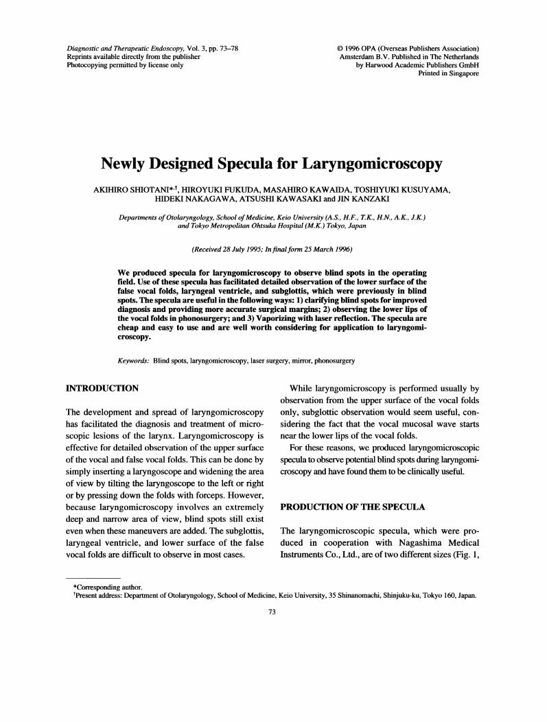
FIGURE The newly designed specula (upper): the larger round speculum has a diameter of 8 mm, and the smaller round one has a diam-eter of 6 mm. The cutoff types are for patients with narrow glottis. They can be used within a direct laryngoscope attached to a scalpel holder.The speculum holder (lower): the speculum can be fixed in any desired position.
SPECULA FOR LARYNGOMICROSCOPY 75
upper). The larger one is 8 mm in diameter, and thesmaller one is 6 mm in diameter. Both can be usedinside a direct laryngoscope. The mirrored surface is
chromium-plated with an aluminum material and hasa slender black brass-plated handle 0.7 mm in diame-
ter and 32 cm in length. The angle between the longaxis of the handle and the mirror is 50 In addition,for patients with narrow glottis, we produced a specu-lum with one-third of the mirror on the left side cut
off, and another with the right side cut off. In practice,use with a direct laryngoscope is made easier byattaching the speculum to a scalpel holder for laryn-gomicroscopy and bending the handle to an appropri-ate angle.
Further, we designed a speculum holder so that the
speculum can be fixed without using the hands (Fig. 1,lower). The holder is screwed to an operating table,and the handle of the speculum is attached to theholder, which can be bent freely to the desired posi-tion. With this device, other procedures can be per-formed while looking at the speculum fixed inside thedirect laryngoscope.
CLINICAL APPLICATIONS
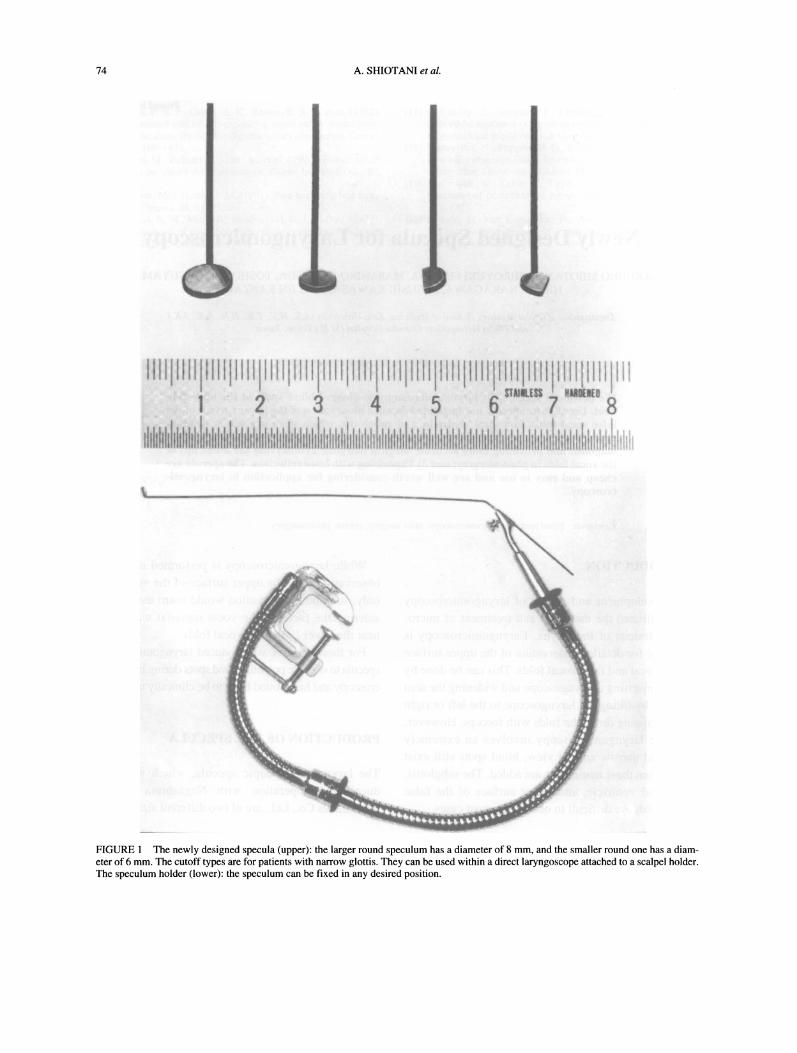
Observation of Blind Spots: Case I: Right GlotticTumor
The tumor appeared to be localized to the glottis fromthe normal field of view. Observation of the laryngealventricle with the speculum revealed tumorous
changes of the mucous membrane deep in the laryn-geal ventricle confirming tumor infiltration (Fig. 2).The diagnosis in this patient was T2 right glottic squa-mous cell carcinoma.
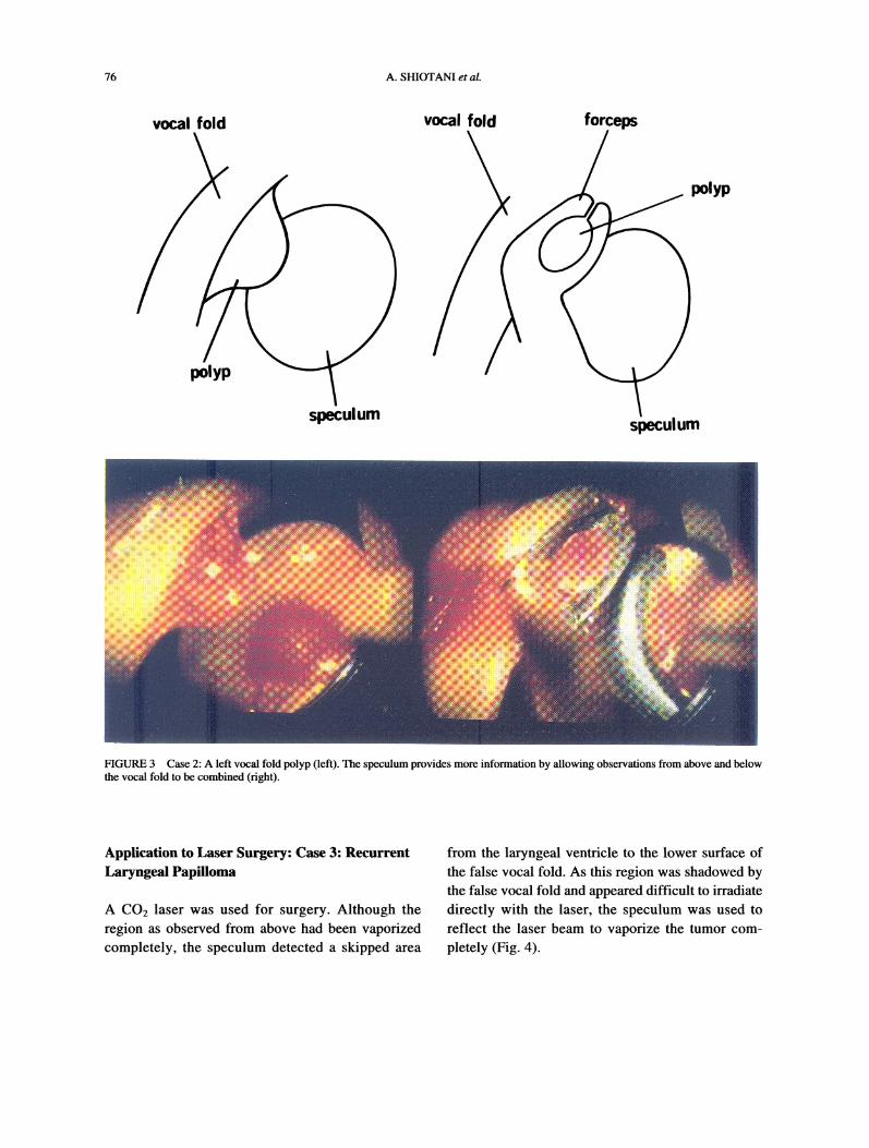
Application in Phonosurgery: Case 2: Left LocalFold Polyp
The speculum was fixed at the lower side of the vocalfold using the holder, and the polyp was resected (Fig.3). Use of the speculum provided detailed observationof the area near the lower lip and facilitated surgerywith combined observations from above and belowthe vocal fold, providing more information.
FIGURE 2 Case 1: a right glottic tumor (left). Observation of the laryngeal ventricle with the speculum reveals tumor infiltration into theventricle (right).
76 A. SHIOTANI et al.
vocal fold vocal fold
polyp
speculum
forceps
polyp
speculum
FIGURE 3 Case 2: A left vocal fold polyp (left). The speculum provides more information by allowing observations from above and belowthe vocal fold to be combined (right).
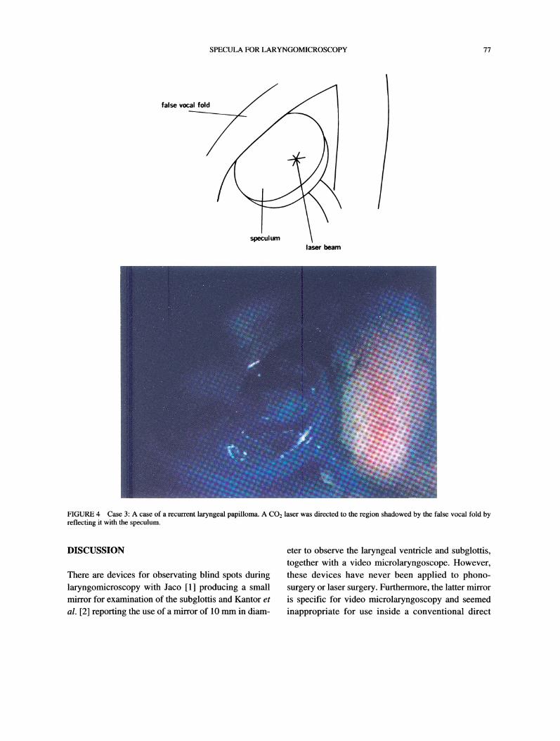
Application to Laser Surgery: Case 3: RecurrentLaryngeal Papilloma
A CO2 laser was used for surgery. Although theregion as observed from above had been vaporizedcompletely, the speculum detected a skipped area
from the laryngeal ventricle to the lower surface ofthe false vocal fold. As this region was shadowed bythe false vocal fold and appeared difficult to irradiate
directly with the laser, the speculum was used to
reflect the laser beam to vaporize the tumor com-
pletely (Fig. 4).
SPECULA FOR LARYNGOMICROSCOPY 77
false vocal fol
speculumlaser beam
FIGURE 4 Case 3: A case of a recurrent laryngeal papilloma. A CO2 laser was directed to the region shadowed by the false vocal fold byreflecting it with the speculum.
DISCUSSION
There are devices for observating blind spots duringlaryngomicroscopy with Jaco [1] producing a smallmirror for examination of the subglottis and Kantor et
al. [2] reporting the use of a mirror of 10 mm in diam-
eter to observe the laryngeal ventricle and subglottis,together with a video microlaryngoscope. However,these devices have never been applied to phono-surgery or laser surgery. Furthermore, the latter mirroris specific for video microlaryngoscopy and seemedinappropriate for use inside a conventional direct
78 A. SHIOTANI et al.
laryngoscope due to its size. While laser reflectors thatcan be used inside an ordinary direct laryngoscopehave been marketed by Storz, it is not certain whetherthey are useful for the observation of blind spots or
phonosurgery.Hence, we have produced our own specula on the
basis of the concepts stated previously and found themuseful in the following clinical applications.
1. Blind spots including the lower surface of thevocal folds, laryngeal ventricle, and subglottis can beobserved easily. The specula are useful for the accu-
rate assessment of the extent of tumor infiltration.
They also provide an accurate estimation of the earlystages of ventricular and subglottic cancers. In addi-
tion, the surgical margin can be confirmed to assure
complete resection.2. In phonosurgery, pathologic regions can be
observed in detail from the subglottis. This permitsdetailed observation of the region near the lowerlips, that is the area where the mucosal wave starts,ensuring a satisfactory surgical result. Thus, a com-bination of usual observation from above and specu-lar observation from the subglottis provides moreinformation and facilitates the performance ofsurgery that allows full appreciation of mucosalwave function to be made. This contributes to theperformance of an ideal resection with a minimumof intervention.
3. Laser irradiation of inaccessible areas underdirect view is possible using reflection.As another method for observing blind spots in
laryngomicroscopy, a rigid laryngotelescope can beused inside a direct laryngoscope. A rigid laryngotele-scope ought to be able to be used in the same way as a
speculum to observe details of the lower surface of thefalse vocal folds, laryngeal ventricle, and subglottis;however, specular observation has advantages interms of price and simplicity of use, in that treatment
can be performed simultaneously.
Acknowledgments
This paper was presented at the 6th Annual Meeting ofthe Laryngological Society of Japan, Saga, March 11,1994; at the 95th Annual Meeting of the Otolaryn-gological Society of Japan, Niigita, May 19, 1994; at
the 3rd International Symposium on Phonosurgery,Kyoto, June 27, 1994; and at the video symposium of1 lth Congress of Pan-Pacific Surgical Association
Japan Chapter, Okinawa, November 24, 1994.
ReferencesJako, G. L. (1970). Laryngoscopy for microscopic observation,surgery, and photography, Arch Otolaryngol, 91, 196-199.
[2] Kantor, E. A., Berci, G., Partlow, E., et al. (1991). Ancillaryinstruments for the video microlaryngoscope, Ann Otol RhinolLaryngol, 100, 317-919.

Submit your manuscripts athttp://www.hindawi.com
Stem CellsInternational
Hindawi Publishing Corporationhttp://www.hindawi.com Volume 2014
Hindawi Publishing Corporationhttp://www.hindawi.com Volume 2014
MEDIATORSINFLAMMATION
of
Hindawi Publishing Corporationhttp://www.hindawi.com Volume 2014
Behavioural Neurology
EndocrinologyInternational Journal of
Hindawi Publishing Corporationhttp://www.hindawi.com Volume 2014
Hindawi Publishing Corporationhttp://www.hindawi.com Volume 2014
Disease Markers
Hindawi Publishing Corporationhttp://www.hindawi.com Volume 2014
BioMed Research International
OncologyJournal of
Hindawi Publishing Corporationhttp://www.hindawi.com Volume 2014
Hindawi Publishing Corporationhttp://www.hindawi.com Volume 2014
Oxidative Medicine and Cellular Longevity
Hindawi Publishing Corporationhttp://www.hindawi.com Volume 2014
PPAR Research
The Scientific World JournalHindawi Publishing Corporation http://www.hindawi.com Volume 2014
Immunology ResearchHindawi Publishing Corporationhttp://www.hindawi.com Volume 2014
Journal of
ObesityJournal of
Hindawi Publishing Corporationhttp://www.hindawi.com Volume 2014
Hindawi Publishing Corporationhttp://www.hindawi.com Volume 2014
Computational and Mathematical Methods in Medicine
OphthalmologyJournal of
Hindawi Publishing Corporationhttp://www.hindawi.com Volume 2014
Diabetes ResearchJournal of
Hindawi Publishing Corporationhttp://www.hindawi.com Volume 2014
Hindawi Publishing Corporationhttp://www.hindawi.com Volume 2014
Research and TreatmentAIDS
Hindawi Publishing Corporationhttp://www.hindawi.com Volume 2014
Gastroenterology Research and Practice
Hindawi Publishing Corporationhttp://www.hindawi.com Volume 2014
Parkinson’s Disease
Evidence-Based Complementary and Alternative Medicine
Volume 2014Hindawi Publishing Corporationhttp://www.hindawi.com