Embed Size (px)
Citation preview
The Nf2 tumor suppressor gene product IS essential for extraembryon.lc development immediately prior to gastrulation Andrea I. McClatchey, 1 Ichiko Saotome, 1"2 Vijaya Ramesh, s James F. Gnsella, s and Tyler Jacks 1'2'4
1Center for Cancer Research, 2Howard Hughes Medical Institute, Massachusetts Institute of Technology, Cambridge, Massachusetts 02139 USA; SMolecular Neurogenetics Unit, Massachusetts General Hospital and Harvard Medical School, Charlestown, Massachusetts 02129 USA
The neurofibromatosis type II (NF2) tumor suppressor encodes a putative cytoskeletal associated protein, the loss of which leads to the development of Schwann cell tumors associated with NF2 in humans. The NF2 protein merlin belongs to the band 4.1 family of proteins that link membrane proteins to the cytoskeleton and are thought to be involved in dynamic cytoskeletal reorganization. Beyond its membership in this family, however, the function of merlin remains poorly understood. In order to analyze the function of merlin during embryogenesis and to develop a system to study merlin function in detail, we have disrupted the mouse Nf2 gene by homologous recombination in embryonic stem cells. Most embryos homozygous for a mutation at the Nf2 locus fail between embryonic days 6.5 and 7.0, exhibiting a collapsed extraembryonic region and the absence of organized extraembryonic ectoderm. The embryo proper continues to develop, but fails to initiate gastrulation. These observations are supported by the expression patterns of markers of the extraembryonic lineage and the lack of expression of mesodermal markers in the mutant embryos. Mosaic studies demonstrate that merlin function is not required cell autonomously in mesoderm, and support the proposition that merlin function is essential for the development of extraembryonic structures during early mouse development.
[Key Words: Merlin; neurofibromatosis type II; tumor suppressor gene; extraembryonic ectoderm; gastrulation; ERM]
Received February 7, 1997; revised version accepted April 8, 1997.
The study of the molecular changes underlying the de- velopment of human cancer has largely focused on per- turbations in normal cellular pathways that converge on the nucleus of the cell. By contrast, very little is under- stood about the interface between the proliferative state of the cell and the cytoskeleton, which must reorganize during the processes of cell division and differentiation, as well as during the transformation and invasion steps of tumorigenesis. In this context, we have focused on the neurofibromatosis type II (NF2) tumor suppressor gene, which encodes a putative cytoskeletal-associated pro- tein. NF2 is a hereditary disorder featuring the develop- ment of benign nervous system tumors including schwannomas, meningiomas, and ependymomas (Hu- son 1994). The hallmark of NF2 is the development of Schwann cell tumors on both eighth cranial (auditory) nerves. Mutations and associated loss of heterozygosity (LOH) have been detected at the NF2 locus in tumors from NF2 patients and in sporadically occurring tumors
4Corresponding author. E-MAIL [email protected]; FAX (617) 253-9863.
of the same types, indicating that NF2 behaves as a clas- sic tumor suppressor gene (for review, see Gusella et al. 1996). In addition to the benign tumors that develop in NF2 patients, mutations in the NF2 gene have been iden- tified in malignant mesotheliomas of the lung (Sekido et al. 1995).
Through positional cloning and LOH studies, the NF2 gene was cloned and its protein product identified by homology as a member of the band 4.1 family of pro- teins, some of which have been demonstrated to link transmembrane proteins to cytoskeletal or cytoskeletal- associated proteins (Seizinger et al. 1986, 1987a,b; Rou- leau et al. 1993; Trofatter et al. 1993). The NF2 protein is most closely related to a subset of this family that con- tains three other members to date: ezrin, radixin, and moesin (the ERM family), and has therefore been dubbed merlin (moesin, ezrin, radixin like protein). ERM family members are localized to cortical actin structures, par- ticularly in areas undergoing active reorganization such as membrane ruffles, neuronal growth cones, or the cleavage furrow of dividing cells, where they are simi- larly thought to link membrane proteins to the cytoskel-
GENES & DEVELOPMENT 11:1253-1265 © 1997 by Cold Spring Harbor Laboratory Press ISSN 0890-9369/97 $5.00 1253
Cold Spring Harbor Laboratory Press on April 30, 2019 - Published by genesdev.cshlp.orgDownloaded from
McClatchey et al.
eton (Arpin et al. 1994). Moesin and ezrin have been reported to bind directly to actin (Turunen et al. 1994; Pestonjamasp et al. 1995), and all three proteins can bind to CD44, a transmembrane receptor for hyaluronic acid in the extracellular matrix (ECM) (Tsukita et al. 1994). More recently, it has been demonstrated that ezrin func- tion is required for aggregation of the cell adhesion mol- ecule ICAM-2 and formation of cell extensions on target cells that are recognized by natural killer (NK) cells dur- ing immune clearance, suggesting a role for ERM pro- teins in cell adhesion (Helander et al. 1996).
Although nothing is known about any protein inter- actions involving merlin, initial studies suggest that both exogenous and endogenous merlin can localize to cortical actin structures, particularly membrane ruffles, microvilli, and the cleavage furrow (den Bakker et al. 1995; Gonzalez-Agosti et al. 1996; R.J. Shaw, A.I. Mc- Clatchey, and T. Jacks, in prep.). Therefore, merlin rep- resents a novel type of tumor suppressor and provides the opportunity to investigate the mechanism by which a protein thought to be involved in cytoskeletal reorga- nization also functions to regulate cell growth and sup- press tumor formation.
To understand how loss of merlin function contributes to tumorigenesis and the development of NF2 in hu- mans, it is important to gain an understanding of the normal function of merlin in various cellular contexts. It is becoming increasingly apparent that the normal func- tions of tumor suppressor proteins are often manifest during development, as revealed by gene targeting ex- periments in mice (for review, see Jacks 1996). The study of a tumor suppressor's function during development provides a framework for understanding how loss of that function in the adult contributes to tumorigenesis. Fur- thermore, data from gene targeting experiments often contribute valuable information toward understanding specific developmental processes. In an effort to investi- gate merlin function in detail, we have generated a tar- geted mutation at the mouse Nf2 locus. We have deter- mined that a homozygous mutation at the Nf2 locus leads to early embryonic failure at embryonic day (E)6.5- 7.0, with embryos displaying poorly developed extraem- bryonic structures and a lack of organized extraembry- onic ectoderm. These observations are supported by the expression patterns of pem-1 protein and L14 lectin mRNA, markers of the extraembryonic lineage, in the mutant embryos. Despite this defect, the embryonic por- tion of the mutant embryo continues to develop, but the embryo does not gastrulate as evidenced both morpho- logically and by the lack of expression of the mesodermal markers brachyury or HNF3[3. In order to investigate this defect further, we developed Nf2 homozygous mu- tant embryonic stem (ES) cells that also carry a ubiqui- tously expressed IacZ transgene and generated chimeric embryos that are composed of both wild-type cells and marked merlin-deficient cells. Using this system, we found that Nf2-deficient cells can become mesoderm, indicating that merlin is not required cell autonomously for mesoderm formation. Furthermore, chimeras receiv- ing high levels of Nf2-deficient contribution to all three
germ layers in the embryo proper gastrulate properly, strongly suggesting that the primary defect in Nf2-defi- cient embryos resides in the extraembryonic lineage.
Results
Targeting strategy
Using a probe corresponding to the entire human NF2 cDNA, we screened a genomic library constructed from the 129/SvJ strain of mice (Stratagene). One isolated clone contained a 18-kb insert corresponding to the ge- nomic region spanning exons 2-5 of the mouse Nf2 gene. This clone was used subsequently to construct the tar- geting vector depicted in Figure 1A. A number of muta- tions, both in the germ line of NF2 patients and in spo- radically occurring schwannomas and meningiomas in humans, have been found in the region of exons 2 and 3, including splice site mutations or deletions that would cause the removal of exon 2 or 3 or both from the NF2 mRNA (for review, see Gusella et al. 1996). We chose to target this region of the mouse Nf2 gene, replacing the 3' half of exon 2, all of exon 3 and intervening intronic sequences with the neomycin resistance gene (neo r) ori- ented in a transcriptional direction opposite to that of Nf2 (Fig. la).
This targeting construct was electroporated into 129/ Sv D3 ES cells (Gossler et al. 1986), which were then subjected to both G418 and gancyclovir selection. A to- tal of 231 clones surviving both types of selection were screened by Southern blotting using probes p5' and p3' (Fig. la,b). Four separate clones gave the correct pattern of bands with both probes, for a final targeting frequency of 4 of 231, or 1.7%. One of these clones was also found to have a second copy of the targeting sequences inserted elsewhere within the genome, and was not used further. The low targeting frequency at this locus may in part reflect a very high concentration of repetitive elements present throughout this region (see Materials and Meth- ods). Furthermore, we identified an intronic polymor- phism between the targeting vector (generated from 129/ SvJ genomic DNA) and the 129/Sv D3 ES cells (see Ma- terials and Methods). It has been observed previously that even subtle differences between input and endog- enous sequences can dramatically affect targeting fre- quency, prompting the use of isogenic DNA in con- structing targeting vectors (re Riele et al. 1992).
In order to characterize the molecular effects of the targeted mutation, we first determined whether mRNA was transcribed from the mutant allele. Northern blot analysis of total RNA isolated from +/+, +/-, and - / - ES cells (see below for derivation of - / - ES cells) revealed the presence of some Nf2 mRNA of approximately the normal size (-4.5 kb) in - / - cells, although at lower lev- els than that seen in +/+ or +/- mutant cells (data not shown). A probe derived from the neo ~ coding region de- tected only the 1.2-kb species expressed from the PGK- neo cassette (see Fig. 1; data not shown), indicating that a composite Nf2-neo mRNA was not generated as has been described for other targeted mutations (e.g., see
1254 GENES & DEVELOPMENT
Cold Spring Harbor Laboratory Press on April 30, 2019 - Published by genesdev.cshlp.orgDownloaded from
Role of Nf2 tumor suppressor in mouse development
a . . . .
w t
X X
I.,-^s.I , X *
NF2 KO vector
w t
I m u t x •
pS' m
r , u , - - - ; / s
i I - t - o o . J ~6d s
a b / n
P +
a c
, a ._o.u I S =
P
p3 ' m
I I
s
4.3 kb
I S - - 3 . 9 k b - -
4.3 kb--
3.9 141)--
÷ ÷ + , ÷ ÷ ÷ ÷ ÷ ÷
÷ ÷ ÷ ÷ ÷ ÷ ÷ ÷ ÷ ÷
1 . . . .
s
i s
, [ . . -
s
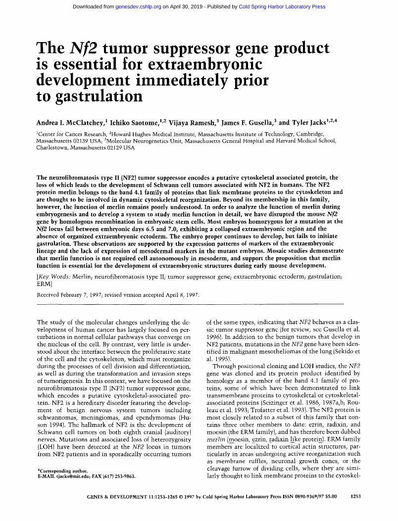
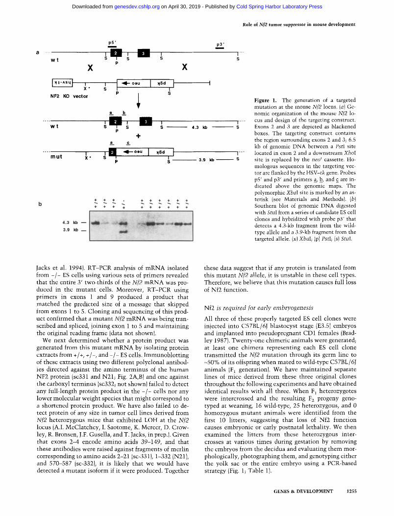
F igure 1. The generation of a targeted mutation at the mouse Nf2 locus. (a) Ge- nomic organization of the mouse Nf2 lo- cus and design of the targeting construct. Exons 2 and 3 are depicted as blackened boxes. The targeting construct contains the region surrounding exons 2 and 3; 6.5 kb of genomic DNA between a PstI site located in exon 2 and a downstream XhoI site is replaced by the neo r cassette. Ho- mologous sequences in the targeting vec- tor are flanked by the HSV-tk gene. Probes p5' and p3' and primers a, b, and c are in- dicated above the genomic maps. The polymorphic XbaI site is marked by an as- terisk (see Materials and Methods). (b) Southern blot of genomic DNA digested with StuI from a series of candidate ES cell clones and hybridized with probe p3' that detects a 4.3-kb fragment from the wild- type allele and a 3.9-kb fragment from the targeted allele. (x)XbaI; (p) PstI; (s) StuI.
Jacks et al. 1994). RT-PCR analysis of m R N A isolated from - / - ES cells using various sets of primers revealed that the entire 3' two-thirds of the Nf2 m R N A was pro- duced in the mutan t cells. Moreover, RT-PCR using p r i m e r s in exons 1 and 9 produced a product that matched the predicted size of a message that skipped from exons 1 to 5. Cloning and sequencing of this prod- uct confirmed that a mutan t Nf2 m R N A was being tran- scribed and spliced, joining exon 1 to 5 and main ta in ing the original reading frame (data not shown).
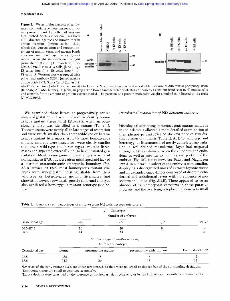
We next determined whether a protein product was generated from this mutan t m R N A by isolating protein extracts from +/+, +/-, and - / - ES cells. Immunoblot t ing of these extracts using two different polyclonal antibod- ies directed against the amino terminus of the h u m a n NF2 protein (sc331 and N21; Fig. 2A, B) and one against the carboxyl terminus (sc332; not shown) failed to detect any full-length protein product in the / cells nor any lower molecular weight species that might correspond to a shortened protein product. We have also failed to de- tect protein of any size in tumor cell lines derived from Nf2 heterozygous mice that exhibited LOH at the Nf2 locus (A.I. McClatchey, I. Saotome, K. Mercer, D. Crow- ley, R. Bronsen, J.F. Gusella, and T. Jacks, in prep.). Given that exons 2-4 encode amino acids 39-149, and that these antibodies were raised against fragments of mer l in corresponding to amino acids 2-21 (sc-331 ), 1-332 (N21), and 570-587 (sc-332), it is l ikely that we would have detected a mutan t isoform if it were produced. Together
these data suggest that if any protein is translated from this mutan t Nf2 allele, it is unstable in these cell types. Therefore, we believe that this muta t ion causes full loss of Nf2 function.
Nf2 is required for early embryogenesis
All three of these properly targeted ES cell clones were injected into C57BL/6J blastocyst stage (E3.5) embryos and implanted into pseudopregnant CD1 females (Brad- ley 1987). Twenty-one chimeric animals were generated; at least one chimera representing each ES cell clone t ransmit ted the Nf2 mutat ion through its germ line to -50% of its offspring when mated to wild-type C57BL/6J animals (F 1 generation). We have mainta ined separate lines of mice derived from these three original clones throughout the following experiments and have obtained identical results wi th all three. When F1 heterozygotes were intercrossed and the resulting F2 progeny geno- typed at weaning, 16 wild-type, 25 heterozygous, and 0 homozygous mutan t animals were identified from the first 10 litters, suggesting that loss of Nf2 function causes embryonic or early postnatal lethality. We then examined the litters from these heterozygous inter- crosses at various t imes during gestation by removing the embryos from the decidua and evaluating them mor- phologically, photographing them, and genotyping either the yolk sac or the entire embryo using a PCR-based strategy (Fig. 1; Table 1).
GENES & DEVELOPMENT 1255
Cold Spring Harbor Laboratory Press on April 30, 2019 - Published by genesdev.cshlp.orgDownloaded from
McClatchey et al.
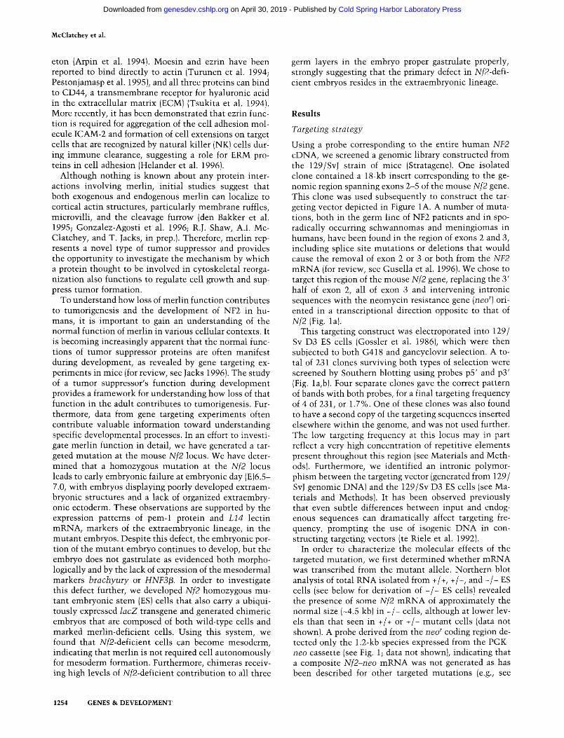
Figure 2. Western blot analysis of cell ly- sates from wild-type, heterozygous, or ho- mozygous mutant ES cells. (A) Western blot probed with monoclonal antibody N21, directed against the human merlin amino terminus (amino acids 1-332), which also detects ezrin and moesin. Po- sitions of merlin, ezrin, and moesin bands are shown on the left, and the positions of molecular weight standards on the right (Amersham). (Lane I) Human fetal fibro- blasts; (lane 2) NIH-3T3 cells; (lane 3) +/+ ES cells; (lane 4) +/- ES cells; (lane 5) - / - ES cells. (B) Western blot was probed with polyclonal antibody SC331 (raised against amino acids 2-21; Santa Cruz). (Lanes 1,2)
ezrin moesin merlin
4- N
97
B
4-
merlin I 70
+/+ ES cells; (lane 3) +/- ES cells; (lane 4) - / - ES cells. Merlin is often detected as a doublet because of differential phosphorylation (R. Shaw, A.I. McClatchey, T. Jacks, in prep.). The lower band detected with this antibody is a constant band seen in all mouse cells and controls for the amount of protein extract loaded. The position of a protein molecular weight standard is indicated to the right (GIBCO BRL).
We examined these litters at progressively earlier stages of gestation and were not able to identify homo- zygous mutan t tissue unti l E8.0-E8.5, when an occa- sional embryo was identified as a mutan t (Table 1). These mutan t s were nearly all in late stages of resorption and were much smaller than their wild-type or hetero- zygous mutan t l i t termates. At E7.5 most homozygous mutan t embryos were intact, but were clearly smaller than their wild-type and heterozygous mutan t litter- mates and appeared externally not to have initiated gas- trulation. Most homozygous mutan t embryos were of normal size at E7.0, but were often misshaped and lacked a distinct extraembryonic-embryonic boundary (Fig. 3A, B, arrow). At E6.5, most homozygous mutan t em- bryos were superficially indistinguishable from their wild-type or heterozygous mutan t l i t termates (not shown); however, a few small, grossly abnormal embryos also exhibited a homozygous mutan t genotype (see be- low).
Histological evaluatiorl of Nf2-deficient embryos
Histological sectioning of homozygous mutan t embryos in their decidua allowed a more detailed examinat ion of their phenotype and revealed the existence of two dis- tinct classes of mutan ts (Table 1). At E7.5, wild-type and heterozygous l i t termates had nearly completed gastrula- tion; a well-defined mesodermal layer had migrated throughout the embryo between the ectoderm and endo- derm as well as into the extraembryonic portion of the embryo (Fig. 3C; for review, see Faust and Magnuson 1993). In contrast, a subset of the embryos were smaller, displaying a disorganized mass of extraembryonic tissue and an expanded egg cylinder composed of discrete ecto- dermal and endodermal layers wi th no evidence of me- soderm induction (Fig. 3D,E). There appeared to be an absence of extraembryonic ectoderm in these putative mutants , and the overlying ectoplacental cone was small
Table 1. Genotypes and phenotypes of embryos from Nf2 heterozygous intercrosses
A. Genotypes Number of embryos
Gestational age +/+ +/- _/_a N.D.b
E6.5-E7.5 10 20 10 5 E8.5 14 23 5 5
B. Phenotypes (paraffin sections) Number of embryos
Gestational age normal presumptive mutant presumptive early mutant Empty deciduum c
E6.5 38 5 6 2 E7.5 118 29 13 13
aEmbryos of the early mutant class are under-represented, as they were too small to dissect free of the surrounding deciduum. bEmbryonic tissue too small to genotype accurately. °Empty decidua were classified by the presence of trophoblast giant cells only or by the lack of any discernible embryonic cells.
1256 GENES & DEVELOPMENT
Cold Spring Harbor Laboratory Press on April 30, 2019 - Published by genesdev.cshlp.orgDownloaded from
Role of Nf2 tumor suppressor in mouse development
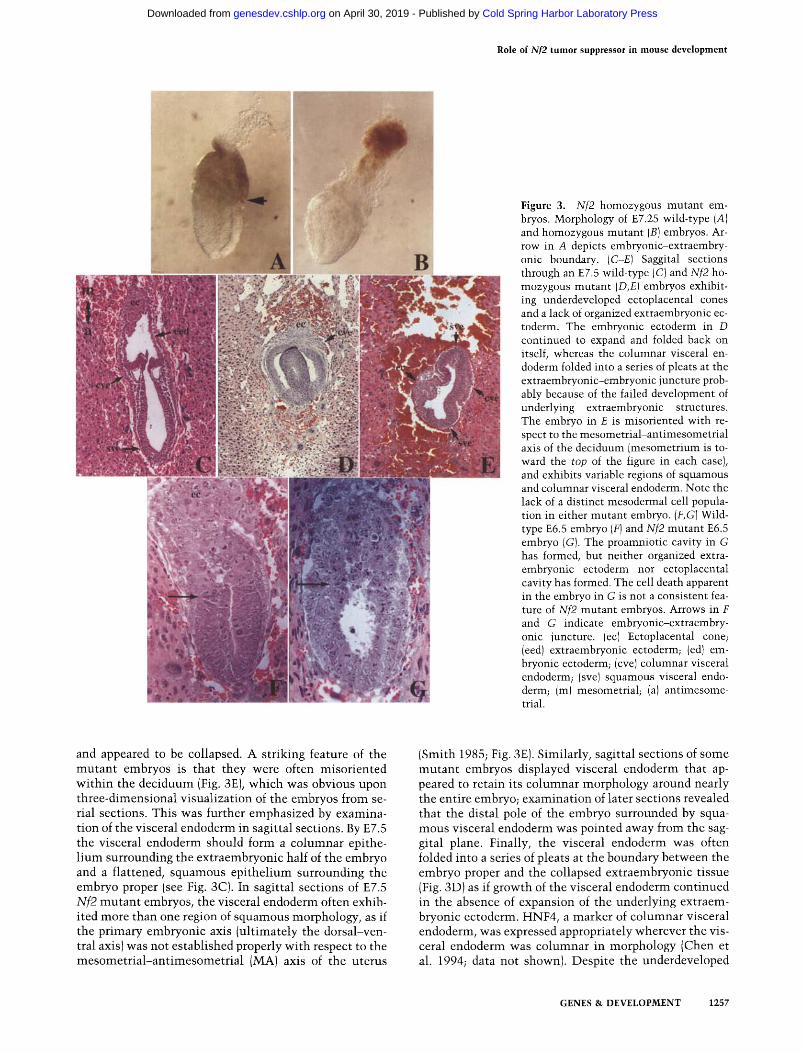
F igure 3. Nf2 homozygous mutant em- bryos. Morphology of E7.25 wild-type (A) and homozygous mutant (B) embryos. Ar- row in A depicts embryonic-extraembry- onic boundary. (C-E) Saggital sections through an E7.5 wild-type (C) and Nf2 ho- mozygous mutant (D,E) embryos exhibit- ing underdeveloped ectoplacental cones and a lack of organized extraembryonic ec- toderm. The embryonic ectoderm in D continued to expand and folded back on itself, whereas the columnar visceral en- doderm folded into a series of pleats at the extraembryonic-embryonic juncture prob- ably because of the failed development of underlying extraembryonic structures. The embryo in E is misoriented with re- spect to the mesometrial-antimesometrial axis of the deciduum (mesometrium is to- ward the top of the figure in each case), and exhibits variable regions of squamous and columnar visceral endoderm. Note the lack of a distinct mesodermal cell popula- tion in either mutant embryo. (F,G)Wild- type E6.5 embryo (F) and Nf2 mutant E6.5 embryo (G). The proamniotic cavity in G has formed, but neither organized extra- embryonic ectoderm nor ectoplacental cavity has formed. The cell death apparent in the embryo in G is not a consistent fea- ture of Nf2 mutant embryos. Arrows in F and G indicate embryonic-extraembry- onic juncture. (ec) Ectoplacental cone; (eed) extraembryonic ectoderm; (ed) em- bryonic ectoderm; (cve) columnar visceral endoderm; (sve) squamous visceral endo- derm; (m)mesometrial; (a) antimesome- trial.
and appeared to be collapsed. A striking feature of the mutan t embryos is that they were often misoriented wi th in the deciduum (Fig. 3E), which was obvious upon three-dimensional visualization of the embryos from se- rial sections. This was further emphasized by examina- tion of the visceral endoderm in sagittal sections. By E7.5 the visceral endoderm should form a columnar epithe- l ium surrounding the extraembryonic half of the embryo and a flattened, squamous epi thel ium surrounding the embryo proper (see Fig. 3C). In sagittal sections of E7.5 Nf2 mutan t embryos, the visceral endoderm often exhib- ited more than one region of squamous morphology, as if the primary embryonic axis (ult imately the dorsal-ven- tral axis) was not established properly wi th respect to the mesometr ia l -an t imesometr ia l (MA) axis of the uterus
(Smith 1985; Fig. 3E). Similarly, sagittal sections of some mutant embryos displayed visceral endoderm that ap- peared to retain its columnar morphology around nearly the entire embryo; examinat ion of later sections revealed that the distal pole of the embryo surrounded by squa- mous visceral endoderm was pointed away from the sag- gital plane. Finally, the visceral endoderm was often folded into a series of pleats at the boundary between the embryo proper and the collapsed extraembryonic tissue (Fig. 3D) as if growth of the visceral endoderm continued in the absence of expansion of the underlying extraem- bryonic ectoderm. HNF4, a marker of columnar visceral endoderm, was expressed appropriately wherever the vis- ceral endoderm was columnar in morphology (Chen et al. 1994; data not shown). Despite the underdeveloped
GENES & DEVELOPMENT 1257
Cold Spring Harbor Laboratory Press on April 30, 2019 - Published by genesdev.cshlp.orgDownloaded from
McClatchey et al.
extraembryonic lineage in the mutan t embryos, the em- bryonic half of the embryo continued to expand, often folding back upon itself, and somet imes displaying a thickening on one side reminiscent of pr imit ive streak formation (Fig. 3D; data not shown). At E6.5, when ex- traembryonic ectoderm is normal ly forming, mutan t embryos were the same size as their wild-type and het- erozygous li t termates, but exhibited underdeveloped ex- traembryonic structures. For example, the homozygous mutan t embryo shown in Figures 3G and 5B (below) which was genotyped by in situ hybridizat ion (see be- low), is dist inguishable histologically by the lack of or- ganized extraembryonic ectoderm.
Only 18% of the embryos at E7.5 were represented by this class of mutants , instead of the expected 25%, im- plying that some mutants were failing at an even earlier stage of development. In addition to vir tual ly empty de- cidua containing only giant cells (-7%; Table 1), which are found with s imilar frequency in control litters, we detected a number of decidua containing only a small mass of disorganized embryonic cells surrounded by tro- phoblast giant cells, suggesting that there might be a second class of homozygous mutan t embryos that failed shortly after implanta t ion (Fig. 4D,E). Interestingly, both types of mutants were identified on inbred 129/Sv and highly enriched C57BL/6J (5 backcross generations; gen- erations 3 and 4 were screened for enriched C57BL/6J
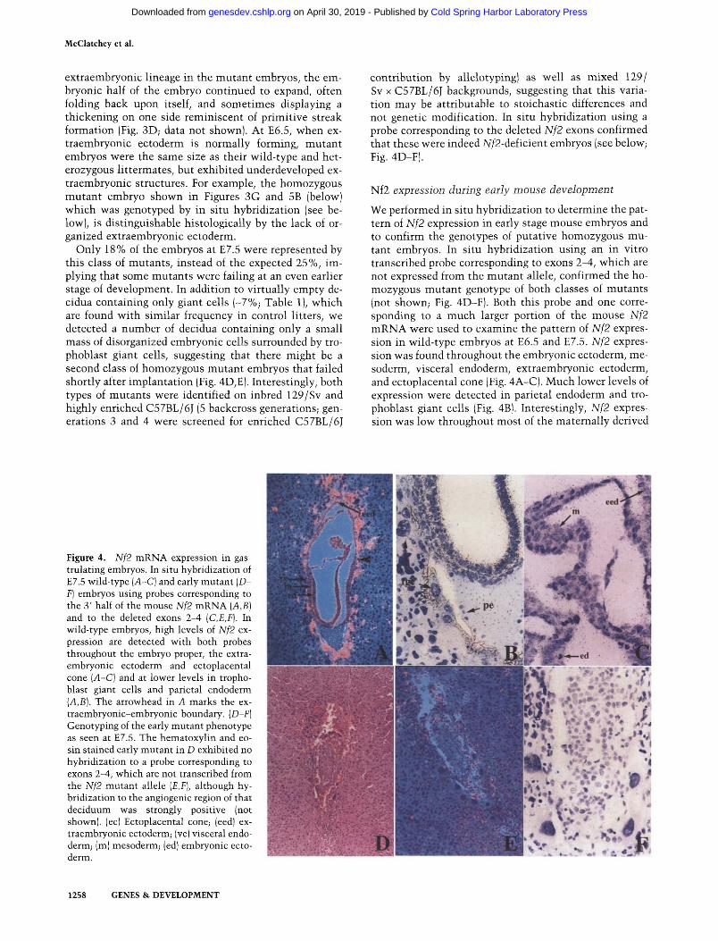
contribution by allelotypingl as well as mixed 129/ Sv x C57BL/6J backgrounds, suggesting that this varia- tion may be attributable to stoichastic differences and not genetic modification. In situ hybridization using a probe corresponding to the deleted Nf2 exons confirmed that these were indeed Nf2-deficient embryos (see below; Fig. 4D-F).
Nf2 expression during early mouse development
We performed in situ hybridization to determine the pat- tern of Nf2 expression in early stage mouse embryos and to confirm the genotypes of putative homozygous mu- tant embryos. In situ hybridizat ion using an in vitro transcribed probe corresponding to exons 2-4, which are not expressed from the mutan t allele, confirmed the ho- mozygous mutan t genotype of both classes of mutants (not shown; Fig. 4D-F). Both this probe and one corre- sponding to a much larger portion of the mouse Nf2 m R N A were used to examine the pattern of Nf2 expres- sion in wild-type embryos at E6.5 and E7.5. Nf2 expres- sion was found throughout the embryonic ectoderm, me- soderm, visceral endoderm, extraembryonic ectoderm, and ectoplacental cone (Fig. 4A-C). Much lower levels of expression were detected in parietal endoderm and tro- phoblast giant cells (Fig. 4B1. Interestingly, Nf2 expres- sion was low throughout most of the maternal ly derived
Figure 4. Nf2 mRNA expression in gas- trulating embryos. In situ hybridization of E7.5 wild-type (A-C) and early mutant (D- F) embryos using probes corresponding to the 3' half of the mouse Nf2 mRNA (A,B} and to the deleted exons 2-4 (C,E,F). In wild-type embryos, high levels of Nf2 ex- pression are detected with both probes throughout the embryo proper, the extra- embryonic ectoderm and ectoplacental cone (A-C) and at lower levels in tropho- blast giant cells and parietal endoderm (A,B). The arrowhead in A marks the ex- traembryonic-embryonic boundary. (D-F) Genotyping of the early mutant phenotype as seen at E7.5. The hematoxylin and eo- sin stained early mutant in D exhibited no hybridization to a probe corresponding to exons 2-4, which are not transcribed from the Nf2 mutant allele (E,F), although hy- bridization to the angiogenic region of that deciduum was strongly positive (not shown). (ec) Ectoplacental cone; (eed) ex- traembryonic ectoderm; lye) visceral endo- derm; (m) mesoderm; (ed) embryonic ecto- derm.
1258 GENES & DEVELOPMENT
Cold Spring Harbor Laboratory Press on April 30, 2019 - Published by genesdev.cshlp.orgDownloaded from
Role of Nf2 tumor suppressor in mouse development
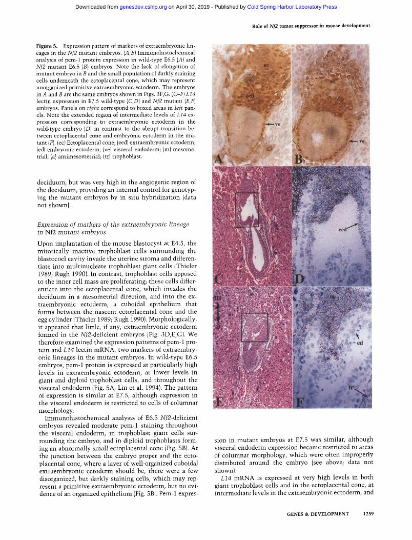
Figure 5. Expression pattern of markers of extraembryonic lin- eages in the Nf2 mutant embryos. (A,B) Immunohistochemical analysis of pem-1 protein expression in wild-type E6.5 (A) and Nf2 mutant E6.5 (B) embryos. Note the lack of elongation of mutant embryo in B and the small population of darkly staining cells underneath the ectoplacental cone, which may represent unorganized primitive extraembryonic ectoderm. The embryos in A and B are the same embryos shown in Figs. 3F, G. (C-F) L14 lectin expression in E7.5 wild-type (C,D) and Nf2 mutant (E,F) embryos. Panels on right correspond to boxed areas in left pan- els. Note the extended region of intermediate levels of L14 ex- pression corresponding to extraembryonic ectoderm in the wild-type embryo (D) in contrast to the abrupt transition be- tween ectoplacental cone and embryonic ectoderm in the mu- tant (F). (ec) Ectoplacental cone; (eed) extraembryonic ectoderm; (ed) embryonic ectoderm; (ve)visceral endoderm; (m)mesome- trial; (al antimesometrial; (tr) trophoblast.
deciduum, but was very high in the angiogenic region of the deciduum, providing an internal control for genotyp- ing the mutant embryos by in situ hybridization (data not shown).
Expression of markers of the extraembryonic lineage in Nf2 mutant embryos
Upon implantation of the mouse blastocyst at E4.5, the mitotically inactive trophoblast cells surrounding the blastocoel cavity invade the uterine stroma and differen- tiate into multinucleate trophoblast giant cells (Thieler 1989; Rugh 1990). In contrast, trophoblast cells apposed to the inner cell mass are proliferating; these cells differ- entiate into the ectoplacental cone, which invades the deciduum in a mesometrial direction, and into the ex- traembryonic ectoderm, a cuboidal epithelium that forms between the nascent ectoplacental cone and the egg cylinder (Thieler 1989; Rugh 1990). Morphologically, it appeared that little, if any, extraembryonic ectoderm formed in the Nf2-deficient embryos (Fig. 3D,E,G). We therefore examined the expression patterns of peru-1 pro- tein and L14 lectin mRNA, two markers of extraembry- onic lineages in the mutant embryos. In wild-type E6.5 embryos, pem-1 protein is expressed at particularly high levels in extraembryonic ectoderm, at lower levels in giant and diploid trophoblast cells, and throughout the visceral endoderm (Fig. 5A; Lin et al. 1994). The pattern of expression is similar at E7.5, although expression in the visceral endoderm is restricted to cells of columnar morphology.
Immunohistochemical analysis of E6.5 Nf2-deficient embryos revealed moderate peru-1 staining throughout the visceral endoderm, in trophoblast giant cells sur- rounding the embryo, and in diploid trophoblasts form- ing an abnormally small ectoplacental cone (Fig. 5B). At the junction between the embryo proper and the ecto- placental cone, where a layer of well-organized cuboidal extraembryonic ectoderm should be, there were a few disorganized, but darkly staining cells, which may rep- resent a primitive extraembryonic ectoderm, but no evi- dence of an organized epithelium (Fig. 5B). Pem-1 expres-
sion in mutant embryos at E7.5 was similar, although visceral endoderm expression became restricted to areas of columnar morphology, which were often improperly distributed around the embryo (see above; data not shown).
L14 mRNA is expressed at very high levels in both giant trophoblast cells and in the ectoplacental cone, at intermediate levels in the extraembryonic ectoderm, and
GENES & DEVELOPMENT 1259
Cold Spring Harbor Laboratory Press on April 30, 2019 - Published by genesdev.cshlp.orgDownloaded from
McClatchey et al.
is undetectable in the embryo proper (Poirier et al. 1992; Fig. 5D). The pattern of L14 expression in Nf2-mutant embryos similarly supported a lack of organized extra- embryonic ectoderm: The heavily staining ectoplacental cone was collapsed onto the embryonic ectoderm, which was completely devoid of expression (Fig. 5F).
Despite the malformation of extraembryonic struc- tures, the mutant embryo proper continued to develop, yet failed to form a distinct mesodermal cell layer. Therefore, it was important to determine whether primi- tive streak formation and mesoderm induction were ever taking place. The transcription factor brachyury is a well-established marker of primitive streak formation and mesoderm induction in the gastrulating embryo. brachyury expression is first detected in the earliest population of mesodermal cells that is induced and in the immediately adjacent primitive ectoderm (Wilkin- son et al. 1990). Therefore, we examined the pattern of brachyury expression in Nf2 mutant embryos and in ad- jacent normal littermates. In multiple sections of several mutant embryos, we were unable to detect any brachy- ury expression. In a single case, a mutant displayed a small patch of cells of ectodermal morphology that ex- pressed moderate levels of brachyury (not shown). This could represent ectopic expression or possibly evidence of early primitive streak formation. This result suggests that mesoderm induction fails to occur in most, if not all Nf2 mutant embryos; however, it is possible that meso- dermal cells are induced but are abnormal and unable to express all mesoderm markers. This notwithstanding, we also failed to detect expression of HNF3[3, a marker of later, more anterior mesoderm (Sasaki and Hogan 1993; data not shown).
Generation of N f 2 - / - lacZ+ ES cells
The lack of mesoderm induction in Nf2 mutant embryos could be attributable to a direct or indirect requirement for merlin in the production of the inductive signal itself or to a cell-autonomous defect in the ability of Nf2-de- ficient cells to become mesoderm. To distinguish be- tween these two possibilities, we generated Nf2 homo- zygous mutant ES cells that are marked and used them to create chimeric embryos in which celt lineages arising from the merlin-deficient cells could be followed.
By crossing our Nf2 mutation into the ROSA26 strain of mice, which carries a ubiquitously expressed lacZ transgene (Friedrich and Soriano 1991 ), we generated Nf2 heterozygous mice that also carried lacZ. We bred these mice to Nf2 heterozygotes and established ES cell lines from individual blastocysts (Robertson 1987; Materials and Methods), 50% of which were expected to be lacZ ÷ and 25% to be homozygous for the Nf2 mutation. Over- all, we were able to establish ES cell lines from 21% of isolated blastocysts from a hybrid C57BL6/J: 129Sv back- ground. We generated four ES cell lines that were homo- zygous for the Nf2 mutation (lines 1076.7, 945.3, 2600E, and 2640D), three of which carried the lacZ transgene (1076.7, 2600E, and 2640D).
Nf2 homozygous mutant contribution to gastrulating embryos
The merlin-deficient ES cells were used to examine the consequences of homozygous mutant contribution to various cell types in the gastrulating embryo. Mutant cells from line 1076.7 or 2600E were injected into wild- type C57BL/6J blastocysts, which were implanted into pseudopregnant females and allowed to develop in utero (Bradley 1987). Chimeric embryos were retrieved at E8.5 (allowing one day of delay for embryo manipulation). Embryos were fixed and stained with X-gal to identify cells derived from the Nf2-/-;lacZ + ES cells. After stain- ing, embryos that exhibited areas of blue color (indica- tive of contribution from the homozygous mutant cells) were photographed and sectioned.
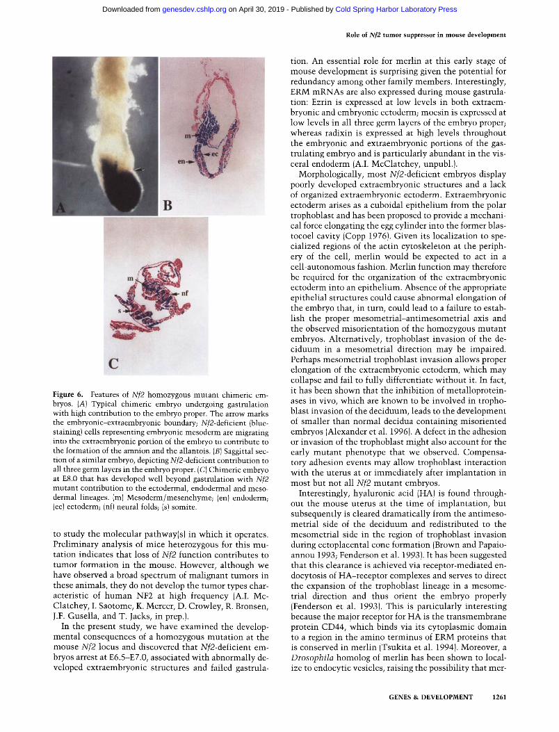
By visual inspection, we were able to identify appar- ently normal, gastrulating embryos that had received ex- tensive contribution from either homozygous mutant cell line, such as the embryo shown in whole mount in Figure 6A. The top half of the embryo with no mutant contribution represents the extraembryonic portion of the embryo, which is largely committed at the blastocyst stage; ES cells introduced into blastocysts contribute al- most exclusively to the embryo proper (Nagy et al. 1990). Blue-staining cells on the left side of the embryo extend- ing upward past the embryonic-extraembryonic bound- ary (arrow) are mesodermal cells that have migrated to contribute to the formation of the amnion and nascent allantois. A section through a similar chimeric embryo is shown in Figure 6B. Sectioning of E7.5-E8.5 chimeric embryos (>20 with varying degrees of chimerism) re- vealed that the homozygous mutant cells were able to contribute extensively to all three germ layers including the mesoderm. Figure 6C depicts a chimeric embryo at E8.0 that has successfully completed gastrulation: Blue- staining cells are prominent in the ectoderm, including the neural folds, in the somites, and extensively through- out the mesenchyme. These results indicate that merlin is not required cell autonomously for the formation of mesoderm. Moreover, they demonstrate that embryos can gastrulate normally with extensive contribution of Nf2 mutant cells to all three embryonic germ layers, consistent with the idea that the primary requirement for merlin in early embryogenesis resides in the extra- embryonic tissues.
Discussion
The identification of the product of the NF2 tumor sup- pressor gene as a member of a cytoskeletal-associated family of proteins affords the possibility of examining a link between the regulation of cytoskeletal reorganiza- tion and tumor development. However, the NF2 gene was identified by positional cloning, and the family to which its encoded protein belongs is poorly understood. Thus, there is currently little understanding of its mechanism of action as a tumor suppressor. We have used gene targeting in the mouse to investigate the nor- mal function of merlin and to generate tools with which
1260 GENES & DEVELOPMENT
Cold Spring Harbor Laboratory Press on April 30, 2019 - Published by genesdev.cshlp.orgDownloaded from
Role of Nf2 tumor suppressor in mouse development
m ~
C
Figure 6. Features of Nf2 homozygous mutant chimeric em- bryos. (A) Typical chimeric embryo undergoing gastrulation with high contribution to the embryo proper. The arrow marks the embryonic-extraembryonic boundary; Nf2-deficient (blue- staining) cells representing embryonic mesoderm are migrating into the extraembryonic portion of the embryo to contribute to the formation of the amnion and the allantois. (B) Saggittal sec- tion of a similar embryo, depicting Nf2-deficient contribution to all three germ layers in the embryo proper. (C) Chimeric embryo at E8.0 that has developed well beyond gastrulation with Nf2 mutant contribution to the ectodermal, endodermal and meso- dermal lineages. (m) Mesoderm/mesenchyme; (en) endoderm; (ec) ectoderm; (nf) neural folds; (s) somite.
to study the molecular pathway(s) in which it operates. Preliminary analysis of mice heterozygous for this mu- tation indicates that loss of Nf2 function contributes to tumor formation in the mouse. However, although we have observed a broad spectrum of malignant tumors in these animals, they do not develop the tumor types char- acteristic of human NF2 at high frequency (A.I. Mc- Clatchey, I. Saotome, K. Mercer, D. Crowley, R. Bronsen, J.F. Gusella, and T. Jacks, in prep.).
In the present study, we have examined the develop- mental consequences of a homozygous mutation at the mouse Nf2 locus and discovered that Nf2-deficient em- bryos arrest at E6.5-E7.0, associated with abnormally de- veloped extraembryonic structures and failed gastrula-
tion. An essential role for merlin at this early stage of mouse development is surprising given the potential for redundancy among other family members. Interestingly, ERM mRNAs are also expressed during mouse gastrula- tion: Ezrin is expressed at low levels in both extraem- bryonic and embryonic ectoderm; moesin is expressed at low levels in all three germ layers of the embryo proper; whereas radixin is expressed at high levels throughout the embryonic and extraembryonic portions of the gas- trulating embryo and is particularly abundant in the vis- ceral endoderm (A.I. McClatchey, unpubl.).
Morphologically, most Nf2-deficient embryos display poorly developed extraembryonic structures and a lack of organized extraembryonic ectoderm. Extraembryonic ectoderm arises as a cuboidal epithelium from the polar trophoblast and has been proposed to provide a mechani- cal force elongating the egg cylinder into the former blas- tocoel cavity (Copp 1976). Given its localization to spe- cialized regions of the actin cytoskeleton at the periph- ery of the cell, merlin would be expected to act in a cell-autonomous fashion. Merlin function may therefore be required for the organization of the extraembryonic ectoderm into an epithelium. Absence of the appropriate epithelial structures could cause abnormal elongation of the embryo that, in turn, could lead to a failure to estab- lish the proper mesometrial-antimesometrial axis and the observed misorientation of the homozygous mutant embryos. Alternatively, trophoblast invasion of the de- ciduum in a mesometrial direction may be impaired. Perhaps mesometrial trophoblast invasion allows proper elongation of the extraembryonic ectoderm, which may collapse and fail to fully differentiate without it. In fact, it has been shown that the inhibition of metalloprotein- ases in vivo, which are known to be involved in tropho- blast invasion of the deciduum, leads to the development of smaller than normal decidua containing misoriented embryos (Alexander et al. 1996). A defect in the adhesion or invasion of the trophoblast might also account for the early mutant phenotype that we observed. Compensa- tory adhesion events may allow trophoblast interaction with the uterus at or immediately after implantation in most but not all Nf2 mutant embryos.
Interestingly, hyaluronic acid (HA) is found through- out the mouse uterus at the time of implantation, but subsequently is cleared dramatically from the antimeso- metrial side of the deciduum and redistributed to the mesometrial side in the region of trophoblast invasion during ectoplacental cone formation (Brown and Papaio- annou 1993; Fenderson et al. 1993). It has been suggested that this clearance is achieved via receptor-mediated en- docytosis of HA-receptor complexes and serves to direct the expansion of the trophoblast lineage in a mesome- trial direction and thus orient the embryo properly (Fenderson et al. 1993). This is particularly interesting because the major receptor for HA is the transmembrane protein CD44, which binds via its cytoplasmic domain to a region in the amino terminus of ERM proteins that is conserved in merlin (Tsukita et al. 1994). Moreover, a Drosophila homolog of merlin has been shown to local- ize to endocytic vesicles, raising the possibility that mer-
GENES & DEVELOPMENT 1261
Cold Spring Harbor Laboratory Press on April 30, 2019 - Published by genesdev.cshlp.orgDownloaded from
McClatchey et al.
lin plays a roll in that process (McCartney and Fehon 1996). Given that HA-CD44 interactions are thought to be involved in cell migration and proliferation, it is pos- sible that loss of merlin function in the trophoblast im- pairs these processes through altered HA-CD44 func- tion. Although it has been reported that CD44 expres- sion cannot be detected immunohis tochemical ly in mouse trophoblast (Brown and Pappaiaonnou 1995; A.I. McClatchey, unpubl.), it is possible nonetheless that a CD44-1ike protein or very low levels of CD44 are present in that cell type. Whether merlin itself binds to or some- how alters the ability of other ERM proteins to bind CD44 or a CD44-1ike protein remains to be determined.
The inability of Nf2-deficient embryos to produce me- soderm most likely results either directly or indirectly from the impaired development of the extraembryonic lineage. Considerable evidence indicates that a prolifera- tive burst in the egg cylinder is required for mesoderm induction (Power and Tam 1993; Mishina et al. 1995; Hakem et al. 1996). However, proliferation of embryonic cells per se does not appear to be defective in the Nf2- deficient embryos. Moreover, Nf2-deficient cells can contribute extensively to the embryonic ectoderm in chimeric embryos that gastrulate successfully. Thus, the underlying defect appears unrelated to the control of pro- liferation in the embryo proper. Instead, the failure to produce proper extraembryonic structures and associ- ated defects in embryonic elongation and orientation may prevent necessary cell-cell contacts required for production of the mesoderm-inducing signal from the embryo proper. Alternatively, the mesoderm-inducing signal may arise from the extraembryonic lineage itself. Indeed, the source of this signal in mouse embryos is not yet known.
In fact, very little is known about the early develop- ment and function of the extraembryonic lineage during mammal i an development. Interestingly, the Nf2 homo- zygous mutan t phenotype strongly resembles that of exed, an uncloned muta t ion that maps to the albino de- letion locus of mouse chromosome 7. Exed mutan t em- bryos fail to produce extraembryonic ectoderm and also fail to gastrulate (Lewis et al. 1976; Niswander et al. 1988, 1989). Therefore it is possible that the product of the exed locus functions analogously or in the same pa thway as merlin. In addition, homozygosity for an un- cloned muta t ion that maps to the short ear locus (se 1) leads to embryonic failure at E7.5 with a dramatic hy- perproliferation of extraembryonic ectoderm and a fail- ure to gastrulate (Dunn 1972). The product of this locus might somehow antagonize the function of merlin or the exed gene product. The study of merlin function and the identi ty of the exed and se I gene products will contribute valuable information toward understanding the develop- ment of extraembryonic structures and their role in the ability of the embryo to proceed through gastrulation. Conversely, the study of merl in function in the morpho- genesis of the extraembryonic portion of the early mouse embryo undoubtedly will help us to understand how merlin loss contributes to tumorigenesis in adult mice and humans.
M a t e r i a l s and m e t h o d s
Construction of targeting vector
A mouse 129/SvJ genomic library was screened with a full- length human NF2 cDNA. Two positive clones were isolated, one of which contained exons 2-5 of the mouse Nf2 gene. After extensive mapping of this clone, 4.2-kb PstI-ApaI and 2.6-kb XbaI-XhoI fragments were subcloned into Bluescript SK II (Stratagene) and excised with KpnI-BamHI and NotI-XhoI re- spectively, to acquire appropriate restriction sites. These frag- ments were then cloned into the targeting vector pPNT (Ty- bulewicz et al. 1991) such that the intervening 6.5 kb of geno- mic sequence, which included the 3' half of exon 2, intron 2, and all of exon 3, was deleted and replaced with the pgk-neo expression cassette oriented in a direction opposite to that of Nf2 transcription. Genomic sequences were flanked on one side by the HSV-tk cassette (Fig. 1A). Electroporation into D3 ES cells and subsequent drug selection was performed as described (Jacks et al. 19921.
Screening of ES cell clones
DNA was isolated from aliquots of surviving ES cell clones and screened by Southern blotting. Both probes p5' and p3' were generated by subcloning 4.1-kb and 800-bp genomic fragments, respectively, sequencing all or part of them, and identifying regions devoid of repetitive elements. For probe p5' a 194-bp PstI-EcoRI restriction fragment represented a unique sequence; for probe p3', primers were generated that would amplify a 233- bp unique sequence by PCR. Southern blotting of StuI-digested genomic DNA was followed by hybridization to probe p3' (Fig. 1B), which detects a 4.3-kb band representing the 3' end of the wild-type allele, and a 3.9-kb band that represents the correctly targeted mutant allele. Of 231 total clones screened, 4 displayed both a wild-type and a mutant band (Fig. 1B). These four clones were further characterized by Southern blotting of XbaI- BamHI-digested DNA and hybridization to probe p5' that should detect 2.4-kb and 1.6-kb bands representing the 5' end of the wild-type and mutant alleles respectively. Careful evalua- tion of the resulting pattern of bands revealed that this XbaI site was not present in the ES cells themselves, and therefore rep- resented a polymorphism between the 129/SvJ genomic library and the 129/Sv-derived D3 ES cells (Fig. 1A, asterisk). Further- more, because the XbaI site was situated in the middle of the arm of homology, depending on the site of recombination, the XbaI site could have been transferred. We found that one clone acquired the XbaI site, two did not, and one displayed a pattern of bands indicating that one copy of the targeting vector recom- bined into the correct locus without the XbaI site, whereas a second copy of the targeting vector recombined into another site in the genome, carrying the XbaI site with it (not shown). This was confirmed using a probe derived from the neo r coding re- gion in the targeting vector and this clone was subsequently not used. Investigation of other restriction sites in this region indi- cated that the 129/SvJ clone and the ES cell DNAs were other- wise identical, and that this was likely to be a single nucleotide or microsequence polymorphism. Hybridization of probe p5' to StuI-digested DNA gave the expected pattern of 1.1-kb wild- type and 2.1-kb mutant bands, confirming the correct integra- tion of the 5' targeting sequences in the three ES cell clones (not shown).
Characterization of the Nf2 mutant allele
For analysis of mRNA transcribed from the mutant allele, total RNA was isolated from several tissues from Nf2+/- mice using
1262 GENES & DEVELOPMENT
Cold Spring Harbor Laboratory Press on April 30, 2019 - Published by genesdev.cshlp.orgDownloaded from
Role of Nf2 tumor suppressor in mouse development
RNAzol-B (Cinna/Bioteckx Laboratories). Ten micrograms of each was run on a formaldehyde gel, transferred to Hybond membrane (Amersham), and hybridized to a 1.7-kb XhoI-XbaI fragment from the pPNT targeting vector that contained the entire neo ~ coding region. Subsequent RT was carried out on 2 lag of total RNA isolated from +/+, +/-, and - / - ES cells and two tumor cell lines from Nf2 heterozygous mice (in preparation) using oligo(dT) as a primer. PCR was performed on the resulting cDNA using the following primer pairs: exon 1 (5'-CCGAGA- TGGAGTTCAACTGCG-3')-exon 9 (5'-TAACACGAAGCTT- TGAGGAG-3'); exon 1-exon 5 (5'-AGAGGGGTCATAGTCG- CC-3'); exon 6 (5'-GCACCGGGGCAGAGCCAG-3')-exon 10 (5'-CTTCTCTTCCCTGGCCTGGGC-3'); exon 10b (5'-CTAT- TTATGAGGCGACGG-3')-exon 12 (5'-CTCTCTGACTCCT- CAGCC-3'); exon 14 (5'-CCACCACCACTGCCTC-3')-exon 17 (5'-AGGCCACTCGGGACTTGGCGC-3'). The RT-PCR prod- ucts corresponding to exons 1-9 from +/+ and - / - ES cells were purified from low-melt agarose, cloned into the TA vector pT7Blue (Novagen), and sequenced.
For Western blot analysis, 9 x 1 0 6 ES cells were trypsinized and resuspended in 250 ~1 of lysis buffer (2% SDS, 0.5 mM PMSF, 1 ~g/ml of leupeptin, 1 ~g/ml of aprotinin, 1 ~g/ml pepstatin) on ice. Following protein quantification using a de- tergent compatible protein assay (BioRad), -400 ~g (80-100 ~1) of total protein was loaded on a 10% SDS-PAGE gel and run for 3 hr at 175 V. Protein was transferred to methanol-activated PVDF membrane (Schleicher and Schuell) overnight in 5 mM Tris, 380 mM glycine, 0.1% SDS, and 20% MeOH at 20 mA (BioRad wet transfer). Membrane was then probed with the polyclonal anti-merlin antibody sc-331 or sc-332 (1 ~g/ml; Santa Cruz Biotechnology) or the monoclonal anti-merlin anti- body N21 (1 ~g/ml; Gonzalez-Agosti et al. 1996) and visualized with horseradish peroxidase-conjugated secondary antibodies and enhanced chemiluminescence (Amersham).
Generation of Nf2 mutant animals
All three correctly targeted ES cell clones were expanded and injected into wild-type C57BL/6J blastocyst stage embryos that were then implanted into pseudopregnant CD 1 females. A total of 21 chimeric animals were generated in this manner, with varying levels of heterozygous mutant contribution (-30% to >90%) as judged by coat color analysis. These animals were bred to wild-type C57BL/6J animals; tail DNA from agouti F~ off- spring was genotyped by PCR amplification using a cocktail of primers: a (5'-GGGGCTTCGGGAAACCTGG-3'), b (5'-GTC- TGGGAAGTCTGTGGAGG-3'), and c (5'-CTATCAGGACA- TAGCGTTGG-3') (see Fig. 1A). Primer pair a-b amplifies a 306- bp product from the wild-type allele, and primer pair a-c am- plifies a 575-bp product from the mutant allele.
Histological analysis of mutant embryos
Uteri containing F 1 intercross embryos were isolated at E6.5, E7.25, and E7.5 in phosphate buffered saline (PBS), fixed over- night in 4% paraformaldehyde, dehydrated, and paraffin-imbed- ded. The staging of embryos was based upon both the timing of embryo isolation (E0 = midnight) and upon morphological cri- teria. Serial sections (4 12) were generated, stained with hema- toxylin and eosin, examined, and photographed under a Nikon Labophot-2 microscope.
In situ hybridization of histological sections
In situ hybridizations were carried out essentially according to Wilkinson (1993) with minor modifications. Unstained sections
(see above) were hybridized to 3sS[UTP]-labeled probes. A plas- mid containing the entire brachyury coding sequence (a gener- ous gift of Andreas Kispert, Harvard University, Cambridge, MA) provided the template for generation of a brachyury probe. Clones containing mouse Nf2 bp 798-2251 (Haase et al. 1993) and bp 91-468 (exon 1-5; see above) served as the templates for generation of Nf2 probes. HNF3~, HNF4, and L14 probes were generated by RT-PCR from total adult liver cDNA using the following primers: HNF3f3, 5'-AGAAGATGGCTTTCAGGCCC- 3' and 5'-CTGCAGATGTCTTGAGAAGC-3', corresponding to bp 1491-1510; HNF4, 5'-ACACGTCCCCATCTGAAGGTG-3' and 5'-CTTCCTTCTTCATGCCAGCCC-3', corresponding to bp 160-429; L14, 5'-GGGAGAGGTGGCCTCGGACGC-3' and 5'-GGCTGGCTTCACTCAAAGGCC-3', corresponding to bp 135-487. Amplified products were cloned into the TA-vector.
In vitro transcription was carried out on 1 ~g of purified, linearized template as described (Wilkinson 1993); probes were not degraded. Section in situ hybridizations were performed overnight at 55°C in a humidified incubation chamber (Omni- slide, Hybaid). Following extensive washing and RNase A treat- ment, slides were dipped in photographic emulsion (Kodak NTB3) and developed after 10-16 days.
Immunohistochemistry
Histological sections were rehydrated, microwaved in citric acid buffer 2x for 5 min each, and rinsed with PBS. Endogenous peroxidase was removed in 30% H202 in MeOH for 30 min. Sections were then blocked with 3% normal goat serum for 30 rain, incubated with a polyclonal pem-1 antibody (1:2000 in 1% NGS; Lin et al. 1994; provided by Dr. C. MacLeod and Jeff Pit- man, University of California, San Diego) for 4 hr and with a biotinylated anti-rabbit IgG secondary antibody (1:500 in 1% NGS; Vectastain ABC kit) for 30 min. Hybridization was visu- alized with an avidin and biotinylated horseradish peroxidase complex (Vectastain ABC kit).
Homozygous mutant ES cell derivation
129/Sv ROSA26 +/- mice (Freidrich and Soriano 1991) were bred to 129/Sv Nf2 +/- animals to generate doubly heterozy- gous mice. For ROSA26 genotyping, ear punches were rinsed in PBS, then incubated for 2 4 hr in X-gal staining solution (0.02% NP-40, 0.01% SDS, 2 mM MgC12, 5 mM K-ferricyanide, 5 mM K-ferrocyanide, 1 mg/ml of X-gal). 129/Sv ROSA26+/-/Nf2+/- males were then mated to mixed background (129/Sv x C57BL/ 6J) Nf2 +/- females. ES cell lines from individual blastocyst- stage embryos were generated as described (Robertson 1987). Briefly, females were ovariectomized 2 days following the de- tection of a vaginal plug. Delayed blastocysts were collected 4 days later and placed on irradiated feeders in a 24-well tissue culture well in ES media. After 4-6 days, blastocysts were picked, disaggregated, and redispersed onto fresh feeders (1 well of a 24-well dish). Undifferentiated colonies that grew up were picked 1-2 weeks later and replated onto fresh feeders (1 well of a 24-well dish). Usually the line grew exponentially at this point and was passaged to 1 well of a 6-well dish and grown to near confluency; three-quarters of the cells were then frozen down and one-quarter used for DNA isolation and genotyping.
Generation of homozygous mutant chimeras
Ten to twelve 1076.7 or 2600E ES cells grown in the presence of irradiated feeders were injected into wild-type C57BL/6J blas- tocysts, which were implanted into pseudopregnant Swiss Web- ster females. Allowing 0.5 to 1.0 day of developmental delay
GENES & DEVELOPMENT 1263
Cold Spring Harbor Laboratory Press on April 30, 2019 - Published by genesdev.cshlp.orgDownloaded from
McClatchey et al.
because of manipulation, embryos were isolated at various times, removed from their decidua, rinsed in 0.1 M NaPO 4 buffer (pH 7.3), fixed (0.2% glutaraldehyde, 5 mM EGTA, 2 mM MgC1, 94 mM NaPO4) for 5-30 rain depending on the size of the em- bryo, washed 3x for 5 to 30 min (2 mM MgC1, 0.02% NP-40, 0.01% deoxycholate, 98 mM NaPO4), and stained overnight in X-gal (1 mg/ml of X-gal, 5 mM K-ferricyanide, 5 mM K-ferrocya- nide) at room temperature. Embryos displaying homozygous (blue) contribution were then photographed, dehydrated, imbed- ded in paraffin, and serial, sectioned (4 ~).
A c k n o w l e d g m e n t s
We are grateful to Reuben Shaw for assistance and stimulating discussions, and to Laura Attardi for critical reading of the manuscript, and to Kim Mercer and Denise Crowley for tireless histological assistance. Elizabeth Farrell is greatly appreciated for her assistance in preparing the figures. We thank Andreas Kispert for providing us with the brachyury cDNA, Carol MacLeod and Jeff Pitman for the pem-1 antibody, and Jill Mc- Mahon for her invaluable assistance with whole mount and in situ hybridization protocols and photography. Gail Martin and Elizabeth Robertson are also gratefully acknowledged for advice and discussion. T.J. is an Assistant Investigator of the Howard Hughes Medical Institute and is supported in part by a grant from the Department of the Army. A.I.M. was supported by fellowships from the National Neurofibromatosis Foundation and the Medallion Foundation and by a Burroughs Wellcome Foundation Career Award in the Biomedical Sciences. J.F.G. and V.R. were supported by a National Institutes of Health grant (NS24279).
The publication costs of this article were defrayed in part by payment of page charges. This article must therefore be hereby marked "advertisement" in accordance with 18 USC section 1734 solely to indicate this fact.
R e f e r e n c e s
Alexander, C.M., E.J. Hansell, O. Behrendtsen, M. Flannery, N.S. Kishnani, S.P. Hawkes, and Z. Werb. 1996. Expression and function of matrix metalloproteinases and their inhibi- tors at the maternal-embryonic boundary during mouse em- bryo implantation. Development 122:1723-1736.
Arpin, M., M. Algrain, and D. Louvard. 1994. Membrane-actin microfilament connections: An increasing diversity of play- ers related to band 4.1. Curr. Opin. Cell Biol. 6: 136-141.
Bradley, A. 1987. Production and analysis of chimeric mice. In Teratocarcinomas and embryonic stem cells: A practical approach (ed. E.J. Robertson), pp. 113-152. IRL Press, Ox- ford, UK.
Brown, J. and V.E. Papaioannou. 1993. Ontogeny of hyaluronan secretion during early mouse development. Development 117: 483-492.
- - . 1995. Extracellular Matrix Remodeling at Implantation: Role of Hyaluronan, Chapter 9. In Serono Symposia: Mo- lecular and cellular aspects of periimplantation processes, pp. 125-132. Springer-Verlag, New York, NY.
Chen, W.S., K. Manova, D. Weinstin, S.A. Duncan, A.S. Plump, V.R. Prezioso, R.F. Bachvarova, and J.E. Darnell, Jr. 1994. Disruption of the HNF-4 gene, expressed in visceral endo- derm, leads to cell death in embryonic ectoderm and im- paired gastrulation of mouse embryos. Genes & Dev. 8: 2466-2477.
Copp, A.J. 1976. Interaction between inner cell mass and troph- ectoderm of the mouse blastocyst II: The fate of the polar trophectoderm. J. Embryol. Exp. Morphol. 51: 109-120.
den Bakker, M.A., P.H.J. Riegman, A.C.P. Hekman, W. Boersma, P.J.A. Janssen, T.H. van der Kwast, and C. Zwar- thoff. 1995. The product of the NF2 tumor suppressor gene localizes near the plasma membrane and is highly expressed in muscle cells. Oncogene 10: 757-763.
Dunn, G.R. 1972. Embryological effects of a minute deficiency in linkage group II of the mouse. J. Embryol. Exp. Morphol. 27: 147-154.
Faust, C. and T. Magnuson. 1993. Genetic control of gastrula- tion in the mouse. Curr. Opin. Genet. Dev. 3: 491-498.
Fenderson, B.A., I. Stamenkovic, and A. Aruffo. 1993. Localiza- tion of hyaluronan in mouse embryos during implantation, gastrulation and organogenesis. Differentiation 54: 85-98.
Friedrich, G. and P. Soriano. 1991. Promoter traps in embryonic stem cells: A genetic screen to identify and mutate develop- mental genes in mice. Genes & Dev. 5" 1513-1523.
Gonzalez-Agosti, C., L. Xu, D. Pinney, R. Beauchamp, W. Hobbs, J. Gusella, and V. Ramesh. 1996. The merlin tumor suppressor localizes preferentially in membrane ruffles. On- cogene 1: 1239-1247.
Gossler, A., T. Doetschman, R. Korn, E. Serfling, and R. Kernier. 1986. Transgenesis by means of blastocyst-derived embry- onic stem cell lines. Proc. Natl. Acad. Sci. 83: 9065-9069.
Gusella, J.F., V. Ramesh, M. MacCollin, and L.B. Jacoby. 1996. Neurofibromatosis 2: Loss of merlin's protective spell. Curr. Opin. Genet. Dev. 6: 87-92.
Hakem, R., J. Luis de la Pompa, C. Sirard, R. Mo, M. Woo, A. Hakem, A. Wakeham, J. Potter, A. Reitmair, F. Billia, E. Firpo, C. Chung Hui, J. Roberts, J. Rossant, and T.W. Mak. 1996. The tumor suppressor gene BRCA1 is required for em- bryonic cellular proliferation in the mouse. Cell 85: 1009- 1023.
Helander, T.S., O. Carpen, O. Turenen, P. Kovanen, A. Vaheri, and T. Timonen. 1996. ICAM-2 redistribution by ezrin as target for killer cells. Nature 382: 265-268.
Huson, S.M. 1994. Neurofibromatosis 2: Clinical features, ge- netic counseling, and management issues. In The neurofi- bromatoses: A practical and clinical overview (ed. S.M. Hu- son and R.A.C. Hughes), pp. 211-233. Chapman and Hall Medical Press, London, UK.
Jacks, T. 1996. Tumor suppressor gene mutations in mice. Annu. Rev. Genet. 30: 603-636.
Jacks, T., A. Fazeli, E.M. Schmitt, R.T. Bronson, M. Goodell, and R.A. Weinberg. 1992. Effects of an Rb mutation in the mouse. Nature 359: 295-300.
Jacks, T., T.S. Shih, E.M. Schmitt, R.T. Bronson, A. Bernards, and R.A. Weinberg. 1994. Tumour predisposition in mice heterozygous for a targeted mutation in Nfl. Nature Genet. 7: 353-361.
Lewis, S., H.A. Turchin, and S. Glueckson-Waelsch. 1976. The developmental analysis of an embryonic lethal (C TM) in the mouse. J. Embryol. Exp. Morphol. 36: 363-371.
Lin, T.-P., P.A. Labosky, L.B. Grabel, C.A. Kozak, J.A. Pitman, J. Kleeman, and C.L. MacLeod. 1994. The Peru homeobox gene is X-linked and exclusively expressed in extraembryonic tis- sues during early murine development. Dev. Biol. 166:170- 179.
McCartney, B. and R.G. Fehon. 1996. Distinct cellular and sub- cellular patterns of expression imply distinct functions for the Drosophila homologues of moesin and the Neurofibro- matosis 2 tumor suppressor, merlin. J. Cell Biol. 133" 843- 852.
Mishina, Y., A. Suzuki, N. Veno, and R.R. Behringer. 1995. BMPR encodes a type I bone morphogenetic protein receptor that is essential for gastrulation during mouse embryogen- esis. Genes & Dev. 9: 3027-3037.
1264 GENES & DEVELOPMENT
Cold Spring Harbor Laboratory Press on April 30, 2019 - Published by genesdev.cshlp.orgDownloaded from
Role of Nf2 tumor suppressor in mouse development
Nagy, A., E. Gocza, E. Merentes Diaz, V. Prideaux, E. Ivanyi, M. Markkula, and J. Rossant. 1990. Embryonic stem cells alone are able to support fetal development in the mouse. Devel- opment 110: 815-821.
Niswander, L., D. Yee, E.M. Rinchik, L.B. Russell, and T. Mag- nuson. 1988. The albino deletion complex and early postim- plantation survival in the mouse. Development 102" 45-53.
Niswander, L., D. Yee, E.M. Rinchik, L.B. Russell, and T. Mag- nuson. 1989. The albino-deletion complex in the mouse de- fines genes necessary for development of the embryonic and xtraembryonic ectoderm. Development 105:175-182.
Pestonjamasp, K., M.R. Amieva, C.P. Strassel, W.M. Nauseef, H. Furthmayr, and E.J. Luna. 1995. Moesin, ezrin and p205 are actin-binding proteins associated with neutrophil plasma membranes. Mol. Biol. Cell 6" 247-259.
Poirier, F., P.M. Timmons, C.-T.J. Chan, J.-L. Guenet, and P.W. Rigby. 1992. Expression of the L14 lectin during mouse em- bryogenesis suggests multiple roles during pre- and post- implantation development. Development 115" 143-155.
Power, M.-A. and P.P.L. Tam 1993. Onset of gastrulation, mor- phogenesis and somitogenesis in mouse embryos displaying compensatory growth. Anat. Embryol. 187: 493-504.
Robertson, E.J. 1987. Embryo-derived stem cell lines. In Tera- tocarcinomas and embryonic stem cells: A practical ap- proach (ed. E.J. Robertson), pp. 71-112. IRL Press, Oxford, UK.
Rouleau, G.A., P. Merel, M. Lutchman, M. Sanson, J. Zucman, C. Marineau, K. Hoang-Xuan, S. Demczuk, C. Desmaze, B. Plougastel, S.M. Pulst, G. Lenoir, E. Bijlsma, R. Fashold, J. Dumanski, P. de Jong, D. Parry, R. Eldridge, A. Aurias, O. Delattre, and G. Thomas. 1993. Alteration in a new gene encoding a putative membrane-organizing protein causes neuro-fibromatosis type 2. Nature 363" 515-521.
Rugh, R. 1990. Normal development of the mouse. In The mouse, pp. 44-101. Oxford University Press, London, UK.
Sasaki, H. and B.L.M. Hogan. 1993. Differential expression of multiple fork head related genes during gastrulation and axial pattern formation in the mouse embryo. Development 118: 47-59.
Seizinger, B.R., R.L. Martuza, and J.F. Gusella. 1986. Loss of genes on chromosome 22 in tumorigenesis of human acous- tic neuroma. Nature 322" 644-647.
Seizinger, B.R., G. Rouleau, L.J. Ozelius, A.H. Lane, P. St. George-Hyslop, S. Huson, J.F. Gusella, and R.L. Martuza. 1987a. Common pathogenetic mechanisms for three tumor types in bilateral acoustic neurofibromatosis. Science 236: 317-319.
Seizinger, KR., S. De la Monte, L. Atkins, J.F. Gusella, and R.L. Maruza. 1987b. Proc. Natl. Acad. Sci. 84: 5419-5423.
Sekido, Y., H.I. Pass, S. Bader, D.J.Y. Mew, M.F. Christman, A.F. Gazdar, and J.D. Minna. 1995. Neurofibromatosis type 2 (NF2) gene is somatically mutated in mesothelioma but not in lung cancer. Cancer Res. 55: 1227-1231.
te Riele, H., E.R. Maandag, and A. Berns 1992. Highly efficient gene targeting in embryonic stem cells through homologous recombination with isogenic DNA constructs. Proc. Natl. Acad. Sci. 89: 5128-5132.
Thieler, K. 1989. The house mouse: Atlas of embryonic devel- opment, pp. 15-25. Springer-Verlag, New York, NY.
Trofatter, J.S., M.M. MacCollin, J.L. Rutter, J.R. Murrell, M.P. Duyao, D.M. Parry, R. Eldridge, N. Kley, A.G. Menon, K. Pulaski, V.H. Haase, C.M. Ambrose, D. Munroe, C. Bove, J.L. Haines, R.L. Martuza, M.E. MacDonald, B.R. Seizinger, M.P. Short, A.J. Buckler, and J.F. Gusella. 1993. A novel moesin-, ezrin-, radixin-like gene is a candidate for the neurofibroma- tosis 2 tumor suppressor. Cell 72" 791-800.
Tsukita, S., K. Oishi, N. Sato, J. Sagara, A. Kawai, and S. Tsu- kita. 1994. ERM family members as molecular linkers be- tween the cell surface glycoprotein CD44 and actin-based cytoskeletons. J. Cell Biol. 126: 391--401.
Turunen, O., T. Wahlstr6m, and A. Vaheri. 1994. Ezrin has a COOH-terminal actin-binding site that is conserved in the ezrin protein family. J. Cell Biol. 126" 1445-1453.
Tybulewicz, V.L.J., C.E. Crawford, P.K. Jackson, R.T. Bronson, and R.C. Mulligan. 1991. Neonatal lethality and lymphope- nia in mice with a homozygous disruption of the c-abl proto- oncogene. Cell 65:1153-1163.
Wilkinson, D.G. 1993. In situ hybridization. In Essential devel- opmental biology: A practical approach (ed. C.D. Stern and P.W. Holland), pp. 257-276. IRL Press, Oxford, UK.
Wilkinson, D.G., S. Bhatt, and B.G. Hermann. 1990. Expression pattern of the mouse T gene and its role in mesoderm for- mation. Nature 343: 657-659.
GENES & DEVELOPMENT 1265
Cold Spring Harbor Laboratory Press on April 30, 2019 - Published by genesdev.cshlp.orgDownloaded from
10.1101/gad.11.10.1253Access the most recent version at doi: 11:1997, Genes Dev.
A I McClatchey, I Saotome, V Ramesh, et al. extraembryonic development immediately prior to gastrulation.The Nf2 tumor suppressor gene product is essential for
References
http://genesdev.cshlp.org/content/11/10/1253.full.html#ref-list-1
This article cites 40 articles, 19 of which can be accessed free at:
License
ServiceEmail Alerting
click here.right corner of the article or
Receive free email alerts when new articles cite this article - sign up in the box at the top
Copyright © Cold Spring Harbor Laboratory Press
Cold Spring Harbor Laboratory Press on April 30, 2019 - Published by genesdev.cshlp.orgDownloaded from