Embed Size (px)
Citation preview
Post-translational cleavage of recombinantly expressednitrilase from Rhodococcus rhodochrous J1 yields astable, active helical formR. Ndoria Thuku1,2, Brandon W. Weber2, Arvind Varsani2 and B. Trevor Sewell1,2
1 Department of Biotechnology, University of the Western Cape, Bellville, South Africa
2 Electron Microscope Unit, University of Cape Town, Rondebosch, South Africa
Nitrilases are useful industrial enzymes that convert
nitriles to the corresponding carboxylic acids and
ammonia. They belong to a superfamily [1] that
includes amidases, acyl transferases and N-carbamoyl-
d-amino acid amidohydrolases, and they occur in both
prokaryotes and eukaryotes. Their applications include
the manufacture of nicotinic acid, ibuprofen and
acrylic acid and the detoxification of cyanide waste
[2,3]. Although nitrilases hydrolyse a variety of nitriles,
their natural substrates are, in general, not known.
Environmental sampling and sequence analysis has
substantially increased our knowledge of the distribu-
tion and specificity of these enzymes [4,5], but detailed
structural information on nitrilases, which would
enable a correlation between sequence and specificity,
is not yet available.
Members of this superfamily have a characteristic
abba-fold, a conserved Glu, Lys, Cys catalytic triad
and divergent N- and C-termini. The atomic structures
of five homologous enzymes in the superfamily are
known, namely the Nit domain of NitFhit fusion
protein (1ems) [6], the N-carbamoyl-d-amino acid
amidohydrolase (1erz, 1uf5) [7,8], the putative CN
hydrolase from yeast (1f89) [9], the hypothetical pro-
tein PH0642 from Pyrococcus horikoshii (1j31) [10] and
the amidase from Geobacillus pallidus RAPc8 [11]. All
the structures are distant homologues having slightly
>20% identity. All have a twofold symmetry that
conserves interactions between two helices at the
subunit interface known as the A surface [12] (see
supplementary Fig. S3). This leads to an extended
abba–abba-fold. Although these nitrilase homologues
exist as dimers or tetramers, microbial nitrilases occur
as higher homo-oligomers [3].
Only in the case of the cyanide dihydratases [13,14]
is there any information about the quaternary struc-
ture of the microbial nitrilases. The Pseudomonas
stutzeri enzyme is an unusual, 14-subunit, self-termin-
ating, homo-oligomeric spiral, whereas that from
Bacillus pumilus shows reversible, pH-dependent,
switching between an 18-subunit, self-terminating, spi-
ral form and a variable-length, regular helix. Docking
a homology model into the 3D reconstruction of the
negative stain envelope of the cyanide dihydratase of
Keywords
electron microscopy; helix; IHRSR; nitrilase;
oligomeric form
Correspondence
B. T. Sewell, Electron Microscope Unit,
University of Cape Town, Private Bag,
Rondebosch 7701, South Africa
Fax: +272 168 91528
Tel: +272 165 02817
E-mail: [email protected]
(Received 23 January 2007, revised 14
February 2007, accepted 20 February 2007)
doi:10.1111/j.1742-4658.2007.05752.x
Nitrilases convert nitriles to the corresponding carboxylic acids and ammo-
nia. The nitrilase from Rhodococcus rhodochrous J1 is known to be inactive
as a dimer but to become active on oligomerization. The recombinant
enzyme undergoes post-translational cleavage at approximately residue 327,
resulting in the formation of active, helical homo-oligomers. Determining
the 3D structure of these helices using electron microscopy, followed by fit-
ting the stain envelope with a model based on homology with other mem-
bers of the nitrilase superfamily, enables the interacting surfaces to be
identified. This also suggests that the reason for formation of the helices is
related to the removal of steric hindrance arising from the 39 C-terminal
amino acids from the wild-type protein. The helical form can be generated
by expressing only residues 1–327.
FEBS Journal 274 (2007) 2099–2108 ª 2007 The Authors Journal compilation ª 2007 FEBS 2099

P. stutzeri led to the identification of four regions in
which the subunits interact to form the spiral – the A,
C, D and E surfaces [12]. The A surface has been des-
cribed above. The C surface is located almost at right
angles to the A surface and leads to elongation of the
spiral. Two sequence insertions relative to the crystallo-
graphically determined homologues are correctly posi-
tioned to contribute to this interface. The D surface
comprises interactions across the groove which can
occur only after the spiral has completed a full turn.
In the case of P. stutzeri cyanide dihydratase, an addi-
tional set of interactions across the groove at the
E surface leads to termination of the spiral.
Rhodococcus rhodochrous J1 nitrilase is known to
form higher oligomers and acquire activity in response
to benzonitrile, heat treatment and ammonium sulfate
[15,16]. Activation of the enzyme on oligomerization
of the dimers is typical of Rhodococcal nitrilases
[17,18], and links formation of the quaternary structure
to the activity. Knowledge of interactions stabilizing
the quaternary structure may lead to an understanding
of the oligomerization-dependent activation and may
enable control of their oligomeric state in the industrial
situation. Here, we report the discovery of a specific
post-translational cleavage of recombinantly expressed
nitrilase from R. rhodochrous J1 which leads to the for-
mation of stable, active helices. 3D reconstruction of
the negatively stained fibres, followed by docking a
homology model into the density, both confirms the
general principles observed in the case of the cyanide
dihydratases, and also leads to definite suggestions
about the interacting residues. Examination of the
complexes formed prior to post-translational cleavage
suggests that steric hindrance resulting from the C-ter-
minal 39 amino acids causes failure of the interactions
leading to helix formation.
Results
Freshly prepared, full-length recombinant enzyme
(expressed in Escherichia coli) was separated by gel-fil-
tration chromatography into fractions containing an
active 480-kDa oligomer and an inactive 80-kDa
dimer, both composed of the same 40-kDa polypeptide
A B
C D
Fig. 1. Gel-filtration chromatography of the recombinant nitrilase from R. rhodochrous J1. (A) Elution of the active 480-kDa oligomer and the
inactive 80-kDa dimer in 100 mM KH2PO4, 200 mM NaCl, pH 7.8 using a Sephacryl S400 HR column. Solid line, protein concentration meas-
ured by D280; red bars, activity measured by D620 according to our assay. Note that the left-hand point of the bar indicates the fraction
assayed. (B) Reducing SDS ⁄ PAGE of the active fraction showed a characteristic nitrilase band of �40 kDa. The contaminating band at
60 kDa was identified as GroEL on the basis of its characteristic appearance in the micrographs. CFE, cell-free extract; MWM, molecular
mass marker. (C) Elution of a 1-month-old active fraction in 100 mM KH2PO4, 200 mM NaCl, pH 7.8 using a TSK G5000PWXL column. The
molecular mass was > 1.5 MDa. (D) Reducing SDS ⁄ PAGE showed a distinct band of subunit atomic mass 36.5 (± 0.6) kDa. The two col-
umns used in (A) and (C) have very similar separation characteristics. The elution profiles have been scaled so that the 80 and 480 kDa elu-
tion positions coincide.
Nitrilase from Rhodococcus rhodochrous J1 R. N. Thuku et al.
2100 FEBS Journal 274 (2007) 2099–2108 ª 2007 The Authors Journal compilation ª 2007 FEBS
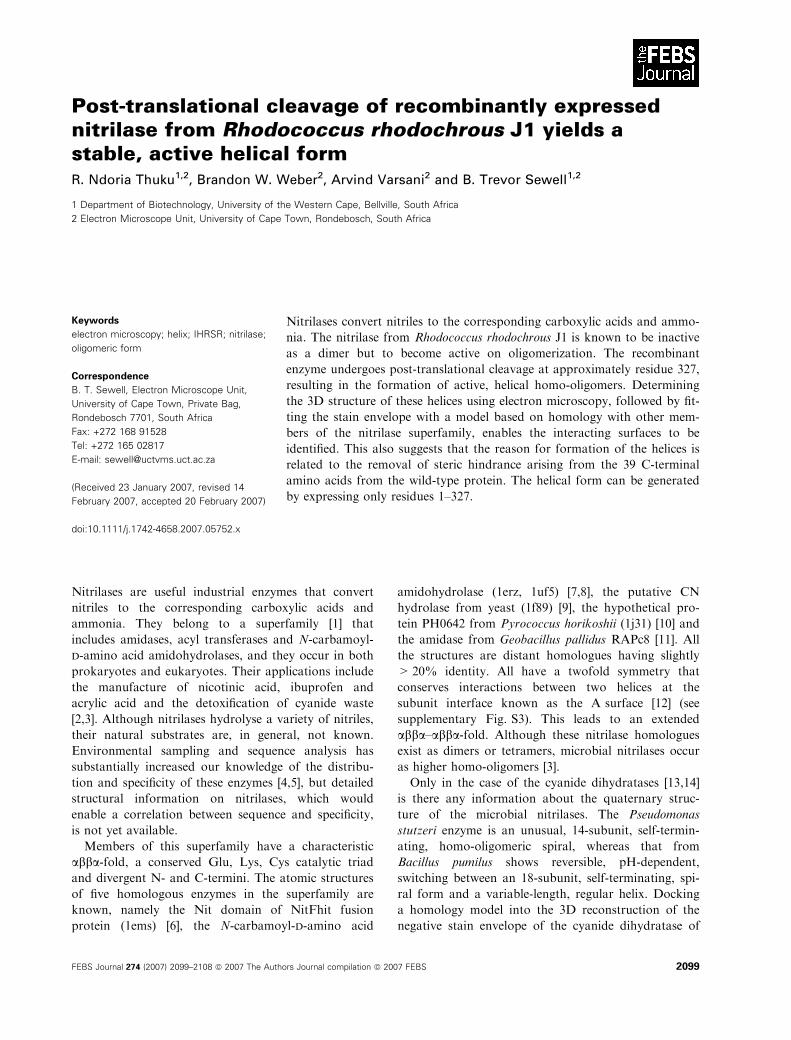
chain (Fig. 1A,B). Even though the protein runs as a
single characteristic band on SDS ⁄PAGE, negative-
stain electron microscopy shows an apparently hetero-
geneous mixture of particles of different shapes and
sizes (Fig. 2A,B). Classification, alignment and avera-
ging of the particles confirms the heterogeneity and, in
addition, demonstrates the existence of a significant
subset of particles that resemble the letter ‘c’ viewed
from different angles (Fig. 2C,D).
After storage at 4 �C for 1 month, the active fraction
eluted from the gel-filtration column in the void volume
indicating a mass >1.5 MDa (Fig. 1C and supplement-
ary Fig. S1). Reducing SDS ⁄PAGE showed that the
subunit atomic mass was 36.5 (± 0.6) kDa (Fig. 1D).
This was confirmed by MS analysis, which gave a sharp
peak at 36 082 Da (R.L. Wolz, Commonwealth
Biotechnologies Inc., Richmond, VA). N-Terminal
sequencing confirmed that the N-terminal was intact
and that C-terminal residues had been removed. The
sharpness of the band on the polyacrylamide gel sug-
gests that this is due to specific cleavage. The calculated
masses of fragments 1–326 and 1–327 are 35 990 and
36 127 Da, respectively, indicating the loss of � 39
amino acids from the C-terminus. Using negative-stain
electron microscopy, the fraction was shown to contain
a homogeneous helical form (Fig. 2E). Analysis of these
helices showed that they had a diameter of 13 nm, a
pitch of 7.7 nm and were of variable length.
3D reconstruction of the fibres using the iterative,
real-space method of Egelman [19] showed that the
helices had 4.9 dimers per turn of the helix, in which
each dimer has an azimuthal rotation of )73.65� and
an axial rise of 1.58 nm (convergence is shown in
supplementary Fig. S2). This corresponds to 78 dimers
Fig. 2. Low-dose electron micrographs of purified and active recombinant nitrilase of R. rhodochrous J1. (A) Quaternary polymorphism of the
480 kDa oligomer. ‘C’-shaped particles (black arrows) and occasional GroEL contamination (white arrow) can be seen. (B) Twenty-five class
averages representing common particle views generated by iterative alignment, sorting and classification of isolated particle images. (C, D)
Putative top and side views of ‘c’-shaped class members and the corresponding class average are shown. The length of the ‘c’ varied
between 9 and 13 nm. (E) Helices formed after storage of the recombinant wild-type nitrilase at 4 �C for 1 month. A power spectrum of the
filament structure (insert) shows a strong layer line, indexed as a Bessel function of order )1, corresponding to a helix with a pitch of
7.7 nm. The layer line at 8.8 nm has been indexed as being a fourth-order Bessel function. The diameter of the helix is 13 nm. (F) Helices
formed from the expression product of J1DC327 which is truncated after residue 327 are indistinguishable from those in (E). White scale
bar ¼ 50 nm.
R. N. Thuku et al. Nitrilase from Rhodococcus rhodochrous J1
FEBS Journal 274 (2007) 2099–2108 ª 2007 The Authors Journal compilation ª 2007 FEBS 2101
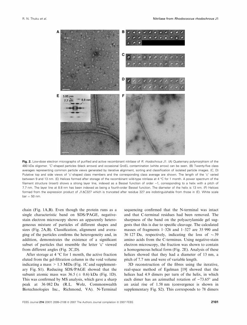
in 16 turns and enables indexing of the power spec-
trum (Fig. 2E, insert) as shown in Fig. 3A. The clear
diffraction spot with a spacing of 7.7 nm is interpreted
as a Bessel function of order )1, corresponding to the
set of left-handed, one-start helices depicted in the heli-
cal net (Fig. 3B). The diffraction spot with a spacing
of 8.8 nm is interpreted as being a Bessel function of
order +4, corresponding to the set of right-handed,
four-start helices depicted in the helical net. It would
be consistent to interpret the diffraction spot with a
spacing of 3.85 nm as corresponding to the unsepa-
rated Bessel functions of orders )2 and +3.
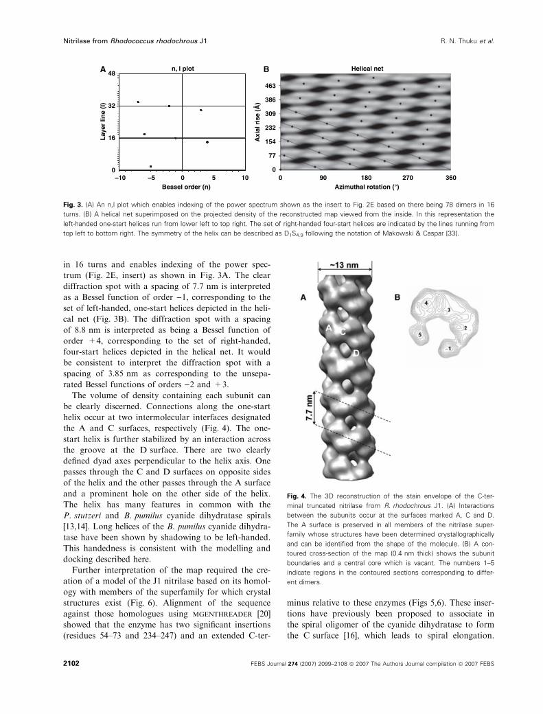
The volume of density containing each subunit can
be clearly discerned. Connections along the one-start
helix occur at two intermolecular interfaces designated
the A and C surfaces, respectively (Fig. 4). The one-
start helix is further stabilized by an interaction across
the groove at the D surface. There are two clearly
defined dyad axes perpendicular to the helix axis. One
passes through the C and D surfaces on opposite sides
of the helix and the other passes through the A surface
and a prominent hole on the other side of the helix.
The helix has many features in common with the
P. stutzeri and B. pumilus cyanide dihydratase spirals
[13,14]. Long helices of the B. pumilus cyanide dihydra-
tase have been shown by shadowing to be left-handed.
This handedness is consistent with the modelling and
docking described here.
Further interpretation of the map required the cre-
ation of a model of the J1 nitrilase based on its homol-
ogy with members of the superfamily for which crystal
structures exist (Fig. 6). Alignment of the sequence
against those homologues using mgenthreader [20]
showed that the enzyme has two significant insertions
(residues 54–73 and 234–247) and an extended C-ter-
minus relative to these enzymes (Figs 5,6). These inser-
tions have previously been proposed to associate in
the spiral oligomer of the cyanide dihydratase to form
the C surface [16], which leads to spiral elongation.
–100
16Lay
er li
ne
(I)
Axi
al r
ise
(Å)32
48
463
386
309
232
154
77
0–5 0
Bessel order (n)
n, l plotA B Helical net
5 10 0 18090 270Azimuthal rotation (°)
360
Fig. 3. (A) An n,l plot which enables indexing of the power spectrum shown as the insert to Fig. 2E based on there being 78 dimers in 16
turns. (B) A helical net superimposed on the projected density of the reconstructed map viewed from the inside. In this representation the
left-handed one-start helices run from lower left to top right. The set of right-handed four-start helices are indicated by the lines running from
top left to bottom right. The symmetry of the helix can be described as D1S4.9 following the notation of Makowski & Caspar [33].
Fig. 4. The 3D reconstruction of the stain envelope of the C-ter-
minal truncated nitrilase from R. rhodochrous J1. (A) Interactions
between the subunits occur at the surfaces marked A, C and D.
The A surface is preserved in all members of the nitrilase super-
family whose structures have been determined crystallographically
and can be identified from the shape of the molecule. (B) A con-
toured cross-section of the map (0.4 nm thick) shows the subunit
boundaries and a central core which is vacant. The numbers 1–5
indicate regions in the contoured sections corresponding to differ-
ent dimers.
Nitrilase from Rhodococcus rhodochrous J1 R. N. Thuku et al.
2102 FEBS Journal 274 (2007) 2099–2108 ª 2007 The Authors Journal compilation ª 2007 FEBS
The strong conservation of the fold, which preserves
all but the peripheral loops, makes it possible to build
a plausible model of the J1 nitrilase which, in turn,
makes it possible to interpret the stain envelope.
At 1.8 nm resolution, the shape of the homologues
(Fig. 7) can be readily discerned in the reconstruction
of the stain envelope. The A surface clearly corres-
ponds to the dimer interface conserved in all the
homologues. Docking of the model [21] (see Experi-
mental procedures) into the stain envelope is simplified
because the symmetry restricts the number of degrees
of freedom. The twofold axis of the dimer model must
coincide with the appropriate twofold axis of the stain
envelope. Thus, the two possible degrees of freedom
(apart from the known helical parameters) are the
radial distance of the model along the dyad axis and
the rotation about the dyad axis. Our docking proce-
dure, which utilized these constraints, produced an
unambiguous optimal fit to the stain envelope.
Four important insights emerge from the docking
(Fig. 7). The C-terminal region is located on the inside
of the helix adjacent to the central channel. There is
some vacant density in this region which may accom-
modate residues 314–327. There is also sufficient
vacant density between the subunits in the C surface
region to accommodate the insertions that have not
been modelled (residues 54–73 and 234–247). The
docking places the bend between beta-sheets b3 and b4(residue 108) in close proximity to alpha-helix a7 (resi-
due 289) suggesting the possibility of an interaction in
this region which contributes to stabilizing the C sur-
face. The D surface is formed by symmetric interac-
tions that occur in the helix a3 having the sequence
-RLLDAARD-. The presence of two arginine and two
Fig. 5. Multiple sequence alignment of the nitrilase from R. rhodochrous J1 (RrJ1) with four nitrilase homologues for which the crystal struc-
tures are known, namely 1ems [6], 1erz [7], 1f89 [9] and 1j31 [10]. Two significant insertions in its sequence (corresponding to residues
54–73 and 234–247) relative to the solved structures are located at the C surface. Furthermore, none of these homologues suggests a
model for the structure of the C-terminal region. The conserved active-site residues are outlined, conserved or homologous residues are in
italics and double underlines indicate the position or the residues which were mutated to stop codons (Table 1). The approximate regions of
interacting surfaces A, C and D are indicated on the top line. Charged residues which are possibly involved in interactions at the D surface
are indicated in bold and the external loop regions are shaded grey. The secondary structural elements identified in 1erz [7] are indicated in
the bottom line.
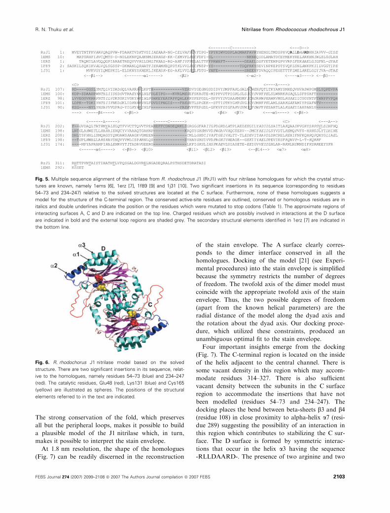
Fig. 6. R. rhodochorus J1 nitrilase model based on the solved
structure. There are two significant insertions in its sequence, relat-
ive to the homologues, namely residues 54–73 (blue) and 234–247
(red). The catalytic residues, Glu48 (red), Lys131 (blue) and Cys165
(yellow) are illustrated as spheres. The positions of the structural
elements referred to in the text are indicated.
R. N. Thuku et al. Nitrilase from Rhodococcus rhodochrous J1
FEBS Journal 274 (2007) 2099–2108 ª 2007 The Authors Journal compilation ª 2007 FEBS 2103
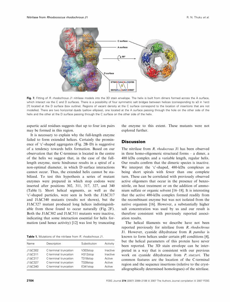
aspartic acid residues suggests that up to four ion pairs
may be formed in this region.
It is necessary to explain why the full-length enzyme
failed to form extended helices. Certainly the promin-
ence of ‘c’-shaped aggregates (Fig. 2B–D) is suggestive
of a tendency towards helix formation. Based on our
observation that the C-terminus is located in the centre
of the helix we suggest that, in the case of the full-
length enzyme, steric hindrance results in a spiral of a
non-optimal diameter, in which D surface interactions
cannot occur. Thus, the extended helix cannot be sta-
bilized. To test this hypothesis a series of mutant
enzymes were prepared in which stop codons were
inserted after positions 302, 311, 317, 327, and 340
(Table 1). Short helical segments, as well as the
‘c’-shaped particles, were seen in both the J1DC317and J1DC340 mutants (results not shown), but the
J1DC327 mutant produced long helices indistinguish-
able from those found to occur naturally (Fig. 2F).
Both the J1DC302 and J1DC311 mutants were inactive,
indicating that some interaction essential for helix for-
mation (and hence activity) [12] was lost by truncating
the enzyme to this extent. These mutants were not
explored further.
Discussion
The nitrilase from R. rhodocrous J1 has been observed
in three homo-oligomeric structural forms – a dimer, a
480 kDa complex and a variable length, regular helix.
Our results confirm that the dimeric species is inactive.
We interpret the ‘c’-shaped, 480-kDa complexes as
being short spirals with fewer than one complete
turn. These can be correlated with previously observed
active oligomers that occur in the presence of benzo-
nitrile, on heat treatment or on the addition of ammo-
nium sulfate or organic solvent [16–18]. It is interesting
that the active 480-kDa complex formed readily from
the recombinant enzyme but was not isolated from the
native organism [16]. However, a substantially higher
salt concentration was used by us and our result is
therefore consistent with previously reported associ-
ation results.
The helical filaments we describe have not been
reported previously for nitrilase from R. rhodochrous
J1. However, cyanide dihydratase from B. pumilus is
known to form helices under certain pH conditions [4],
but the helical parameters of this protein have never
been reported. The 3D stain envelope can be inter-
preted in a way that is consistent with our previous
work on cyanide dihydratase from P. stutzeri. The
common features are the location of the C-terminal
region and the sequence insertions (relative to the cryst-
allographically determined homologues) of the nitrilase.
Fig. 7. Fitting of R. rhodochrous J1 nitrilase models into the 3D stain envelope. The helix is built from dimers formed across the A surface,
which interact via the C and D surfaces. There is a possibility of four symmetric salt bridges between helices (corresponding to a3 in 1erz)
[7] located at the D surface (box outline). Regions of vacant density at the C surface correspond to the location of insertions that are not
modelled. There are two horizontal dyads (yellow ellipses), one located at the A surface passing through the hole on the other side of the
helix and the other at the D surface passing through the C surface on the other side of the helix.
Table 1. Mutations of the nitrilase from R. rhodochrous J1.
Name Description Substitution Activity
J1DC302 C-terminal truncation V303stop Inactive
J1DC311 C-terminal truncation H312stop Inactive
J1DC317 C-terminal truncation T318stop Active
J1DC327 C-terminal truncation T328stop Active
J1DC340 C-terminal truncation E341stop Active
Nitrilase from Rhodococcus rhodochrous J1 R. N. Thuku et al.
2104 FEBS Journal 274 (2007) 2099–2108 ª 2007 The Authors Journal compilation ª 2007 FEBS
The location of the C-terminal region on the inside
of the helix immediately suggests a reason for the
failure of the wild-type enzyme to form the long heli-
ces. Namely, that steric hindrance resulting from the
C-terminal 39 amino acids prevents completion of the
turn and formation of the D surface. Our observation
that constructs on either side of the experimentally
observed cleavage at approximately residue 327 do not
form stable long helices suggests that the truncation at
residue 327 is the ‘optimal point’ for cleavage, produ-
cing a homogeneous population of long helices of the
nitrilase. At this position, the packing results in the
putative D surface salt bridges being optimally aligned
and the helix elongates readily, whereas on either side
of this optimum the packing is too tight or too loose,
resulting in unstable, short helices.
Vacant density in the reconstruction corresponds in
location and volume to the 28 residues per dimer omit-
ted from the model at the C-terminus and the 68 resi-
dues per dimer at the C surface. The sequence
insertions and C-terminal extension also occur in the
cyanide dihydratases and cyanide hydratases [12],
indeed these features are common to a large number
of the microbial nitrilases. The location of the
sequence insertions implicates at least some of these
residues in the C surface interactions and points to
them being necessary for helix or spiral formation.
This, in turn, implicates structural changes resulting
from this interaction in the activation of the enzyme
that occurs on oligomerization.
An open question is the reason for the cleavage at
position 326 or 327. We cannot rule out the presence
of a contaminating proteinase arising from the E. coli,
but this seems unlikely given the specificity of the cut,
that the only known proteinases likely to cut at either
of these locations have a very broad specificity, and
that no further degradation takes place over a period
exceeding 1 year. We therefore suggest that this is an
autolysis. The residues responsible for the cleavage
remain unknown.
The biological role of nitrilases is suggested to be
the metabolism of cyanosugars, hormone precursors
containing nitriles and other organocyanide com-
pounds produced by prokaryotes and eukaryotes [4].
Several specific gene clusters containing the nitrilase
gene have been identified in both cultured bacteria and
environmentally sampled DNA [5]. If we postulate that
the tendency to form spirals or helices is widespread in
nitrilases, then a possible role for the helices could be
to act as a scaffold for proteins expressed by genes in
the cluster, leading to an organelle-like assembly.
Assembly of dimers following post-translational clea-
vage into an enzymatically active complex suggests a
regulatory mechanism. This presumably occurs in the
cells in addition to the transcriptional regulation des-
cribed previously [22]. This multistep process leads to
a concentration of the active sites the role of which
could be to protect the organism from harmful nitriles.
Even though the functional significance in vivo is
unknown, biotechnological applications of artificially
made helices are suggested. The long helices provide a
concentration of active sites that can easily be purified,
immobilized and stored for long periods.
In conclusion, we have discovered how to produce
active, long helical oligomers of the nitrilase from
R. rhodochrous J1. We have identified surfaces involved
in forming and maintaining the helix, and suggest that
oligomerization utilizing these or similar interactions
may be common among microbial nitrilases, cyanide
dihydratases and cyanide hydratases. The active
R. rhodochrous J1 nitrilase helix has a 4.9-fold screw
axis, which would preclude the formation of three
dimensional crystals. Although crystallization and
X-ray structure determination of this enzyme remains
elusive, strategies for preventing helix formation by
mutating the residues involved in helix stabilization are
suggested by this study. High-resolution structure
determination will provide more insight into the confi-
guration of the active site, the link between oligomeri-
zation and activity, as well as an understanding of
how the versatile chemistry of this enzyme class arises
from the Glu, Lys, Cys catalytic triad.
Experimental procedures
Expression of recombinant R. rhodochrous J1
nitrilase
Recombinant nitrilase from R. rhodochrous J1 was exp-
ressed in E. coli strain BL21 (DE3) pLysS cells carrying
plasmid pET30a (Novagen, Madison, WI), in which the
gene for the wild-type enzyme was incorporated. Mutant
J1DC327 was recombinantly expressed using pET29b, and
J1DC302, J1DC311, J1DC317 and J1DC340 were expressed
using pET26b, all in the same E. coli strain. A small
amount of transformed host cells was used to inoculate
5 mL of Luria–Bertani broth containing 25 lgÆmL)1 kana-
mycin and 200 lgÆmL)1 chloramphenicol. This was grown
overnight and then diluted into 1 L of Luria–Bertani broth
containing 25 lgÆmL)1 kanamycin and grown at 37 �C.When cells reached an D600 of � 1, isopropyl-b-d-thiogal-actopyranoside was added to a final concentration of 1 mm
to induce protein expression. Cells were grown overnight,
pelleted (4000 g, 10 min, 4 �C) and resuspended in 40 mL
of 100 mm KH2PO4 pH 7.8 (buffer A) containing one tab-
let of protease cocktail inhibitors (Roche Diagnostics
R. N. Thuku et al. Nitrilase from Rhodococcus rhodochrous J1
FEBS Journal 274 (2007) 2099–2108 ª 2007 The Authors Journal compilation ª 2007 FEBS 2105
GmbH, Mannheim, Germany). Cells were disrupted using a
Misonix� 3000, sonicator (Misonix Inc., Farmingdale, NY)
with pauses for cooling, for a total of 4 min and then
harvested by centrifugation (20 000 g, 4 �C, 30 min). The
supernatant was filtered through a 0.45 lm Millipore
membrane and then applied to an anion-exchange column
(Q-Sepharose XK 26 ⁄ 20; Amersham Biosciences, Piscata-
way, NJ) previously equilibrated with 100 mm KH2PO4,
200 mm KCl, 10% (v ⁄ v) ethanol, pH 7.8 (the J1DC327 was
subjected to 30–40% ammonium sulfate precipitation prior
to this step). The protein was eluted using 100 mm
KH2PO4, 400 mm KCl, 10% (v ⁄ v) ethanol, pH 7.8,
whereas the J1DC327 mutant was eluted using a linear gra-
dient from 0.1 to 1 m KCl in the same buffer. Active peak
fractions were analysed by reducing SDS ⁄PAGE. Protein
concentration was determined using either Bradford assay
or photometrically at k ¼ 280 nm using the known extinc-
tion coefficient of R. rhodochrous J1 nitrilase [15] of
0.93 mg)1Æcm)1ÆmL)1. Active fractions were pooled and
concentrated to 8.5 mgÆmL)1 using an Amicon stirred cell
(Millipore, Billerica, MA) with a 10 kDa exclusion mem-
brane (Millipore PM10) and ultrafiltration subsequently
applied to the gel filtration column (Sephacryl S400 HRXk
16 ⁄ 70; Amersham Biosciences). Proteins were eluted with
100 mm KH2PO4, 200 mm NaCl, pH 7.8 (buffer B) and
where necessary, this step was repeated to rid the protein of
contaminating GroEL-like particles. All gel filtration col-
umns were previously calibrated using Bio-Rad standards
(supplementary Fig. S1) at the same flow rate. Active peak
fractions were separated on reducing SDS ⁄PAGE and
bands visualized by silver staining. An active sample of
1-month-old purified enzyme kept refrigerated at 4 �C was
filtered through a 0.22 lm membrane and applied to gel fil-
tration (TSK G5000PWXL column; Tosoh Bioscience,
GmbH, Stuttgart, Germany) previously equilibrated and
eluted using buffer B. At the end of each chromatographic
step, the active protein was investigated by negative-stain
electron microscopy.
Assay for enzyme activity
Nitrilase activity was analysed by assaying the release of
ammonia as described previously [23]. Reactions were car-
ried out in 1 mL volumes containing 2 lL of enzyme solu-
tion, 988 lL of buffer A and 10 lL of 100 mm benzonitrile
(dissolved in 1 mL ethanol to increase its solubility). The
reaction was allowed to occur for 1 h at room temperature
followed by the addition of 40 lL phenol–alcohol, 40 lLsodium nitroprusside and 100 lL of freshly prepared oxid-
izing solution [1 part NaOCl to 4 parts alkaline complexing
agent (10 g sodium citrate, 0.5 g sodium hydroxide made
up to 50 mL with distilled water)]. Reaction mixtures were
incubated for 1 h at room temperature and the colour
change was recorded by measuring the absorbance at
620 nm. One unit of the enzyme was defined as the amount
that converts 1 lmole of benzonitrile in 1 min to produce
an equivalent amount of benzoate and ammonia. Every
second fraction eluted from the columns was assayed for
activity.
Negative-stain electron microscopy
Four microlitres of purified enzyme solution was pippetted
onto a fresh glow-discharged grid previously coated with a
thin carbon support film under vacuum. In order to reduce
precipitation between phosphate buffer and uranyl acetate,
grids were subjected to two successive water washes fol-
lowed by staining with 2% uranyl acetate. At each step,
excess sample, wash and stain were blotted. Grids were air-
dried before electron microscopy. The salt concentration in
the buffer was reduced by a 5–10-fold dilution with distilled
water. All staining was carried out at room temperature.
Micrographs for image processing were recorded slightly
under focus on Kodak S0163 film under low-dose condi-
tions on a JEOL 1200EX II transmission electron micro-
scope operating at 120 kV.
Image processing
Good-quality negatives were scanned using a Leafscan� 45
scanner at pixel size of 10 lm, giving 2 A per pixel at the
specimen level. The oligomeric particles were extracted in
160 · 160 pixel boxes and later binned by a factor of two.
Raw images (� 11 000) were band-pass filtered (20 to
1.5 nm) and masked and then iteratively aligned and classi-
fied using routines in the spider program suite [24]. A 3D
reconstruction of the oligomeric state was not pursued
because of sample heterogeneity. Filament segments
(13 506) were windowed in 256 · 256 pixel boxes using
boxer, a program from the eman package [25], and then
binned by a factor of two. The overlap between boxes
along the length of a single filament was 96%. 3D recon-
struction was carried out using the iterative helical real
space reconstruction method [19]. The reconstruction was
based on 13 506 segments, each 12.8 nm long. After several
cycles of iteration, the twofold axis perpendicular to the
helix axis, which corresponded to the dyad axis of the
dimer, became readily apparent and twofold symmetry was
imposed on the reconstruction in subsequent cycles. The
reconstruction was low-pass filtered to 1.8 nm and visual-
ized using ucsf chimera [26].
Homology modelling and docking
The search for structural homologues and sequence align-
ment was done using mgenthreader [20]. Pair-wise align-
ment of the solved structures was done using align [27].
Based on the alignment (slightly modified by hand), a 3D
model of the J1 nitrilase dimer having 313 residues (of
Nitrilase from Rhodococcus rhodochrous J1 R. N. Thuku et al.
2106 FEBS Journal 274 (2007) 2099–2108 ª 2007 The Authors Journal compilation ª 2007 FEBS
366 due to lack of a template for its extended C-terminus)
was constructed using modeller [28]. Side-chain orienta-
tion was optimized using scwrl [29]. The model was eval-
uated using procheck [30] and prosa [31] and visualized
with pymol [32]. Automatic fitting of a helix model
comprising two turns made up of nine dimers of the J1
nitrilase model without the insertions or the C-terminal
extension, was carried out using the contour-based low-
resolution (colores) program implemented in the situs
package [21]. The nine-dimer helix model was generated
by applying the helical symmetry operators to a single
dimer model whose twofold axis was located on the x-
axis. Once the helical parameters were determined, the
dihedral (D1) symmetry of the helix allows only two addi-
tional degrees of freedom for fitting such a model, namely
the azimuthal rotation about the x-axis and translation
along the same axis. All surface renderings were carried
using ucsf chimera [26].
N-Terminal sequencing and mass spectrometry
Following results from gel filtration and reducing
SDS ⁄PAGE, a purified 1-month-old sample (0.75 mgÆmL)1)
of the nitrilase was sent to Commonwealth Biotechnologies,
Inc. (Richmond, Virginia) for N-terminal sequencing and
MALDI-TOF MS. One hundred microlitres of sample was
subjected to 10 cycles of Edman degradation to determine
the amino acid sequence. For MS, 1 lL of undiluted sam-
ple was mixed with 1 lL of matrix (ferulic acid) and then
spotted onto a sample plate. The sample was desalted to
improve the signal.
Acknowledgements
We thank Professor Charles Brenner for the generous
gift of the recombinant expression plasmid, Professor
Edward H. Egelman for his assistance with the itera-
tive helical real-space reconstruction programs, Dr
Dean Brady for access to the HPLC equipment at
CSIR Bio ⁄Chemtek and Professor Michael Benedik
for his comments on the manuscript. We greatly
appreciate the substantial support we have received
from the Carnegie Corporation of New York. RNT
was funded by an international scholarship from UCT
as well as a studentship from CSIR (Bio ⁄Chemtek).
References
1 Pace H & Brenner C (2001) The nitrilase superfamily:
classification, structure and function. Genome Biol
2, 1–9.
2 O’Reilly C & Turner PD (2003) The nitrilase family of
CN hydrolyzing enzymes – a comparative study. J Appl
Microbiol 95, 1161–1174.
3 Banerjee A, Sharma R & Banerjee UC (2002) The
nitrile-degrading enzymes: current status and future
prospects. Appl Microbiol Biotechnol 60, 30–44.
4 Robertson DE, Chaplin JA, DeSantis G, Podar M,
Madden M, Chi E, Richardson T, Milan A, Miller M,
Weiner DP et al. (2004) Exploring nitrilase sequence
space for enantioselective catalysis. Appl Environ Micro-
biol 70, 2429–2436.
5 Podar M, Eads JR & Richardson TH (2005) Evolution
of a microbial nitrilase gene family: a comparative and
environmental genomics study. BMC Evol Biol 5, 1–13.
6 Pace HC, Hodawadekar SC, Draganescu A, Huang J,
Bieganowski P, Pekarsky Y, Croce CM & Brenner C
(2000) Crystal structure of the worm NitFhit Rosetta
Stone protein reveals a Nit tetramer binding two Fhit
dimers. Curr Biol 10, 907–917.
7 Nakai T, Hasegawa T, Yamashita E, Yamamoto M,
Kumasaka T, Ueki T, Nanba H, Ikenaka Y, Takahashi
S, Sato M et al. (2000) Crystal structure of N-carbamyl-
d-amino acid amidohydrolase with a novel catalytic
framework common to amidohydrolases. Structure 8,
729–737.
8 Hashimoto H, Aoki M, Shimizu T, Nakai T, Morikawa
H, Ikenaka Y, Takahashi S & Sato M (2004) Crystal
Structure of C171A ⁄V236A Mutant of N-Carbamyl-
D-Amino Acid Amidohydrolase. RCSB Protein Databank
(1uf5).
9 Kumaran D, Eswaramoorthy S, Gerchman SE, Kycia
H, Studier FW & Swaminathan S (2003) Crystal struc-
ture of putative CN hydrolase from yeast. Proteins:
Struct Funct Genet 52, 283–291.
10 Sakai N, Tajika Y, Yao M, Watanabe N & Tanaka I
(2004) Crystal structure of hypothetical protein PH0642
from Pyrococcus horikoshii at 1.6 A resolution. Proteins:
Struct Funct Bioinform 57, 869–873.
11 Agarkar VB, Kimani SW, Cowan DA, Sayed MF-R &
Sewell BT (2006) The quaternary structure of the ami-
dase from Geobacillus pallidus RAPc8 is revealed by its
crystal packing. Acta Crystallogr F62, 1174–1178.
12 Sewell BT, Thuku RN, Zhang X & Benedik MJ (2005)
The oligomeric structure of nitrilases: the effect of
mutating interfacial residues on activity. Ann NY Acad
Sci 1056, 153–159.
13 Sewell BT, Berman MN, Meyers PR, Jandhyala D &
Benedik MJ (2003) The cyanide degrading nitrilase from
Pseudomonas stutzeri AK61 is a two-fold symmetric,
14-subunit spiral. Structure 11, 1–20.
14 Jandhyala D, Berman M, Meyers PR, Sewell BT,
Willson RC & Benedik MJ (2003) Cyn D, the cyanide
dihydratase from Bacillus pumillus: gene cloning and
structural studies. Appl Environ Microbiol 69,
4794–4805.
15 Kobayashi M, Nagasawa T & Yamada H (1989) Nitri-
lase of Rhodococcus rhodochrous J1: purification and
characterization. Eur J Biochem 182, 349–356.
R. N. Thuku et al. Nitrilase from Rhodococcus rhodochrous J1
FEBS Journal 274 (2007) 2099–2108 ª 2007 The Authors Journal compilation ª 2007 FEBS 2107
16 Nagasawa T, Wieser M, Nakamura T, Iwahara H,
Yoshida T & Gekko K (2000) Nitrilase of Rhodococcus
rhodochrous J1: conversion into the active form by sub-
unit association. Eur J Biochem 267, 138–144.
17 Stevenson DE, Feng R, Dumas F, Groleau D, Mihoc A
& Storer AC (1992) Mechanistic and structural studies
on Rhodococcus ATCC 39484 nitrilase. Biotechn Appl
Biochem 15, 283–302.
18 Harper DB (1977) Microbial metabolism of aromatic
nitriles: enzymology of C–N cleavage by Norcadia sp.
(Rhodochrous group) NCIB 11216. Biochem J 165,
309–319.
19 Egelman EH (2000) A robust algorithm for the recon-
struction of helical filaments using single-particle meth-
ods. Ultramicroscopy 85, 225–234.
20 Jones DT (1999) GenTHREADER: an efficient and
reliable protein fold recognition method for genomic
sequences. J Mol Biol 287, 797–815.
21 Chacon P & Wriggers W (2002) Multi-resolution con-
tour-based fitting of macromolecular structures. J Mol
Biol 317, 375–384.
22 Komeda H, Hori Y, Kobayashi M & Shimizu S (1996)
Transcriptional regulation of the Rhodococcus
rhodochrous J1 nitA gene enconding a nitrilase. Proc
Natl Acad Sci USA 93, 10572–10577.
23 Piotrowski M, Schonfelder S & Weiler EW (2001) The
Arabidopsis thaliana isogene NIT4 and its orthologs in
tobacco encode b-cyano-l-alanine hydratase ⁄ nitrilase.J Biol Chem 276, 2616–2621.
24 Frank J, Radermacher M, Penczek P, Zhu J, Li Y,
Ladjadj M & Leith A (1996) SPIDER and WEB:
processing and visualization of images in 3D electron
microscopy and related fields. J Struct Biol 116,
190–199.
25 Ludtke SJ, Baldwin PR & Chiu W (1999) EMAN:
semi-automated software for high-resolution single
particle reconstructions. J Struct Biol 128,
82–96.
26 Pettersen EF, Goddard TD, Huang CC, Couch GS,
Greenblatt DM, Meng EC & Ferrin TE (2004) UCSF
Chimera – a visualization system for exploratory
research and analysis. J Comput Chem 25,
1605–1612.
27 Cohen GH (1997) ALIGN: a program to superimpose
protein coordinates, accounting for insertions and dele-
tions. J Appl Crystallogr 30, 1160–1161.
28 Sali A & Blundell TL (1993) Comparative protein mod-
eling by satisfaction of spatial restraints. J Mol Biol
234, 779–815.
29 Bower JM, Cohen FE & Dunbrack RL Jr (1997) Pre-
diction of protein side-chain rotamers from a backbone-
dependent rotamer library: a new homology modeling
tool. J Mol Biol 267, 1268–1282.
30 Laskowski RA, MacArthur MW, Moss DS & Thornton
JM (1993) PROCHECK: a program to check the stereo-
chemical quality of protein structures. J Appl Crystal-
logr 26, 283–291.
31 Sippl M (1993) Recognition of errors in three-dimen-
sional structures of proteins. Proteins: Struct Funct
Genet 17, 355–362.
32 DeLano WL (2002) The PyMOL Molecular Graphics
System. DeLano Scientific, San Carlos, CA. http://
www.pymol.org.
33 Makowski L & Caspar DLD (1981) The symmetries of
filamentous phage particles. J Mol Biol 145, 611–617.
Supplementary material
The following supplementary material is available
online:
Fig. S1. Calibration of the TSK G5000PWXL column
used for the elution of 1-month-old Rhodococcus
rhodochrous J1 (J1 nitrilase).
Fig. S2. Convergence of the IHRSR algorithm after
22 cycles.
Fig. S3. Cartoon representation of the structural
homologues and 3D model of the nitrilase from Rho-
dococcus rhodochrous J1.
This material is available as part of the online article
from http://www.blackwell-synergy.com
Please note: Blackwell Publishing is not responsible
for the content or functionality of any supplementary
material supplied by the authors. Any queries (other
than missing material) should be directed to the corres-
ponding author for the article.
Nitrilase from Rhodococcus rhodochrous J1 R. N. Thuku et al.
2108 FEBS Journal 274 (2007) 2099–2108 ª 2007 The Authors Journal compilation ª 2007 FEBS