Embed Size (px)
Citation preview
Research ArticleNickel Nanoparticle-Modified Electrode for the ElectrochemicalSensory Detection of Penicillin G in Bovine Milk Samples
Suleiman Salihu,1 Nor Azah Yusof ,1,2 Faruq Mohammad ,3 Jaafar Abdullah ,1,2
and Hamad A. Al-Lohedan 3
1Department of Chemistry, Faculty of Science, Universiti Putra Malaysia, 43400 Serdang, Selangor, Malaysia2Institute of Advanced Technology, Universiti Putra Malaysia, 43400 Serdang, Selangor, Malaysia3Department of Chemistry, College of Science, King Saud University, Riyadh, 11451, Saudi Arabia
Correspondence should be addressed to Nor Azah Yusof; [email protected] and Faruq Mohammad; [email protected]
Received 20 August 2019; Revised 14 October 2019; Accepted 29 October 2019; Published 26 November 2019
Academic Editor: Ilaria Armentano
Copyright © 2019 Suleiman Salihu et al. This is an open access article distributed under the Creative Commons AttributionLicense, which permits unrestricted use, distribution, and reproduction in any medium, provided the original work isproperly cited.
The monitoring of chemical and antibiotic residues like amoxicillin, penicillin, tetracycline, and vancomycin in the food originatingfrom the animal and plant sources can prevent the humans from getting exposed to the antibiotic-induced allergic reactions andalso decreased immunity towards the microbial population. By taking into consideration the necessity of developing effectiveand sensitive techniques for milk containing Penicillin G antibiotics in an easy and cost-effective mode, the present work dealswith the electrochemical sensor made up of nickel nanoparticles (NiNPs). In order to enhance the chemical stability andbiocompatibility, the NiNPs were crosslinked with (3-aminopropyl)triethoxysilane (APTES) and the formed composite wasthoroughly characterized using the physical characterization techniques. In addition, the qualitative analysis results confirmedthe nanocomposite’s synergetic effect towards the oxidation of Penicillin G. Further, the quantitative analysis towards the use ofa nanocomposite electrode due to the changes in pH, scan rate, accumulation time and potential, nanoparticle (NP) amount,etc. was optimized. The limit of detection and limit of quantitation of Penicillin G with this composite were detected to be0.00031 μM and 0.00100μM, respectively. Overall, from the results, it can be indicated that the fabricated NiNP sensor can findits applications as a potential electrode material for the qualitative and quantitative analysis of Penicillin G in liquid samples.
1. Introduction
Penicillin G (benzylpenicillin) is an antibiotic commonlyused for the treatment of bacterial infections in humansand animals where a substantial increase in the amount ofthis drug at trace levels in the samples of edible meat, dairyproducts, blood, and urine (from nanograms to microgramsper liter) in recent years has been a concern to the regulatoryagencies. Penicillin G as shown in Figure 1 has a molecularformula of C16H18N2O4S that corresponds to the molecularweight of 334.4 g/mol. The wider application of penicillin asa medicine is due to its active pharmacological effects thatcan cure the bacterial infections like pneumonia, diphtheria,leptospirosis, strep throat, and gas gangrene. Because of thewider therapeutic uses of this drug in both humans and farmanimals, it has been produced by many biotechnology, drug,
and cosmetic industries where the critical step involves thepurification, and if anything is done wrong, it will end upin water and soil contamination [1–3]. The wider applicationof penicillin as a direct medicine or unhindered utilization(meat, milk, and eggs) in both humans and farm animals isresponsible for many negative effects like proliferation ofdrug-resistant bacteria and drug-involved hypersensitization(fever, urticaria, rashes, and joint pains) [4–6]. Hence,controlling the amount of the penicillin drug in food, milk,meat, and water samples is of utmost importance wheremany different instrumental techniques are involved for thequalitative and quantitative analysis.
With regard to penicillin adulteration, there have been anumber of reports which confirm the presence of this drugin food, meat, milk, and water samples, and the majorconcern is that this amount is getting raised each year. These
HindawiJournal of NanomaterialsVolume 2019, Article ID 1784154, 11 pageshttps://doi.org/10.1155/2019/1784154
multiple routes and associated increase in the amounts ofPenicillin G or its degradation products in the wastewaterhave raised a substantial concern in the public and regulatoryagencies since these have a significant effect to the humanand ecological health. Since the byproducts of Penicillin Gare associated with some potential risks to the humans andwildlife if they enter into the food and water cycle, the detec-tion of Penicillin G has likely become more significant toprotect the environment and human health. The commonlyused techniques for the detection of Penicillin G in samplesare instrumental [7], such as capillary electrophoresis [8, 9],gas chromatography [10, 11], liquid chromatography [11,12] coupled with mass spectrometry [13], Fenton’s process[14–16], UV/ZnO photocatalytic process [17], and someadvanced oxidation processes [18]. However, each methodhas its own limitations like the requirement of skilled techni-cians, complex instruments, expensive method development,time-consuming process, unpredictable analysis time, usageof toxic solvents, and multiple washing steps, thereby makinga strong need for the development of novel techniques withminimum of these limitations [19, 20]. In that view, theelectrochemical-based sensory methods proved to beinteresting candidates and also low-cost alternatives for thebiomolecular detection in liquid samples, since the electro-chemical sensors can provide quick results and it is possibleto miniature these devices to portable sizes that can traceeven very low levels of Penicillin G where the sensitivity hasnot to be compromised. In that way, the electrochemicalsensory detection of Penicillin G, therefore, is extrapolatedfrom the electrochemical sensory technique in milk sampleswhere the method involves the detection of current bymaking use of the simultaneous oxidoreduction processoccurring at the electrode surface.
In recent years, the engagement of electrode materialswith enriched electrochemical properties in many differentelectrolytes, physicochemical and thermal stabilities, andsmall background currents made them a fashionable choicein the analytical chemistry field for the qualitative and quan-titative analysis [21], since a majority of commonly usedelectrode materials like mercury, lead, arsenic, and cadmiumare unsuitable for such analytical applications because oftheir intrinsic toxicity, aggressive nature, chemical instability,and high costs. Interestingly, the nickel nanoparticles(NiNPs) offer some of the unique properties which make thismaterial overcome the problems of affordability (notexpensive like Pt, Pd, or Ag), instability (characteristics aresimilar to noble metals), and toxicity (not aggressive as
compared to Hg2+, As3+, Pb2+, and Cd2+) and maintainsoxidoreductive properties. The screen-printed carbonelectrodes (SPCE) have shown very promising results in thefield of electroanalysis [22–24], and so, by taking advantageof its effective nature, the present work is aimed at evaluatingthe effectiveness of NiNP-modified SPCE for the electrode-position of Penicillin G using an electrooxidation method.The formed composite was characterized thoroughly usingdifferent instrumental methods, and further, the effect ofvarious physicochemical parameters towards the detectionof Penicillin G in milk samples was evaluated.
2. Materials and Methods
2.1. Chemicals and Supplies. All the chemicals were used of thehighest analytical reagent grades; reagents and solvents used inthis study were purchased from a company (Finn ChemicalsSdn Bhd, Malaysia) and used without further purification.Penicillin G ((2S,5R,6R)-3,3-dimethyl-7-oxo-6-(2-phenylaceta-mido)-4-thia-1-azabicyclo[3.2.0]heptane-2-carboxylic acid) isreagent grade and was obtained from Fluka (Ronkonkoma,NY, USA). Polyvinylpyrrolidone (PVP) was purchased fromR&M Chemicals (Essex, UK), while nickel(II) chloride and(3-aminopropyl)triethoxysilane (APTES) were from Sigma-Aldrich (St. Louis, MO, USA). Deionized water was obtainedfrom a Millipore water purification system. Phosphate-buffered solution (PBS) was prepared in a usual way. A stocksolution of 0.01mol/L Penicillin G was prepared by dis-solving 0.384 g Penicillin G sodium salt of the puresubstance in a 100mL volumetric flask with deionizedwater. More diluted solutions of penicillin were preparedby precise dilution of stock solutions. All glassware waswashed and rinsed several times with deionized water.
2.2. Instrumentation. In this study, the electrochemicalmeasurements were recorded using the potentiostatMetrohm@μAutolab type III (Eco Chemie, Utrecht, TheNetherlands) which was integrated with a screen-printedjunction cable and is controlled by the NOVA 1.11 software.The SPCE was obtained from DropSens company (Oviedo,Spain), and the disk-shaped working electrode has a diameterof 4mm; the working and auxiliary electrodes were made ofgold, while the reference electrode was made of silver. Allthe electrodes used for this study were printed on a ceramicsupport (L 33 ×W 10 ×H 0:5mm), and the studies weredone at room temperature conditions only. The DropSenspotentiostat μStat 8000 electrochemical system connectedto PC and the SPCE made of a three-electrode system, i.e.,working electrode, counter electrode, and Ag/AgCl referenceelectrode, was used. The electrochemical cell consists of aSPCE inserted in a glass medium mounted on a stirrercontaining supporting electrolyte and analyte (Rapid LabsSdn Bhd, Malaysia); a pH meter was used to adjust the pHof solutions while centrifugation was performed on a Kubotacentrifuge (Japan). The Malvern Nano-sizer or ZetasizerNano range providing both exceptionally high performanceand entry-level systems that incorporate combinations ofparticle size analyzer measurements was used. The surfacefunctionality of the synthesized composite was studied by
HS CH3
CH3
OHO
O
O
R
N
NH
Figure 1: Chemical structure of Penicillin G.
2 Journal of Nanomaterials
using Fourier transform infrared spectroscopy (FTIR)(Perkin Elmer) in the wavenumber range of 4000-400 cm-1
at a resolution of 4 cm-1. The size, shape, surface morphology,and elemental composition in dry powdered form were stud-ied using field emission scanning electron microscopycoupled with energy dispersive X-ray (FESEM-EDX) (JEOLJSM 7600F). The Philips X-ray diffraction (XRD) instrumentwas used for understanding the crystal structure, and thesamples were analyzed by placing the powder on a flat amor-phous silica sample holder. The patterns were recorded in the2θ range of 10-60 degrees at room temperature using Cu kαradiation (λ = 1:5418Å).
2.3. Synthesis of NiNPs. The NiNPs were synthesized fromthe nickel chloride-hydrazine (NiCl2-N2H4) mixture in anaqueous sodium hydroxide (NaOH) solution that wasmaintained at a high pH ~12.5 [25]. The N2H4 in thealkaline NaOH solution is represented by the followingoxidation reaction:
N2H4 + 4OH− ⟶ 4e− + N2 + 4H2O, Eθ = −0:16V ð1Þ
When the Ni ions from NiCl2 salt get mixed withN2H4 and NaOH hydroxide solutions, it consequently getsreduced [26, 27]:
2Ni2+ + 4e− ⟶ 2Ni, Eθ = −0:25V ð2Þ
Therefore, precipitation of Ni with N2H4 behaving as areductant in alkaline solution depends on pH of the solu-tion which can be simply explained by the equation
2Ni2+ +N2H4 + 4OH− ⟶ 2Ni +N2 + 4H2O, Eθ = +0:09Vð3Þ
The discharge of NiNPs was enhanced by the additionof strong NaOH base which increased the solution pH andby constant maintenance of Ni ion concentration. Addi-tionally, hydrazine in this reaction mixture acts as asurface ligand to the Ni particles by means of forming astable complex in the Ni solution with respect to themolar ratio of Ni to N2H4. The colloidal synthesis offersa route for the synthesis of nanoparticles (NPs) with con-trolled size, composition, and structural features by takingadvantage of the PVP molecules. In the present case, theNiNPs were added with equal amount of PVP moleculesso that the surfaces of the NiNPs are protected as soonas they are formed from the agglomeration where thePVP serves as the surface stabilizer, growth modifier,dispersant, and also reducing agent. The PVP moleculesshow a high degree of compatibility both in solution andin film form, with most of the inorganic salt solutions.The PVP is widely used in a broad variety of industriesdue to its unique physicochemical properties, offeringgood solubility in water and many organic solvents whereits chemical is stable and providing affinity towardscomplex hydrophobic and hydrophilic substances withnontoxic character.
2.4. Synthesis of Amine-Functionalized NiNPs. For the aminefunctionalization of earlier formed PVP-NiNPs, we haveused the cocondensation method by the incorporation ofAPTES. In brief, about 0.5 g of PVP-NiNPs was dispersedin 50mL DMSO under sonication for about 1 h, and afterthe period, the mixture was transferred to a round bottomflask connected to a reflux condenser maintained at inertatmosphere. About 400μL of 3-APTES was added to thismixture, refluxed at 120°C for at least 3 h, and after thecompletion of reaction, the resulting solution was cooled toroom temperature. The solution was added with ethanol(degassed), separating the precipitate by means of centrifuga-tion at 12000 rpm for about 15min, and this washing processwith ethanol was continued several times so as to completelyremove the unreacted 3-APTES and any other unwantedbyproducts. Finally, the amine functionalized NiNPs(APTES-NiNPs) were formed by drying the product at60°C overnight.
2.5. Real Sample Extraction. For the real sample extraction,we have employed the same method as described by Moorsand Massart [28] with little modifications. Briefly, about30mL raw milk was spiked with a known Penicillin G con-centration, vortexed for 2min at room temperature so as toallow for the equilibration of β-lactams with that of milkmatrix before their actual extraction. Now, the contents weresubjected to centrifugation at 3000 rpm for 10min, and then,10mL of defatted milk was deproteinized by the addition of10mL of acetonitrile in a dropwise manner with continuousvortex so as to allow for the complete mixing and centrifugedagain at 4000 rpm for 15min. Following the centrifugation,10mL of supernatant was transferred to a beaker, heated to60°C in order to evaporate the added acetonitrile, and theheating was continued until the volume reduces approxi-mately to 1mL. Then, 1mL of PBS was added immediatelyso as to avoid the degradation of analyte, the mixture wascentrifuged at 4000 rpm for 20min, and the supernatantwas collected and filtered through a 0.22μm filter paperand used further for the analysis.
2.6. Analytical Procedure. For the detection of Penicillin G,the oxidation peak current was measured, and for that, thevoltammetric response in the potential range of -0.6-0.6V,0.1M PBS (pH 7.0), as the supporting electrolyte was applied.The differential pulse voltammetry (DPV) was performed byapplying -0.4V accumulation potential for about 180 sec andequilibrium time of 10 sec. For each voltammogrammeasurement, a scan rate of 0.1V/s was applied and was con-ducted at room temperature (25°C) only.
3. Results and Discussion
The dynamic light scattering studies using the Zetasizernanoseries were performed for measuring the size of particlesin the solution phase, and for that, the colloidal solutions ofNiNPs settled at the bottom side of the flask during thechemical synthesis before the drying was taken. The zetapotential analysis of NiNPs formed by the wet chemicalmethod provided the information that the hydrodynamic
3Journal of Nanomaterials
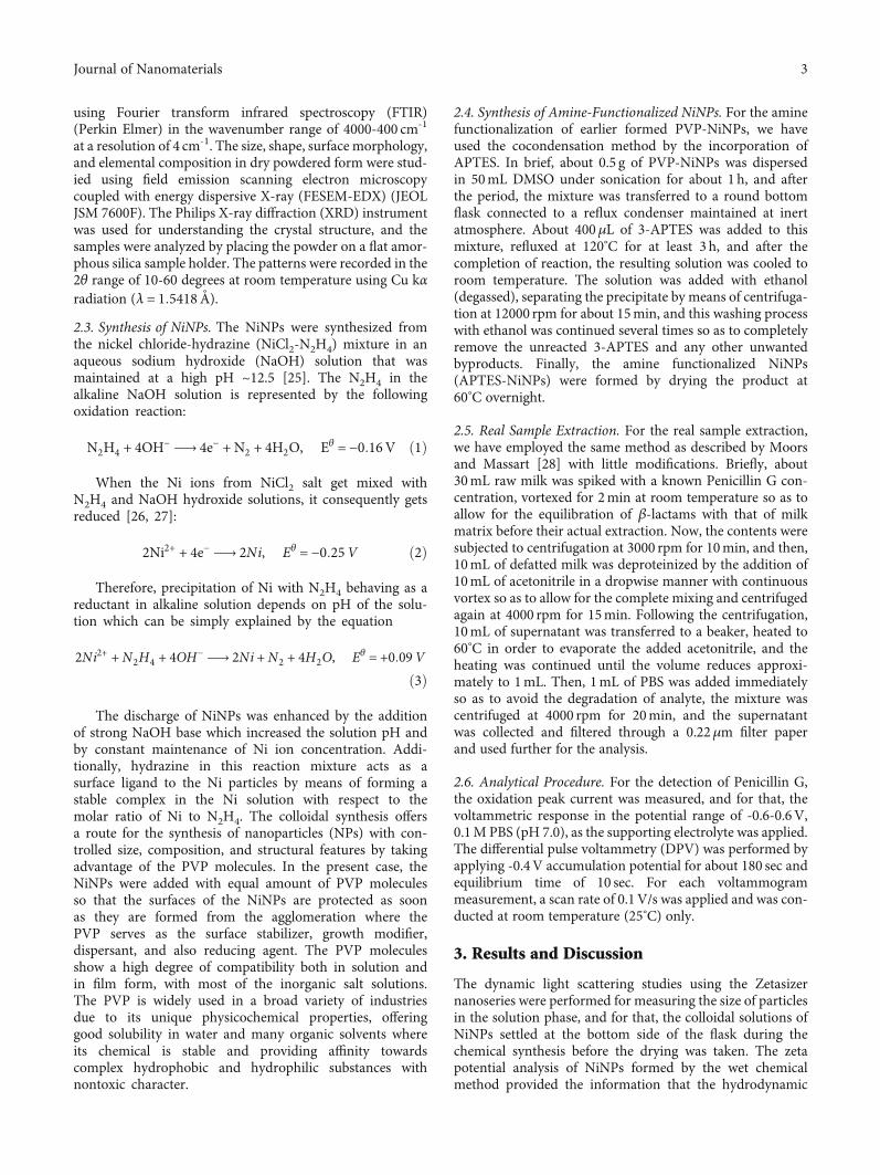
size of particles is about 15nm (data not shown here). Theobtained size is the combination of NiNP core along withthe APTES-coated material and a little portion of the solventlayer attached to the surface as the particles move under theinfluence of Brownian movement. Following the zeta poten-tial studies, the particle size and surface morphology ofsynthesized NPs were determined by FESEM; Figures 2(a)and 2(b) shows the FESEM and EDX images of NiNPs(APTES coated) produced by the simplified chemicalreduction method. It is indicated from the FESEM image(Figure 2(a)) that the NiNPs are of spherical shape havingthe rough surface and the particles are within the instru-ment’s range of size detection, as the particles are formed inthe nanometer size (scale of 500nm was used). Further, theresults of the EDX spectroscopic detector connected to theFESEM instrument used for the investigation of elementalcomposition in the synthesized NiNPs (Figure 2(b))confirmed the presence of corresponding elements. TheNiNP spectrum displayed the eminent peaks of Ni; the NPsare coated with gold during the sample preparation, whilethe presence of oxygen can be from the APTES and surfaceoxidation of the particles [29]. Thus, from the analysis, theavailability of Ni in the NiNPs can aid for the catalyticproperty of the modified electrode.
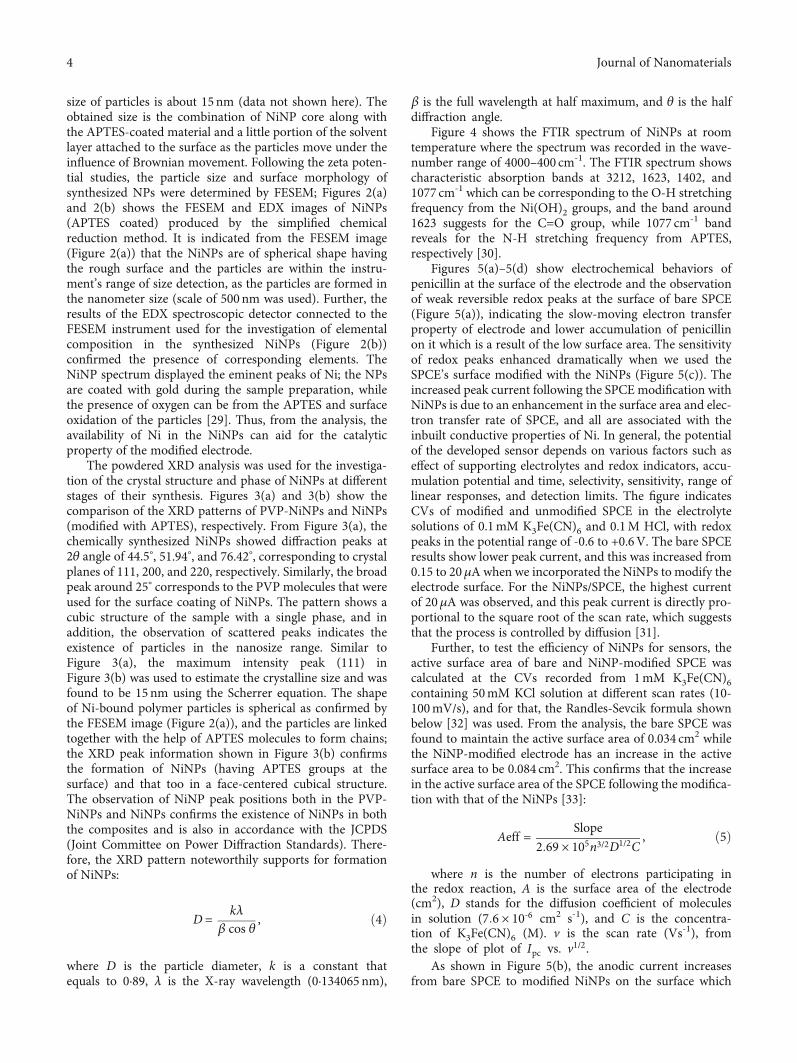
The powdered XRD analysis was used for the investiga-tion of the crystal structure and phase of NiNPs at differentstages of their synthesis. Figures 3(a) and 3(b) show thecomparison of the XRD patterns of PVP-NiNPs and NiNPs(modified with APTES), respectively. From Figure 3(a), thechemically synthesized NiNPs showed diffraction peaks at2θ angle of 44.5°, 51.94°, and 76.42°, corresponding to crystalplanes of 111, 200, and 220, respectively. Similarly, the broadpeak around 25° corresponds to the PVP molecules that wereused for the surface coating of NiNPs. The pattern shows acubic structure of the sample with a single phase, and inaddition, the observation of scattered peaks indicates theexistence of particles in the nanosize range. Similar toFigure 3(a), the maximum intensity peak (111) inFigure 3(b) was used to estimate the crystalline size and wasfound to be 15 nm using the Scherrer equation. The shapeof Ni-bound polymer particles is spherical as confirmed bythe FESEM image (Figure 2(a)), and the particles are linkedtogether with the help of APTES molecules to form chains;the XRD peak information shown in Figure 3(b) confirmsthe formation of NiNPs (having APTES groups at thesurface) and that too in a face-centered cubical structure.The observation of NiNP peak positions both in the PVP-NiNPs and NiNPs confirms the existence of NiNPs in boththe composites and is also in accordance with the JCPDS(Joint Committee on Power Diffraction Standards). There-fore, the XRD pattern noteworthily supports for formationof NiNPs:
D = kλβ cos θ , ð4Þ
where D is the particle diameter, k is a constant thatequals to 0·89, λ is the X-ray wavelength (0·134065 nm),
β is the full wavelength at half maximum, and θ is the halfdiffraction angle.
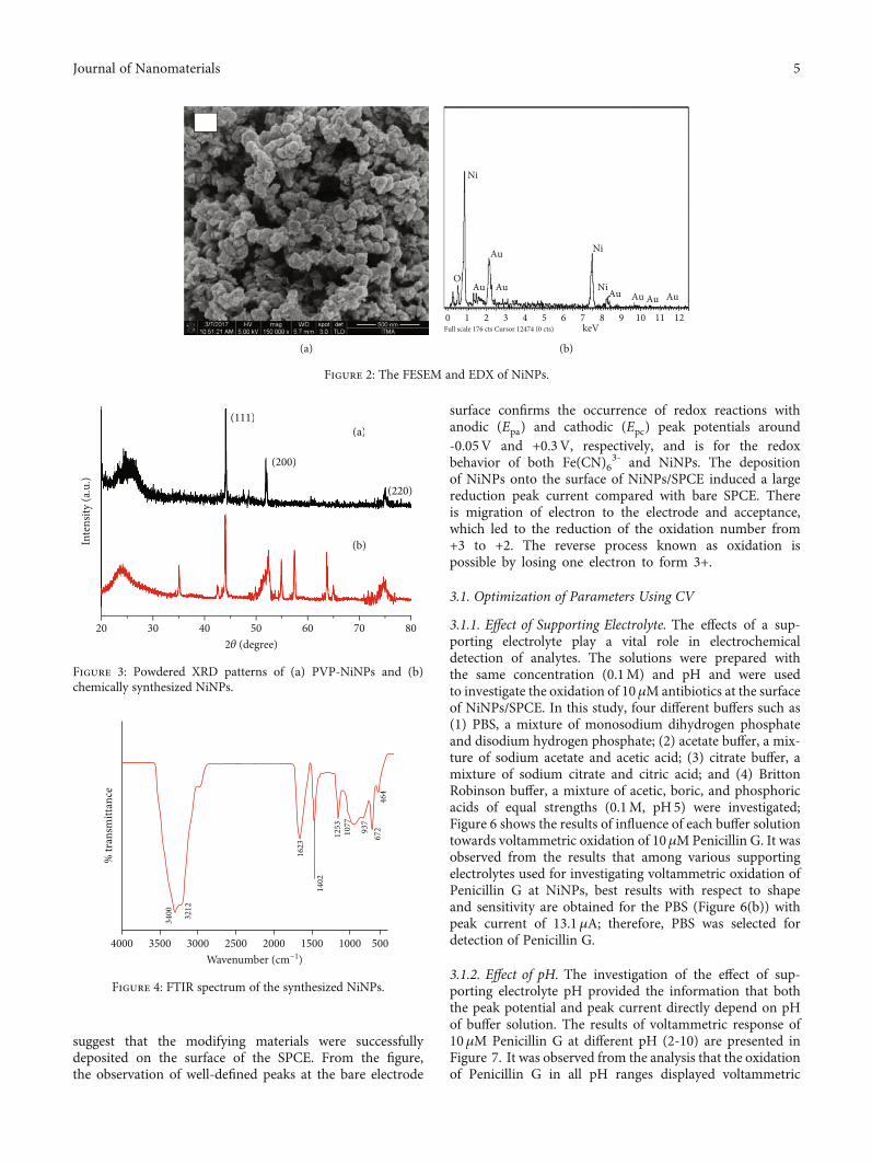
Figure 4 shows the FTIR spectrum of NiNPs at roomtemperature where the spectrum was recorded in the wave-number range of 4000–400 cm-1. The FTIR spectrum showscharacteristic absorption bands at 3212, 1623, 1402, and1077 cm-1 which can be corresponding to the O-H stretchingfrequency from the Ni(OH)2 groups, and the band around1623 suggests for the C=O group, while 1077 cm-1 bandreveals for the N-H stretching frequency from APTES,respectively [30].
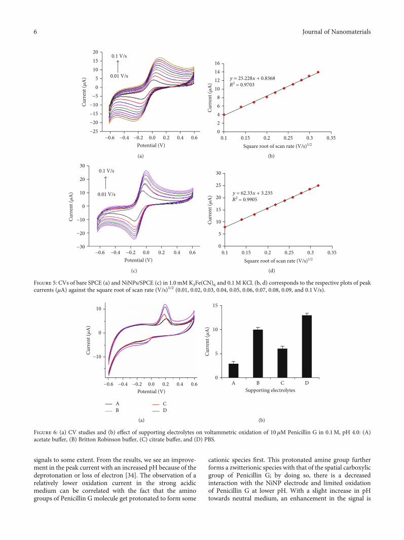
Figures 5(a)–5(d) show electrochemical behaviors ofpenicillin at the surface of the electrode and the observationof weak reversible redox peaks at the surface of bare SPCE(Figure 5(a)), indicating the slow-moving electron transferproperty of electrode and lower accumulation of penicillinon it which is a result of the low surface area. The sensitivityof redox peaks enhanced dramatically when we used theSPCE’s surface modified with the NiNPs (Figure 5(c)). Theincreased peak current following the SPCE modification withNiNPs is due to an enhancement in the surface area and elec-tron transfer rate of SPCE, and all are associated with theinbuilt conductive properties of Ni. In general, the potentialof the developed sensor depends on various factors such aseffect of supporting electrolytes and redox indicators, accu-mulation potential and time, selectivity, sensitivity, range oflinear responses, and detection limits. The figure indicatesCVs of modified and unmodified SPCE in the electrolytesolutions of 0.1mM K3Fe(CN)6 and 0.1M HCl, with redoxpeaks in the potential range of -0.6 to +0.6V. The bare SPCEresults show lower peak current, and this was increased from0.15 to 20μA when we incorporated the NiNPs to modify theelectrode surface. For the NiNPs/SPCE, the highest currentof 20μA was observed, and this peak current is directly pro-portional to the square root of the scan rate, which suggeststhat the process is controlled by diffusion [31].
Further, to test the efficiency of NiNPs for sensors, theactive surface area of bare and NiNP-modified SPCE wascalculated at the CVs recorded from 1mM K3Fe(CN)6containing 50mM KCl solution at different scan rates (10-100mV/s), and for that, the Randles-Sevcik formula shownbelow [32] was used. From the analysis, the bare SPCE wasfound to maintain the active surface area of 0.034 cm2 whilethe NiNP-modified electrode has an increase in the activesurface area to be 0.084 cm2. This confirms that the increasein the active surface area of the SPCE following the modifica-tion with that of the NiNPs [33]:
Aeff = Slope2:69 × 105n3/2D1/2C
, ð5Þ
where n is the number of electrons participating inthe redox reaction, A is the surface area of the electrode(cm2), D stands for the diffusion coefficient of moleculesin solution (7:6 × 10‐6 cm2 s-1), and C is the concentra-tion of K3Fe(CN)6 (M). v is the scan rate (Vs-1), fromthe slope of plot of Ipc vs. v1/2.
As shown in Figure 5(b), the anodic current increasesfrom bare SPCE to modified NiNPs on the surface which
4 Journal of Nanomaterials
suggest that the modifying materials were successfullydeposited on the surface of the SPCE. From the figure,the observation of well-defined peaks at the bare electrode
surface confirms the occurrence of redox reactions withanodic (Epa) and cathodic (Epc) peak potentials around-0.05V and +0.3V, respectively, and is for the redoxbehavior of both Fe(CN)6
3- and NiNPs. The depositionof NiNPs onto the surface of NiNPs/SPCE induced a largereduction peak current compared with bare SPCE. Thereis migration of electron to the electrode and acceptance,which led to the reduction of the oxidation number from+3 to +2. The reverse process known as oxidation ispossible by losing one electron to form 3+.
3.1. Optimization of Parameters Using CV
3.1.1. Effect of Supporting Electrolyte. The effects of a sup-porting electrolyte play a vital role in electrochemicaldetection of analytes. The solutions were prepared withthe same concentration (0.1M) and pH and were usedto investigate the oxidation of 10μMantibiotics at the surfaceof NiNPs/SPCE. In this study, four different buffers such as(1) PBS, a mixture of monosodium dihydrogen phosphateand disodium hydrogen phosphate; (2) acetate buffer, a mix-ture of sodium acetate and acetic acid; (3) citrate buffer, amixture of sodium citrate and citric acid; and (4) BrittonRobinson buffer, a mixture of acetic, boric, and phosphoricacids of equal strengths (0.1M, pH5) were investigated;Figure 6 shows the results of influence of each buffer solutiontowards voltammetric oxidation of 10μMPenicillin G. It wasobserved from the results that among various supportingelectrolytes used for investigating voltammetric oxidation ofPenicillin G at NiNPs, best results with respect to shapeand sensitivity are obtained for the PBS (Figure 6(b)) withpeak current of 13.1μA; therefore, PBS was selected fordetection of Penicillin G.
3.1.2. Effect of pH. The investigation of the effect of sup-porting electrolyte pH provided the information that boththe peak potential and peak current directly depend on pHof buffer solution. The results of voltammetric response of10μM Penicillin G at different pH (2-10) are presented inFigure 7. It was observed from the analysis that the oxidationof Penicillin G in all pH ranges displayed voltammetric
(a)
0 1 2 3 4
Au
Au Au Au Au Au Au
O
Ni
Ni
Ni
5 6 7 8keVFull scale 176 cts Cursor 12474 (0 cts)
9 10 11 12
(b)
Figure 2: The FESEM and EDX of NiNPs.
20 30 40
Inte
nsity
(a.u
.)
50
(111)
(200)
(220)
602𝜃 (degree)
70
(b)
(a)
80
(111)
(200)
(220)
(b)
(a)
Figure 3: Powdered XRD patterns of (a) PVP-NiNPs and (b)chemically synthesized NiNPs.
4000 3500 3000
3400 32
12
1402
1623
1253
1077
937
672
464
2500 2000 1500Wavenumber (cm−1)
1000 500
% tr
ansm
ittan
ce
Figure 4: FTIR spectrum of the synthesized NiNPs.
5Journal of Nanomaterials
signals to some extent. From the results, we see an improve-ment in the peak current with an increased pH because of thedeprotonation or loss of electron [34]. The observation of arelatively lower oxidation current in the strong acidicmedium can be correlated with the fact that the aminogroups of Penicillin G molecule get protonated to form some
cationic species first. This protonated amine group furtherforms a zwitterionic species with that of the spatial carboxylicgroup of Penicillin G; by doing so, there is a decreasedinteraction with the NiNP electrode and limited oxidationof Penicillin G at lower pH. With a slight increase in pHtowards neutral medium, an enhancement in the signal is
−0.6
0.01 V/s
0.1 V/s
−25−20−15−10
−505
Curr
ent (𝜇
A)
101520
−0.4 −0.2 0.0 0.2Potential (V)
0.4 0.6
(a)
0.102468
10121416
0.15
Curr
ent (𝜇
A)
0.2
y = 25.228x + 0.8368R2 = 0.9703
0.25 0.3 0.35Square root of scan rate (V/s)1/2
(b)
−0.6
0.01 V/s
0.1 V/s
−20
−30
−10
0
Curr
ent (𝜇
A) 10
20
30
−0.4 −0.2 0.0 0.2Potential (V)
0.4 0.6
(c)
Curr
ent (𝜇
A)
0.10
5
10
15
20
25
30
0.15 0.2
y = 62.33x + 3.235R2 = 0.9905
0.25 0.3 0.35Square root of scan rate (V/s)1/2
(d)
Figure 5: CVs of bare SPCE (a) and NiNPs/SPCE (c) in 1.0mM K3Fe(CN)6 and 0.1M KCl. (b, d) corresponds to the respective plots of peakcurrents (μA) against the square root of scan rate (V/s)1/2 (0.01, 0.02, 0.03, 0.04, 0.05, 0.06, 0.07, 0.08, 0.09, and 0.1V/s).
−10
−0.6 −0.4 −0.2 0.0 0.2
A
0.4Potential (V)
0.6
0
10
Curr
ent (𝜇
A)
BCD
(a)
0
5
10
15
Supporting electrolytesA B C D
Curr
ent (𝜇
A)
(b)
Figure 6: (a) CV studies and (b) effect of supporting electrolytes on voltammetric oxidation of 10μM Penicillin G in 0.1M, pH 4.0: (A)acetate buffer, (B) Britton Robinson buffer, (C) citrate buffer, and (D) PBS.
6 Journal of Nanomaterials
observed which can be attributed to a better interactionbetween analyte and electrode corresponding to the fact ofdeprotonation of the amino group and no such zwitterionformation with the carboxylic group of Penicillin G, and allsupported for enhanced oxidation and associated currentincrease. With further increase of pH towards more basicconditions, we observed a decreased oxidation current,and this can be attributed to the formation of zwitterionspecies again by the migration of proton intramolecularlyfrom the amino group to the carboxylic group of PenicillinG. Under basic conditions, a greater number of free OH-
are available and they can readily grab the proton fromthe acidic group of Penicillin G to form COO- which rapidlyforms zwitterion by taking proton from the amine group,and this process occurs reversibly that finally reduces theoxidation. Thus, the best results with respect to sensitivityand shape of voltammogram is obtained at pH 7.0 with peakcurrent of 14μA for Penicillin G. Therefore, 0.1M PBS ofpH 7.0 was chosen as optimum medium for voltammetricoxidation of Penicillin G at NiNPs/SPCE. The observationof PBS of pH 7.0 as the optimum condition for the voltam-metric oxidation of Penicillin G is in close conformity withthe previous report by [35].
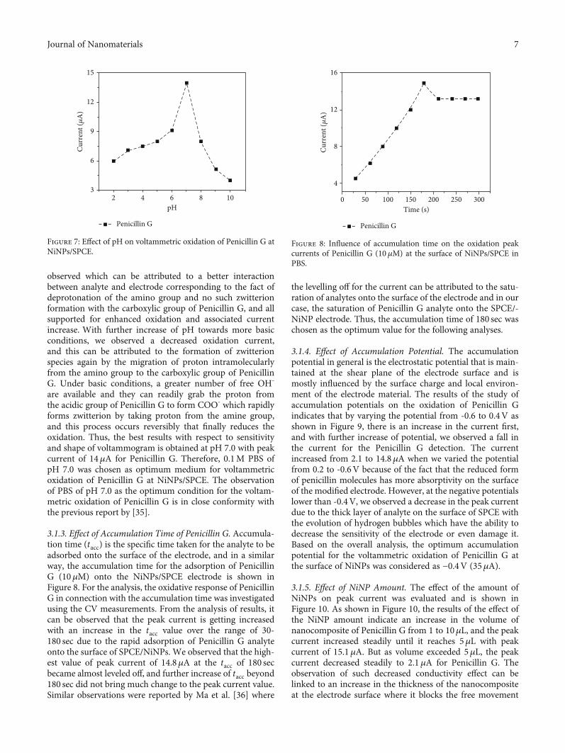
3.1.3. Effect of Accumulation Time of Penicillin G. Accumula-tion time (tacc) is the specific time taken for the analyte to beadsorbed onto the surface of the electrode, and in a similarway, the accumulation time for the adsorption of PenicillinG (10μM) onto the NiNPs/SPCE electrode is shown inFigure 8. For the analysis, the oxidative response of PenicillinG in connection with the accumulation time was investigatedusing the CV measurements. From the analysis of results, itcan be observed that the peak current is getting increasedwith an increase in the tacc value over the range of 30-180 sec due to the rapid adsorption of Penicillin G analyteonto the surface of SPCE/NiNPs. We observed that the high-est value of peak current of 14.8μA at the tacc of 180 secbecame almost leveled off, and further increase of tacc beyond180 sec did not bring much change to the peak current value.Similar observations were reported by Ma et al. [36] where
the levelling off for the current can be attributed to the satu-ration of analytes onto the surface of the electrode and in ourcase, the saturation of Penicillin G analyte onto the SPCE/-NiNP electrode. Thus, the accumulation time of 180 sec waschosen as the optimum value for the following analyses.
3.1.4. Effect of Accumulation Potential. The accumulationpotential in general is the electrostatic potential that is main-tained at the shear plane of the electrode surface and ismostly influenced by the surface charge and local environ-ment of the electrode material. The results of the study ofaccumulation potentials on the oxidation of Penicillin Gindicates that by varying the potential from -0.6 to 0.4V asshown in Figure 9, there is an increase in the current first,and with further increase of potential, we observed a fall inthe current for the Penicillin G detection. The currentincreased from 2.1 to 14.8μA when we varied the potentialfrom 0.2 to -0.6V because of the fact that the reduced formof penicillin molecules has more absorptivity on the surfaceof the modified electrode. However, at the negative potentialslower than -0.4V, we observed a decrease in the peak currentdue to the thick layer of analyte on the surface of SPCE withthe evolution of hydrogen bubbles which have the ability todecrease the sensitivity of the electrode or even damage it.Based on the overall analysis, the optimum accumulationpotential for the voltammetric oxidation of Penicillin G atthe surface of NiNPs was considered as −0.4V (35μA).
3.1.5. Effect of NiNP Amount. The effect of the amount ofNiNPs on peak current was evaluated and is shown inFigure 10. As shown in Figure 10, the results of the effect ofthe NiNP amount indicate an increase in the volume ofnanocomposite of Penicillin G from 1 to 10μL, and the peakcurrent increased steadily until it reaches 5μL with peakcurrent of 15.1μA. But as volume exceeded 5μL, the peakcurrent decreased steadily to 2.1μA for Penicillin G. Theobservation of such decreased conductivity effect can belinked to an increase in the thickness of the nanocompositeat the electrode surface where it blocks the free movement
23
6
9
12
Penicillin G
Curr
ent (𝜇
A)
15
4 6pH
8 10
Figure 7: Effect of pH on voltammetric oxidation of Penicillin G atNiNPs/SPCE.
0 50 100 150 200Time (s)
250 300
4
8
12
16
Curr
ent (𝜇
A)
Penicillin G
Figure 8: Influence of accumulation time on the oxidation peakcurrents of Penicillin G (10 μM) at the surface of NiNPs/SPCE inPBS.
7Journal of Nanomaterials
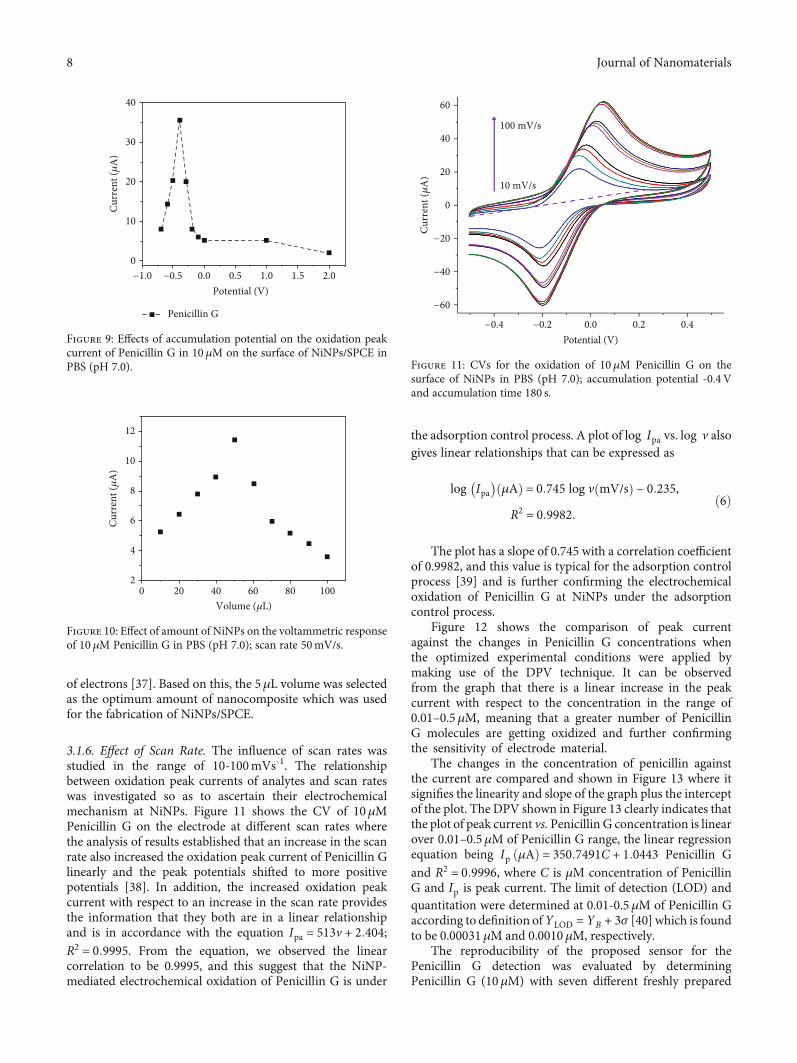
of electrons [37]. Based on this, the 5μL volume was selectedas the optimum amount of nanocomposite which was usedfor the fabrication of NiNPs/SPCE.
3.1.6. Effect of Scan Rate. The influence of scan rates wasstudied in the range of 10-100mVs-1. The relationshipbetween oxidation peak currents of analytes and scan rateswas investigated so as to ascertain their electrochemicalmechanism at NiNPs. Figure 11 shows the CV of 10μMPenicillin G on the electrode at different scan rates wherethe analysis of results established that an increase in the scanrate also increased the oxidation peak current of Penicillin Glinearly and the peak potentials shifted to more positivepotentials [38]. In addition, the increased oxidation peakcurrent with respect to an increase in the scan rate providesthe information that they both are in a linear relationshipand is in accordance with the equation Ipa = 513v + 2:404;R2 = 0:9995. From the equation, we observed the linearcorrelation to be 0.9995, and this suggest that the NiNP-mediated electrochemical oxidation of Penicillin G is under
the adsorption control process. A plot of log Ipa vs. log v alsogives linear relationships that can be expressed as
log Ipa� �
μAð Þ = 0:745 log v mV/sð Þ − 0:235,
R2 = 0:9982:ð6Þ
The plot has a slope of 0.745 with a correlation coefficientof 0.9982, and this value is typical for the adsorption controlprocess [39] and is further confirming the electrochemicaloxidation of Penicillin G at NiNPs under the adsorptioncontrol process.
Figure 12 shows the comparison of peak currentagainst the changes in Penicillin G concentrations whenthe optimized experimental conditions were applied bymaking use of the DPV technique. It can be observedfrom the graph that there is a linear increase in the peakcurrent with respect to the concentration in the range of0.01–0.5μM, meaning that a greater number of PenicillinG molecules are getting oxidized and further confirmingthe sensitivity of electrode material.
The changes in the concentration of penicillin againstthe current are compared and shown in Figure 13 where itsignifies the linearity and slope of the graph plus the interceptof the plot. The DPV shown in Figure 13 clearly indicates thatthe plot of peak current vs. Penicillin G concentration is linearover 0.01–0.5μM of Penicillin G range, the linear regressionequation being Ip ðμAÞ = 350:7491C + 1:0443 Penicillin Gand R2 = 0:9996, where C is μM concentration of PenicillinG and Ip is peak current. The limit of detection (LOD) andquantitation were determined at 0.01-0.5μM of Penicillin Gaccording to definition ofYLOD = YB + 3σ [40] which is foundto be 0.00031μM and 0.0010μM, respectively.
The reproducibility of the proposed sensor for thePenicillin G detection was evaluated by determiningPenicillin G (10μM) with seven different freshly prepared
−1.0 −0.5 0.0 0.5 1.0Potential (V)
1.5 2.00
10
20
30
40Cu
rren
t (𝜇
A)
Penicillin G
Figure 9: Effects of accumulation potential on the oxidation peakcurrent of Penicillin G in 10 μM on the surface of NiNPs/SPCE inPBS (pH 7.0).
0 20 40 60 80 1002
4
6
8
10
12
Curr
ent (𝜇
A)
Volume (𝜇L)
Figure 10: Effect of amount of NiNPs on the voltammetric responseof 10 μM Penicillin G in PBS (pH 7.0); scan rate 50mV/s.
−0.4
−60
−40
−20
0
20
40100 mV/s
10 mV/s
60
−0.2 0.0 0.2Potential (V)
0.4
Curr
ent (𝜇
A)
Figure 11: CVs for the oxidation of 10μM Penicillin G on thesurface of NiNPs in PBS (pH 7.0); accumulation potential -0.4 Vand accumulation time 180 s.
8 Journal of Nanomaterials
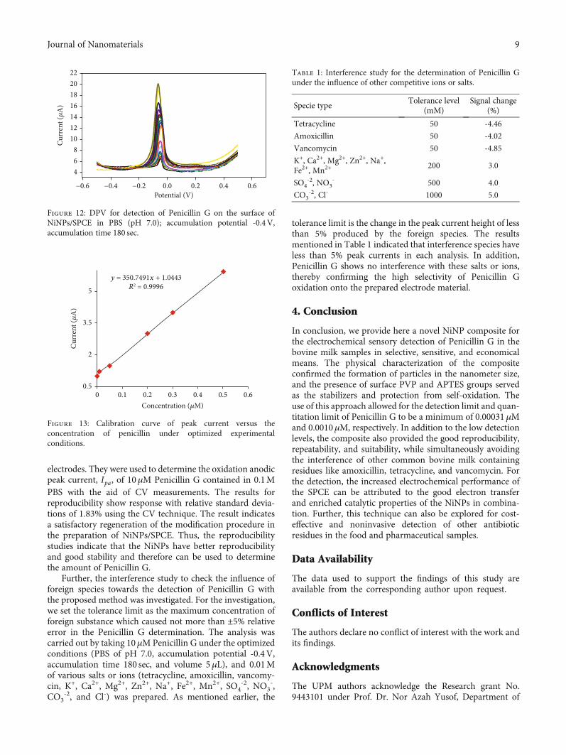
electrodes. They were used to determine the oxidation anodicpeak current, Ipa, of 10μM Penicillin G contained in 0.1MPBS with the aid of CV measurements. The results forreproducibility show response with relative standard devia-tions of 1.83% using the CV technique. The result indicatesa satisfactory regeneration of the modification procedure inthe preparation of NiNPs/SPCE. Thus, the reproducibilitystudies indicate that the NiNPs have better reproducibilityand good stability and therefore can be used to determinethe amount of Penicillin G.
Further, the interference study to check the influence offoreign species towards the detection of Penicillin G withthe proposed method was investigated. For the investigation,we set the tolerance limit as the maximum concentration offoreign substance which caused not more than ±5% relativeerror in the Penicillin G determination. The analysis wascarried out by taking 10μMPenicillin G under the optimizedconditions (PBS of pH 7.0, accumulation potential -0.4V,accumulation time 180 sec, and volume 5μL), and 0.01Mof various salts or ions (tetracycline, amoxicillin, vancomy-cin, K+, Ca2+, Mg2+, Zn2+, Na+, Fe2+, Mn2+, SO4
-2, NO3-,
CO3-2, and Cl-) was prepared. As mentioned earlier, the
tolerance limit is the change in the peak current height of lessthan 5% produced by the foreign species. The resultsmentioned in Table 1 indicated that interference species haveless than 5% peak currents in each analysis. In addition,Penicillin G shows no interference with these salts or ions,thereby confirming the high selectivity of Penicillin Goxidation onto the prepared electrode material.
4. Conclusion
In conclusion, we provide here a novel NiNP composite forthe electrochemical sensory detection of Penicillin G in thebovine milk samples in selective, sensitive, and economicalmeans. The physical characterization of the compositeconfirmed the formation of particles in the nanometer size,and the presence of surface PVP and APTES groups servedas the stabilizers and protection from self-oxidation. Theuse of this approach allowed for the detection limit and quan-titation limit of Penicillin G to be a minimum of 0.00031μMand 0.0010μM, respectively. In addition to the low detectionlevels, the composite also provided the good reproducibility,repeatability, and suitability, while simultaneously avoidingthe interference of other common bovine milk containingresidues like amoxicillin, tetracycline, and vancomycin. Forthe detection, the increased electrochemical performance ofthe SPCE can be attributed to the good electron transferand enriched catalytic properties of the NiNPs in combina-tion. Further, this technique can also be explored for cost-effective and noninvasive detection of other antibioticresidues in the food and pharmaceutical samples.
Data Availability
The data used to support the findings of this study areavailable from the corresponding author upon request.
Conflicts of Interest
The authors declare no conflict of interest with the work andits findings.
Acknowledgments
The UPM authors acknowledge the Research grant No.9443101 under Prof. Dr. Nor Azah Yusof, Department of
4
−0.6 −0.4 −0.2 0.0 0.2Potential (V)
0.4 0.6
68
10121416182022
Curr
ent (𝜇
A)
Figure 12: DPV for detection of Penicillin G on the surface ofNiNPs/SPCE in PBS (pH 7.0); accumulation potential -0.4 V,accumulation time 180 sec.
Curr
ent (𝜇
A)
00.5
2
3.5
5
0.1 0.2 0.3 0.4Concentration (𝜇M)
0.5 0.6
y = 350.7491x + 1.0443R2 = 0.9996
Figure 13: Calibration curve of peak current versus theconcentration of penicillin under optimized experimentalconditions.
Table 1: Interference study for the determination of Penicillin Gunder the influence of other competitive ions or salts.
Specie typeTolerance level
(mM)Signal change
(%)
Tetracycline 50 -4.46
Amoxicillin 50 -4.02
Vancomycin 50 -4.85
K+, Ca2+, Mg2+, Zn2+, Na+,Fe2+, Mn2+
200 3.0
SO4-2, NO3
- 500 4.0
CO3-2, Cl- 1000 5.0
9Journal of Nanomaterials
Chemistry, Faculty of Science, Universiti Putra Malaysia, andextend their thanks to the Faculty of Science, Universiti PutraMalaysia. The King Saud University authors extend theirappreciation to the Deanship of Scientific Research at KingSaud University for funding the work through the researchgroup project no. RG-148.
References
[1] M. D. H. Wirzal, A. R. M. Yusoff, J. Zima, and J. Barek,“Degradation of ampicillin and penicillin G using anodic oxi-dation,” International Journal of Electrochemical Science,vol. 8, pp. 8978–8988, 2013.
[2] P. D. Anderson, V. J. D'Aco, P. Shanahan et al., “Screeninganalysis of human pharmaceutical compounds in U.S. surfacewaters,” Environmental Science & Technology, vol. 38, no. 3,pp. 838–849, 2004.
[3] M. Rabiet, A. Togola, F. Brissaud, J. L. Seidel, H. Budzinski,and F. Elbaz-Poulichet, “Consequences of treated water recy-cling as regards pharmaceuticals and drugs in surface andground waters of a medium-sized Mediterranean catchment,”Environmental Science & Technology, vol. 40, no. 17, pp. 5282–5288, 2006.
[4] W. Du, H. Zhou, Z. Luo et al., “Selective determination ofpenicillin G from tap water and milk samples using surfacemolecularly imprinted polymers as solid-phase extraction sor-bent,” Molecular Imprinting, vol. 2, no. 1, pp. 18–29, 2014.
[5] D. HELLER, M. SMITH, and O. CHIESA, “LC/MS/MS mea-surement of penicillin G in bovine plasma, urine, and biopsysamples taken from kidneys of standing animals,” Journal ofChromatography B, vol. 830, no. 1, pp. 91–99, 2006.
[6] A. Junza, R. Amatya, D. Barrón, and J. Barbosa, “Comparativestudy of the LC-MS/MS and UPLC-MS/MS for the multi-residue analysis of quinolones, penicillins and cephalosporinsin cow milk, and validation according to the regulation2002/657/EC,” Journal of Chromatography B, vol. 879,no. 25, pp. 2601–2610, 2011.
[7] M. Seifrtová, L. Nováková, C. Lino, A. Pena, and P. Solich, “Anoverview of analytical methodologies for the determination ofantibiotics in environmental waters,” Analytica Chimica Acta,vol. 649, no. 2, pp. 158–179, 2009.
[8] A. V. Herrera-Herrera, L. M. Ravelo-Pérez, J. Hernández-Borges, M. M. Afonso, J. A. Palenzuela, and M. Á. Rodrí-guez-Delgado, “Oxidized multi-walled carbon nanotubes forthe dispersive solid-phase extraction of quinolone antibi-otics from water samples using capillary electrophoresisand large volume sample stacking with polarity switching,”Journal of Chromatography A, vol. 1218, no. 31, pp. 5352–5361, 2011.
[9] S. Ge, W. Tang, R. Han et al., “Sensitive analysis of amino-glycoside antibiotics via hyphenation of transient movingsubstitution boundary with field-enhanced sample injectionin capillary electrophoresis,” Journal of Chromatography A,vol. 1295, pp. 128–135, 2013.
[10] P. Lacina, L. Mravcová, and M. Vávrová, “Application ofcomprehensive two-dimensional gas chromatography withmass spectrometric detection for the analysis of selecteddrug residues in wastewater and surface water,” Journal ofEnvironmental Sciences, vol. 25, no. 1, pp. 204–212, 2013.
[11] N. Migowska, M. Caban, P. Stepnowski, and J. Kumirska,“Simultaneous analysis of non-steroidal anti-inflammatorydrugs and estrogenic hormones in water and wastewater sam-
ples using gas chromatography-mass spectrometry and gaschromatography with electron capture detection,” Science ofthe Total Environment, vol. 441, pp. 77–88, 2012.
[12] F. J. Lara, M. del Olmo-Iruela, C. Cruces-Blanco,C. Quesada-Molina, and A. M. García-Campaña, “Advancesin the determination of β-lactam antibiotics by liquid chro-matography,” Trends in Analytical Chemistry, vol. 38,pp. 52–66, 2012.
[13] B. Le Bizec, G. Pinel, and J. P. Antignac, “Options for vet-erinary drug analysis using mass spectrometry,” Journal ofChromatography A, vol. 1216, no. 46, pp. 8016–8034, 2009.
[14] E. S. Elmolla and M. Chaudhuri, “Degradation of amoxicillin,ampicillin and cloxacillin antibiotics in aqueous solution bythe UV/ZnO photocatalytic process,” Journal of HazardousMaterials, vol. 173, no. 1-3, pp. 445–449, 2010.
[15] E. Isarain-Chávez, R. M. Rodríguez, P. L. Cabot et al., “Degra-dation of pharmaceutical beta-blockers by electrochemicaladvanced oxidation processes using a flow plant with a solarcompound parabolic collector,” Water Research, vol. 45,no. 14, pp. 4119–4130, 2011.
[16] A. Dirany, I. Sirés, N. Oturan, and M. A. Oturan, “Electro-chemical abatement of the antibiotic sulfamethoxazole fromwater,” Chemosphere, vol. 81, no. 5, pp. 594–602, 2010.
[17] E. S. Elmolla and M. Chaudhuri, “Photocatalytic degradationof amoxicillin, ampicillin and cloxacillin antibiotics in aqueoussolution using UV/TiO2 and UV/H2O2/TiO2 photocatalysis,”Desalination, vol. 252, no. 1-3, pp. 46–52, 2010.
[18] M. Klavarioti, D. Mantzavinos, and D. Kassinos, “Removal ofresidual pharmaceuticals from aqueous systems by advancedoxidation processes,” Environment International, vol. 35,no. 2, pp. 402–417, 2009.
[19] N. Sanvicens, I. Mannelli, J. P. Salvador, E. Valera, and M. P.Marco, “Biosensors for pharmaceuticals based on noveltechnology,” Trends in Analytical Chemistry, vol. 30, no. 3,pp. 541–553, 2011.
[20] C. Cristea, M. Tertis, and R. Galatus, “Magnetic nanoparticlesfor antibiotics detection,” Nanomaterials, vol. 7, no. 6, p. 119,2017.
[21] P. de Lima-Neto, A. N. Correia, R. R. Portela, M. da SilvaJulião, G. F. Linhares-Junior, and J. E. S. de Lima, “Square wavevoltammetric determination of nitrofurantoin in pharmaceu-tical formulations on highly boron-doped diamond electrodesat different boron- doping contents,” Talanta, vol. 80, no. 5,pp. 1730–1736, 2010.
[22] R. Gomes, “Determination of endocrine disrupters in sewagetreatment and receiving waters,” Trends in Analytical Chemis-try, vol. 22, no. 10, pp. 697–707, 2003.
[23] M. Panizza and G. Cerisola, “Application of diamondelectrodes to electrochemical processes,” Electrochimica Acta,vol. 51, no. 2, pp. 191–199, 2005.
[24] N. W. Khun and E. Liu, “Linear sweep anodic strippingvoltammetry of heavy metals from nitrogen doped tetrahedralamorphous carbon thin films,” Electrochimica Acta, vol. 54,no. 10, pp. 2890–2898, 2009.
[25] P. N. Dave, P. N. Ram, and S. Chaturvedi, “Transition metaloxide nanoparticles: potential nano-modifier for rocketpropellants,” Particulate Science and Technology, vol. 34,no. 6, pp. 676–680, 2016.
[26] J. W. Park, E. H. Chae, S. H. Kim et al., “Preparation of fine Nipowders from nickel hydrazine complex,”Materials Chemistryand Physics, vol. 97, no. 2-3, pp. 371–378, 2006.
10 Journal of Nanomaterials
[27] T. Goto, Y. Ito, S. Yamada, H. Matsumoto, and H. Oka, “High-throughput analysis of tetracycline and penicillin antibiotics inanimal tissues using electrospray tandem mass spectrometrywith selected reaction monitoring transition,” Journal of Chro-matography A, vol. 1100, no. 2, pp. 193–199, 2005.
[28] M. Moors and D. L. Massart, “Evaluation of solid-phaseextraction of basic drugs from human milk,” Journal ofPharmaceutical and Biomedical Analysis, vol. 9, no. 2,pp. 129–139, 1991.
[29] R. G. Chaudhary, J. A. Tanna, N. V. Gandhare, A. R. Rai, andH. D. Juneja, “Synthesis of nickel nanoparticles: microscopicinvestigation, an efficient catalyst and effective antibacterialactivity,” Advanced Materials Letters, vol. 6, no. 11, pp. 990–998, 2015.
[30] S. Sudhasree, A. S. Banu, P. Brindha, and G. A. Kurian,“Synthesis of nickel nanoparticles by chemical and greenroute and their comparison in respect to biological effectand toxicity,” Toxicological & Environmental Chemistry,vol. 96, no. 5, pp. 743–754, 2014.
[31] H. Zhang, R. Xiao, B. Jin, D. Shen, R. Chen, and G. Xiao,“Catalytic fast pyrolysis of straw biomass in an internallyinterconnected fluidized bed to produce aromatics andolefins: effect of different catalysts,” Bioresource Technology,vol. 137, pp. 82–87, 2013.
[32] J. Du, R. Yue, F. Ren et al., “Novel graphene flowers modifiedcarbon fibers for simultaneous determination of ascorbic acid,dopamine and uric acid,” Biosensors and Bioelectronics, vol. 53,pp. 220–224, 2014.
[33] L. Zhang, F. Zhang, X. Yang et al., “Porous 3D graphene-basedbulk materials with exceptional high surface area and excellentconductivity for supercapacitors,” Scientific Reports, vol. 3,no. 1, article 1408, 2013.
[34] H. Bagheri, A. Afkhami, Y. Panahi, H. Khoshsafar, andA. Shirzadmehr, “Facile stripping voltammetric determinationof haloperidol using a high performance magnetite/carbonnanotube paste electrode in pharmaceutical and biologicalsamples,” Materials Science and Engineering C, vol. 37,pp. 264–270, 2014.
[35] M. S. Ibrahim, D. A. Kulesh, S. S. Saleh et al., “Real-time PCRassay to detect smallpox virus,” Journal of Clinical Microbiol-ogy, vol. 41, no. 8, pp. 3835–3839, 2003.
[36] X. Ma, J. Geiser-Lee, Y. Deng, and A. Kolmakov, “Interactionsbetween engineered nanoparticles (ENPs) and plants: phyto-toxicity, uptake and accumulation,” Science of the Total Envi-ronment, vol. 408, no. 16, pp. 3053–3061, 2010.
[37] W. Li, X. Z. Tang, H. B. Zhang et al., “Simultaneous surfacefunctionalization and reduction of graphene oxide withoctadecylamine for electrically conductive polystyrene com-posites,” Carbon, vol. 49, no. 14, pp. 4724–4730, 2011.
[38] A. Muhammad, R. Hajian, N. A. Yusof et al., “A screen printedcarbon electrode modified with carbon nanotubes and goldnanoparticles as a sensitive electrochemical sensor for deter-mination of thiamphenicol residue in milk,” RSC Advances,vol. 8, no. 5, pp. 2714–2722, 2018.
[39] G. Singh, I. Y. Kim, K. S. Lakhi, P. Srivastava, R. Naidu,and A. Vinu, “Single step synthesis of activated bio-carbons with a high surface area and their excellent CO2adsorption capacity,” Carbon, vol. 116, pp. 448–455, 2017.
[40] S. Cherian, R. K. Gupta, B. C. Mullin, and T. Thundat, “Detec-tion of heavy metal ions using protein-functionalized micro-cantilever sensors,” Biosensors and Bioelectronics, vol. 19,no. 5, pp. 411–416, 2003.
11Journal of Nanomaterials

CorrosionInternational Journal of
Hindawiwww.hindawi.com Volume 2018
Advances in
Materials Science and EngineeringHindawiwww.hindawi.com Volume 2018
Hindawiwww.hindawi.com Volume 2018
Journal of
Chemistry
Analytical ChemistryInternational Journal of
Hindawiwww.hindawi.com Volume 2018
Scienti�caHindawiwww.hindawi.com Volume 2018
Polymer ScienceInternational Journal of
Hindawiwww.hindawi.com Volume 2018
Hindawiwww.hindawi.com Volume 2018
Advances in Condensed Matter Physics
Hindawiwww.hindawi.com Volume 2018
International Journal of
BiomaterialsHindawiwww.hindawi.com
Journal ofEngineeringVolume 2018
Applied ChemistryJournal of
Hindawiwww.hindawi.com Volume 2018
NanotechnologyHindawiwww.hindawi.com Volume 2018
Journal of
Hindawiwww.hindawi.com Volume 2018
High Energy PhysicsAdvances in
Hindawi Publishing Corporation http://www.hindawi.com Volume 2013Hindawiwww.hindawi.com
The Scientific World Journal
Volume 2018
TribologyAdvances in
Hindawiwww.hindawi.com Volume 2018
Hindawiwww.hindawi.com Volume 2018
ChemistryAdvances in
Hindawiwww.hindawi.com Volume 2018
Advances inPhysical Chemistry
Hindawiwww.hindawi.com Volume 2018
BioMed Research InternationalMaterials
Journal of
Hindawiwww.hindawi.com Volume 2018
Na
nom
ate
ria
ls
Hindawiwww.hindawi.com Volume 2018
Journal ofNanomaterials
Submit your manuscripts atwww.hindawi.com


![Ultrafine Nickel‐Nanoparticle‐Enabled SiO2 Hierarchical ...mai.group.whut.edu.cn/chs/lw/slected/201802/P...x/NiSi x with enhanced structural stability and kinetics.[34] However,](https://img.pdfslide.net/doc/110x75/600aa20a049ab32e231cee93/ultrafine-nickelananoparticleaenabled-sio2-hierarchical-maigroupwhuteducnchslwslected201802p.jpg)


![Platinum Nanoparticle Electrode Modified Iodine using ...€¦ · has also been used to simultaneously detect vitamin C and glucose [30].Recent combinations of various voltammetry](https://img.pdfslide.net/doc/110x75/6074f326a005a0047a162b64/platinum-nanoparticle-electrode-modified-iodine-using-has-also-been-used-to.jpg)