Embed Size (px)
Citation preview
nutrients
Communication
Night Blindness in Cystic Fibrosis: The Key Role ofVitamin A in the Digestive System
Lorenzo Norsa 1,†,* , Laura Zazzeron 2,†, Marialaura Cuomo 3, Laura Claut 2,Anna Marta Clotilde Bulfamante 2, Arianna Biffi 2 and Carla Colombo 2,4
1 Pediatric Hepatology Gastroenterology and Transplantation, ASST Papa Giovanni XXIII,24027 Bergamo, Italy
2 Cystic Fibrosis Center, Fondazione IRCCS Ca’ Granda Ospedale Maggiore Policlinico, University of Milan,20121 Milan, Italy
3 Department of Pediatrics, Fondazione MBBM Onlus/Ospedale San Gerardo, University of Milano-Bicocca,20121 Milan, Italy
4 Dipartimento di Fisiopatologia Medico-Chirurgica e dei Trapianti, Università degli Studi di Milano,20121 Milan, Italy
* Correspondence: [email protected]; Tel.: +39-035-267-3185† The authors equally contributed to the paper.
Received: 29 June 2019; Accepted: 8 August 2019; Published: 13 August 2019�����������������
Abstract: Vitamin A is a fundamental micronutrient that regulates various cellular patterns. VitaminA deficiency (VAT) is a worldwide problem and the primary cause of nocturnal blindness especially inlow income countries. Cystic fibrosis (CF) is a known risk factor of VAD because of liposoluble vitaminmalabsorption due to pancreatic insufficiency. We describe a case of a 9-year-old girl who experiencedrecurrent episodes of nocturnal blindness due to profound VAD. This little girl is paradigmatic forthe explanation of the key role of the gut–liver axis in vitamin A metabolism. She presents withmeconium ileus at birth, requiring intestinal resection that led to a transient intestinal failure withparenteral nutrition need. In addition, she suffered from cholestatic liver disease due to CF andintestinal failure-associated liver disease. The interaction of pancreatic function, intestinal absorptionand liver storage is fundamental for the correct metabolism of vitamin A.
Keywords: cystic fibrosis; vitamin A deficiency; short bowel syndrome
1. Introduction
Humans, and in general higher animals, must obtain vitamin A from their diet, either as apre-formed vitamin (retinol) from meat, eggs and milk, or as pro-vitamin carotenoids, primarily foundin yellow fruit, vegetables and carrots [1].
These are both fat soluble molecules that need bile acid micelle solubilization to be digested. Inthe lumen of the small intestine, long chain fatty acid retinol esters (the most represented pre-formedvitamin in the diet) are hydrolyzed by pancreatic lipase and by enzymes on the brush border, andtherefore free retinol can be absorbed by enterocytes. Beta carotene, the most abundant of carotenoidsin the diet, does not require digestion to enter intestinal cells and diffuses through the cell membranevia facilitated transport linked to a surface receptor. In the cell, retinol is esterified again to retinylester (mainly with palmitate) and is embedded in chylomicrons together with beta carotene, othercarotenoids and dietary lipids to be secreted in lymphatic vessels. Chylomicrons undergo remodelingprocesses resulting in chylomicron remnants, which are richer in retinyl esters and are up taken by theliver; here, retinol can be stored or can be bound to serum retinol binding protein (RBP4), a protein ofhepatic origin, and delivered to peripheral tissues [2].
Nutrients 2019, 11, 1876; doi:10.3390/nu11081876 www.mdpi.com/journal/nutrients
Nutrients 2019, 11, 1876 2 of 7
Normally the liver contains a two-year store of vitamin A, mainly as retinyl palmitate and stearatewhich can be hydrolyzed to retinol and secreted when needed [3].
Differently, carotenoids in the bloodstream are bound to high-density lipoprotein (HDL) andlow-density lipoprotein (LDL) with other lipids [1].
These micronutrient compounds are either individually or collectively required throughout life andregulate many processes including cell differentiation, proliferation and development, embryogenesis,growth, normal metabolism and immune function. Moreover, beta carotene also has an importantrole as an antioxidant too. In retinal pigment epithelium retinol is efficiently up taken by a membranereceptor, stimulated by retinoic acid 6 (STRA6), with high affinity for RBP4, and while bound to acellular RBP, is converted into 11-cis retinaldehyde, a chromophore that joins with the protein opsin toform rhodopsin, the molecular complex involved in vision [4].
Vitamin A deficiency (VAD) can be defined as serum retinol concentration lower than 0.70 µmol/L(less than 0.20 mg/L) [5] and is especially prevalent in low income countries [6], where malnutritionfrequently occurs. Nevertheless, hypovitaminosis A due to inadequate intake is the leading cause ofchildhood blindness worldwide; while rare in developed countries [7], it can arise due to intestinallipid malabsorption, pancreatic insufficiency, liver and bowel disease or surgical shortening.
According to these premises, we describe the case of a 9-year-old girl with a neonatal diagnosis ofcystic fibrosis (CF) with cholestatic liver disease and a history of bowel surgical shortening presentingwith night blindness secondary to VAD.
Data were collected retrospectively and anonymized after parents’ approval with informedconsent signature. No ethical clearance was applicable according to national regulations.
2. Case Report
A full-term newborn girl developed abdominal distention with emesis and failure to passmeconium 12 h after birth. The pregnancy had been complicated by an antenatal diagnosis ofpolyhydramnios and dilated bowel loops. The mother was in good clinical condition without anyhistory of vitamin deficit before or during pregnancy.
The baby underwent an extensive enterectomy with small bowel resection (involving 30 cm of theproximal ileum, the terminal atresic ileum, the ileocecal valve and the cecum) and ileostomy formationon day one of life.
The diagnosis of CF was confirmed by DNA analysis, showing compound heterozygosity forknown disease-causing mutations (F508del/L1077del).
During the first months of life, the patient presented with abnormal liver enzyme levels with normaltotal and direct bilirubin levels. Abdominal ultrasound revealed mild hepatomegaly and steatosis.
Despite a severe intestinal malabsorption with steatorrhea (steatocrit of about 20%), both weightand height increased, and growth pattern was satisfactory.
Minimal enteral feeds were tolerated, and the baby was dependent on parenteral nutrition (PN)including fat-soluble vitamin supplementation (A 1300 UI/die, D 240 UI, E 4.2 UI, K 120 µg per day).According to CF nutritional guidelines [4], she received oral nutrition support and multivitaminsupplementation (including vitamin A at 4000 UI daily).
The patient remained under close clinical follow-up until six months of age, when she developeda bowel obstruction due to adhesions, which required additional bowel surgery. After surgery, a totallength of 102 cm of small bowel was measured with a termino-terminal ileocolic anastomosis withoutileocecal valve.
For this condition of type 2 short bowel with intestinal failure, she was trained for home parenteralnutrition and discharged. Home PN (HPN) was successfully administered for the next 15 monthswhen it was gradually stopped because of normal growth.
She was then regularly followed-up at the CF center with adequate oral dietary intake viaoral nutrition, pancreatic enzyme replacement therapy and liposoluble vitamin supplementation athigh dose.
Nutrients 2019, 11, 1876 3 of 7
At four years of age, she started complaining of slow dark adaptation and nyctalopia. No othersymptoms were reported. An ophthalmic examination was performed: slit lamp examinationwas remarkable for a Bitot’s spot without retinal dystrophy; electroretinography (ERG) was withinnormal limits.
Night blindness due to systemic VAD was suspected and confirmed by a serum level of 0 mg/L(normal range of 0.30–0.70 mg/L), measured by high performance liquid chromatography (HPLC).
Fasting hypoglycemia was also documented, suggesting impaired intestinal absorption; thus,despite being within normal growth parameters, complementary HPN was restarted in order toprovide additional calories and micronutrients.
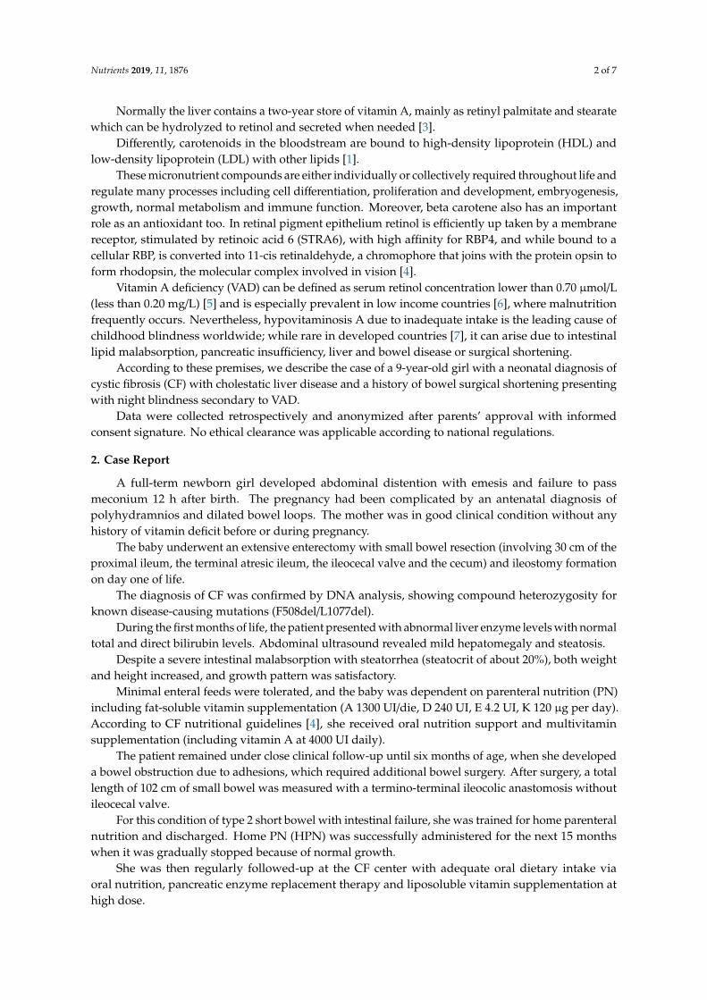
Within the first month of HPN, the night blindness resolved.After 10 months of parenteral supplementation, despite treatment with ursodeoxycholic acid
and efforts to optimize enteral feeding and cycling of HPN, severe intrahepatic cholestasis developed(Figure 1).
Nutrients 2019, 11, x FOR PEER REVIEW 3 of 7
At four years of age, she started complaining of slow dark adaptation and nyctalopia. No other symptoms were reported. An ophthalmic examination was performed: slit lamp examination was remarkable for a Bitot’s spot without retinal dystrophy; electroretinography (ERG) was within normal limits.
Night blindness due to systemic VAD was suspected and confirmed by a serum level of 0 mg/L (normal range of 0.30–0.70 mg/L), measured by high performance liquid chromatography (HPLC).
Fasting hypoglycemia was also documented, suggesting impaired intestinal absorption; thus, despite being within normal growth parameters, complementary HPN was restarted in order to provide additional calories and micronutrients.
Within the first month of HPN, the night blindness resolved. After 10 months of parenteral supplementation, despite treatment with ursodeoxycholic acid
and efforts to optimize enteral feeding and cycling of HPN, severe intrahepatic cholestasis developed (Figure 1).
Figure 1. Correlation between liver function tests (AST—ALT—direct bilirubin), vitamin A levels, parenteral nutrition (PN) administration and vitamin A supplementation.
Therefore, in consideration of her optimal clinical conditions and normal levels of micronutrients, HPN was stopped again.
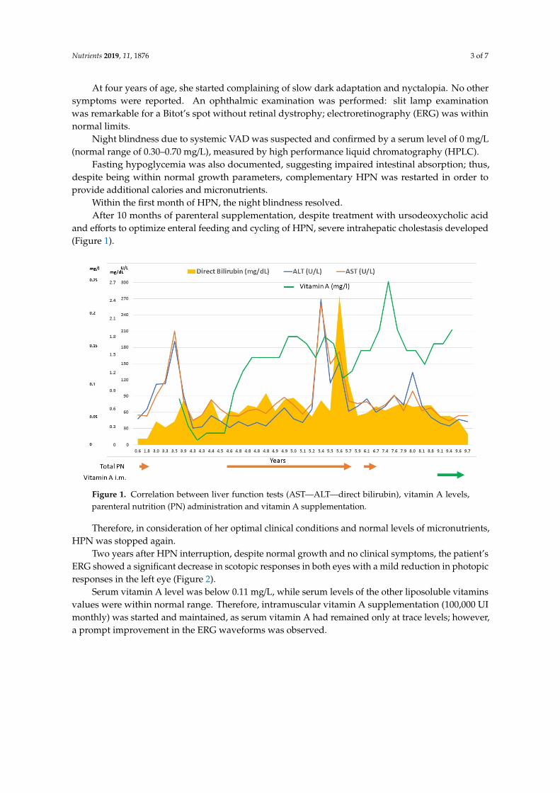
Two years after HPN interruption, despite normal growth and no clinical symptoms, the patient’s ERG showed a significant decrease in scotopic responses in both eyes with a mild reduction in photopic responses in the left eye (Figure 2).
Figure 1. Correlation between liver function tests (AST—ALT—direct bilirubin), vitamin A levels,parenteral nutrition (PN) administration and vitamin A supplementation.
Therefore, in consideration of her optimal clinical conditions and normal levels of micronutrients,HPN was stopped again.
Two years after HPN interruption, despite normal growth and no clinical symptoms, the patient’sERG showed a significant decrease in scotopic responses in both eyes with a mild reduction in photopicresponses in the left eye (Figure 2).
Serum vitamin A level was below 0.11 mg/L, while serum levels of the other liposoluble vitaminsvalues were within normal range. Therefore, intramuscular vitamin A supplementation (100,000 UImonthly) was started and maintained, as serum vitamin A had remained only at trace levels; however,a prompt improvement in the ERG waveforms was observed.
Nutrients 2019, 11, 1876 4 of 7Nutrients 2019, 11, x FOR PEER REVIEW 4 of 7
Figure 2. Patient’s full-field electroretinogram (ERG) (A) before vitamin A supplementation: decreased scotopic responses in both eyes and depressed photopic response in the left eye; (B) after vitamin A supplementation: normal.
Serum vitamin A level was below 0.11 mg/L, while serum levels of the other liposoluble vitamins values were within normal range. Therefore, intramuscular vitamin A supplementation (100,000 UI monthly) was started and maintained, as serum vitamin A had remained only at trace levels; however, a prompt improvement in the ERG waveforms was observed.
3. Discussion
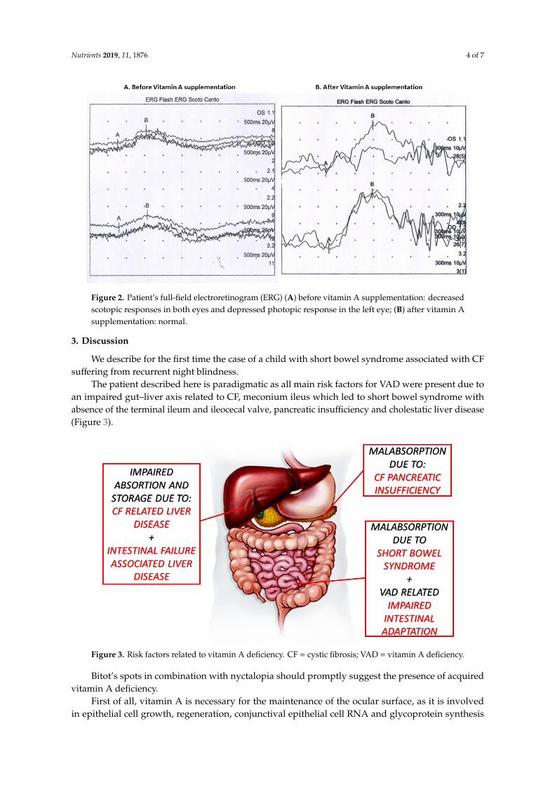
We describe for the first time the case of a child with short bowel syndrome associated with CF suffering from recurrent night blindness
The patient described here is paradigmatic as all main risk factors for VAD were present due to an impaired gut–liver axis related to CF, meconium ileus which led to short bowel syndrome with absence of the terminal ileum and ileocecal valve, pancreatic insufficiency and cholestatic liver disease (Figure 3).
Figure 2. Patient’s full-field electroretinogram (ERG) (A) before vitamin A supplementation: decreasedscotopic responses in both eyes and depressed photopic response in the left eye; (B) after vitamin Asupplementation: normal.
3. Discussion
We describe for the first time the case of a child with short bowel syndrome associated with CFsuffering from recurrent night blindness.
The patient described here is paradigmatic as all main risk factors for VAD were present due toan impaired gut–liver axis related to CF, meconium ileus which led to short bowel syndrome withabsence of the terminal ileum and ileocecal valve, pancreatic insufficiency and cholestatic liver disease(Figure 3).
Nutrients 2019, 11, x FOR PEER REVIEW 4 of 7
Figure 2. Patient’s full-field electroretinogram (ERG) (A) before vitamin A supplementation: decreased scotopic responses in both eyes and depressed photopic response in the left eye; (B) after vitamin A supplementation: normal.
Serum vitamin A level was below 0.11 mg/L, while serum levels of the other liposoluble vitamins values were within normal range. Therefore, intramuscular vitamin A supplementation (100,000 UI monthly) was started and maintained, as serum vitamin A had remained only at trace levels; however, a prompt improvement in the ERG waveforms was observed.
3. Discussion
We describe for the first time the case of a child with short bowel syndrome associated with CF suffering from recurrent night blindness
The patient described here is paradigmatic as all main risk factors for VAD were present due to an impaired gut–liver axis related to CF, meconium ileus which led to short bowel syndrome with absence of the terminal ileum and ileocecal valve, pancreatic insufficiency and cholestatic liver disease (Figure 3).
Figure 3. Risk factors related to vitamin A deficiency. CF = cystic fibrosis; VAD = vitamin A deficiency.
Bitot’s spots in combination with nyctalopia should promptly suggest the presence of acquiredvitamin A deficiency.
First of all, vitamin A is necessary for the maintenance of the ocular surface, as it is involvedin epithelial cell growth, regeneration, conjunctival epithelial cell RNA and glycoprotein synthesis
Nutrients 2019, 11, 1876 5 of 7
helping to maintain the conjunctival mucosa and corneal stroma. Secondly, in the retina, retinol actsas the backbone of the photosensitive visual pigment in both rod and cone photoreceptors. The rodsystem is much more sensitive to vitamin A deficiency than the cone system. Therefore, nyctalopia isthe most common and earliest symptom of vitamin A deficiency, whereas Bitot’s spot and conjunctivaland corneal xerosis tend to occur after long periods of deficiency [8].
Approximately 85–90% of patients with CF suffer from pancreatic insufficiency, which predisposesthem to a reduced enterohepatic circulation of bile acids leading to malabsorption of fat and fat-solublevitamins (A, D, E, K), and reduced levels of RBP, which is essential for transport of retinol from theliver to tissues [9].
Night blindness in children with CF has been reported since the late 1980s [10]. Those reportsdrove the interest in liposoluble vitamins with the publication of the largest prospective study onchildren with CF reporting vitamin A deficiency in 29% of the studied population whereas vitamin Eand D deficiencies were detected in 22% and 25%, respectively [11].
More recently, the prevalence of vitamin A deficiency in adolescents with CF has been assessed.A large retrospective study from Australia found an increasing prevalence from 10% in 2007 to 13%in 2010 [12], while a more recent study on a Dutch pediatric population found a prevalence of only2% [13]. However, those studies do not report any patients with clinical symptoms.
Night blindness has been also attributed to conditions of intestinal malabsorption, in particularin patients who have undergone intestinal bypass or bariatric surgery (even a long time aftersurgery) [14,15]. Regarding short bowel syndrome in particular, a prospective study on adultsdemonstrated that subjects undergoing intestinal rehabilitation have a high risk of developing vitaminA deficiency, especially after HPN weaning [16].
Furthermore, the residual intestine can undergo adaptation due to structural and functionalchanges, leading to an increase in surface area and enhanced nutrient absorptive capacity. This adaptiveprocess is characterized by increased villus height and crypt depth as a result of cellular hyperplasia.Several lines of evidence have implicated vitamin A as a potential modulator of intestinal mucosaladaptive growth in rat models of massive small bowel resection. The speculative mechanism seemsto be via retinoid-responsive genes, which were shown to be upregulated in the adapting remnantsmall intestine at early time points after resection [17,18]. In addition, the same group of researchersdemonstrated that VAD seems to inhibit intestinal adaptation after small bowel resection because ofcrypt cell proliferation reduction, crypt cell apoptosis augmentation and a reduction in enterocytemigration rates [19].
Finally, liver disease could also be a cause of bile acid deficiency and reduced micellar solubilizationfat malabsorption, or the inability to synthetize RBP which is needed for the transport of vitamin A [2].Furthermore, VAD may also exacerbate cholestasis due to possible excessive intrahepatic bile ductproliferation, which has a negative effect on the digestion of lipids [20]. Cholestatic liver disease inour patient is probably multifactorial and mediated by the sum of CF related liver disease [21], whichcould account for the precocity of liver damage, and the development of intestinal failure-associatedliver disease [22] (Figure 1).
Normal findings in the other lipid soluble vitamins (namely D, E, K) demonstrates once more thepeculiarity of vitamin A metabolism.
The intestinal absorption of lipid soluble vitamins has been studied in a cellular model and foundto be different for vitamin A in comparison with the other lipid soluble vitamins. This model alsodemonstrated that vitamin A has a competitive absorption pathway with the other three lipid solublevitamins, which could partially explain the different values found in our patient [23].
Another mechanism for the other lipid soluble vitamins is probably related to the central role ofliver absorption and storage, which has been demonstrated to be impaired in patients with chronicliver diseases [24].
In conclusion, this case report nicely illustrates the central issue of vitamin A deficiency andthe key factors leading to the “vicious cycle of vitamin A deficiency” [25]. Patients with conditions
Nutrients 2019, 11, 1876 6 of 7
impairing intestinal absorption associated with liver disease could be at risk of VAD development; thus,it is essential to monitor serum vitamin A level. When these conditions are particularly severe, oralsupplementation could be insufficient, and the parenteral route of administration could be required.
Author Contributions: L.N., L.Z. conceptualized the paper and wrote the original draft; M.C. wrote the originaldraft; A.B., L.C., A.M.C.B. curated the data; C.C. wrote, reviewed and edited the manuscript.
Funding: This research received no external funding.
Acknowledgments: Silvia Gabrielle Osnaghi for the oculist follow-up, Lauretta Valmarana for the dieteticfollow-up and Lorenzo D’Antiga, Director of the Pediatric Hepatology, Gastroenterology and Transplantation unitof Bergamo.
Conflicts of Interest: The authors declare no conflict of interest.
References
1. Weber, D.; Grune, T. The contribution of β-carotene to vitamin A supply of humans. Mol. Nutr. Food Res.2012, 56, 251–258. [CrossRef] [PubMed]
2. Blaner, W.S.; Li, Y.; Brun, P.-J.; Yuen, J.J.; Lee, S.-A.; Clugston, R.D. Vitamin A Absorption, Storage andMobilization. Subcell. Biochem. 2016, 81, 95–125. [PubMed]
3. Senoo, H.; Mezaki, Y.; Fujiwara, M. The stellate cell system (vitamin A-storing cell system). Anat. Sci. Int.2017, 92, 387–455. [CrossRef] [PubMed]
4. Harrison, E.H. Mechanisms of Transport and Delivery of Vitamin A and Carotenoids to the Retinal PigmentEpithelium. Mol. Nutr. Food Res. 2019, e1801046. [CrossRef] [PubMed]
5. Adeli, K.; Higgins, V.; Trajcevski, K.; White-Al Habeeb, N. The Canadian laboratory initiative on pediatricreference intervals: A CALIPER white paper. Crit. Rev. Clin. Lab. Sci. 2017, 54, 358–413. [CrossRef][PubMed]
6. Song, P.; Wang, J.; Wei, W.; Chang, X.; Wang, M.; An, L. The Prevalence of Vitamin A Deficiency in ChineseChildren: A Systematic Review and Bayesian Meta-Analysis. Nutrients 2017, 9, 1285. [CrossRef] [PubMed]
7. Chiu, M.; Dillon, A.; Watson, S. Vitamin A deficiency and xerophthalmia in children of a developed country.J. Paediatr. Child Health 2016, 52, 699–703. [CrossRef]
8. Fiore, P.; De Marco, R.; Sacco, O.; Priolo, E. Nightblindness, xerophthalmia, and severe loss of visual acuitydue to unnecessary dietary restriction. Nutrition 2004, 20, 477. [CrossRef]
9. Dodge, J.A.; Turck, D. Cystic fibrosis: Nutritional consequences and management. Best Pract. Res. Clin.Gastroenterol. 2006, 20, 531–546. [CrossRef]
10. Rayner, R.J.; Tyrrell, J.C.; Hiller, E.J.; Marenah, C.; Neugebauer, M.A.; Vernon, S.A.; Brimlow, G. Nightblindness and conjunctival xerosis caused by vitamin A deficiency in patients with cystic fibrosis. Arch. Dis.Child. 1989, 64, 1151–1156. [CrossRef]
11. Feranchak, A.P.; Sontag, M.K.; Wagener, J.S.; Hammond, K.B.; Accurso, F.J.; Sokol, R.J. Prospective, long-termstudy of fat-soluble vitamin status in children with cystic fibrosis identified by newborn screen. J. Pediatr.1999, 135, 601–610. [CrossRef]
12. Rana, M.; Wong-See, D.; Katz, T.; Gaskin, K.; Whitehead, B.; Jaffe, A.; Coakley, J.; Lochhead, A. Fat-solublevitamin deficiency in children and adolescents with cystic fibrosis. J. Clin. Pathol. 2014, 67, 605–608.[CrossRef] [PubMed]
13. Woestenenk, J.W.; Broos, N.; Stellato, R.K.; Arets, H.G.M.; van der Ent, C.K.; Houwen, R.H.J. Vitamin Aintake and serum retinol levels in children and adolescents with cystic fibrosis. Clin. Nutr. 2016, 35, 654–659.[CrossRef] [PubMed]
14. Spits, Y.; De Laey, J.-J.; Leroy, B.P. Rapid recovery of night blindness due to obesity surgery after vitamin Arepletion therapy. Br. J. Ophthalmol. 2004, 88, 583–585. [CrossRef] [PubMed]
15. Chae, T.; Foroozan, R. Vitamin A deficiency in patients with a remote history of intestinal surgery. Br. J.Ophthalmol. 2006, 90, 955–956. [CrossRef] [PubMed]
16. Luo, M.; Estívariz, C.F.; Schleicher, R.L.; Bazargan, N.; Leader, L.M.; Galloway, J.R.; Ziegler, T.R. Prospectiveanalysis of serum carotenoids, vitamin A, and tocopherols in adults with short bowel syndrome undergoingintestinal rehabilitation. Nutrition 2009, 25, 400–407. [CrossRef] [PubMed]
Nutrients 2019, 11, 1876 7 of 7
17. Swartz-Basile, D.A.; Rubin, D.C.; Levin, M.S. Vitamin A status modulates intestinal adaptation after partialsmall bowel resection. JPEN J. Parenter. Enteral Nutr. 2000, 24, 81–88. [CrossRef] [PubMed]
18. Wang, L.; Tang, Y.; Rubin, D.C.; Levin, M.S. Chronically administered retinoic acid has trophic effects in therat small intestine and promotes adaptation in a resection model of short bowel syndrome. Am. J. Physiol.Gastrointest. Liver Physiol. 2007, 292, G1559–G1569. [CrossRef]
19. Swartz-Basile, D.A.; Wang, L.; Tang, Y.; Pitt, H.A.; Rubin, D.C.; Levin, M.S. Vitamin A deficiency inhibitsintestinal adaptation by modulating apoptosis, proliferation, and enterocyte migration. Am. J. Physiol.Gastrointest. Liver Physiol. 2003, 285, G424–G432. [CrossRef]
20. Weiss, B.; Barshack, I.; Onaca, N.; Goldberg, I.; Berkovich, Z.; Melzer, E.; Jonas, A.; Reifen, R. Vitamin Adeficiency associated with enhanced proliferation of bile duct epithelial cells in the rat. Isr. Med. Assoc. J.IMAJ 2010, 12, 82–86.
21. Debray, D.; Narkewicz, M.R.; Bodewes, F.A.J.A.; Colombo, C.; Housset, C.; de Jonge, H.R.; Jonker, J.W.;Kelly, D.A.; Ling, S.C.; Poynard, T.; et al. Cystic Fibrosis-related Liver Disease: Research Challenges andFuture Perspectives. J. Pediatr. Gastroenterol. Nutr. 2017, 65, 443–448. [CrossRef]
22. Norsa, L.; Nicastro, E.; Di Giorgio, A.; Lacaille, F.; D’Antiga, L. Prevention and Treatment of IntestinalFailure-Associated Liver Disease in Children. Nutrients 2018, 10, 664. [CrossRef]
23. Goncalves, A.; Roi, S.; Nowicki, M.; Dhaussy, A.; Huertas, A.; Amiot, M.-J.; Reboul, E. Fat-soluble vitaminintestinal absorption: Absorption sites in the intestine and interactions for absorption. Food Chem. 2015, 172,155–160. [CrossRef]
24. Saeed, A.; Dullaart, R.P.F.; Schreuder, T.C.M.A.; Blokzijl, H.; Faber, K.N. Disturbed Vitamin A Metabolism inNon-Alcoholic Fatty Liver Disease (NAFLD). Nutrients 2017, 10, 29. [CrossRef]
25. Wiseman, E.M.; Bar-El Dadon, S.; Reifen, R. The vicious cycle of vitamin a deficiency: A review. Crit. Rev.Food Sci. Nutr. 2017, 57, 3703–3714. [CrossRef]
© 2019 by the authors. Licensee MDPI, Basel, Switzerland. This article is an open accessarticle distributed under the terms and conditions of the Creative Commons Attribution(CC BY) license (http://creativecommons.org/licenses/by/4.0/).