Embed Size (px)
Citation preview
Effect of Digestion and Storage of Human Milk on Free FattyAcid Concentration and Cytotoxicity
Alexander H. Penn, Ph.D.1, Angelina E. Altshuler, Ph.D.1, James W. Small, B.S.1, Sharon F.Taylor, M.D.2, Karen R. Dobkins, Ph.D.3, and Geert W. Schmid-Schönbein, Ph.D.1
1Department of Bioengineering, University of California San Diego, La Jolla, CA 92093-0412
2Department of Pediatrics, Rady Children’s Hospital San Diego, San Diego, CA 92123
3Department of Psychology, University of California San Diego, La Jolla, CA 92093-0412
Abstract
Objectives—Fat is digested in the intestine into free fatty acids (FFAs), which are detergents and
therefore toxic to cells at micromolar concentration. The mucosal barrier protects cells in the adult
intestine, but this barrier may not be fully developed in premature infants. Lipase-digested infant
formula, but not fresh human milk, has elevated FFAs and is cytotoxic to intestinal cells, and
therefore could contribute to intestinal injury in necrotizing enterocolitis (NEC). But even infants
exclusively fed breast milk may develop NEC. Our objective was to determine if stored milk and
milk from donor milk banks (DM) could also become cytotoxic, especially after digestion.
Methods—We exposed cultured rat intestinal epithelial cells or human neutrophils to DM and
milk collected fresh and stored at 4 or −20 °C for up to 12 weeks and then treated for 2 hours
(37°C) with 0.1 or 1 mg/ml pancreatic lipase and/or trypsin and chymotrypsin.
Results—DM and milk stored 3 days (at 4 or −20 °C) and then digested were cytotoxic. Storage
at −20 °C for 8 and 12 weeks resulted in an additional increase in cytotoxicity. Protease digestion
decreased, but did not eliminate cell death.
Conclusions—Current storage practices may allow milk to become cytotoxic and contribute to
intestinal damage in NEC.
Additional keywords
Lipase; necrotizing enterocolitis; breast milk donor banks; necrosis; intestinal cell damage
Introduction
In many ways, the infant intestine is more vulnerable to damage than the mature intestine.
One example of this is necrotizing enterocolitis (NEC), the leading cause of gastrointestinal
related death in premature infants. NEC is characterized by intestinal hemorrhagic necrosis
Correspondence: Alexander H. Penn, Department of Bioengineering, The Institute of Engineering in Medicine, University ofCalifornia San Diego, La Jolla, CA 92093-0412 USA, Tel: 858 534-2714, Fax: 858 534-5722, [email protected].
Conflicts of InterestThe authors have no financial interests or conflicts of interest to declare related to the contents of this manuscript
NIH Public AccessAuthor ManuscriptJ Pediatr Gastroenterol Nutr. Author manuscript; available in PMC 2015 September 01.
Published in final edited form as:J Pediatr Gastroenterol Nutr. 2014 September ; 59(3): 365–373. doi:10.1097/MPG.0000000000000441.
NIH
-PA
Author M
anuscriptN
IH-P
A A
uthor Manuscript
NIH
-PA
Author M
anuscript
and is rarely seen before onset of enteral nutrition (1). This vulnerability to intestinal
damage likely results from the immature state at birth of the intestinal mucosal barrier,
observable as increased permeability to luminal content compared to mature intestines (2,
3). The intestines of premature infants are even more permeable (3), suggesting,
unsurprisingly, they are even less mature.
The intestinal mucosal barrier controls permeability and is comprised of an inner barrier of
epithelial cells with tight inter-epithelial junctions and an outer barrier of attached and
secreted mucin (i.e. mucus). In the mature intestine, the mucosal barrier serves to prevent
pathogens in the intestinal lumen from entering the body. The mucin component also
prevents digestive enzymes and certain products of food digestion from directly contacting
and damaging the epithelium. One such product is free fatty acid (FFA) released by lipase
digestion of ingested fats. As detergents, FFAs may insert themselves into and disrupt (4) all
types of cell membranes (5, 6) (i.e. a receptor independent event). As a result, they may be
cytotoxic in even micromolar concentration (7). Yet FFA concentrations in the intestinal
lumen after a meal may easily surpass 10 mM in humans (8). The mucin barrier prevents
FFAs from damaging the epithelium (9). However, it has been shown that at higher
concentrations (e.g. 40 mM), FFAs cause villi damage even in healthy, mature intestine (9).
Likewise, inhibition of the enzymes that create and release FFAs prevents intestinal
hemorrhagic necrosis and improves mortality after mucosal barrier disruption by intestinal
ischemia in adult rats (7).
In neonates the mucin barrier is still forming and less mature intestines may be damaged by
lower concentrations of FFA (e.g. 5 mM) (10). Mouse models suggest that the ability of the
intestine to replenish its mucin barrier after a challenge is deficient in pre-term neonates
(11). This possibility is supported by findings of fewer mucin-containing goblet cells in
histological sections from NEC infants in humans, suggesting depletion of mucin stores
(11). In contrast, addition of dietary epidermal growth factor (EGF), a mucin-stimulating
factor, protects against NEC in mouse models (12, 13). If mucin deficit is a major factor
predisposing the premature infant to NEC, then it becomes critical to determine what dietary
factors increase FFA concentration and thus cytotoxicity in the intestinal lumen.
There is a 10-fold higher risk for development of NEC in premature infants fed infant
formula instead of breast milk (14). We have shown that infant formula digested in vitro
generates 10 times the concentration of unbound (i.e. not attached to protein and therefore
cytotoxic) FFA generated by fresh human milk under the same digestion conditions (5).
Furthermore, the digested formulas but not the digested fresh human milk were cytotoxic to
multiple cell types, since they cause physical disruption of lipid membranes. This evidence
suggests that the increased risk for NEC associated with formula feeding could be
attributable to concentrations of FFA after digestion exceeding the protective capacity of the
mucin barrier.
However, milk from donor banks has not always performed better than formula in
preventing NEC (15, 16) nor does exclusive breast milk feeding provide full protection from
NEC or other GI problems (14, 17). One possible reason may be the practice of storing
breast milk. Breast milk contains bile salt-sensitive lipase (BSSL) and lipoprotein lipase
Penn et al. Page 2
J Pediatr Gastroenterol Nutr. Author manuscript; available in PMC 2015 September 01.
NIH
-PA
Author M
anuscriptN
IH-P
A A
uthor Manuscript
NIH
-PA
Author M
anuscript
(LPL), and, if stored, forms FFAs that taste “soapy” (18). This may occur even if the milk is
stored frozen at −20 °C (19). Stored breast milk has been shown to become cytotoxic to a
number of cell types and this death was attributed to FFAs (6), but the focus was on the
effects this could have on immune cells or pathogens present in the milk, rather than on
potential damage to the intestine. Nor have previous studies taken into account the
additional FFA formation and thus cytotoxicity that may occur during digestion in an
infant’s intestine. Current guidelines suggest that parents may store milk up to eight days at
4 °C or 6 to 12 months at −20 °C (20) and allow milk banks 3 to 6 months of milk storage at
−20 °C prior to pasteurization (21). This extended storage could lead to cytotoxicity in
donor milk that may be detrimental to the premature infant’s intestine. Even otherwise
healthy full-term neonates could potentially be affected by cytotoxicity in their mother’s
own stored milk if their mucosal barrier is not fully developed.
The objectives of this study were to: 1) determine if intestinal epithelial cells (IECs) can be
killed by stored breast milk, 2) investigate general cytotoxicity as a function of storage time,
storage temperature, and subsequent digestion, and 3) determine if donor milk (DM)
obtained from human milk banks is cytotoxic with or without subsequent digestion.
Materials and Methods
To achieve our objectives we obtained DM and collected fresh milk from volunteers and
stored it at 4°, −20°, or −80° C for varying periods then digested the milk with pancreatic
lipase and/or proteases. When possible, we tested cytotoxicity on cultured IECs. However,
that assay is limited in that cells were not always available at the same time as fresh human
milk and because the presence of even small amounts of protease would cause cell
detachment from the culture wells. Since the latter interferes with quantification of cell
death, the exposure time of IECs to milk was limited to 5 minutes (well below the expected
in vivo exposure time of ~50 min, based on gastric emptying rate (22)). Therefore for some
studies a general assay of cytotoxicity was performed using 60-minute exposure to rapidly
obtainable, freshly isolated human neutrophils as the test cell type.
Ethics Statement
The Institutional Review Board of the University of California, San Diego, approved all
protocols involving human subjects.
Human Milk Collection and Storage
Fresh human milk was obtained from healthy volunteer mothers after written consent.
Participants were asked to pump a full expression to allow mixing of fore and hind milk.
Milk was kept at 4°C and aliquoted within 2 hours. Aliquots for the fresh milk group were
immediately digested for measurements of cytotoxicity on human neutrophils, an uncultured
human cell source available on short notice. Milk that was not slated for immediate testing
was stored at 4 °C, −20 °C, and/or −80 °C. For selected studies, duplicate aliquots of milk
received 0.25 mg/ml of the lipase inhibitor, orlistat (Sigma-Aldrich; St. Louis, MO), prior to
storage. Because IECs were not always available when fresh milk arrived, the primary
determination of cytotoxicity at fresh, 3 day, and 7 day time points was performed using the
Penn et al. Page 3
J Pediatr Gastroenterol Nutr. Author manuscript; available in PMC 2015 September 01.
NIH
-PA
Author M
anuscriptN
IH-P
A A
uthor Manuscript
NIH
-PA
Author M
anuscript

general cytotoxicity (e.g. neutrophil) assay. The “fresh” and “7 day” time points were then
replicated on IECs using milk stored at −80 °C until the day of the assay or thawed and
stored at 4 °C 7 days prior to the IEC assay. To study long-term storage at −20 °C, fresh
milk was stored for 0, 4, 8, or 12 weeks at −20 °C before transfer to −80 °C prior to testing
on IECs (all samples assayed on the same day).
Raw (N=6) and pasteurized (i.e. as provided for consumption; N=6) DM was obtained from
the San Jose Human Milk Bank. DM was frozen and shipped in batches to the milk bank by
mothers after collection over an 8 to 130 day period. The milk remained frozen at −20 °C
until pasteurization by the Milk Bank between 2 and 16 days of receipt by the bank before it
was shipped to us (written communication from milk bank). Samples were shipped frozen
on dry ice. DM was immediately thawed and aliquoted upon delivery (6 to 8 days after
pasteurization). Within minutes, raw and pasteurized DMs were assayed for lipase activity
and pasteurized DMs were digested and tested for cytotoxicity on neutrophils. The
remaining aliquots were frozen (−80 °C) for later (14 days) cytotoxicity measurements on
IECs.
Milk Digestion with Pancreatic Lipase and Protease
For studies involving digestion, human milk was incubated for 2 h (37 °C) (similar to the
transit time through an infant jejunum) after mixing 4:1 (v:v) with PBS, porcine pancreatic
lipase (Sigma Aldrich), bovine pancreatic trypsin and chymotrypsin (Sigma Aldrich), or
lipase, trypsin, and chymotrypsin. Final enzyme concentrations were either 0.1 (this, along
with reducing exposure time to 5 minutes, was designed to reduce IEC cell detachment) or 1
mg/ml. These concentrations bracket and are on the order of concentrations in the intestinal
lumen of human infants (23). Note we avoided the use of bile salts in the digestion, as bile
salt concentration is highly variable in neonates and often well below the critical micelle
concentration (i.e. the theoretical concentration at which bile would be able to assist in
digestion) (24).
After digestion, selected samples were defatted (centrifugation at 16000g, 20 min, 4 °C,
collecting the supernatant under the solid fat layer). Defatting to remove fat globules was
required for all cytotoxicity assays involving neutrophils to allow measurement by flow
cytometry and for all plate reader assays (i.e. lipase activity and FFA concentration
measurements).
Some samples were filtered after digestion and defatting through glass fiber pre-filters (Pall-
Gellman; Port Washington, NY) to remove unbound FFAs, as previously described (5).
Cytotoxicity Assays
Freshly isolated human neutrophil and cultured rat IEC cytotoxicity measurements were
performed as previously described (5). In brief, after a 60 minute exposure to sample for
neutrophils (to approximate the expected bolus transit time of food passing through the
intestine as suggested by gastric emptying rate (22)) or a 5 minute exposure for IECs (to
avoid cell detachment), death was determined using propidium iodide (PI; Sigma), a
fluorescent life/death indicator, and analyzed immediately via flow cytometer (neutrophils)
or fluorescent imaging (IECs). % Cell death was calculated as the percentage of gated cells
Penn et al. Page 4
J Pediatr Gastroenterol Nutr. Author manuscript; available in PMC 2015 September 01.
NIH
-PA
Author M
anuscriptN
IH-P
A A
uthor Manuscript
NIH
-PA
Author M
anuscript
that were PI-positive in the neutrophil assay and by using the ratio of PI-positive cells to the
average number of PI-positive cells in wells treated with 0.1% Triton X-100 in PBS for 30
seconds (100% cell death). A sample was defined as cytotoxic if it caused more than 17%
(neutrophils) or 5% (IECs) cell death (i.e. at least one standard deviation above the mean
death of cells exposed to PBS alone − 10±7% and 1.6±1.7%, respectively).
Lipase Activity Assay
Lipase activity of defatted milk was assayed fresh, after 1 week of refrigeration (4 °C) or
freezing (−20 °C), after long-term (16±9 weeks) storage at −80 °C, and in the raw and
pasteurized DM upon arrival. Lipase activity was determined via digestion of the fluorescent
substrate, O-pivaloyloxymethyl umbelliferone (C-POM; Invitrogen; Carlsbad, CA) as
previously described (7).
FFA Measurement
Total FFA concentration was determined in a 96-well plate using the Free Fatty Acids Half-
Micro Test kit (Roche Applied Sciences; Indianapolis, IN). All samples were pre-diluted 10-
fold to bring concentrations within the linear range of the assay (<1 mM), with reported
values corrected to the original concentrations. The kit measures total FFAs regardless of
binding to fatty acid binding proteins such as bovine serum albumin (BSA) (5). Filtration
through glass fiber pre-filters removes unbound but not bound FFAs (5) and reduces
cytotoxicity (7, 25). Therefore, to determine the approximate amounts of bound and
unbound FFAs, we filtered samples through 3 glass fiber filters in series, as previously
described (5). Filtered samples were then assayed for total FFAs yielding the bound FFA
concentration of the original unfiltered sample.
Statistics
Data are presented as means ± standard deviation. Single or multi-factor ANOVA (with
repeated measures for paired samples) was completed for experiments with multiple groups
(α=0.05) followed by Bonferroni correction for pair-wise t-test comparisons. Statistical
analysis was performed in Excel and SPSS (IBM corporation; Armonk, NY).
Results
Cytotoxicity of digested fresh vs. stored breast milk to human neutrophils
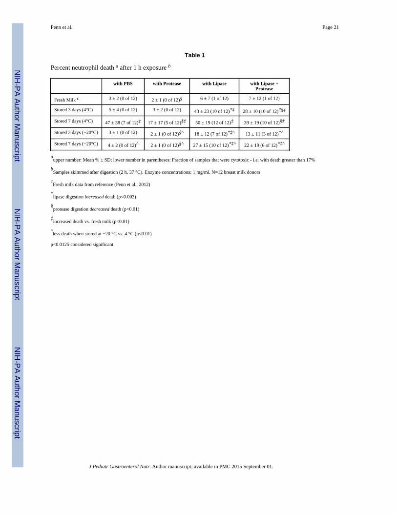
Defatted fresh human milk was on average not cytotoxic in the neutrophil assay, regardless
of digestion group (only 1 of 12 samples became cytotoxic after lipase digestion; Table 1).
However, milk stored for 3 days at 4 °C or −20 °C showed a significant increase in
cytotoxicity after lipase digestion, with more cell death in the 4 °C group than in the −20 °C
group. After 1 week of storage, 11 of 12 milk samples stored at 4 °C and 10 of 12 samples
stored at −20 °C were cytotoxic after lipase digestion. Neutrophils exposed to 4 °C, but not
−80 °C, stored milk (1 week) show visible disruptions of the cell membrane (e.g. large
blebs) (Figure 1), similar to previously observed bleb formations that precede cell membrane
rupture and death by a FFA mechanism (5, 7, 25).
Penn et al. Page 5
J Pediatr Gastroenterol Nutr. Author manuscript; available in PMC 2015 September 01.
NIH
-PA
Author M
anuscriptN
IH-P
A A
uthor Manuscript
NIH
-PA
Author M
anuscript
Protease digestion, shown previously to increase the capacity of breast milk to resist death
from exogenously added FFA (5), either decreased the cell death and/or the number of
samples that were cytotoxic in stored milk (Table 1). This did not appear to be due to
digestion of endogenous or exogenous lipase since lipase activity was unchanged with
protease addition (1259±585 vs. 1196±487 fluorescent units in the 7 day, 4 °C, milk with
lipase vs. lipase + protease groups). Lipase- and protease-only controls were not cytotoxic to
neutrophils (not shown).
The post-natal age of the child at the time of milk donation (range: 22–507 days) did not
correlate to the cytotoxicity of milk stored 7 days at 4 °C (PBS digestion, correlation
coefficient = 0.15, p=0.32) or initial lipase activity in the sample (correlation coefficient =
−0.01, p=0.49). Mother’s age at time of expression was unavailable for 2 samples, but we
did not find a correlation between mother’s age and cytotoxicity of milk stored 7 days at 4
°C (PBS digestion, correlation coefficient = −0.17, p=0.32) or initial lipase activity in the
sample (correlation coefficient = 0.13, p=0.35) in the remaining 10 samples. Mothers’ race
was primarily Caucasian (7 Caucasian/non-Hispanic, 1 Black, 1 Asian, 1 half-Asian/half-
Caucasian, 2 unavailable), so no effects of race are determinable for this data.
FFA Cytotoxic Mechanism
We measured lipase activity in samples stored for 7 days at 4 °C or −20 °C, or long-term at
−80 °C, normalized by their activities when fresh, as well as that of raw and pasteurized DM
(Figure 2A). Fresh milk alone had 3.1±1.4 times the activity as 1 mg/ml of exogenous
pancreatic lipase (not shown). Pasteurized DM had no detectable lipase activity, in
agreement with previous studies (26), but all other samples had activity. Activity was
significantly elevated with freezing (−20 °C) compared to refrigeration (4 °C). Raw DM and
−80 °C stored milk also trended towards increased activity compared to 4 °C (p=0.046 for
raw DM; p=0.076 for −80 °C stored milk). This agrees with previous studies showing lipase
activation by cooling (2 °C) cow’s milk (27) and by freezing human milk (BSSL normally
needs bile salt to become active, but this requirement is lost after freezing) (28). No
correlation was seen between cytotoxicity of the Milk with PBS after 7 days storage at 4 °C
and the initial (correlation coefficient = −0.04, p=0.45) or final lipase activity (correlation
coefficient = −0.35, p=0.16), suggesting that variations in cytotoxicity were due more to
differences in substrate (e.g. milk fat globules) between samples than differences in lipase
activity.
Despite a relatively short 5 minutes exposure, milk stored at 4 °C for 7 days killed 41% of
IECs, as compared to 5% from milk stored at −80 °C (Figure 2B). Exogenous lipase
digestion increased both of these values a small but significant (by paired t-test) amount (see
inset for raw data of 4 °C groups). Cytotoxicity was prevented when lipase was inhibited
with orlistat prior to storage at 4 °C. Thus, as with digested formula (5), FFAs created by
lipase digestion in stored milk are cytotoxic to intestinal cells. We confirmed that lipase
inhibition would also prevent cytotoxicity to neutrophils (Figure 2C) and that passage of
cytotoxic stored milk through glass fiber filters, which bind and remove unbound, but not
bound, FFAs (5), significantly reduced cytotoxicity (Figure 2D).
Penn et al. Page 6
J Pediatr Gastroenterol Nutr. Author manuscript; available in PMC 2015 September 01.
NIH
-PA
Author M
anuscriptN
IH-P
A A
uthor Manuscript
NIH
-PA
Author M
anuscript
We measured total and unbound FFAs in aliquots of the same defatted solutions that were
previously tested for cytotoxicity on human neutrophils (Table 1) and stored at –80 °C until
FFA analysis. We detected little to no FFAs in fresh milk incubated with PBS or proteases
(Figure 3). Pancreatic lipase digestion significantly increased the total and unbound FFAs in
fresh breast milk. Milk stored for 7 days at 4 °C had significantly higher unbound and total
FFA levels compared to fresh milk in all cases, except for unbound FFAs in the Milk with
Lipase groups which approached, but did not reach significance (P=0.02). Lipase digestion
of stored milk increased neither the total nor the unbound concentrations of FFAs over the
Stored milk with PBS control unless protease digestion also occurred, in agreement with the
cytotoxicity findings (Table 1). It is possible that fat digestion or FFA aqueous solubility in
breast milk are near their maximal levels in the stored milk already, minimizing the effect of
exogenous lipase unless proteases are present to increase the binding of FFA to proteins (see
below) removing them from solution and making room for additional FFA (in vivo,
solubility is increased via FFA incorporation into bile acid micelles (29)). Protease digestion
decreased total FFA levels in all cases. Overall, unbound FFA concentration correlated
significantly with cytotoxicity (correlation coefficient = 0.706, p<0.001).
Long-term milk storage at −20 °C
To explore the effects of long-term milk storage at −20 °C on IEC cytotoxicity, we stored
fresh milk at −20 °C for 0, 4, 8, or 12 weeks, transferring the aliquots to −80 °C until
simultaneous digestion and testing on IECs (total storage = 20.7 ± 8.3 weeks, max 37.6
weeks). Every group, including the 0 week group that was kept at −80 °C for the duration,
increased their cytotoxicity with lipase digestion (Figure 4 also apparent in Figure 2B). This
suggests that frozen milk, even at −80 °C, loses some of the resistance to lipolysis present in
fresh milk, subject to variations between donors and/or assay conditions. However, after 8
weeks we saw an additional cytotoxic effect of storage at −20 °C. This increase was seen
even in the absence of exogenous pancreatic lipase digestion.
To determine whether the secondary increase in cytotoxicity during storage at −20 °C is due
to retention of residual lipase activity even while frozen, we tested the effect of lipase
inhibition on milk prior to storage. Milk from 7 individual donors was stored 28 days or
approximately 2 months (60.1 ± 3.0 days) at −20 °C with or without 0.25 mg/ml orlistat,
then moved to −80 °C before thawing and immediate defatting and testing for cytotoxicity to
neutrophils without additional digestion or incubation at 37 °C (total storage time 33.1 ± 3.0
and 61.1 ± 3.0 days, respectively). Aliquots of the defatted milk were immediately returned
to −80 °C until testing for FFA concentration (total storage time 38.1 ± 3.0 and 68.1 ± 3.0
days, respectively). We found no appreciable cell death at either time point (3 ± 1% with or
without orlistat at 28 day time point, 2 ± 1% with or without orlistat at 2 month time point).
At 28 days we found approximately the same low concentration of FFAs as we observed in
the fresh milk group (Figure 3) in both groups (0.13 ± 0.12 mM FFA without orlistat and
0.10 ± 0.04 mM FFA with orlistat). However, at the 2-month time point, FFAs were doubled
(0.22 ± 0.11 mM FFA), and this increase was prevented by pretreatment with orlistat (0.10 ±
0.04 mM FFA; p<0.011).
Penn et al. Page 7
J Pediatr Gastroenterol Nutr. Author manuscript; available in PMC 2015 September 01.
NIH
-PA
Author M
anuscriptN
IH-P
A A
uthor Manuscript
NIH
-PA
Author M
anuscript
Milk from Donor Bank
Given that 8 weeks of storage at −20 °C can increase FFAs and cytotoxicity and that milk
banks allow donors to store milk for longer periods before shipping to their facilities, we
determined the cytotoxicity of DM, with and without exogenous lipase digestion. We found
that 2 of 6 DM samples were initially cytotoxic to neutrophils, but after lipase digestion all 6
samples were cytotoxic (Figure 5 left). The IEC assay gave similar results for the control
and lipase digested DM although by that method, 4 of the samples were considered
cytotoxic to start with, increasing to 5 of 6 with lipase digestion (Figure 5 right). The
addition of proteases to the digestion of DM resulted in a decrease in cytotoxicity by IEC
assay, but increased cytotoxicity as measured by the neutrophil assay, possibly due to the
higher protease concentration and extra ~1.5 hours of digestion (55 minutes longer
incubation with neutrophils plus ~30 minutes for defatting step) in that assay (see below).
FFA binding capacity as measured by cytotoxicity response to exogenous FFA
Because FFAs are likely to insert themselves into milk fat globule membranes, the fat layer
itself may provide some protection from cytotoxicity. We therefore compared the ability of
defatted and whole milk to lower the cytotoxicity of exogenous oleic acid (e.g. via binding
the FFA) and determined how those capacities are affected by protease digestion. Similar to
previous results with milk (5), digestion by proteases significantly improved the ability of
milk to lower the cytotoxicity of exogenous FFA, whether or not the milk was defatted prior
to oleic acid addition (Figure 6). This suggests that protease digestion increases FFA binding
capacity not just in whole milk but also in the components that remain in defatted milk.
There was also a significant protection against cytotoxicity if oleic acid was added to milk
before, as opposed to after, defatting. This suggests that the fat layer and/or pellet also have
a significant FFA binding capacity. There was a significant interaction by ANOVA between
defatting and digestion, implying that the binding capacity of the fat layer/pellet is also
increased by protease digestion. This agrees with the FFA measurement (Figure 3) showing
a decrease in total FFA concentration in milk digested by proteases before defatting, despite
no decrease in lipase activity.
To determine if the differential response to protease digestion shown in Figure 5 could be
the result of differences in the degree of protein digestion, we kinetically tested the effect of
protease digestion on FFA binding capacity in DM. 5mM exogenous oleic acid was added to
defatted DM, which was then digested for 0, 0.5, 1, 2, or 4 hours with 1 mg/ml trypsin and
chymotrypsin before inhibiting protease activity with 1 mM phenylmethanesulfonylfluoride
and incubating with neutrophils. Unlike in Figure 5 left we saw a significant decrease in
neutrophil cytotoxicity compared to 0 hours digestion (59±11%) at 0.5 (47±11%, p<0.0001)
and 1 hours (46±11%, p<0.015) digestion (N=6). However, this was followed by a near
significant increase in cytotoxicity at the 2-hour time point (55±21%, p=0.052 versus 1-hour
time point, with one sample having a 10% increase in cytotoxicity compared to its 0-hour
time point) and a second significant decrease at 4 hours (33±11%, p<0.01 versus 2-hour
time point). The complexity of the kinetics suggests that more than one protein is likely to
be involved in this process.
Penn et al. Page 8
J Pediatr Gastroenterol Nutr. Author manuscript; available in PMC 2015 September 01.
NIH
-PA
Author M
anuscriptN
IH-P
A A
uthor Manuscript
NIH
-PA
Author M
anuscript
Discussion
The current results indicate that 1) stored human milk is capable of killing IECs with as little
as 5 minutes exposure, 2) 3 days storage at either 4 °C or −20 °C will cause milk to become
cytotoxic after lipase digestion similar to that in the infant intestine, and 3) some milk from
donor banks is already cytotoxic upon distribution and, unlike fresh milk, gains significant
cytotoxicity after lipase digestion.
Cytotoxicity could be prevented by pre-treatment with the lipase inhibitor, orlistat, prior to
milk storage, confirming that the products of lipolysis are the cause of cell death.
Cytotoxicity could be decreased by protease digestion, and by filtering through glass fiber
pre-filters, a procedure that removes unbound FFAs but not FFAs bound to proteins such as
albumin (5, 7). Both total and unbound FFAs are increased in stored milk and unbound FFA
concentration correlates with levels of cell death. These findings support the hypothesis that
unbound FFAs are the main cytotoxic mediator in stored milk, though monoglycerides may
also contribute.
We observed membrane bleb formation and rupture and eventually total cell destruction,
similar to that observed with other FFA sources (7), and consistent with the idea of physical
disruption of the bi-lipid membrane. Cell destruction by FFAs is cell type independent (e.g.
epithelial cells (5), endothelial cells (5), leukocytes (6), red cells (6)). Though neutrophils
were used here primarily as a convenient measure of general cytotoxicity, neutrophils are
present in intestinal tissue and, like other cell types in the intestine that remain to be
investigated, may be affected by FFA cytotoxicity when the mucosal barrier is breached. In
our previous work, the cell sources were human, rat, and bovine, all of which gave
qualitatively similar results (5), making it unlikely that there are major species differences
with regards to response of cells exposed to FFAs.
Freezer stored milk (−20 °C) caused cell death that appears to increase in a biphasic fashion.
In the first phase, we saw that freezing for 3 or more days resulted in increased cytotoxicity
after subsequent exogenous lipase digestion. We saw no increase in cytotoxicity, and levels
of FFA that approximated that of fresh milk, in milk stored for 4 weeks at −20 °C without
subsequent digestion, suggesting that no appreciable fat digestion occurs while milk is
frozen for less than 4 weeks. Therefore, the increased cytotoxicity versus fresh milk we
observed at 3 and 7 days (Table 1) after exogenous lipase digestion is likely due to an
increase in the susceptibility of the milk fat to lipolysis (e.g. disruption of the milk fat
globule membrane). Alternatively, BSSL activated by freezing (28) (Figure 2A) may
interact synergistically with the exogenous lipase to increase lipolysis.
The secondary increase in cytotoxicity of milk stored for 8 and 12 weeks at −20 °C but not
milk kept at −80 °C is likely the result of lipase activity while the milk is frozen, since FFA
concentration was doubled in milk stored at −20 °C for 8 weeks without orlistat. Partial
digestion of milk fat or the milk fat globule membrane may also prime the fat for increased
lipolysis after thawing. The non-negligible death in milk stored long-term −80 °C (Figure 4
0 week Control group) and the small but significant increase in cytotoxicity with exogenous
lipase digestion after short-term storage at −80 °C (Figure 2B) suggest that the same changes
Penn et al. Page 9
J Pediatr Gastroenterol Nutr. Author manuscript; available in PMC 2015 September 01.
NIH
-PA
Author M
anuscriptN
IH-P
A A
uthor Manuscript
NIH
-PA
Author M
anuscript
to milk that occur at −20 °C may also occur at −80 °C, but to a lesser extent that varies
between individuals.
Two-thirds of the milk samples obtained from the donor bank were cytotoxic to IECs
without additional lipid digestion, increasing to five-sixths after exogenous lipase digestion.
This is in line with our findings of increased cytotoxicity compared to fresh milk after long-
term storage at −20 °C.
In vivo intestinal environment
Though we tried to match factors such as digestion time and temperature, enzyme
concentrations, and cell exposure time, our in vitro protocols are not, of course, a perfect
representation of digestion as it occurs in an infant in vivo. For example, lipase profiles
change during early development and in response to weaning (30). For this study, we chose
to use pancreatic lipase so our findings would be comparable with our previous study
comparing formula and fresh milk (5). This is likely to match the conditions in infants that
have begun to wean, but are still consuming some human milk, but may not match
conditions in infants on a milk-only diet, as prior to weaning most of the lipase activity in a
neonatal intestine is from pancreatic lipase-related protein 2 (PLRP2) or BSSL (30). PLRP2
may be less efficient at lipid digestion in milk than pancreatic lipase (31), which could lead
to lower FFA levels and cytotoxicity in the intestine than demonstrated in the current
experiments. In contrast, lingual and gastric lipases, also not included in this study, may
prime milk fats for easier digestion in the intestine, leading to higher FFA levels and greater
cytotoxicity. Interestingly, since BSSL and PLRP2 have a lower affinity for large saturated
fatty acids like palmitic acid (32, 33), which is almost exclusively located at the sn-2
position in human milk triglycerides and is found there over 3 times as often as any other
fatty acid (34), and since pancreatic lipase also avoids cleaving the sn-2 triglyceride
position, it is unlikely that the composition of FFAs will differ substantially regardless of
which lipase dominates. Though we clearly demonstrate here that subsequent lipase
digestion may cause non-cytotoxic stored milk to become cytotoxic (Table 1), testing
cytotoxicity in infant intestinal aspirates will be necessary to resolve the question of the
degree to which digestion increases stored milk cytotoxicity in-vivo.
Likewise, we did not include bile acids in our digestions, as their concentration is low and
variable in neonates (24). However, since bile acids are small, amphiphilic molecules like
FFAs, and are themselves cytotoxic (35), with the in vivo purposes of activating BSSL (28),
emulsifying fats, and increasing FFA solubility and transport of FFAs across the mucin
barrier by incorporating them into bile acid micelles (36), it is likely that bile acids would
only increase overall cytotoxicity from stored milk.
Lastly, while milk supplied to donor banks is expected to come from later in lactation and
while older infants could also be affected by milk stored later in lactation, it is possible that
colostrum obtained in the first week or two may develop cytotoxicity either faster or slower
with storage than the milk used for this study.
Penn et al. Page 10
J Pediatr Gastroenterol Nutr. Author manuscript; available in PMC 2015 September 01.
NIH
-PA
Author M
anuscriptN
IH-P
A A
uthor Manuscript
NIH
-PA
Author M
anuscript
Protease Digestion of Stored Milk
Similar to our findings with fresh milk and a few infant formulas (5), cytotoxicity of stored
milk also decreased significantly with protease digestion, which increases the FFA binding
capacity of the milk. This is opposite to the effects of protease digestion on FFA binding
proteins we have previously studied (e.g. albumin and the binding proteins in intestinal wall
homogenates) (7, 25), suggesting the involvement of a specialized protein. One possible
candidate is the casein micelle, which is disrupted upon protease digestion (37) to expose
hydrophobic portions of the individual casein molecules and which is known to bind to
hydrophobic lipid after disruption (31, 38). Alternatively, partial digestion could induce the
conformational change in alpha lactalbumin that allows it to bind FFAs after the removal of
its calcium ion (39).
This putative specialized protein is likely not the only protein involved in this process,
however, as Figure 4 indicates that milk has the capacity to bind mM quantities of FFA even
without protease digestion. For example, serum albumin is also present in human milk, and
as mentioned above is capable of binding FFAs unless first digested with protease (7). A
simultaneous decrease in binding capacity of some proteins and increase in binding capacity
of other proteins with protease digestion could easily explain our findings in Figure 5 and
the kinetic experiment on DM. We have preliminary data suggesting that the protection
provided by protease digestion of milk eventually disappears altogether as protease digestion
continues. It is possible that the pasteurization step that occurs during preparation of DM
may accelerate the rate at which the protective protein is degraded compared to the
unpasteurized milk in Table 1. The effect of protease on the fat layer may also have an
effect.
Cytotoxicity reduction strategies
Our findings suggest a number of possible strategies to limit cytotoxicity in stored breast
milk. Storage of breast milk at −80 °C rather than −20 °C or above, or earlier pasteurization
for donor milk to denature the lipase, may reduce the potential production of cytotoxic
mediators prior to, and during, digestion of the milk. Since freezing activates milk lipases,
more limited storage of thawed milk before consumption or pasteurization may also serve as
a preventive measure. If pancreatic insufficiency is present, pretreatment of breast milk with
trypsin and/or chymotrypsin may decrease cytotoxicity. Guidelines for milk storage times at
4 °C or −20 °C could be more limited. If long-term storage is required and colder storage is
unavailable, the efficacy and safety of glass fiber filtration or addition of a lipase inhibitor
could be considered.
In summary, we determined that milk stored according to current practices, whether
obtained from a fresh source or from a donor bank can become cytotoxic, though to a lesser
degree than nearly every infant formula we previously tested (5). It remains to be examined
to what degree the cytotoxicity is increased during digestion in-vivo, however, the incidence
of cytotoxicity in stored milk may explain why NEC can occur in milk fed infants and why
DM has not always performed better than formula in preventing NEC.
Penn et al. Page 11
J Pediatr Gastroenterol Nutr. Author manuscript; available in PMC 2015 September 01.
NIH
-PA
Author M
anuscriptN
IH-P
A A
uthor Manuscript
NIH
-PA
Author M
anuscript
Acknowledgments
We wish to thank Tiffany Lai for help in recruiting milk donors, Lynn Han and Leena Kurre for help in analysis ofIEC death, Parth Chokshi for assistance with IEC culturing, and Dr. Emily Blumenthal for her statistical assistance.
Funding Disclosure: Supported by NIH grants NS071580 and GM85072.
References
1. Anand RJ, Leaphart CL, Mollen KP, et al. The role of the intestinal barrier in the pathogenesis ofnecrotizing enterocolitis. Shock. 2007; 27(2):124–133. [PubMed: 17224785]
2. Catassi C, Bonucci A, Coppa GV, et al. Intestinal permeability changes during the first month:effect of natural versus artificial feeding. J Pediatr Gastroenterol Nutr. 1995; 21(4):383–386.[PubMed: 8583288]
3. Weaver LT, Laker MF, Nelson R. Intestinal permeability in the newborn. Arch Dis Child. 1984;59(3):236–241. [PubMed: 6424583]
4. Badwey JA, Curnutte JT, Robinson JM, et al. Effects of free fatty acids on release of superoxide andon change of shape by human neutrophils. Reversibility by albumin. J Biol Chem. 1984; 259(12):7870–7877. [PubMed: 6330088]
5. Penn AH, Altshuler AE, Small JW, et al. Digested formula but not digested fresh human milkcauses death of intestinal cells in vitro: implications for necrotizing enterocolitis. PediatricResearch. 2012
6. Sakaguchi M, Tomomasa T, Kuroume T. Cytolytic action of stored human milk on blood cells invitro. J Perinat Med. 1995; 23(4):293–300. [PubMed: 8537859]
7. Penn AH, Schmid-Schönbein GW. The intestine as source of cytotoxic mediators in shock: freefatty acids and degradation of lipid-binding proteins. Am J Physiol Heart Circ Physiol. 2008;294(4):H1779–H1792. [PubMed: 18263716]
8. Hofmann AF, Borgstroem B. The Intraluminal Phase of Fat Digestion in Man: The Lipid Content ofthe Micellar and Oil Phases of Intestinal Content Obtained during Fat Digestion and Absorption. JClin Invest. 1964; 43:247–257. [PubMed: 14162533]
9. Ishikawa S, Cepinskas G, Specian RD, et al. Epidermal growth factor attenuates jejunal mucosalinjury induced by oleic acid: role of mucus. Am J Physiol. 1994; 267(6 Pt 1):G1067–G1077.[PubMed: 7810653]
10. Velasquez OR, Tso P, Crissinger KD. Fatty acid-induced injury in developing piglet intestine:effect of degree of saturation and carbon chain length. Pediatr Res. 1993; 33(6):543–547.[PubMed: 8378108]
11. McElroy SJ, Prince LS, Weitkamp JH, et al. Tumor necrosis factor receptor 1-dependent depletionof mucus in immature small intestine: a potential role in neonatal necrotizing enterocolitis. Am JPhysiol Gastrointest Liver Physiol. 2011; 301(4):G656–G666. [PubMed: 21737776]
12. Clark JA, Doelle SM, Halpern MD, et al. Intestinal barrier failure during experimental necrotizingenterocolitis: protective effect of EGF treatment. Am J Physiol Gastrointest Liver Physiol. 2006;291(5):G938–G949. [PubMed: 16798726]
13. Halpern MD, Holubec H, Saunders TA, et al. Bile acids induce ileal damage during experimentalnecrotizing enterocolitis. Gastroenterology. 2006; 130(2):359–372. [PubMed: 16472592]
14. Lucas A, Cole TJ. Breast milk and neonatal necrotising enterocolitis. Lancet. 1990; 336(8730):1519–1523. [PubMed: 1979363]
15. Boyd CA, Quigley MA, Brocklehurst P. Donor breast milk versus infant formula for preterminfants: systematic review and meta-analysis. Arch Dis Child Fetal Neonatal Ed. 2007;92(3):F169–F175. [PubMed: 16556615]
16. Schanler RJ, Lau C, Hurst NM, et al. Randomized trial of donor human milk versus pretermformula as substitutes for mothers' own milk in the feeding of extremely premature infants.Pediatrics. 2005; 116(2):400–406. [PubMed: 16061595]
17. Kolacek S, Puntis JW, Lloyd DR, et al. Ontogeny of pancreatic exocrine function. Arch Dis Child.1990; 65(2):178–181. [PubMed: 2317062]
Penn et al. Page 12
J Pediatr Gastroenterol Nutr. Author manuscript; available in PMC 2015 September 01.
NIH
-PA
Author M
anuscriptN
IH-P
A A
uthor Manuscript
NIH
-PA
Author M
anuscript
18. Deeth, HCaF-G.; CH. Lipolytic Enzymes and Hydrolytic Rancidity. In: Fox, PFaM; PLH, editors.Advanced Dairy Chemistry, Volume 2: Lipids. New York: Springer; 2006.
19. Bitman J, Wood DL, Mehta NR, et al. Lipolysis of triglycerides of human milk during storage atlow temperatures: a note of caution. J Pediatr Gastroenterol Nutr. 1983; 2(3):521–524. [PubMed:6620059]
20. Eglash A. ABM clinical protocol #8: human milk storage information for home use for full-terminfants (original protocol March 2004;revision #1 March 2010). Breastfeed Med. 2010; 5(3):127–130. [PubMed: 21548822]
21. Arslanoglu S, Bertino E, Tonetto P, et al. Guidelines for the establishment and operation of a donorhuman milk bank. J Matern Fetal Neonatal Med. 2010; 23(Suppl 2):1–20. [PubMed: 20840052]
22. Van Den Driessche M, Peeters K, Marien P, et al. Gastric emptying in formula-fed and breast-fedinfants measured with the 13C-octanoic acid breath test. J Pediatr Gastroenterol Nutr. 1999; 29(1):46–51. [PubMed: 10400103]
23. Norman A, Strandvik B, Ojamae O. Bile acids and pancreatic enzymes during absorption in thenewborn. Acta Paediatr Scand. 1972; 61(5):571–576. [PubMed: 5053134]
24. Andersson EL, Hernell O, Blackberg L, et al. BSSL and PLRP2: key enzymes for lipid digestion inthe newborn examined using the Caco-2 cell line. J Lipid Res. 2011; 52(11):1949–1956. [PubMed:21865348]
25. Penn AH, Hugli TE, Schmid-Schönbein GW. Pancreatic enzymes generate cytotoxic mediators inthe intestine. Shock. 2007; 27(3):296–304. [PubMed: 17304111]
26. Henderson TR, Fay TN, Hamosh M. Effect of pasteurization on long chain polyunsaturated fattyacid levels and enzyme activities of human milk. J Pediatr. 1998; 132(5):876–878. [PubMed:9602205]
27. Krukovsky VN, Sharp PF. Effect of the properties of the fat and of the fat globule surface onlipolytic activity in milk. Journal of Dairy Science. 1940; 23:1109–1118.
28. Mehta NR, Jones JB, Hamosh M. Lipases in preterm human milk: ontogeny and physiologicsignificance. J Pediatr Gastroenterol Nutr. 1982; 1(3):317–326. [PubMed: 6892252]
29. Tso P, Nauli A, Lo CM. Enterocyte fatty acid uptake and intestinal fatty acid-binding protein.Biochem Soc Trans. 2004; 32(Pt 1):75–78. [PubMed: 14748716]
30. Li X, Lindquist S, Lowe M, et al. Bile salt-stimulated lipase and pancreatic lipase-related protein 2are the dominating lipases in neonatal fat digestion in mice and rats. Pediatr Res. 2007; 62(5):537–541. [PubMed: 17805199]
31. Berton A, Sebban-Kreuzer C, Rouvellac S, et al. Individual and combined action of pancreaticlipase and pancreatic lipase-related proteins 1 and 2 on native versus homogenized milk fatglobules. Mol Nutr Food Res. 2009; 53(12):1592–1602. [PubMed: 19824014]
32. Wang CS, Kuksis A, Manganaro F, et al. Studies on the substrate specificity of purified humanmilk bile salt-activated lipase. J Biol Chem. 1983; 258(15):9197–9202. [PubMed: 6874684]
33. Xiao X, Ross LE, Miller RA, et al. Kinetic properties of mouse pancreatic lipase-related protein-2suggest the mouse may not model human fat digestion. J Lipid Res. 2011; 52(5):982–990.[PubMed: 21382969]
34. Innis SM, Dyer R, Nelson CM. Evidence that palmitic acid is absorbed as sn-2 monoacylglycerolfrom human milk by breast-fed infants. Lipids. 1994; 29(8):541–545. [PubMed: 7990660]
35. Campbell NB, Ruaux CG, Shifflett DE, et al. Physiological concentrations of bile salts inhibitrecovery of ischemic-injured porcine ileum. Am J Physiol Gastrointest Liver Physiol. 2004;287(2):G399–G407. [PubMed: 15087278]
36. Thomson AB, Keelan M, Garg ML, et al. Intestinal aspects of lipid absorption: in review. Can JPhysiol Pharmacol. 1989; 67(3):179–191. [PubMed: 2663123]
37. Ono T, Takagi Y, Kunishi I. Casein phosphopeptides from casein micelles by successive digestionwith pepsin and trypsin. Biosci Biotechnol Biochem. 1998; 62(1):16–21. [PubMed: 9501513]
38. Michalski MC, Januel C. Does homogenization affect the health properties of cow's milk? Trendsin Food Science & Technology. 2006; 17:423–437.
39. Barbana C, Perez MD, Pocovi C, et al. Interaction of human alpha-lactalbumin with fatty acids:determination of binding parameters. Biochemistry (Mosc). 2008; 73(6):711–716. [PubMed:18620538]
Penn et al. Page 13
J Pediatr Gastroenterol Nutr. Author manuscript; available in PMC 2015 September 01.
NIH
-PA
Author M
anuscriptN
IH-P
A A
uthor Manuscript
NIH
-PA
Author M
anuscript
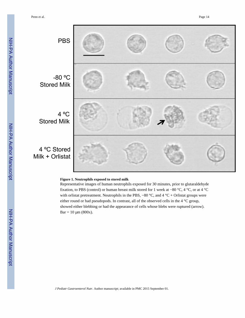
Figure 1. Neutrophils exposed to stored milkRepresentative images of human neutrophils exposed for 30 minutes, prior to glutaraldehyde
fixation, to PBS (control) or human breast milk stored for 1 week at −80 °C, 4 °C, or at 4 °C
with orlistat pretreatment. Neutrophils in the PBS, −80 °C, and 4 °C + Orlistat groups were
either round or had pseudopods. In contrast, all of the observed cells in the 4 °C group,
showed either blebbing or had the appearance of cells whose blebs were ruptured (arrow).
Bar = 10 µm (800x).
Penn et al. Page 14
J Pediatr Gastroenterol Nutr. Author manuscript; available in PMC 2015 September 01.
NIH
-PA
Author M
anuscriptN
IH-P
A A
uthor Manuscript
NIH
-PA
Author M
anuscript

Figure 2. IEC death after 4 °C storage, lipase activity and effects of lipase inhibition orhydrophobic filtering on cell death(A) Milks stored (duration and temperature as shown) or obtained from the milk bank before
and after pasteurization. Values normalized by activity prior to storage (or the mean of the
fresh milk activity, in the case of DM). N=12 donors of fresh milk, N=6 batches of raw DM
and 6 batches of pasteurized DM. * p < 0.0007 vs. Day 7 4°C milk. **p< 0.003 vs. raw milk
or Day 7 4°C milk. (B) Milk stored at −80 °C or 7 days at 4 °C (with or without orlistat)
before transfer to −80 °C (total storage time 13 ± 5 days – min 7, max 20), then digested
(PBS (white) or 0.1 mg/ml lipase (black)) and exposed to IEC for 5 minutes. N=15; ‡
p<0.006 vs. without lipase; * p<0.0007 vs. −80 °C storage only; § p<0.0007 vs. without
orlistat. Inset: raw data from Day 7 4°C milk digested with PBS or Lipase. (C&D) Milk
stored 7 days at 4 °C (non-cytotoxic samples excluded) (C) with (black) or without (white)
orlistat pretreatment or (D) before (white) or after (black) removing unbound FFAs by
filtering three times with glass fibers. (C) * p<0.02 (N=3 for milk w/ PBS; N=4 for milk w/
lipase and protease groups), (D) * p<0.05 (N=3 in milk w/ PBS; N=5 in milk w/ lipase and
protease).
Penn et al. Page 15
J Pediatr Gastroenterol Nutr. Author manuscript; available in PMC 2015 September 01.
NIH
-PA
Author M
anuscriptN
IH-P
A A
uthor Manuscript
NIH
-PA
Author M
anuscript
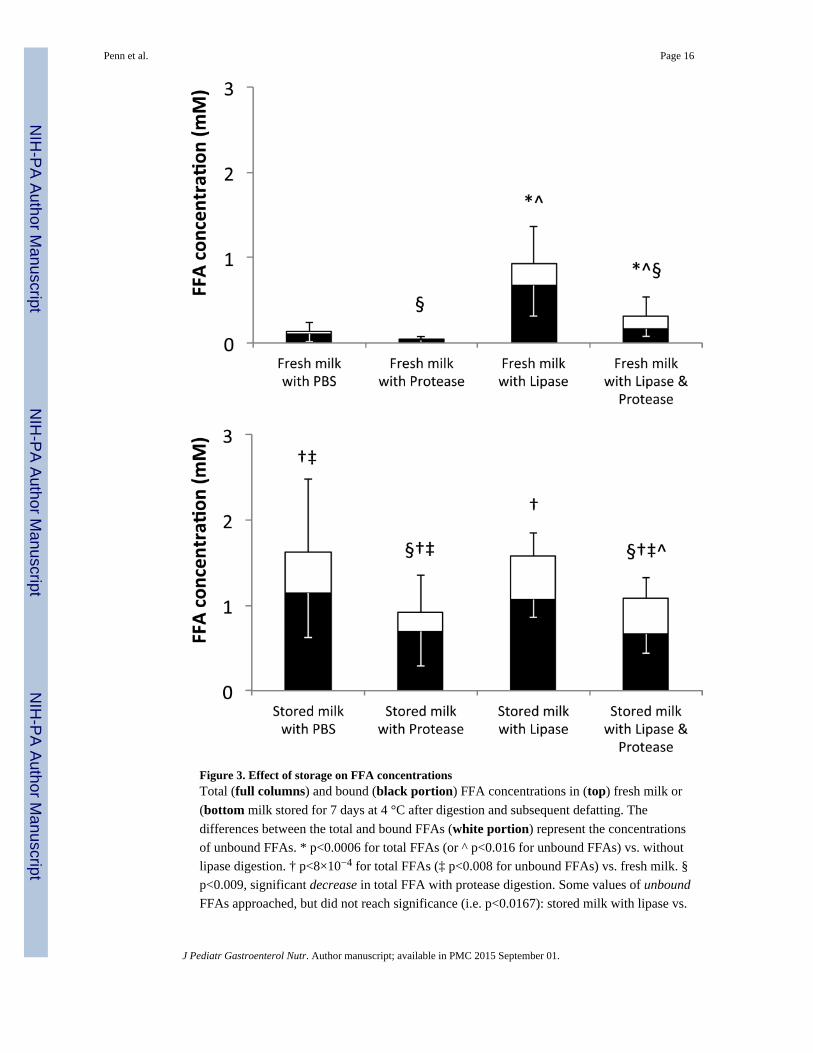
Figure 3. Effect of storage on FFA concentrationsTotal (full columns) and bound (black portion) FFA concentrations in (top) fresh milk or
(bottom milk stored for 7 days at 4 °C after digestion and subsequent defatting. The
differences between the total and bound FFAs (white portion) represent the concentrations
of unbound FFAs. * p<0.0006 for total FFAs (or ^ p<0.016 for unbound FFAs) vs. without
lipase digestion. † p<8×10⁻⁴ for total FFAs (‡ p<0.008 for unbound FFAs) vs. fresh milk. §
p<0.009, significant decrease in total FFA with protease digestion. Some values of unbound
FFAs approached, but did not reach significance (i.e. p<0.0167): stored milk with lipase vs.
Penn et al. Page 16
J Pediatr Gastroenterol Nutr. Author manuscript; available in PMC 2015 September 01.
NIH
-PA
Author M
anuscriptN
IH-P
A A
uthor Manuscript
NIH
-PA
Author M
anuscript
fresh milk with lipase (p=0.02) and stored milk with PBS vs. stored milk with protease
(p=0.05). N=12. Fresh milk data from reference (5).
Penn et al. Page 17
J Pediatr Gastroenterol Nutr. Author manuscript; available in PMC 2015 September 01.
NIH
-PA
Author M
anuscriptN
IH-P
A A
uthor Manuscript
NIH
-PA
Author M
anuscript
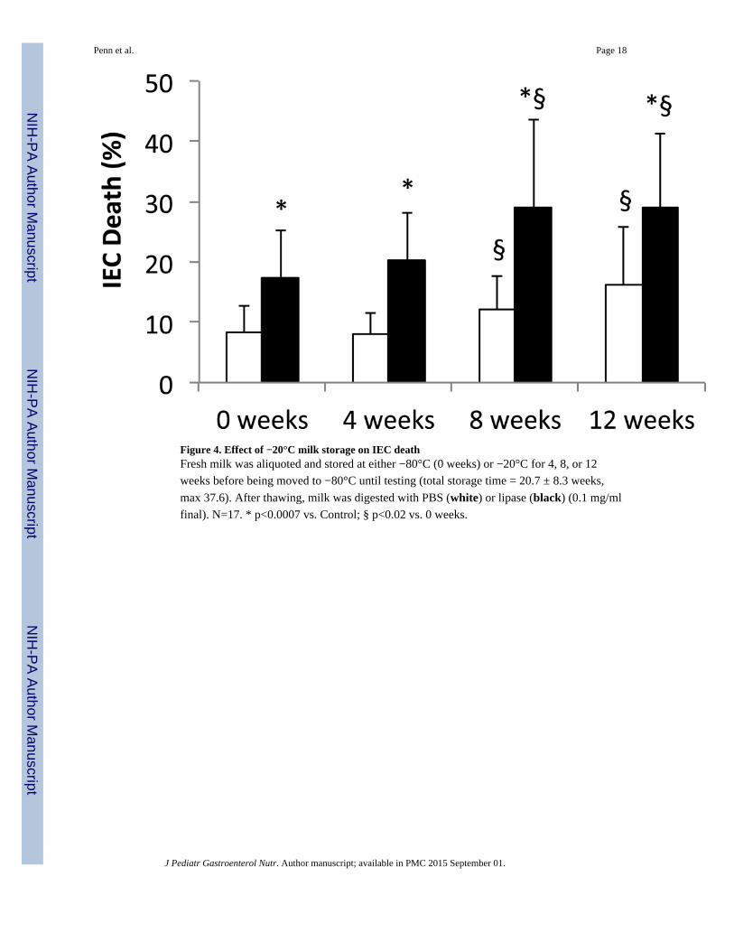
Figure 4. Effect of −20°C milk storage on IEC deathFresh milk was aliquoted and stored at either −80°C (0 weeks) or −20°C for 4, 8, or 12
weeks before being moved to −80°C until testing (total storage time = 20.7 ± 8.3 weeks,
max 37.6). After thawing, milk was digested with PBS (white) or lipase (black) (0.1 mg/ml
final). N=17. * p<0.0007 vs. Control; § p<0.02 vs. 0 weeks.
Penn et al. Page 18
J Pediatr Gastroenterol Nutr. Author manuscript; available in PMC 2015 September 01.
NIH
-PA
Author M
anuscriptN
IH-P
A A
uthor Manuscript
NIH
-PA
Author M
anuscript
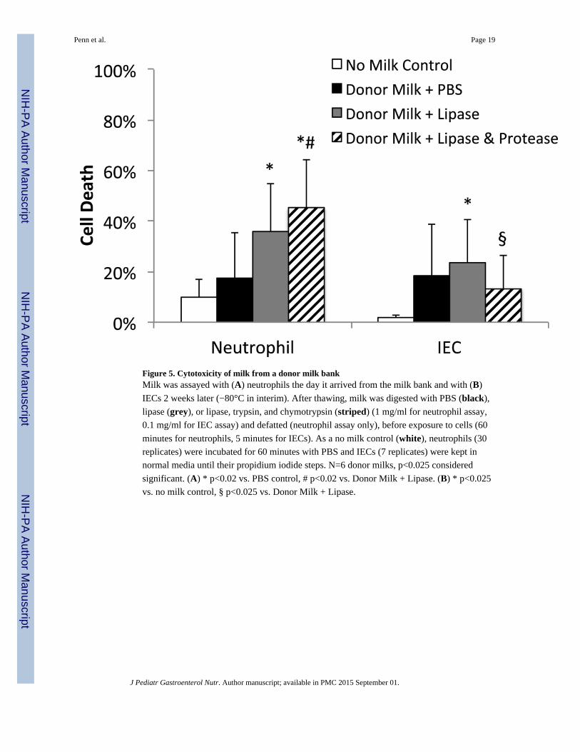
Figure 5. Cytotoxicity of milk from a donor milk bankMilk was assayed with (A) neutrophils the day it arrived from the milk bank and with (B)
IECs 2 weeks later (−80°C in interim). After thawing, milk was digested with PBS (black),
lipase (grey), or lipase, trypsin, and chymotrypsin (striped) (1 mg/ml for neutrophil assay,
0.1 mg/ml for IEC assay) and defatted (neutrophil assay only), before exposure to cells (60
minutes for neutrophils, 5 minutes for IECs). As a no milk control (white), neutrophils (30
replicates) were incubated for 60 minutes with PBS and IECs (7 replicates) were kept in
normal media until their propidium iodide steps. N=6 donor milks, p<0.025 considered
significant. (A) * p<0.02 vs. PBS control, # p<0.02 vs. Donor Milk + Lipase. (B) * p<0.025
vs. no milk control, § p<0.025 vs. Donor Milk + Lipase.
Penn et al. Page 19
J Pediatr Gastroenterol Nutr. Author manuscript; available in PMC 2015 September 01.
NIH
-PA
Author M
anuscriptN
IH-P
A A
uthor Manuscript
NIH
-PA
Author M
anuscript
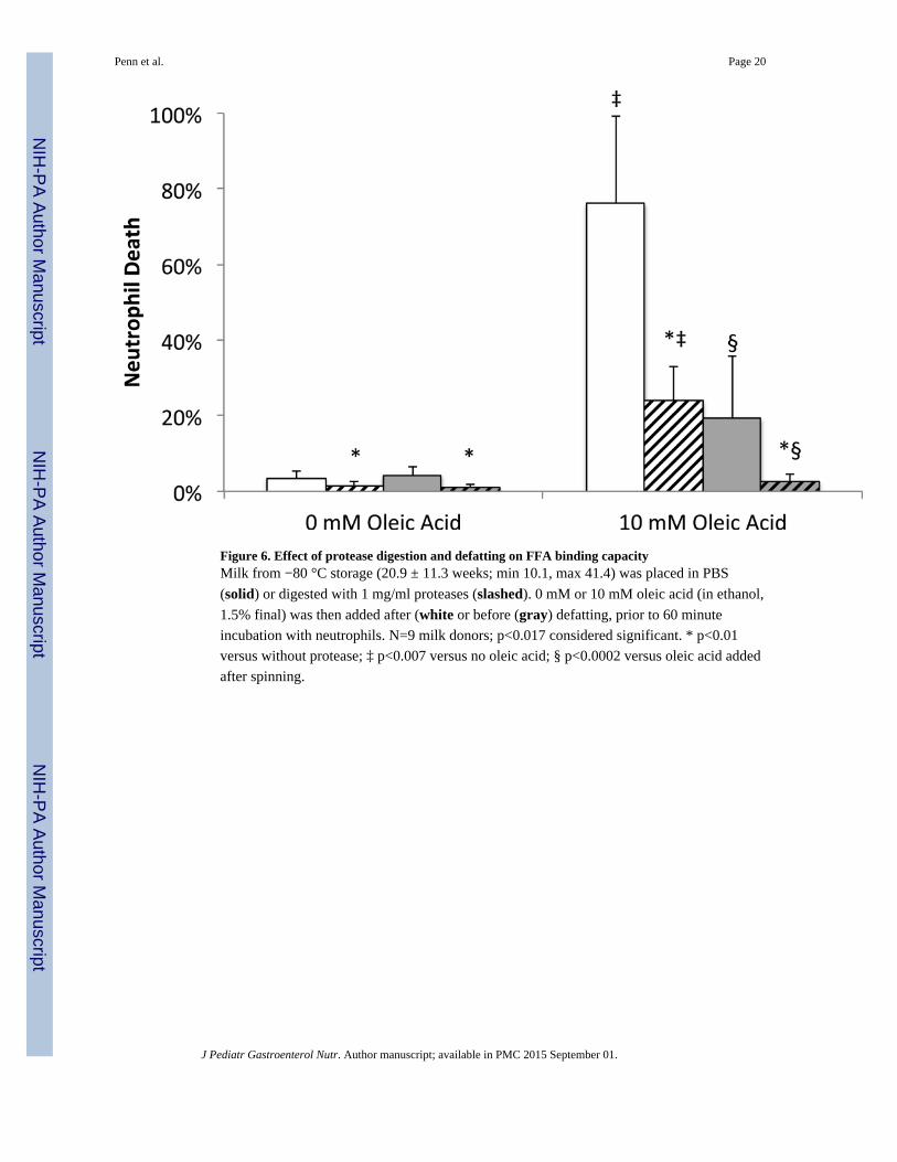
Figure 6. Effect of protease digestion and defatting on FFA binding capacityMilk from −80 °C storage (20.9 ± 11.3 weeks; min 10.1, max 41.4) was placed in PBS
(solid) or digested with 1 mg/ml proteases (slashed). 0 mM or 10 mM oleic acid (in ethanol,
1.5% final) was then added after (white or before (gray) defatting, prior to 60 minute
incubation with neutrophils. N=9 milk donors; p<0.017 considered significant. * p<0.01
versus without protease; ‡ p<0.007 versus no oleic acid; § p<0.0002 versus oleic acid added
after spinning.
Penn et al. Page 20
J Pediatr Gastroenterol Nutr. Author manuscript; available in PMC 2015 September 01.
NIH
-PA
Author M
anuscriptN
IH-P
A A
uthor Manuscript
NIH
-PA
Author M
anuscript
NIH
-PA
Author M
anuscriptN
IH-P
A A
uthor Manuscript
NIH
-PA
Author M
anuscript
Penn et al. Page 21
Table 1
Percent neutrophil death a after 1 h exposure b
with PBS with Protease with Lipase with Lipase +Protease
Fresh Milk c 3 ± 2 (0 of 12) 2 ± 1 (0 of 12)§ 6 ± 7 (1 of 12) 7 ± 12 (1 of 12)
Stored 3 days (4°C) 5 ± 4 (0 of 12) 3 ± 2 (0 of 12) 43 ± 23 (10 of 12)*‡ 28 ± 10 (10 of 12)*§‡
Stored 7 days (4°C) 47 ± 38 (7 of 12)‡ 17 ± 17 (5 of 12)§‡ 50 ± 19 (12 of 12)‡ 39 ± 19 (10 of 12)§‡
Stored 3 days (−20°C) 3 ± 1 (0 of 12) 2 ± 1 (0 of 12)§^ 18 ± 12 (7 of 12)*‡^ 13 ± 11 (3 of 12)*^
Stored 7 days (−20°C) 4 ± 2 (0 of 12)^ 2 ± 1 (0 of 12)§^ 27 ± 15 (10 of 12)*‡^ 22 ± 19 (6 of 12)*‡^
aupper number: Mean % ± SD; lower number in parentheses: Fraction of samples that were cytotoxic - i.e. with death greater than 17%
bSamples skimmed after digestion (2 h, 37 °C). Enzyme concentrations: 1 mg/ml. N=12 breast milk donors
cFresh milk data from reference (Penn et al., 2012)
*lipase digestion increased death (p<0.003)
§protease digestion decreased death (p<0.01)
‡increased death vs. fresh milk (p<0.01)
^less death when stored at −20 °C vs. 4 °C (p<0.01)
p<0.0125 considered significant
J Pediatr Gastroenterol Nutr. Author manuscript; available in PMC 2015 September 01.


![Reactions in the System Nitro-cellulose/ …...occurring polymers with nitric acid grew [5]. In 1845, Schönbein, paid special attention to the properties of the product he obtained](https://img.pdfslide.net/doc/110x75/5e9072afb97582734f3624d5/reactions-in-the-system-nitro-cellulose-occurring-polymers-with-nitric-acid.jpg)