Embed Size (px)
Citation preview

of April 25, 2018.This information is current as
Jagged1 and Notch1 SignalingActivated− Bacillus Calmette-Guérinbovis
MycobacteriumNitric Oxide Is Involved in
Kithiganahalli N. BalajiNisha Kapoor, Yeddula Narayana, Shripad A. Patil and
ol.0903174http://www.jimmunol.org/content/early/2010/02/10/jimmun
published online 10 February 2010J Immunol
MaterialSupplementary
4.DC1http://www.jimmunol.org/content/suppl/2010/02/09/jimmunol.090317
average*
4 weeks from acceptance to publicationFast Publication! •
Every submission reviewed by practicing scientistsNo Triage! •
from submission to initial decisionRapid Reviews! 30 days* •
Submit online. ?The JIWhy
Subscriptionhttp://jimmunol.org/subscription
is online at: The Journal of ImmunologyInformation about subscribing to
Permissionshttp://www.aai.org/About/Publications/JI/copyright.htmlSubmit copyright permission requests at:
Email Alertshttp://jimmunol.org/alertsReceive free email-alerts when new articles cite this article. Sign up at:
Print ISSN: 0022-1767 Online ISSN: 1550-6606. All rights reserved.1451 Rockville Pike, Suite 650, Rockville, MD 20852The American Association of Immunologists, Inc.,
is published twice each month byThe Journal of Immunology
by guest on April 25, 2018
http://ww
w.jim
munol.org/
Dow
nloaded from
by guest on April 25, 2018
http://ww
w.jim
munol.org/
Dow
nloaded from
The Journal of Immunology
Nitric Oxide Is Involved in Mycobacterium bovis BacillusCalmette-Guerin–Activated Jagged1 and Notch1 Signaling
Nisha Kapoor,* Yeddula Narayana,* Shripad A. Patil,† and Kithiganahalli N. Balaji*
Pathogenic mycobacteria have evolved unique strategies to survive within the hostile environment of macrophages. Modulation of
key signaling cascades by NO, generated by the host during infection, assumes critical importance in overall cell-fate decisions. We
show that NO is a critical factor inMycobacterium bovis bacillus Calmette-Guerin–mediated Notch1 activation, as the generation of
activated Notch1 or expression of Notch1 target genes matrix metalloproteinase-9 (MMP-9) or Hes1 was abrogated in macro-
phages derived from inducible NO synthase (iNOS) knockout (iNOS2/2), but not from wild-type, mice. Interestingly, expression of
the Notch1 ligand Jagged1 was compromised in M. bovis bacillus Calmette-Guerin–stimulated iNOS2/2 macrophages, and loss of
Jagged1 expression or Notch1 signaling could be rescued by NO donors. Signaling perturbations or genetic approaches implicated
that robust expression of MMP-9 or Hes1 required synergy and cross talk between TLR2 and canonical Notch1-PI3K cascade.
Further, CSL/RBP-Jk contributed to TLR2-mediated expression of MMP-9 or Hes1. Correlative evidence shows that, in a murine
model for CNS tuberculosis, this mechanism operates in vivo only in brains derived from WT but not from iNOS2/2 mice.
Importantly, we demonstrate the activation of Notch1 signaling in vivo in granulomatous lesions in the brains of Mycobacterium
tuberculosis-infected human patients with tuberculous meningitis. Current investigation identifies NO as a pathological link that
modulates direct cooperation of TLR2 with Notch1-PI3K signaling or Jagged1 to regulate specific components of TLR2 responses.
These findings provide new insights into mechanisms by which Notch1, TLR2, and NO signals are integrated in a cross talk that
modulates a defined set of effector functions in macrophages. The Journal of Immunology, 2010, 184: 000–000.
Mycobacteria species including Mycobacterium tuber-culosis, M. bovis, etc., have evolved diverse sets ofsophisticated survival mechanisms to interfere with
the ensuing immune response (1). Macrophages are vital com-ponents of both innate as well as adaptive immunity, and amongmacrophages, classically activated macrophages play crucialroles in resisting a range of infections by producing key effectormolecules like NO and are known to participate in host controlof the spread of various intracellular pathogens including My-cobacterium sp. (2, 3).
NO is a product of arginine metabolism and produced by differentisoforms of NO synthases. Among three isoforms, inducible NOsynthase (iNOS) is expressed in a variety of immune cells, includingmacrophages, in response to a diverse set of proinflammatory stimuli(4). NO resulting from iNOS enzymatic activity is suggested tointegrate fundamental cell fate decisions during immune responses.It is well known that NO or its derivatives like peroxynitrite(ONOO2) modify diverse cellular proteins by processes of nitro-sylation, nitrosation, nitration, or oxidation (5). NO mediatedS-nitrosylation of cysteine residues of target proteins includingvarious signaling molecules or the nitration of key tyrosine residuesto nitrotyrosine is implicated as one of the novel strategies tomodulate a variety of signaling events during initiation or activationof inflammatory responses (6, 7).Among various signaling events, Notch signaling pathway is
suggested to execute an important function during the onset ofinflammatory immune responses (8, 9). Notch signaling is sug-gested to play an important role in the development of the immunesystem and seems to act in a significant manner in critical cell-fatedecisions among diverse immune cells including macrophages ordendritic cells (9, 10). The binding of Jagged or Delta, specificligands of Notch receptor, results in the release of Notch in-tracellular domain (NICD/cleaved Notch) from the membrane byproteolytic cleavage executed by g-secretase complex. The NICDthen translocates to the nucleus and forms a complex with DNA-binding protein CSL/RBP-Jk. In general, CSL/RBP-Jk, along withmany corepressors, binds to promoters of Notch target genes,resulting in the inhibition of transcription. Upon nuclear trans-location, NICD binds to the CSL/RBP-Jk corepressor complex,resulting in the displacement of the corepressors and recruitmentof various coactivators, including Mastermind, p300, and/or CBP,leading to CSL/RBP-Jk–dependent transcription of target genes(11, 12). However, CSL/RBP-Jk–independent activation of itstarget genes by Notch or activation of CSL/RBP-Jk by signalingcascades other than Notch signaling have been documented (8).
*Department of Microbiology and Cell Biology, Indian Institute of Science; and†Department of Microbiology, National Institute of Mental Health and Neuroscien-ces, Bangalore, India
Received for publication September 29, 2009. Accepted for publication January 10,2010.
This work was supported by the Department of Biotechnology, Department of Sci-ence and Technology, Indian Council of Medical Research, and Council for Scientificand Industrial Research, and infrastructure support was received from the IndianCouncil of Medical Research (Center for Advanced Study in Molecular Medicine),Department of Science and Technology, and University Grants Commission (specialassistance). N.K. and Y.N. received fellowships from the Indian Institute of Scienceand Council for Scientific and Industrial Research, Government of India, respec-tively.
Address correspondence and reprint requests to Dr. Kithiganahalli N. Balaji, De-partment of Microbiology and Cell Biology, Indian Institute of Science, Bangalore560012, India. E-mail address: [email protected]
The online version of this article contains supplemental material.
Abbreviations used in this paper: BCG, bacillus Calmette-Guerin; ChIP, chromatinimmunoprecipitation; COX-2, cyclooxygenase-2; DN, dominant-negative; 4EBP1, eu-karyotic initiation factor 4E-binding protein 1; ECM, extracellular matrix; GSI-I,g-secretase inhibitor I; HC, healthy control; IHC, immunohistochemistry; iNOS, in-ducible NO synthase; Med, medium; MMP-9, matrix metalloproteinase-9; NICD,Notch intracellular domain; PEI, polyethylenimine; SIN1, 3-morpholinosydnonimine;siRNA, small interfering RNA; SNAP, S-nitroso-N-acetylpenicillamine; SOCS3, sup-pression of cytokine signaling 3; TBM, tuberculous meningitis; WT, wild-type.
Copyright� 2010 by The American Association of Immunologists, Inc. 0022-1767/10/$16.00
www.jimmunol.org/cgi/doi/10.4049/jimmunol.0903174
Published February 10, 2010, doi:10.4049/jimmunol.0903174 by guest on A
pril 25, 2018http://w
ww
.jimm
unol.org/D
ownloaded from

Notch signaling is known to play key roles in the regulation ofvarious developmental stages of T and B cells, T cell activation, ordifferentiation of Th cells (10). Further, TLR stimulation or var-ious proinflammatory stimuli induce the expression of Notch re-ceptor and ligands on macrophages and dendritic cells (8, 12). Inthis context, TLR-mediated induction of Notch receptor and li-gand expression on myeloid cells has resulted in activation ofa noncanonical Notch signaling cascade, leading to NF-kB acti-vation and TNF-a production (8). However, precise moleculardetails on the TLR-mediated Notch activation remain unexplored.Further, information on the effects of Notch signaling in myeloid
lineage cells still remains limited. In this perspective, matrixmetalloproteinase-9 (MMP-9), an important member of Zn2+- andCa2+-dependent endopeptidases, participates in a significantmanner in several aspects of host immune responses to myco-bacterial infection, such as graunloma formation, extracellularmatrix (ECM) reorganization, lymphocyte trafficking, infiltrations,and inflammation, etc. MMP-9 is expressed at various clinicalcategories of tuberculosis disease like active cavitary tuberculosis,meningitis, and pleuritis (13–15). Notably, in the case of pulmo-nary tuberculosis, breakdown of ECM by MMP-9 forms an in-tegral part of the granuloma formation (16).The current study demonstrates for the first time that NO,
a product of iNOS activity, is responsible for M. bovis bacillusCalmette-Guerin (BCG)-triggered activation of Notch1 in mac-rophages through direct regulation of Jagged1 expression as wellas in generation of activated Notch1. We present the evidence thatiNOS activity is a critical factor in TLR2-mediated Notch1 acti-vation as macrophages derived from iNOS knockout (iNOS2/2)but not from wild-type (WT) mice failed to activate Jagged1 ex-pression as well as Notch1 signaling upon M. bovis BCG in-fection. The loss of TLR2-mediated Jagged1 expression or Notch1activation in iNOS2/2 macrophages could be rescued by treatmentwith NO donor 3-morpholinosydnonimine (SIN1) or S-nitroso-N-acetylpenicillamine (SNAP). Signaling perturbations stronglyimplicated the role for cross talk among members of Notch1-PI3Kand MAPK cascades in M. bovis BCG-TLR2–mediated activationof Notch1 target genes MMP-9 or Hes1. Chromatin immunopre-cipitation (ChIP) experiments demonstrate that M. bovis BCG’sability to trigger increased binding of CSL/RBP-Jk to MMP-9promoter was severely compromised in macrophages derived fromiNOS2/2 mice compared to WT mice. These results are consistentwith the observation that NO-triggered Notch1 signaling-mediatedCSL/RBP-Jk recruitment has a positive regulatory role in M. bovisBCG-induced MMP-9 transcription. We show correlative evidencethat this mechanism operates in vivo by immunohistochemicalexpression analysis of activated Notch1 or its target gene productsHes1 or MMP-9 in brains of WT or iNOS2/2 mice that wereintracerebrally infected with M. bovis BCG. Further, activation ofNotch1 signaling in vivo could be demonstrated only in granulo-matous lesions in brains derived from human patients with tu-berculous meningitis (TBM) as opposed to healthy individualsvalidating the role of Notch1 signaling in Mycobacterium patho-genesis. Briefly, the current investigation identifies NO as thepathological link between TLR2 and Notch1 signaling, whichregulates the relative abundance of various immunopathologicalparameters including MMP-9 in macrophages.
Materials and MethodsCells and bacteria
Peritoneal macrophages were isolated from peritoneal exudates of C57BL/6or iNOS2/2 C57BL/6 mice that were maintained at the Central AnimalFacility, Indian Institute of Science, Bangalore, India. RAW 264.7 andHEK 293 cell lines (obtained from National Centre for Cell Science, Pune,
India) were cultivated in DMEM supplemented with 10% heat-inactivatedFBS (Sigma-Aldrich, St. Louis, MO). The experiments with mouse mac-rophages were carried out after approval was obtained from the In-stitutional Ethics Committee for Animal Experimentation as well as fromthe Institutional Bioethics Committee, Indian Institute of Science.M. bovisBCG Pasteur 1173P2 was grown to midlog phase in Souton’s medium, andaliquots were stored as described previously (17, 18). M. bovis BCG wasused at 10 multiplicity of infection for infection in all of the experiments.
Chemicals and reagents
General laboratory reagents were purchased from Sigma-Aldrich or Merck(Darmstadt, Germany). NO donors, SIN1, and SNAP were obtained fromCalbiochem (San Diego, CA).
Anti-Ser473 Akt, anti-Akt, anti-Thr70 eukaryotic initiation factor 4E-binding protein 1 (4EBP1), anti-4EBP1, anti-Thr202/Tyr204 pERK1/2,anti-ERK1/2, Thr180/Tyr182 pp38 MAPK, anti-p38 MAPK, anti-Ser536NF-kB, anti–NF-kB, anti-Jagged1, and anti-cleaved Notch1 Abs werepurchased from Cell Signaling Technology (Beverly, MA). Anti-Hes1 andanti–RBP-Jk Abs were purchased from Santa Cruz Biotechnology (SantaCruz, CA). HRP conjugated anti-mouse IgG Abs were obtained fromCalbiochem. Anti–b-actin (AC-15) and anti MMP-9 Abs were obtainedfrom Sigma-Aldrich. Anti-CD16/32 (Fc blocker) Abs were purchased fromeBioscience, San Diego, CA. Cy2 or Cy5 or HRP conjugated anti-rat andanti-rabbit IgG Abs were purchased from Jackson ImmunoResearchLaboratories (West Grove, PA).
In vivo infection of mice with M. bovis BCG
The experiments involving infection of mice were performed afterthe approval from the Institutional Ethics Committee for Animal Experi-mentation as well as from the Institutional Biosafety Committee. C57BL/6mice and iNOS2/2 C57BL/6 mice used in the current investigation were5 to 6 wk old. For intracranial infection, 1 3 106 M. bovis BCG Pasteur1173P2 bacteria were washed in PBS, resuspended in 50 ml sterile PBS,and then inoculated intracranially using 1-ml syringes and a 26-gaugeneedle. One set of iNOS2/2 mice received 20 mg SIN1/mice in addition toinfection withM. bovis BCG. Control mice were injected with 50 ml sterilePBS using the same protocol. Before intracranial inoculation, mice wereanesthetized with an i.p. injection of ketamine (6 mg) in 80 ml. In ex-periments involving TLR2 Ab, WT mice were injected with anti-TLR2 orcontrol IgG Ab (200 mg/kg) 24 h prior to infection with M. bovis BCG(19). For in vivo silencing of MyD88, WT mice received 0.6 nmol MyD88or control small interfering RNA (siRNA) complexed with low m.w.polyethylenimine (PEI) 24 h before intracranial inoculation with M. bovisBCG (20–22). After 5 d of inoculations, brains were harvested from ex-perimental mice. Microtome sections (4 mm) were cut from formalin-fixed,decalcified, and paraffin-embedded tissue samples. These paraffin-embedded sections were first deparaffinized followed by Ag retrieval withboiling 10 mM citrate buffer (pH 6.0) in a microwave for 10 min, treatedwith 1% H2O2 10 min, incubated with 0.1 M glycine for 15 min, andblocked with 5% BSA for 1h at room temperature. Primary Abs wereincubated overnight, and HRP conjugated secondary Abs for 90 min. TheHRP reaction was detected with 0.05% diaminobenzidine and 0.03%H2O2. Sections were counterstained with hematoxylin, dehydrated, andmounted. Stained tissue sections were analyzed with a Leica DMLB mi-croscope (Leica Microsystems, Wetzlar, Germany). All experiments wereperformed with appropriate isotype-matched control Abs.
Human patients with TBM
Human brain tissue samples were collected from patients with TBM at theNational Institute of Mental Health and Neurosciences, Bangalore, India,according to the guidelines of the Institutional Ethics Committee. Eachpatient provided informed written consent to participation in this study inaccordance with institutional and regulatory guidelines. Brain tissuesamples from patients that had died due to accident served as controls.Microtome sections (4 mm) of brain tissue samples were stained with in-dicated Abs for immunohistochemistry (IHC) as described above.
Treatment with pharmacological inhibitors
The pharmacological inhibitors were obtained from Calbiochem andwere reconstituted in sterile DMSO and used at following concentrations:1400W (100 mM), LY294002 (50 mM), wortmannin (100 nM), rapamycin(100 nM), Akt inhibitor II (Akt I-II, 10 mM), g-secretase inhibitor I (GSI-I)(10 mM), SB203580 (20 mM), U0126 (10 mM), SP600125 (50 mM),manumycin (20 mM), and Bay 11-7082 (20 mM). DMSO at 0.1% con-centration was used as the vehicle control. In all experiments with in-hibitors, a tested concentration was used after careful titration experimentsassessing the viability of the macrophages.
2 ROLE FOR NO IN Notch1 SIGNALING
by guest on April 25, 2018
http://ww
w.jim
munol.org/
Dow
nloaded from

Stable transfection of NICD
RAW 264.7 macrophages stably expressing Notch1 intracellular domain(RAW-NICD) were generated as described previously (17, 18).
siRNA transfections
RAW264.7 cells were transfected with 100 nM siRNA using Lipofectamine2000 (Invitrogen, Carlsbad, CA) according to the manufacturer’s in-structions. Seventy-two hours posttransfection, the cells were infected with10 multiplicity of infection M. bovis BCG for 12 h. Later, protein lysateswere prepared and analyzed for the targeted proteins by Western blotting.siRNAs specific to Notch1, Akt, MyD88, CSL/RBP-Jk, control siRNA,and siGLO Lamin A/C were obtained from Dharmacon (Chicago, IL) assiGENOME SMARTpool reagents, which contains a pool of four differentdsRNA oligonucleotides (siRNA).
Transient transfections and luciferase assays
RAW 264.7 macrophages were transiently transfected with MMP-9 orHes1 promoter constructs or TLR2-dominant negative cDNA constructusing PEI (Sigma-Aldrich), and assays were carried out as describedpreviously (18).
RNA isolation and quantitative real-time PCR
Macrophages were treated as indicated, and the transcript levels of the targetmoleculeswere assessed by quantitative real-time PCR analysis as describedpreviously (17). The forward and reverse primer pairs used were as follows:GAPDH forward 59-gagccaaacgggtcatcatct-39, reverse 59-gaggggccatccaca-gtctt-39; MMP-9 forward 59-gagctgtgcgtcttccccttc-39, reverse 59-ggaatgat-ctaagcccagtgc-39; Notch1 forward 59-agaatggcatggtgcccag-39, reverse 59-tg-gtggagaggctgctgtgtag-39; Jagged1 forward 59-gaagtcagagttcagaggcgtcc-39,reverse 59-agtagaaggctgtcaccaagcaac-39; and Hes1 forward 59-gagagg-ctgccaaggtttttg-39, and reverse 59-cactggaaggtgacactgcg-39. All the primerswere purchased from Sigma Genosys (Bangalore, India).
Immunoblotting
Cell lysates were prepared after washing briefly with ice-cold PBS in 13radioimmunoprecipitation assay lysis buffer (50 mM Tris-HCl [pH 7.4],1% Nonidet P-40, 0.25% Na-deoxycholate, 150 mM NaCl, 1 mM ED-TA, 1 mM PMSF, 1 mg/ml each aprotinin, leupeptin, and pepstatin,1 mM Na3VO4, and 1 mM NaF). An equal amount of protein wassubjected to SDS-PAGE, and then proteins were transferred to poly-vinylidene difluoride membranes (Millipore, Bedford, MA), which wereblocked in TBST buffer (0.02 M Tris-Hcl [pH 7.5], 0.15 M NaCl, and0.1% Tween 20) containing 5% nonfat dried milk and probed witha primary Ab for overnight at 4˚C. After washing with TBST, mem-branes were incubated with secondary Ab conjugated to HRP (JacksonImmunoResearch Laboratories). The blots were then visualized with anECL detection system (PerkinElmer, Wellesley, MA) as per the manu-facturer’s instructions.
Immunofluorescence
Resident mouse peritoneal macrophages were seeded on coverslips andtreated as indicated. The cells were fixed with 3% formaldehyde for 10min at room temperature. Primary Abs were incubated for 1 h at roomtemperature following three washes with PBS. Secondary Abs (Cy2-conjugated anti-rabbit IgG or Cy5-conjugated anti-rat IgG) were in-cubated in the dark for 1 h at room temperature and then washed threetimes with PBS. Coverslips with cells were mounted on a slide withfluoromount G. Confocal images were taken on Zeiss LSM 510 Metaconfocal laser scanning microscope using plan Apochromat 633/1.4 oilDIC objective (Zeiss, Oberkochen, Germany) and Argon/2 458, 477,488, and 514 nm and HeNe 543 lasers. During colocalization studies,a series of images at an interval of 0.37-mm focal planes was collectedinto a z-stack to ascertain the actual colocalization. Every single layer ofz-stack was subjected to image analysis by LSM 5 image examinersoftware to visualize and locate protein expression.
FIGURE 1. Involvement of iNOS inM. bovisBCG-triggered activation ofNotch1 signaling and Jagged1 expression.Macrophageswere treatedwith 1400W
prior to infection withM. bovisBCG followed by analysis of protein levels of NICD and Jagged1 by immunoblotting (A), cell surface expression of Jagged1 by
flow cytometry (B), and transcript levels of Notch1 by quantitative real-time PCR (C).D, Macrophages derived fromWTmice were pretreated with 1400Wor
vehicle control followed by infection withM. bovisBCG. In case ofmacrophages derived from iNOS2/2mice, cells were either infected withM. bovisBCGor
treated with SIN1 or SNAP for different time points and transcript levels of Jagged1were analyzed by quantitative real-time PCR. iNOS2/2macrophages were
either infectedwithM. bovisBCGor treatedwith SIN1 or SNAP and analyzed for cell surface expression of Jagged1 by flow cytometry (E) and transcript levels
of Notch1 by quantitative real-time PCR (F).G, Macrophages derived from iNOS2/2mice were treated with SIN1 and SNAP, and protein level expression of
Jagged1 or NICD was evaluated by immunoblotting. H, WT and iNOS2/2 macrophages were treated as indicated and nuclear translocation of NICD was
analyzed by confocal microscopy. The data presented in the figure is representative of three independent experiments. Med, medium.
The Journal of Immunology 3
by guest on April 25, 2018
http://ww
w.jim
munol.org/
Dow
nloaded from

Flow cytometry analysis
The cells were washed with PBS and fixed in 0.1% formaldehyde. To avoidnonspecific binding, cells were incubated with 0.5 mg Fc blocker (per 106
cells) for 30 min on ice followed by staining with rabbit anti-mouse MMP-9/rabbit anti-mouse Jagged1 Ab. This was followed by staining with FITCor Cy2-conjugated anti-rabbit IgG. Cells were analyzed using FACScan(BD Biosciences, San Jose, CA). Dead cells were excluded from theanalysis by their forward and sideways light-scattering properties.
ChIP assay
ChIP assays were carried out following a protocol provided by UpstateBiotechnology (Lake Placid, NY), with modifications. Peritoneal macro-
phageswere leftuninfectedor infectedwithBCGfor12h.Thecellswerefixedwith 1.42% formaldehyde for 15 min at room temperature followed by in-activation of formaldehyde with addition of 125 mM glycine. Chromatinextracts containing DNA fragments with an average size of 500 bp wereimmunoprecipitated using anti-Notch1 or RBP-Jk Ab. Purified DNA wasanalyzed by quantitative real-time PCR by using the SYBR Green method(Finnzymes, Espoo, Finland). Regions with the RBP-Jk binding site in themouseMMP-9 promoter were amplified using primer pairs RBP-Jk forward,59-atcagtcagggccgtcagac-39, and RBP-Jk reverse, 59-gacccacaggaaaccaca-gaac-39. 28S rRNAwasusedascontrol in thePCR, and theprimerswere forward,59-ctgggtataggggcgaaagac-39, and reverse, 59-ggccccaagacctctaatcat-39. All re-sults were normalized either by respective input values or by amplification of 28SrRNA. All ChIP experiments were repeated at least three times.
FIGURE 3. Activation of Notch1
signaling in vivo. Serial sections of
human brain tissue samples from pa-
tients with TBM or healthy subjects
were stained for expression levels of
activated Notch1/NICD (A) as well as
Notch1 target gene Hes1 (B) by IHC.
Hematoxylin (blue) was used for nu-
clear staining. HC, healthy control.
FIGURE 2. Essential role of NO inM. bovisBCG-induced expression of Notch1 target genes MMP-9 and Hes1. Macrophages derived fromWTmice were
pretreatedwith 1400Wfollowedby infectionwithM.bovisBCG, and transcript levels ofMMP-9 (A) andHes1 (B)were analyzed byquantitative real-timePCR,
whereas cell surface expression of MMP-9 was analyzed by flow cytometry (C). Macrophages from iNOS2/2mice were either infected withM. bovisBCG or
treated with SIN1 or SNAP followed by analysis of transcript level expression of MMP-9 (D), Hes1 (E), and cell surface expression of MMP-9 (F).G, Nuclear
Hes1 expression inWTor iNOS2/2macrophages treated as indicated as analyzed by confocalmicroscopy.H, Recruitment ofNotch1 (i) andCSL/RBP-Jk (ii) at
MMP-9 promoter was analyzed by ChIP assay with Abs to Notch1 or RBP-Jk, respectively, in M. bovis BCG-infected WT or iNOS2/2 macrophages. The
recruitment of Notch1 and RBP-Jk at MMP-9 promoter was assessed by quantitative real-time PCR. Data are representative of three independent experiments.
Med, medium.
4 ROLE FOR NO IN Notch1 SIGNALING
by guest on April 25, 2018
http://ww
w.jim
munol.org/
Dow
nloaded from
Statistical analysis
Levels of significance for comparison between samples were determined bythe Student t test distribution. The data in the graphs is expressed as themean 6 SEM. Graphpad Prism 3.0 software (GraphPad, San Diego, CA)was used for all the statistical analysis.
ResultsNO is required for M. bovis BCG-induced Notch1 signalingand Jagged1 expression
M. bovis BCG challenge of macrophages could provokea spectrum of cellular activities including TLR2-MyD88–de-pendent expression of key Notch1 target genes like MMP-9(Supplemental Fig. 1A–D), an effector molecule that partic-ipates in macrophage cell motility during inflammatory re-sponses (16). We show that TLR2 stimulation by M. bovis BCGleads to upregulation of Notch1 and activation of the Notch1signaling pathway by inducing the formation of a cleavageproduct of Notch1 (NICD) (Fig. 1A, Supplemental Fig. 1E, 1F).Further, M. bovis BCG triggers robust activation of Jagged1expression, a Notch1 receptor ligand (Fig. 1A, 1B, Supple-mental Fig. 1F, 1G). In this perspective, we addressed whetherM. bovis BCG-TLR2–mediated expression of iNOS participatesin activation of Notch1 signaling. Interestingly, the ability of M.bovis BCG to induce the formation of NICD, upregulation ofNotch1, or expression of the Notch1 receptor ligand Jagged1was abrogated in the presence of 1400W, an iNOS-selective
inhibitor (Fig. 1A–C). To substantiate the importance of iNOSin induction of NICD or Jagged1 expression, macrophages de-rived from WT and iNOS2/2 mice challenged with or withoutM. bovis BCG were analyzed. As shown in Fig. 1D, 1F, com-pared with WT macrophages, marked inhibition of Jagged1 orNotch1 expression both at RNA and protein levels evoked byM. bovis BCG challenge was observed in iNOS null macro-phages. This iNOS deficiency caused defect in the ability of M.bovis BCG to trigger Jagged1 or Notch1 expression was not dueto the general inability of cells to mobilize, because analogs ofiNOS downstream mediators, SNAP, or SIN1 (NO donor) couldaugment Jagged1, NICD, or Notch1 expression in iNOS-de-ficient macrophages comparable to that in WT macrophages(Fig. 1D–G). Confocal microscopy studies further validated therequirement of NO in M. bovis BCG-triggered Notch1 signalingactivation as inhibition of iNOS activity or deficiency in iNOSexpression resulted in marked reduction in nuclear translocationof NICD in macrophages (Fig. 1H). However, treatment ofiNOS2/2 macrophages with NO donor SIN1 could reverse theinhibition in M. bovis BCG-triggered nuclear translocation ofNICD (Fig. 1H). Accordingly, expression of Notch1 targetgenes MMP-9 or Hes1 was regulated by NO, as inhibition ofiNOS activity by iNOS-selective inhibitor 1400W abrogated theM. bovis BCG-induced MMP-9 or Hes1 expression (Fig. 2A–C).Further, M. bovis BCG failed to induce MMP-9 or Hes1 expressionin iNOS null macrophages, which could be rescued by treatmentwith NO donors, SNAP, or SIN1 (Fig. 2D–F). Similar to above
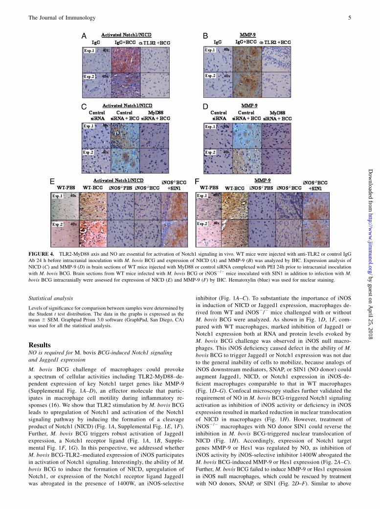
FIGURE 4. TLR2-MyD88 axis and NO are essential for activation of Notch1 signaling in vivo. WT mice were injected with anti-TLR2 or control IgG
Ab 24 h before intracranial inoculation with M. bovis BCG and expression of NICD (A) and MMP-9 (B) was analyzed by IHC. Expression analysis of
NICD (C) and MMP-9 (D) in brain sections of WT mice injected with MyD88 or control siRNA complexed with PEI 24h prior to intracranial inoculation
with M. bovis BCG. Brain sections from WT mice infected with M. bovis BCG or iNOS2/2 mice inoculated with SIN1 in addition to infection with M.
bovis BCG intracranially were assessed for expression of NICD (E) and MMP-9 (F) by IHC. Hematoxylin (blue) was used for nuclear staining.
The Journal of Immunology 5
by guest on April 25, 2018
http://ww
w.jim
munol.org/
Dow
nloaded from

results on NICD nuclear translocation, data derived from confocalmicroscopy studies suggested that the inhibition of iNOS activity ordeficiency in iNOS expression abrogated the ability of M. bovisBCG to induce nuclear expression of Hes1 (Fig. 2G). However,SIN1 treatment could augment nuclear expression of Hes1 in iNOSnull macrophages in contrast to significant reduction triggered byM. bovis BCG challenge (Fig. 2G).To further validate, by sequence analysis,we identified threeCSL/
RBP-Jk–binding consensus in themouseMMP-9 promoter (23) andshowed that the recruitment of CSL/RBP-Jk or NICD was severelycompromised in macrophages derived from iNOS2/2 mice com-pared withWTmice by ChIP experiments (Fig. 2H). However, totalexpression levels of CSL/RBP-Jk were unaltered in iNOS2/2 orWT mice (data not shown). These results are consistent with theobservation that NO-triggered Notch1 signaling-mediated CSL/RBP-Jk recruitment has a positive regulatory role inM. bovis BCG-induced MMP-9 transcription. These findings suggest the criticalparticipation of the NO pathway inM. bovis BCG-induced Jagged1expression and activation of Notch1 signaling in macrophages.
High expression levels of activated Notch1 or its target genesHes1 or MMP-9 in brains of human TBM patients
We investigated whether this mechanism for Notch1 activation wasrelevant to the biology ofMycobacterium infection in vivo. Humanbrain tissue samples of patients with TBM or from healthy sub-jects were assessed for the expression of activated Notch1 (NICD)or its target genes Hes1 or MMP-9. By immunohistochemicalexpression analysis, we found that NICD protein levels weresignificantly increased in the brain of most patients with TBMcompared with healthy subjects brains (Fig. 3A). Similarly, Hes1or MMP-9 expression levels were significantly enhanced in brainsamples of patients with TBM compared with healthy subjects(Fig. 3B, Supplemental Fig. 2).
TLR2-MyD88-iNOS/NO axis is a critical link in M. bovisBCG-triggered activation of Notch1 signaling in vivo
To ascertain the role of TLR2-MyD88 and NO signaling in acti-vation of Notch1 signaling in vivo, a suggested murine model forthe study of CNS tuberculosis or TBM involving intracranial in-oculation of M. bovis was used (24). Blocking TLR2 signaling byneutralizing Ab to TLR2 in vivo clearly abrogated M. bovis BCG-triggered Jagged1 expression, Notch1 activation, and expressionof Notch1 signaling target genes, Hes1 and MMP-9 (Fig. 4A, 4B,Supplemental Fig. 3).Further, siRNA-mediated knockdown of MyD88 in vivo abol-
ished M. bovis BCG-induced Jagged1 expression, Notch1 activa-tion, and Notch1 target genes Hes1 and MMP-9 expression (Fig.4C, 4D, Supplemental Fig. 4), potentiating a critical role of TLR2-MyD88 axis in activation of Notch1 signaling in vivo. Further, inaccordance with results obtained with macrophages, IHC data ofbrain sections demonstrates that iNOS deficiency in iNOS2/2
mice severely compromised M. bovis BCG potential to triggeraugmented expression of NICD and Jagged1 compared with WTmice (Fig. 4E, Supplemental Fig. 5). Similarly, expression levelsof Nocth1 target genes Hes1 or MMP-9 were markedly reduced inbrain sections derived from infected iNOS2/2 mice comparedwith WT mice (Fig. 4F, Supplemental Fig. 5). In addition, NOdonor (SIN1) treatment of iNOS2/2 mice in vivo rescued acti-vation of Notch1 and expression of Jagged1 as well as Notch1target genes, Hes1 and MMP-9 (Fig. 4E, 4F, Supplemental Fig. 5).These results imply that NO acts as an essential mediator of M.bovis BCG-dependent Notch1 signaling activation and is respon-sible for expression of specific transcriptional target proinfam-matory gene signatures (such as MMP-9) in macrophages.
NO-triggered Jagged1 expression and Notch1 signalingrequires Ca2+
NO can directly influence diverse signaling cascades through nitro-sylation (6) and can exert its capacity to act as a Ca2+-mobilizingintracellular messenger (7). This critical interplay between NO andCa2+ might act as key modulator of signaling related processes thusexecuting important roles in cell-fate decisions. In addition toa number of Ca2+-permeable channels, NO modulates upstreamcomponents such as cGMP, p21ras, or protein phosphatases (25, 26).In this perspective, blockade of Ca2+ mobilization by BAPTA-AMsignificantly inhibited SNAP/SIN1 (NO donor) or M. bovis BCG-triggered activation of Jagged1 expression or Notch1 signaling iniNOSnullmacrophages andWTmacrophages, respectively. (Fig. 5A,5C). Accordingly, a Notch1 target gene, MMP-9, expression bySNAP or SIN1 in iNOS null macrophages could be blocked by Ca2+
chelator (Fig. 5B). Similarly, Hes1 promoter activity triggered byM.bovis BCG-mediated TLR2 stimulation could be blocked by in-tracellularCa2+chelatorBAPTA/AM(Fig. 5D).Confocalmicroscopyanalysis further validated the requirement of Ca2+ as blocking effectsof BAPTA-AMon intracellularCa2+wave resulted in the inhibition inM. bovis BCG-triggered nuclear translocation of NICD (Supple-mental Fig. 6A) aswell as expression ofHes1 (Supplemental Fig. 6B).As described subsequently, blockade of Ca2+ mobilization by BAP-TA-AM abrogated the Notch1-mediated Akt, 4EBP1, or ERK1/2activation as well as NF-kB nuclear translocation, which ascertainsthe critical role of NO-Ca2+ axis in overall M. bovis BCG-TLR2signaling (Supplemental Fig. 6C, 6D). However, addition of cGMPcould not rescue Jagged1, NICD, or Notch1, MMP-9 expression in
FIGURE 5. Requirement of Ca2+ in NO-dependent activation of Notch1
signaling. A and B, Macrophages derived from iNOS2/2 mice were pre-
treated with BAPTA-AM or vehicle control (DMSO) prior to treatment
with NO donors. The cell-surface expression levels of Jagged1 (A) or
MMP-9 (B) were analyzed by flow cytometry. Results are represented as
mean fluorescence intensity (MFI). Addition of cGMP or infection of
iNOS2/2 macrophages with M. bovis BCG did not alter cell-surface ex-
pression levels of Jagged1 (A) or MMP-9 (B). C, WT macrophages were
pretreated with BAPTA-AM or DMSO followed by infection withM. bovis
BCG. The generation of NICD at indicated time points was assessed by
immunoblotting. D, Chelation of calcium or TLR2 dominant-negative
(DN) construct significantly reduced M. bovis BCG-triggered Hes1 ex-
pression as evaluated by Hes1 promoter activity. The results are expressed
as mean 6 SEM of three independent experiments, and the blots are
representative of three independent experiments. Med, medium.
6 ROLE FOR NO IN Notch1 SIGNALING
by guest on April 25, 2018
http://ww
w.jim
munol.org/
Dow
nloaded from

iNOS-deficient macrophages comparable to that inWTmacrophages(Fig. 5A, 5B and data not shown). These findings rule out the partic-ipation of the NO/cGMP pathway in M. bovis BCG-mediated acti-vation of Notch1 signaling as well as Jagged1 or MMP-9 expression.
Expression of MMP-9 involves iNOS-Notch1–mediatedactivation of PI3K signaling
The currently available data on the diverse array of cellular processesunder the control of Notch1 clearly illustrate the physiologicalimportance of Notch1 signaling. Notch was described to play pivotalroles in upregulation of antiapototic genes, vasodilatation, etc. (27,28). The Notch-specific genetic signature is significantly sufficientin the development of immune system and in imparting survivalbenefits in many cell types (10, 27). An increasing body of evidenceindicates that Notch1 signaling-mediated survival effects in manycell types often require participation of the members of PI3Kpathway (29, 30). It is known that mycobacteria trigger the acti-vation of PI3K pathway in macrophages (17, 18).To examine an involvement of Notch1 in NO/Notch1/PI3K-me-
diated regulation of Notch1 target genesMMP-9 or Hes1 expression,signaling perturbations were carried out with Notch1 activation in-hibitor GSI-I, siRNA-mediated knockdown of Notch1 or CSL/RBP-Jk. The inhibitor of Notch1 signaling, GSI-I, abolished NO-inducedMMP-9 expression both at RNA level and cell surface expression onmacrophages (Fig. 6A, 6B). Data derived from siRNA-mediatedknockdown of Notch1 or CSL/RBP-Jk significantly reducedM.bovisBCG-triggered induction of MMP-9 (Fig. 6C). Stable expression ofNICD in RAW264.7 macrophages resulted in augmented expressionof Notch1 target genes Hes1 or MMP-9 (Fig. 6D).As described, cross talk between Notch1 and PI3K pathways
including genetic interactions, the physical binding of Notch1 toPI3K, or their association to common, yet unique cofactors havebeen described (17, 27). In this perspective, GSI-I abrogated M.bovis BCG-triggered 4EBP1 or Akt activation, and attenuation of4EBP1 activation was quite comparable to LY294002, a known
inhibitor of PI3K (Fig. 6E and data not shown). In concurrencewith these results, inhibition of PI3K by LY294002 or Wortmanin,Akt by Akt inhibitor or siRNA, and mTOR by rapamycin sig-nificantly abrogated M. bovis BCG-triggered MMP-9 expression(Fig. 7A–C). We next studied whether Notch1-PI3K signaling axisregulates MMP-9 expression by activating MAPKs. Our analysisutilizing pharmacological inhibitors revealed that both ERK1/2and p38 MAPK are involved in Notch1-PI3K–driven MMP-9expression (Fig. 7D). In addition, LY294002 pretreatment down-regulated M. bovis BCG-triggered ERK1/2 or p38 MAPK acti-vation (Fig. 7E). Together, these results strongly suggest thatactivated Notch1 exert a direct effect in regulating PI3K-MAPKsignaling, thus exerting cooperative effects in modulating specificgene transcription.
Notch1 signaling inhibition attenuates NF-kB activation
MMP-9 promoter contains many cis-acting NF-kB consensus ele-ments, and Notch1 mediated activation of its target genes often in-volves the active recruitment of transcription factor NF-kB (8, 11, 17).In this perspective, we demonstrate that besides the physical parti-tioning ofNF-kB, nuclear translocation ofNF-kBwas abrogated uponinhibition ofNotch1 signaling byGSI-I (Fig. 7F). Interference in PI3Kor MAPK activation reversedM. bovis BCG-mediated nuclear trans-location of NF-kB from cytosol (Fig. 7F). Blocking the NF-kB sig-naling pathway using Bay 11-7082, a specific inhibitor of IkB-aphosphorylation markedly diminished induced expression of MMP-9(Fig. 7G). These results suggest that NO, upstream of Notch1 signal-ing, acts as an essential mediator of TLR2-dependent responses andmodulates direct cooperation of TLR2with Notch1-PI3K signaling toregulate a distinct set of effector functions in macrophages.
DiscussionNO is described to play critical roles during diverse pathophysi-ological responses associated with inflammation during host-pathogen interactions (31). NO is reported to be microbicidal
FIGURE 6. NO-Notch1-PI3K signaling cross talk involved in infection triggered MMP-9 expression. Notch1 activation inhibitor, GSI-I, abrogated M.
bovis BCG-induced MMP-9 expression as analyzed by real-time PCR (A), immunoblotting (A), or flow cytometry (B). C, RAW 264.7 macrophages were
transiently transfected with either control siRNA or Notch1 (i) and RBP-Jk siRNA (ii). After 3 d of transfection, expression levels of MMP-9 were an-
alyzed. D, RAW 264.7 macrophages stably transfected with pCMV-NICD (RAW-NICD) or pCMV (RAW-Vec) were analyzed for expression of Hes1 (i)
and MMP-9 (ii) by quantitative real-time PCR. E, Pretreatment of macrophages with GSI-I or PI3K inhibitor, LY294002, abrogated M. bovis BCG-induced
phosphorylation of 4EBP1. The data presented is representative of three independent experiments. Med, medium.
The Journal of Immunology 7
by guest on April 25, 2018
http://ww
w.jim
munol.org/
Dow
nloaded from

in vitro, which presumed to play a role in antimicrobial action.However, mycobacteria have developed the ability to resist ROIand RNI within the hostile environment of host phagocytes (32–34). In addition, NO represents as a crucial molecular signal, andwe had previously reported that NO/iNOS-mediated regulation ofproinflammatory gene expression involves multiple pathways inmacrophages (18). This could account for elicitation of differen-tial macrophage responses to high-level NO microenvironment atthe site of inflammation compared with low NO levels duringcontraction phase of the inflammatory responses (35). In macro-phages, different levels of NO flux or levels could be triggereddepending on the state of activation as well as initiation of specificsignaling pathways (35, 36), clearly implicating the local con-centration and duration of NO exposure as significant elements inregulation of a variety of key genes involved in immune responsesand macrophage transition from execution of cellular apoptosis ormatrix degradation to cellular proliferation or matrix regeneration,processes required for contraction of inflammation and woundhealing (37).We had earlier reported that M. bovis BCG upregulates Notch1
and activates Notch1 signaling pathway, leading to the expressionof suppression of cytokine signaling 3 (SOCS3) and cyclo-oxygenase-2 (COX-2) in macrophages (17, 18). Infection-trig-gered Notch1-signaling required a role for TLR2-MyD88 axis andcross talk among members of PI3K and MAPK cascades withNotch1 culminating in SOCS3 and COX-2 expression. SOCS3 isknown to function as a negative regulator of multiple cytokine andToll receptor-induced signaling (38) as well as a negative regulatorof inflammatory responses (39, 40). One of the critical mecha-nisms for modulation of host responses by mycobacteria lies in
their ability to impart refractoriness of the infected macrophagesto many cytokines, including IFN-g (41–44). In this perspective,the induction of SOCS3 by mycobacteria represents a novelstrategy to render unresponsiveness of the infected macrophagesto IFN-g amid robust host immunity. Moreover, mycobacterialinfection triggered COX-2, a key enzyme catalyzing the rate-limiting step in the inducible production of PGE2, to act as animportant factor influencing the overall host immune response(18). PGE2 is reported to exert immunosuppressive functions suchas inhibition of human T lymphocyte activation and proliferation,production of Th1 cytokines (such as IL-2 and IFN-g, but not ofTh2 cytokines IL-4 and IL-5), induction of IL-10 production, etc.(45, 46). These results reveal a crucial role for NO and Notch1signaling in modulation of immune responses against invadingpathogen-like mycobacteria.In this context, we investigated regulation of a key Notch1-
signaling target gene, MMP-9, that codes for ECM protease in-volved in granuloma formation, lymphocyte trafficking, angio-genesis, inflammation, etc. (47, 48). Detailed understanding of itstranscriptional regulation assumes a critical importance for un-derstanding of mycobacterial pathogenesis.Previous work has shown that stimulation of various TLRs or
diverse inflammatory stimuli trigger induced expression of Notchreceptors or ligands (8, 49). Similarly, inactivation of the Notchpathway leads to abrogation of TLR2-induced expression ofproinflammatory genes (8, 17, 27). In this study, our results definethe mechanism that mediates the cross talk between the TLR2 andNotch1 pathway in macrophages and determines its functionalrelevance. In the current study, we identified NO as a pathologicallink between TLR2 and Notch1 that may be pertinent to initiation
FIGURE 7. Notch1-PI3K signaling axis controls MAPK-dependent NF-kB activation. Pretreatment of macrophages with pharmacological inhibitors of
PI3K pathwaymembers inhibitsM. bovisBCG-inducedMMP-9 expression as evaluated by real-time PCR (A), flow cytometry (B), or immunoblotting (B).C,
siRNA targeted to Akt blocksM. bovis BCG-induced MMP-9 expression. D, Pharmacological inhibition of MAPKs modulates MMP-9 expression. E, PI3K
inhibitor, LY294002, reducedM. bovisBCG-inducedERK1/2 and p38MAPKactivation.F, Pharmacological inhibition ofNotch1 activation byGSI-I, PI3Kby
LY294002,MAPKs byU0126, or SB203580 abrogates infection-triggered nuclear translocation ofNF-kBas analyzed by confocalmicroscopy. Colocalization
correlation coefficient was generated from scatter plots derived from numerical analysis on 15 cells in each group. G, Bay 11-7082 abrogatesM. bovis BCG-
induced cell surface expression of MMP-9. Data represents two independent experiments. Med, medium.
8 ROLE FOR NO IN Notch1 SIGNALING
by guest on April 25, 2018
http://ww
w.jim
munol.org/
Dow
nloaded from

or expression of key proinflammatory mediators like MMP-9.Upon TLR2 stimulation of macrophages, Notch1 ligand Jagged1is directly regulated by NO, thus leading to positive regulation ofNotch1 activation. Identification of NO as a vital link in triggeringNotch1 signaling activation in a TLR2-dependent manner may beof significance in our explant model or in vivo studies and duringthe activation of inflammation and initiation of granuloma for-mation. The iNOS deficiency caused a defect in M. bovis BCG-triggered expression of Notch1 target genes, MMP-9 or Hes1,which lies in the inability to activate/modulate Notch1-PI3K-MAPK signaling axis in macrophages. We also demonstrate thatM. bovis BCG-induced expression of MMP-9 requires Notch1-mediated recruitment of suppressor of hairless (CSL/RBP-Jk) andNF-kB to the MMP-9 promoter.In the perspective of bringing clinical relevance to the biology of
Mycobacterium infection in vivo, we found that activated Notch1expression is upregulated in brain samples derived from patientswith TBM. As expected, the expression levels of one of the well-documented Notch1 target genes, Hes1, were augmented in TBMbrains as opposed to brain samples derived from healthy in-dividuals. Accordingly, similar to Hes1 expression, MMP-9 ex-pression was significantly enhanced in TBM brain samples.Furthermore, data from infection studies in iNOS null mice clearlyimplicates NO as a critical link in M. bovis BCG-TLR2–triggeredactivation of Notch1 signaling in vivo.In conclusion, our study demonstrates that NO can regulate
Notch1 signaling in a direct cooperative manner with TLR2 byregulating Jagged1 expression and leading to activation of Notch1-PI3K signaling events, which can modulate a defined set of mac-rophage effector functions. This integrated cross talk of Notch1,TLR2, and NO signals can be of importance during transition ofmacrophages to classically activated state during inflammatorypathologies ofMycobacterium infection. Because our data suggestthe involvement of NO-Notch1–specific genetic signature inMMP-9 expression, we believe that our work will extend current mech-anistic understanding of inflammatory parameters associated withhost-mycobacteria interactions and perhaps might lead to betterdesign and evaluation of therapeutic potential of novel agents tar-geted at diverse mycobacterial diseases.
AcknowledgmentsWe thank the Central Animal facility, Indian Institute of Science, for pro-
viding mice for experimentation and Kushagra Bansal for generous help
and support during various stages of the current study. We acknowledge
Drs. Douglas Boyd, MD Anderson Cancer Center, Houston, TX; Moto-
haru Seiki, University of Tokyo, Tokyo, Japan; and Douglas Golenbock,
University of Massachusetts Medical School, Worcester, MA, for the kind
gift of reagents. The assistance of Dr. Omana Joy, Vamshi, Department of
Biotechnology- Fluorescence-Activated Cell Sorting facility, and Sneha,
Department of Biotechnology-Confocal facility, is highly appreciated.
We also thank Drs. Kumarvel Somasundaram, Apurva Sarin, Annapoorni
Rangarajan, Dipshikha Chakravortty, Polani B. Seshagiri, Utpal Nath,
and Mainak Dasgupta for their help during the course of the current
investigation.
DisclosuresThe authors have no financial conflicts of interest.
References1. Cosma, C. L., D. R. Sherman, and L. Ramakrishnan. 2003. The secret lives of
the pathogenic mycobacteria. Annu. Rev. Microbiol. 57: 641–676.2. Tripathi, P., P. Tripathi, L. Kashyap, and V. Singh. 2007. The role of nitric oxide
in inflammatory reactions. FEMS Immunol. Med. Microbiol. 51: 443–452.3. Bredt, D. S., and S. H. Snyder. 1994. Nitric oxide: a physiologic messenger
molecule. Annu. Rev. Biochem. 63: 175–195.
4. Griffith, O. W., and D. J. Stuehr. 1995. Nitric oxide synthases: properties andcatalytic mechanism. Annu. Rev. Physiol. 57: 707–736.
5. Bogdan, C., M. Rollinghoff, and A. Diefenbach. 2000. The role of nitric oxide ininnate immunity. Immunol. Rev. 173: 17–26.
6. Mannick, J. B., and C. M. Schonhoff. 2002. Nitrosylation: the next phosphor-ylation? Arch. Biochem. Biophys. 408: 1–6.
7. Beck, K. F., W. Eberhardt, S. Frank, A. Huwiler, U. K. Messmer, H. Muhl, andJ. Pfeilschifter. 1999. Inducible NO synthase: role in cellular signalling. J. Exp.Biol. 202: 645–653.
8. Palaga, T., C. Buranaruk, S. Rengpipat, A. H. Fauq, T. E. Golde, S. H. Kaufmann,and B. A. Osborne. 2008. Notch signaling is activated by TLR stimulation andregulates macrophage functions. Eur. J. Immunol. 38: 174–183.
9. Hu, X., A. Y. Chung, I. Wu, J. Foldi, J. Chen, J. D. Ji, T. Tateya, Y. J. Kang,J. Han, M. Gessler, et al. 2008. Integrated regulation of Toll-like receptor re-sponses by Notch and interferon-gamma pathways. Immunity 29: 691–703.
10. Maillard, I., T. Fang, and W. S. Pear. 2005. Regulation of lymphoid development,differentiation, and function by the Notch pathway. Annu. Rev. Immunol. 23:945–974.
11. Bray, S. J. 2006. Notch signalling: a simple pathway becomes complex. Nat. Rev.Mol. Cell Biol. 7: 678–689.
12. Ehebauer, M., P. Hayward, and A. Martinez-Arias. 2006. Notch signalingpathway. Sci. STKE 2006: cm7.
13. Chang, J. C., A. Wysocki, K. M. Tchou-Wong, N. Moskowitz, Y. Zhang, andW. N. Rom. 1996. Effect of Mycobacterium tuberculosis and its components onmacrophages and the release of matrix metalloproteinases. Thorax 51: 306–311.
14. Price, N. M., R. H. Gilman, J. Uddin, S. Recavarren, and J. S. Friedland. 2003.Unopposed matrix metalloproteinase-9 expression in human tuberculous gran-uloma and the role of TNF-alpha-dependent monocyte networks. J. Immunol.171: 5579–5586.
15. Lee, K. Y., E. H. Kim, W. S. Yang, H. Ryu, S. N. Cho, B. I. Lee, and J. H. Heo.2004. Persistent increase of matrix metalloproteinases in cerebrospinal fluid oftuberculous meningitis. J. Neurol. Sci. 220: 73–78.
16. Taylor, J. L., J. M. Hattle, S. A. Dreitz, J. M. Troudt, L. S. Izzo, R. J. Basaraba,I. M. Orme, L. M. Matrisian, and A. A. Izzo. 2006. Role for matrix metal-loproteinase 9 in granuloma formation during pulmonary Mycobacterium tu-berculosis infection. Infect. Immun. 74: 6135–6144.
17. Narayana, Y., and K. N. Balaji. 2008. NOTCH1 up-regulation and signalinginvolved in Mycobacterium bovis BCG-induced SOCS3 expression in macro-phages. J. Biol. Chem. 283: 12501–12511.
18. Bansal, K., Y. Narayana, S. A. Patil, and K. N. Balaji. 2009. M. bovis BCGinduced expression of COX-2 involves nitric oxide-dependent and -independentsignaling pathways. J. Leukoc. Biol. 85: 804–816.
19. Yang, H. Z., B. Cui, H. Z. Liu, S. Mi, J. Yan, H. M. Yan, F. Hua, H. Lin,W. F. Cai, W. J. Xie, et al. 2009. Blocking TLR2 activity attenuates pulmonarymetastases of tumor. PLoS One 4: e6520.
20. Akhtar, S., and I. F. Benter. 2007. Nonviral delivery of synthetic siRNAs in vivo.J. Clin. Invest. 117: 3623–3632.
21. Kircheis, R., L. Wightman, and E. Wagner. 2001. Design and gene deliveryactivity of modified polyethylenimines. Adv. Drug Deliv. Rev. 53: 341–358.
22. Urban-Klein, B., S. Werth, S. Abuharbeid, F. Czubayko, and A. Aigner. 2005. RNAi-mediated gene-targeting through systemic application of polyethylenimine (PEI)-complexed siRNA in vivo. Gene Ther. 12: 461–466.
23. Cartharius, K., K. Frech, K. Grote, B. Klocke, M. Haltmeier, A. Klingenhoff,M. Frisch, M. Bayerlein, and T. Werner. 2005. MatInspector and beyond: pro-moter analysis based on transcription factor binding sites. Bioinformatics 21:2933–2942.
24. Rock, R. B., M. Olin, C. A. Baker, T. W. Molitor, and P. K. Peterson. 2008.Central nervous system tuberculosis: pathogenesis and clinical aspects. Clin.Microbiol. Rev. 21: 243–261.
25. Looms, D. K., K. Tritsaris, B. Nauntofte, and S. Dissing. 2001. Nitric oxide andcGMP activate Ca2+-release processes in rat parotid acinar cells. Biochem. J.355: 87–95.
26. Courtois, C., A. Besson, J. Dahan, S. Bourque, G. Dobrowolska, A. Pugin, andD. Wendehenne. 2008. Nitric oxide signalling in plants: interplays with Ca2+and protein kinases. J. Exp. Bot. 59: 155–163.
27. Sade, H., S. Krishna, and A. Sarin. 2004. The anti-apoptotic effect of Notch-1requires p56lck-dependent, Akt/PKB-mediated signaling in T cells. J. Biol.Chem. 279: 2937–2944.
28. Rehman, A. O., and C. Y. Wang. 2006. Notch signaling in the regulation oftumor angiogenesis. Trends Cell Biol. 16: 293–300.
29. Liu, Z. J., M. Xiao, K. Balint, K. S. Smalley, P. Brafford, R. Qiu, C. C. Pinnix,X. Li, and M. Herlyn. 2006. Notch1 signaling promotes primary melanomaprogression by activating mitogen-activated protein kinase/phosphatidylinositol3-kinase-Akt pathways and up-regulating N-cadherin expression. Cancer Res.66: 4182–4190.
30. Chan, S. M., A. P. Weng, R. Tibshirani, J. C. Aster, and P. J. Utz. 2007. Notchsignals positively regulate activity of the mTOR pathway in T-cell acute lym-phoblastic leukemia. Blood 110: 278–286.
31. Nathan, C., and M. U. Shiloh. 2000. Reactive oxygen and nitrogen intermediatesin the relationship between mammalian hosts and microbial pathogens. Proc.Natl. Acad. Sci. USA 97: 8841–8848.
32. Bryk, R., C. D. Lima, H. Erdjument-Bromage, P. Tempst, and C. Nathan. 2002.Metabolic enzymes of mycobacteria linked to antioxidant defense by a thioredoxin-like protein. Science 295: 1073–1077.
33. Chan, J., T. Fujiwara, P. Brennan, M. McNeil, S. J. Turco, J. C. Sibille, M. Snapper,P. Aisen, and B. R. Bloom. 1989. Microbial glycolipids: possible virulence factorsthat scavenge oxygen radicals. Proc. Natl. Acad. Sci. USA 86: 2453–2457.
The Journal of Immunology 9
by guest on April 25, 2018
http://ww
w.jim
munol.org/
Dow
nloaded from

34. Miller, B. H., R. A. Fratti, J. F. Poschet, G. S. Timmins, S. S. Master, M. Burgos,M. A. Marletta, and V. Deretic. 2004. Mycobacteria inhibit nitric oxide synthase re-cruitment to phagosomesduringmacrophage infection. Infect. Immun.72:2872–2878.
35. Duffield, J. S. 2003. The inflammatory macrophage: a story of Jekyll and Hyde.Clin. Sci. (Lond.) 104: 27–38.
36. Peppin, G. J., and S. J. Weiss. 1986. Activation of the endogenous metal-loproteinase, gelatinase, by triggered human neutrophils. Proc. Natl. Acad. Sci.USA 83: 4322–4326.
37. MacMicking, J., Q. W. Xie, and C. Nathan. 1997. Nitric oxide and macrophagefunction. Annu. Rev. Immunol. 15: 323–350.
38. Yoshimura, A., T. Naka, and M. Kubo. 2007. SOCS proteins, cytokine signallingand immune regulation. Nat. Rev. Immunol. 7: 454–465.
39. Yasukawa, H., M. Ohishi, H. Mori, M. Murakami, T. Chinen, D. Aki, T. Hanada,K. Takeda, S. Akira, M. Hoshijima, et al. 2003. IL-6 induces an anti-inflammatoryresponse in the absence of SOCS3 in macrophages. Nat. Immunol. 4: 551–556.
40. Yoshimura, A., H. Mori, M. Ohishi, D. Aki, and T. Hanada. 2003. Negativeregulation of cytokine signaling influences inflammation. Curr. Opin. Immunol.15: 704–708.
41. Banaiee, N., E. Z. Kincaid, U. Buchwald, W. R. Jacobs, Jr., and J. D. Ernst. 2006.Potent inhibition of macrophage responses to IFN-gamma by live virulent Myco-bacterium tuberculosis is independent of mature mycobacterial lipoproteins butdependent on TLR2. J. Immunol. 176: 3019–3027.
42. Nagabhushanam,V.,A. Solache, L.M.Ting, C. J. Escaron, J. Y. Zhang, and J. D. Ernst.2003. Innate inhibitionof adaptive immunity:Mycobacterium tuberculosis-induced IL-6 inhibits macrophage responses to IFN-gamma. J. Immunol. 171: 4750–4757.
43. Fortune, S. M., A. Solache, A. Jaeger, P. J. Hill, J. T. Belisle, B. R. Bloom,E. J. Rubin, and J. D. Ernst. 2004. Mycobacterium tuberculosis inhibits mac-rophage responses to IFN-gamma through myeloid differentiation factor 88-dependent and -independent mechanisms. J. Immunol. 172: 6272–6280.
44. Pai, R. K., M. E. Pennini, A. A. Tobian, D. H. Canaday, W. H. Boom, andC. V. Harding. 2004. Prolonged toll-like receptor signaling by Mycobacteriumtuberculosis and its 19-kilodalton lipoprotein inhibits gamma interferon-inducedregulation of selected genes in macrophages. Infect. Immun. 72: 6603–6614.
45. Betz, M., and B. S. Fox. 1991. Prostaglandin E2 inhibits production of Th1lymphokines but not of Th2 lymphokines. J. Immunol. 146: 108–113.
46. Shibata, Y., R. A. Henriksen, I. Honda, R. M. Nakamura, and Q. N. Myrvik.2005. Splenic PGE2-releasing macrophages regulate Th1 and Th2 immune re-sponses in mice treated with heat-killed BCG. J. Leukoc. Biol. 78: 1281–1290.
47. Chakrabarti, S., and K. D. Patel. 2005. Matrix metalloproteinase-2 (MMP-2) andMMP-9 in pulmonary pathology. Exp. Lung Res. 31: 599–621.
48. St-Pierre, Y., C. Van Themsche, and P. O. Esteve. 2003. Emerging features in theregulation of MMP-9 gene expression for the development of novel moleculartargets and therapeutic strategies. Curr. Drug Targets Inflamm. Allergy 2: 206–215.
49. Monsalve, E., M. A. Perez, A. Rubio, M. J. Ruiz-Hidalgo, V. Baladron, J. J. Garcıa-Ramırez, J. C. Gomez, J. Laborda, and M. J. Dıaz-Guerra. 2006. Notch-1 up-regulation and signaling following macrophage activation modulates gene ex-pression patterns known to affect antigen-presenting capacity and cytotoxic ac-tivity. J. Immunol. 176: 5362–5373.
10 ROLE FOR NO IN Notch1 SIGNALING
by guest on April 25, 2018
http://ww
w.jim
munol.org/
Dow
nloaded from