Embed Size (px)
Citation preview

REVIEW
NMR and computational methods for molecular resolutionof allosteric pathways in enzyme complexes
Kyle W. East1 & Erin Skeens1 & Jennifer Y. Cui1 & Helen B. Belato1& Brandon Mitchell2 & Rohaine Hsu2
&
Victor S. Batista3 & Giulia Palermo2& George P. Lisi1
Received: 16 September 2019 /Accepted: 5 December 2019# International Union for Pure and Applied Biophysics (IUPAB) and Springer-Verlag GmbH Germany, part of Springer Nature 2019
AbstractAllostery is a ubiquitous biological mechanism in which a distant binding site is coupled to and drastically alters the function of acatalytic site in a protein. Allostery provides a high level of spatial and temporal control of the integrity and activity ofbiomolecular assembles composed of proteins, nucleic acids, or small molecules. Understanding the physical forces that driveallosteric coupling is critical to harnessing this process for use in bioengineering, de novo protein design, and drug discovery.Current microscopic models of allostery highlight the importance of energetics, structural rearrangements, and conformationalfluctuations, and in this review, we discuss the synergistic use of solution NMR spectroscopy and computational methods toprobe these phenomena in allosteric systems, particularly protein-nucleic acid complexes. This combination of experimental andtheoretical techniques facilitates an unparalleled detection of subtle changes to structural and dynamic equilibria in biomoleculeswith atomic resolution, and we provide a detailed discussion of specialized NMR experiments as well as the complementarymethods that provide valuable insight into allosteric pathways in silico. Lastly, we highlight two case studies to demonstrate theadaptability of this approach to enzymes of varying size and mechanistic complexity.
Keywords Allostery . NMR .Molecular dynamics . Protein dynamics . Community network analysis
Introduction
Allostery is a fundamental biomolecular regulatory mecha-nism characterized by communication between spatially dis-tinct sites within a protein. The binding of an allosteric effector(i.e., peptide, small molecule) modulates substrate bindingaffinity (Kd, K-type) and/or enzymatic activity (Vmax, V-type)by altering the structure and/or dynamics of the protein matrix(Fenton 2008). The idea that subtle conformational motionsaffect the energetic landscape of a protein to transmit chemicalinformation has evolved with experimental technology, as
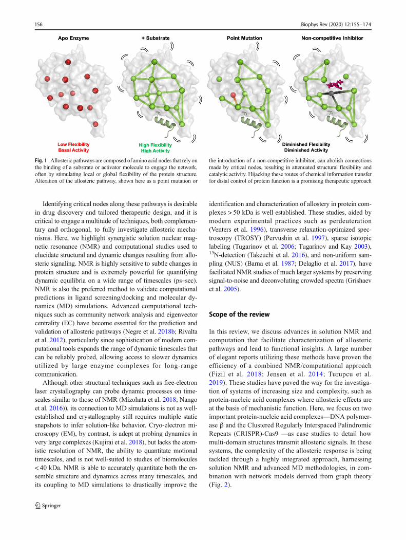
novel allosteric systems do not always conform to classicalparadigms from phenomenological models (Koshland Jr. et al.1966; Monod et al. 1965). Coupled to this observation is thenotion that amino acid “networks” intrinsic to the protein areactivated by endogenous or exogenous stimuli (Fig. 1). Theseallosteric pathways present an opportunity for fine-tuning orcontrolling biological responses; thus, ensemble models ofallostery, where proteins sample microstates along a free en-ergy continuum (Motlagh et al. 2014), have replaced a purelystructural view of discrete conformational changes. However,a unifying model for all allosteric systems remains elusive.Ensemble models describe differing proteins with the samethermodynamic parameters, but such models generally ex-clude communicative pathways between active and regulatorysites, even though such a connection is necessary from anexperimental point-of-view. Coupled communication orga-nizes the active and allosteric sites of enzymes for substratebinding and mediates proper functionality. Despite advance-ments in biochemical and biophysical probes, the complexityof these mechanisms is such that allosteric pathways remainlargely uncharacterized, especially in high molecular weightproteins.
* George P. [email protected]
1 Department of Molecular Biology, Cell Biology & Biochemistry,Brown University, Providence, RI 02903, USA
2 Department of Bioengineering and of Chemistry, University ofCalifornia Riverside, 900 University Avenue, Riverside, CA 92521,USA
3 Department of Chemistry, Yale University, New Haven, CT 06520,USA
https://doi.org/10.1007/s12551-019-00609-zBiophysical Reviews (2020) 12: –174155
Published online: 14 December 2019/
Identifying critical nodes along these pathways is desirablein drug discovery and tailored therapeutic design, and it iscritical to engage a multitude of techniques, both complemen-tary and orthogonal, to fully investigate allosteric mecha-nisms. Here, we highlight synergistic solution nuclear mag-netic resonance (NMR) and computational studies used toelucidate structural and dynamic changes resulting from allo-steric signaling. NMR is highly sensitive to subtle changes inprotein structure and is extremely powerful for quantifyingdynamic equilibria on a wide range of timescales (ps–sec).NMR is also the preferred method to validate computationalpredictions in ligand screening/docking and molecular dy-namics (MD) simulations. Advanced computational tech-niques such as community network analysis and eigenvectorcentrality (EC) have become essential for the prediction andvalidation of allosteric pathways (Negre et al. 2018b; Rivaltaet al. 2012), particularly since sophistication of modern com-putational tools expands the range of dynamic timescales thatcan be reliably probed, allowing access to slower dynamicsutilized by large enzyme complexes for long-rangecommunication.
Although other structural techniques such as free-electronlaser crystallography can probe dynamic processes on time-scales similar to those of NMR (Mizohata et al. 2018; Nangoet al. 2016)), its connection to MD simulations is not as well-established and crystallography still requires multiple staticsnapshots to infer solution-like behavior. Cryo-electron mi-croscopy (EM), by contrast, is adept at probing dynamics invery large complexes (Kujirai et al. 2018), but lacks the atom-istic resolution of NMR, the ability to quantitate motionaltimescales, and is not well-suited to studies of biomolecules< 40 kDa. NMR is able to accurately quantitate both the en-semble structure and dynamics across many timescales, andits coupling to MD simulations to drastically improve the
identification and characterization of allostery in protein com-plexes > 50 kDa is well-established. These studies, aided bymodern experimental practices such as perdeuteration(Venters et al. 1996), transverse relaxation-optimized spec-troscopy (TROSY) (Pervushin et al. 1997), sparse isotopiclabeling (Tugarinov et al. 2006; Tugarinov and Kay 2003),15N-detection (Takeuchi et al. 2016), and non-uniform sam-pling (NUS) (Barna et al. 1987; Delaglio et al. 2017), havefacilitated NMR studies of much larger systems by preservingsignal-to-noise and deconvoluting crowded spectra (Grishaevet al. 2005).
Scope of the review
In this review, we discuss advances in solution NMR andcomputation that facilitate characterization of allostericpathways and lead to functional insights. A large numberof elegant reports utilizing these methods have proven theefficiency of a combined NMR/computational approach(Fizil et al. 2018; Jensen et al. 2014; Turupcu et al.2019). These studies have paved the way for the investiga-tion of systems of increasing size and complexity, such asprotein-nucleic acid complexes where allosteric effects areat the basis of mechanistic function. Here, we focus on twoimportant protein-nucleic acid complexes—DNA polymer-ase β and the Clustered Regularly Interspaced PalindromicRepeats (CRISPR)-Cas9 —as case studies to detail howmulti-domain structures transmit allosteric signals. In thesesystems, the complexity of the allosteric response is beingtackled through a highly integrated approach, harnessingsolution NMR and advanced MD methodologies, in com-bination with network models derived from graph theory(Fig. 2).
Fig. 1 Allosteric pathways are composed of amino acid nodes that rely onthe binding of a substrate or activator molecule to engage the network,often by stimulating local or global flexibility of the protein structure.Alteration of the allosteric pathway, shown here as a point mutation or
the introduction of a non-competitive inhibitor, can abolish connectionsmade by critical nodes, resulting in attenuated structural flexibility andcatalytic activity. Hijacking these routes of chemical information transferfor distal control of protein function is a promising therapeutic approach
Biophys Rev (2020) 12:155–174156
Solution NMR studies of protein dynamicsand allostery
Conformational rearrangements occur frequently during enzy-matic mechanisms, often as a result of ligand binding where theassociated conformational changes can be rate-limiting to catal-ysis (Benkovic and Hammes-Schiffer 2003; Lisi and Loria2017; Watt et al. 2007; Whittier et al. 2013). Thus, characteri-zation of relevant dynamics associated with these transitions isessential for understanding the biochemical mechanism. Theequilibria between energetically similar apo (ligand-free) andliganded enzymatic states can be characterized by numerousbiophysical techniques; however, in many cases, it is unclearwhether the enzyme in question is intrinsically capable of sam-pling the relevant conformations required for allosteric activa-tion. To address this question in molecular detail, experimentaland computational approaches that can accurately quantitatemulti-timescale protein motions are required (Yuan et al.2015). It is now well-established that allosteric communicationcontains enthalpic and entropic thermodynamic components(Tsai et al. 2009), the latter of which is strongly influenced byconformational fluctuations (Henzler-Wildman and Kern 2007;Tsai et al. 2008; Volkman et al. 2001; Wand 2001). Well-vettedNMR methods for the study of protein dynamics have beenreported (Dyson and Wright 2004; Loria et al. 2008; Palmer3rd 2004; Palmer III 2015), and we will briefly describe howthese methods can be used in concert with molecular simula-tions to probe biological processes that access weakly populat-ed conformational states (Baldwin and Kay 2009; Loria et al.2008; Mittermaier and Kay 2009; Palmer 3rd 2004; Palmer III2015) and equilibrium dynamics that contribute to the config-urational entropy of the system (Igumenova et al. 2006;Mittermaier and Kay 2006; Trbovic et al. 2009).
Solution NMR spectroscopy is performed under physio-logically relevant conditions and is capable of determiningatomic resolution protein structures, capturing site-specificconformational changes, and quantifying protein motionsspanning the ps–sec timescales. The contributions of manylaboratories to the application of novel NMR experimentsand elucidation of mechanisms by which proteins propagatechemical signals have demonstrated the diversity of allostericsystems, though only a few examples can be discussed indetail. Novel examples of dynamically-driven allostery con-tinue to surface where NMR experiments and/or moleculardynamics (MD) simulations have been used to establish themechanism (Dhulesia et al. 2008; Hwang et al. 2004; Jacobyet al. 1996; Jarymowycz and Stone 2008; Kalodimos 2011;Liu et al. 2008;Wiesner et al. 2006; Zhang et al. 2012; Fuenteset al. 2004; Masterson et al. 2010; Petit et al. 2009; Coyne andGiedroc 2013; Lee 2015; Chakravorty et al. 2013; Shi andKay 2014).
NMR probes local and global biomolecular structure anddynamics (Shi and Kay 2014) (Velyvis et al. 2007, 2009;Lipchock and Loria 2010) to illuminate pathways of allostericcross-talk (Figs. 1 and 3). Dynamic processes specifically re-lated to allostery occur primarily in the μs–ms time regime,though contributions of faster (ps–ns) timescale motions havebeen noted, particularly as it relates to the optimization of theprotein scaffold for effector binding and quantitation of con-figurational entropy in protein systems (Capdevila et al. 2017;Caro et al. 2017; Tzeng and Kalodimos 2012). The suite ofNMR spin relaxation experiments designed to probe thesetime regimes have been used to interrogate internal proteinmotions (Akke and Palmer 1996; Loria et al. 1999a; Mulderet al. 2002) (Grey et al. 2003; Massi et al. 2005), proteinfolding (Hill et al. 2000; Korzhnev et al. 2004c; Tang et al.
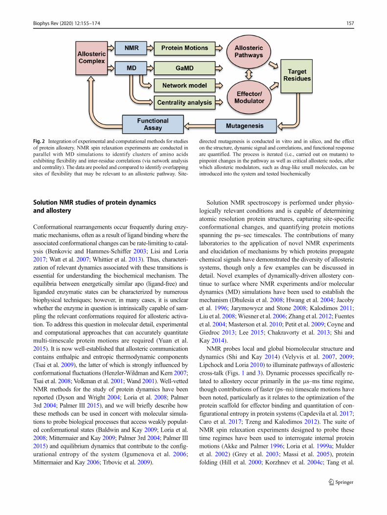
Fig. 2 Integration of experimental and computational methods for studiesof protein allostery. NMR spin relaxation experiments are conducted inparallel with MD simulations to identify clusters of amino acidsexhibiting flexibility and inter-residue correlations (via network analysisand centrality). The data are pooled and compared to identify overlappingsites of flexibility that may be relevant to an allosteric pathway. Site-
directed mutagenesis is conducted in vitro and in silico, and the effecton the structure, dynamic signal and correlations, and functional responseare quantified. The process is iterated (i.e., carried out on mutants) topinpoint changes in the pathway as well as critical allosteric nodes, afterwhich allosteric modulators, such as drug-like small molecules, can beintroduced into the system and tested biochemically
Biophys Rev (2020) 12:155–174 157
2006), ligand binding events (Mittag et al. 2003; Tolkatchevet al. 2003), and enzymatic mechanisms (Cole and Loria2002; Beach et al. 2005; Kovrigin and Loria 2006a, b;Kempf and Loria 2002; Berlow et al. 2007). The theoreticaldetails of these solution NMR experiments are discussed inthe next section, as are useful NMR observables that havepowerful synergy with computation.
Chemical shift perturbations
The resonant frequency (ω0), or Larmor precession, of anNMR-active nucleus is dependent on the local magnetic field(B), with contributions from the external field (B0) and localelectronic environment (Bi), where B =B0 +Bi. In practice,resonant frequencies are referenced to a standard frequencyin order to quantitate the chemical shift (δ, in parts-per-millionor Hertz) and minimize the contribution of B0. Thus, the
chemical shift is extremely sensitive to changes in the localelectronic environment and can be measured with high preci-sion. Small changes in protein structure caused by ligandbinding or a transition between inactive and active states(T→R in allosteric nomenclature) are easily detectable withNMR chemical shifts. In a simple two-site exchange modeldescribing the interaction of an enzyme with an allosteric ef-fector, NMR-detectable nuclei sample two unique structural(and magnetic) environments as the allosteric effector binds/unbinds. The populations of each state, pT (tense or inactive)and pR (relaxed or active), have chemical shifts δT and δR,respectively. The chemical shift difference between thesetwo states is given byΔω = |δR - δT| and the rate of chemicalexchange between states T and R is given by kex = kT→R +kR→ T. In the case of fast-to-intermediate exchange betweenconformers, where kex ≥Δω, the observed chemical shift(δobs = PRδR + PTδT) is a population weighted average of the
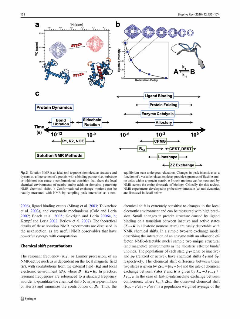
Fig. 3 Solution NMR is an ideal tool to probe biomolecular structure anddynamics. a Interaction of a protein with a binding partner (i.e., substrateor inhibitor) can cause a conformational transition that alters the localchemical environments of nearby amino acids or domains, perturbingNMR chemical shifts. b Conformational exchange motions can bereadily measured with NMR by sampling peak intensities as a non-
equilibrium state undergoes relaxation. Changes in peak intensities as afunction of a variable relaxation delay provide signatures of flexible ami-no acids within a protein matrix. c Protein motions can be measured byNMR across the entire timescale of biology. Critically for this review,NMR experiments developed to probe slow timescale (μs-ms) dynamicsare discussed in detail below
Biophys Rev (2020) 12:155–174158
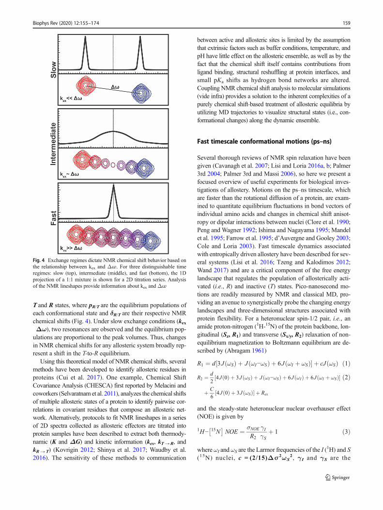
T and R states, where pR/T are the equilibrium populations ofeach conformational state and δR/T are their respective NMRchemical shifts (Fig. 4). Under slow exchange conditions (kexΔω), two resonances are observed and the equilibrium pop-ulations are proportional to the peak volumes. Thus, changesin NMR chemical shifts for any allosteric system broadly rep-resent a shift in the T-to-R equilibrium.
Using this theoretical model of NMR chemical shifts, severalmethods have been developed to identify allosteric residues inproteins (Cui et al. 2017). One example, Chemical ShiftCovariance Analysis (CHESCA) first reported by Melacini andcoworkers (Selvaratnam et al. 2011), analyzes the chemical shiftsof multiple allosteric states of a protein to identify pairwise cor-relations in covariant residues that compose an allosteric net-work. Alternatively, protocols to fit NMR lineshapes in a seriesof 2D spectra collected as allosteric effectors are titrated intoprotein samples have been described to extract both thermody-namic (K and ΔG) and kinetic information (kex, kT→R, andkR→ T) (Kovrigin 2012; Shinya et al. 2017; Waudby et al.2016). The sensitivity of these methods to communication
between active and allosteric sites is limited by the assumptionthat extrinsic factors such as buffer conditions, temperature, andpH have little effect on the allosteric ensemble, as well as by thefact that the chemical shift itself contains contributions fromligand binding, structural reshuffling at protein interfaces, andsmall pKa shifts as hydrogen bond networks are altered.Coupling NMR chemical shift analysis to molecular simulations(vide infra) provides a solution to the inherent complexities of apurely chemical shift-based treatment of allosteric equilibria byutilizing MD trajectories to visualize structural states (i.e., con-formational changes) along the dynamic ensemble.
Fast timescale conformational motions (ps–ns)
Several thorough reviews of NMR spin relaxation have beengiven (Cavanagh et al. 2007; Lisi and Loria 2016a, b; Palmer3rd 2004; Palmer 3rd and Massi 2006), so here we present afocused overview of useful experiments for biological inves-tigations of allostery. Motions on the ps–ns timescale, whichare faster than the rotational diffusion of a protein, are exam-ined to quantitate equilibrium fluctuations in bond vectors ofindividual amino acids and changes in chemical shift anisot-ropy or dipolar interactions between nuclei (Clore et al. 1990;Peng and Wagner 1992; Ishima and Nagayama 1995; Mandelet al. 1995; Farrow et al. 1995; d’Auvergne and Gooley 2003;Cole and Loria 2003). Fast timescale dynamics associatedwith entropically driven allostery have been described for sev-eral systems (Lisi et al. 2016; Tzeng and Kalodimos 2012;Wand 2017) and are a critical component of the free energylandscape that regulates the population of allosterically acti-vated (i.e., R) and inactive (T) states. Pico-nanosecond mo-tions are readily measured by NMR and classical MD, pro-viding an avenue to synergistically probe the changing energylandscapes and three-dimensional structures associated withprotein flexibility. For a heteronuclear spin-1/2 pair, i.e., anamide proton-nitrogen (1H-15N) of the protein backbone, lon-gitudinal (Sz, R1) and transverse (Sx/y, R2) relaxation of non-equilibrium magnetization to Boltzmann equilibrium are de-scribed by (Abragam 1961)
R1 ¼ d 3J ωSð Þ þ J ωI−ωSð Þ þ 6J ωI þ ωSð Þ½ � þ cJ ωSð Þ ð1ÞR2 ¼ d
24J 0ð Þ þ 3J ωSð Þ þ J ωI−ωSð Þ þ 6J ωIð Þ þ 6J ωI þ ωSð Þ½ �
þ C6
4J 0ð Þ þ 3J ωSð Þ½ � þ Rex
ð2Þ
and the steady-state heteronuclear nuclear overhauser effect(NOE) is given by
1H− 15N� �
NOE ¼ σNOE
R2
γIγS
þ 1 ð3Þ
where ωI and ωS are the Larmor frequencies of the I (1H) and S(15N) nuclei, c = (2/15)Δσ2ωS
2, γI and γS are the
Fig. 4 Exchange regimes dictate NMR chemical shift behavior based onthe relationship between kex and Δω. For three distinguishable timeregimes: slow (top), intermediate (middle), and fast (bottom), the 1Dprojection of a 1:1 mixture is shown for a 2D titration series. Analysisof the NMR lineshapes provide information about kex and Δω
Biophys Rev (2020) 12:155–174 159
gyromagnetic ratios of the I (1H) and S (15N) nuclei,Δσ is thechemical shift anisotropy of the S (15N) nucleus and σNOE isthe cross-correlation relaxation term from I→ S (1H→ 15N).The contribution of μs–ms conformational motion to trans-verse relaxation is contained in Rex, which is negligible inmany cases. The dipolar coupling constant (d) in the R1 andR2 cases is described by Eq. 4,
d ¼ 1
10
μ0
4π
� �2ℏ2γ1
2γS2 rIS−6� � ð4Þ
where μo is the permeability of free space, ħ is Planck’s con-stant divided by 2π, γI and γS are again the gyromagnetic ratiosof the I (1H) and S (15N) nuclei, and ⟨rIS⟩ is the average bondlength between I and S. The spectral density function describingoverall and internal bond vector fluctuations is written as
J ωð Þ ¼ 2
5
S2τc1þ τcωð Þ2 þ
1−S2�
τ
1þ τωð Þ2 !
ð5Þ
where τc is the rotational correlation time of the biomolecule,τ¼τ−1
c þτ−1e and τe is the effective correlation time for internal
motions. S2 is a generalized order parameter ranging fromzero-to-one that is commonly related to the configurationalentropy of protein bond vectors, where lower values indicateheightened flexibility (Akke et al. 1993; Li et al. 1996; Yangand Kay 1996). Qualitative assessments of conformational en-tropy through the average order parameter, ⟨S2⟩, have beendescribed by Bracken et al. (Kneller et al. 2002) using theproduct of experimentally measured R1 and R2 relaxation rates
S2� � ¼
ffiffiffiffiffiffiffiffiffiffiffiffiffiffiffiffiR1R2h i
R1R2max
sð6Þ
where ⟨R1R2⟩ is the mean value and R1R2max is the calculated
maximum value. Thus, R1R2 values below the mean of thedata correspond to sites with lower order parameters (i.e.,heightened flexibility).
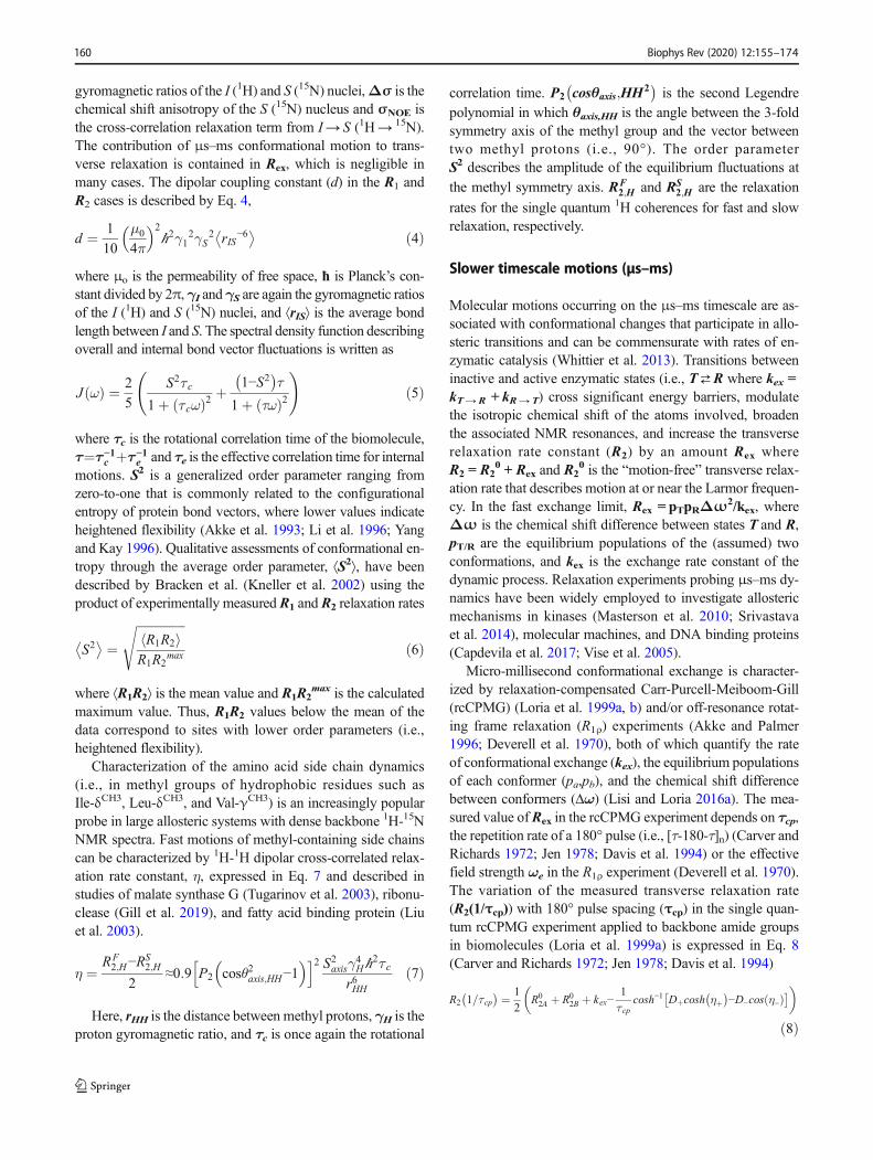
Characterization of the amino acid side chain dynamics(i.e., in methyl groups of hydrophobic residues such asIle-δCH3, Leu-δCH3, and Val-γCH3) is an increasingly popularprobe in large allosteric systems with dense backbone 1H-15NNMR spectra. Fast motions of methyl-containing side chainscan be characterized by 1H-1H dipolar cross-correlated relax-ation rate constant, η, expressed in Eq. 7 and described instudies of malate synthase G (Tugarinov et al. 2003), ribonu-clease (Gill et al. 2019), and fatty acid binding protein (Liuet al. 2003).
η ¼ RF2;H−R
S2;H
2≈0:9 P2 cosθ2axis;HH−1
� �h i2 S2axisγ4Hℏ2τ cr6HH
ð7Þ
Here, rHH is the distance between methyl protons, γH is theproton gyromagnetic ratio, and τc is once again the rotational
correlation time. P2 cosθaxis;HH2�
is the second Legendrepolynomial in which θaxis,HH is the angle between the 3-foldsymmetry axis of the methyl group and the vector betweentwo methyl protons (i.e., 90°). The order parameterS2 describes the amplitude of the equilibrium fluctuations at
the methyl symmetry axis. RF2;H and RS
2;H are the relaxation
rates for the single quantum 1H coherences for fast and slowrelaxation, respectively.
Slower timescale motions (μs–ms)
Molecular motions occurring on the μs–ms timescale are as-sociated with conformational changes that participate in allo-steric transitions and can be commensurate with rates of en-zymatic catalysis (Whittier et al. 2013). Transitions betweeninactive and active enzymatic states (i.e., T⇄R where kex =kT→R + kR→ T) cross significant energy barriers, modulatethe isotropic chemical shift of the atoms involved, broadenthe associated NMR resonances, and increase the transverserelaxation rate constant (R2) by an amount Rex whereR2 = R2
0 + Rex and R20 is the “motion-free” transverse relax-
ation rate that describes motion at or near the Larmor frequen-cy. In the fast exchange limit, Rex = pTpRΔω2/kex, whereΔω is the chemical shift difference between states T and R,pT/R are the equilibrium populations of the (assumed) twoconformations, and kex is the exchange rate constant of thedynamic process. Relaxation experiments probing μs–ms dy-namics have been widely employed to investigate allostericmechanisms in kinases (Masterson et al. 2010; Srivastavaet al. 2014), molecular machines, and DNA binding proteins(Capdevila et al. 2017; Vise et al. 2005).
Micro-millisecond conformational exchange is character-ized by relaxation-compensated Carr-Purcell-Meiboom-Gill(rcCPMG) (Loria et al. 1999a, b) and/or off-resonance rotat-ing frame relaxation (R1ρ) experiments (Akke and Palmer1996; Deverell et al. 1970), both of which quantify the rateof conformational exchange (kex), the equilibrium populationsof each conformer (pa,pb), and the chemical shift differencebetween conformers (Δω) (Lisi and Loria 2016a). The mea-sured value ofRex in the rcCPMG experiment depends on τcp,the repetition rate of a 180° pulse (i.e., [τ-180-τ]n) (Carver andRichards 1972; Jen 1978; Davis et al. 1994) or the effectivefield strength ωe in the R1ρ experiment (Deverell et al. 1970).The variation of the measured transverse relaxation rate(R2(1/τcp)) with 180° pulse spacing (τcp) in the single quan-tum rcCPMG experiment applied to backbone amide groupsin biomolecules (Loria et al. 1999a) is expressed in Eq. 8(Carver and Richards 1972; Jen 1978; Davis et al. 1994)
R2 1=τcp� ¼ 1
2R02A þ R0
2B þ kex−1
τ cpcosh−1 Dþcosh ηþ
� −D−cos η−ð Þ� �� �
ð8Þ
Biophys Rev (2020) 12:155–174160
where R02A and R0
2B are the intrinsic (i.e., motion-free) trans-verse relaxation rates of the two sites and D± and η± are
D� ¼ 1
2�1þ Ψþ 2Δω2
Ψ2 þ ζ2� 1=2" #" #
; η� ¼ τ cpffiffiffi2
p �Ψþ Ψ2 þ ζ2� 1=2h i1=2
ð9Þ
and Ψ ¼ R02A−R
02B−pAkex þ pBkex
� 2−Δω2 þ 4pApBk2ex; ζ ¼
2Δω R02A−R
02B−pAkex þ pBkex
� .
Equation 8 is required when conformational motion is inthe slow-to-intermediate exchange regime (kex ≤ Δω); howev-er, under fast exchange conditions (kex > Δω), the expressionsimplifies to (Luz and Meiboom 1963)
R2 1=τ cp� ¼ R0
2 þ ϕex=kex 1−2tanhkexτ cp2
� �= kexτ cp� �
ð10Þ
whereϕex = pApBΔω2 (Ishima and Torchia 1999; Millet et al.2000; Kovrigin et al. 2006; Lisi and Loria 2016a). Adaptationof the rcCPMG experiment for multiple quantum (MQ) relax-ation dispersion studies of Ile-δCH3, Leu-δCH3, and Val-γCH3
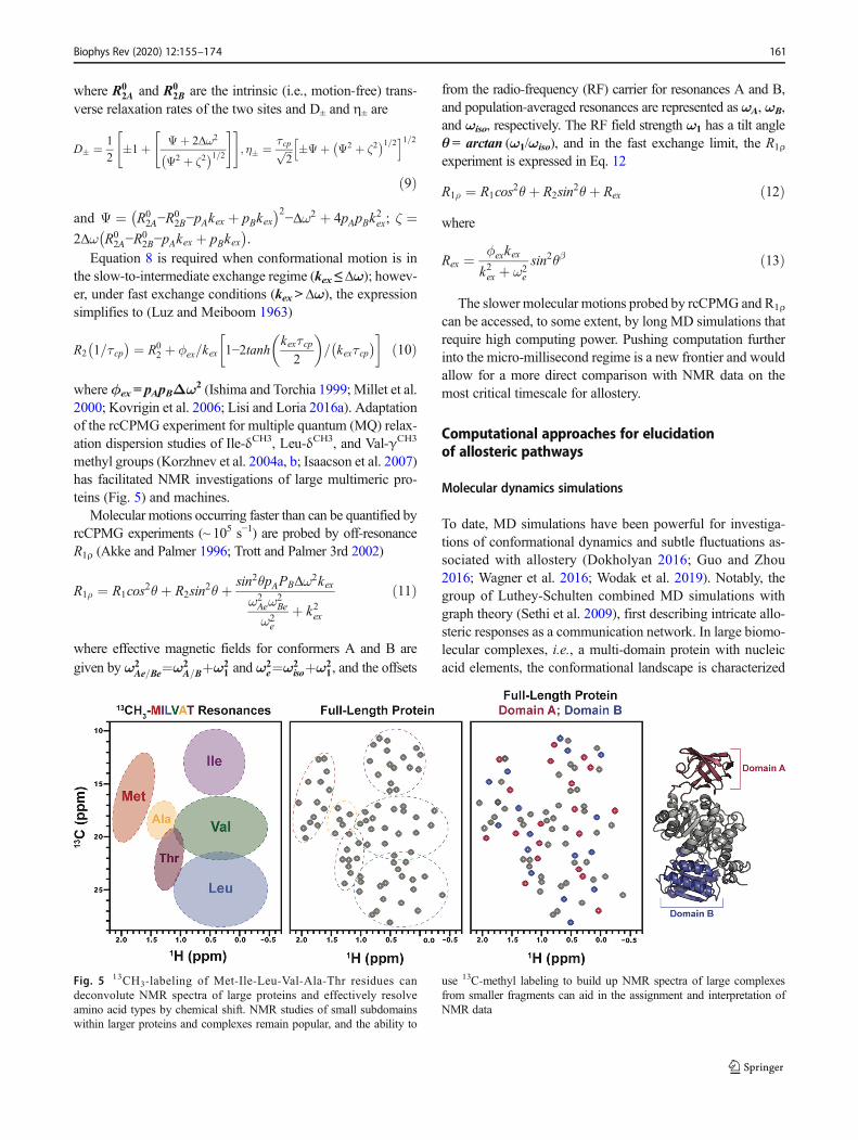
methyl groups (Korzhnev et al. 2004a, b; Isaacson et al. 2007)has facilitated NMR investigations of large multimeric pro-teins (Fig. 5) and machines.
Molecular motions occurring faster than can be quantified byrcCPMG experiments (~ 105 s−1) are probed by off-resonanceR1ρ (Akke and Palmer 1996; Trott and Palmer 3rd 2002)
R1ρ ¼ R1cos2θþ R2sin2θþ sin2θpAPBΔω2kexω2Aeω
2Be
ω2e
þ k2ex
ð11Þ
where effective magnetic fields for conformers A and B aregiven by ω2
Ae=Be¼ω2A=Bþω2
1 and ω2e¼ω2
isoþω21, and the offsets
from the radio-frequency (RF) carrier for resonances A and B,and population-averaged resonances are represented as ωA, ωB,and ωiso, respectively. The RF field strength ω1 has a tilt angleθ = arctan (ω1/ωiso), and in the fast exchange limit, the R1ρexperiment is expressed in Eq. 12
R1ρ ¼ R1cos2θþ R2sin2θþ Rex ð12Þ
where
Rex ¼ ϕexkexk2ex þ ω2
e
sin2θβ ð13Þ
The slower molecular motions probed by rcCPMG and R1ρ
can be accessed, to some extent, by long MD simulations thatrequire high computing power. Pushing computation furtherinto the micro-millisecond regime is a new frontier and wouldallow for a more direct comparison with NMR data on themost critical timescale for allostery.
Computational approaches for elucidationof allosteric pathways
Molecular dynamics simulations
To date, MD simulations have been powerful for investiga-tions of conformational dynamics and subtle fluctuations as-sociated with allostery (Dokholyan 2016; Guo and Zhou2016; Wagner et al. 2016; Wodak et al. 2019). Notably, thegroup of Luthey-Schulten combined MD simulations withgraph theory (Sethi et al. 2009), first describing intricate allo-steric responses as a communication network. In large biomo-lecular complexes, i.e., a multi-domain protein with nucleicacid elements, the conformational landscape is characterized
Fig. 5 13CH3-labeling of Met-Ile-Leu-Val-Ala-Thr residues candeconvolute NMR spectra of large proteins and effectively resolveamino acid types by chemical shift. NMR studies of small subdomainswithin larger proteins and complexes remain popular, and the ability to
use 13C-methyl labeling to build up NMR spectra of large complexesfrom smaller fragments can aid in the assignment and interpretation ofNMR data
Biophys Rev (2020) 12:155–174 161
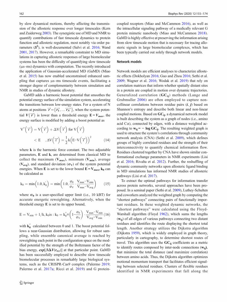
by slow dynamical motions, thereby affecting the transmis-sion of the allosteric response over longer timescales (Kernand Zuiderweg 2003). The synergistic use ofMD and NMR toquantify contributions of fast timescale dynamics to proteinfunction and allosteric regulation, most notably via order pa-rameters (S2), is well-documented (Salvi et al. 2016; Wand2001, 2017). However, a remarkable constraint to MD simu-lations in capturing allosteric responses of large biomolecularsystems has been the difficulty of quantifying slow timescale(μs–ms) dynamics with computation. The recently introducedthe application of Gaussian-accelerated MD (GaMD) (Miaoet al. 2015) has now enabled unconstrained enhanced sam-pling that captures μs–ms timescale events, facilitating astronger degree of complementarity between simulation andNMR in studies of dynamic allostery.
GaMD adds a harmonic boost potential that smoothes thepotential energy surface of the simulation system, acceleratingthe transitions between low-energy states. For a system of Natoms at positions r!¼ r1
!;… rN�!� �
, when the system poten-
tial V r!� is lower than a threshold energy E = Vmax, the
energy surface is modified by adding a boost potential as
V* r!� �
¼ V r!� �
þΔV r!� �
for V r!� �
< E ;ΔV r!� �
¼ 1
2k E−V r!
� �� �2ð14Þ
where k is the harmonic force constant. The two adjustableparameters, E and k, are determined from classical MD tocollect the maximum (Vmax), minimum (Vmin), average(Vavg), and standard deviation (σV) of the system potentialenergies. When E is set to the lower bound E =Vmax, k0 canbe calculated as
k0 ¼ min 1:0; k00
� �¼ min 1:0;
σ0
σV� Vmax−Vmin
Vmax−Vavg
� �ð15Þ
where σ0 is a user-specified upper limit (i.e., 10 kBT) foraccurate energetic reweighting. Alternatively, when thethreshold energy E is set to its upper bound,
E ¼ Vmin þ 1=k; k0is : k0 ¼ k0 00≡ 1−
σ0
σV
� �� Vmax−Vmin
Vavg−Vminð16Þ
with k0 00 calculated between 0 and 1. The boost potential fol-
lows a near-Gaussian distribution, allowing for robust sam-pling, while ensemble canonical average is reached byreweighting each point in the configuration space on the mod-ified potential by the strength of the Boltzmann factor of thebias energy, exp[βΔV(rt(i))] at that particular point. GaMDhas been successfully employed to describe slow timescalebiomolecular processes in remarkably large biological sys-tems, such as the CRISPR-Cas9 complex (Palermo 2019;Palermo et al. 2017a; Ricci et al. 2019) and G protein-
coupled receptors (Miao and McCammon 2016), as well asthe intracellular signaling pathway of a medically relevant Gprotein mimetic nanobody (Miao and McCammon 2018).GaMD is highly effective at preserving the information arisingfrom slow timescale motion that is necessary for tracing allo-steric signals in large biomolecular complexes, which hasbeen typically carried out solely through network models.
Network models
Network models are efficient analyses to characterize alloste-ric effects (Dokholyan 2016; Guo and Zhou 2016; Sethi et al.2009; Wagner et al. 2016; Wodak et al. 2019) that rely oncorrelation matrices that inform whether spatially distant sitesin a protein are coupled in motion over dynamic trajectories.Generalized correlation (GCij) methods (Lange andGrubmuller 2006) are often employed to capture non-collinear correlations between residue pairs (i, j) based onShannon’s entropy and describe both linear and non-linearcoupled motions. Based onGCij, a dynamical network modelis built describing the system as a graph of nodes (i.e., aminoacid Cα), connected by edges, with a distance weighted ac-cording to wij = − log GCij. The resulting weighted graph isused to structure the system’s correlations through communitynetwork analysis (CNA) (Sethi et al. 2009), which definesgroups of highly correlated residues and the strength of theirinterconnectivity to quantify chemical information flow.Residues clustered together by CNA have shown similar con-formational exchange parameters in NMR experiments (Lisiet al. 2016; Rivalta et al. 2012). Further, the reshuffling ofdynamic community networks upon allosteric ligand bindingin MD simulations has informed NMR studies of allostericpathways (Lisi et al. 2017).
To extract the optimal pathways for information transferacross protein networks, several approaches have been pro-posed. In a seminal paper (Sethi et al. 2009), Luthey-Schultenand coworkers analyzed the weighted graph by computing the“shortest pathways” connecting pairs of functionally impor-tant residues. In these weighted dynamic networks, the“shortest pathways” were calculated using the Floyd-Warshall algorithm (Floyd 1962), which sums the lengths(wij) of all edges of various pathways connecting two distantresidues and identifies the route displaying the shortest totallength. Another strategy utilizes the Dijkstra algorithm(Dijkstra 1959), which is widely employed in graph theory,particularly in cartography, to determine shortest routes oftravel. This algorithm uses the GCij coefficients as a metricto identify routes composed by inter-node connections (wij),that minimize the total distance (and maximize correlation)between amino acids. Thus, the Dijkstra algorithm optimizesmotional momentum transport that facilitates efficient signal-ing between selected residues. Clusters of flexible residuesidentified in NMR experiments that fall along the
Biophys Rev (2020) 12:155–174162
computationally derived route of information transfer are like-ly to be directly involved in the allosteric mechanism.
Eigenvector centrality
One of the cornerstones of network theory is the concept ofcentrality (Doshi et al. 2016), i.e., the relative influence of anode or cluster of nodes to the network (Alvarez-Socorro et al.2015; Borgatti 2005; Hanke and Foraita 2017; Newman2010). A recently proposed method harnesses the so-calledeigenvector centrality (EC) (Negre et al. 2018b), where theEC of a node, ci, is defined as the sum of the centralities ofall nodes that are connected to it by an edge, Aij ci ¼ 1
λ∑nj¼1
Aijc j;where the edgesAij are elements of the adjacency matrixA (based onGCij), and λ is the eigenvalue associated with theeigenvector composed by ci elements. This approach relies onassigning functional dynamics to the major collective mode ofthe system (i.e., the first eigenvector of A), and hence, λ is thehighest eigenvalue of A. The EC estimation quantifies thedegree of connectivity of each amino acid or nucleobase inthe system, measuring how well the nodes of the protein net-work are connected to other well-connected nodes. This en-riches the information derived from betweenness centrality(BC) metrics, which measures instead how information flowsbetween nodes (or edges) in a network (Sethi et al. 2009).Distinct from other centrality descriptors, EC also serves asa measure of the connectivity against a fixed scale when nor-malized and can reliably compare the connectivity of a systemfollowing allosteric effector binding. This concept has beenespecially critical to identifying targets for site-directed muta-genesis and inhibitor docking within allosteric networks.Residues with high EC may also be dynamic transducers inproteins, where mutations at these sites stimulate or attenuateprotein motions (Lisi et al. 2017; Negre et al. 2018b). EC isalso valuable to obtain the main mode of collective correlationresponsible for the allosteric signal, beyond the capabilities ofstandard principal component methods.
Reliability of simulated ensembles with respect to NMRexperiments
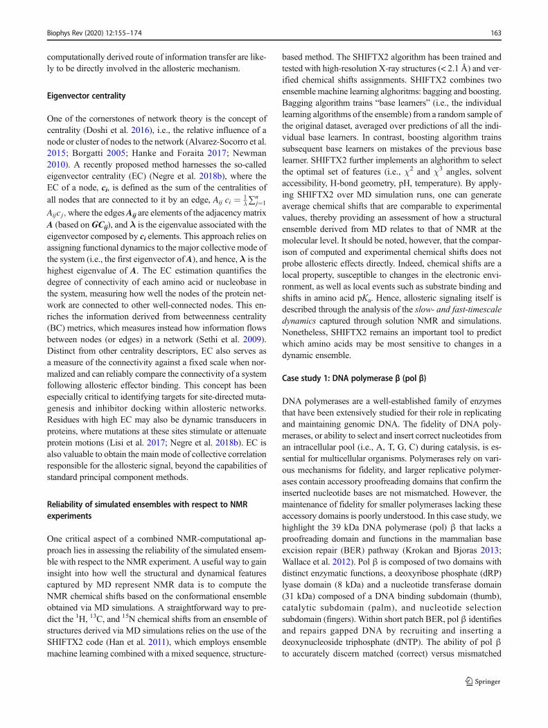
One critical aspect of a combined NMR-computational ap-proach lies in assessing the reliability of the simulated ensem-ble with respect to the NMR experiment. A useful way to gaininsight into how well the structural and dynamical featurescaptured by MD represent NMR data is to compute theNMR chemical shifts based on the conformational ensembleobtained via MD simulations. A straightforward way to pre-dict the 1H, 13C, and 15N chemical shifts from an ensemble ofstructures derived via MD simulations relies on the use of theSHIFTX2 code (Han et al. 2011), which employs ensemblemachine learning combined with a mixed sequence, structure-
based method. The SHIFTX2 algorithm has been trained andtested with high-resolution X-ray structures (< 2.1 Å) and ver-ified chemical shifts assignments. SHIFTX2 combines twoensemble machine learning alghoritms: bagging and boosting.Bagging algorithm trains “base learners” (i.e., the individuallearning algorithms of the ensemble) from a random sample ofthe original dataset, averaged over predictions of all the indi-vidual base learners. In contrast, boosting algorithm trainssubsequent base learners on mistakes of the previous baselearner. SHIFTX2 further implements an alghorithm to selectthe optimal set of features (i.e., χ2 and χ3 angles, solventaccessibility, H-bond geometry, pH, temperature). By apply-ing SHIFTX2 over MD simulation runs, one can generateaverage chemical shifts that are comparable to experimentalvalues, thereby providing an assessment of how a structuralensemble derived from MD relates to that of NMR at themolecular level. It should be noted, however, that the compar-ison of computed and experimental chemical shifts does notprobe allosteric effects directly. Indeed, chemical shifts are alocal property, susceptible to changes in the electronic envi-ronment, as well as local events such as substrate binding andshifts in amino acid pKa. Hence, allosteric signaling itself isdescribed through the analysis of the slow- and fast-timescaledynamics captured through solution NMR and simulations.Nonetheless, SHIFTX2 remains an important tool to predictwhich amino acids may be most sensitive to changes in adynamic ensemble.
Case study 1: DNA polymerase β (pol β)
DNA polymerases are a well-established family of enzymesthat have been extensively studied for their role in replicatingand maintaining genomic DNA. The fidelity of DNA poly-merases, or ability to select and insert correct nucleotides froman intracellular pool (i.e., A, T, G, C) during catalysis, is es-sential for multicellular organisms. Polymerases rely on vari-ous mechanisms for fidelity, and larger replicative polymer-ases contain accessory proofreading domains that confirm theinserted nucleotide bases are not mismatched. However, themaintenance of fidelity for smaller polymerases lacking theseaccessory domains is poorly understood. In this case study, wehighlight the 39 kDa DNA polymerase (pol) β that lacks aproofreading domain and functions in the mammalian baseexcision repair (BER) pathway (Krokan and Bjoras 2013;Wallace et al. 2012). Pol β is composed of two domains withdistinct enzymatic functions, a deoxyribose phosphate (dRP)lyase domain (8 kDa) and a nucleotide transferase domain(31 kDa) composed of a DNA binding subdomain (thumb),catalytic subdomain (palm), and nucleotide selectionsubdomain (fingers). Within short patch BER, pol β identifiesand repairs gapped DNA by recruiting and inserting adeoxynucleoside triphosphate (dNTP). The ability of pol βto accurately discern matched (correct) versus mismatched
Biophys Rev (2020) 12:155–174 163
(incorrect) base pairs is integral to its function, highlighted bythe fact that over 30% of human cancers have mutations in thegene encoding pol β (Marsden et al. 2017; Starcevic et al.2004). Many of these cancer-linked mutations lead to a lossof function; however, several mutations maintain near wild-type (WT) catalytic efficiency with vastly altered fidelity andhave been termed “mutator variants.” Expression of pol βmutants within healthy mammalian cells has been shown tocause cellular transformation and metastasis (Marsden et al.2017; Murphy et al. 2012; Wallace et al. 2012); thus, there isgreat interest in elucidating the mechanistic underpinnings ofthe pol β selection process, particularly the allosteric compo-nent by which binding of the appropriate nucleotide is regu-lated. Several reviews (Barakat et al. 2012; Beard and Wilson2006, 2014; Hakem 2008; Yamtich and Sweasy 2010) haverigorously described the known molecular mechanism andstructure of pol β and related polymerases; therefore, we willonly highlight important NMR and computational studies thathave improved the understanding of allosteric regulation un-derlying its nucleotide selection.
Foundational understanding of four precatalytic states ofpol β; apo enzyme, gapped DNA bound (binary), as well astwo ternary states (gapped DNA + dNTP), in “open” and“closed” forms, has been established through X-ray crystal-lography and Förster Resonance Enenergy Transfer (FRET)(Towle-Weicksel et al. 2014). The ternary states are distin-guished by 7–10 Å closure of the C-terminal nucleotide selec-tion domain around the active site complex (Fig. 6a).However, time-averaged snapshots of these complexes inopen and closed conformations do not inform the allostericmechanism of nucleotide selection (Freudenthal et al. 2012;Sawaya et al. 1997). FRETstudies explored global motions ofthe precatalytic transitions of pol β following dNTP bindingto the binary (i.e., DNA-bound) enzyme (Towle-Weickselet al. 2014) and suggested an induced fit mechanism for allo-steric control of pol β, where the allosteric effector, a matcheddNTP, causes a conformational change and subsequent non-covalent mechanistic step that regulates catalysis (Towle-Weicksel et al. 2014). This obligate non-covalent step hasbeen suggested to be the binding of a magnesium ion or anadditional conformational change. Recent NMR and compu-tational investigations have provided further insight into intra-and intermolecular structural and dynamical processes under-lying nucleotide selection and pol β activation.
Due to the size of pol β (~ 40 kDa), early NMR studiesutilized sparse 1H,13C-methyl labeling of methionine residuesto probe DNA binding to the apo enzyme and subsequentbinding of a non-hydrolyzable dNTP to the binary complex.This work highlighted specific residues within pol β that par-ticipate in the formation of binary and ternary complexes aswell as spectral signatures of the “open” and “closed” alloste-ric states sampled in the presence of mismatched and matcheddNTPs, respectively (Bose-Basu et al. 2004). More recent
1H-15N experiments probing backbone amides highlightedactivation of the pol β allosteric network upon matcheddNTP binding due to changes in the flexibility of the proteinbackbone (Berlow et al. 2012; Loria et al. 2008), where NMRrelaxation dispersion revealed specific amino acids in apo andbinary pol β exhibiting millisecond timescale motions.Interestingly, several of these dynamic sites are coincidentwith mutations found in certain cancers (Starcevic et al.2004), suggesting that flexibility of pol β precatalytic states,allosteric regulation of pol β, and its role in disease may belinked (Berlow et al. 2012; Moscato et al. 2016).
Additional support for an allosteric network in polβ comesfrom MD simulations of cancer-associated mutants withdisrupted hydrogen bonding between N279 and a bounddCTP (C = cytosine) that showed strongly attenuated catalyticefficiencies (kcat/KM) despite the fact that N279 does not makedirect contact with the primer or α-phosphate of dCTP(Martinek et al. 2007). Corresponding kinetic studies notethe overall change in free energy (ΔG) more closely approx-imatesΔG of dNTP binding than that of the catalytic reaction(Xiang et al. 2006), supporting a mechanism in which pol β ispreorganized for dNTP binding and subsequent allostericchanges propagate to improve or impede pol β catalytic effi-ciency. In addition to the importance of N279 as an allostericmodulator of pol β, computational studies have establishedthe importance of magnesium ions in both the active site andthe allosteric network in general (Palermo et al. 2015).Computational modeling of pre- and post-chemistry steps bySchlick and coworkers showed that in the absence of magne-sium, dCTP triphosphate oxygens interact with different ami-no acid side chains than in the presence of magnesium andsuggested that closure of the nucleotide selection domainaround the active site requires two magnesium ions (Yanget al. 2004). Opening of the enzyme post-chemistry is con-comitant with the release of one magnesium ion from theactive site in MD simulations.
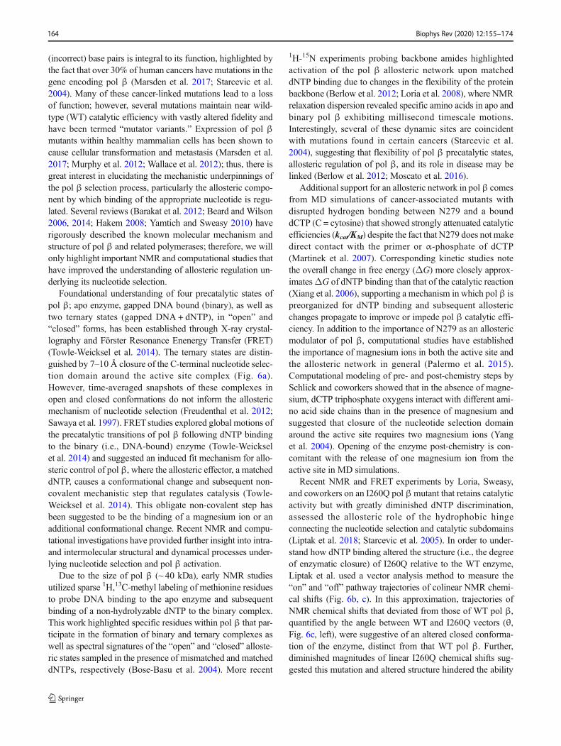
Recent NMR and FRET experiments by Loria, Sweasy,and coworkers on an I260Q pol βmutant that retains catalyticactivity but with greatly diminished dNTP discrimination,assessed the allosteric role of the hydrophobic hingeconnecting the nucleotide selection and catalytic subdomains(Liptak et al. 2018; Starcevic et al. 2005). In order to under-stand how dNTP binding altered the structure (i.e., the degreeof enzymatic closure) of I260Q relative to the WT enzyme,Liptak et al. used a vector analysis method to measure the“on” and “off” pathway trajectories of colinear NMR chemi-cal shifts (Fig. 6b, c). In this approximation, trajectories ofNMR chemical shifts that deviated from those of WT pol β,quantified by the angle between WT and I260Q vectors (θ,Fig. 6c, left), were suggestive of an altered closed conforma-tion of the enzyme, distinct from that WT pol β. Further,diminished magnitudes of linear I260Q chemical shifts sug-gested this mutation and altered structure hindered the ability
Biophys Rev (2020) 12:155–174164
of pol β to sample a fully closed structure (Fig. 6c, right).Diminished closure in the nucleotide selection subdomain inthe presence of dNTP mismatches is known to be importantfor fidelity of the WT enzyme, as addition of a mismatchednucleotide does not induce large chemical shift perturbationsin its 1H-13CH3 NMR spectrum. However, addition of a mis-matched nucleotide to the I260Q mutant induces large chem-ical shift perturbations in the nucleotide selection domain,many of which fall along the pathway to closure seen forWT pol β in the presence of a matched nucleotide. The chem-ical shift vector methodology in Fig. 6c allowed for identifi-cation of specific residues that contribute to the pol β fidelitybased on their ability (or lack thereof) to sample closed con-formations in various dNTP-bound states.
The use of NMR and computation in the study of DNA polβ fidelity was crucial to refining the current view of allosterythat governs the transferase activity of the enzyme. Initial X-ray crystallographic work highlighted the induced fit of adNTP into the active site via closure of the nucleotide selec-tion domain, while NMR and computation clarified the mo-lecular details of the process, namely the regions of pol β thatcomposed an allosteric network and displayed the flexibilitynecessary to alter the open/closed ensemble. In the currentmodel of pol β nucleotide selection, allosteric activation isachieved by formation of a Watson-Crick pair between theincoming nucleotide and templating base, which is controlledby the affinity of pol β for matched/mismatched nucleotides.A chemical signal is then propagated through the hydrophobic
Fig. 6 a X-ray crystal structures of open (black) and closed (gray) pol β.DNA molecules are shown in orange shades. b NMR chemical shiftsignatures of the open (black, WT without nucleotide), closed (gray,WTwith matched nucleotide), and “off-pathway” (purple, I260Q mutantwith mismatched nucleotide) structural states of pol β. c Schematic rep-resentation of chemical shift vector analysis reported by Liptak et al.,accounting for various degrees of enzymatic closure around matchedand mismatched nucleotides in WT and I260Q mutant pol β. WT pol βadopts its native, closed structure upon matched dNTP binding (black,
gray), while the cancer-associated I260Q variant populates altered struc-tures with various dNTPs (red, blue, green) that deviate from the WTpathway. The normalized vector magnitude was calculated as Δδn =Δδexptl/Δδref, where Δδref is the WT trajectory. The degree of I260Q polβ closure and its overall structure relative to WT pol β is summarized onthe correlation plot (right). I260Q data points deviating from the 1,1-cross-section display either altered structures (horizontal) or altered levelsof enzymatic closure (vertical). Dots are colored according to theI260Q:DNA complexes indicated in the vector diagram at left
Biophys Rev (2020) 12:155–174 165
hinge to the active site, where the incorporation of magnesiumions modulates catalytic function. Since enzymatic catalysis(kcat, kpol) is affected by preceding steps in this informationtransfer, pol β appears to display mixed K-type and V-typeallostery.
Case study 2: CRISPR-Cas9
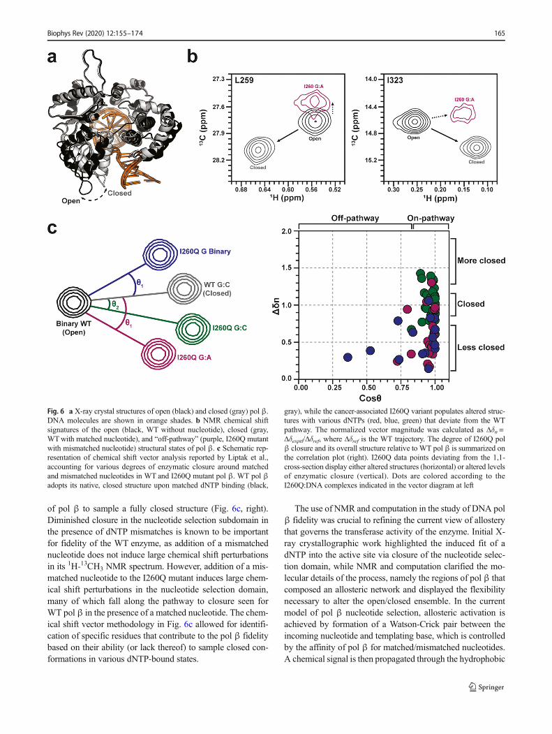
The CRISPR-Cas9 (clustered regularly interspaced short pal-indromic repeats–associated protein 9) system is crucial toprokaryotic organisms as an adaptive immune responseagainst invading bacteriophages (Doudna and Charpentier2014; Jinek et al. 2012) and is widely utilized as a genomeediting tool in biotechnology and medicine (Adli 2018). Cas9is a 160 kDa RNA-guided endonuclease that creates double-strand breaks in DNA upon site-specific recognition and bind-ing of a short 2–5 nucleotide Protospacer Adjacent Motif(PAM) that precedes the cleavage site (Fig. 7). The multi-domain Cas9 enzyme is composed of a large recognition lobe(REC) that accommodates the formation of a RNA:DNA hy-brid through three subdomains (REC1-3), and a nuclease lobeincluding two domains HNH and RuvC that cleave the DNAstrand complementary to the guide RNA and the non-complementary strand, respectively.
The spatially separated, yet functionally connected recog-nition and nuclease sites of Cas9 implied an allosteric relay,which was initially probed by the insertion of exogenous PDZdomains into the native Cas9 structure by Doudna and co-workers, highlighting regions of the enzyme as “hotspots”(Oakes et al. 2016). Biochemical studies by Doudna and co-workers (Sternberg et al. 2014, 2015) have indicated that al-lostery in Cas9 synchronizes DNA binding, recognition, andconcerted double-stranded cleavage. Early computational in-vestigations revealed that allosteric cross-talk between theHNH and RuvC catalytic domains is essential for activationof concerted DNA cleavage (Palermo et al. 2017b) and morerecently, Chen and colleagues revealed byMD that motions ofthe REC lobe govern the conformational changes of the HNH
domain toward cleavage (Chen et al. 2017). While X-raystructures have shown “open” (apo) or “closed” (RNA/DNA-bound) snapshots of Cas9 (Huai et al. 2017; Jiang andDoudna 2015; Jinek et al. 2014; Nishimasu et al. 2014), it iswidely hypothesized that Cas9, like other allosteric proteins(Koshland Jr. et al. 1966; Lukin et al. 2003; Monod et al.1965; Yuan et al. 2015) populates numerous microstates be-tween these structures (Dagdas et al. 2017).
In this respect, the nature of allosteric communication with-in this protein-nucleic acid complex is poorly understood,though recent investigations have provided intricate detailabout how nucleic acid binding information is transmittedacross the multi-domain Cas9 structure to control the activa-tion of the catalytic HNH nuclease. HNH displays a remark-able conformational mobility, observed by cryo-EM(CryoEM; EMD-3277) to be at lower resolution (8–10Å) thanthe overall structure (6 Å) (Jiang et al. 2016). The first all-atomMD simulation of Cas9 (Palermo et al. 2016) also highlightedthe “striking” flexibility of HNHwith respect to the remainingprotein subunits that enable HNH to rapidly sample its acti-vated state primed for DNA cleavage. HNH activation is de-pendent on the conformational dynamics of the REC lobe ofCas9, where allosteric function relies on the opening of theREC3 region (amino acids 480–718) to integrate the incomingRNA:DNA hybrid (Chen et al. 2017). This interaction isthought to propagate a biological signal to REC2 (amino acids180–308), which directly contacts HNH, enabling its ap-proach to the DNA cleavage site on the complementarystrand. In spite of extensive structural characterization ofCas9, high-resolution snapshots of its fully activated statehave yet to be observed, perhaps due to the inherent flexibilityof HNHwhen engaged in enzymatic function. However, earlycomputational studies employing targeted approaches and ac-celeratedMD have provided the first structural information onthe activated complex, including the structure of an activatedHNH domain (Palermo et al. 2017a; Zuo and Liu 2017).Extensive MD simulations have also revealed that theREC1-3 regions collectively move with HNH during its
Fig. 7 Structure of the SpCRISPR-Cas9 system, composedof the endonuclease Cas9 incomplex with a guide RNA and atarget DNA. The Cas9 enzyme isshown in molecular surface,highlighting the HNH (green),RuvC (blue), and REC (gray)domains with different colors.The RNA (orange) as well as thetarget DNA strand (TS, cyan) andthe non-target DNA strand (NTS,violet) are shown as ribbons. Aclose-up view of the PAM-binding region is shown on theright
Biophys Rev (2020) 12:155–174166
activation (Palermo et al. 2018), supporting the “allosterichypothesis” of Chen and colleagues (Chen et al. 2017).However, the nature of the allosteric control that REC exertson HNH and the mechanism of its signal transduction remainsunclear. Each of these functionalities relies on molecular mo-tion and is thus amenable to characterization by synergisticNMR and MD approaches. Thus, the most recent biophysicalprobes of Cas9 are integrating solution NMR (Lisi and Loria2016a) and MD or novel GaMD (Miao et al. 2015) with net-work models (Sethi et al. 2009) and newly developed central-ity analysis methods (Negre et al. 2018a) to track the molec-ular motions at the core of the allosteric communication be-tween REC, HNH, and RuvC.
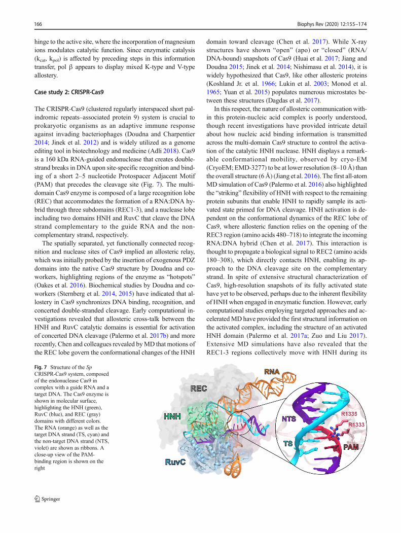
Since coupling of NMR data to all-atom MD simulationshas significantly increased our understanding of dynamic al-lostery in smaller protein-nucleic acid complexes (Adhireksanet al. 2017; Lisi and Loria 2016a; Wodak et al. 2019), thesemethodologies are being extended to the Cas9 machinery(Belato et al. 2019), building on outcomes of prior MD sim-ulations (Palermo 2019; Palermo et al. 2016, 2017a, b, 2018;Ricci et al. 2019; Zuo and Liu 2017). Several domains criticalto Cas9 allosteric control have been studied by 1H-15N and1H-13CH3 NMR (Belato 2019) (Fig. 8) in order to generatefingerprints of the key players in Cas9 allostery and connect
with previous computational work highlighting the dynamicinterplay of HNH, REC2, and REC3. NMR spin relaxationexperiments reveal multi-timescale dynamics within this axisof Cas9, which may be involved in information transfer forconcerted cleavage of the two DNA strands. Regions of fast(ps–ns) dynamics are consistent with those proposed by earlyMD studies of Cas9 allostery, but further investigation of thesedynamic properties is required to decipher the mechanism ofcommunication between these domains.
The role of the PAM sequence is also intriguing, as itsbinding initiates DNA association and cleavage, triggeringinter-dependent molecular motions of the Cas9 domains(Palermo et al. 2017b; Sternberg et al. 2014). Specifically,PAM has been identified as an “allosteric activator” of Cas9function fromMD simulations performed of Cas9 binding to a‘5–TGG–3’ PAM sequence (i.e., Cas9–PAM) (Anders et al.2014), and on its analogue, crystallized without PAM(Nishimasu et al. 2014). A total of ~ 13 μs of aggregate MDruns probing the impact of PAM binding on the dynamics ofCas9 revealed that PAM induces an “open–to–close” confor-mational transition, as indicated by principal component anal-ysis. This conformational change agrees with the transitionhypothesized for nucleic acid binding (Jiang et al. 2016;Palermo et al. 2017a) and reveals that PAM is essential in
Fig. 8 NMR spectra of critical domains of Cas9 implicated in itsallosteric mechanism. 1H-15N correlation spectra of backbone amideresidues are shown for the REC2 and REC3 regions, while a 1H-13CH3
methyl spectrum reporting on Ile, Leu, Val, Ala, Met, and Thr side chains
is shown for HNH. NMR studies of the structure and dynamics of thesecritical domains are required to confirm the allosteric relay proposed byMD simulations
Biophys Rev (2020) 12:155–174 167
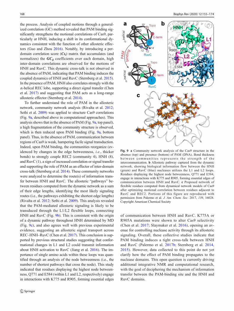
the process. Analysis of coupled motions through a general-ized correlation (GC) method revealed that PAM binding sig-nificantly strengthens the motional correlations of Cas9, par-ticularly at HNH, inducing a shift in its conformational dy-namics consistent with the function of other allosteric effec-tors (Guo and Zhou 2016). Notably, by introducing a per-domain correlation score (Csi) matrix that accumulates (andnormalizes) the GCij coefficients over each domain, highinter-domain correlations are observed for the motions ofHNH and RuvC. This dynamic cross-talk is not observed inthe absence of PAM, indicating that PAM binding induces thecoupled dynamics of HNH and RuvC (Sternberg et al. 2015).In the presence of PAM,HNH also correlates strongly with theα-helical REC lobe, supporting a direct signal transfer (Chenet al. 2017) and suggesting that PAM acts as a long-rangeallosteric effector (Sternberg et al. 2014).
To further understand the role of PAM in the allostericnetwork, community network analysis (Rivalta et al. 2012;Sethi et al. 2009) was applied to structure Cas9 correlations(Fig. 9a, described above in computational approaches). Thisanalysis shows that in the absence of PAM (Fig. 9a, top panel),a high fragmentation of the community structure is observed,which is then reduced upon PAM binding (Fig. 9a, bottompanel). Thus, in the absence of PAM, communication betweenregions of Cas9 is weak, hampering facile signal transduction.Indeed, upon PAM binding, the communities reorganize (ev-idenced by changes in the edge betweenness, i.e., thickerbonds) to strongly couple REC2 (community 4), HNH (8),and RuvC (1), a sign of increased correlation or signal transferand supporting the role of PAM as an effector of inter-domaincross-talk (Sternberg et al. 2014). These community networkswere analyzed to determine the route(s) of information trans-fer between HNH and RuvC. The allosteric “pathways” be-tween residues computed from the dynamic network as a sumof their edge lengths, identifying the most likely signalingroutes (i.e., the pathways exhibiting the shortest edge lengths)(Rivalta et al. 2012; Sethi et al. 2009). This analysis revealedthat the PAM-mediated allosteric signaling is likely to betransduced through the L1/L2 flexible loops, connectingHNH and RuvC (Fig. 9b). This is consistent with the originof a dynamic pathway throughout HNH determined by MD(Fig. 9c), and also agrees well with previous experimentalevidence, suggesting an allosteric signal transport acrossREC–HNH–RuvC (Chen et al. 2017). This conclusion is sup-ported by previous structural studies suggesting that confor-mational changes in L1 and L2 could transmit informationabout HNH activation to RuvC (Jiang et al. 2016). The im-portance of single amino acids within these loops was quan-tified through an analysis of the node betweenness (i.e., thenumber of shortest pathways that cross the node). This studyindicated that residues displaying the highest node between-ness, Q771 and E584 (within L1 and L2, respectively) engagein interactions with K775 and R905, forming essential edges
of communication between HNH and RuvC. K775A orR905A mutations were shown to alter Cas9 selectivity(Chen et al. 2017; Slaymaker et al. 2016), opening an av-enue for controlling nuclease activity through its allostericsignaling. Overall, these collective studies indicate thatPAM binding induces a tight cross-talk between HNHand RuvC (Palermo et al. 2017b; Sternberg et al. 2014,2015). However, data collected to this point do not yetclarify how the effect of PAM binding propagates to thenuclease domains. This open question is currently drivingadditional integrative NMR and computational research,with the goal of deciphering the mechanism of informationtransfer between the PAM-binding site and the HNH andRuvC domains.
Fig. 9 a Community network analysis of the Cas9 structure in theabsence (top) and presence (bottom) of PAM (DNA). Bond thicknessb e twe en commun i t i e s r e p r e s e n t s t h e s t r e n g t h o f t h eintercommunication. b Allosteric pathway captured from the dynamicnetwork, showing biological information flow between the HNH(green) and RuvC (blue) nucleases utilizes the L1 and L2 loops.Residues displaying the highest node betweenness, Q771 and E584,engage in interactions with K775 and R905, forming essential edges ofcommunication between HNH and RuvC. c Proposed network offlexibile residues computed from dynamical network models of Cas9after optimizing motional correlation between residues adjacent toRuvC and REC2. Portions of this figure are reproduced withpermission from Palermo et al. J. Am. Chem. Soc. 2017, 139, 16028.Copyright American Chemical Society
Biophys Rev (2020) 12:155–174168
Summary
Allostery in biomolecular assemblies provides a high level offunctional control over vital biological processes. The combi-nation of experimental solution NMRwith in silico techniquescan elucidate the mechanisms by which allosteric control ismaintained in nucleoprotein assemblies. The highlighted casestudies illustrate a broad range of molecular size and structuralcomplexity that can be explored with these methodologies.
Acknowledgments G.P. acknowledges funding from the NationalScience Foundation (NSF), as this material is based upon work supportedby the National Science Foundation under Grant No. CHE-1905374.GPL acknowledges funding from the COBRE Center forComputational Biology of Human Disease (NIGMS P20GM109035).
References
Abragam A (1961) Principles of nuclear magnetism. Clarendon Press,Oxford
Adhireksan Z, Palermo G, Riedel T, Ma Z, Muhammad R, RothlisbergerU, Dyson PJ, Davey CA (2017) Allosteric cross-talk in chromatincan mediate drug-drug synergy. Nat Commun 8:14860
Adli M (2018) The CRISPR tool kit for genome editing and beyond. NatCommun 9:1911. https://doi.org/10.1038/s41467-018-04252-2
Akke M, Palmer AG (1996) Monitoring macromolecular motions onmicrosecond-millisecond time scales by R1r-R1 constant-relaxation-time NMR spectroscopy. J Am Chem Soc 118:911
AkkeM, Brüschweiler R, Palmer AG (1993) NMR order parameters andfree energy: an analytic approach and application to cooperativeCa2+ binding by calbindin D9k. J Am Chem Soc 115:9832–9833
Alvarez-Socorro AJ, Herrera-Almarza GC, Gonzalez-Diaz LA (2015)Eigencentrality based on dissimilarity measures reveals centralnodes in complex networks. Sci Rep 5:17095. https://doi.org/10.1038/srep17095
Anders C, Niewoehner O, Duerst A, Jinek M (2014) Structural basis ofPAM-dependent target DNA recognition by the Cas9 endonuclease.Nature 513:569–573
Baldwin AJ, Kay LE (2009) NMR spectroscopy brings invisible proteinstates into focus. Nat Chem Biol 5:808–814. https://doi.org/10.1038/nchembio.238
Barakat KH, Gajewski MM, Tuszynski JA (2012) DNA polymerase beta(pol beta) inhibitors: a comprehensive overview. Drug DiscovToday 17:913–920. https://doi.org/10.1016/J.Drudis.2012.04.008
Barna JCJ, Laue ED, Mayger MR, Skilling J, Worrall SJP (1987)Exponential sampling, an alternative method for sampling in two-dimensional Nmr experiments. J Magn Reson 73:69–77. https://doi.org/10.1016/0022-2364(87)90225-3
Beach H, Cole R, Gill M, Loria JP (2005) Conservation ofμs-ms enzymemotions in the apo- and substrate-mimicked state. J Am Chem Soc127:9167–9176. https://doi.org/10.1021/ja0514949
Beard WA, Wilson SH (2006) Structure and mechanism of DNA poly-merase beta. Chem Rev 106:361–382. https://doi.org/10.1021/cr0404904
Beard WA, Wilson SH (2014) Structure and mechanism of DNA poly-merase beta. Biochemistry 53:2768–2780. https://doi.org/10.1021/bi500139h
Belato HB, East KW, Lisi GP (2019) (1)H, (13)C, (15)N backbone andside chain resonance assignment of the HNH nuclease from strep-tococcus pyogenes CRISPR-Cas9. Biomol Nmr Assign 13:367–370. https://doi.org/10.1007/s12104-019-09907-9
Benkovic SJ, Hammes-Schiffer S (2003) A perspective on enzyme catal-ysis. Science 301:1196–1202. https://doi.org/10.1126/science.1085515
Berlow RB, Igumenova TI, Loria JP (2007) Value of a hydrogen bond intriosephosphate isomerase loop motion. Biochemistry 46:6001–6010. https://doi.org/10.1021/bi700344v
Berlow RB, Swain M, Dalal S, Sweasy JB, Loria JP (2012) Substrate-dependent millisecond domain motions in DNA polymerase beta. JMol Biol 419:171–182. https://doi.org/10.1016/j.jmb.2012.03.013
Borgatti SP (2005) Centrality and network flow. Soc Networks 27:55–71.https://doi.org/10.1016/j.socnet.2004.11.008
Bose-Basu B, DeRose EF, Kirby TW, Mueller GA, Beard WA, WilsonSH, London RE (2004) Dynamic characterization of a DNA repairenzyme: NMR studies of [methyl-13C]methionine-labeled DNApolymerase beta. Biochemistry 43:8911–8922. https://doi.org/10.1021/bi049641n
Capdevila DA, Braymer JJ, Edmonds KA, Wu H, Giedroc DP (2017)Entropy redistribution controls allostery in a metalloregulatory pro-tein. Proc Natl Acad Sci U S A 114:4424–4429. https://doi.org/10.1073/pnas.1620665114
Caro JA, Harpole KW, Kasinath V, Lim J, Granja J, Valentine KG, SharpKA, Wand AJ (2017) Entropy in molecular recognition by proteins.Proc Natl Acad Sci U S A 114:6563–6568. https://doi.org/10.1073/pnas.1621154114
Carver JP, Richards RE (1972) General two-site solution for the chemicalexchange produced dependence of T2 upon the Carr-Purcell pulseseparation. J Magn Reson 6:89–105
Cavanagh J, Fairbrother WJ, Palmer AG, Rance M, Skelton NJ (2007)Protein NMR Spectroscopy: Principles and Practice
Chakravorty DK, Parker TM, Guerra AJ, Sherrill CD, Giedroc DP, MerzKMJ (2013) Energetics of zinc-mediated interactions in the alloste-ric pathways of metal sensor proteins. J Am Chem Soc 135:30–33.https://doi.org/10.1021/ja309170g
Chen JS, Dagdas YS, Kleinstiver BP, Welch MM, Harrington LB,Sternberg SH, Joung JK, Yildiz A, Doudna JA (2017) Enhancedproofreading governs CRISPR-Cas9 targeting accuracy. Nature550:407–410. https://doi.org/10.1101/160036
Clore GM, Szabo A, Bax A, Kay LE, Driscoll PC, Gronenborn AM(1990) Deviations from the simple two-parameter model-free ap-proach to the interpretation of nitrogen-15 nuclear magnetic relaxa-tion of proteins. J Am Chem Soc 112:4989–4991
Cole R, Loria JP (2002) Evidence for flexibility in the function of ribo-nuclease A. Biochemistry 41:6072–6081
Cole R, Loria JP (2003) FAST-modelfree: a program for rapid automatedanalysis of solution NMR spin-relaxation data. J Biomol NMR 26:203–213
Coyne HJ, Giedroc DP (2013) Backbone resonance assignments of thehomotetrameric (48 kD) copper sensor CsoR from Geobacillusthermodenitrificans in the apo- and Cu(I)-bound states: insights intocopper-mediated allostery. Biomol Nmr Assign 7:279–283. https://doi.org/10.1007/s12104-012-9428-4
Cui DS, Beaumont V, Ginther PS, Lipchock JM, Loria JP (2017)Leveraging reciprocity to identify and characterize unknown allo-steric sites in protein tyrosine phosphatases. J Mol Biol 429:2360–2372. https://doi.org/10.1016/j.jmb.2017.06.009
d’Auvergne EJ, Gooley PR (2003) The use of model selection in themodel-free analysis of protein dynamics. J Biomol NMR 25:25–39. https://doi.org/10.1023/A:1021902006114
Dagdas YS, Chen JS, Sternberg SH, Doudna JA (2017)A conformationalcheckpoint between DNA binding and cleavage by CRISPR-Cas9.Sci Adv 3:eaao002
Davis DG, Perlman ME, London RE (1994) Direct measurements of thedissociation-rate constant for inhibitor-enzyme complexes via theT1ρ and T2 (CPMG) methods. J Magn Reson 104
Biophys Rev (2020) 12:155–174 169
Delaglio F, Walker GS, Farley KA, Sharma R, Hoch JC, Arbogast LW,Brinson RG, Marino JP (2017) Non-uniform sampling for all: moreNMR spectral quality, Less Measurement Time. Am Pharm Rev 20
Deverell C, Morgan RE, Strange JH (1970) Chemical exchange by nu-clear magnetic relaxation in the rotating frame. Mol Phys 18:553–559
Dhulesia A, Gsponer J, Vendruscolo M (2008) Mapping of two networksof residues that exhibit structural and dynamical changes upon bind-ing in a PDZ domain protein. J Am Chem Soc 130:8931–8939.https://doi.org/10.1021/ja0752080
Dijkstra EW (1959) Numer Math 1:269–271Dokholyan NV (2016) Controlling allosteric networks in proteins. Chem
Rev 116:6463–6487. https://doi.org/10.1021/acs.chemrev.5b00544Doshi U, Holliday MJ, Eisenmesser EZ, Hamelberg D (2016) Dynamical
network of residue-residue contacts reveals coupled allosteric effectsin recognition, catalysis, and mutation. Proc Natl Acad Sci U S A113:4735–4740. https://doi.org/10.1073/pnas.1523573113
Doudna JA, Charpentier E (2014) Genome editing. The new frontier ofgenome engineering with CRISPR-Cas9. Science 346:1258096.https://doi.org/10.1126/science.1258096
Dyson HJ, Wright PE (2004) Unfolded proteins and protein folding stud-ied by NMR. Chem Rev 104:3607–3622. https://doi.org/10.1021/cr030403s
Farrow NA, Zhang O, Szabo A, Torchia DA, Kay LE (1995) Spectraldensity function mapping using 15N relaxation data exclusively. JBiomol NMR 6:153–162
Fenton AW (2008) Allostery: an illustrated definition for the ‘secondsecret of life’. Trends Biochem Sci 33:420–425. https://doi.org/10.1016/j.tibs.2008.05.009
Fizil A, Sonderegger C, Czajlik A, Fekete A, Komaromi I, Hajdu D,Marx F, Batta G (2018) Calcium binding of the antifungal proteinPAF: structure, dynamics and function aspects by NMR and MDsimulations. PLoS One 13:e0204825. https://doi.org/10.1371/journal.pone.0204825
Floyd RW (1962) Algorithm-97 - shortest path. Commun Acm 5:345.https://doi.org/10.1145/367766.368168
Freudenthal BD, Beard WA, Wilson SH (2012) Structures of dNTP in-termediate states during DNA polymerase active site assembly.Structure 20:1829–1837. https://doi.org/10.1016/j.str.2012.08.008
Fuentes EJ, Der CJ, Lee AL (2004) Ligand-dependent dynamics andIntramolecular signaling in a PDZ domain. J Mol Biol 335:1105–1115
Gill ML, Hsu A, Palmer AG (2019) Detection of chemical exchange inmethyl groups of macromolecules. J Biomol NMR 73:443–450.https://doi.org/10.1007/s10858-019-00240-w
Grey MJ, Wang C, Palmer AG 3rd (2003) Disulfide bond isomerizationin basic pancreatic trypsin inhibitor: multisite chemical exchangequantified by CPMG relaxation dispersion and chemical shiftmodeling. J Am Chem Soc 125:14324–14335. https://doi.org/10.1021/ja0367389
GrishaevA,Wu J, Trewhella J, Bax A (2005) Refinement ofmultidomainprotein structures by combination of solution small-angle X-rayscattering and NMR data. J Am Chem Soc 127:16621–16628.https://doi.org/10.1021/ja054342m
Guo J, Zhou HX (2016) Protein allostery and conformational dynamics.Chem Rev 116:6503–6515. https://doi.org/10.1021/acs.chemrev.5b00590
Hakem R (2008) DNA-damage repair; the good, the bad, and the ugly.EMBO J 27:589–605. https://doi.org/10.1038/emboj.2008.15
Han B, Liu Y, Ginzinger SW,Wishart DS (2011) SHIFTX2: significantlyimproved protein chemical shift prediction. J Biomol NMR 50:43–57. https://doi.org/10.1007/s10858-011-9478-4
Hanke M, Foraita R (2017) Clone temporal centrality measures for in-complete sequences of graph snapshots. BMC Bioinformatics 18:261. https://doi.org/10.1186/s12859-017-1677-x
Henzler-Wildman K, Kern D (2007) Dynamic personalities of proteins.Nature 450:964–972. https://doi.org/10.1038/nature06522
Hill RB, Bracken C, DeGrado WF, Palmer AG 3rd (2000) Molecularmotions and protein folding: characterization of the backbone dy-namics and folding equilibrium of alpha D-2 using C-13 NMR spinrelaxation. J Am Chem Soc 122:11610–11619. https://doi.org/10.1021/ja001129b
Huai G, Li G, Yao R, Zhang Y, Cao M, Kong L, Jia C, Yuan H, Chen H,Lu D, Huang Q (2017) Structural insights into DNA cleavage acti-vation of CRISPR-Cas9 system. Nat Commun 8:1–9
Hwang PM, Bishop RE, Kay LE (2004) The integral membrane enzymePagP alternates between two dynamically distinct states. Proc NatlAcad Sci U S A 101:9618–9623. https://doi.org/10.1073/pnas.0402324101
Igumenova TI, Frederick KK, Wand AJ (2006) Characterization of thefast dynamics of protein amino acid side chains using NMR relax-ation in solution. Chem Rev 106:1672–1699. https://doi.org/10.1021/cr040422h
Isaacson RL, Simpson PJ, Liu M, Cota E, Zhang X, Freemont P,Matthews S (2007) A new labeling method for methyl transverserelaxation-optimized spectroscopyNMR spectra of alanine residues.J Am Chem Soc 129:15428–15429. https://doi.org/10.1021/ja0761784
Ishima R, Nagayama K (1995) Protein backbone dynamics revealed byquasi spectral density function analysis of amide N-15 nuclei.Biochemistry 34:3162–3171
Ishima R, Torchia DA (1999) Estimating the time scale of chemical ex-change of proteins frommeasurements of transverse relaxation ratesin solution. J Biomol NMR 14:369–372
Jacoby E, Hua QX, Stern AS, Frank BH,Weiss MA (1996) Structure anddynamics of a protein assembly. 1H-NMR studies of the 36 kDa R6insulin hexamer. J Mol Biol 258:136–157. https://doi.org/10.1006/jmbi.1996.0239
Jarymowycz VA, Stone MJ (2008) Remote changes in the dynamics ofthe phosphotyrosine-binding domain of insulin receptor substrate-1induced by phosphopeptide binding. Biochemistry 47:13371–13382. https://doi.org/10.1021/bi801096b
Jen J (1978) Chemical exchange and NMR T2 relaxation – the multisitecase. J Magn Reson 30:111
Jensen MR, Zweckstetter M, Huang JR, Blackledge M (2014) Exploringfree-energy landscapes of intrinsically disordered proteins at atomicresolution using NMR spectroscopy. Chem Rev 114:6632–6660.https://doi.org/10.1021/cr400688u
Jiang F, Doudna JA (2015) The structural biology of CRISPR-Cas sys-tems. Curr Opin Struct Biol 30:100–111. https://doi.org/10.1016/j.sbi.2015.02.002
Jiang FG, Taylor DW, Chen JS, Kornfeld JE, Zhou KH, Thompson AJ,Nogales E, Doudna JA (2016) Structures of a CRISPR-Cas9 R-loopcomplex primed for DNA cleavage. Science 351:867–871
Jinek M, Chylinski K, Fonfara I, Hauer M, Doudna JA, Charpentier E(2012) A programmable dual-RNA-guided DNA endonuclease inadaptive bacterial immunity. Science 337:816–821
Jinek M, Jiang F, Taylor DW, Sternberg SH, Kaya E, Ma E, Anders C,Hauer M, Zhou K, Lin S, Kaplan M, Iavarone AT, Charpentier E,Nogales E, Doudna JA (2014) Structures of Cas9 endonucleasesreveal RNA-mediated conformational activation. Science 343:12479971–12479911
Kalodimos CG (2011) NMR reveals novel mechanisms of protein activ-ity regulation. Protein Sci 20:773–782. https://doi.org/10.1002/pro.614
Kempf JG, Loria JP (2002) Theory and applications of protein dynamicsfrom solution NMR. Cell Biochem Biophys 37:187–211
Kern D, Zuiderweg ER (2003) The role of dynamics in allosteric regula-tion. Curr Opin Struct Biol 13:748–757
Biophys Rev (2020) 12:155–174170
Kneller JM, Lu M, Bracken C (2002) An effective method for the dis-crimination of motional anisotropy and chemical exchange. J AmChem Soc 124:1852–1853. https://doi.org/10.1021/ja017461k
Korzhnev DM, Kloiber K, Kanelis V, Tugarinov V, Kay LE (2004a)Probing slow dynamics in high molecular weight proteins bymethyl-trosy NMR spectroscopy: application to a 723-residue en-zyme. J Am Chem Soc 126:3964–3973. https://doi.org/10.1021/ja039587i
Korzhnev DM, Kloiber K, Kay LE (2004b) Multiple-quantum relaxationdispersion NMR spectroscopy probing millisecond time-scale dy-namics in proteins: theory and application. J Am Chem Soc 126:7320–7329. https://doi.org/10.1021/ja049968b
Korzhnev DM, Salvatella X, Vendruscolo M, Di Nardo AA, DavidsonAR, Dobson CM, Kay LE (2004c) Low-populated folding interme-diate of Fyn SH3 characterized by relaxation dispersion NMR.Nature 430:586–590
Koshland DE Jr, Nemethy G, Filmer D (1966) Comparison of experi-mental binding data and theoretical models in proteins containingsubunits. Biochemistry 5:365–385. https://doi.org/10.1021/bi00865a047
Kovrigin EL (2012) NMR line shapes and multi-state binding equilibria.J Biomol NMR 53:257–270. https://doi.org/10.1007/s10858-012-9636-3
Kovrigin EL, Loria JP (2006a) Characterization of the transition state offunctional enzyme dynamics. J Am Chem Soc 128:7724–7725.https://doi.org/10.1021/ja061435a
Kovrigin EL, Loria JP (2006b) Enzyme dynamics along the reactioncoordinate: critical role of a conserved residue. Biochemistry 45:2636–2647. https://doi.org/10.1021/bi0525066
Kovrigin EL, Kempf JG, GreyMJ, Loria JP (2006) Faithful estimation ofdynamics parameters from CPMG relaxation dispersion measure-ments. J Magn Reson 180:93–104. https://doi.org/10.1016/j.jmr.2006.01.010
Krokan HE, Bjoras M (2013) Base excision repair. Cold Spring HarbPerspect Biol 5:a012583. https://doi.org/10.1101/cshperspect.a012583
Kujirai T, Ehara H, Fujino Y, Shirouzu M, Sekine SI, Kurumizaka H(2018) Structural basis of the nucleosome transition during RNApolymerase II passage. Science 362:595–598. https://doi.org/10.1126/science.aau9904
Lange OF, Grubmuller H (2006)Generalized correlation for biomoleculardynamics. Proteins 62:1053–1061. https://doi.org/10.1002/prot.20784
Lee AL (2015) Contrasting roles of dynamics in protein allostery: NMRand structural studies of CheYand the third PDZ domain from PSD-95. Biophys Rev 7:217–226
Li Z, Raychaudhuri S, Wand AJ (1996) Insights into the local residualentropy of proteins provided by NMR relaxation. Protein Sci 5:2647–2650
Lipchock JM, Loria JP (2010) Nanometer propagation of millisecondmotions in V-type allostery. Structure 18:1596–1607. https://doi.org/10.1016/j.str.2010.09.020
Liptak C, Mahmoud MM, Eckenroth BE, Moreno MV, East K, AlnajjarKS, Huang J, Towle-Weicksel JB, Doublie S, Loria JP, Sweasy JB(2018) I260Q DNA polymerase beta highlights precatalytic confor-mational rearrangements critical for fidelity. Nucleic Acids Res.https://doi.org/10.1093/nar/gky825
Lisi GP, Loria JP (2016a) Solution NMR spectroscopy for the study ofenzyme allostery. Chem Rev 116:6323–6369. https://doi.org/10.1021/acs.chemrev.5b00541
Lisi GP, Loria JP (2016b) Using NMR spectroscopy to elucidate the roleof molecular motions in enzyme function. Prog Nucl Magn ResonSpectrosc 92-93:1–17. https://doi.org/10.1016/j.pnmrs.2015.11.001
Lisi GP, Loria JP (2017) Allostery in enzyme catalysis. Curr Opin StructBiol 47:123–130. https://doi.org/10.1016/j.sbi.2017.08.002
Lisi GP, Manley GA, Hendrickson H, Rivalta I, Batista VS, Loria JP(2016) Dissecting dynamic allosteric pathways using chemicallyrelated small-molecule activators. Structure 24:1155–1166. https://doi.org/10.1016/j.str.2016.04.010
Lisi GP, East KW, Batista VS, Loria JP (2017) Altering the allostericpathway in IGPS suppresses millisecond motions and catalytic ac-tivity. Proc Natl Acad Sci U S A 114:E3414–E3423. https://doi.org/10.1073/pnas.1700448114
LiuW, ZhengY, Cistola DP, YangD (2003)Measurement ofmethyl 13C-1H cross-correlation in uniformly 13C-, 15N-, labeled proteins. JBiomol NMR 27:351–364. ht tps: / /doi .org/10.1023/a:1025884922203
Liu JX, Zhang JH, YangYS, HuangHD, ShenWQ, HuQ,Wang XS,WuJH, Shi YY (2008) Conformational change upon ligand binding anddynamics of the PDZ domain from leukemia-associated rho guaninenucleotide exchange factor. Protein Sci 17:1003–1014. https://doi.org/10.1110/ps.073416508
Loria JP, Rance M, Palmer AG 3rd (1999a) A relaxation-compensatedCarr-Purcell-Meiboom-Gill sequence for characterizing chemicalexchange by NMR spectroscopy. J Am Chem Soc 121:2331–2332. https://doi.org/10.1021/ja983961a
Loria JP, Rance M, Palmer AG 3rd (1999b) ATROSY CPMG sequencefor characterizing chemical exchange in large proteins. J BiomolNMR 15:151–155
Loria JP, Berlow RB, Watt ED (2008) Characterization of enzyme mo-tions by solution NMR relaxation dispersion. Acc Chem Res 41:214–221. https://doi.org/10.1021/ar700132n
Lukin JA, Kontaxis G, Simplaceanu V, Yuan Y, Bax A, Ho C (2003)Quaternary structure of hemoglobin in solution. Proc Natl AcadSci U S A 100:517–520. https://doi.org/10.1073/pnas.232715799
Luz Z, Meiboom S (1963) Nuclear Magnetic Resonance (N.M.R.) Studyof the protolysis of trimethylammonium ion in aqueous solution.Order of the reaction with respect to solvent. J Chem Phys 39:366
Mandel A, Akke M, Palmer AG 3rd (1995) Backbone dynamics ofEscherichia coli ribonuclease HI: correlations with structure andfunction in an active enzyme. J Mol Biol 246:144–163
Marsden CG, Dragon JA, Wallace SS, Sweasy JB (2017) Base excisionrepair variants in cancer. DNA Repair Enzymes: Cell, Molecular,and Chemical Biology 591:119–157. https://doi.org/10.1016/bs.mie.2017.03.003
Martinek V, Bren U, Goodman MF, Warshel A, Florian J (2007) DNApolymerase beta catalytic efficiency mirrors the Asn279-dCTP H-bonding strength. Febs Lett 581:775–780. https://doi.org/10.1016/j.febslet.2007.01.042
Massi F, Grey MJ, Palmer AG 3rd (2005) Microsecond timescale back-bone conformational dynamics in ubiquitin studied by NMR R1rhorelaxation experiments. Protein Sci 14:735–742. https://doi.org/10.1110/ps.041139505
Masterson LR, Cheng C, Yu T, Tonelli M, Kornev A, Taylor SS, Veglia G(2010) Dynamics connect substrate recognition to catalysis in pro-tein kinase A. Nat Chem Biol 6:821–828. https://doi.org/10.1038/nchembio.452
Miao YL, McCammon JA (2016) Graded activation and free energylandscapes of a muscarinic G-protein-coupled receptor. P NatlAcad Sci USA 113:12162–12167. https://doi.org/10.1073/pnas.1614538113
Miao YL, McCammon JA (2018) Mechanism of the G-protein mimeticnanobody binding to a muscarinic G-protein-coupled receptor. PNatl Acad Sci USA 115:3036–3041. https://doi.org/10.1073/pnas.1800756115
Miao Y, Feher VA, McCammon JA (2015) Gaussian accelerated molec-ular dynamics: unconstrained enhanced sampling and free energycalculation. J Chem Theory Comput 11:3584–3595. https://doi.org/10.1021/acs.jctc.5b00436
Millet O, Loria JP, Kroenke CD, Pons M, Palmer AG 3rd (2000) Thestatic magnetic field dependence of chemical exchange
Biophys Rev (2020) 12:155–174 171
linebroadening defines the NMR chemical shift time scale. J AmChem Soc 122:2867–2877. https://doi.org/10.1021/ja993511y
Mittag T, Schaffhausen B, Gunther UL (2003) Direct observation ofprotein-ligand interaction kinetics. Biochemistry 42:11128–11136.https://doi.org/10.1021/bi0347499
Mittermaier AK, Kay LE (2006) New tools provide new insights in NMRstudies of protein dynamics. Science 312:224–228. https://doi.org/10.1126/science.1124964
Mittermaier AK, Kay LE (2009) Observing biological dynamics at atom-ic resolution using NMR. Trends Biochem Sci 34:601–611. https://doi.org/10.1016/j.tibs.2009.07.004
Mizohata E, Nakane T, Fukuda Y, Nango E, Iwata S (2018) Serial fem-tosecond crystallography at the SACLA: breakthrough to dynamicstructural biology. Biophys Rev 10:209–218. https://doi.org/10.1007/s12551-017-0344-9
Monod J, Wyman J, Changeux JP (1965) On the nature of allosterictransitions: a plausible model. J Mol Biol 12:88–118. https://doi.org/10.1016/s0022-2836(65)80285-6
Moscato B, Swain M, Loria JP (2016) Induced fit in the selection ofcorrect versus incorrect nucleotides by DNA polymerase beta.Biochemistry 55:382–395. https://doi.org/10.1021/acs.biochem.5b01213
Motlagh HN, Wrabl JO, Li J, Hilser VJ (2014) The ensemble nature ofallostery. Nature 508:331–339. https://doi.org/10.1038/nature13001
Mulder FA, Hon B,Mittermaier AK, Dahlquist FW, Kay LE (2002) Slowinternal dynamics in proteins: application of NMR relaxation dis-persion spectroscopy to methyl groups in a cavity mutant of T4lysozyme. J Am Chem Soc 124:1443–1451
Murphy DL, Donigan KA, Jaeger J, Sweasy JB (2012) The E288K colontumor variant of DNA polymerase beta is a sequence specificmutator. Biochemistry 51:5269–5275. https://doi.org/10.1021/bi3003583
Nango E, Royant A, Kubo M, Nakane T, Wickstrand C, Kimura T,Tanaka T, Tono K, Song C, Tanaka R, Arima T, Yamashita A,Kobayashi J, Hosaka T, Mizohata E, Nogly P, Sugahara M, NamD, Nomura T, Shimamura T, Im D, Fujiwara T, Yamanaka Y, JeonB, Nishizawa T, Oda K, Fukuda M, Andersson R, Bath P, Dods R,Davidsson J, Matsuoka S, Kawatake S, Murata M, Nureki O,Owada S, Kameshima T, Hatsui T, Joti Y, Schertler G, Yabashi M,Bondar AN, Standfuss J, Neutze R, Iwata S (2016) A three-dimensional movie of structural changes in bacteriorhodopsin.Science 354:1552–1557. https://doi.org/10.1126/science.aah3497
Negre CFA, Morzan UN, Hendrickson HP, Pal R, Lisi GP, Loria JP,Rivalta I, Batista VS (2018a) Eigenvector centrality distributionfor characterization of protein allosteric pathways. Proc Natl AcadSci U S A 115:12201–12208
Negre CFA, Morzan UN, Hendrickson HP, Pal R, Lisi GP, Loria JP,Rivalta I, Ho J, Batista VS (2018b) Eigenvector centrality for char-acterization of protein allosteric pathways. ProcNatl Acad Sci U SA115:E12201–E12208. https://doi.org/10.1073/pnas.1810452115
Newman M (2010) Networks: an introduction. Oxford University Press,Inc.
Nishimasu H, Ran FA, Hsu PD, Konemann S, Shehata SI, Dohmae N,Ishitani R, Zhang F, Nureki O (2014) Crystal Structure of Cas9 incomplex with guide RNA and target DNA. Cell 156:935–949
Oakes BL, Nadler DC, Flamholz A, Fellmann C, Staahl BT, Doudna JA,Savage DF (2016) Profiling of engineering hotspots identifies anallosteric CRISPR-Cas9 switch. Nat Biotechnol 34:646–651.https://doi.org/10.1038/nbt.3528
Palermo G (2019) Structure and dynamics of the CRISPR-Cas9 catalyticcomplex. J Chem Inf Model 59:2394–2406. https://doi.org/10.1021/acs.jcim.8b00988
Palermo G, Cavalli A, Klein ML, Alfonso-Prieto M, Dal Peraro M, DeVivo M (2015) Catalytic metal ions and enzymatic processing ofDNA and RNA. Acc Chem Res 48:220–228. https://doi.org/10.1021/ar500314j
Palermo G, Miao Y, Walker RC, Jinek M, McCammon JA (2016)Striking plasticity of CRISPR-Cas9 and key role of non-targetDNA, as revealed by molecular simulations. ACS Cent Sci 2:756–763
Palermo G, Miao Y, Walker RC, Jinek M, McCammon JA (2017a)CRISPR-Cas9 conformational activation as elucidated from en-hanced molecular simulations. Proc Natl Acad Sci U S A 114:7260–7265. https://doi.org/10.1073/pnas.1707645114
Palermo G, Ricci CG, Fernando A, Basak R, Jinek M, Rivalta I, BatistaVS, McCammon JA (2017b) Protospacer adjacent motif-inducedallostery activates CRISPR-Cas9. J Am Chem Soc 139:16028–16031
Palermo G, Chen JS, Ricci CG, Rivalta I, Jinek M, Batista VS, DoudnaJA, McCammon JA (2018) Key role of the REC lobe duringCRISPR-Cas9 activation by “sensing”, “regulating” and “locking”the catalytic HNH domain. Q Rev Biophys 51:e9
Palmer AG 3rd (2004) NMR characterization of the dynamics ofbiomacromolecules. Chem Rev 104:3623–3640. https://doi.org/10.1021/cr030413t
Palmer AG 3rd, Massi F (2006) Characterization of the dynamics ofbiomacromolecules using rotating-frame spin relaxation NMR spec-troscopy. Chem Rev 106:1700–1719
Palmer AG III (2015) Enzyme dynamics from NMR spectroscopy. AccChem Res 48:457–465
Peng JW, Wagner G (1992) Mapping of spectral density functions usingheteronuclear NMR relaxation measurements. J Magn Reson 98:308–332. https://doi.org/10.1016/0022-2364(92)90135-T
Pervushin K, Riek R, Wider G, Wuthrich K (1997) Attenuated T2 relax-ation bymutual cancellation of dipole-dipole coupling and chemicalshift anisotropy indicates an avenue to NMR structures of very largebiological macromolecules in solution. Proc Natl Acad Sci USA 94:12366–12371
Petit CM, Zhang J, Sapienza PJ, Fuentes EJ, Lee AL (2009) Hiddendynamic allostery in a PDZ domain. Proc Natl Acad Sci USA106:18249–18254. https://doi.org/10.1073/pnas.0904492106
Ricci CG, Chen JS, Miao Y, Jinek M, Doudna JA, McCammon JA,Palermo G (2019) Deciphering off-target effects in CRISPR-Cas9through accelerated molecular dynamics. ACS Cent Sci 5:651–662.https://doi.org/10.1021/acscentsci.9b00020
Rivalta I, Sultan MM, Lee NS, Manley G, Loria JP, Batista VS (2012)Allosteric pathways in imidazole glycerol phosphate synthase. ProcNatl Acad Sci U S A 109:E1428–E1436. https://doi.org/10.1073/pnas.1120536109
Salvi N, Abyzov A, Blackledge M (2016) Multi-timescale dynamics inintrinsically disordered proteins from NMR relaxation and molecu-lar simulation. J Phys Chem Lett 7:2483–2489. https://doi.org/10.1021/acs.jpclett.6b00885
Sawaya MR, Prasad R, Wilson SH, Kraut J, Pelletier H (1997) Crystalstructures of human DNA polymerase beta complexed with gappedand nicked DNA: evidence for an induced fit mechanism.Biochemistry 36:11205–11215. https://doi.org/10.1021/bi9703812
Selvaratnam R, Chowdhury S, VanSchouwen B, Melacini G (2011)Mapping allostery through the covariance analysis of NMR chemi-cal shifts. Proc Natl Acad Sci U S A 108:6133–6138. https://doi.org/10.1073/pnas.1017311108
Sethi A, Eargle J, Black AA, Luthey-Schulten Z (2009) Dynamical net-works in tRNA: protein complexes. Proc Natl Acad Sci USA 106:6620–6625. https://doi.org/10.1073/Pnas.0810961106
Shi L, Kay LE (2014) Tracing an allosteric pathway regulating the activ-ity of the HsIV protease. Proc Natl Acad Sci USA 111:2140–2145
Shinya S, Ghinet MG, Brzezinski R, Furuita K, Kojima C, Shah S,Kovrigin EL, Fukamizo T (2017) NMR line shape analysis of amulti-state ligand binding mechanism in chitosanase. J BiomolNMR 67:309–319. https://doi.org/10.1007/s10858-017-0109-6
Biophys Rev (2020) 12:155–174172
Slaymaker IM, Gao L, Zetsche B, Scott DA, Yan WX, Zhang F (2016)Rationally engineered Cas9 nucleases with improved specificity.Science 351:84–88. https://doi.org/10.1126/science.aad5227
Srivastava AK, McDonald LR, Cembran A, Kim J, Masterson LR,McClendon CL, Taylor SS, Veglia G (2014) Synchronous openingand closing motions are essential for cAMP-dependent protein ki-nase A signaling. Structure 22:1735
Starcevic D, Dalal S, Sweasy JB (2004) Is there a link between DNApolymerase beta and cancer? Cell Cycle 3:998–1001
Starcevic D, Dalal S, Sweasy J (2005) Hinge residue Ile260 of DNApolymerase beta is important for enzyme activity and fidelity.Biochemistry 44:3775–3784. https://doi.org/10.1021/bi047956x
Sternberg SH, Redding S, Jinek M, Greene EC, Doudna JA (2014) DNAinterrogation by the CRISPR RNA-guided endonuclease Cas9.Nature 507:62–6+. https://doi.org/10.1038/Nature13011
Sternberg SH, LaFrance B, Kaplan M, Doudna JA (2015)Conformational control of DNA target cleavage by CRISPR-Cas9.Nature 527:110–113. https://doi.org/10.1038/nature15544
Takeuchi K, Arthanari H, Imai M,Wagner G, Shimada I (2016) Nitrogen-detected TROSY yields comparable sensitivity to proton-detectedTROSY for non-deuterated, large proteins under physiological saltconditions. J Biomol NMR 64:143–151
Tang Y, Grey MJ, McKnight J, Palmer AG 3rd, Raleigh DP (2006)Multistate folding of the villin headpiece domain. J Mol Biol 355:1066–1077. https://doi.org/10.1016/j.jmb.2005.10.066
Tolkatchev D, Xu P, Ni F (2003) Probing the kinetic landscape of tran-sient peptide-protein interactions by use of peptide 15(N) NMRrelaxation dispersion spectroscopy: binding of an antithrombin pep-tide to human prothrombin. J Am Chem Soc 125:12432–12442.https://doi.org/10.1021/ja021238l
Towle-Weicksel JB, Dalal S, Sohl CD, Doublie S, Anderson KS, SweasyJB (2014) Fluorescence resonance energy transfer studies of DNApolymerase beta the critical role of fingers domain movements and anovel non-covalent step during nucleotide selection. J Biol Chem289:16541–16550. https://doi.org/10.1074/jbc.M114.561878
Trbovic N, Cho J-H, Abel R, Friesner RA, Rance M, Palmer AG 3rd(2009) Protein side-chain dynamics and residual conformational en-tropy. J Am Chem Soc 131:615–622. https://doi.org/10.1021/ja806475k
Trott O, Palmer AG 3rd (2002) R1ρ Relaxation outside of the fast-exchange limit. J Magn Reson 154:157–160. https://doi.org/10.1006/jmre.2001.2466
Tsai C-J, del Sol A, Nussinov R (2008) Allostery: Absence of a change inshape does not imply that allostery is not at play. J Mol Biol 378
Tsai CJ, Del Sol A, Nussinov R (2009) Protein allostery, signal transmis-sion and dynamics: a classification scheme of allosteric mecha-nisms. Mol Biosyst 5:207–216. https://doi.org/10.1039/b819720b
Tugarinov V, Kay LE (2003) Ile, Leu, and ValMethyl Assignments of the723-residue malate synthase G using a new labeling strategy andnovel NMR methods. J Am Chem Soc 125:13868–13878. https://doi.org/10.1021/ja030345s
Tugarinov V, Hwang PM, Ollerenshaw JE, Kay LE (2003) Cross-correlated relaxation enhanced 1H[bond]13C NMR spectroscopyof methyl groups in very highmolecular weight proteins and proteincomplexes. J Am Chem Soc 125:10420–10428. https://doi.org/10.1021/ja030153x
Tugarinov V, Kanelis V, Kay LE (2006) Isotope labeling strategies for thestudy of high-molecular-weight proteins by solution NMR spectros-copy. Nat Protoc 1:749–754. https://doi.org/10.1038/nprot.2006.101
Turupcu A, Bowen AM, Di Paolo A, Matagne A, Oostenbrink C,Redfield C, Smith LJ (2019) An NMR and MD study of complexesof bacteriophage lambda lysozyme with tetra- and hexa-N-acetylchitohexaose. Proteins. https://doi.org/10.1002/prot.25770
Tzeng SR, Kalodimos CG (2012) Protein activity regulation by confor-mational entropy. Nature 488:236–240. https://doi.org/10.1038/nature11271
Velyvis A, Yang YR, Schachman HK, Kay LE (2007) A solution NMRstudy showing that active site ligands and nucleotides directly per-turb the allosteric equilibrium in aspartate transcarbamoylase. ProcNatl Acad Sci USA 104:8815–8820. https://doi.org/10.1073/pnas.0703347104
Velyvis A, Schachman HK, Kay LE (2009) Application of methyl-TROSY NMR to test allosteric models describing effects of nucle-otide binding to aspartate transcarbamoylase. J Mol Biol 387:540–547. https://doi.org/10.1016/j.jmb.2009.01.066
Venters RA, Farmer BTI, Fierke CA, Spicer LD (1996) Characterizingthe use of perdeuteration in NMR studies of large proteins: 13C,15N and 1H assignments of human carbonic anhydrase II. J MolBiol 264:1101–1106
Vise PD, Baral B, Latos AJ, Daughdrill GW (2005) NMR chemical shiftand relaxation measurements provide evidence for the coupled fold-ing and binding of the p53 transactivation domain. Nucleic AcidsRes 33:2061–2077. https://doi.org/10.1093/nar/gki336
Volkman BF, Lipson D,Wemmer DE, Kern D (2001) Two-state allostericbehavior in a single-domain signaling protein. Science 291:2429–2433. https://doi.org/10.1126/science.291.5512.2429
Wagner JR, Lee CT, Durrant JD, Malmstrom RD, Feher VA, Amaro RE(2016) Emerging computational methods for the rational discoveryof allosteric drugs. Chem Rev 116:6370–6390. https://doi.org/10.1021/acs.chemrev.5b00631
Wallace SS, Murphy DL, Sweasy JB (2012) Base excision repair andcancer. Cancer Lett 327:73–89. https://doi.org/10.1016/j.canlet.2011.12.038
WandAJ (2001) Dynamic activation of protein function: a view emergingfromNMR spectroscopy. Nat Struct Biol 8:926–931. https://doi.org/10.1038/nsb1101-926
Wand AJ (2017) Bringing disorder and dynamics in protein allostery intofocus. Proc Natl Acad Sci U S A 114:4278–4280. https://doi.org/10.1073/pnas.1703796114
Watt ED, Shimada H, Kovrigin EL, Loria JP (2007) The mechanism ofrate-limiting motions in enzyme function. Proc Natl Acad Sci U S A104:11981–11986. https://doi.org/10.1073/pnas.0702551104
Waudby CA, Ramos A, Cabrita LD, Christodoulou J (2016) Two-dimensional NMR lineshape analysis. Sci Rep 6:24826. https://doi.org/10.1038/srep24826
Whittier SK, Hengge AC, Loria JP (2013) Conformational motions reg-ulate phosphoryl transfer in related protein tyrosine phosphatases.Science 341:899–903. https://doi.org/10.1126/science.1241735
Wiesner S, Wybenga-Groot LE, Warner N, Lin H, Pawson T, Forman-Kay JD, Sicheri F (2006) A change in conformational dynamicsunderlies the activation of Eph receptor tyrosine kinases. EMBO J25:4686–4696. https://doi.org/10.1038/sj.emboj.7601315
Wodak SJ, Paci E, Dokholyan NV, Berezovsky IN, Horovitz A, Li J,Hilser VJ, Bahar I, Karanicolas J, Stock G, Hamm P, Stote RH,Eberhardt J, Chebaro J, Dejaegere A, Cecchini M, Changeux JP,Bolhuis PJ, Vreede J, Faccioli P, Orioli S, Ravasio R, Yan L, BritoC, Wyart M, Gkeka P, Rivalta I, Palermo G, McCammon JA,Panecka-Hofman J, Wade RC, Di Pizio A, Niv MY, Nussinov R,Tsai CJ, Jang H, Padhorny D, Kozakov D, McLeish T (2019)Allostery in its many disguises: from theory to applications.Structure 27:566–578. https://doi.org/10.1016/j.str.2019.01.003
Xiang Y, Oelschlaeger P, Florian J, Goodman MF, Warshel A (2006)Simulating the effect of DNA polymerase mutations on transition-state energetics and fidelity: evaluating amino acid group contribu-tion and allosteric coupling for ionized residues in human pol beta.Biochemistry 45:7036–7048. https://doi.org/10.1021/bi060147o
Yamtich J, Sweasy JB (2010) DNA polymerase family X: function, struc-ture, and cellular roles. Bba-Proteins Proteom 1804:1136–1150.https://doi.org/10.1016/J.Bbapap.2009.07.008
Biophys Rev (2020) 12:155–174 173

Yang D, Kay LE (1996) Contributions to conformational entropy arisingfrom bond vector fluctuations measured from NMR-derived orderparameters: application to protein folding. J Mol Biol 263:369–382
Yang L, Arora K, BeardWA,Wilson SH, Schlick T (2004) Critical role ofmagnesium ions in DNA polymerase beta’s closing and active siteassembly. J AmChem Soc 126:8441–8453. https://doi.org/10.1021/ja049412o
Yuan Y, Tam MF, Simplaceanu V, Ho C (2015) New look at hemoglobinallostery. Chem Rev 115:1702–1724. https://doi.org/10.1021/cr500495x
Zhang L, Bouget-Bonnet S, Buck M (2012) Combining NMR and mo-lecular dynamics for insights into the allostery of small GTPase-protein interactions. Methods Mol Biol 796:235–259. https://doi.org/10.1007/978-1-61779-334-9_13
Zuo Z, Liu J (2017) Structure and dynamics of Cas9 HNH domain cata-lytic state. Sci Rep 7:17271. https://doi.org/10.1038/s41598-017-17578-6
Publisher’s note Springer Nature remains neutral with regard tojurisdictional claims in published maps and institutional affiliations.
Biophys Rev (2020) 12:155–174174