Embed Size (px)
Citation preview
449RESEARCH ARTICLE
INTRODUCTIONTo ensure faithful transmission of the genome upon cell division,eukaryotic cells have developed checkpoints, regulatory pathways thatdelay cell-cycle progression until completion of prior events. TheDNA damage/replication checkpoint plays a crucial role in preservinggenomic integrity (Branzei and Foiani, 2008). Upon detection of DNAdefects, the kinases ATM (ataxia telangiectasia mutated) and ATR(ATM-Rad3-related) are recruited to sites of damage and activated.ATM and ATR substrates include checkpoint kinases CHK1 andCHK2, which phosphorylate proteins that mediate cell-cycle arrest.The ensuing delay, resulting from engagement of this checkpoint,presumably allows cells time to correct defects.
Research over the past decade has highlighted major roles forprotein ubiquitination in regulating cellular responses to DNAdamage (Harper and Elledge, 2007). This post-translationalmodification, which involves covalent linkage of one or moreubiquitin molecules to another protein, regulates many fundamentalcellular processes (Pickart, 2001). Ubiquitination may alter the fateof a protein in numerous ways, such as targeting it for destruction bythe 26S proteasome, changing its subcellular location, or changingits protein-protein interactions.
Ubiquitination is a highly dynamic, multi-step process thatrequires three components: ubiquitin-activating enzyme (E1),ubiquitin-conjugating enzyme (E2 or Ubc) and ubiquitin ligase (E3).E3s can be divided into two main classes: HECT and RING domain-containing proteins. RING-type E3 ubiquitin ligases (Freemont,
2000; Jackson et al., 2000) contain a specialized motif of 40 to 60residues that binds two zinc atoms. Many RING-type E3s bind topartnering E2 conjugating enzymes via their RING domains(Passmore and Barford, 2004). Database searches of the Drosophilagenome predict that it contains one E1, 36 E2s and ~130 E3s, whichrepresents ~40% of the ubiquitination machinery in humans (Ditzeland Meier, 2005).
Significant insights into the roles of many cell-cycle regulatorshave come from studying their functions in Drosophila. Drosophilais well suited for studying cell-cycle regulation during the formationof a multicellular organism, in large part because of its developmentaluse of cell cycles that differ in structure from canonical G1–S–G2–Mcycles and the availability of genetic tools (Garcia et al., 2007; Leeand Orr-Weaver, 2003). The first thirteen cell cycles of Drosophilaembryogenesis involve nearly synchronous nuclear divisions drivenby stockpiles of maternally expressed mRNA and protein (Foe et al.,1993). These rapid cycles (~10 minutes in length) consist ofoscillating S–M (DNA replication–mitosis) phases withoutintervening gap phases or cytokinesis. Minimal gene transcriptionoccurs during this developmental stage, so cell cycles are regulatedby post-transcriptional mechanisms. At cycle 14, the embryocellularizes and initiates zygotic transcription at the midblastulatransition (MBT).
We report here the identification and characterization of aDrosophila maternal-effect lethal mutant that we have named ‘nopoles’ (nopo). Embryos from nopo females undergo mitotic arrestwith acentrosomal, barrel-shaped spindles during syncytialdivisions. Our results indicate that this arrest is secondary to theactivation of a CHK2-mediated DNA checkpoint in early embryos.We show that NOPO, a predicted E3 ubiquitin ligase, interacts withan E2 component, BEN. ben females are sterile, producing embryoswith nopo-like defects. We propose that BEN-UEV1A and NOPOfunction together as an E2-E3 complex required for genomicintegrity during Drosophila embryogenesis.
no poles encodes a predicted E3 ubiquitin ligase required forearly embryonic development of DrosophilaJulie A. Merkle1, Jamie L. Rickmyre1, Aprajita Garg2, Erin B. Loggins1, Jeanne N. Jodoin1, Ethan Lee1,Louisa P. Wu2 and Laura A. Lee1,*
In a screen for cell-cycle regulators, we identified a Drosophila maternal effect-lethal mutant that we named ‘no poles’ (nopo).Embryos from nopo females undergo mitotic arrest with barrel-shaped, acentrosomal spindles during the rapid S–M cycles ofsyncytial embryogenesis. We identified CG5140, which encodes a candidate RING domain-containing E3 ubiquitin ligase, as thenopo gene. A conserved residue in the RING domain is altered in our EMS-mutagenized allele of nopo, suggesting that E3 ligaseactivity is crucial for NOPO function. We show that mutation of a DNA checkpoint kinase, CHK2, suppresses the spindle anddevelopmental defects of nopo-derived embryos, revealing that activation of a DNA checkpoint operational in early embryoscontributes significantly to the nopo phenotype. CHK2-mediated mitotic arrest has been previously shown to occur in response tomitotic entry with DNA damage or incompletely replicated DNA. Syncytial embryos lacking NOPO exhibit a shorter interphaseduring cycle 11, suggesting that they may enter mitosis prior to the completion of DNA replication. We show that Bendless (BEN),an E2 ubiquitin-conjugating enzyme, interacts with NOPO in a yeast two-hybrid assay; furthermore, ben-derived embryos arrestwith a nopo-like phenotype during syncytial divisions. These data support our model that an E2-E3 ubiquitination complexconsisting of BEN-UEV1A (E2 heterodimer) and NOPO (E3 ligase) is required for the preservation of genomic integrity during earlyembryogenesis.
KEY WORDS: Drosophila, Embryogenesis, Cell cycle, Mitosis, DNA checkpoint, E3 ubiquitin ligase
Development 136, 449-459 (2009) doi:10.1242/dev.027599
1Department of Cell and Developmental Biology, Vanderbilt University MedicalCenter, U-4200 MRBIII, 465 21st Avenue South, Nashville, TN 37232, USA. 2Centerfor Biosystems Research, University of Maryland Biotechnology Institute, 5115 PlantSciences Building, College Park, MD 20742, USA.
*Author for correspondence (e-mail: [email protected])
Accepted 25 November 2008 DEVELO
PMENT
450
MATERIALS AND METHODSDrosophila stocksFlies were maintained at 25°C using standard techniques. y w was used aswild type unless otherwise indicated. cn Z2-1447 bw/CyO was a gift fromCharles Zuker (UC San Diego); ben1 and mnk6006 stocks were from MarkTanouye (UC Berkeley) and Bill Theurkauf (UMass Worcester),respectively; and the EYG5845 stock was from GenExel (Seoul, Korea).Other fly stocks were from the Bloomington or Szeged stock centers.
Quantification of egg hatch ratesFive newly eclosed females of the indicated genotype and five wild-typemales were incubated in yeast-pasted vials for two days and transferredto egg-collection chambers at 25°C. Eggs were collected daily over fivedays and scored for hatching ~40 hours post-collection (>500 eggs pergenotype). Hatch rate is the ratio of hatched to total eggs expressed as apercentage.
Genetic and molecular mapping of nopoWe screened a second chromosome deficiency collection for non-complementation of female sterility of nopoZ1447. Females carryingnopoZ1447 in trans to any of several overlapping deficiencies (Df(2R)Pcl-11B,Df(2R)Pcl-XM82, Df(2R)Pcl-7B or Df(2R)PC4) were sterile, placing nopoin the 55A1-C1 interval.
We further mapped nopoZ1447 by P-element-induced malerecombination (Chen et al., 1998) relative to several insertions:lolalEP2169, Dgp-1BG00396, CG5721EY03388, fjKG03419 and EP(2)1081.Multiple independent recombinant chromosomes were recovered for eachP-element tested. We narrowed nopo to five candidates in the 55B11-12region (Dgp-1, CG10916, CG5726, CG5140 and CG5721) distal toDgp-1BG00396 and proximal to CG5721EY03388, as annotated onFlyBase (Grumbling and Strelets, 2006).
For each candidate gene, coding regions were sequenced as described(Rickmyre et al., 2007). nopoZ1447 is a missense mutation in CG5140 causinga glutamic acid to lysine change at residue 11 of the predicted protein.Df(2R)Exel7153, which deletes 15 genes in this region, was subsequentlyfound to uncover nopo. Putative nopo homologs were identified usingHomoloGene (release 56), and the RING domain of NOPO was identifiedusing ScanProsite.
Generation of the nopo-null alleleA nopo-null allele was generated by imprecise excision of P-elementEYG5845. The 771-bp deletion nopoExc142 lacks part of the 5�-UTR andexons encoding residues 1-181.
cDNA clonescDNA encoding NOPO, BEN and UEV1A (GH03577, LD24448 andLD28904, respectively) were from the Drosophila Gene Collection. HumanTRIP cDNA (ID 2821007) was from Open Biosystems.
TransgenesisA 3.8-kb genomic fragment containing CG5140 and flanking regions(Fig. 2A) was PCR-amplified from BAC clone BACR15G20 (DrosophilaGenomics Resource Center) and subcloned into pCaSpeR4. A transgenicline carrying pCaSpeR4-CG5140 was generated by P-element-mediatedtransformation via embryo injection (Rubin and Spradling, 1982).
Embryo immunostaining and microscopyMethods for fixation, staining and fluorescence microscopy of embryos (1.5-2.5 hours unless otherwise indicated) and live-image analysis werepreviously described (Rickmyre et al., 2007). P-values for live-image datawere obtained using a two-tailed, unpaired Student’s t-test.
NOPO polyclonal antibodiesA fusion consisting of an N-terminal MBP tag and C-terminal NOPO wasused to generate anti-NOPO antibodies. DNA encoding C-terminal NOPO(residues 224 to 435) was PCR amplified and subcloned into pMAL (NewEngland Biolabs). MBP-C-NOPO was produced in bacteria, purified usingamylose resin, and injected into guinea pigs (Covance).
Protein extracts and immunoblotsProtein extracts were made by homogenizing embryos (1-2 hours) ordissected tissues in urea sample buffer (Tang et al., 1998). Proteins weretransferred to nitrocellulose for immunoblotting using standard techniques.Antibodies used were as follows: guinea pig anti-NOPO (1:1000), mouseanti-GAPDH (1:1000, Abcam), mouse anti-α-tubulin (DM1a, 1:5000,Sigma), mouse anti-Cyclin B (F2F4, 1:200, Developmental StudiesHybridoma Bank), and rabbit anti-pY15-CDK1 (1:1000, Upstate).
Mammalian cell transfection, staining and microscopyHeLa cells were maintained in Dulbecco’s modified Eagle Medium(DMEM) containing 10% fetal bovine serum. Plasmids encoding N-terminally tagged (eGFP or mCherry) versions of NOPO, TRIP and BENgenerated by subcloning into pCS2 were transfected into cells usingLipofectamine 2000 (Invitrogen) according to the manufacturer’s directions.
Cells were plated on fibronectin-coated coverslips 21 hours post-transfection and fixed three hours later. For direct fluorescence andcentromere staining, cells were fixed for 20 minutes with 4% formaldehydein CBS [10 mM MES (pH 6.1), 138 mM KCl, 3 mM MgCl2, 2 mM EGTA,0.32 M sucrose]. For PCNA staining, cells were fixed for 5 minutes in 70%methanol/30% acetone. For Cyclin A staining, cells were fixed for 20minutes in 3% paraformaldehyde/20% sucrose in phosphate-buffered saline.Cells were permeabilized for 10 minutes with 0.5% Triton X-100 in Tris-buffered saline. Primary antibodies used were as follows: humanautoimmune (CREST) serum (1:1000, ImmunoVision), Cyclin A (H-432,1:100, Santa Cruz Biotechnology), and PCNA (PC10, 1:200 Santa CruzBiotechnology). To visualize actin, cells were stained for one hour withfluorescently conjugated phalloidin (1:1000, Invitrogen). Fluorescentlyconjugated secondary antibodies were used at a dilution of 1:5000. Slideswere mounted in Vectashield with DAPI (Vector Laboratories). Images wereacquired using a Nikon Eclipse 80i microscope equipped with a CoolSNAPES camera (Photometrics) and Plan-Apo 60� objective. For experimentsinvolving quantification, at least 400 cells per condition were scored.
Yeast two-hybrid assaysYeast two-hybrid assays were performed as described (James et al., 1996).Plasmids expressing wild-type and mutant versions of NOPO, BEN andUEV1A fused to the Gal4 DNA-binding domain (‘bait’ vector pGBD-C) orGal4-activation domain (‘prey’ vector pGAD-C) were transformed intoSaccharomyces cerevisiae strain PJ69-4A. Cells containing both bait andprey plasmids were selected by growth on synthetic complete (SC) plateslacking tryptophan and leucine, and spotted onto SC plates lackingtryptophan, leucine and histidine; growth on the latter (scored after two daysat 30°C) indicates physical interaction between the fusion proteins tested.
DNA damage-response assaysThe sensitivity of nopo larvae to hydroxyurea or irradiation was tested asdescribed (Rickmyre et al., 2007).
Behavioral assays and TDT morphologyTo assess the visually mediated jump response, white-eyed control (w1118)and mutant flies (two days old) were dark adapted, transferred withoutanesthesia to a Petri dish covered in vellum, and exposed to a ‘lights off’stimulus using an LED light apparatus as described (Fayyazuddin et al.,2006). Ten males per genotype were each tested in 10 trials separated by 30seconds. The climbing ability of adult males was assessed as described(Silva et al., 2004), with three replicates per genotype. P-values wereobtained using two-tailed, unpaired Student’s t-tests. To visualize TDTmuscle attachment sites, adult males (30 per genotype) were ventrally trans-illuminated with a dissecting microscope lamp as described (Edgecomb etal., 1993).
Innate immunity assayAdult males (5- to 7-days old) were injected using a Drummond Nanojectwith ~50 nl of an overnight culture of Escherichia coli resuspended inphosphate-buffered saline. Six hours later, RNA was isolated byhomogenizing flies in STAT-60 buffer according to the manufacturer’sdirections (Isotex Diagnostics). Following DNase treatment, cDNA wasprepared by reverse transcription using Superscript II (Invitrogen). A
RESEARCH ARTICLE Development 136 (3)
DEVELO
PMENT
diptericin-specific LUX primer (Invitrogen) was used to performquantitative real-time PCR with the 7300 Real-Time PCR System (AppliedBiosystems). diptericin levels were normalized to Rp49 levels as anendogenous control. Results from three independent experiments wereaveraged and further normalized against buffer-injected Canton S flies. P-values were obtained using a two-tailed, unpaired Student’s t-test.
RESULTSThe nopo phenotype in the early embryoWe previously screened the maternal-effect lethal subset of theZuker collection to identify genes that regulate S–M cycles ofDrosophila early embryogenesis (Koundakjian et al., 2004; Lee etal., 2003; Rickmyre et al., 2007). We identified an allele (Z1447) ofa gene that we have named ‘no poles’ (nopo), based on thephenotype of acentrosomal mitotic spindles in mutant-derivedembryos (see below). nopoZ1447 females are completely sterile(Table 1). DNA staining of the egg chambers of nopoZ1447 femalesrevealed no obvious oogenesis defects, and the presence of polarbodies in their unfertilized eggs indicated that meiosis wascompleted (data not shown).
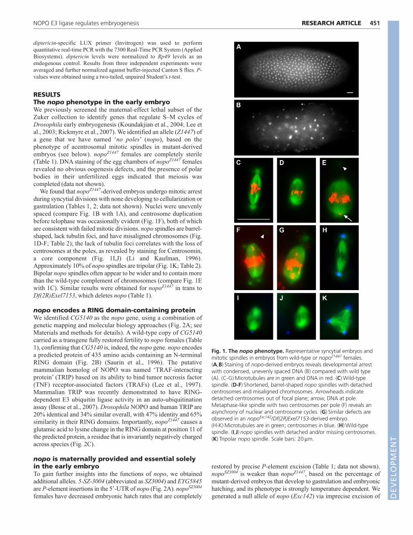
We found that nopoZ1447-derived embryos undergo mitotic arrestduring syncytial divisions with none developing to cellularization orgastrulation (Tables 1, 2; data not shown). Nuclei were unevenlyspaced (compare Fig. 1B with 1A), and centrosome duplicationbefore telophase was occasionally evident (Fig. 1F), both of whichare consistent with failed mitotic divisions. nopo spindles are barrel-shaped, lack tubulin foci, and have misaligned chromosomes (Fig.1D-F; Table 2); the lack of tubulin foci correlates with the loss ofcentrosomes at the poles, as revealed by staining for Centrosomin,a core component (Fig. 1I,J) (Li and Kaufman, 1996).Approximately 10% of nopo spindles are tripolar (Fig. 1K; Table 2).Bipolar nopo spindles often appear to be wider and to contain morethan the wild-type complement of chromosomes (compare Fig. 1Ewith 1C). Similar results were obtained for nopoZ1447 in trans toDf(2R)Exel7153, which deletes nopo (Table 1).
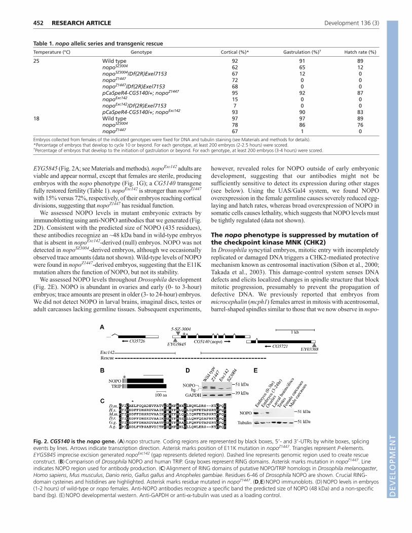
nopo encodes a RING domain-containing proteinWe identified CG5140 as the nopo gene, using a combination ofgenetic mapping and molecular biology approaches (Fig. 2A; seeMaterials and methods for details). A wild-type copy of CG5140carried as a transgene fully restored fertility to nopo females (Table1), confirming that CG5140 is, indeed, the nopo gene. nopo encodesa predicted protein of 435 amino acids containing an N-terminalRING domain (Fig. 2B) (Saurin et al., 1996). The putativemammalian homolog of NOPO was named ‘TRAF-interactingprotein’ (TRIP) based on its ability to bind tumor necrosis factor(TNF) receptor-associated factors (TRAFs) (Lee et al., 1997).Mammalian TRIP was recently demonstrated to have RING-dependent E3 ubiquitin ligase activity in an auto-ubiquitinationassay (Besse et al., 2007). Drosophila NOPO and human TRIP are20% identical and 34% similar overall, with 47% identity and 65%similarity in their RING domains. Importantly, nopoZ1447 causes aglutamic acid to lysine change in the RING domain at position 11 ofthe predicted protein, a residue that is invariantly negatively chargedacross species (Fig. 2C).
nopo is maternally provided and essential solelyin the early embryoTo gain further insights into the functions of nopo, we obtainedadditional alleles. 5-SZ-3004 (abbreviated as SZ3004) and EYG5845are P-element insertions in the 5�-UTR of nopo (Fig. 2A). nopoSZ3004
females have decreased embryonic hatch rates that are completely
restored by precise P-element excision (Table 1; data not shown).nopoSZ3004 is weaker than nopoZ1447, based on the percentage ofmutant-derived embryos that develop to gastrulation and embryonichatching, and its phenotype is strongly temperature dependent. Wegenerated a null allele of nopo (Exc142) via imprecise excision of
451RESEARCH ARTICLENOPO E3 ligase regulates embryogenesis
Fig. 1. The nopo phenotype. Representative syncytial embryos andmitotic spindles in embryos from wild-type or nopoZ1447 females.(A,B) Staining of nopo-derived embryos reveals developmental arrestwith condensed, unevenly spaced DNA (B) compared with wild type(A). (C-G) Microtubules are in green and DNA in red. (C) Wild-typespindle. (D-F) Shortened, barrel-shaped nopo spindles with detachedcentrosomes and misaligned chromosomes. Arrowheads indicatedetached centrosomes out of focal plane; arrow, DNA at pole.Metaphase-like spindle with two centrosomes per pole (F) reveals anasynchrony of nuclear and centrosome cycles. (G) Similar defects areobserved in an nopoExc142/Df(2R)Exel7153-derived embryo.(H-K) Microtubules are in green; centrosomes in blue. (H) Wild-typespindle. (I,J) nopo spindles with detached and/or missing centrosomes.(K) Tripolar nopo spindle. Scale bars: 20μm.
DEVELO
PMENT
452
EYG5845 (Fig. 2A; see Materials and methods). nopoExc142 adults areviable and appear normal, except that females are sterile, producingembryos with the nopo phenotype (Fig. 1G); a CG5140 transgenefully restored fertility (Table 1). nopoExc142 is stronger than nopoZ1447
with 15% versus 72%, respectively, of their embryos reaching corticaldivisions, suggesting that nopoZ1447 has residual function.
We assessed NOPO levels in mutant embryonic extracts byimmunoblotting using anti-NOPO antibodies that we generated (Fig.2D). Consistent with the predicted size of NOPO (435 residues),these antibodies recognize an ~48 kDa band in wild-type embryosthat is absent in nopoExc142-derived (null) embryos. NOPO was notdetected in nopoSZ3004-derived embryos, although we occasionallyobserved trace amounts (data not shown). Wild-type levels of NOPOwere found in nopoZ1447-derived embryos, suggesting that the E11Kmutation alters the function of NOPO, but not its stability.
We assessed NOPO levels throughout Drosophila development(Fig. 2E). NOPO is abundant in ovaries and early (0- to 3-hour)embryos; trace amounts are present in older (3- to 24-hour) embryos.We did not detect NOPO in larval brains, imaginal discs, testes oradult carcasses lacking germline tissues. Subsequent experiments,
however, revealed roles for NOPO outside of early embryonicdevelopment, suggesting that our antibodies might not besufficiently sensitive to detect its expression during other stages(see below). Using the UAS/Gal4 system, we found NOPOoverexpression in the female germline causes severely reduced egg-laying and hatch rates, whereas broad overexpression of NOPO insomatic cells causes lethality, which suggests that NOPO levels mustbe tightly regulated (data not shown).
The nopo phenotype is suppressed by mutation ofthe checkpoint kinase MNK (CHK2)In Drosophila syncytial embryos, mitotic entry with incompletelyreplicated or damaged DNA triggers a CHK2-mediated protectivemechanism known as centrosomal inactivation (Sibon et al., 2000;Takada et al., 2003). This damage-control system senses DNAdefects and elicits localized changes in spindle structure that blockmitotic progression, presumably to prevent the propagation ofdefective DNA. We previously reported that embryos frommicrocephalin (mcph1) females arrest in mitosis with acentrosomal,barrel-shaped spindles similar to those that we now observe in nopo-
RESEARCH ARTICLE Development 136 (3)
Table 1. nopo allelic series and transgenic rescueTemperature (°C) Genotype Cortical (%)* Gastrulation (%)† Hatch rate (%)
25 Wild type 92 91 89nopoSZ3004 62 65 12nopoSZ3004/Df(2R)Exel7153 67 12 0nopoZ1447 72 0 0nopoZ1447/Df(2R)Exel7153 68 0 0pCaSpeR4-CG5140/+; nopoZ1447 95 92 87nopoExc142 15 0 0nopoExc142/Df(2R)Exel7153 7 0 0pCaSpeR4-CG5140/+; nopoExc142 93 90 83
18 Wild type 97 97 89nopoSZ3004 78 86 76nopoZ1447 67 1 0
Embryos collected from females of the indicated genotypes were fixed for DNA and tubulin staining (see Materials and methods for details).*Percentage of embryos that develop to cycle 10 or beyond. For each genotype, at least 200 embryos (2-2.5 hours) were scored.†Percentage of embryos that develop to the initiation of gastrulation or beyond. For each genotype, at least 200 embryos (3-4 hours) were scored.
Fig. 2. CG5140 is the nopo gene. (A) nopo structure. Coding regions are represented by black boxes, 5�- and 3�-UTRs by white boxes, splicingevents by lines. Arrows indicate transcription direction. Asterisk marks position of E11K mutation in nopoZ1447. Triangles represent P-elements.EYG5845 imprecise excision generated nopoExc142 (gap represents deleted region). Dashed line represents genomic region used to create rescueconstruct. (B) Comparison of Drosophila NOPO and human TRIP. Gray boxes represent RING domains. Asterisk marks mutation in nopoZ1447. Lineindicates NOPO region used for antibody production. (C) Alignment of RING domains of putative NOPO/TRIP homologs in Drosophila melanogaster,Homo sapiens, Mus musculus, Danio rerio, Gallus gallus and Anopheles gambiae. Residues 6-46 of Drosophila NOPO are shown. Crucial RING-domain cysteines and histidines are highlighted. Asterisk marks residue mutated in nopoZ1447. (D,E) NOPO immunoblots. (D) NOPO levels in embryos(1-2 hours) of wild-type or nopo females. Anti-NOPO antibodies recognize a specific band the predicted size of NOPO (48 kDa) and a non-specificband (bg). (E) NOPO developmental western. Anti-GAPDH or anti-α-tubulin was used as a loading control. D
EVELO
PMENT
derived embryos (Rickmyre et al., 2007). We demonstrated thatthese mcph1 defects were suppressed by mutation of maternalnuclear kinase (mnk), also known as loki, which encodes DrosophilaCHK2 (Abdu et al., 2002; Brodsky et al., 2004; Masrouha et al.,2003; Xu et al., 2001). mnk nulls exhibit increased sensitivity toionizing radiation, but are viable and fertile. Suppression of mcph1by mnk revealed that centrosomal inactivation significantlycontributes to the mcph1 phenotype.
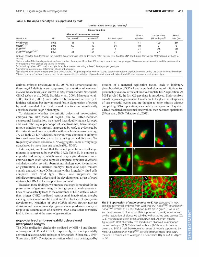
To determine whether the mitotic defects of nopo-derivedembryos are, like those of mcph1, due to CHK2-mediatedcentrosomal inactivation, we created lines doubly mutant for nopoand mnk. The nopo phenotype of acentrosomal, barrel-shapedmitotic spindles was strongly suppressed by mnk, as evidenced bythe restoration of normal spindles with attached centrosomes (Fig.3A-C; Table 2). DNA defects, however, were common in embryosfrom mnk nopo females, particularly during cortical divisions. Wefrequently observed abnormal DNA aggregates, some of very largesize, shared by more than one spindle (Fig. 3D,E).
Like mcph1, we found that the developmental arrest of nopomutants is suppressed by mnk (Fig. 3F,G; Table 2). In contrast tonopo-derived embryos, which arrest in syncytial divisions, mostembryos from mnk nopo females complete syncytial divisions,cellularize, and arrest with aberrant morphology upon the initiationof gastrulation. Cellularized embryos from mnk nopo femalescontain unusually large DNA masses within irregularly sized cellscompared with wild type. Thus, mnk suppresses thespindle/centrosomal defects and the developmental arrest of nopomutants, but DNA defects appear to accumulate.
Based on these findings, we propose that nopo is required for thepreservation of genomic integrity during syncytial embryogenesis.Lack of nopo activity leads to the occurrence of DNA defects, whichthen trigger CHK2-mediated centrosomal inactivation, therebycausing widespread mitotic arrest and the blockade of embryonicdevelopment. Mutation of mnk (Chk2) allows further nucleardivisions and developmental progression in nopo-derived embryos,despite the accumulation of extensive DNA defects that eventuallylead to their arrest at the onset of gastrulation.
nopo-derived embryos exhibit decreasedinterphase lengthThe DNA-replication checkpoint mediated by MEI-41 and Grapes,orthologs of ATR and CHK1, respectively, is developmentallyactivated in late syncytial embryos of Drosophila (Sibon et al., 1999;Sibon et al., 1997). Checkpoint activation, which may be triggered by
titration of a maternal replication factor, leads to inhibitoryphosphorylation of CDK1 and a gradual slowing of mitotic entry,presumably to allow sufficient time to complete DNA replication. AtMBT (cycle 14), the first G2 gap phase is introduced. Embryos frommei-41 or grapes (grp) mutant females fail to lengthen the interphasesof late syncytial cycles and are thought to enter mitosis withoutcompleting DNA replication; a secondary damage-control system,CHK2-mediated centrosomal inactivation, then becomes operational(Sibon et al., 2000; Takada et al., 2003).
453RESEARCH ARTICLENOPO E3 ligase regulates embryogenesis
Table 2. The nopo phenotype is suppressed by mnk
Mitotic spindle defects (% spindles)†
Bipolar spindles
Abnormal centrosome number Tripolar Gastrulation HatchGenotype MI* Decreased‡ Increased§ Barrel-shaped spindles (% embryos)¶ rate (%)
Wild type 0.63 <1 0 <1 <1 100 89nopoZ1447 0.95 62 13 64 10 0 0mnk6006 0.64 1 <1 1 <1 99 80mnk6006 nopoZ1447 0.68 <1 <1 <1 <1 77 0
Embryos collected from females of the indicated genotypes were used to determine hatch rates or were fixed for DNA and tubulin staining (see Materials and methods fordetails).*Mitotic index (MI)=% embryos in mitosis/total number of embryos. More than 300 embryos were scored per genotype. Chromosome condensation and the presence of amitotic spindle were used as the criteria for mitosis.†All mitotic spindles (>500 total) in a single focal plane were scored using at least 25 embryos per genotype. ‡Spindles with centrosomal detachment at one or both poles.§Spindles with >1 centrosome per pole (one or both poles). Telophase spindles were not scored because centrosome duplication occurs during this phase in the early embryo. ¶Stained embryos (3-4 hours) were scored for development to the initiation of gastrulation (or beyond). More than 200 embryos were scored per genotype.
Fig. 3. Suppression of nopo by mnk. (A-E) Representative mitoticspindles in syncytial embryos from wild-type (A), nopoZ1447 (B) and mnknopoZ1447 females (C-E). (A-C) Microtubules are in green, DNA in red,and centrosomes in blue. nopo (B) is suppressed by mnk, as evidencedby the restoration of elongated spindles with attached centrosomes (C).(D,E) Microtubules are in green and DNA in red. Aberrant mitoticfigures with DNA shared by two spindles are observed in mnk nopo-derived embryos. (F,G) Cellularized embryos (2-3 hours). Actin is ingreen and DNA in red. Developmental arrest of nopo is suppressed bymnk. Cellularized mnk nopoZ1447-derived embryos show large DNAmasses (G) compared to wild type (F). Scale bars: 10μm in A-E; 20μmin F,G. D
EVELO
PMENT
454
Mitotic entry with incompletely replicated DNA can cause CHK2-mediated centrosomal inactivation in syncytial embryos (Sibon et al.,2000; Takada et al., 2003). Control mechanisms to ensure completionof DNA replication prior to mitosis may be particularly critical duringrapid S–M cycles. Oscillating CDK1-Cyclin B activity plays a keyrole in coordinating these cycles (Edgar et al., 1994; Su et al., 1998).S–M transitions appear to be controlled by Cyclin B levels prior tocycle 10, and by both Cyclin B levels and a DNA-replicationcheckpoint in cycles 10-13 (Ji et al., 2004; Sibon et al., 1997).
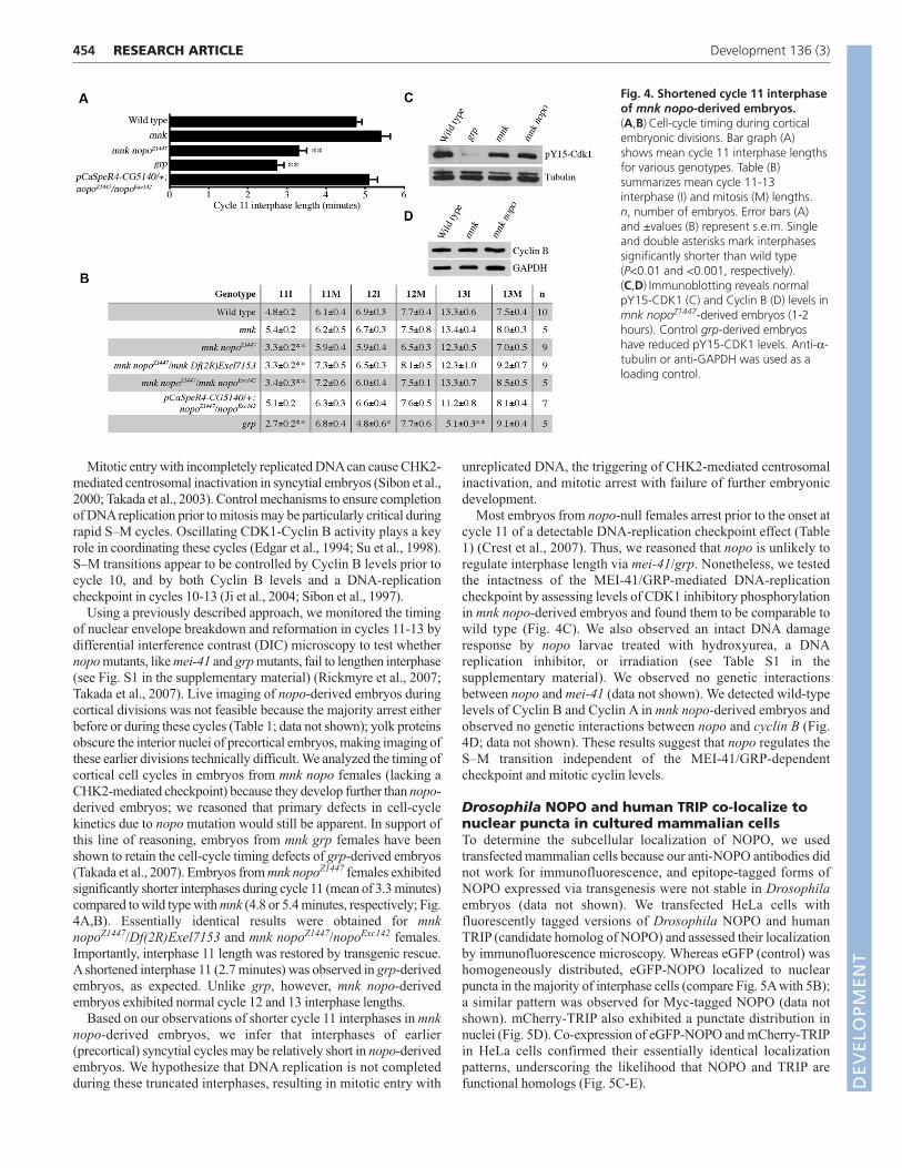
Using a previously described approach, we monitored the timingof nuclear envelope breakdown and reformation in cycles 11-13 bydifferential interference contrast (DIC) microscopy to test whethernopo mutants, like mei-41 and grp mutants, fail to lengthen interphase(see Fig. S1 in the supplementary material) (Rickmyre et al., 2007;Takada et al., 2007). Live imaging of nopo-derived embryos duringcortical divisions was not feasible because the majority arrest eitherbefore or during these cycles (Table 1; data not shown); yolk proteinsobscure the interior nuclei of precortical embryos, making imaging ofthese earlier divisions technically difficult. We analyzed the timing ofcortical cell cycles in embryos from mnk nopo females (lacking aCHK2-mediated checkpoint) because they develop further than nopo-derived embryos; we reasoned that primary defects in cell-cyclekinetics due to nopo mutation would still be apparent. In support ofthis line of reasoning, embryos from mnk grp females have beenshown to retain the cell-cycle timing defects of grp-derived embryos(Takada et al., 2007). Embryos from mnk nopoZ1447 females exhibitedsignificantly shorter interphases during cycle 11 (mean of 3.3 minutes)compared to wild type with mnk (4.8 or 5.4 minutes, respectively; Fig.4A,B). Essentially identical results were obtained for mnknopoZ1447/Df(2R)Exel7153 and mnk nopoZ1447/nopoExc142 females.Importantly, interphase 11 length was restored by transgenic rescue.A shortened interphase 11 (2.7 minutes) was observed in grp-derivedembryos, as expected. Unlike grp, however, mnk nopo-derivedembryos exhibited normal cycle 12 and 13 interphase lengths.
Based on our observations of shorter cycle 11 interphases in mnknopo-derived embryos, we infer that interphases of earlier(precortical) syncytial cycles may be relatively short in nopo-derivedembryos. We hypothesize that DNA replication is not completedduring these truncated interphases, resulting in mitotic entry with
unreplicated DNA, the triggering of CHK2-mediated centrosomalinactivation, and mitotic arrest with failure of further embryonicdevelopment.
Most embryos from nopo-null females arrest prior to the onset atcycle 11 of a detectable DNA-replication checkpoint effect (Table1) (Crest et al., 2007). Thus, we reasoned that nopo is unlikely toregulate interphase length via mei-41/grp. Nonetheless, we testedthe intactness of the MEI-41/GRP-mediated DNA-replicationcheckpoint by assessing levels of CDK1 inhibitory phosphorylationin mnk nopo-derived embryos and found them to be comparable towild type (Fig. 4C). We also observed an intact DNA damageresponse by nopo larvae treated with hydroxyurea, a DNAreplication inhibitor, or irradiation (see Table S1 in thesupplementary material). We observed no genetic interactionsbetween nopo and mei-41 (data not shown). We detected wild-typelevels of Cyclin B and Cyclin A in mnk nopo-derived embryos andobserved no genetic interactions between nopo and cyclin B (Fig.4D; data not shown). These results suggest that nopo regulates theS–M transition independent of the MEI-41/GRP-dependentcheckpoint and mitotic cyclin levels.
Drosophila NOPO and human TRIP co-localize tonuclear puncta in cultured mammalian cellsTo determine the subcellular localization of NOPO, we usedtransfected mammalian cells because our anti-NOPO antibodies didnot work for immunofluorescence, and epitope-tagged forms ofNOPO expressed via transgenesis were not stable in Drosophilaembryos (data not shown). We transfected HeLa cells withfluorescently tagged versions of Drosophila NOPO and humanTRIP (candidate homolog of NOPO) and assessed their localizationby immunofluorescence microscopy. Whereas eGFP (control) washomogeneously distributed, eGFP-NOPO localized to nuclearpuncta in the majority of interphase cells (compare Fig. 5A with 5B);a similar pattern was observed for Myc-tagged NOPO (data notshown). mCherry-TRIP also exhibited a punctate distribution innuclei (Fig. 5D). Co-expression of eGFP-NOPO and mCherry-TRIPin HeLa cells confirmed their essentially identical localizationpatterns, underscoring the likelihood that NOPO and TRIP arefunctional homologs (Fig. 5C-E).
RESEARCH ARTICLE Development 136 (3)
Fig. 4. Shortened cycle 11 interphaseof mnk nopo-derived embryos.(A,B) Cell-cycle timing during corticalembryonic divisions. Bar graph (A)shows mean cycle 11 interphase lengthsfor various genotypes. Table (B)summarizes mean cycle 11-13interphase (I) and mitosis (M) lengths.n, number of embryos. Error bars (A)and ±values (B) represent s.e.m. Singleand double asterisks mark interphasessignificantly shorter than wild type(P<0.01 and <0.001, respectively).(C,D) Immunoblotting reveals normalpY15-CDK1 (C) and Cyclin B (D) levels inmnk nopoZ1447-derived embryos (1-2hours). Control grp-derived embryoshave reduced pY15-CDK1 levels. Anti-α-tubulin or anti-GAPDH was used as aloading control.
DEVELO
PMENT
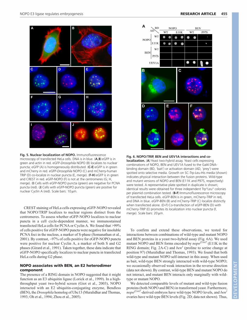
CREST staining of HeLa cells expressing eGFP-NOPO revealedthat NOPO/TRIP localizes to nuclear regions distinct from thecentromeres. To assess whether eGFP-NOPO localizes to nuclearpuncta in a cell cycle-dependent manner, we immunostainedtransfected HeLa cells for PCNA or Cyclin A. We found that >99%of cells positive for eGFP-NOPO puncta were negative for insolublePCNA foci in the nucleus, a marker of S-phase (Somanathan et al.,2001). By contrast, ~97% of cells positive for eGFP-NOPO punctawere positive for nuclear Cyclin A, a marker of both S and G2phases (Girard et al., 1991). Taken together, these data indicate thateGFP-NOPO specifically localizes to nuclear puncta in transfectedHeLa cells during G2 phase.
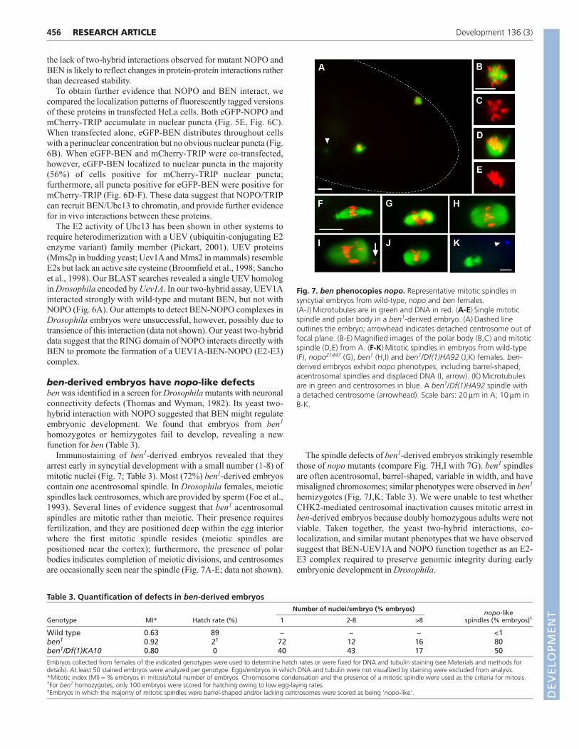
NOPO associates with BEN, an E2 heterodimercomponentThe presence of a RING domain in NOPO suggested that it mightfunction as an E3 ubiquitin ligase (Lorick et al., 1999). In a high-throughput yeast two-hybrid screen (Giot et al., 2003), NOPOinteracted with an E2 ubiquitin-conjugating enzyme, Bendless(BEN), the Drosophila homolog of Ubc13 (Muralidhar and Thomas,1993; Oh et al., 1994; Zhou et al., 2005).
To confirm and extend these observations, we tested forinteractions between combinations of wild-type and mutant NOPOand BEN proteins in a yeast two-hybrid assay (Fig. 6A). We usedmutant NOPO and BEN forms encoded by nopoZ1447 (E11K in theRING domain; Fig. 2A-C) and ben1 (proline to serine change atposition 97) (Muralidhar and Thomas, 1993). We found that bothwild-type and mutant NOPO self-interact in this assay. When usedas bait, wild-type BEN strongly interacted with wild-type NOPO;we occasionally observed weak interaction in the reverse direction(data not shown). By contrast, wild-type BEN and mutant NOPO donot interact, and mutant BEN interacts only marginally with wild-type or mutant NOPO.
We detected comparable levels of mutant and wild-type fusionproteins (both NOPO and BEN) in transformed yeast. Furthermore,nopoZ1447-derived embryos have wild-type NOPO levels, and ben1
ovaries have wild-type BEN levels (Fig. 2D; data not shown). Thus,
455RESEARCH ARTICLENOPO E3 ligase regulates embryogenesis
Fig. 5. Nuclear localization of NOPO. Immunofluorescencemicroscopy of transfected HeLa cells. DNA is in blue. (A,B) eGFP is ingreen and actin in red. eGFP-Drosophila NOPO (B) localizes to nuclearpuncta; eGFP (A) is homogeneously distributed. (C-E) eGFP is in greenand mCherry in red. eGFP-Drosophila NOPO (C) and mCherry-humanTRIP (D) co-localize in nuclear puncta (E, merge). (F-H) eGFP is in greenand CREST in red. eGFP-NOPO (F) is not at the centromeres (G; H,merge). (I) Cells with eGFP-NOPO puncta (green) are negative for PCNApuncta (red). (J) Cells with eGFP-NOPO puncta (green) are positive fornuclear Cyclin A (red). Scale bars: 10μm.
Fig. 6. NOPO/TRIP, BEN and UEV1A interactions and co-localization. (A) Yeast two-hybrid assay. Yeast cells expressingcombinations of NOPO, BEN and UEV1A fused to the Gal4 DNA-binding domain (BD, ‘bait’) or activation domain (AD, ‘prey’) werespotted onto selective media. Growth on SC-Trp-Leu-His media (shown)indicates physical interaction between the fusion proteins. Wild-typeand mutant versions of NOPO and BEN (E11K and P97S, respectively)were tested. A representative plate spotted in duplicate is shown;identical results were obtained for three independent Trp+Leu+ coloniesper plasmid combination tested. (B-F) Immunofluorescence microscopyof transfected HeLa cells. eGFP-BEN is in green, mCherry-TRIP in red,and DNA in blue. eGFP-BEN (B) and mCherry-TRIP (C) localize distinctlywhen transfected alone. (D-F) Co-transfection of eGFP-BEN (D) withmCherry-TRIP (E) promotes its localization into nuclear puncta (F,merge). Scale bars: 20μm.
DEVELO
PMENT
456
the lack of two-hybrid interactions observed for mutant NOPO andBEN is likely to reflect changes in protein-protein interactions ratherthan decreased stability.
To obtain further evidence that NOPO and BEN interact, wecompared the localization patterns of fluorescently tagged versionsof these proteins in transfected HeLa cells. Both eGFP-NOPO andmCherry-TRIP accumulate in nuclear puncta (Fig. 5E, Fig. 6C).When transfected alone, eGFP-BEN distributes throughout cellswith a perinuclear concentration but no obvious nuclear puncta (Fig.6B). When eGFP-BEN and mCherry-TRIP were co-transfected,however, eGFP-BEN localized to nuclear puncta in the majority(56%) of cells positive for mCherry-TRIP nuclear puncta;furthermore, all puncta positive for eGFP-BEN were positive formCherry-TRIP (Fig. 6D-F). These data suggest that NOPO/TRIPcan recruit BEN/Ubc13 to chromatin, and provide further evidencefor in vivo interactions between these proteins.
The E2 activity of Ubc13 has been shown in other systems torequire heterodimerization with a UEV (ubiquitin-conjugating E2enzyme variant) family member (Pickart, 2001). UEV proteins(Mms2p in budding yeast; Uev1A and Mms2 in mammals) resembleE2s but lack an active site cysteine (Broomfield et al., 1998; Sanchoet al., 1998). Our BLAST searches revealed a single UEV homologin Drosophila encoded by Uev1A. In our two-hybrid assay, UEV1Ainteracted strongly with wild-type and mutant BEN, but not withNOPO (Fig. 6A). Our attempts to detect BEN-NOPO complexes inDrosophila embryos were unsuccessful, however, possibly due totransience of this interaction (data not shown). Our yeast two-hybriddata suggest that the RING domain of NOPO interacts directly withBEN to promote the formation of a UEV1A-BEN-NOPO (E2-E3)complex.
ben-derived embryos have nopo-like defectsben was identified in a screen for Drosophila mutants with neuronalconnectivity defects (Thomas and Wyman, 1982). Its yeast two-hybrid interaction with NOPO suggested that BEN might regulateembryonic development. We found that embryos from ben1
homozygotes or hemizygotes fail to develop, revealing a newfunction for ben (Table 3).
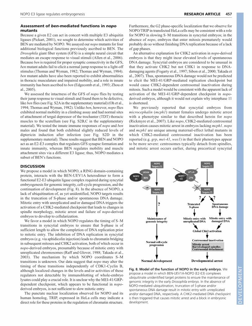
Immunostaining of ben1-derived embryos revealed that theyarrest early in syncytial development with a small number (1-8) ofmitotic nuclei (Fig. 7; Table 3). Most (72%) ben1-derived embryoscontain one acentrosomal spindle. In Drosophila females, meioticspindles lack centrosomes, which are provided by sperm (Foe et al.,1993). Several lines of evidence suggest that ben1 acentrosomalspindles are mitotic rather than meiotic. Their presence requiresfertilization, and they are positioned deep within the egg interiorwhere the first mitotic spindle resides (meiotic spindles arepositioned near the cortex); furthermore, the presence of polarbodies indicates completion of meiotic divisions, and centrosomesare occasionally seen near the spindle (Fig. 7A-E; data not shown).
The spindle defects of ben1-derived embryos strikingly resemblethose of nopo mutants (compare Fig. 7H,I with 7G). ben1 spindlesare often acentrosomal, barrel-shaped, variable in width, and havemisaligned chromosomes; similar phenotypes were observed in ben1
hemizygotes (Fig. 7J,K; Table 3). We were unable to test whetherCHK2-mediated centrosomal inactivation causes mitotic arrest inben-derived embryos because doubly homozygous adults were notviable. Taken together, the yeast two-hybrid interactions, co-localization, and similar mutant phenotypes that we have observedsuggest that BEN-UEV1A and NOPO function together as an E2-E3 complex required to preserve genomic integrity during earlyembryonic development in Drosophila.
RESEARCH ARTICLE Development 136 (3)
Fig. 7. ben phenocopies nopo. Representative mitotic spindles insyncytial embryos from wild-type, nopo and ben females.(A-J) Microtubules are in green and DNA in red. (A-E) Single mitoticspindle and polar body in a ben1-derived embryo. (A) Dashed lineoutlines the embryo; arrowhead indicates detached centrosome out offocal plane. (B-E) Magnified images of the polar body (B,C) and mitoticspindle (D,E) from A. (F-K) Mitotic spindles in embryos from wild-type(F), nopoZ1447 (G), ben1 (H,I) and ben1/Df(1)HA92 (J,K) females. ben-derived embryos exhibit nopo phenotypes, including barrel-shaped,acentrosomal spindles and displaced DNA (I, arrow). (K) Microtubulesare in green and centrosomes in blue. A ben1/Df(1)HA92 spindle witha detached centrosome (arrowhead). Scale bars: 20 μm in A; 10 μm inB-K.
Table 3. Quantification of defects in ben-derived embryos Number of nuclei/embryo (% embryos) nopo-like
Genotype MI* Hatch rate (%) 1 2-8 >8 spindles (% embryos)‡
Wild type 0.63 89 – – – <1ben1 0.92 2† 72 12 16 80ben1/Df(1)KA10 0.80 0 40 43 17 50
Embryos collected from females of the indicated genotypes were used to determine hatch rates or were fixed for DNA and tubulin staining (see Materials and methods fordetails). At least 50 stained embryos were analyzed per genotype. Eggs/embryos in which DNA and tubulin were not visualized by staining were excluded from analysis.*Mitotic index (MI) = % embryos in mitosis/total number of embryos. Chromosome condensation and the presence of a mitotic spindle were used as the criteria for mitosis.†For ben1 homozygotes, only 100 embryos were scored for hatching owing to low egg-laying rates.‡Embryos in which the majority of mitotic spindles were barrel-shaped and/or lacking centrosomes were scored as being ‘nopo-like’. D
EVELO
PMENT
Assessment of ben-mediated functions in nopomutantsBecause a given E2 can act in concert with multiple E3 ubiquitinligases (Pickart, 2001), we sought to determine which activities ofBEN are mediated by NOPO. We assayed our nopo mutants for fouradditional biological functions previously ascribed to BEN. TheDrosophila giant fiber system (GFS) is a simple neural circuit thatmediates an escape response to visual stimuli (Allen et al., 2006).Because ben is required for proper synaptic connectivity in the GFS,ben mutant adults fail to elicit a normal jump response to a light-offstimulus (Thomas and Wyman, 1982; Thomas and Wyman, 1984).ben mutant adults have also been reported to exhibit abnormalitiesin thoracic musculature and impaired mobility, and a role in innateimmunity has been ascribed to ben (Edgecomb et al., 1993; Zhou etal., 2005).
We assessed the intactness of the GFS of nopo flies by testingtheir jump response to visual stimuli and found them to be defective,like ben flies (see Fig. S2A in the supplementary material) (Oh et al.,1994; Thomas and Wyman, 1982). Unlike ben, however, nopo fliesexhibited normal mobility in a climbing assay and had normal sitesof attachment of tergal depressor of the trochanter (TDT) thoracicmuscles to the scutellum (see Fig. S2B,C in the supplementarymaterial). We tested the innate immune responses of nopo and benmales and found that both exhibited slightly reduced levels ofdiptericin induction after infection (see Fig. S2D in thesupplementary material). These results suggest that BEN and NOPOact as an E2-E3 complex that regulates GFS synapse formation andinnate immunity, whereas BEN regulates mobility and muscleattachment sites via a different E3 ligase; thus, NOPO mediates asubset of BEN’s functions.
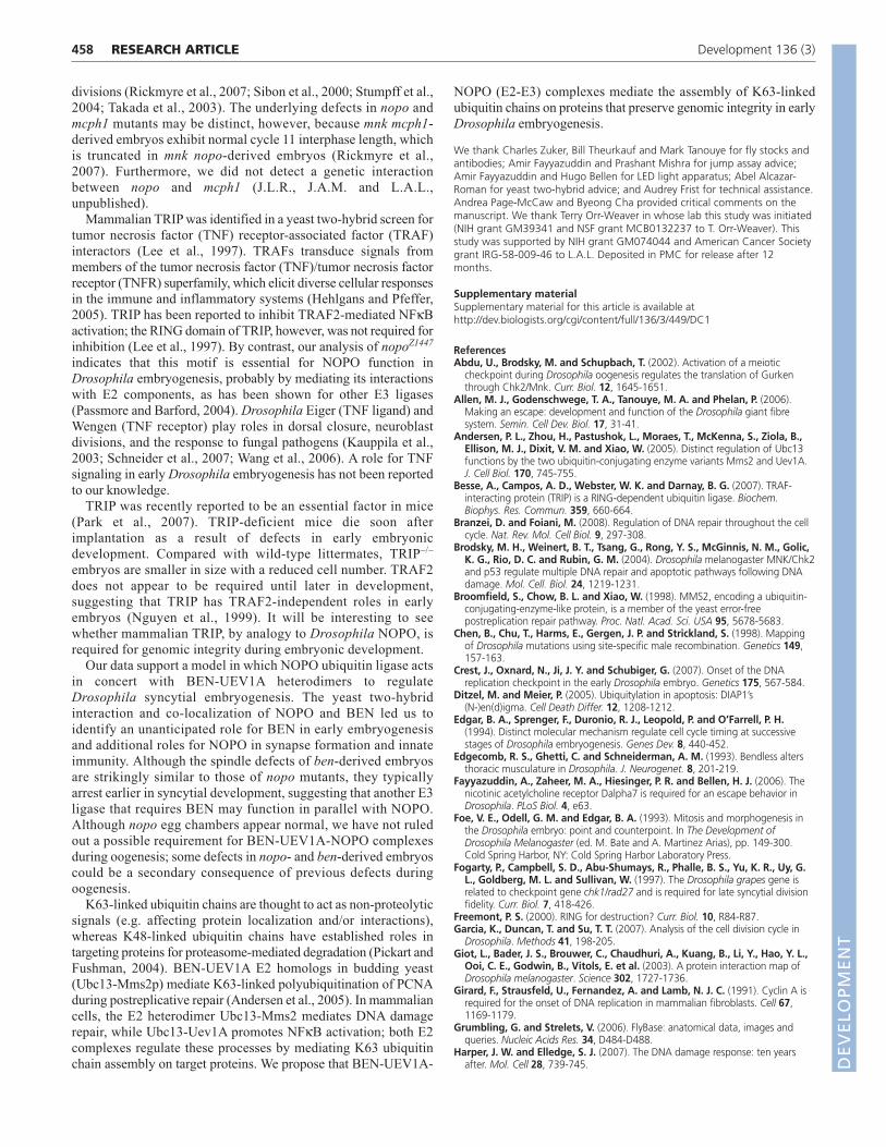
DISCUSSIONWe propose a model in which NOPO, a RING domain-containingprotein, interacts with the BEN-UEV1A heterodimer to form afunctional E2-E3 ubiquitin ligase complex required during syncytialembryogenesis for genomic integrity, cell-cycle progression, and thecontinuation of development (Fig. 8). In the absence of NOPO, alack of ubiquitination of, as yet unidentified, NOPO targets resultsin the truncation of S-phase and/or spontaneous DNA damage.Mitotic entry with unreplicated and/or damaged DNA triggers theactivation of a CHK2-mediated checkpoint that leads to changes inspindle morphology, mitotic arrest and failure of nopo-derivedembryos to develop to cellularization.
We favor a model in which NOPO regulates the timing of S–Mtransitions in syncytial embryos to ensure that S-phase is ofsufficient length to allow the completion of DNA replication priorto mitotic entry. The inhibition of DNA replication in syncytialembryos (e.g. via aphidicolin injection) leads to chromatin bridgingin subsequent mitoses and CHK2 activation, both of which occur innopo-derived embryos, presumably because of mitotic entry withunreplicated chromosomes (Raff and Glover, 1988; Takada et al.,2003). The mechanism by which NOPO coordinates S–Mtransitions is unknown. Our data suggest that nopo may alter thetiming of these transitions independently of CDK1-Cyclin B,although localized changes in the levels and/or activities of theseregulators not detectable by immunoblotting of whole-embryolysates could play a crucial role. It is unclear why the MEI-41/GRP-dependent checkpoint, which appears to be functional in nopo-derived embryos, is not sufficient to slow mitotic entry.
The punctate nuclear localization observed for NOPO and itshuman homolog, TRIP, expressed in HeLa cells may indicate adirect role for these proteins in the regulation of chromatin structure.
Furthermore, the G2 phase-specific localization that we observe forNOPO/TRIP in transfected HeLa cells may be consistent with a rolefor NOPO in slowing S–M transitions in syncytial embryos; in theabsence of nopo, embryos that enter mitosis prematurely wouldprobably do so without finishing DNA replication because of a lackof gap phases.
An alternative explanation for CHK2 activation in nopo-derivedembryos is that they might incur elevated levels of spontaneousDNA damage. Syncytial embryos are considered to be unusual inthat they activate CHK2 but not CHK1 in response to DNA-damaging agents (Fogarty et al., 1997; Sibon et al., 2000; Takada etal., 2007). Thus, spontaneous DNA damage would not be predictedto elicit the MEI-41/GRP-mediated replication checkpoint butwould cause CHK2-dependent centrosomal inactivation duringmitosis. Such a model would be consistent with the apparent lack ofactivation of the MEI-41/GRP-dependent checkpoint in nopo-derived embryos, although it would not explain why interphase 11is shortened.
We previously reported that syncytial embryos frommicrocephalin (mcph1) mutant females undergo mitotic arrestwith a phenotype similar to that described herein for nopo(Rickmyre et al., 2007). Like nopo, CHK2-mediated centrosomalinactivation causes mitotic arrest in embryos lacking mcph1. nopoand mcph1 are unique among maternal-effect lethal mutants inwhich CHK2-mediated centrosomal inactivation has beenreported (e.g. grp, mei-41, wee1) in that their phenotypes appearto be more severe: centrosomes typically detach from spindles,and mitotic arrest occurs earlier, during precortical syncytial
457RESEARCH ARTICLENOPO E3 ligase regulates embryogenesis
Fig. 8. Model of the function of NOPO in the early embryo. Wepropose a model in which BEN-UEV1A-NOPO (E2-E3) complexesubiquitinate unidentified target proteins to ensure the maintenance ofgenomic integrity in the early Drosophila embryo. In the absence ofNOPO-mediated ubiquitination, truncation of S-phase and/orspontaneous DNA damage result in mitotic entry with unreplicatedand/or damaged DNA, respectively. A CHK2-mediated DNA checkpointis then triggered that causes mitotic arrest and a block in embryonicdevelopment. D
EVELO
PMENT
458
divisions (Rickmyre et al., 2007; Sibon et al., 2000; Stumpff et al.,2004; Takada et al., 2003). The underlying defects in nopo andmcph1 mutants may be distinct, however, because mnk mcph1-derived embryos exhibit normal cycle 11 interphase length, whichis truncated in mnk nopo-derived embryos (Rickmyre et al.,2007). Furthermore, we did not detect a genetic interactionbetween nopo and mcph1 (J.L.R., J.A.M. and L.A.L.,unpublished).
Mammalian TRIP was identified in a yeast two-hybrid screen fortumor necrosis factor (TNF) receptor-associated factor (TRAF)interactors (Lee et al., 1997). TRAFs transduce signals frommembers of the tumor necrosis factor (TNF)/tumor necrosis factorreceptor (TNFR) superfamily, which elicit diverse cellular responsesin the immune and inflammatory systems (Hehlgans and Pfeffer,2005). TRIP has been reported to inhibit TRAF2-mediated NFκBactivation; the RING domain of TRIP, however, was not required forinhibition (Lee et al., 1997). By contrast, our analysis of nopoZ1447
indicates that this motif is essential for NOPO function inDrosophila embryogenesis, probably by mediating its interactionswith E2 components, as has been shown for other E3 ligases(Passmore and Barford, 2004). Drosophila Eiger (TNF ligand) andWengen (TNF receptor) play roles in dorsal closure, neuroblastdivisions, and the response to fungal pathogens (Kauppila et al.,2003; Schneider et al., 2007; Wang et al., 2006). A role for TNFsignaling in early Drosophila embryogenesis has not been reportedto our knowledge.
TRIP was recently reported to be an essential factor in mice(Park et al., 2007). TRIP-deficient mice die soon afterimplantation as a result of defects in early embryonicdevelopment. Compared with wild-type littermates, TRIP–/–
embryos are smaller in size with a reduced cell number. TRAF2does not appear to be required until later in development,suggesting that TRIP has TRAF2-independent roles in earlyembryos (Nguyen et al., 1999). It will be interesting to seewhether mammalian TRIP, by analogy to Drosophila NOPO, isrequired for genomic integrity during embryonic development.
Our data support a model in which NOPO ubiquitin ligase actsin concert with BEN-UEV1A heterodimers to regulateDrosophila syncytial embryogenesis. The yeast two-hybridinteraction and co-localization of NOPO and BEN led us toidentify an unanticipated role for BEN in early embryogenesisand additional roles for NOPO in synapse formation and innateimmunity. Although the spindle defects of ben-derived embryosare strikingly similar to those of nopo mutants, they typicallyarrest earlier in syncytial development, suggesting that another E3ligase that requires BEN may function in parallel with NOPO.Although nopo egg chambers appear normal, we have not ruledout a possible requirement for BEN-UEV1A-NOPO complexesduring oogenesis; some defects in nopo- and ben-derived embryoscould be a secondary consequence of previous defects duringoogenesis.
K63-linked ubiquitin chains are thought to act as non-proteolyticsignals (e.g. affecting protein localization and/or interactions),whereas K48-linked ubiquitin chains have established roles intargeting proteins for proteasome-mediated degradation (Pickart andFushman, 2004). BEN-UEV1A E2 homologs in budding yeast(Ubc13-Mms2p) mediate K63-linked polyubiquitination of PCNAduring postreplicative repair (Andersen et al., 2005). In mammaliancells, the E2 heterodimer Ubc13-Mms2 mediates DNA damagerepair, while Ubc13-Uev1A promotes NFκB activation; both E2complexes regulate these processes by mediating K63 ubiquitinchain assembly on target proteins. We propose that BEN-UEV1A-
NOPO (E2-E3) complexes mediate the assembly of K63-linkedubiquitin chains on proteins that preserve genomic integrity in earlyDrosophila embryogenesis.
We thank Charles Zuker, Bill Theurkauf and Mark Tanouye for fly stocks andantibodies; Amir Fayyazuddin and Prashant Mishra for jump assay advice;Amir Fayyazuddin and Hugo Bellen for LED light apparatus; Abel Alcazar-Roman for yeast two-hybrid advice; and Audrey Frist for technical assistance.Andrea Page-McCaw and Byeong Cha provided critical comments on themanuscript. We thank Terry Orr-Weaver in whose lab this study was initiated(NIH grant GM39341 and NSF grant MCB0132237 to T. Orr-Weaver). Thisstudy was supported by NIH grant GM074044 and American Cancer Societygrant IRG-58-009-46 to L.A.L. Deposited in PMC for release after 12months.
Supplementary materialSupplementary material for this article is available athttp://dev.biologists.org/cgi/content/full/136/3/449/DC1
ReferencesAbdu, U., Brodsky, M. and Schupbach, T. (2002). Activation of a meiotic
checkpoint during Drosophila oogenesis regulates the translation of Gurkenthrough Chk2/Mnk. Curr. Biol. 12, 1645-1651.
Allen, M. J., Godenschwege, T. A., Tanouye, M. A. and Phelan, P. (2006).Making an escape: development and function of the Drosophila giant fibresystem. Semin. Cell Dev. Biol. 17, 31-41.
Andersen, P. L., Zhou, H., Pastushok, L., Moraes, T., McKenna, S., Ziola, B.,Ellison, M. J., Dixit, V. M. and Xiao, W. (2005). Distinct regulation of Ubc13functions by the two ubiquitin-conjugating enzyme variants Mms2 and Uev1A.J. Cell Biol. 170, 745-755.
Besse, A., Campos, A. D., Webster, W. K. and Darnay, B. G. (2007). TRAF-interacting protein (TRIP) is a RING-dependent ubiquitin ligase. Biochem.Biophys. Res. Commun. 359, 660-664.
Branzei, D. and Foiani, M. (2008). Regulation of DNA repair throughout the cellcycle. Nat. Rev. Mol. Cell Biol. 9, 297-308.
Brodsky, M. H., Weinert, B. T., Tsang, G., Rong, Y. S., McGinnis, N. M., Golic,K. G., Rio, D. C. and Rubin, G. M. (2004). Drosophila melanogaster MNK/Chk2and p53 regulate multiple DNA repair and apoptotic pathways following DNAdamage. Mol. Cell. Biol. 24, 1219-1231.
Broomfield, S., Chow, B. L. and Xiao, W. (1998). MMS2, encoding a ubiquitin-conjugating-enzyme-like protein, is a member of the yeast error-freepostreplication repair pathway. Proc. Natl. Acad. Sci. USA 95, 5678-5683.
Chen, B., Chu, T., Harms, E., Gergen, J. P. and Strickland, S. (1998). Mappingof Drosophila mutations using site-specific male recombination. Genetics 149,157-163.
Crest, J., Oxnard, N., Ji, J. Y. and Schubiger, G. (2007). Onset of the DNAreplication checkpoint in the early Drosophila embryo. Genetics 175, 567-584.
Ditzel, M. and Meier, P. (2005). Ubiquitylation in apoptosis: DIAP1’s(N-)en(d)igma. Cell Death Differ. 12, 1208-1212.
Edgar, B. A., Sprenger, F., Duronio, R. J., Leopold, P. and O’Farrell, P. H.(1994). Distinct molecular mechanism regulate cell cycle timing at successivestages of Drosophila embryogenesis. Genes Dev. 8, 440-452.
Edgecomb, R. S., Ghetti, C. and Schneiderman, A. M. (1993). Bendless altersthoracic musculature in Drosophila. J. Neurogenet. 8, 201-219.
Fayyazuddin, A., Zaheer, M. A., Hiesinger, P. R. and Bellen, H. J. (2006). Thenicotinic acetylcholine receptor Dalpha7 is required for an escape behavior inDrosophila. PLoS Biol. 4, e63.
Foe, V. E., Odell, G. M. and Edgar, B. A. (1993). Mitosis and morphogenesis inthe Drosophila embryo: point and counterpoint. In The Development ofDrosophila Melanogaster (ed. M. Bate and A. Martinez Arias), pp. 149-300.Cold Spring Harbor, NY: Cold Spring Harbor Laboratory Press.
Fogarty, P., Campbell, S. D., Abu-Shumays, R., Phalle, B. S., Yu, K. R., Uy, G.L., Goldberg, M. L. and Sullivan, W. (1997). The Drosophila grapes gene isrelated to checkpoint gene chk1/rad27 and is required for late syncytial divisionfidelity. Curr. Biol. 7, 418-426.
Freemont, P. S. (2000). RING for destruction? Curr. Biol. 10, R84-R87.Garcia, K., Duncan, T. and Su, T. T. (2007). Analysis of the cell division cycle in
Drosophila. Methods 41, 198-205.Giot, L., Bader, J. S., Brouwer, C., Chaudhuri, A., Kuang, B., Li, Y., Hao, Y. L.,
Ooi, C. E., Godwin, B., Vitols, E. et al. (2003). A protein interaction map ofDrosophila melanogaster. Science 302, 1727-1736.
Girard, F., Strausfeld, U., Fernandez, A. and Lamb, N. J. C. (1991). Cyclin A isrequired for the onset of DNA replication in mammalian fibroblasts. Cell 67,1169-1179.
Grumbling, G. and Strelets, V. (2006). FlyBase: anatomical data, images andqueries. Nucleic Acids Res. 34, D484-D488.
Harper, J. W. and Elledge, S. J. (2007). The DNA damage response: ten yearsafter. Mol. Cell 28, 739-745.
RESEARCH ARTICLE Development 136 (3)
DEVELO
PMENT
Hehlgans, T. and Pfeffer, K. (2005). The intriguing biology of the tumournecrosis factor/tumour necrosis factor receptor superfamily: players, rules andthe games. Immunology 115, 1-20.
Jackson, P. K., Eldridge, A. G., Freed, E., Furstenthal, L., Hsu, J. Y., Kaiser, B.K. and Reimann, J. D. (2000). The lore of the RINGs: substrate recognition andcatalysis by ubiquitin ligases. Trends Cell Biol. 10, 429-439.
James, P., Halladay, J. and Craig, E. A. (1996). Genomic libraries and a hoststrain designed for highly efficient two-hybrid selection in yeast. Genetics 144,1425-1436.
Ji, J. Y., Squirrell, J. M. and Schubiger, G. (2004). Both cyclin B levels and DNA-replication checkpoint control the early embryonic mitoses in Drosophila.Development 131, 401-411.
Kauppila, S., Maaty, W. S., Chen, P., Tomar, R. S., Eby, M. T., Chapo, J.,Chew, S., Rathore, N., Zachariah, S., Sinha, S. K. et al. (2003). Eiger and itsreceptor, Wengen, comprise a TNF-like system in Drosophila. Oncogene 22,4860-4867.
Koundakjian, E. J., Cowan, D. M., Hardy, R. W. and Becker, A. H. (2004). TheZuker collection: a resource for the analysis of autosomal gene function inDrosophila melanogaster. Genetics 167, 203-206.
Lee, L. A. and Orr-Weaver, T. L. (2003). Regulation of cell cycles in Drosophiladevelopment: intrinsic and extrinsic cues. Annu. Rev. Genet. 37, 545-578.
Lee, L. A., Van Hoewyk, D. and Orr-Weaver, T. L. (2003). The Drosophila cellcycle kinase PAN GU forms an active complex with PLUTONIUM and GNU toregulate embryonic divisions. Genes Dev. 17, 2979-2991.
Lee, S. Y., Lee, S. Y. and Choi, Y. (1997). TRAF-interacting protein (TRIP): a novelcomponent of the tumor necrosis factor receptor (TNFR)- and CD30-TRAFsignaling complexes that inhibits TRAF2-mediated NF-kappaB activation. J. Exp.Med. 185, 1275-1285.
Li, K. and Kaufman, T. C. (1996). The homeotic target gene centrosomin encodesan essential centrosomal component. Cell 85, 585-596.
Lorick, K. L., Jensen, J. P., Fang, S., Ong, A. M., Hatakeyama, S. andWeissman, A. M. (1999). RING fingers mediate ubiquitin-conjugating enzyme(E2)-dependent ubiquitination. Proc. Natl. Acad. Sci. USA 96, 11364-11369.
Masrouha, N., Yang, L., Hijal, S., Larochelle, S. and Suter, B. (2003). TheDrosophila chk2 gene loki is essential for embryonic DNA double-strand-breakcheckpoints induced in S phase or G2. Genetics 163, 973-982.
Muralidhar, M. G. and Thomas, J. B. (1993). The Drosophila bendless geneencodes a neural protein related to ubiquitin-conjugating enzymes. Neuron 11,253-266.
Nguyen, L. T., Duncan, G. S., Mirtsos, C., Ng, M., Speiser, D. E., Shahinian,A., Marino, M. W., Mak, T. W., Ohashi, P. S. and Yeh, W. C. (1999). TRAF2deficiency results in hyperactivity of certain TNFR1 signals and impairment ofCD40-mediated responses. Immunity 11, 379-389.
Oh, C. E., McMahon, R., Benzer, S. and Tanouye, M. A. (1994). bendless, aDrosophila gene affecting neuronal connectivity, encodes a ubiquitin-conjugating enzyme homolog. J. Neurosci. 14, 3166-3179.
Park, E. S., Choi, S., Kim, J. M., Jeong, Y., Choe, J., Park, C. S., Choi, Y. andRho, J. (2007). Early embryonic lethality caused by targeted disruption of theTRAF-interacting protein (TRIP) gene. Biochem. Biophys. Res. Commun. 363,971-977.
Passmore, L. A. and Barford, D. (2004). Getting into position: the catalyticmechanisms of protein ubiquitylation. Biochem. J. 379, 513-525.
Pickart, C. M. (2001). Mechanisms underlying ubiquitination. Annu. Rev. Biochem.70, 503-533.
Pickart, C. M. and Fushman, D. (2004). Polyubiquitin chains: polymeric proteinsignals. Curr. Opin. Chem. Biol. 8, 610-616.
Raff, J. W. and Glover, D. M. (1988). Nuclear and cytoplasmic mitotic cyclescontinue in Drosophila embryos in which DNA synthesis is inhibited withaphidicolin. J. Cell Biol. 107, 2009-2019.
Rickmyre, J. L., Dasgupta, S., Ooi, D. L., Keel, J., Lee, E., Kirschner, M. W.,Waddell, S. and Lee, L. A. (2007). The Drosophila homolog of MCPH1, ahuman microcephaly gene, is required for genomic stability in the early embryo.J. Cell Sci. 120, 3565-3577.
Rubin, G. M. and Spradling, A. C. (1982). Genetic transformation of Drosophilawith transposable element vectors. Science 218, 348-353.
Sancho, E., Vila, M. R., Sanchez-Pulido, L., Lozano, J. J., Paciucci, R., Nadal,M., Fox, M., Harvey, C., Bercovich, B., Loukili, N. et al. (1998). Role of UEV-1, an inactive variant of the E2 ubiquitin-conjugating enzymes, in in vitrodifferentiation and cell cycle behavior of HT-29-M6 intestinal mucosecretorycells. Mol. Cell. Biol. 18, 576-589.
Saurin, A. J., Borden, K. L., Boddy, M. N. and Freemont, P. S. (1996). Does thishave a familiar RING? Trends Biochem. Sci. 21, 208-214.
Schneider, D. S., Ayres, J. S., Brandt, S. M., Costa, A., Dionne, M. S., Gordon,M. D., Mabery, E. M., Moule, M. G., Pham, L. N. and Shirasu-Hiza, M. M.(2007). Drosophila eiger mutants are sensitive to extracellular pathogens. PLoSPathog. 3, e41.
Sibon, O. C., Stevenson, V. A. and Theurkauf, W. E. (1997). DNA-replicationcheckpoint control at the Drosophila midblastula transition. Nature 388, 93-97.
Sibon, O. C., Laurencon, A., Hawley, R. and Theurkauf, W. E. (1999). TheDrosophila ATM homologue Mei-41 has an essential checkpoint function at themidblastula transition. Curr. Biol. 9, 302-312.
Sibon, O. C., Kelkar, A., Lemstra, W. and Theurkauf, W. E. (2000). DNA-replication/DNA-damage-dependent centrosome inactivation in Drosophilaembryos. Nat. Cell Biol. 2, 90-95.
Silva, E., Tiong, S., Pedersen, M., Homola, E., Royou, A., Fasulo, B., Siriaco, G.and Campbell, S. D. (2004). ATM is required for telomere maintenance andchromosome stability during Drosophila development. Curr. Biol. 14, 1341-1347.
Somanathan, S., Suchyna, T. M., Siegel, A. J. and Berezney, R. (2001).Targeting of PCNA to sites of DNA replication in the mammalian cell nucleus. J.Cell. Biochem. 81, 56-67.
Stumpff, J., Duncan, T., Homola, E., Campbell, S. D. and Su, T. T. (2004).Drosophila Wee1 kinase regulates Cdk1 and mitotic entry duringembryogenesis. Curr. Biol. 14, 2143-2148.
Su, T. T., Sprenger, F., DiGregorio, P. J., Campbell, S. D. and O’Farrell, P. H.(1998). Exit from mitosis in Drosophila syncytial embryos requires proteolysis andcyclin degradation, and is associated with localized dephosphorylation. GenesDev. 12, 1495-1503.
Takada, S., Kelkar, A. and Theurkauf, W. E. (2003). Drosophila checkpointkinase 2 couples centrosome function and spindle assembly to genomicintegrity. Cell 113, 87-99.
Takada, S., Kwak, S., Koppetsch, B. S. and Theurkauf, W. E. (2007). grp (chk1)replication-checkpoint mutations and DNA damage trigger a Chk2-dependentblock at the Drosophila midblastula transition. Development 134, 1737-1744.
Tang, T. T., Bickel, S. E., Young, L. M. and Orr-Weaver, T. L. (1998).Maintenance of sister-chromatid cohesion at the centromere by the DrosophilaMEI-S332 protein. Genes Dev. 12, 3843-3856.
Thomas, J. B. and Wyman, R. J. (1982). A mutation in Drosophila alters normalconnectivity between two identified neurones. Nature 298, 650-651.
Thomas, J. B. and Wyman, R. J. (1984). Mutations altering synaptic connectivitybetween identified neurons in Drosophila. J. Neurosci. 4, 530-538.
Wang, H., Cai, Y., Chia, W. and Yang, X. (2006). Drosophila homologs ofmammalian TNF/TNFR-related molecules regulate segregation ofMiranda/Prospero in neuroblasts. EMBO J. 25, 5783-5793.
Xu, J., Xin, S. and Du, W. (2001). Drosophila Chk2 is required for DNA damage-mediated cell cycle arrest and apoptosis. FEBS Lett. 508, 394-398.
Zhou, R., Silverman, N., Hong, M., Liao, D. S., Chung, Y., Chen, Z. J. andManiatis, T. (2005). The role of ubiquitination in Drosophila innate immunity. J.Biol. Chem. 280, 34048-34055.
459RESEARCH ARTICLENOPO E3 ligase regulates embryogenesis
DEVELO
PMENT


![SIZ1 Small Ubiquitin-Like Modifier E3 Ligase …...SIZ1 Small Ubiquitin-Like Modifier E3 Ligase Facilitates Basal Thermotolerance in Arabidopsis Independent of Salicylic Acid1[W][OA]](https://img.pdfslide.net/doc/110x75/5f808b34f08f5c13890b6672/siz1-small-ubiquitin-like-modiier-e3-ligase-siz1-small-ubiquitin-like-modiier.jpg)