Embed Size (px)
Citation preview
1
Agrawal, R; Lavric, A; Restori, M; Pavesio, C; Sagoo, MS; (2015) Nodular Posterior Scleritis: Clinico-Sonographic Characteristics and Proposed Diagnostic Criteria. Retina 10.1097/IAE.0000000000000699. (In press). Downloaded from UCL Discovery: http://discovery.ucl.ac.uk/1470774. ARTICLE
Nodular posterior scleritis: Clinico-sonographic characteristics and proposed diagnostic criteria Rupesh Agrawal, FRCS1,2,4, Alenka Lavric, MD1,3,5, Marie Restori, MSc1, Carlos Pavesio, FRCOphth1, Mandeep S. Sagoo, FRCS (Ed), FRCOphth1,2,3,* 1 Biomedical Research Centre, Moorfields Eye Hospital, London, UK 2 UCL Institute of Ophthalmology, London, UK 3 Ocular Oncology Service, St. Bartholomew’s Hospital, London, UK 4 National Healthcare Group Eye Institute, Tan Tock Seng Hospital, Singapore 5 Eye Hospital, University Medical Centre, Ljubljana, Slovenia * Corresponding Author: Mandeep S. Sagoo, MB, PhD, FRCS (Ed), FRCOphth, Moorfields Eye Hospital. City Road, London EC1V 2PD, UK; Telephone: 020 346 56863; Fax: 020 346 55936; E-mail: [email protected]. Abstract Purpose: To report the clinical and ultrasound features and outcomes of a series of nodular posterior scleritis. Methods: Retrospective medical record review of 11 consecutive patients with nodular posterior scleritis. Patient demographics, ocular and systemic findings, ultrasound features and final anatomical and visual outcomes were recorded. Results: There were 9 females and 2 males (11 eyes) with mean age at presentation of 57 years (range 30-84 years). Underlying systemic inflammatory disease was present in 73%. Symptoms included pain in 73% and blurred vision in 45%. A solitary amelanotic mass without the presence of lipofuscin was found in all cases. Associated ocular features included retinal pigment epithelial changes (67%), intraocular inflammation (55%), subretinal fluid (50%), macular edema (50%) and choroidal folds (30%). B-Mode ultrasound showed a high internal reflectivity sclerochoroidal mass (100%) with mean elevation of 4.1 mm. There was nodular thickening of the sclera (100%) and fluid in Tenon’s space or ‘T’ sign (36%). A complete regression of the nodule after the treatment was observed only in one patient (11 %), and partial regression in 4 patients (44 %). Conclusion: Nodular posterior scleritis should be considered in the differential diagnosis of a single amelanotic choroidal mass showing high internal reflectivity on ultrasound B-scan. It can produce intraocular inflammation in 50% of the cases and may be painless in 25%. It has a high association with a systemic underlying disease. Keywords: Choroidal mass lesion, Masquerade syndrome, Nodular posterior scleritis, Posterior scleritis, Pseudomelanoma, Ultrasound Summary statement We report the clinical and ultrasound features and outcomes of a series of nodular posterior scleritis. It should be considered as one of the differential diagnosis for choroidal mass
2
lesion. Based on our observations in 11 cases and review of the reports in the literature a proposed diagnostic criteria for posterior nodular scleritis are presented. “This work was supported by the National Institute for Health Research (NIHR) Biomedical Research Centre based at Moorfields Eye Hospital NHS Foundation Trust and UCL Institute of Ophthalmology. The views expressed are those of the author(s) and not necessarily those of the NHS, the NIHR or the Department of Health.” None of the authors have any financial or conflicting interests to disclose. Introduction The presence of a mass lesion in the choroid has a wide differential diagnosis, including tumours and pseudotumours. Among the pseudotumours, posterior scleritis is a scleral inflammatory disease which may lead to the formation of a choroidal mass and can be the result of an infectious process, trauma, prior ocular surgery, associated systemic disease or can be idiopathic. Posterior scleritis has two clinical and sonographic forms: diffuse and nodular.1 In the diffuse type, there is generalised thickening of posterior sclera, usually most obvious temporally. Localised nodular thickening of the eye coats is exceedingly rare and only sporadic case reports are published in the literature. 2–5 The first reported case suspecting nodular involvement was described in 1915 by Derby, in a patient with brawny scleritis with massive granuloma of the sclera.2 Clinically such a choroidal mass can masquerade as a choroidal melanoma, lymphoma, metastatic deposit or hemangioma. Its clinical differential diagnosis from choroidal tumours is sometimes difficult and occasionaly a chorioretinal biopsy is needed.3 Inappropriate diagnosis can lead to erroneous invasive diagnostic tests and treatment and there are reports of enucleation due to suspicion of a choroidal malignant melanoma.4,5 The present study aims to describe the patient characteristics, systemic associations, clinical and ultrasound features and outcomes of a large series of patients with posterior nodular scleritis. Methods The medical records of patients with nodular posterior scleritis presenting to a tertiary referral centre in the UK from January 2001 to March 2014 were reviewed. This study adhered to the tenets of the Declaration of Helsinki and was conducted after obtaining institutional review board approval for retrospective analysis of data (ROAD 14/003). Patient demographics, ocular and systemic findings, ultrasound features, ocular and systemic treatments and final anatomical and visual outcomes were recorded. All patients underwent detailed anterior and posterior segment slit-lamp and ultrasound examination. Fluorescein angiography (FA) and optical coherence tomography (OCT) was performed as needed to establish the diagnosis. The follow-up period and complications were documented. The ultrasound examinations were carried out by one author (M.R.) using the whole body Siemens Acuson 512 scanner and the probes used were the 15L8 and 10V4. The findings recorded included location, shape, reflectivity and height of the lesion, number of lobes, associated subretinal fluid, choroidal excavation and extraocular features (myositis, mass in orbit, ‘T’ sign of fluid in sub-Tenon’s space). A lesion shape was described as sessile when the ratio of base to maximal elevation was ≥ 7:1. A dome shaped lesion had a curved anterior surface, minimum elevation at the base edges and with a base to maximal elevation ratio < 7:1. Regression of the clinical and ultrasound signs for the nodule at different follow-up visits was also recorded.
3
Results During a 13 year period (2001-2014), there were 11 patients, (9 female and 2 male) with posterior nodular scleritis presenting to our department. The mean age (± SD) at presentation was 57 (±17) years (median 60, range 30 to 84 years). There were no asymptomatic cases at presentation. The most common symptom was pain (8 eyes, 73 %), followed by blurred vision (5 eyes, 45 %) and headache (1 patient, 9 %). In almost half the patients the referring diagnosis was choroidal mass (5 eyes, 45 %), followed by posterior scleritis (3 eyes, 27 %), and 1 eye (9%) each of lymphoma, vasculitis and uveitis. There was a family history of breast cancer in 1 patient and lung cancer in another. In 5 patients, there was associated systemic inflammatory disease (arthritis, ulcerative colitis, temporal arteritis, Wegener’s granulomatosis) and in 3 patients there was a prior history of ocular inflammation (uveitis and bilateral retinal vasculitis). The mean follow up period (±SD) was 50 (± 48) months (range 5-146 months). The clinical features are summarised in Table 1. In all cases the disease was unilateral. The mean logMAR visual acuity (±SD) at presentation was 0.75 (±1.05). Out of 11 patients, 7 had a red eye on examination (64 %). There was associated anterior scleritis in 4 eyes (36 %) and anterior uveitis in 6 eyes (55 %). Examination of the fundus was not possible in 1 eye, due to corneal decompensation and in 1 eye, the fundus examination appeared normal, but the nodular component was found on B-Mode ultrasound testing. Therefore, in 9 patients a choroidal mass was seen clinically on fundus examination (Figure 1). All nodules were non-pigmented without the presence of lipofuscin or drusen. Other features included choroidal folds in 3 eyes (30%), subretinal fluid in 5 eyes (50%), macular edema in 5 eyes (50%) and retinal pigment epithelial changes in 6 eyes (67%). There were 5 patients that required ocular oncology review in order to exclude possible ocular malignancy. Two of these underwent chorioretinal biopsy with histology report showing no signs of neoplasia and consistent with reactive inflammatory process. On ocular B-Mode ultrasound all lesions were solitary, had high internal reflectivity and no internal blood flow. (Table 2). The shape of the mass was sessile in 7 eyes (64 %) and dome shaped in 4 eyes (36 %). There were no collar stud shaped lesions and choroidal excavation was also absent in all cases (Figure 2). Subretinal fluid was detected in 3 eyes (27 %) and fluid was found in Tenon’s space (‘T’ sign) in 4 eyes (36 %). During the follow-up period, 1 eye developed choroidal effusion and 1 eye had choroidal effusion with total retinal detachment. The mean baseline thickness of the scleral nodule (± SD) was 4.1 (± 2.1) mm (range: 2.1 to 8.6 mm). Ultrasound was used as a follow up tool to assess and monitor the progression of the lesion (Figure 3). FA and OCT scans were used as required to assist the diagnosis in 5 and 3 eyes respectively. (Figure 4 and Figure 5). FA features included staining of the lesion (4 eyes) in the late phase, optic disc leakage (4 eyes), angiographic macular edema (4 eyes) and leakage from retinal vessels (1 eye). The fellow eye was normal on FA in 3 out of 5 cases, whereas 2 fellow eyes had disc staining and cystoid macular oedema. A fellow eye was affected in one patient with bilateral vasculitis and in a patient with Wegener’s granulomatosis. In the 3 eyes that underwent OCT, it was possible to scan the nodule in 2 eyes. Features on OCT scan included cystoid macular oedema in 2 eyes, loss of ellipsoid layer over the lesion in 2 eyes, hypereflective retinal epithelial irregularity in 2 eyes or hypereflective subretinal thickening 1 eye. Laboratory workup included chest x-ray, complete blood count, angiotensin-converting enzyme (ACE), QuantiFERONR-TB Gold test, venereal disease research laboratory (VDRL) test, antinuclear antibody (ANA), rheumatoid factor (RF), anti-neutrophil cytoplasmic antibody (ANCA). ANA was positive in 2 patients (18 %), RF in one (9 %) and cytoplasmic-ANCA (c-ANCA) in one patient (9 %). All other tests were normal. Other systemic tests included mammography and Positron Emission Tomography – Computed Tomography (PET-CT) to
4
rule out a metastatic deposit from breast cancer as there was a strong family history. There was no metabolic activity detected on PET scan. One patient had magnetic resonance imaging (MRI) of the orbit. Local treatment included topical steroids alone (36 %) or in combination with steroidal anti-inflammatory drugs (NSAIDs) in 36 % and subTenon steroid injection in in 18%. Systemic NSAIDs were used in 45 %. In patients non-responsive to oral NSAIDs and those with more severe presentation, systemic steroids were used. Oral corticosteroids were used in 82 % of cases and in 36 % immunosuppressive agents were required to control the disease. Total regression of the nodule was observed only in one patient (11 %) and partial regression in 4 (44 %) patients. Documented growth by clinical examination or interval B-mode ultrasound scans was not apparent in any case. However, on B scan a fluctuation of scleral thickness was commonly observed. Figure 3 shows regression of the nodule on B-mode ultrasound scans in one case that had a follow up of 146 months. Local complications included cataract (27 %), glaucoma (18 %) and epiretinal membrane formation (18 %). Systemic complications were osteoporosis, diabetes and chronic pain in 9 % each. Discussion Nodular posterior scleritis is an important but benign ocular disorder, that can pose a diagnostic conundrum. It is rare, as demonstrated in the series of Shields et al.7 In that report, out of 400 intraocular tumours and pseudotumours, only 1.5 % were nodular scleritis, the remainder the eyes were 27% with choroidal naevus, 13% with age related macular degeneration, 11% with extramacular age related macular degeneration, 10% with congenital hypertrophy of the retinal pigment epithelium and 8% with choroidal haemangioma. Scleritis can be diffuse, but the more unusual nodular type masquerades as an intraocular tumour, and is a discrete thickened sclerochoridal area. Presenting symptoms, clinical and imaging features, especially ultrasound can help to differentiate nodular posterior scleritis from other pathology to allow optimal treatment, and importantly to avoid erroneous management. In the current study we aim to add to the literature our experience of a large series of nodular posterior scleritis, to detail the diagnostic features for this unique entity. In our study, the majority of patients diagnosed with nodular posterior scleritis were female (82%). This agrees with the the literature on posterior scleritis1 as well as its association with systemic inflammatory diseases. The most common associated systemic disease in posterior scleritis patients is rheumatiod arthritis 1,6 and this was confirmed in our series of the posterior nodular variant (18 %). Pain is a key presenting symptom in patients with scleritis, but may be absent in posterior scleritis. In choroidal tumours such as melanoma, retinal symptoms such as photopsiae or blurred vision predominate. However, intraocular tumours can cause an inflammatory reaction. For example, necrotic choroidal melanomas are associated with episcleritis and scleritis, and can be painful.8 In our series important clinical clues to the true diagnosis included associated anterior scleritis (36 %), anterior chamber or vitreous inflammation (55% and 40%), suggesting inflammatory eye disease. Other characteristic fundus features included choroidal folds (30%), subretinal fluid (50%) and cystoid macular oedema (50%). The real challenge comes in differentiating this benign sclero-choroidal mass or lesion from a malignant one. In addition to demographic profile and presenting history, the clinical and ultraosonographic characteristics of the mass help to establish a diagnosis in favour of nodular posterior scleritis. Choroidal melanoma usually has a dome or collar stud shape, the latter occurring when the tumour ruptures Bruch’s membrane. None of our cases had a collar stud shape and the predominant shape noted in our cohort was sessile (63%). Other tumours such as metastatic deposits or choroidal lymphoma can be bilobed or multilobed. In contrast, all our cases were a solitary nodule.
5
The colour of the mass in nodular posterior scleritis was pale in all cases in our series. Choroidal melanomas are pigmented in 85%, but the remainder are non-pigmented tumours. Furthermore, lipofuscin which is one of the five risk factors of growth and a hallmark for melanoma, 9 was a feature that was absent in all our patients with nodular posterior scleritis. Commonly encountered pale tumours include metastatic deposits. These tend to have a larger amount of subretinal fluid than nodular posterior scleritis, and in two-thirds of cases, the primary malignancy is known.10 Optic disc oedema is reported in posterior scleritis in upto 45%,1,11 but this was not detected in our patients. Possible reasons for this include a less widespread inflammatory pathology for this nodular type of scleritis as compared to diffuse scleritis and a location in all but 1 eye away from the juxtapapillary location. Choroidal lymphoma (previously termed benign reactive lymphoid hyperplasia) is a low grade MALT (mucosa-associated lymphoid tissue) type lymphoproliferative disorder. 12 It presents with a unilateral amelanoic mass with diffuse or nodular appearance that can mimic nodular scleritis. Occasionally subretinal fluid is found or infiltration of anterior uveal tract, but often there is a subconjunctival salmon patch, that is readiliy biopsied to make the diagnosis. In choroidal MALT lymphoma, ultrasound B-scan finds choroidal thickening, often with an extraocular extension, with typically low to medium internal reflectivity and internal vascularity.12 This is different to nodular posterior scleritis, where there is thickened sclero-choroidal tissue exhibiting high internal reflectivity. B-Mode ultrasound scan is the key investigation in the workup of posterior segment tumours and pseudotumours. In the Collaborative Ocular Melanoma Study (COMS), 88% of choroidal melanomas had mixed echogenicity with mainly medium and low amplitude echoes, a collar stud shape or both features.13 In our nodular posterior scleritis series, all eyes had a sessile or dome shaped lesion. There was absence of choroidal excavation in all eyes. Oedema in Tenon’s space (‘T’ sign) indicating an inflammatory aetiology was present in 36%, in agreement with other reports.5 Choroidal effusion, also reported in posterior scleritis,1 was detected in 18% of our cases. The differential diagnosis of a pale choroidal elevation with high internal refectivity on B-scan includes choroidal metastasis, choroidal haemangioma and posterior nodular scleritis. The latter has been reported to simulate choroidal metastasis.14 In 520 cases with uveal metastases, choroidal metastases were yellow, plateau shaped and had subretinal fluid.15 In that report, 92% of metastases were found in locations posterior to the equator and two-thirds of patients had a previous diagnosis of systemic cancer. Often the retinal detachement is greater in active matastatic deposits than posterior nodular scleritis. Choroidal haemangiomas have high internal reflectivity on B-scan, and often present with visual loss from an exudative retinal detachment.16 On B-scan, there is usually fast internal blood flow to distinguish these from nodular posterior scleritis, and indocyanine green angiography and enhanced depth spectral domain OCT is useful in the diagnosis of choroidal haemangiomas. Fluorescein angiograpy performed in 5 patients showed evidence of inflammation with disc staining (80%) and macular oedema (80%) and vasculitis (20%). No cases demonstrated the dual circulation sign that is associated with choroidal melanomas. OCT was of limited use in posterior noodular scleritis due to size and location of the thickening, but was helpful in confirming cystoid macular oedema. Magnetic resonance imaging has been reported as additional diagnostic tool in differentiating posterior nodular scleritis from choroidal melanoma.3 It was required in only 1 case in our series, where thickenning and enhancement of sclera was found. Earlier reports have shown magnetic resonance imaging can be helpful in larger tumors (>2mm thick) and
6
shows presence of low signal in comparison to vitreous in both T1 and T2-weighted images.17 In melanoma, MRI scans usually show a high signal on T1-weighted images and a low signal on T2 weighted images.18 Choroidal biopsy is a valuable diagnostic procedure in patients with suspected malignant or inflammatory origin of the lesion.19 Two cases in the current study required this to exclude suspected lymphoma in one patient and a malignant choroidal tumour in the other. A few reports describe scleral3,20 or vitrectomy-assisted biopsy19,21 as diagnostic tool to obtain histolopathological diagnosis of scleritis. Though biopsy can be conclusive, the risk involved with the invasive nature of the procedure, should leave it as an option only for cases where the diagnosis remains uncertain despite non-invasive testing. Different treatment options were explored including topical therapy with corticosteroids and/or NSAIDs, periocular adjuvant therapy with depot steroids, systemic corticosteroids and immunosuppressive agents for varying periods of time. Systemic immunosuppressive agents tried included Mycophenolate mofetil, Azathioprine, Cyclophospamide, Rituximab and Methotrexate. The inflammation and pain associated with scleritis improved with treatment. However, contrary to published reports, no treatment led to complete resolution of the nodule, only a reduction in its size. Other reports20,22 of isolated cases or small series have shown that steroid treatment resolved the nodule completely. In the current series, symptoms resolved, inflammatory signs reduced, but a nodule persisited in all but one eye (89%). An interesting finding was fluctuation of nodule thickness during observation, possibly reflecting disease activity. Demirci et al3 reported a stable nodule even without any treatment during 12 years of follow up. Often systemic steroids and NSAIDs are used in posterior scleritis patients, sometimes in combination with local topical or periocular treatment. Use of immunosupressive agents is reported to be required between 24 %11 and 85 %.23 Four patients (36 %) in this study with observed fluctuation of the scleral nodule thickness despite steroid treatment needed immunosuppressive medication to control activity. In all cases regression or stability of the scleral nodule on ultrasound was observed after the treatment with mean final elevation of 3.4 mm measured with ultrasound. In two patients treatment was stopped after recovery of symptoms and no growth of the residual scleral nodule was observed after a follow-up of 20 months in one patient and 6 months in the second patient. Therefore once the inflammatory symptoms and signs have resolved, a stable residual nodule may be persist, and on its own doen not merit additional treatment. A literature review of publications, summarized in Table 3, revealed reports of isolated cases or small series with posterior nodualr scleritis, 3,5,14,20,22,24–27 stimulating the present study to evaluate the features of our series. In the published reports, most of the cases were adult females with a unilateral yellow-orange subretinal or choroidal mass with subretinal fluid or choroidal folds, in agreement with our results. Only Kranias et al27 reported bilateral sequential choroidal masses, occurring 41 months apart. Optic disc edema was present in 2 cases.24,26 In the reports there were 2 cases5,20 out of 10 that had associated systemic disease, whereas in the present study, there were 5 out of 11 that had associated inflammatory disease. On ultrasound B-scan the common features were a nodule of medium to high internal echogenicity, with ‘T’ sign in 70%. In contrast, we found a ‘T’ sign on B-scan in only 36 %. Furthermore, most cases in the literature show complete resolution of the mass with steroid treatment, whereas we found it was common to observe a fluctuating nodule. Based on our observations in 11 cases and review of the reports in the literature (Table 3), clinical characteristics of nodular posterior scleritis include (Table 4): strong female preponderance; strictly unilateral; pain (but not always); associated systemic inflammatory disease; Intraocular inflammatory signs; solitary, non-pigmented subretinal mass with
7
choroidal folds and absence of lipofuscin. Ultrasound features are of sessile, unilobed lesion with high reflectivity and oedema in sub-Tenon’s space. (‘T’ sign). Fluorescein angiography may confirm inflammatory origin with optic disc staining and macular oedema, and will also exclude dual circulation. It is important to recognize these features to diagnose nodular posterior scleritis and to realize that this entity responds to therapy although a residual scleral nodule may persist. References 1. McCluskey PJ, Watson PG, Lightman S, et al. Posterior scleritis: clinical features,
systemic associations, and outcome in a large series of patients. Ophthalmology 1999;106(12):2380–2386.
2. Derby GS. Massive Granuloma of the Sclera (Brawny Scleritis), with the report of an unusual case: pathologic examination by Dr. F. H. Verhoeff. Trans Am Ophthalmol Soc 1915;14:110–124. Available at: http://www.pubmedcentral.nih.gov/articlerender.fcgi?artid=1318024&tool=pmcentrez&rendertype=abstract. Accessed September 20, 2014.
3. Demirci H, Shields CL, Honavar SG, et al. Long-term follow-up of giant nodular posterior scleritis simulating choroidal melanoma. Arch Ophthalmol 2000;118:1290–1292. Available at: http://www.ncbi.nlm.nih.gov/pubmed/10980778. Accessed August 5, 2014.
4. Feldon SE, Sigelman J, Albert DM, Smith TR. Clinical manifestations of brawny scleritis. Am J Ophthalmol 1978;85:781–787. Available at: http://www.ncbi.nlm.nih.gov/pubmed/677204. Accessed August 5, 2014.
5. Finger PT, Perry HD, Packer S, et al. Posterior scleritis as an intraocular tumour. Br J Ophthalmol 1990;74:121–122. Available at: http://www.pubmedcentral.nih.gov/articlerender.fcgi?artid=1042007&tool=pmcentrez&rendertype=abstract. Accessed August 5, 2014.
6. Gonzalez-Gonzalez LA, Molina-Prat N, Doctor P, Tauber J, et al. Clinical features and presentation of posterior scleritis: a report of 31 cases. Ocul Immunol Inflamm 2014;22:203–207.
7. Shields JA, Augsburger JJ, Brown GC, Stephens RF. The differential diagnosis of posterior uveal melanoma. Ophthalmology 1980;87:518–522. Available at: http://www.ncbi.nlm.nih.gov/pubmed/7413140. Accessed August 9, 2014.
8. Moshari A, Cheeseman EW, McLean IW. Totally necrotic choroidal and ciliary body melanomas: associations with prognosis, episcleritis, and scleritis. Am J Ophthalmol 2001;131:232–236. Available at: http://www.ncbi.nlm.nih.gov/pubmed/11228300. Accessed August 9, 2014.
9. Shields CL, Shields JA, Kiratli H, et al. Risk factors for growth and metastasis of small choroidal melanocytic lesions. Ophthalmology 1995;102:1351–1361. Available at: http://www.ncbi.nlm.nih.gov/pubmed/9097773. Accessed August 9, 2014.
10. Stephens RF, Shields JA. Diagnosis and management of cancer metastatic to the uvea: a study of 70 cases. Ophthalmology 1979;86:1336–1349. Available at: http://www.ncbi.nlm.nih.gov/pubmed/233866. Accessed October 17, 2014.
11. Calthorpe CM, Watson PG, McCartney AC. Posterior scleritis: a clinical and histological survey. Eye (Lond) 1988;2:267–277.
12. Chang TS, Byrne SF, Gass JD, et al. Echographic findings in benign reactive lymphoid hyperplasia of the choroid. Arch Ophthalmol 1996;114:669–675. Available at: http://www.ncbi.nlm.nih.gov/pubmed/8639077. Accessed September 10, 2014.
13. Boldt HC, Byrne SF, Gilson MM, et al. Baseline echographic characteristics of tumors in eyes of patients enrolled in the Collaborative Ocular Melanoma Study: COMS report no. 29. Ophthalmology 2008;115:1390–1397.
14. Hage R, Jean-Charles A, Guyomarch J, et al. Nodular posterior scleritis mimicking choroidal metastasis: a report of two cases. Clin Ophthalmol 2011;5:877–880.
8
15. Shields CL, Shields JA, Gross NE, et al. Survey of 520 eyes with uveal metastases. Ophthalmol 1997;104:1265–1276. Available at: http://www.ncbi.nlm.nih.gov/pubmed/9261313. Accessed October 17, 2014.
16. Singh AD, Kaiser PK, Sears JE. Choroidal hemangioma. Ophthalmol Clin North Am 2005;18:151–161.
17. Shields JA, Shields CL, De Potter P, Singh AD. Diagnosis and treatment of uveal melanoma. Semin Oncol 1996;23:763–767. Available at: http://www.ncbi.nlm.nih.gov/pubmed/8970600. Accessed September 20, 2014.
18. Peyster RG, Augsburger JJ, Shields JA, et al. Intraocular tumors: evaluation with MR imaging. Radiology 1988;168:773–779.
19. Johnston RL, Tufail A, Lightman S, et al. Retinal and choroidal biopsies are helpful in unclear uveitis of suspected infectious or malignant origin. Ophthalmology 2004;111:522–528.
20. Pérez-Campagne E, Guex-Crosier Y, Schalenbourg A, et al. Giant nodular posterior scleritis compatible with ocular sarcoidosis simulating choroidal melanoma. Arch Soc Esp Oftalmol 2007;82:563–566. Available at: http://www.ncbi.nlm.nih.gov/pubmed/17846948. Accessed August 9, 2014.
21. Kvanta A, Seregard S, Kopp ED, et al. Choroidal biopsies for intraocular tumors of indeterminate origin. Am J Ophthalmol 2005;140:1002–1016.
22. Shukla D, Kim R. Giant nodular posterior scleritis simulating choroidal melanoma. Indian J Ophthalmol 2006;54:120–122. Available at: http://www.ncbi.nlm.nih.gov/pubmed/16770031. Accessed August 6, 2014.
23. Cheung CMG, Chee S-P. Posterior scleritis in children: clinical features and treatment. Ophthalmology 2012;119:59–65.
24. Brod RD, Saul RF. Nodular posterior scleritis. Arch Ophthalmol 1990;108:1170–1171. Available at: http://www.ncbi.nlm.nih.gov/pubmed/2200388. Accessed October 17, 2014.
25. Osman Saatci A, Saatci I, Kocak N, Durak I. Magnetic resonance imaging characteristics of posterior scleritis mimicking choroidal mass. Eur J Radiol 2001;39:88–91. Available at: http://www.ncbi.nlm.nih.gov/pubmed/11522416. Accessed October 17, 2014.
26. Wang J-K, Lai P-C, Yang C-H. Subretinal mass as a presenting sign of posterior scleritis: a case report. Kaohsiung J Med Sci 2003;19:522–525.
27. Kranias G, Tyradellis C, Krebs TP, Augsburger JJ. Bilateral atypical nodular posterior scleritis. Eur J Ophthalmol 16:614–617. Available at: http://www.ncbi.nlm.nih.gov/pubmed/16952104. Accessed October 17, 2014.
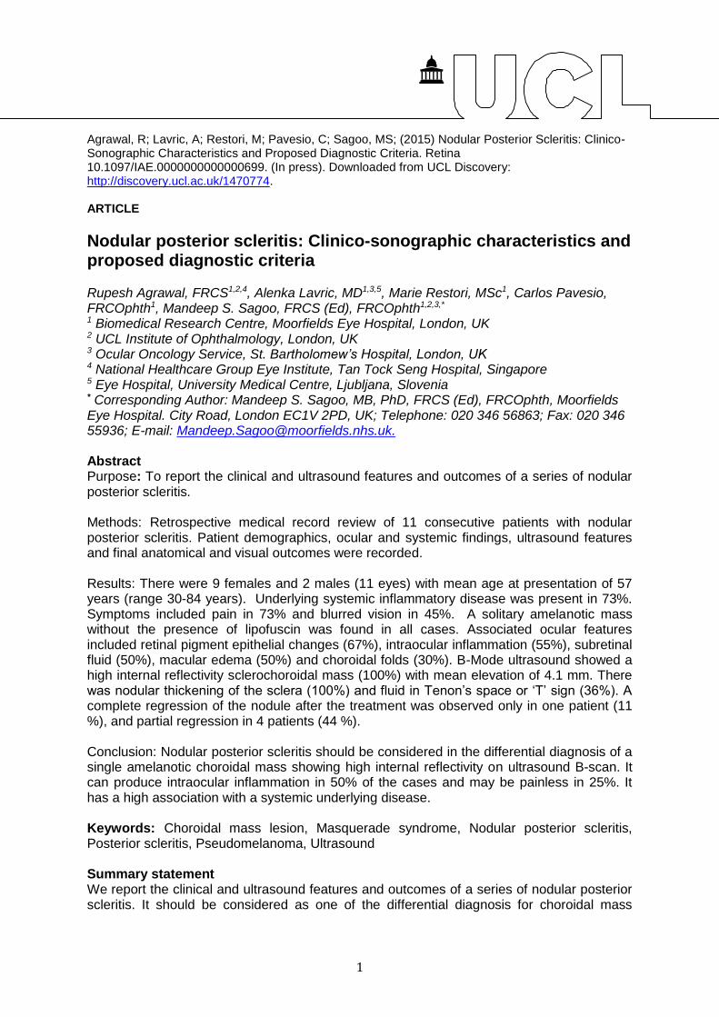
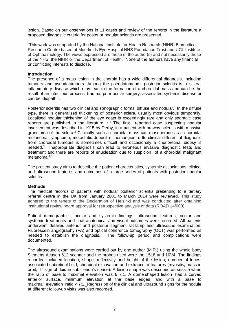
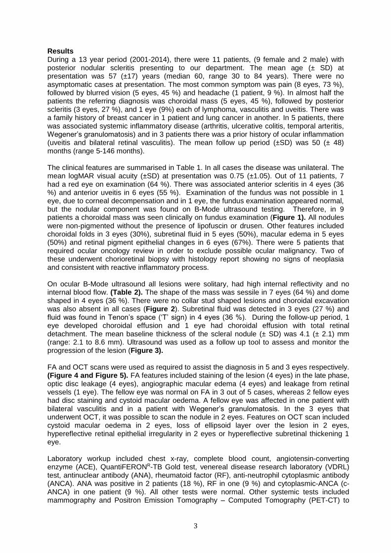
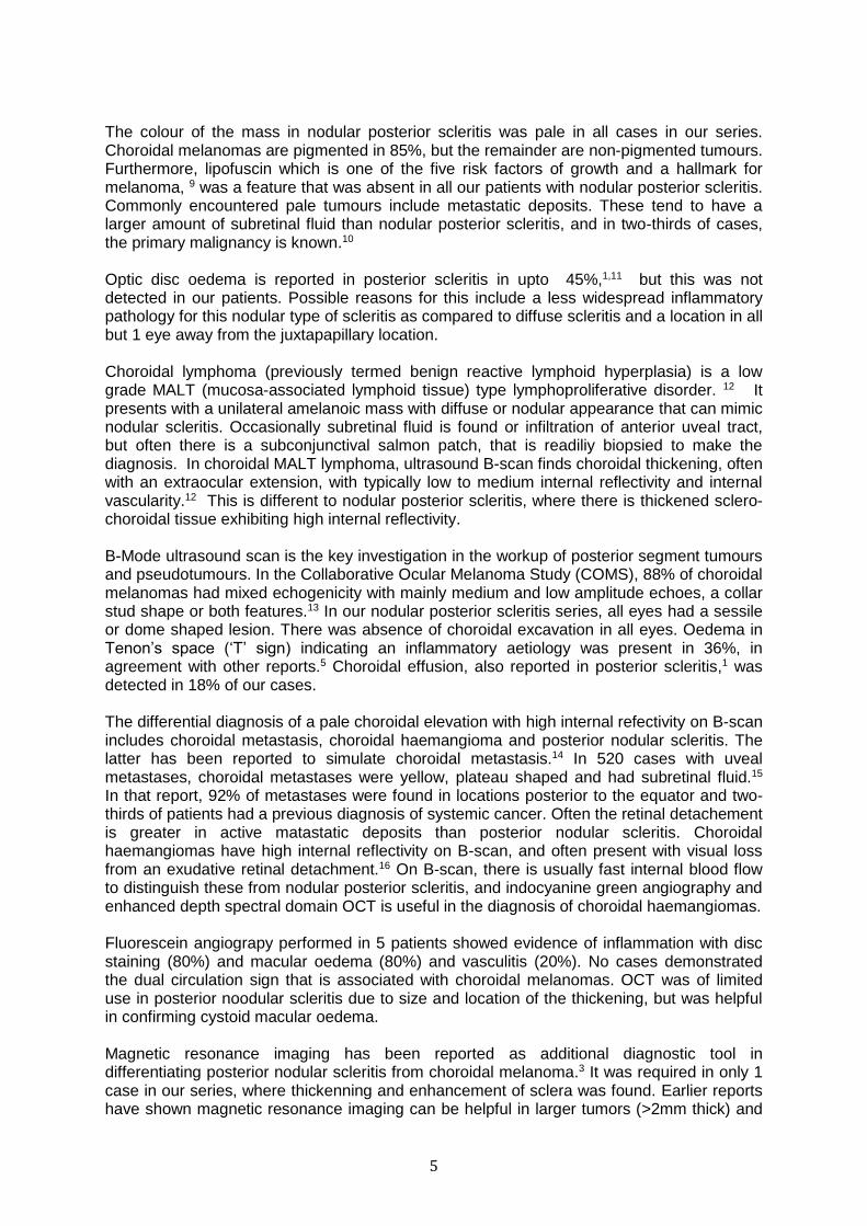
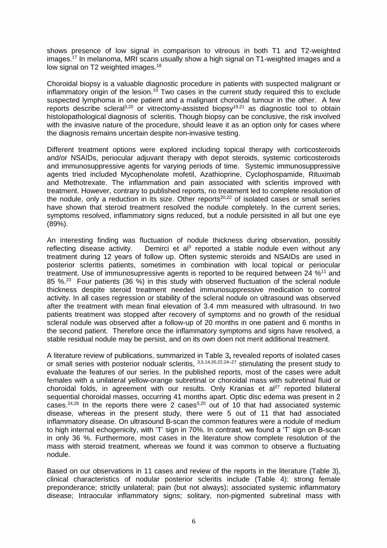
Figure legends Figure 1: Color fundus photos of choroidal lesions. Fig 1A and 1B shows elevated solitary subretinal lesion. Fig 1C shows solitary irregular greyish white well defined lesion at subretinal level with no pigmentation and no lipofuscin. Fig 1D: presence of elevated amleanotic choroidal lesion. Figure 2: Ultrasonographic chracteristics of the nodular posterior scleritis and adjacent normal ocular coats. 2A: Transverse B-Mode image of nasal scleral nodule (thickness: 3.5mm). 2B: Transverse B-Mode image of nasal scleral nodule (thickness: 6.6 mm). 2C: Transverse B-Mode image of inferior scleral nodule (thickness: 7.3mm). 2D: same eye as Fig 2C in longitudinal B-Mode image showing inferior nodule (cursors). Figure 3: Representative follow up ultrasound images of two of the eleven patients with posterior nodular scleritis. 3A: (a-d) Nasal nodular scleritis: Transverse B-Mode images showing reduction in thickness of nodule over time. Thickness - (a): July 2011 - 7.5mm, (b): September 2011-3.2mm, (c): January 2012 - 2.6mm, (d) October 2012 - 2.3mm. 3B: (a-d): Inferior nodular scleritis: Longitudinal B-Mode images taken on same eye with inferior gaze. Images taken at different time period over a 4 year period. Thickness - (a) 2008: 7.5mm (b) 2009: 5.4 mm, (c) 2010: 6.9mm, (d) 2011: 5.3mm
9
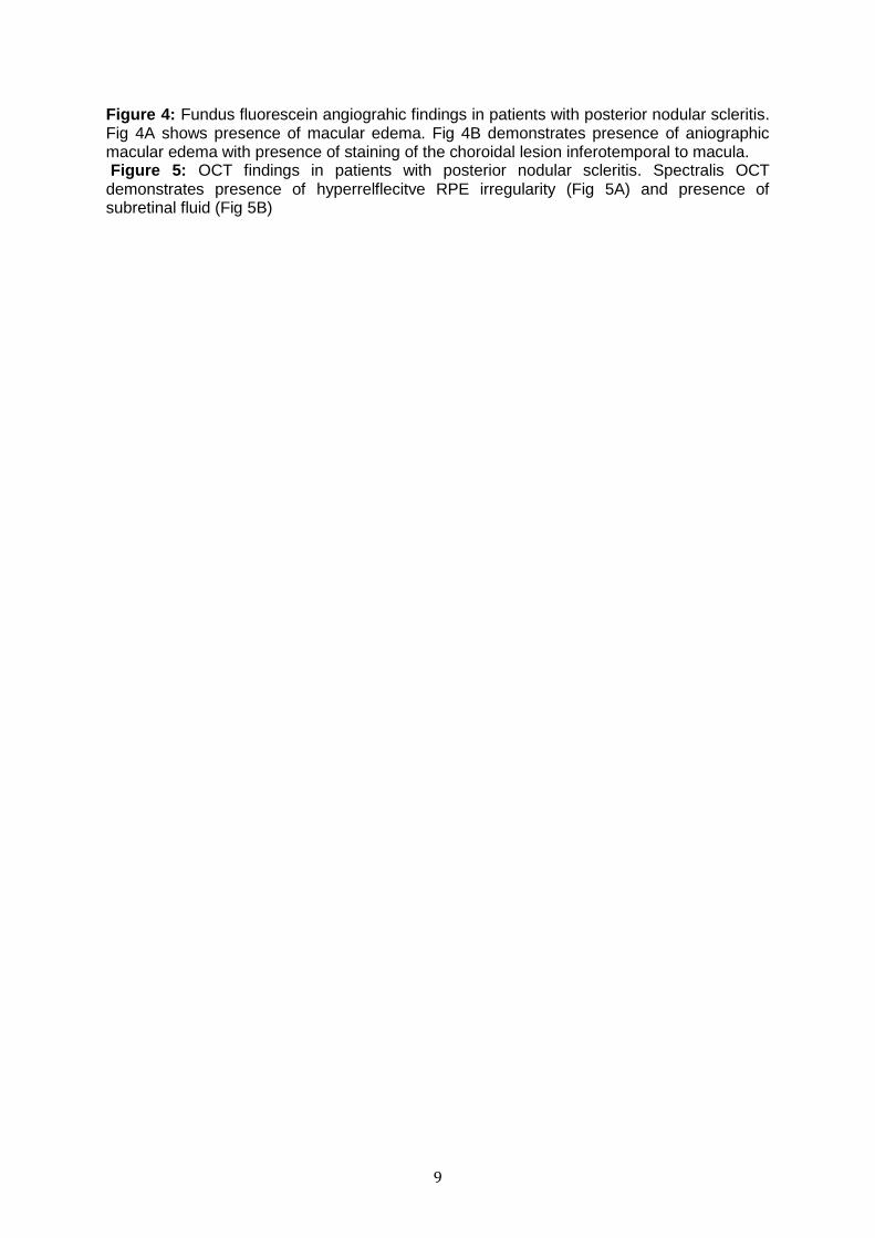
Figure 4: Fundus fluorescein angiograhic findings in patients with posterior nodular scleritis. Fig 4A shows presence of macular edema. Fig 4B demonstrates presence of aniographic macular edema with presence of staining of the choroidal lesion inferotemporal to macula. Figure 5: OCT findings in patients with posterior nodular scleritis. Spectralis OCT demonstrates presence of hyperrelflecitve RPE irregularity (Fig 5A) and presence of subretinal fluid (Fig 5B)

10
Tables Table 1. Clinical features and systemic investigations
Feature Number of patients (%)
Laterality
Unilateral 11 (100)
Initial visual acuity
6/6 (20/20) 4 (36)
6/7.5 - 6/12 (20/25 – 20/40) 1 (9)
6/15 - 6/48 (20/50 – 20/160) 3 (27)
6/60 (20/200) or worse 3 (27)
Anterior segment findings (n = 11 patients)
Proptosis, reduced motility 0
Conjunctival injection, chemosis 7 (64)
Peripheral keratitis 0
Corneal decompensation 1 (9)
Flare 3 (27)
Anterior scleritis 4 (36)
Anterior uveitis 6 (54)
Glaucoma, ocular hypertension 2 (18)
Posterior segment findings (n = 10 patients)
Vitreous cells 4 (40)
Choroidal folds, retinal striae 3 (30)
Annular choroidal detachment 1* (10)
Retinal detacment
Total 1* (10)
Subtotal 0
No RD 9 (90)
Non-RD subretinal fluid
Clinical 4 (40)
On OCT 1 (10)
None 5 (50)
Macular oedema 5 (50)
Macular star 0
Optic nerve swelling 0
Vasculitis 1 (10)
Choroidal mass features ( n = 9 patients)†
Location
Inferotemporal 4 (36)
Inferior 2 (18)
Superonasal 3 (27)
Inferonasal 2 (18)
Colour
Nonpigmented 9 (100)
Pigmented 0
Position of apex
Macular 0
Juxtapapillary 1 (11)
Equatorial 8 (89)
Lipofuscin 0
Drusen 0
RPE changes 6 (67)
11
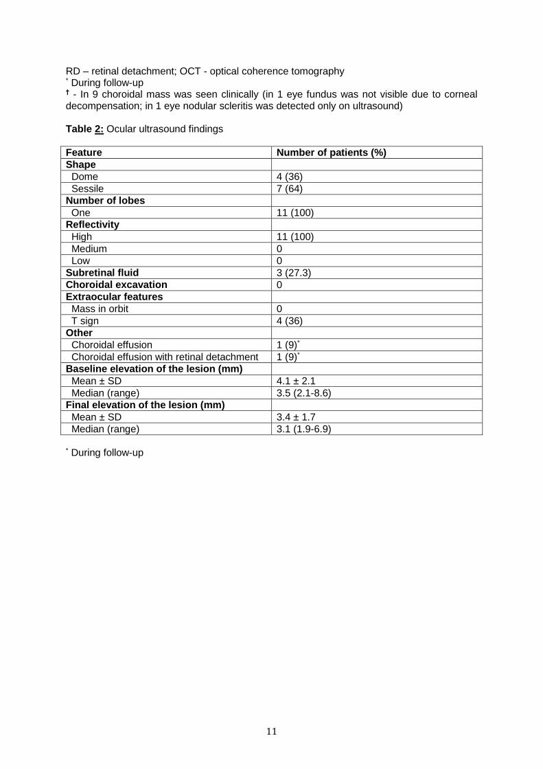
RD – retinal detachment; OCT - optical coherence tomography * During follow-up † - In 9 choroidal mass was seen clinically (in 1 eye fundus was not visible due to corneal decompensation; in 1 eye nodular scleritis was detected only on ultrasound) Table 2: Ocular ultrasound findings
Feature Number of patients (%)
Shape
Dome 4 (36)
Sessile 7 (64)
Number of lobes
One 11 (100)
Reflectivity
High 11 (100)
Medium 0
Low 0
Subretinal fluid 3 (27.3)
Choroidal excavation 0
Extraocular features
Mass in orbit 0
T sign 4 (36)
Other
Choroidal effusion 1 (9)*
Choroidal effusion with retinal detachment 1 (9)*
Baseline elevation of the lesion (mm)
Mean ± SD 4.1 ± 2.1
Median (range) 3.5 (2.1-8.6)
Final elevation of the lesion (mm)
Mean ± SD 3.4 ± 1.7
Median (range) 3.1 (1.9-6.9)
* During follow-up
12
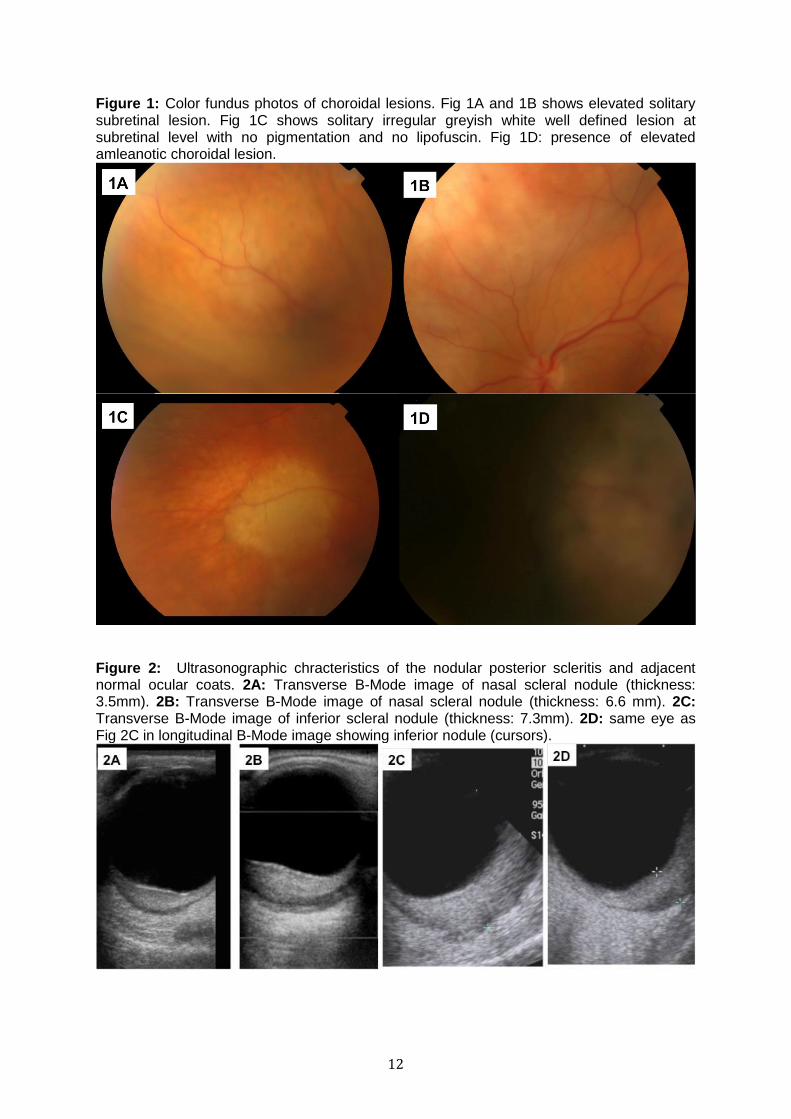
Figure 1: Color fundus photos of choroidal lesions. Fig 1A and 1B shows elevated solitary subretinal lesion. Fig 1C shows solitary irregular greyish white well defined lesion at subretinal level with no pigmentation and no lipofuscin. Fig 1D: presence of elevated amleanotic choroidal lesion.
Figure 2: Ultrasonographic chracteristics of the nodular posterior scleritis and adjacent normal ocular coats. 2A: Transverse B-Mode image of nasal scleral nodule (thickness: 3.5mm). 2B: Transverse B-Mode image of nasal scleral nodule (thickness: 6.6 mm). 2C: Transverse B-Mode image of inferior scleral nodule (thickness: 7.3mm). 2D: same eye as Fig 2C in longitudinal B-Mode image showing inferior nodule (cursors).
13
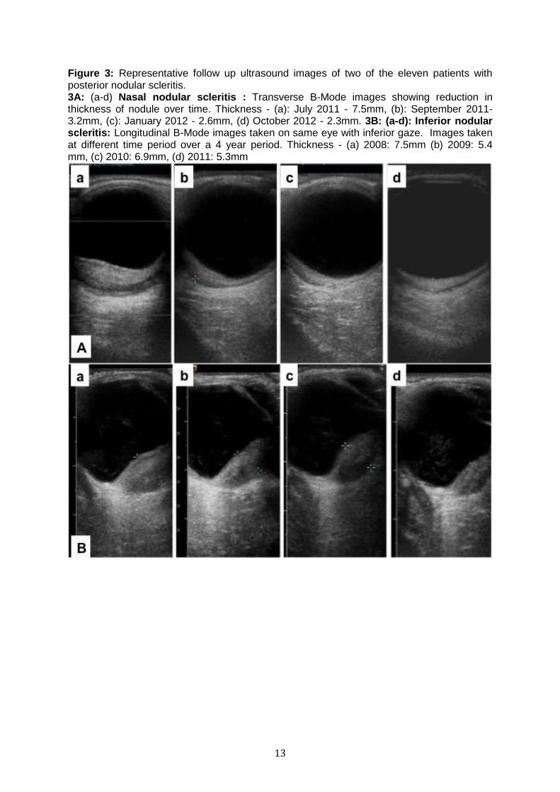
Figure 3: Representative follow up ultrasound images of two of the eleven patients with posterior nodular scleritis. 3A: (a-d) Nasal nodular scleritis : Transverse B-Mode images showing reduction in thickness of nodule over time. Thickness - (a): July 2011 - 7.5mm, (b): September 2011-3.2mm, (c): January 2012 - 2.6mm, (d) October 2012 - 2.3mm. 3B: (a-d): Inferior nodular scleritis: Longitudinal B-Mode images taken on same eye with inferior gaze. Images taken at different time period over a 4 year period. Thickness - (a) 2008: 7.5mm (b) 2009: 5.4 mm, (c) 2010: 6.9mm, (d) 2011: 5.3mm
14
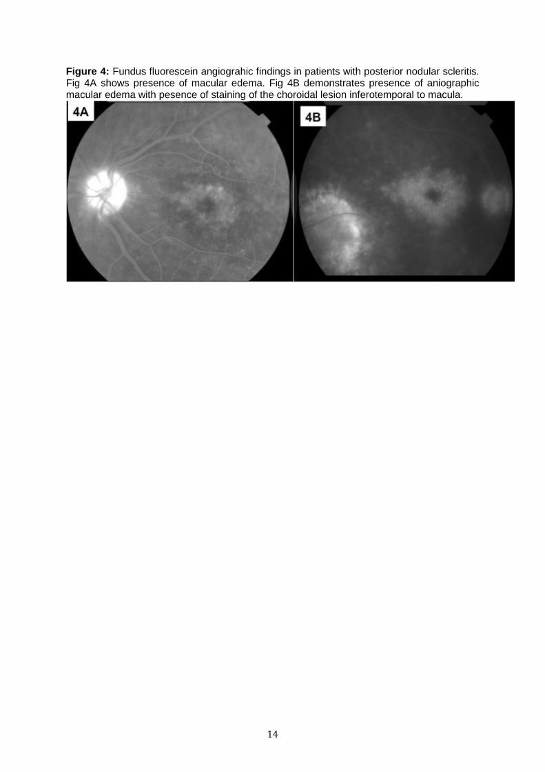
Figure 4: Fundus fluorescein angiograhic findings in patients with posterior nodular scleritis. Fig 4A shows presence of macular edema. Fig 4B demonstrates presence of aniographic macular edema with pesence of staining of the choroidal lesion inferotemporal to macula.
15
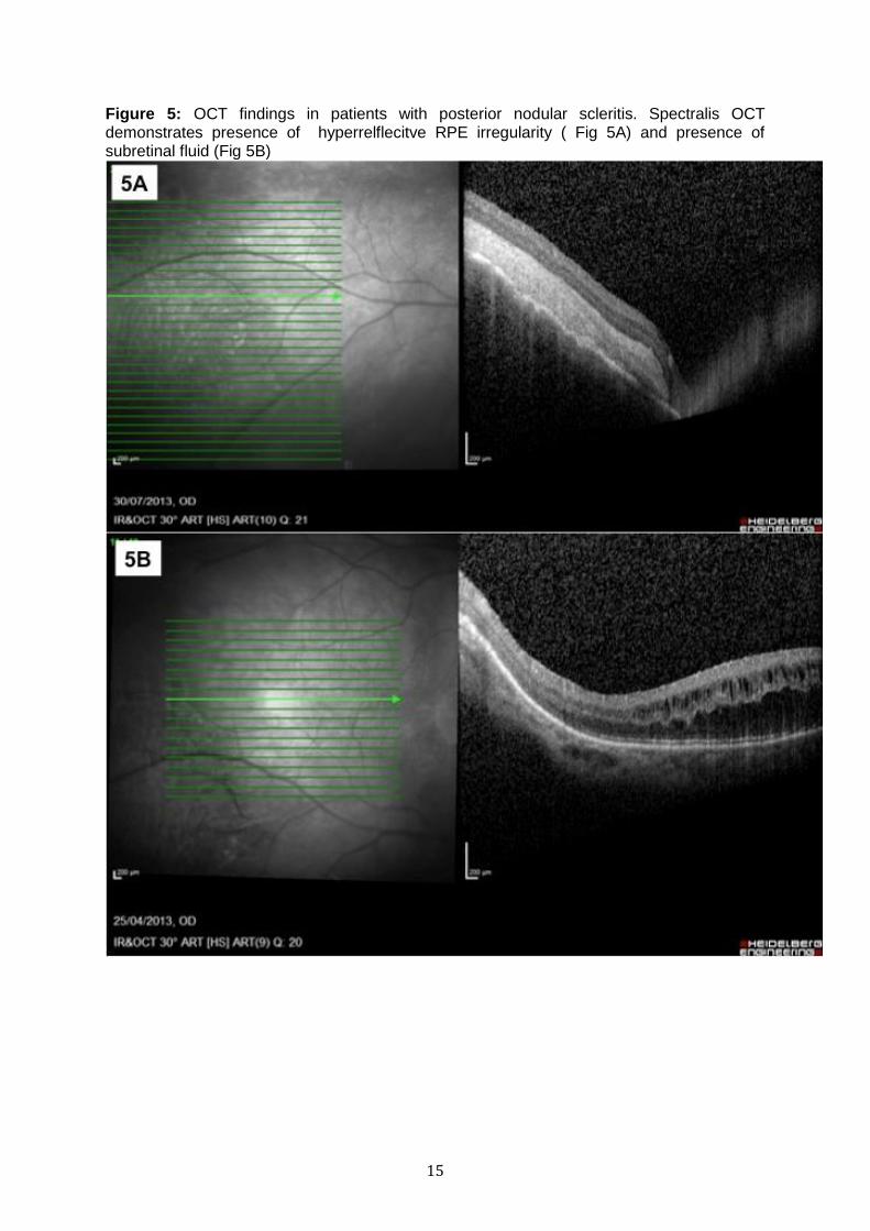
Figure 5: OCT findings in patients with posterior nodular scleritis. Spectralis OCT demonstrates presence of hyperrelflecitve RPE irregularity ( Fig 5A) and presence of subretinal fluid (Fig 5B)
16
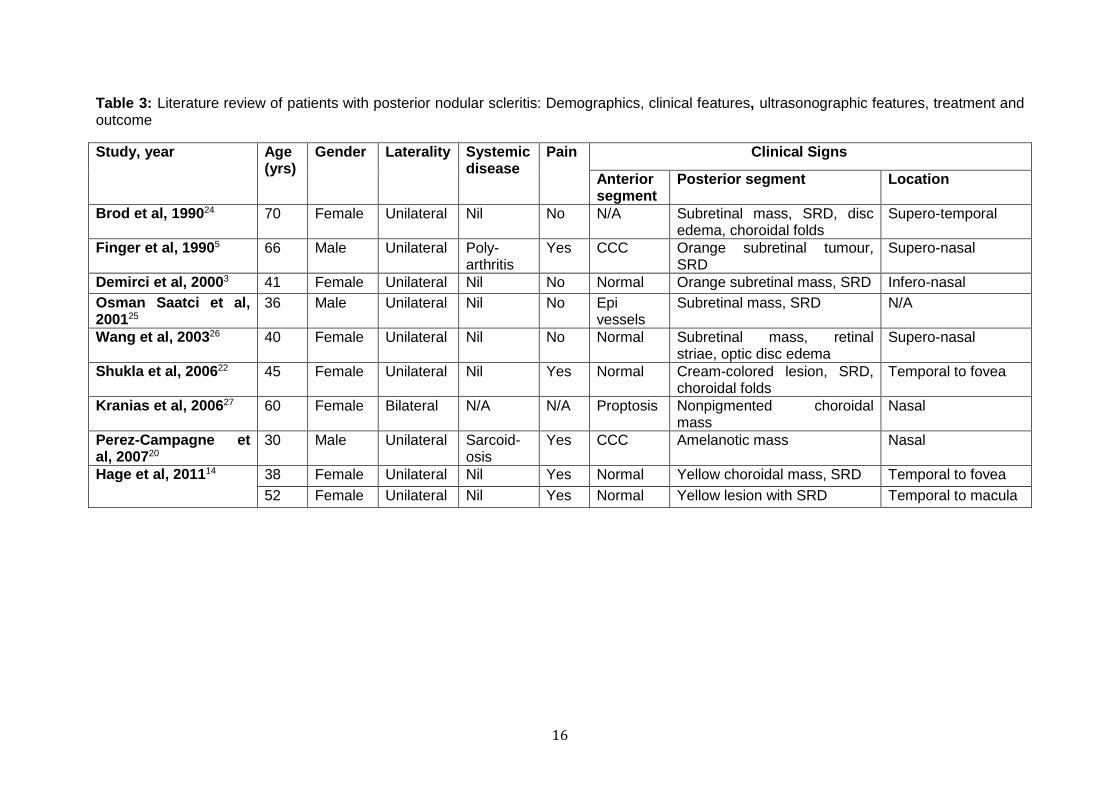
Table 3: Literature review of patients with posterior nodular scleritis: Demographics, clinical features, ultrasonographic features, treatment and outcome
Study, year Age (yrs)
Gender Laterality Systemic disease
Pain Clinical Signs
Anterior segment
Posterior segment Location
Brod et al, 199024 70 Female Unilateral Nil No N/A Subretinal mass, SRD, disc edema, choroidal folds
Supero-temporal
Finger et al, 19905 66 Male Unilateral Poly-arthritis
Yes CCC Orange subretinal tumour, SRD
Supero-nasal
Demirci et al, 20003 41 Female Unilateral Nil No Normal Orange subretinal mass, SRD Infero-nasal
Osman Saatci et al, 200125
36 Male Unilateral Nil No Epi vessels
Subretinal mass, SRD N/A
Wang et al, 200326 40 Female Unilateral Nil No Normal Subretinal mass, retinal striae, optic disc edema
Supero-nasal
Shukla et al, 200622 45 Female Unilateral Nil Yes Normal Cream-colored lesion, SRD, choroidal folds
Temporal to fovea
Kranias et al, 200627 60 Female Bilateral N/A N/A Proptosis Nonpigmented choroidal mass
Nasal
Perez-Campagne et al, 200720
30 Male Unilateral Sarcoid-osis
Yes CCC Amelanotic mass Nasal
Hage et al, 201114 38 Female Unilateral Nil Yes Normal Yellow choroidal mass, SRD Temporal to fovea
52 Female Unilateral Nil Yes Normal Yellow lesion with SRD Temporal to macula

17
Study, year Ultrasonographic characteristics Treatment Follow-up Response to treatment - Lesion Thickness
(mm) Shape Internal
echogenicity T- sign
Brod et al, 199024 4 N/A Moderate Yes Oral steroids 4 months Resolution
Finger et al, 19905 8.3 Multinodular High Yes Enucleation
Demirci et al, 20003 7.5 Dome-shaped High yes Nil 12 years No change
Osman Saatci et al, 200125 N/A N/A Moderate Yes Oral steroids 1 year Resolution
Wang et al, 200326 4.97 Dome-shaped High Yes Oral steroids 7 months Resolution
Shukala et al, 200622 4.5 Dome-shaped Moderate Yes IV steroids 15 months Resolution
Kranias et al, 200627 N/A Bi-convex Medium N/A Oral steroids N/A
Perez-Campagne et al, 200720 5 Nodular N/A Yes Oral steroids 6 months Resolution
Hage et al., 201114 4.6 Dome-shaped N/A N/A Oral steroids 2 years Resolution
N/A Dome-shaped Topical NSAID
5 months Resolution
N/A – information not available, SRD- serous retinal detachment, CCC- circum corneal congestion, IV – intravenous, NSAID- Non steroidal anti inflammatory drug Table 4: Clinical characteristics of posterior nodular scleritis
Gender Female preponderance
Symptoms Periocular Pain and blurred vision
Systemic disease Predominantly associated with systemic inflammatory disorder
Laterality Unilateral
Location More commonly - Inferotemporal quadrant around equator, can be present in any quadrant
Associated ocular signs
Anterior scleritis, and conjunctival chemosis
Ultrasound characteristics
Nodular swelling ,‘T’ sign and subretinal fluid
Treatment response
Nodular swelling gets smaller but residual swelling often persists but can disappear completely


![A case of scleritis associated rheumatoid arthritis ...syphilis, caused by nodular infectious uveitis [9, 10]. Biswas et al. reported a case of tuberculous uveitis associated with](https://img.pdfslide.net/doc/110x75/60997293e4fd5e2ef7072fd8/a-case-of-scleritis-associated-rheumatoid-arthritis-syphilis-caused-by-nodular.jpg)


![Understanding the relationship between diabetes, retinopathy, …Paper] uveitis scleritis and glycaemia.pdf · diffuse/nodular). It has been shown to cause vision loss (a permanent](https://img.pdfslide.net/doc/110x75/5ca0773788c99350178d3ca3/understanding-the-relationship-between-diabetes-retinopathy-paper-uveitis-scleritis.jpg)