Embed Size (px)
Citation preview

Non-coplanar trajectories to improve organ at risk sparing in volumetric modulated arc
therapy for primary brain tumours
Gregory Smyth 1, Philip M. Evans 2, Jeffrey C. Bamber 1, Henry C. Mandeville 3, Liam C.
Welsh 3, Frank H. Saran 3, James L. Bedford 1
1 Joint Department of Physics at The Institute of Cancer Research and The Royal Marsden
NHS Foundation Trust, London, SM2 5PT, United Kingdom.
2 Centre for Vision, Speech and Signal Processing, University of Surrey, Guildford, Surrey,
GU2 7HX, United Kingdom.
3 The Royal Marsden NHS Foundation Trust, London, SM2 5PT, United Kingdom.
Corresponding author: Gregory Smyth, Joint Department of Physics, The Institute of
Cancer Research and The Royal Marsden NHS Foundation Trust, Downs Road, Sutton,
London, SM2 5PT, United Kingdom. Email: [email protected] Tel: +44 20 8661 3472
Fax: +44 20 8643 3812
27 pages, 1 table, 5 figures
Running head: Non-coplanar trajectories for brain VMAT
Keywords: VMAT, non-coplanar, trajectory, optimization, treatment planning, hippocampal
sparing

Smyth et al. Non-coplanar trajectories for brain VMAT 2
Abstract
Background and purpose: To evaluate non-coplanar volumetric modulated arc radiotherapy
(VMAT) trajectories for organ at risk (OAR) sparing in primary brain tumour radiotherapy.
Materials and methods: Fifteen patients were planned using coplanar VMAT and compared
against non-coplanar VMAT plans for three trajectory optimization techniques. A geometric
heuristic technique (GH) combined beam scoring and Dijkstra's algorithm to minimize the
importance-weighted sum of OAR volumes irradiated. Fluence optimization was used to
perform a local search around coplanar and GH trajectories, producing fluence-based local
search (FBLS) and FBLS+GH trajectories respectively.
Results: GH, FBLS, and FBLS+GH trajectories reduced doses to the contralateral globe,
optic nerve, hippocampus, temporal lobe, and cochlea. However, FBLS increased dose to the
ipsilateral lens, optic nerve and globe. Compared to GH, FBLS+GH increased dose to the
ipsilateral temporal lobe and hippocampus, contralateral optics, and the brainstem and body.
GH and FBLS+GH trajectories reduced bilateral hippocampi normal tissue complication
probability (p = 0.028 and p = 0.043, respectively). All techniques reduced PTV conformity;
GH and FBLS+GH trajectories reduced homogeneity but less so for FBLS+GH.
Conclusions: The geometric heuristic technique best spared OARs and reduced normal tissue
complication probability, however incorporating fluence information into non-coplanar
trajectory optimization maintained PTV homogeneity.

Smyth et al. Non-coplanar trajectories for brain VMAT 3
Introduction
Sparing organs at risk (OAR) in intracranial radiotherapy reduces the risk of side effects that
affect quality of life, such as cranial and optic neuropathy, hearing loss, and neurocognitive
impairment [1-6]. Using non-coplanar beam orientations has been shown to improve OAR
dosimetry in conformal [7], intensity modulated (IMRT) [8], and volumetric modulated arc
(VMAT) [9] radiation therapy. However, non-coplanar geometries are fixed during delivery
for a given beam, limiting their application to VMAT. New linear accelerators can perform
dynamic couch rotation during beam delivery, making possible non-coplanar VMAT
trajectories that use more of the 4π space around the patient [10-12] and enabling potential
additional reductions in normal tissue complication probability (NTCP).
Early research into the clinical benefit of non-coplanar VMAT mainly focused on planner-
defined trajectories [13-15], while recent work has investigated trajectory optimization
techniques [10, 16-19]. Published optimization techniques have used one of two approaches:
geometric heuristics or fluence optimization. Geometric heuristics score individual beam
orientations and determine trajectories that minimize the overall score [10, 16, 17]. Fluence-
based techniques identify a smaller group of optimal candidate beam orientations, which are
then connected via intermediate paths [18, 19]. Geometric heuristics are appealing due to the
computational complexity of a full fluence search for a VMAT arc but lack the dosimetric
information that can be utilized in fluence optimization.
Smyth et al. Non-coplanar trajectories for brain VMAT 4
This paper proposes and evaluates three different trajectory optimization techniques - a
geometric heuristic technique and two incorporating fluence optimization - for primary brain
tumour radiotherapy using non-coplanar VMAT. We aim to answer three questions:
1. Does a geometric heuristic technique improve OAR sparing over coplanar VMAT?
2. Does a fluence-based local search technique improve OAR sparing over coplanar VMAT?
3. Is there a synergistic effect if the geometric heuristic and fluence-based local search
techniques are combined?
This work quantifies the clinical effect of new techniques for optimizing non-coplanar
VMAT and aims to widen the therapeutic window of radiotherapy for primary brain tumours.
We demonstrate that a less computationally intense geometric heuristic technique is sufficient
to produce high quality plans. Our goal is to facilitate the introduction of non-coplanar
VMAT into neuro-oncology clinical practice.
Materials and Methods
Patient selection and treatment planning
Fifteen patients treated with radiotherapy for primary brain tumours were planned using
VMAT. Mean and standard deviation planning target volume (PTV) size was 336.6 ± 214.1
cc (range 5.5 – 723.6 cc), with a CTV-PTV margin of 3 mm in all cases. Original PTV
prescription doses were 60 Gy in 2 Gy fractions, and 54 Gy or 59.4 Gy in 1.8 Gy fractions.
One patient had palliative treatment (30 Gy in 6 Gy fractions) but was replanned to an
appropriate radical dose (60 Gy in 2 Gy fractions) for this study. Further information for

Smyth et al. Non-coplanar trajectories for brain VMAT 5
each patient case is contained in Supplementary Table A1. Coplanar and non-coplanar
radiotherapy plans were produced for a 6 MV Elekta Synergy linear accelerator (Elekta AB,
Stockholm, Sweden) with Agility multi-leaf collimator [20]. Coplanar VMAT planning used
our standard clinical technique of a single arc with 180 control points, however to avoid bias
due to the additional degrees of freedom available to non-coplanar methods, dual arc coplanar
plans with 360 control points were also produced.
Plans were optimized using an in-house VMAT planning system [21, 22] (AutoBeam v5.5a),
adapted to import complex couch trajectories [16]. The planning process is summarized here,
with the detailed workflow included in Supplementary Figure A1. AutoBeam performed
fluence optimization at each control point before sequencing the fluence maps into
deliverable connected VMAT apertures. As sequencing degraded the dose distribution, direct
aperture optimization was performed subject to machine limits for VMAT delivery. Further
detail on AutoBeam and the optimization techniques used at each stage can be found
elsewhere [21, 22].
All cases used the same optimization objectives (Supplementary Table A2) to ensure a fair
comparison. AutoBeam plans were reconstructed in Pinnacle3 (Pinnacle3 v9.8, Philips
Medical, Madison, WI) for final dose calculation in line with clinical practice. Dose was
prescribed to the PTV mean value and calculated on a 2.5 x 2.5 x 2.5 mm3 resolution dose
grid using the Adaptive Convolve algorithm.
Trajectory optimization
Smyth et al. Non-coplanar trajectories for brain VMAT 6
Three non-coplanar VMAT trajectory optimization techniques were developed in MATLAB
(R2010b, The MathWorks, Natick, MA): a geometric heuristic technique (GH), a fluence-
based local search technique (FBLS), and the combination of GH and FBLS (FBLS+GH).
Organs at risk used in trajectory optimization were the brainstem, globes, optic nerves, optic
chiasm, lenses, hippocampi, temporal lobes, cochleae, and the volume of brain excluding the
PTV and other OARs. A patient voxel size of 5 x 5 x 5 mm3 was used during trajectory
optimization. For ray tracing, a beam aperture was defined as the projection of the PTV onto
the isocentre plane and rays were cast through the centre of 2.5 x 2.5 mm2 beam elements. A
2 mm margin was applied to the optic nerves, lenses, optic chiasm, and cochleae during
trajectory optimization to prevent small OARs being missed in this step.
Geometric heuristic technique
The geometric heuristic technique (Figure 1(a)) is an extension of the algorithm described in
[16]; further detail is provided in Supplementary Figure A1. Ray tracing was performed
through the patient to determine a cost based on OAR geometry for all achievable isocentric
beam orientations (Figure 1(a), step 1). The trajectory optimization was formulated as a
graph search problem, with the cost for a given beam orientation being the penalty applied for
adding that orientation to the VMAT trajectory, and solved using Dijkstra’s least-cost path
algorithm [23] (Figure 1(a), step 2). Single arc trajectories were produced through 358° of
gantry rotation, from 179° to 181°, with control points spaced every 2° of gantry or couch
rotation. Sections of trajectory with continuous couch rotation but static gantry rotation were
allowed, provided the overall trajectory cost was minimised.

Smyth et al. Non-coplanar trajectories for brain VMAT 7
For this study the technique was extended to incorporate multiple OARs of different relative
importance and prevent large or less important OARs from dominating the cost for a given
beam orientation, a limitation of the previous method [16]. The cost, C, for each orientation
is given by the sum of the relative volumes of each OAR intersected during ray tracing,
weighted by their relative importance (Equation 1).
Eq. 1
where an OAR, v, from all clinically important OARs, V, with relative importance i, has n of
N voxels intersected by rays cast from a beam orientation with couch angle, c, and gantry
angle, g. The importance factors, i, were those used during plan optimization
(Supplementary Table A2) and chosen based on relative clinical priority.
Fluence-based local search
One limitation of GH is that individual beam orientation costs, and therefore trajectories, do
not evaluate the effect of fluence modulation around the arc. In some cases it may be
beneficial to deliver dose to the PTV from beam angles which irradiate through an OAR,
provided modulation is used to reduce the fluence directed at the OAR e.g. through the
contralateral optics. GH would overlook these high cost beam orientations even if they might
be included in a dosimetrically optimal trajectory. FBLS was developed to investigate the
effect on plan dosimetry of local modifications, based on fluence modulation, to a supplied
trajectory.
FBLS was applied to a coplanar VMAT trajectory to determine if it alone could significantly
improve dosimetry. FBLS was also applied to a GH trajectory to investigate nearby
∑∈
=Vv v
vvgc NniC ,
Smyth et al. Non-coplanar trajectories for brain VMAT 8
trajectories that, although not optimal in terms of geometric avoidance over the whole arc,
might improve overall plan dosimetry.
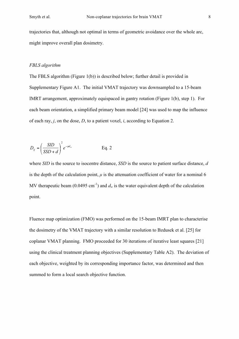
FBLS algorithm
The FBLS algorithm (Figure 1(b)) is described below; further detail is provided in
Supplementary Figure A1. The initial VMAT trajectory was downsampled to a 15-beam
IMRT arrangement, approximately equispaced in gantry rotation (Figure 1(b), step 1). For
each beam orientation, a simplified primary beam model [24] was used to map the influence
of each ray, j, on the dose, D, to a patient voxel, i, according to Equation 2.
Eq. 2
where SID is the source to isocentre distance, SSD is the source to patient surface distance, d
is the depth of the calculation point, µ is the attenuation coefficient of water for a nominal 6
MV therapeutic beam (0.0495 cm-1) and dw is the water equivalent depth of the calculation
point.
Fluence map optimization (FMO) was performed on the 15-beam IMRT plan to characterise
the dosimetry of the VMAT trajectory with a similar resolution to Bzdusek et al. [25] for
coplanar VMAT planning. FMO proceeded for 30 iterations of iterative least squares [21]
using the clinical treatment planning objectives (Supplementary Table A2). The deviation of
each objective, weighted by its corresponding importance factor, was determined and then
summed to form a local search objective function.
wdij e
dSSDSIDD µ−⎟
⎠
⎞⎜⎝
⎛+
=2

Smyth et al. Non-coplanar trajectories for brain VMAT 9
The couch rotation of the first beam orientation was perturbed by a step size of ±10°, with
new FMO performed, and the change that most improved the objective function was accepted
(Figure 1(b), step 2). For each beam in turn, repeated perturbations were performed and
accepted until there was no absolute improvement in the objective function from adjusting
the current beam (Figure 1(b), step 3). The perturbation step size was then reduced
incrementally from 10° to 2° and the perturbation stage repeated for each beam at its new
couch angle (Figure 1(b), step 4). The set of all new beam orientations was then incorporated
into the original trajectory using MATLAB’s piecewise cubic hermite polynomial
interpolation [26]. Checks were performed to ensure the interpolated trajectory avoided
collision regions and did not extend beyond the initial arc start and stop gantry angles.
Finally, the trajectory was resampled to maintain the same number of control points as the
input trajectory (Figure 1(b), step 5).
Plan evaluation
Dose statistics were compared for all plans, with OAR doses judged against relevant
QUANTEC constraints [1]. Dose-volume statistics linked to cognitive performance, V10Gy
and V40Gy for the hippocampi, and V40Gy and V60Gy for the temporal lobes [27], were also
compared. The probability of radiation induced cognitive impairment, as measured by the
Wechsler Memory Scale-III Word List Delayed Recall (WMS-III WL-DR) test, was
calculated from the equivalent dose in 2 Gy fractions to 40% of the bilateral hippocampi
(EQD2 40%) according to the NTCP model proposed by Gondi et al. [6]. All plans were
compared against monitor units required, homogeneity index [28] (HI):
!" = !!%!!!"%!!"%
×100 Eq. 3
Smyth et al. Non-coplanar trajectories for brain VMAT 10
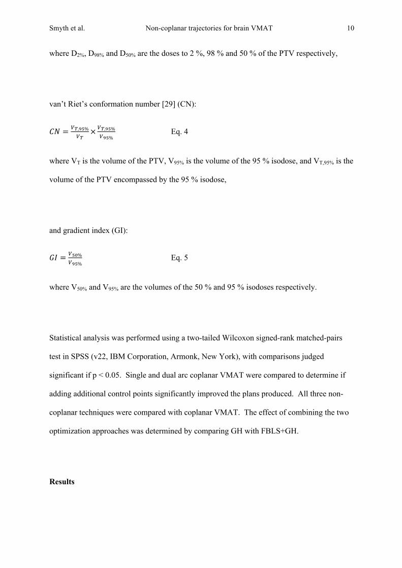
where D2%, D98% and D50% are the doses to 2 %, 98 % and 50 % of the PTV respectively,
van’t Riet’s conformation number [29] (CN):
!" = !!,!"%!!
× !!,!"%!!"%
Eq. 4
where VT is the volume of the PTV, V95% is the volume of the 95 % isodose, and VT,95% is the
volume of the PTV encompassed by the 95 % isodose,
and gradient index (GI):
!" = !!"%!!"%
Eq. 5
where V50% and V95% are the volumes of the 50 % and 95 % isodoses respectively.
Statistical analysis was performed using a two-tailed Wilcoxon signed-rank matched-pairs
test in SPSS (v22, IBM Corporation, Armonk, New York), with comparisons judged
significant if p < 0.05. Single and dual arc coplanar VMAT were compared to determine if
adding additional control points significantly improved the plans produced. All three non-
coplanar techniques were compared with coplanar VMAT. The effect of combining the two
optimization approaches was determined by comparing GH with FBLS+GH.
Results

Smyth et al. Non-coplanar trajectories for brain VMAT 11
Dual arc coplanar plans were not significantly different from single arc coplanar for most
metrics studied (Supplementary Table A3). However, the contralateral optic nerve,
hippocampus and temporal lobe, and body excluding PTV dose statistics and gradient index
showed modest improvements. In all cases where metrics were improved by dual arc
planning, statistical significance tests against non-coplanar VMAT were unaffected by the
number of coplanar arcs. As the number of control points used for single arc coplanar plans
(180) was similar to non-coplanar plans (median 180, range 180 – 202 for GH and
FBLS+GH; 180 for FBLS), remaining comparisons are against single arc coplanar VMAT
only.
Coronal sections through all plans for a representative patient case are presented in Figure 2.
Note how the orientation of the isodose levels in the non-coplanar plans differs from the
coplanar case to avoid the OARs, particularly for the 50 % and 20 % isodoses. Trajectories
for all plans for one patient case are shown in Figure 3 overlaid on the cost map of the
geometric heuristic technique. Regions of the cost map with high cost indicate beam
orientations where a large proportion of clinically important organs at risk would be
irradiated by a beam aperture conforming to the projection of the PTV. Low cost regions
indicate beam orientations where no OARs, or a small proportion of low importance OARs,
would be irradiated. Potential collision regions, estimated with a volunteer lying on the
treatment couch, are shown in white.
Mean relative dose or volume deviations of clinically relevant OAR statistics from the
coplanar VMAT plan are presented in Figure 4, with negative values indicating reductions
and error bars indicating ±1 standard deviation. Complete results, including absolute dose
Smyth et al. Non-coplanar trajectories for brain VMAT 12
statistics and statistical comparisons, are presented in Table 1. All three non-coplanar
trajectory optimization techniques significantly reduced doses to the contralateral globe, optic
nerve, hippocampus (mean dose and V40Gy), temporal lobe (mean dose and V40Gy), and
cochlea. Additionally GH and FBLS+GH significantly reduced the contralateral lens dose
and contralateral hippocampus V10Gy; GH also reduced the mean brainstem dose. FBLS
significantly increased doses to the ipsilateral globe, optic nerve (mean), and lens. Compared
to GH, FBLS+GH significantly increased dose to the brainstem, contralateral optics,
ipsilateral hippocampus (mean dose and V40Gy), ipsilateral temporal lobe (mean), and body
excluding PTV.
PTV homogeneity index increased for GH (p = 0.004) and FBLS+GH (p = 0.013) plans
compared with coplanar VMAT. However, FBLS+GH improved homogeneity over GH (p =
0.009). PTV coverage was less conformal for GH (p = 0.033), FBLS (p = 0.002), and
FBLS+GH (p = 0.015) compared with coplanar VMAT. Gradient index was improved by
FBLS (p = 0.001) and FBLS+GH (p = 0.029) plans compared with coplanar; FBLS+GH
plans also improved over GH (p = 0.004). Non-coplanar plans required more monitor units
than coplanar VMAT (p = 0.001 for all techniques) but there was no difference between GH
and FBLS+GH.
Predicted clinical effect
Whole body D1cc to patients with 59.4 Gy and 60 Gy prescriptions exceeded 60 Gy for all
techniques (maximum 63.5 Gy), suggesting a risk of brain necrosis of 3-5 % [2]. Although
non-coplanar techniques showed small increases in D1cc, these are unlikely to result in

Smyth et al. Non-coplanar trajectories for brain VMAT 13
clinically significant differences in symptomatic necrosis risk. No patient received 59 Gy to
more than 10 cc of the brainstem, complying with QUANTEC constraints [1].
QUANTEC optic nerve or chiasm constraints (maximum dose < 55 Gy) were exceeded in
eight patients. Constraints were exceeded for the ipsilateral optic nerve in four patients, and
the contralateral optic nerve in one patient, for all coplanar and non-coplanar plans. For
patient 2, the ipsilateral optic nerve maximum dose increased to 55.3 Gy for GH from 54.8
Gy for coplanar VMAT, but reduced to 54.4 Gy with FBLS+GH. Seven patients exceeded
the optic chiasm constraint, of which five exceeded the constraint for all coplanar and non-
coplanar plans. For patient 4, the maximum chiasm dose increased to 56.6 Gy and 55.8 Gy
for GH and FBLS+GH respectively from the initial coplanar VMAT dose of 54.4 Gy. For
patient 6, the maximum chiasm dose was reduced to below the constraint for all non-coplanar
plans from the initial coplanar VMAT dose of 55.1 Gy. QUANTEC suggests the threshold
for optic neuropathy may be 59 Gy for non-pituitary tumours at these fraction sizes [4]. One
case with a prescription dose of 60 Gy (patient 8) exceeded 59 Gy to the ipsilateral optic
nerve and chiasm; this was breached for all plans including coplanar VMAT. None of these
changes in dose would be expected to significantly affect the likelihood of radiation induced
optic neuropathy.
There was no difference in the number of patients breaching the QUANTEC cochlear dose
constraints between techniques at either the standard 45 Gy or conservative 35 Gy levels,
corresponding to a 30 % chance of hearing loss. However, the significant reduction in
contralateral cochlear dose demonstrated by the non-coplanar techniques could reduce the
likelihood of hearing loss.

Smyth et al. Non-coplanar trajectories for brain VMAT 14
Dose volume histograms (DVH) of the contralateral hippocampus and temporal lobe for three
representative patient cases are shown in Figure 5. Contralateral hippocampus and temporal
lobe V40Gy were significantly reduced by all non-coplanar techniques, while contralateral
hippocampus V10Gy was reduced by GH and FBLS+GH. Median overlap of the bilateral
hippocampi with the PTV was 29.2 % (range 0 - 59.5 %), with six patients having an overlap
of more than 40 %. The probability of cognitive impairment for GH plans (mean ± 1 S.D.,
0.875 ± 0.263) was significantly reduced from coplanar VMAT (0.936 ± 0.183, p = 0.028).
FBLS+GH significantly increased cognitive impairment probability (0.898 ± 0.229, p =
0.028) over GH, but remained significantly reduced over coplanar VMAT (p = 0.043).
Discussion
This work evaluates three non-coplanar VMAT trajectory optimization techniques for a
cohort of primary brain tumour patients. Having performed a systematic comparison of these
techniques, alone and in combination, we can draw some specific conclusions regarding their
relative merits. FBLS achieved additional contralateral OAR sparing over coplanar VMAT
plan, while maintaining PTV homogeneity, but increased dose to ipsilateral OARs.
FBLS+GH maintained much of the OAR sparing of GH while recovering some lost PTV
dose homogeneity. The trade-off between PTV dose homogeneity and OAR sparing depends
on the planning objectives used, while the extent to which individual OARs are spared
depends on their relative importance for the specific clinical case. For this cohort, it is
important to maximize OAR sparing and therefore the geometric heuristic technique is
recommended for primary brain tumours.

Smyth et al. Non-coplanar trajectories for brain VMAT 15
Recent work has proposed different heuristic or fluence-based methods of trajectory
optimization [10, 16-19]. Fluence-based techniques solved non-coplanar IMRT beam
orientation problems for up to 20 beams but did not evaluate the dosimetry of the connecting
paths [18, 19]; therefore the final VMAT trajectories may not be globally optimal. FBLS
accepted only local changes that decreased the local search objective function and therefore
did not guarantee the altered VMAT trajectory was optimal. However, it did allow the
dosimetry of alternate trajectories to be investigated while maintaining the quality of the rest
of the connected trajectory. The complexity of FBLS was limited by using a simplified beam
model; further work to incorporate a clinical dose model and determine its effect on trajectory
optimization is planned.
More general issues regarding the potential clinical implementation of dynamic couch non-
coplanar VMAT have yet to be fully addressed. While modern linear accelerators can deliver
non-coplanar VMAT [10, 17], there has been no systematic investigation of its delivery
accuracy or efficiency. Although statistically significant, coplanar and non-coplanar monitor
units were sufficiently similar that we expect delivery efficiency to depend on couch rotation
speed. Potential differences in delivery efficiency between non-coplanar trajectories will
depend on gantry rotation, couch rotation, and dose rate limits for the specific machine.
Dynamic couch rotation requires extra quality assurance testing [30] and advanced collision
prediction and detection methods [31]. Patient rotation during treatment could introduce
intra-fractional motion, with the effect dependent on treatment site and couch trajectory.
Although additional immobilization is unlikely to be necessary for intracranial treatments,
this may be a significant issue for other body sites and requires investigation. An alternative

Smyth et al. Non-coplanar trajectories for brain VMAT 16
linac configuration capable of rotation around the vertical axis allows non-coplanar treatment
without patient movement and would address some of these problems [32, 33]. However, the
reduced range of rotation achievable [32] may limit the utility of this approach for
intracranial sites. The trajectory optimization techniques described in this work are
applicable to all delivery platforms, within machine limitations.
Non-coplanar VMAT demonstrated improved sparing of functionally important OARs,
notably significantly decreasing dose to the contralateral temporal lobe and hippocampus
(Figure 5). Neurocognitive decline has been linked to higher doses to the hippocampi [6, 27,
34, 35] and temporal lobes [27, 34, 36]. Redmond et al. found reductions in children’s motor
speed and dexterity were correlated with increased dose to the hippocampi and temporal
lobes, while visual perception decreased with increasing dose to the left temporal lobe [34].
Jalili et al. found children and young adults receiving greater than 43.2 Gy to 13 % of the left
temporal lobe were significantly more likely to demonstrate a reduction in intelligence
quotient (IQ) of 10 % or more [36]. The effect of hippocampal sparing in whole brain
radiotherapy is currently under active investigation in several clinical trials worldwide, and
has shown promising results in the RTOG 0933 phase 2 trial [35]. Gondi et al. modelled the
probability of impaired learned-word recall with increasing dose to the hippocampi for a
cohort of adult patients [6]. Gondi et al.’s NTCP model of cognitive injury due to
hippocampal dose results in a rather stringent hippocampal dose sparing requirement (D40% to
the bilateral hippocampi leading to a 50% probability of WMS-III WL-DR impairment
(EQD502) was estimated to be 14.88 Gy [95% CI, 12.86-17.06 Gy]) that may be difficult to
achieve in practice for patients treated for primary brain tumours where intra-cranial tumour
control is the priority. While the modelled hippocampal NTCP reductions achieved by non-
coplanar VMAT were modest, the absolute dose reductions achieved for the contralateral

Smyth et al. Non-coplanar trajectories for brain VMAT 17
temporal lobe and hippocampus (Figure 5) are likely to be clinically significant [27]. These
studies suggest that non-coplanar VMAT using dynamic couch rotation should reduce the
incidence and severity of neurocognitive side effects by limiting dose to the contralateral
temporal lobe and hippocampus. The potential benefit of non-coplanar VMAT for a
homogeneous cohort of primary brain tumour patients should now be evaluated within a
clinical trial.
Conclusions
Non-coplanar VMAT trajectories using GH significantly spared contralateral OARs over
coplanar VMAT for primary brain tumours. Both fluence-based trajectories emphasized PTV
homogeneity over OAR sparing, although FBLS+GH maintained most of the OAR sparing
achieved by GH. However, for primary brain tumour patients, organ at risk sparing is
clinically more important than the relatively small differences in PTV homogeneity.
Therefore, non-coplanar VMAT using the geometric heuristic technique is recommended to
reduce normal tissue complication probability for primary brain tumour patients.
Conflict of interest statement
JCB reports grants from Cancer Research UK, grants from NHS funding to the NIHR
Biomedical Research Center at The Royal Marsden and the ICR, during the conduct of the
study. JLB reports grants, non-financial support and other from Elekta AB (Stockholm),
outside the submitted work. GS, LCW, HCM, FHS and PME have nothing to disclose.

Smyth et al. Non-coplanar trajectories for brain VMAT 18
Acknowledgements
The authors thank Dr Simona Gaito for assistance with organ at risk delineation. We are
grateful for NHS funding to the NIHR Biomedical Research Centre at The Royal Marsden
and The Institute of Cancer Research. Research at The Institute of Cancer Research is also
supported by Cancer Research UK under Programs C46/A10588 and C33589/A19727.

Smyth et al. Non-coplanar trajectories for brain VMAT 19
References
[1] Jackson A, Marks L B, Bentzen S M et al. The lessons of QUANTEC: recommendations
for reporting and gathering data on dose–volume dependencies of treatment outcome. Int. J.
Radiat. Oncol. Biol. 2010; 76: S155-60.
[2] Lawrence Y R, Li X A, El Naqa I et al. Radiation dose–volume effects in the brain. Int. J.
Radiat. Oncol. Biol. 2010; 76: S20-7
[3] Mayo C, Yorke E, Merchant T E. Radiation associated brainstem injury. Int. J. Radiat.
Oncol. Biol. 2010; 76: S36-41.
[4] Mayo C, Martel M K, Marks L B, Flickinger J, Nam J, Kirkpatrick J. Radiation dose–
volume effects of optic nerves and chiasm. Int. J. Radiat. Oncol. Biol. 2010; 76: S28-35.
[5] Bhandare N, Jackson A, Eisbruch A et al. Radiation therapy and hearing loss. Int. J.
Radiat. Oncol. Biol. 2010; 76: S50-7.
[6] Gondi V, Hermann B P, Mehta M P, Tomé W A. Hippocampal dosimetry predicts
neurocognitive function impairment after fractionated stereotactic radiotherapy for benign or
low-grade adult brain tumours. Int. J. Radiat. Oncol. Biol. Phys. 2013; 85: 348-54

Smyth et al. Non-coplanar trajectories for brain VMAT 20
[7] Rowbottom C G, Oldham, M Webb S. Constrained customization of non-coplanar beam
orientations in radiotherapy of brain tumours. Phys. Med. Biol. 1999; 44: 383.
[8] Bangert M, Ziegenhein Z, Oelfke U. Comparison of beam angle selection strategies for
intracranial IMRT. Med. Phys. 2013; 40: 011706.
[9] Beltran C, Gray J, Merchant T E. Intensity-modulated arc therapy for pediatric posterior
fossa tumours. Int. J. Radiat. Oncol. Biol. 2012; 82: e299-304.
[10] Yang Y, Zhang P, Happersett L et al. Choreographing couch and collimator in
volumetric modulated arc therapy. Int. J. Radiat. Oncol. Biol. Phys. 2011; 80: 1238–47.
[11] Fahimian B, Yu V, Horst K, Xing L, Hristov D. Trajectory modulated prone breast
irradiation: A linac-based technique combining intensity modulated delivery and motion of
the couch. Radiother. Oncol. 2013; 109: 475–81.
[12] Dong P, Lee P, Ruan D et al. 4π non-coplanar liver SBRT: A novel delivery technique.
Int. J. Radiat. Oncol. Biol. Phys. 2013; 85: 1360-6.

Smyth et al. Non-coplanar trajectories for brain VMAT 21
[13] Krayenbuehl J, Davis J B, Ciernik I F. Dynamic intensity-modulated non-coplanar arc
radiotherapy (INCA) for head and neck cancer. Radiother. Oncol. 2006; 81: 151-7.
[14] Shaitelman S F, Kim L H, Yan D, Martinez A A, Vicini F A, Grills I S. Continuous arc
rotation of the couch therapy for the delivery of accelerated partial breast irradiation: a
treatment planning analysis. Int. J. Radiat. Oncol. Biol. Phys. 2011; 80: 771-8
[15] Popescu C C, Beckham W A, Patenaude V V, Olivotto I A, Vlachaki M T. Simultaneous
couch and gantry dynamic arc rotation (CG-DARC) in the treatment of breast cancer with
accelerated partial breast irradiation (APBI): a feasibility study. J. Appl. Clin. Med. Phys.
2013; 14: 161–75.
[16] Smyth G, Bamber J C, Evans P M, Bedford J L. Trajectory optimisation for dynamic
couch rotation during volumetric modulated arc radiotherapy. Phys. Med. Biol. 2013; 58:
8163–77.
[17] MacDonald R L, Thomas C G. Dynamic trajectory-based couch motion for improvement
of radiation therapy trajectories in cranial SRT. Med. Phys. 2015; 42: 2317-25
[18] Wild E, Bangert M, Nill S, Oelfke U. Noncoplanar VMAT for nasopharyngeal tumours:
Plan quality versus treatment time. Med. Phys. 2015; 42: 2157-68.

Smyth et al. Non-coplanar trajectories for brain VMAT 22
[19] Papp D, Bortfeld T, Unkelbach J. A modular approach to intensity-modulated arc
therapy optimization with noncoplanar trajectories. Phys. Med. Biol. 2015; 60: 5179
[20] Bedford J L, Thomas M D, Smyth G. Beam modeling and VMAT performance with the
Agility 160-leaf multileaf collimator. J. Appl. Clin. Med. Phys. 2013; 14: 172-85
[21] Bedford J L. Treatment planning for volumetric modulated arc therapy. Med. Phys.
2009; 36: 5128–38.
[22] Bedford J L. Sinogram analysis of aperture optimization by iterative least-squares in
volumetric modulated arc therapy. Phys. Med. Biol. 2013; 58: 1235–50.
[23] Dijkstra E W. A note on two problems in connexion with graphs. Numer. Math. 1959; 1:
269–71.
[24] Rowbottom C G, Webb S, Oldham M. Improvements in prostate radiotherapy from the
customization of beam directions. Med. Phys. 1998; 25: 1171-9.
Smyth et al. Non-coplanar trajectories for brain VMAT 23
[25] Bzdusek K, Friberger H, Eriksson K, Hårdemark B, Robinson D, Kaus M. Development
and evaluation of an efficient approach to volumetric arc therapy planning. Med. Phys. 2009;
36: 2328-39.
[26] Fritsch F N, Carlson R E. Monotone piecewise cubic interpolation. SIAM J. Numer.
Anal. 1980;17: 238-46.
[27] Peiffer A M, Leyrer C M, Greene-Schloesser D M et al. Neuroanatomical target theory
as a predictive model for radiation-induced cognitive decline. Neurology 2013; 80: 747-53
[28] The International Commission on Radiation Units and Measurements. Prescribing,
Recording, and Reporting Intensity-Modulated Photon-Beam Therapy (IMRT). ICRU Report
83. J ICRU 2010;10
[29] Van't Riet A, Mak A C, Moerland M A, Elders L H, van der Zee W. A conformation
number to quantify the degree of conformality in brachytherapy and external beam
irradiation: application to the prostate. Int. J. Radiat. Oncol. Biol. Phys. 1997; 37: 731-6
[30] Yu V Y, Fahimian B P, Xing L, Hristov D H. Quality control procedures for dynamic
treatment delivery techniques involving couch motion. Med. Phys. 2014; 41: 081712.
Smyth et al. Non-coplanar trajectories for brain VMAT 24
[31] Yu V Y, Tran A, Nguyen D et al. The development and verification of a highly accurate
collision prediction model for automated noncoplanar plan delivery. Med. Phys. 2015; 42:
6457-67.
[32] Mizowaki T, Takayama K, Nagano K et al. Feasibility evaluation of a new irradiation
technique: three-dimensional unicursal irradiation with the VERO4DRT (MHI-TM2000). J.
Radiat. Res. 2013; 54: 330–6.
[33] Burghelea M, Verellen D, Poels K et al. Geometric verification of Dynamic Wave Arc
delivery with the Vero system using orthogonal X-ray fluoroscopic imaging. Int. J. Radiat.
Oncol. Biol. Phys. 2015;92: 754–61.
[34] Redmond K J, Mahone E M, Terezakis S et al. Association between radiation dose to
neuronal progenitor cell niches and temporal lobes on performance on neuropsychological
testing in children: a prospective study. Neuro. Oncol. 2013; 15: 360-9
[35] Gondi V, Pugh S L, Tome W A, Caine C et al. Preservation of memory with conformal
avoidance of the hippocampal neural stem-cell compartment during whole-brain radiotherapy
for brain metastases (RTOG 0933): a phase II multi-institutional trial. 2014; 32: 3810-6

Smyth et al. Non-coplanar trajectories for brain VMAT 25
[36] Jalili R, Mallick I, Dutta D et al. Factors influencing neurocognitive outcomes in young
patients with benign and low-grade brain tumours treated with stereotactic conformal
radiotherapy. Int. J. Radiat. Oncol. Biol. Phys. 2010;77:974-9

Smyth et al. Non-coplanar trajectories for brain VMAT 26
Table 1. Mean and standard deviations of dose statistics for coplanar and geometric heuristic
technique (GH), fluence-based local search (FBLS), and combined GH and FBLS
(FBLS+GH) non-coplanar VMAT trajectories. Significant statistical comparisons (p < 0.05)
are indicated as follows: (a) coplanar vs GH, (b) coplanar vs FBLS, (c) coplanar vs
FBLS+GH, (d) GH vs FBLS+GH, (e) GH vs FBLS, (f) FBLS vs FBLS+GH. Ipsi and contra
= ipsilateral and contralateral. Dx = dose to volume x. VxGy = relative volume receiving x
Gy.
Figure 1. Non-coplanar trajectory optimization methods for (a) the geometric heuristic
technique (GH), and (b) the fluence-based local search (FBLS) algorithm. Followed left to
right, (b) shows how the FBLS algorithm updates at each numbered step. As the example
shown uses GH as its initial trajectory, (b) would produce a FBLS+GH trajectory. All
trajectories are overlaid on the normalised GH cost map. White regions indicate excluded
potential collision regions; high cost regions indicate orientations where a beam aperture
conforming to the PTV would irradiate multiple or high importance organs at risk. In (b),
black and grey circles indicate current and previously considered beam orientations
respectively; the dashed line indicates the new trajectory.
Figure 2. Coronal sections for Case 4, showing plans for (a) coplanar VMAT, (b) the
geometric heuristic technique (GH), (c) the fluence-based local search (FBLS), and (d) the
combination of GH and FBLS (FBLS+GH). PTV (pink colorwash), hippocampus (orange),
Smyth et al. Non-coplanar trajectories for brain VMAT 27
temporal lobe (yellow), brainstem (red), and cochlea (purple) are shown. Isodose lines are
95% (green), 80% (blue), 50% (lilac), and 20% (brown) of prescription dose.
Figure 3. VMAT trajectories for (a) the geometric heuristic technique (GH), (b) fluence-
based local search (FBLS), and (c) and the combination of GH and FBLS (FBLS+GH). All
trajectories are overlaid on the normalised cost map for the geometric heuristic technique.
White regions indicate excluded potential collision regions; high cost regions indicate
orientations where a beam aperture conforming to the PTV would irradiate multiple or high
importance organs at risk.
Figure 4. Mean relative deviations from single arc VMAT of clinically relevant organ at risk
(a) maximum dose, (b) mean dose, and (c) dose volume statistics for the geometric heuristic
technique (GH), fluence-based local search (FBLS), and combined GH and FBLS
(FBLS+GH). Error bars show ± 1 standard deviation. Vx = relative volume receiving x Gy;
I. = ipsilateral; C. = contralateral; temp. = temporal lobe; hippo. = hippocampus.
Figure 5. PTV and contralateral hippocampus (a, c, e) and contralateral temporal lobe (b, d,
f) dose volume histograms for the geometric heuristic technique (GH), fluence-based local
search (FBLS), and combined GH and FBLS (FBLS+GH), for representative patient cases
with small (top; Case 1), medium (middle; Case 9), and large (bottom; Case 15) PTV
volumes.

Gantry angle (°)
Cou
ch a
ngle
(°)
−80−60−40−20
020406080
−150 −100 −50 0 50 100 150
0.0
0.2
0.4
0.6
0.8
1.0
1. Calculate single-beam cost map 2. Determine least-cost trajectory through the cost map
(a)
Gantry angle (°)
Cou
ch a
ngle
(°)
−80−60−40−20
020406080
−150 −100 −50 0 50 100 150
0.0
0.2
0.4
0.6
0.8
1.0
1. Convert initial trajectory into 15 fixed beams
2. Accept changes to each beam in turn
4. Refine search on each pass
3. Repeat for all beams
5. Interpolate new trajectory
(b)


(a)
Gantry angle (°)
Cou
ch a
ngle
(°)
−80−60−40−20
020406080
−150 −100 −50 0 50 100 150
0.0
0.2
0.4
0.6
0.8
1.0
(b)
Gantry angle (°)
Cou
ch a
ngle
(°)
−80−60−40−20
020406080
−150 −100 −50 0 50 100 150
0.0
0.2
0.4
0.6
0.8
1.0
(c)
Gantry angle (°)
Cou
ch a
ngle
(°)
−80−60−40−20
020406080
−150 −100 −50 0 50 100 150
0.0
0.2
0.4
0.6
0.8
1.0

Brai
nste
m m
ax
I. gl
obe
max
C. g
lobe
max
I. op
tic n
erve
max
C. o
ptic
ner
ve m
ax
Chi
asm
max
I. le
ns m
ax
C. l
ens
max
Rel
ative
dos
e de
viat
ion
(%)
−30
−20
−10
0
10
20
FBLS+GHFBLSGH
(a)
I. hi
ppo.
mea
n
C. h
ippo
. mea
n
I. te
mp.
lobe
mea
n
C. t
emp.
lobe
mea
n
I. co
chle
a m
ean
C. c
ochl
ea m
ean
Rel
ative
dos
e de
viat
ion
(%)
−50
−40
−30
−20
−10
0
10
20(b)
I. hi
ppo.
V10
Gy
I. hi
ppo.
V40
Gy
C. h
ippo
. V10
Gy
C. h
ippo
. V40
Gy
I. te
mp.
lobe
V40
Gy
I. te
mp.
lobe
V60
Gy
C. t
emp.
lobe
V40
Gy
C. t
emp.
lobe
V60
Gy
Rel
ative
vol
ume
devi
atio
n (%
)
−80
−60
−40
−20
0
20
(c)

0 10 20 30 40 50 600
20
40
60
80
100
Dose (Gy)
Volu
me
(%)
PTV
Hippocampus
(a)
0 10 20 30 40 50 600
20
40
60
80
100
Dose (Gy)
Volu
me
(%)
CoplanarGHFBLSFBLS+GH
(b)
0 10 20 30 40 50 600
20
40
60
80
100
Dose (Gy)
Volu
me
(%)
PTV
Hippocampus
(c)
0 10 20 30 40 50 600
20
40
60
80
100
Dose (Gy)
Volu
me
(%)
(d)
0 10 20 30 40 50 600
20
40
60
80
100
Dose (Gy)
Volu
me
(%)
PTV
Hippocampus
(e)
0 10 20 30 40 50 600
20
40
60
80
100
Dose (Gy)
Volu
me
(%)
(f)

Pinnacle3 calculation
Trajectory optimization
Patient data
VMAT trajectory
VMAT apertures
AutoBeam planning
Stop angle
reached?
Perform ray tracing for current orientation
Output trajectory
Determine beam cost and add to cost map
All couch rotations visited?
NO
NO
YES
YES
(a)
(b)
Final beam?
Input trajectory
Convert to IMRT problem & optimize fluence
Perturb couch rotation of current beam
Minimum step size?
Interpolate new trajectory
Move to next beam
Optimize fluence and evaluate cost function
Cost function
reduced?
NO
NO
NO
YES
YES
YES
(c)Start
angle for gantry
arc
Stop angle for
gantry arc
Convert cost map into graph problem
Run Dijkstra’s algorithm on graph problem
Increment gantry angle
Increment couch angle
Initial couch angle
Reduce step size and return to first beam
Fluence optimization
Arc sequencing
Aperture optimization
VMAT arc construction
Dose calculation
Accept perturbation
Figure A1. Flowchart showing (a) the non-coplanar trajectory planning workflow, (b) the geometric heuristic technique, and (c)
the fluence-based local search algorithm.
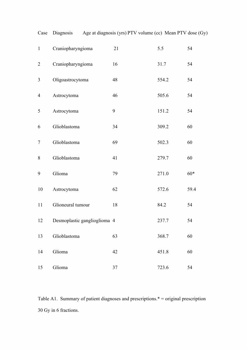
Case Diagnosis Age at diagnosis (yrs) PTV volume (cc) Mean PTV dose (Gy)
1 Craniopharyngioma 21 5.5 54
2 Craniopharyngioma 16 31.7 54
3 Oligoastrocytoma 48 554.2 54
4 Astrocytoma 46 505.6 54
5 Astrocytoma 9 151.2 54
6 Glioblastoma 34 309.2 60
7 Glioblastoma 69 502.3 60
8 Glioblastoma 41 279.7 60
9 Glioma 79 271.0 60*
10 Astrocytoma 62 572.6 59.4
11 Glioneural tumour 18 84.2 54
12 Desmoplastic ganglioglioma 4 237.7 54
13 Glioblastoma 63 368.7 60
14 Glioma 42 451.8 60
15 Glioma 37 723.6 54
Table A1. Summary of patient diagnoses and prescriptions.* = original prescription
30 Gy in 6 fractions.

Region of interest Objective Relative importance PTV Minimize RMS around prescription dose 100 Brainstem Minimize maximum dose 10 Globes Minimize maximum dose 5 Optic nerves Minimize maximum dose 5 Optic chiasm Minimize maximum dose 5 Lenses Minimize mean dose 5 Hippocampi Minimize mean dose 3 Temporal lobes Minimize mean dose 2 Cochleae Minimize mean dose 1 Brain excluding other ROIs Minimize mean dose 1 Table A2. AutoBeam optimization parameters. RMS = root mean square deviation. ROI = region of interest. Lens mean dose, not maximum dose, is used during optimization in routine clinical VMAT planning at our centre.
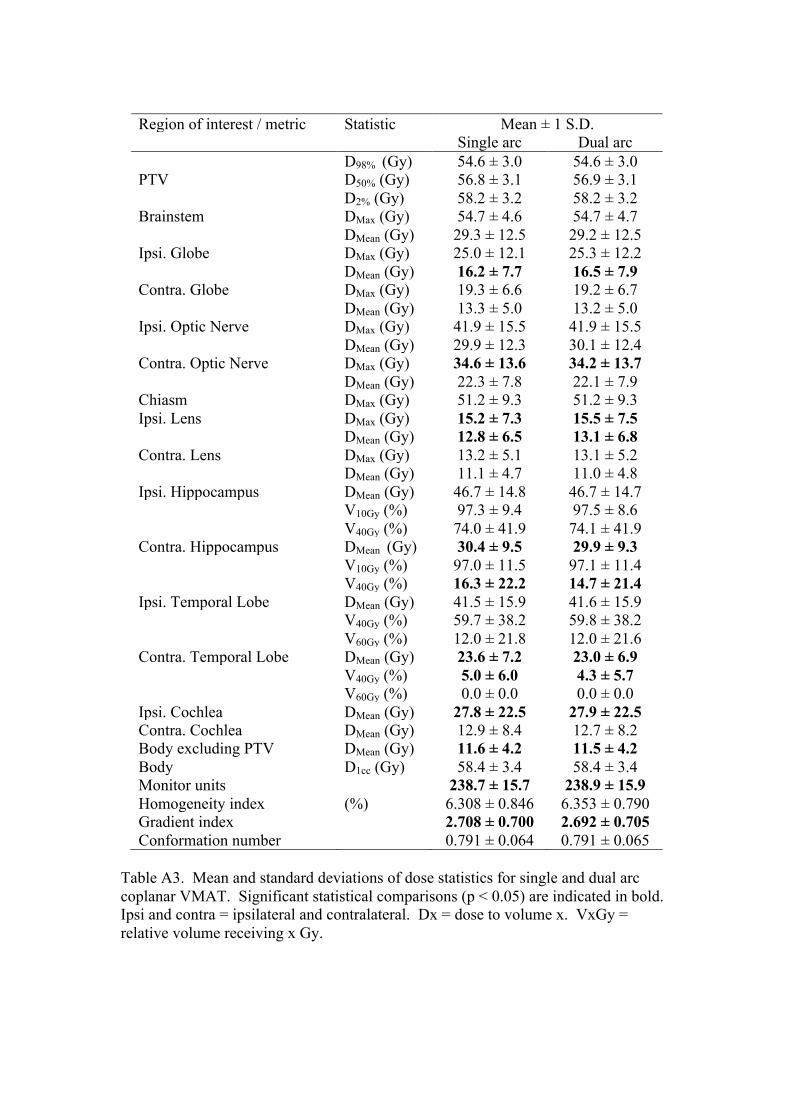
Region of interest / metric Statistic Mean ± 1 S.D. Single arc Dual arc D98% (Gy) 54.6 ± 3.0 54.6 ± 3.0 PTV D50% (Gy) 56.8 ± 3.1 56.9 ± 3.1 D2% (Gy) 58.2 ± 3.2 58.2 ± 3.2 Brainstem DMax (Gy) 54.7 ± 4.6 54.7 ± 4.7 DMean (Gy) 29.3 ± 12.5 29.2 ± 12.5 Ipsi. Globe DMax (Gy) 25.0 ± 12.1 25.3 ± 12.2 DMean (Gy) 16.2 ± 7.7 16.5 ± 7.9 Contra. Globe DMax (Gy) 19.3 ± 6.6 19.2 ± 6.7 DMean (Gy) 13.3 ± 5.0 13.2 ± 5.0 Ipsi. Optic Nerve DMax (Gy) 41.9 ± 15.5 41.9 ± 15.5 DMean (Gy) 29.9 ± 12.3 30.1 ± 12.4 Contra. Optic Nerve DMax (Gy) 34.6 ± 13.6 34.2 ± 13.7 DMean (Gy) 22.3 ± 7.8 22.1 ± 7.9 Chiasm DMax (Gy) 51.2 ± 9.3 51.2 ± 9.3 Ipsi. Lens DMax (Gy) 15.2 ± 7.3 15.5 ± 7.5 DMean (Gy) 12.8 ± 6.5 13.1 ± 6.8 Contra. Lens DMax (Gy) 13.2 ± 5.1 13.1 ± 5.2 DMean (Gy) 11.1 ± 4.7 11.0 ± 4.8 Ipsi. Hippocampus DMean (Gy) 46.7 ± 14.8 46.7 ± 14.7 V10Gy (%) 97.3 ± 9.4 97.5 ± 8.6 V40Gy (%) 74.0 ± 41.9 74.1 ± 41.9 Contra. Hippocampus DMean (Gy) 30.4 ± 9.5 29.9 ± 9.3 V10Gy (%) 97.0 ± 11.5 97.1 ± 11.4 V40Gy (%) 16.3 ± 22.2 14.7 ± 21.4 Ipsi. Temporal Lobe DMean (Gy) 41.5 ± 15.9 41.6 ± 15.9 V40Gy (%) 59.7 ± 38.2 59.8 ± 38.2 V60Gy (%) 12.0 ± 21.8 12.0 ± 21.6 Contra. Temporal Lobe DMean (Gy) 23.6 ± 7.2 23.0 ± 6.9 V40Gy (%) 5.0 ± 6.0 4.3 ± 5.7 V60Gy (%) 0.0 ± 0.0 0.0 ± 0.0 Ipsi. Cochlea DMean (Gy) 27.8 ± 22.5 27.9 ± 22.5 Contra. Cochlea DMean (Gy) 12.9 ± 8.4 12.7 ± 8.2 Body excluding PTV DMean (Gy) 11.6 ± 4.2 11.5 ± 4.2 Body D1cc (Gy) 58.4 ± 3.4 58.4 ± 3.4 Monitor units 238.7 ± 15.7 238.9 ± 15.9 Homogeneity index (%) 6.308 ± 0.846 6.353 ± 0.790 Gradient index 2.708 ± 0.700 2.692 ± 0.705 Conformation number 0.791 ± 0.064 0.791 ± 0.065
Table A3. Mean and standard deviations of dose statistics for single and dual arc coplanar VMAT. Significant statistical comparisons (p < 0.05) are indicated in bold. Ipsi and contra = ipsilateral and contralateral. Dx = dose to volume x. VxGy = relative volume receiving x Gy.