Embed Size (px)
Citation preview
2/12/2018
1
Non-melanocytic Patterns
Michelle Tarbox, MD
Assistant Professor of Dermatology and Dermatopathology Texas Tech University Health Sciences Center
2018
Non-melanocytic Lesions
• Seborrheic keratoses• Acanthotic• Macular
• Vascular lesions• Angiomas• Angiokeratomas• Lymphangiomas
• Dermatofibromas• Conventional • Cellular
• Basal cell carcinoma• Pigmented and non-pigmented
• Actinic keratoses• Pigmented actinic keratoses
• Squamous cell carcinomas
• Merkel cell carcinoma
• Bugs• Cutaneous infections• Cutaneous infestations
• BONUS cases!
I have no conflicts of interest to disclose
Except that I LOVE dermoscopy
Non-Melanocytic Lesion Dermoscopy
• Problem: Often No Pigment!
• Solution: Use your clues!• Vascular structures• Chrysalis structures• Texture• Structureless areas• Scale
Comma-like - IDN Dotted -Spitz Linear irregular - AMM
Hairpin – SK, SCC, KA Glomerular - SCCIS Arborizing - BCC
Crown vessels – Seb H Strawberry pattern - AK
Milky red areas/globules – Thick AMM
J Am Acad Dermatol - 01-SEP-2010;
63(3)Vascular morphology
A regularB in a stringC clusteredD radialE irregularly branchedF irregular
J Am Acad Dermatol - 01-SEP-2010; 63(3)
2/12/2018
2
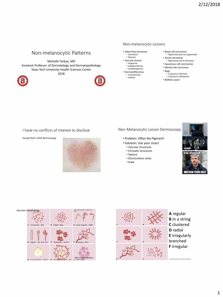
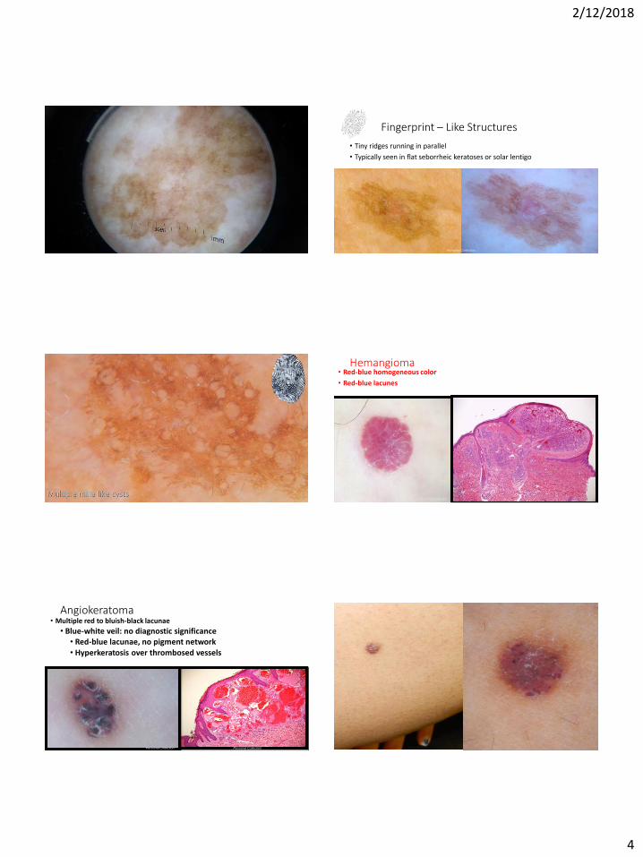
Seborrheic Keratosis
• Multiple milia like cysts
• Irregular crypts / comedo-like openings
• Fissures/ridges
• Fingerprint-like structures
Milia-like cysts = pseudo-horn cysts
(black arrows)
Comedo-like openings = comedostructures
(red arrows)
Personal Collection
Personal Collection
Multiple milia like cysts
Irregular crypts and comedo like openings
Personal Collection
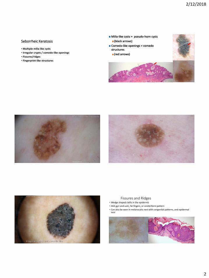
Fissures and Ridges • Wedge shaped clefts in the epidermis
• AKA gyri and sulci, fat fingers, or cerebriform pattern
• Can also be seen in melanocytic nevi with congenital patterns, and epidermal nevi
Personal Collection
2/12/2018
3
Fissures/ridgesPersonal Collection
Congenital type nevus with Fissures/ridges Personal Collection
Fissures/ridgesPersonal Collection
Personal Collection, patient granted special permission to show
tattoo
Personal Collection Personal Collection
2/12/2018
4
Fingerprint – Like Structures
• Tiny ridges running in parallel
• Typically seen in flat seborrheic keratoses or solar lentigo
Personal Collection
Multiple milia like cysts
Hemangioma• Red-blue homogeneous color
• Red-blue lacunes
Personal Collection
• Multiple red to bluish-black lacunae
• Blue-white veil: no diagnostic significance• Red-blue lacunae, no pigment network • Hyperkeratosis over thrombosed vessels
Angiokeratoma
Personal Collection Personal Collection Personal Collection
2/12/2018
5
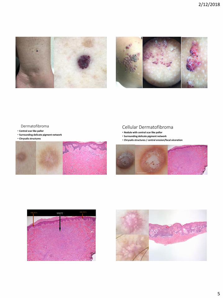
Dermatofibroma• Central scar like pallor
• Surrounding delicate pigment network
• Chrysalis structures
Personal Collection
Cellular Dermatofibroma• Nodule with central scar like pallor
• Surrounding delicate pigment network
• Chrysalis structures / central erosion/focal ulceration
Personal Collection
BROWN BROWN
Personal Collection
2/12/2018
6
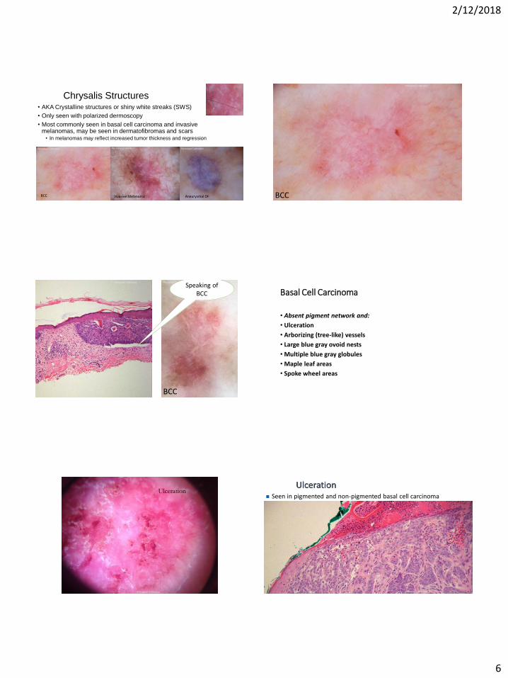
Chrysalis Structures • AKA Crystalline structures or shiny white streaks (SWS)
• Only seen with polarized dermoscopy
• Most commonly seen in basal cell carcinoma and invasive melanomas, may be seen in dermatofibromas and scars
• In melanomas may reflect increased tumor thickness and regression
BCC Invasive Melanoma Aneurysmal DF
Personal Collection Personal Collection Personal Collection
BCC
Personal Collection
BCC
Personal Collection Personal Collection
Speaking of BCC Basal Cell Carcinoma
• Absent pigment network and:
• Ulceration
• Arborizing (tree-like) vessels
• Large blue gray ovoid nests
• Multiple blue gray globules
• Maple leaf areas
• Spoke wheel areas
Ulceration
Personal Collection
Ulceration Seen in pigmented and non-pigmented basal cell carcinoma
Personal Collection
2/12/2018
7
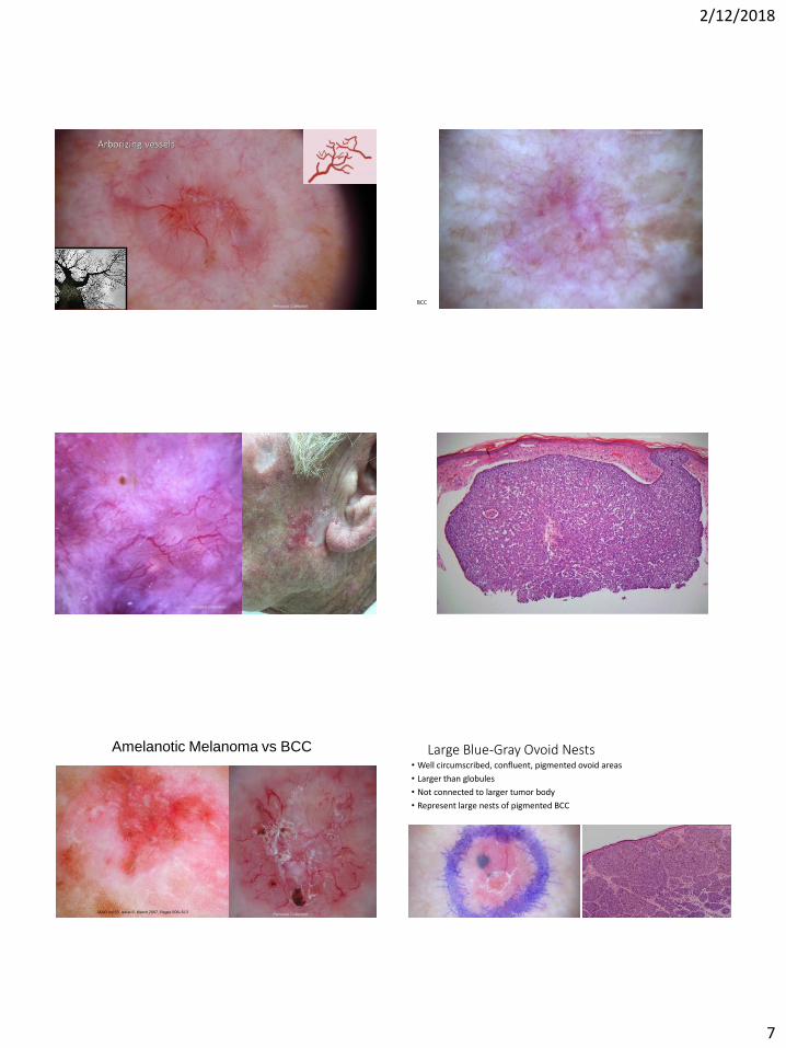
Arborizing vessels
Personal Collection BCC
Personal Collection
Personal Collection
Personal Collection
Amelanotic Melanoma vs BCC
Personal Collection JAAD Vol 53, Issue 6, March 2007, Pages 508–513
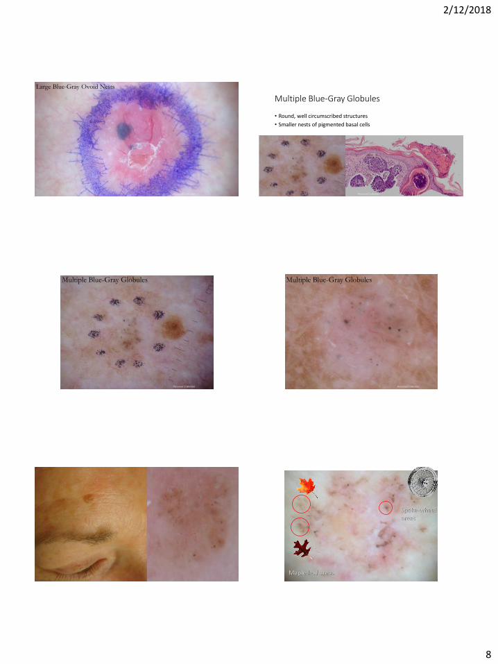
Large Blue-Gray Ovoid Nests • Well circumscribed, confluent, pigmented ovoid areas
• Larger than globules
• Not connected to larger tumor body
• Represent large nests of pigmented BCC
Personal Collection
2/12/2018
8
Large Blue-Gray Ovoid Nests
Personal Collection
Multiple Blue-Gray Globules
• Round, well circumscribed structures
• Smaller nests of pigmented basal cells
Personal Collection
Personal Collection
Multiple Blue-Gray Globules
Personal Collection
Multiple Blue-Gray Globules
Maple leaf areas
Spoke-wheelareas
2/12/2018
9
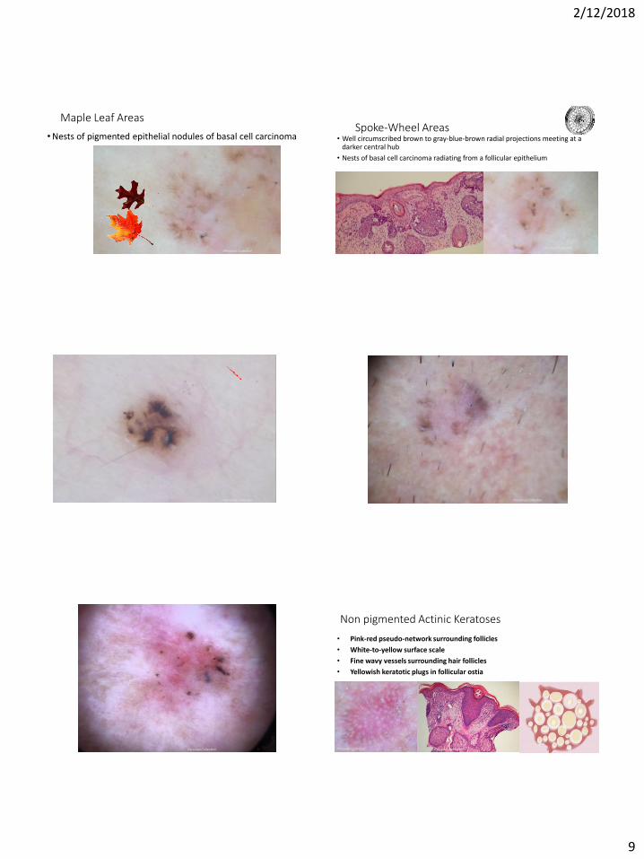
Maple Leaf Areas
• Nests of pigmented epithelial nodules of basal cell carcinoma
Personal Collection
Spoke-Wheel Areas• Well circumscribed brown to gray-blue-brown radial projections meeting at a
darker central hub
• Nests of basal cell carcinoma radiating from a follicular epithelium
Personal Collection
Personal Collection Personal Collection
Personal Collection
Non pigmented Actinic Keratoses
• Pink-red pseudo-network surrounding follicles
• White-to-yellow surface scale
• Fine wavy vessels surrounding hair follicles
• Yellowish keratotic plugs in follicular ostia
Personal Collection Personal Collection
2/12/2018
10
Actinic keratosis
Personal Collection
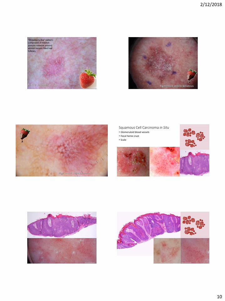
“Strawberry-like” pattern
composed of reddish
pseudo network around
whitish keratin filled hair
follicles
Pigmented actinic keratosis
Personal Collection
Personal Collection
Pigmented actinic keratosis
Squamous Cell Carcinoma in Situ • Glomeruloid blood vessels
• Focal heme crust
• Scale
Personal Collection
Personal Collection
Personal Collection Personal Collection Personal Collection
2/12/2018
11
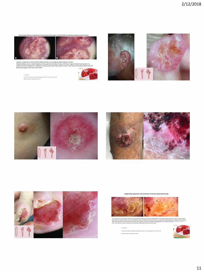
Lobules composed of central whitish yellow keratin surrounded by polymorphous vesselsDotted (yellow arrow), arborizing (blue arrow), glomerular (black circle), and linear irregular (red arrow) vessels on a yellowish white background in addition to perivascular white halos (black circle). Focal areas of ulceration (blue star) and blood spots (green star) were also noted.
A. Tülin Güleç
Dermoscopic features of squamous cell carcinoma of the tongue: It looks similar to cutaneous squamous cell carcinoma
Journal of the American Academy of Dermatology, Volume 75, Issue 2, 2016, e53–e54
http://dx.doi.org/10.1016/j.jaad.2016.01.030
Personal Collection Personal Collection Personal Collection
Fig 2. Squamous cell carcinoma of the lip. Dermoscopy revealed central whitish yellow keratin surrounded by polymorphous vessels, namely, dotted (yellow arrow), arborizing (blue arrow), coiled (black circle), and hairpin (red arrow) vessels on a yellowish white background in addition to perivascular white halos (purple circle) and white structureless areas (black arrow) that correlated to aggregated mass of highly keratinized malignant squamous cells. Focal areas of surface scale (blue star) and blood spots (green star) were also noted.
A. Tülin Güleç
Diagnosing squamous cell carcinoma of the lip using dermoscopy
Journal of the American Academy of Dermatology, Volume 76, Issue 2, Supplement 1, 2017, S82–S83
http://dx.doi.org/10.1016/j.jaad.2016.10.026
2/12/2018
12
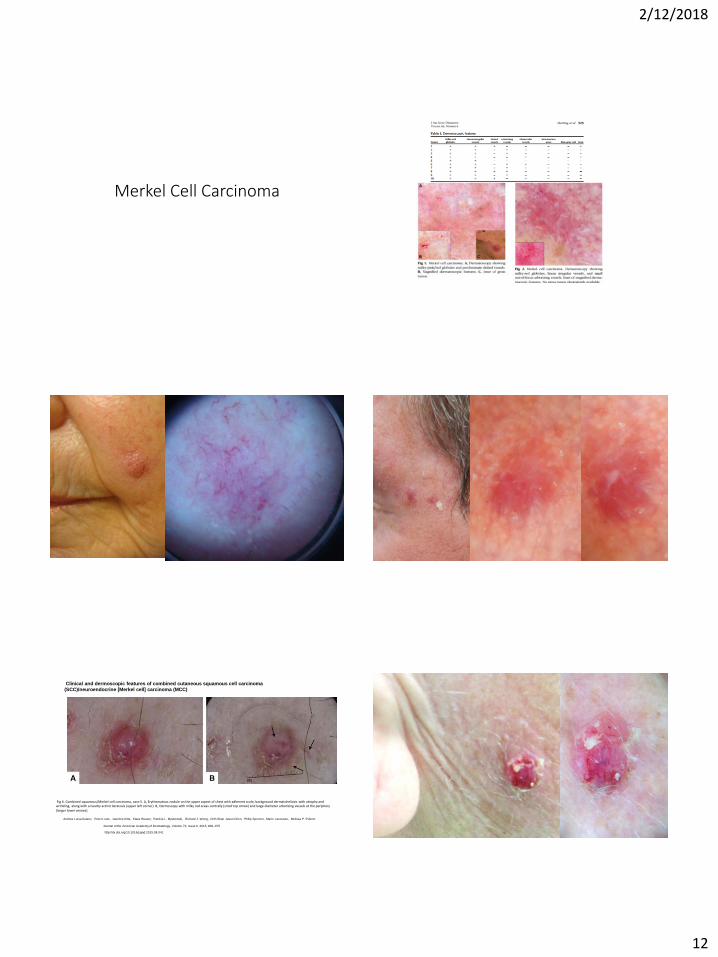
Merkel Cell Carcinoma
Fig 6. Combined squamous/Merkel cell carcinoma, case 5. A, Erythematous nodule on the upper aspect of chest with adherent scale; background dermatoheliosis with atrophy and wrinkling, along with a nearby actinic keratosis (upper left corner). B, Dermoscopy with milky red areas centrally (small top arrow) and large-diameter arborizing vessels at the periphery (larger lower arrows).
Andrea Luísa Suárez, Peter Louis, Jasmine Kitts, Klaus Busam, Patricia L. Myskowski, Richard J. Wong, Chih-Shan Jason Chen, Philip Spencer, Mario Lacouture, Melissa P. Pulitzer
Clinical and dermoscopic features of combined cutaneous squamous cell carcinoma
(SCC)/neuroendocrine [Merkel cell] carcinoma (MCC)
Journal of the American Academy of Dermatology, Volume 73, Issue 6, 2015, 968–975
http://dx.doi.org/10.1016/j.jaad.2015.08.041
2/12/2018
13
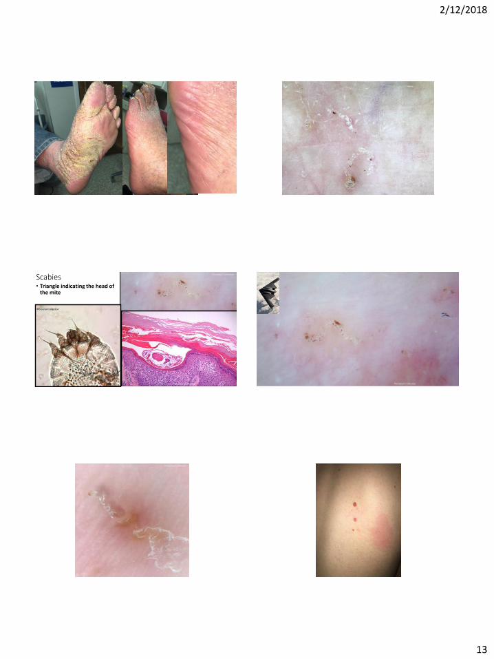
Scabies • Triangle indicating the head of
the mite
Personal Collection
Personal Collection
Personal Collection
Personal Collection
Personal Collection
Personal Collection
Personal Collection
2/12/2018
14
Personal Collection
Thank you! Michelle Tarbox, MD
Assistant Professor of Dermatology
Texas Tech University Health Sciences Center
Fun Benign things . . .
2/12/2018
15
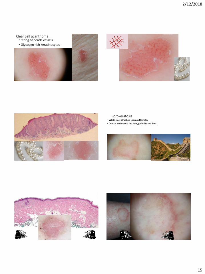
Clear cell acanthoma •String of pearls vessels
•Glycogen rich keratinocytes
Personal Collection
Personal Collection
Personal Collection
Personal Collection
Personal Collection
Personal Collection Personal Collection
Porokeratosis• White tract structure =cornoid lamella
• Central white area, red dots, globules and lines
Personal Collection
Personal Collection
Personal Collection
Personal Collection
2/12/2018
16
Personal Collection Personal Collection
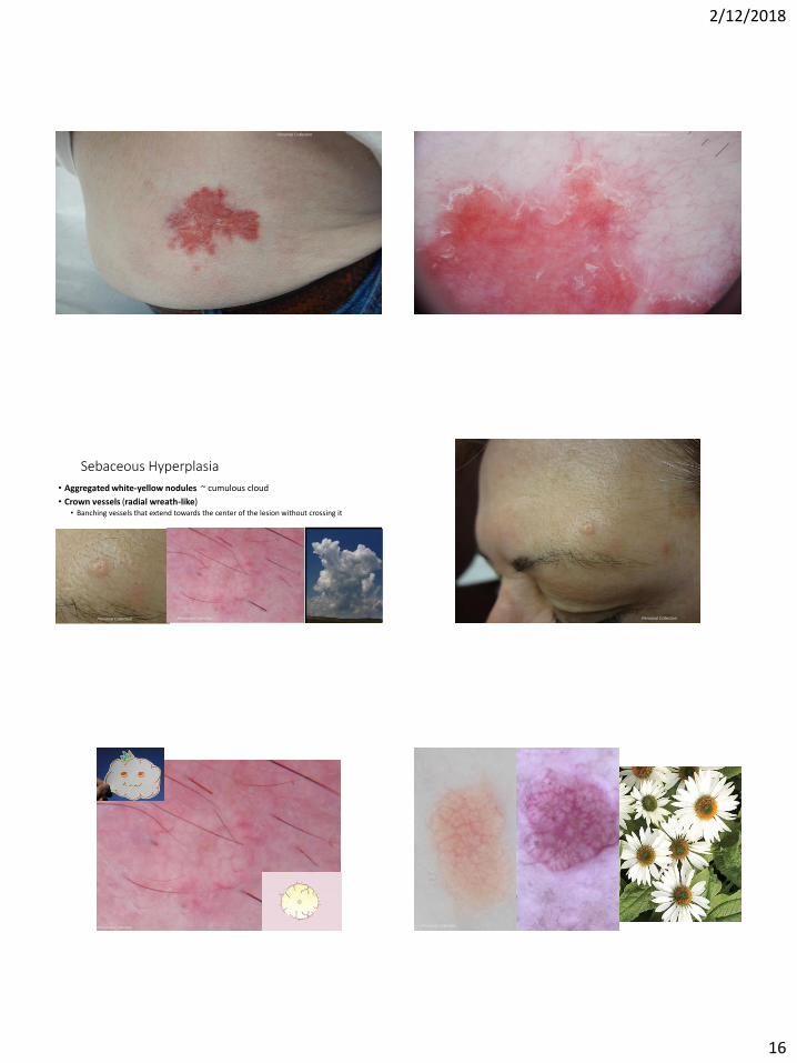
Sebaceous Hyperplasia
• Aggregated white-yellow nodules ~ cumulous cloud
• Crown vessels (radial wreath-like) • Banching vessels that extend towards the center of the lesion without crossing it
Personal Collection Personal Collection Personal Collection
Personal Collection Personal Collection
Personal Collection
2/12/2018
17
Nevus Sebaceous
Personal Collection
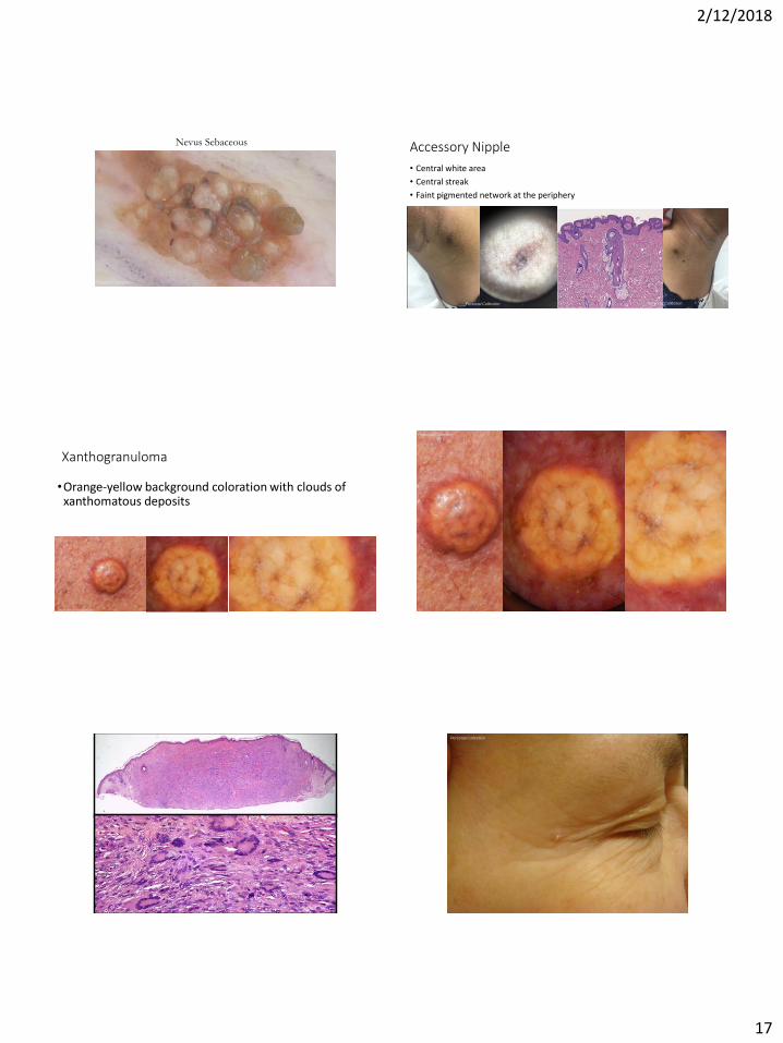
Accessory Nipple
• Central white area
• Central streak
• Faint pigmented network at the periphery
Personal Collection Personal Collection
Xanthogranuloma
•Orange-yellow background coloration with clouds of xanthomatous deposits
Personal Collection
Personal Collection
Personal Collection
Personal Collection
2/12/2018
18
Personal Collection
BONUS Cases
Personal Collection Personal Collection


![In vivo assessment of optical properties of melanocytic ... · melanocytic lesions complementary to that of RCM [9]. However, the diagnostic potential of HD-OCT seems to be not high](https://img.pdfslide.net/doc/110x75/5f081df47e708231d4206c1d/in-vivo-assessment-of-optical-properties-of-melanocytic-melanocytic-lesions.jpg)