Embed Size (px)
Citation preview

Nonhypotensive Dose of Telmisartan Attenuates CognitiveImpairment Partially Due to Peroxisome
Proliferator-Activated Receptor-� Activation in Mice WithChronic Cerebral Hypoperfusion
Kazuo Washida, MD; Masafumi Ihara, MD; Keiko Nishio, MD; Youshi Fujita, MD;Takakuni Maki, MD; Mahito Yamada, MD; Jun Takahashi, MD; Xiaofeng Wu, MS;
Takeshi Kihara, MD; Hidefumi Ito, MD; Hidekazu Tomimoto, MD; Ryosuke Takahashi, MD
Background and Purpose—The effect of telmisartan, an angiotensin II Type 1 receptor blocker with peroxisomeproliferator-activated receptor-�-modulating activity, was investigated against spatial working memory disturbances inmice subjected to chronic cerebral hypoperfusion.
Methods—Adult C57BL/6J male mice were subjected to bilateral common carotid artery stenosis using externalmicrocoils. Mice received a daily oral administration of low-dose telmisartan (1 mg/kg per day), high-dose telmisartan(10 mg/kg per day), or vehicle with or without peroxisome proliferator-activated receptor-� antagonist GW9662(1 mg/kg per day) for all treatments for 30 days after bilateral common carotid artery stenosis. Cerebral mRNAexpression of monocyte chemoattractant protein-1 and tumor necrosis factor-� was measured 30 days after bilateralcommon carotid artery stenosis, and postmortem brains were analyzed for demyelinating change with Kluver-Barrerastaining and immunostained for glial, oxidative stress, and vascular endothelial cell markers. Spatial working memorywas assessed by the Y-maze test.
Results—Mean systolic blood pressure and cerebral blood flow did not decrease with low-dose telmisartan but significantlydecreased with high-dose telmisartan. Low-dose telmisartan significantly attenuated, but high-dose telmisartanprovoked, spatial working memory impairment with glial activation, oligodendrocyte loss, and demyelinating change inthe white matter. Such positive effects of low-dose telmisartan were partially offset by cotreatment with GW9662.Consistent with this, low-dose telmisartan reduced the degree of oxidative stress of vascular endothelial cells and themRNA levels of monocyte chemoattractant protein-1 and tumor necrosis factor-� compared with vehicle.
Conclusions—Anti-inflammatory and antioxidative effects of telmisartan that were exerted in part by peroxisomeproliferator-activated receptor-� activation, but not its blood pressure-lowering effect, have protective roles againstcognitive impairment and white matter damage after chronic cerebral hypoperfusion. (Stroke. 2010;41:1798-1806.)
Key Words: chronic cerebral hypoperfusion � oligovascular niche � oxidative stress � PPAR-� � telmisartan
Drugs that target the renin–angiotensin system seem tohave particular potential for prevention of dementias,
including Alzheimer disease and vascular dementia. ThePerindopril Protection Against Recurrent Stroke Study(PROGRESS) has suggested a protective effect of angioten-sin-converting enzyme inhibitors on cognitive function inpatients with stroke.1 Moreover, the Study on Cognition andPrognosis in the Elderly (SCOPE) trial demonstrated apositive effect of the angiotensin II Type 1 receptor blocker(ARB), candesartan, in a subgroup of elderly hypertensivepatients with mild cognitive impairment.2 Notably, a prospec-tive cohort analysis of 819 491 participants suggested that
ARBs are associated with a significant reduction in theincidence and progression of dementia, even compared withangiotensin-converting enzyme inhibitors.3
No benefit was found in cognitive performance afteradministration of the ARB, telmisartan, at the subacute stage(within 15 days) after stroke in the Prevention Regimen forEffectively Avoiding Second Strokes (PRoFESS) study.4
However, in vitro studies have suggested that telmisartan, thestrongest peroxisome proliferator-activated receptor-�(PPAR-�) activator among ARBs,5 may protect oligodendro-cytes and neurons through a reduction of brain inflammationthrough PPAR-� activation and AT1 receptor blockade.
Received March 9, 2010; final revision received April 13, 2010; accepted May 5, 2010.From the Department of Neurology (K.W., M.I., K.N., Y.F., T.M., M.Y., H.I., R.T.), Graduate School of Medicine, Kyoto University, Sakyo-ku, Kyoto,
Japan; the Department of Neurology (H.T.), Graduate School of Medicine, Mie University, Tsu, Japan; the Department of Biological Repair (J.T.),Institute for Frontier Medical Sciences, Kyoto University, Kyoto, Japan; and the Department of Neuroscience for Drug Discovery Research (X.W., T.K.),Kyoto University, Kyoto, Japan.
Correspondence to Masafumi Ihara, MD, Kyoto University, 54 Kawahara-cho, Shogoin, Sakyo, Kyoto 606-8507, Japan. E-mail [email protected]© 2010 American Heart Association, Inc.
Stroke is available at http://stroke.ahajournals.org DOI: 10.1161/STROKEAHA.110.583948
1798
by guest on August 2, 2017
http://stroke.ahajournals.org/D
ownloaded from

Telmisartan has structural similarities to the PPAR-� ligandpioglitazone and thus could act as a partial agonist ofPPAR-�.5 PPAR-�, one of the nuclear receptors, plays acritical role in a variety of biological processes, includingangiogenesis, inflammation, oxidative stress, glucose metab-olism, and adipogenesis.5 Moreover, PPAR-� activation inthe brain has been suggested as a protective effect againstAlzheimer disease through its multifaceted effects, includinganti-inflammation and amyloid-� clearance.6 Therefore, inthe PRoFESS study, excessive lowering of blood pressure(BP) in the period with cerebrovascular autoregulatory dys-function may have affected the cerebral circulation andneuronal function, although other factors could also beinvolved.
The present study is therefore designed to explore themultifaceted effects of telmisartan on cognitive disturbancesin a mouse model of vascular dementia by administering ahypotensive or a nonhypotensive dose of telmisartan. Thismodel of chronic cerebral hypoperfusion, which is producedby placing microcoils bilaterally on the common carotidarteries, invariably exhibits glial activation, oxidative stress,inflammation, demyelinating change, and axonal loss in thewhite matter with resultant spatial working memory defi-cits.7,8 This in vivo system will help determine whethertelmisartan affects vascular autoregulatory function andwhether and how telmisartan exerts its protective effectagainst cognitive impairment related to white matter damage.
Materials and MethodsExperimental ProtocolThe experimental protocol is shown in Figure 1. Nine-week-old maleC57BL/6J mice (weighing 24 to 29 g; CLEA, Tokyo, Japan) werefed with the pelleted chow (MF) containing low-dose telmisartan (1mg/kg per day), high-dose telmisartan (10 mg/kg per day), or vehiclewith or without PPAR-� antagonist GW9662 (1 mg/kg per day;Sigma-Aldrich) for all treatments, beginning from 7 days before thebilateral common carotid artery stenosis (BCAS) surgery until 30
days post-BCAS. Immediately after spatial working memory wasassessed by the Y-maze test, mice were euthanized for histologicaland real-time reverse transcriptase–polymerase chain reaction exam-ination 30 days post-BCAS.
Surgical Procedure of BCASUnder anesthesia with halothane (2%), both common carotid arterieswere exposed through a midline cervical incision, and a microcoilwith an inner diameter of 0.18 mm was applied to the bilateralcommon carotid arteries. See Supplemental Method I for details(available at http://stroke.ahajournals.org).7,8
Systolic BP and Cerebral BloodFlow MeasurementsVarious doses of telmisartan (0 to 100 mg/kg per day) wereadministered and the BP measured for determining nonhypotensiveand hypotensive doses of telmisartan. Then, in mice receiving thepredetermined nonhypotensive or hypotensive dose of telmisartan orvehicle, systolic BP and cerebral blood flow (CBF) were monitoredat 7 days before BCAS (before starting telmisartan treatment),immediately before BCAS, and 2 hours, 1 day, 3 days, 7 days, 14days, and 30 days after BCAS. See Supplemental Method II fordetails.
Histochemical Evaluation of White MatterLesions, Glial Activation, and Oxidative StressThe mouse brains were analyzed for demyelinating change withKluver-Barrera staining and immunostained for glial fibrillary acidicprotein (a marker of astrocyte), ionized calcium binding adaptormolecule-1 (Iba-1; microglia), glutathione S-transferase-� (GST-�;oligodendrocyte), 8-hydroxy-deoxyguanosine (8-OHdG; oxidativestress), and CD31 (vascular endothelial cell). See SupplementalMethod III for details.7
Quantitative Real-Time ReverseTranscriptase–Polymerase Chain ReactionCerebral mRNA levels of monocyte chemoattractant protein-1(MCP-1) and tumor necrosis factor-� (TNF-�) were assessed byquantitative real-time reverse transcriptase–polymerase chain reac-tion pre-BCAS and 30 days post-BCAS. Detailed procedures aredescribed in Supplemental Method IV.
Figure 1. Experimental protocol. Tel(Low) indicates low-dose telmisartan(1 mg/kg per day); Tel (High), high-dosetelmisartan (10 mg/kg per day); GW,GW9662 (1 mg/kg per day); RT-PCR,reverse transcriptase–polymerase chainreaction.
Washida et al Effect of Telmisartan on a Vascular Dementia Model 1799
by guest on August 2, 2017
http://stroke.ahajournals.org/D
ownloaded from
Y-Maze Test for Spatial WorkingMemory AssessmentSpatial working memory was assessed by the Y-maze test. The detailof the Y-maze test protocol is described in Supplemental Method V.
Blood Concentration of TelmisartanSee Supplemental Method VI for details.
Statistical AnalysisAll values are expressed as means�SEM in the text and figures.One-way analysis of variance was used to evaluate significantdifferences among groups except when otherwise stated. When astatistically significant effect was found, a post hoc Tukey test orTukey-Kramer test was performed to detect the difference betweenthe groups. Temporal profiles of systolic BP and CBF were analyzedby 2-way repeated-measures analysis of variance followed by a posthoc Tukey test. Differences with P�0.05 were considered statisti-cally significant in all statistical analyses used.
Results
Systolic BP and CBF AfterTelmisartan AdministrationTreatment with telmisartan, �1 mg/kg per day, did not resultin a significant reduction in BP, whereas treatment withtelmisartan �3 mg/kg per day resulted in a significantreduction in BP (Figure 2A). CBF was not significantlyreduced �1 mg/kg per day but began to decrease at 3 mg/kgper day (Figure 2B). A nonhypotensive dose of 1 mg/kg perday or a hypotensive dose of 10 mg/kg per day wassubsequently administered. Temporal profiles of systolic BPand CBF were not affected by administration of a nonhypo-tensive dose of telmisartan or addition of GW9662 (Figure2C–E). CBF gradually recovered after BCAS in mice withvehicle or a nonhypotensive dose of telmisartan but not inthose with a hypotensive dose of telmisartan (Figure 2E).
The mortality rates were 10% at �1 mg/kg per day in thetelmisartan-treated group after BCAS surgery. The mortalityrate increased to 30% at 3 mg/kg per day in the telmisartan-treated group and 50% at 10 mg/kg per day in the telmisartan-treated group. Eighty percent at 50 or 100 mg/kg per day inthe telmisartan-treated group died within 3 days post-BCAS.
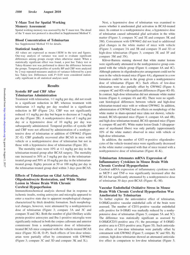
Effects of Telmisartan on Glial Activation,Oligodendrocyte Restoration, and White MatterLesion in Mouse Brain With ChronicCerebral HypoperfusionImmunohistochemical analysis showed that in response toischemic insults, resting astrocytes and microglia appeared toenter a reactive state due to apparent morphological changescharacterized by thick dendritic formation. Such morpholog-ical changes, however, were attenuated by a nonhypotensivedose of telmisartan (Figure 3; compare 3A and 3C andcompare 3I and 3K). Both the number of glial fibrillary acidicprotein-positive astrocytes and Iba-1-positive microglia weresignificantly reduced in both the corpus callosum and anteriorcommissure from a nonhypotensive dose of telmisartan-treated BCAS mice compared with the vehicle-treated BCASmice (Figure 3G–H, O–P). Such effects of low-dose telmis-artan were partially offset by cotreatment with GW9662(Figure 3; compare 3C and 3D and compare 3K and 3L).
Next, a hypotensive dose of telmisartan was examined toassess whether it ameliorated glial activation in BCAS-treatedmice. In contrast to a nonhypotensive dose, a hypotensive doseof telmisartan caused substantial glial activation in the whitematter (Figure 3; compare 3C and 3E and compare 3K and3M). Cotreatment with GW9662 did not lead to additionalglial changes in the white matter of mice with vehicle(Figure 3; compare 3A and 3B and compare 3I and 3J) orhigh-dose telmisartan (Figure 3; compare 3E and 3F andcompare 3M and 3N).
Kluver-Barrera staining showed that white matter lesionswere significantly attenuated in the nonhypotensive group com-pared with the vehicle group (Figure 4; compare 4A and 4C).Although patterns in oligodendrocytes arrangement could not beseen in the vehicle-treated mice (Figure 4A), alignment in a rowformation could be seen in the group given a nonhypotensivedose of telmisartan (Figure 4C). Such effects of low-dosetelmisartan were also partially offset by GW9662 (Figure 4;compare 4C and 4D) with significant differences (Figure 4G–H).In contrast, high-dose telmisartan did not attenuate white matterlesions (Figure 4; compare 4A and 4E). There were no signifi-cant histological differences between vehicle and high-dosetelmisartan-treated mice with or without GW9662. In addition,administration of GW9662 had no effects on morphology of thewhite matter in sham-operated mice (data not shown), vehicle-treated, BCAS-operated mice (Figure 4; compare 4A and 4B),and high-dose telmisartan-treated, BCAS-operated mice (Figure4; compare 4E and 4F). White matter lesion Grade 3 (disappear-ance of myelinated fibers) was only partially (approximately10% of the white matter) observed in mice with vehicle orhigh-dose telmisartan.
In addition, the number of GST-�-positive oligodendro-cytes of the vehicle-treated mice were significantly decreasedin the white matter compared with that of mice treated with anonhypotensive dose of telmisartan (Figure 4I–K).
Telmisartan Attenuates mRNA Expression ofInflammatory Cytokines in Mouse Brain WithChronic Cerebral HypoperfusionCerebral mRNA expression of inflammatory cytokines suchas MCP-1 and TNF-� was significantly increased after theBCAS but significantly attenuated by a nonhypotensive doseof telmisartan 30 days post-BCAS (Figure 4L–M).
Vascular Endothelial Oxidative Stress in MouseBrain With Chronic Cerebral Hypoperfusion WasAmeliorated by TelmisartanTo further explore the antioxidative effect of telmisartan,8-OHdG-positive vascular endothelial cells of the brain wereassessed. The number of CD31-positive vascular endothelialcells positive for 8-OHdG was markedly reduced by a nonhy-potensive dose of telmisartan (Figure 5; compare 5A and 5C).The difference was statistically significant as assessed by8-OHdG/CD31-positive area (%; the percentage of 8-OHdG-positive area to CD31-positive area; Figure 5J). Such antioxida-tive effects of low-dose telmisartan were partially offset bycotreatment with GW9662 (Figure 5; compare 5C and 5D). Bycontrast, high-dose telmisartan showed an attenuated antioxida-tive effect in comparison to low-dose telmisartan (Figure 5;
1800 Stroke August 2010
by guest on August 2, 2017
http://stroke.ahajournals.org/D
ownloaded from

Figure 2. Systolic BP (A) and CBF (B) in mice treated with various doses of telmisartan (n�10 each). Representative CBF images of the6 groups of mice as assessed by laser speckle flowmetry 7 days before (Day �7) and immediately before (Day 0) BCAS and 30 dayspost-BCAS (Day 30; C). Temporal profiles of systolic BP (D) and CBF (E) of the 6 groups of mice (n�5 each). CBF was expressed as apercentage of baseline flow. F5,24�84.100 (D), F5,24�37.441 (E), *P�0.01 versus vehicle.
Washida et al Effect of Telmisartan on a Vascular Dementia Model 1801
by guest on August 2, 2017
http://stroke.ahajournals.org/D
ownloaded from
Figure 3. Representative images of immu-nohistochemistry for glial fibrillary acidicprotein (GFAP; A–F) and Iba-1 (I–N) in theparamedian parts of the corpus callosum ofthe BCAS-operated mice treated with vehi-cle (A, I), vehicle�GW (B, J), Tel (Low; C, K),Tel (Low)�GW (D, L), Tel (High; E, M), andTel (High)�GW (F, N) 30 days post-BCAS(n�7 each). Insets indicate enlarged imagesof astrocytes (A–F) and microglia (I–N).Scale bar, 100 �m. Histogram showing thedensity of GFAP-positive astrocytes (G–H)and Iba-1-positive microglia (O–P) in thecorpus callosum (G, O) and anterior com-missure (H, P) of the 6 groups of mice. Tel(Low) indicates low-dose telmisartan(1 mg/kg per day); Tel (High), high-dosetelmisartan (10 mg/kg per day); GW,GW9662 (1 mg/kg per day).
1802 Stroke August 2010
by guest on August 2, 2017
http://stroke.ahajournals.org/D
ownloaded from

Figure 4. Representative images of the Kluver-Barrera staining in the paramedian parts of the cor-pus callosum of the BCAS-operated mice treatedwith vehicle (A), vehicle�GW (B), Tel (Low; C), Tel(Low)�GW (D), Tel (High; E), and Tel (High)�GW (F)30 days post-BCAS (n�7 each). Insets indicateenlarged images of oligodendrocytes. Histogramshowing the grading of the white matter lesions ofthe 6 groups of mice (G–H; see SupplementalMethod III for details). Representative images ofimmunohistochemistry for GST-�-positive oligoden-drocytes in the medial parts of the corpus callosumof the BCAS-operated mice treated with vehicle (I)and Tel (Low; J) 30 days post-BCAS (n�7 each).Scale bar, 100 �m. Histogram showing the densityof GST-�-positive oligodendrocytes (K) of the 2groups of mice. Cerebral mRNA expressions ofMCP-1 (L) and TNF-� (M) pre-BCAS and 30 dayspost-BCAS in the sham-operated or BCAS-operated mice treated with vehicle or Tel (Low; n�5each). Tel (Low) indicates low-dose telmisartan(1 mg/kg per day); Tel (High), high-dose telmisartan(10 mg/kg per day); GW, GW9662 (1 mg/kg per day).
Washida et al Effect of Telmisartan on a Vascular Dementia Model 1803
by guest on August 2, 2017
http://stroke.ahajournals.org/D
ownloaded from

compare 5A, 5C, and 5E). Cotreatment with GW9662 did notlead to additional histological changes in mice with vehicle(Figure 5; compare 5A and 5B) or high-dose telmisartan (Figure5; compare 5E and 5F).
Spatial Working Memory in Mice With ChronicCerebral Hypoperfusion Was Restored by aNonhypotensive Dose of TelmisartanFinally, we analyzed spatial working memory of BCAS miceby the Y-maze test as the final functional output. The percentageof alternation behaviors significantly decreased in vehicle-treated BCAS mice compared with the sham-operated mice butsignificantly increased in a nonhypotensive dose of telmisartan-
treated mice. Such effects of low-dose telmisartan were partiallyoffset by cotreatment with GW9662. Hypotensive doses oftelmisartan-treated mice manifested in further impaired workingmemory (Figure 6A). There were no significant differences inthe number of entries to each arm, which was considered toreflect locomotor activity, among the 5 groups (Figure 6B).These results suggest that a nonhypotensive dose of telmisartan,but not a hypotensive dose, improved spatial working memoryof BCAS-operated mice.
Blood Concentration of TelmisartanPlasma concentration after 1 mg/kg per day administration oftelmisartan for 7 days was 142.86�14.85 ng/mL (n�4, values
Figure 5. Representative images of the immunoflu-orescent staining for 8-OHdG (red) in the medialparts of the corpus callosum of the BCAS-operated mice treated with vehicle (A),vehicle�GW (B), Tel (Low; C), Tel (Low)�GW (D),Tel (High; E), and Tel (High)�GW (F) 30 days post-BCAS (n�7 each). Capillaries double-positive forCD31 (G, green) and 8-OHdG (H, red) and mergedimage (I) in vehicle-treated mice. Scale bars,100 �m (A–F), 50 �m (G–I). Histogram showing thepercentage of 8-OHdG-positive area to CD31-positive area of the 6 groups of mice (J). Tel (Low)indicates low-dose telmisartan (1 mg/kg per day);Tel (High), high-dose telmisartan (10 mg/kg perday); GW, GW9662 (1 mg/kg per day).
1804 Stroke August 2010
by guest on August 2, 2017
http://stroke.ahajournals.org/D
ownloaded from

are expressed as mean�SEM), a lower limit of BP-loweringeffect in humans.
DiscussionThis study showed that (1) a nonhypotensive dose of telmis-artan alleviated microglial/astroglial activation, endothelialoxidative stress, oligodendrocyte loss, and demyelinatingchanges in the white matter of the mice with chronic cerebralhypoperfusion; (2) such protective effects against the whitematter lesions were at least partially mediated by anti-inflammatory and antioxidative effects that were exerted inpart by PPAR-� activation; (3) by contrast, a hypotensivedose of telmisartan did not induce such positive effects inBCAS-operated mice in the white matter; and (4) a nonhy-potensive, but not a hypotensive, dose of telmisartan amelio-rated cognitive decline of the BCAS-operated mice. Thus, theprotective effects of telmisartan against white matter damageand cognitive impairment are exerted by its multifacetedeffects, partially through PPAR-� activation, but these areabolished by its BP-lowering effects when given at a higherdose. Thus, telmisartan should be considered for putativetreatment for subcortical vascular dementia, although strictmonitoring of BP is required.
Telmisartan is an ARB with a high degree of lipophilicityand is able to cross the blood–brain barrier. Telmisartaninhibits TNF-�-induced nuclear factor-�B activation, mainly
through AT1 receptor blockade.9 In addition, telmisartansuppresses MCP-1 expression through AT1 receptor blockadeand PPAR-� activation.10 Genetic deletion of AT1 receptorprotects against damage due to brain ischemia.11 With itssynergistic effects of AT1 receptor blockade and PPAR-�activation, telmisartan may exert multiple beneficial effects,including an antioxidative and anti-inflammatory effect, asshown in this study.
The nonhypotensive dose of telmisartan suppressed super-oxide production from the vessel wall without lowering CBF.Telmisartan reduces NADPH oxidase activity10; therefore,administration of telmisartan in such a low dose seems todecelerate the free radical system in the chronically hypoper-fused mouse brain. However, a hypotensive dose of telmis-artan substantially increased the degree of endothelial oxida-tive stress together with glial activation, white matter lesions,and spatial working memory deficits. Because mice withsevere CBF reduction (approximately lower than 50% of thebaseline level) died and were subsequently excluded from theanalysis, such detrimental effects of high-dose telmisartanmay even be underestimated. ARBs protect against ischemiccerebral injury, independently of BP.12 The benefit of normo-tension over hypotension lies in the absence of hypotension-induced aggravation of ischemic change. In the PRoFESS study,lowering BP at the subacute phase after stroke (within 15 days)may have decreased CBF. The Acute Candesartan CilexetilTherapy in Stroke Survivors (ACCESS) study indicated thatcandesartan treatment immediately after stroke, without signifi-cant lowering of BP, decreased the rate of recurrent stroke.13
Therefore, the beneficial pleiotropic effect of telmisartan seemsto be overwhelmed by BP-lowering if given in a high dose.Caution should therefore be exercised when lowering BP at anacute to subacute stage when cerebrovascular autoregulation isdamaged. In clinical practice, appropriate timing and dose oftelmisartan should be considered.
The effect of telmisartan on cognitive function was alsoassessed. The Y-maze tests showed that spatial working memorywas significantly disrupted by chronic cerebral hypoperfusion.Nonhypotensive doses of telmisartan significantly attenuated thedeterioration of spatial working memory together with histolog-ical improvement in the white matter. By contrast, hypotensivedoses of telmisartan further aggravated spatial working mem-ory together with histological deterioration. In this mousemodel, pathological changes were restricted in the whitematter only. The cerebral cortex and hippocampus, which areassociated with spatial working memory,14 were not damagedin a relatively short period (for example, 30 days post-BCAS). Thus, telmisartan may have restored frontal–subcor-tical circuitry function, which is also associated with spatialworking memory.8,14 However, hippocampal changes appearat 3 months post-BCAS15; it would therefore be of interest toextend the observation period to investigate effect of telmis-artan on the gray matter in a future study.
Recently, it has been shown that cerebral endothelial cellssecrete trophic factors that support the survival and prolifer-ation of oligodendrocyte precursor cells.16 Such oligodendro-cyte precursor cell-supportive phenomena in endothelial cellsare mediated by Akt and Src signaling pathways. Noncyto-toxic levels of oxidative stress downregulate the production
Figure 6. Spatial working memory assessed by the Y-maze testshowing alternation behavior (%; A) and number of arm entries(B) of the indicated groups of mice (sham, vehicle�BCAS, Tel(Low)�BCAS, Tel (Low)�GW�BCAS; n�20, Tel (High) �BCAS;n�10). Tel (Low) indicates low-dose telmisartan (1 mg/kg perday); Tel (High), high-dose telmisartan (10 mg/kg per day); GW,GW9662 (1 mg/kg per day).
Washida et al Effect of Telmisartan on a Vascular Dementia Model 1805
by guest on August 2, 2017
http://stroke.ahajournals.org/D
ownloaded from
of trophic factors such as brain-derived neurotrophic factorand fibroblast growth factor and disrupt the ability of cerebralendothelial cells to support oligodendrocyte precursor cells.Furthermore, GST-�-positive oligodendrocytes were restoredby low-dose telmisartan. These data suggest that a novel func-tion of telmisartan is to maintain “oligovascular niche” bysustaining oligodendrocyte homeostasis in mammalian brain.
In conclusion, long-term AT1 receptor blockade and PPAR-�activation with telmisartan should be considered as a noveltherapeutic approach for protection from damage associated withchronic cerebral hypoperfusion or subcortical vascular dementia.
AcknowledgmentsWe thank Prof Kalaria for his excellent advice on this work and DrKhundakar for his editorial assistance and comments. We areindebted to Ms Nakabayashi, Ms Gomibuchi, Ms Katsukawa, andMr Kubota for their excellent technical assistance. Telmisartan wasprovided by Boehringer Ingelheim (Ingelheim, Germany). Boehring-er Ingelheim provided no other support for this study, exceptsupplying telmisartan.
Sources of FundingThis work was supported by a Grant-in-Aid for Scientific Researchon Priority Areas from the Japanese Ministry of Education, Scienceand Culture (to J.T.) and a grant from the Suzuken MemorialFoundation (to M.I., J.T.).
DisclosuresNone.
References1. Tzourio C, Anderson C, Chapman N, Woodward M, Neal B, MacMahon
S, Chalmers J; PROGRESS Collaborative Group. Effects of bloodpressure lowering with perindopril and indapamide therapy on dementiaand cognitive decline in patients with cerebrovascular disease. ArchIntern Med. 2003;163:1069–1075.
2. Saxby BK, Harrington F, Wesnes KA, McKeith IG, Ford GA. Cande-sartan and cognitive decline in older patients with hypertension: asubstudy of the SCOPE trial. Neurology. 2008;70:1858–1866.
3. Li NC, Lee A, Whitmer RA, Kivipelto M, Lawler E, Kazis LE, Wolozin B.Use of angiotensin receptor blockers and risk of dementia in a predominantlymale population: prospective cohort analysis. BMJ. 2010;340:b5465.
4. Diener HC, Sacco RL, Yusuf S, Cotton D, Ounpuu S, Lawton WA,Palesch Y, Martin RH, Albers GW, Bath P, Bornstein N, Chan BP, ChenST, Cunha L, Dahlof B, De Keyser J, Donnan GA, Estol C, Gorelick P,Gu V, Hermansson K, Hilbrich L, Kaste M, Lu C, Machnig T, Pais P,Roberts R, Skvortsova V, Teal P, Toni D, VanderMaelen C, Voigt T,Weber M, Yoon BW; Prevention Regimen for Effectively AvoidingSecond Strokes (PRoFESS) study group. Effects of aspirin plus extended-release dipyridamole versus clopidogrel and telmisartan on disability andcognitive function after recurrent stroke in patients with ischaemic strokein the Prevention Regimen for Effectively Avoiding Second Stroke(PRoFESS) trial: a double-blind, active and placebo-controlled study.Lancet Neurol. 2008;7:875–884.
5. Benson SC, Pershadsingh HA, Ho CI, Chittiboyina A, Desai P, PravenecM, Qi N, Wang J, Avery MA, Kurtz TW. Identification of telmisartan asa unique angiotensin II receptor antagonist with selective PPAR�-modulating activity. Hypertension. 2004;43:993–1002.
6. Tsukuda K, Mogi M, Iwanami J, Min LJ, Sakata A, Jing F, Iwai M,Horiuchi M. Cognitive deficit in amyloid-�-injected mice was improvedby pretreatment with a low dose of telmisartan partly because of per-oxisome proliferator-activated receptor-� activation. Hypertension. 2009;54:782–787.
7. Shibata M, Ohtani R, Ihara M, Tomimoto H. White matter lesions andglial activation in a novel mouse model of chronic cerebral hypoper-fusion. Stroke. 2004;35:2598–2603.
8. Shibata M, Yamasaki N, Miyakawa T, Kalaria RN, Fujita Y, Ohtani R,Ihara M, Takahashi R, Tomimoto H. Selective impairment of workingmemory in a mouse model of chronic cerebral hypoperfusion. Stroke.2007;38:2826–2832.
9. Nakano A, Hattori Y, Aoki C, Jojima T, Kasai K. Telmisartan inhibitscytokine-induced nuclear factor-�B activation independently of the per-oxisome proliferator-activated receptor-�. Hypertens Res. 2009;32:765–769.
10. Matsui T, Yamagishi S, Ueda S, Nakamura K, Imaizumi T, Takeuchi M,Inoue H. Telmisartan, an angiotensin II type 1 receptor blocker, inhibitsadvanced glycation end-product (AGE)-induced monocyte chemoat-tractant protein-1 expression in mesangial cells through downregulationof receptor for AGEs via peroxisome proliferator-activated receptor-�activation. J Int Med Res. 2007;35:482–489.
11. Walther T, Olah L, Harms C, Maul B, Bader M, Hortnagl H, SchultheissHP, Mies G. Ischemic injury in experimental stroke depends on angio-tensin II. FASEB J. 2002;16:169–176.
12. Takizawa S, Dan T, Uesugi T, Nagata E, Takagi S, van Ypersele deStrihou C, Miyata T. A sartan derivative with a very low angiotensin IIreceptor affinity ameliorates ischemic cerebral damage. J Cereb BloodFlow Metab. 2009;29:1665–1672.
13. Schrader J, Luders S, Kulschewski A, Berger J, Zidek W, Treib J,Einhaupl K, Diener HC, Dominiak P; Acute Candesartan CilexetilTherapy in Stroke Survivors Study Group. The ACCESS Study: eval-uation of acute candesartan cilexetil therapy in stroke survivors. Stroke.2003;34:1699–1703.
14. Sarti C, Pantoni L, Bartolini L, Inzitari D. Persistent impairment of gaitperformances and working memory after bilateral common carotid arteryocclusion in the adult Wistar rat. Behav Brain Res. 2002;136:13–20.
15. Nishio K, Ihara M, Yamasaki N, Kalaria RN, Maki T, Fujita Y, Ito H,Oishi N, Fukuyama H, Miyakawa T, Takahashi R, Tomimoto H. A mousemodel characterizing features of vascular dementia with hippocampalatrophy. Stroke. 2010;41:1278–1284.
16. Arai K, Lo EH. An oligovascular niche: cerebral endothelial cellspromote the survival and proliferation of oligodendrocyte precursor cells.J Neurosci. 2009;29:4351–4355.
1806 Stroke August 2010
by guest on August 2, 2017
http://stroke.ahajournals.org/D
ownloaded from

Ryosuke TakahashiYamada, Jun Takahashi, Xiaofeng Wu, Takeshi Kihara, Hidefumi Ito, Hidekazu Tomimoto and
Kazuo Washida, Masafumi Ihara, Keiko Nishio, Youshi Fujita, Takakuni Maki, MahitoHypoperfusion
Activation in Mice With Chronic CerebralγPeroxisome Proliferator-Activated Receptor-Nonhypotensive Dose of Telmisartan Attenuates Cognitive Impairment Partially Due to
Print ISSN: 0039-2499. Online ISSN: 1524-4628 Copyright © 2010 American Heart Association, Inc. All rights reserved.
is published by the American Heart Association, 7272 Greenville Avenue, Dallas, TX 75231Stroke doi: 10.1161/STROKEAHA.110.583948
2010;41:1798-1806; originally published online July 1, 2010;Stroke.
http://stroke.ahajournals.org/content/41/8/1798World Wide Web at:
The online version of this article, along with updated information and services, is located on the
http://stroke.ahajournals.org//subscriptions/
is online at: Stroke Information about subscribing to Subscriptions:
http://www.lww.com/reprints Information about reprints can be found online at: Reprints:
document. Permissions and Rights Question and Answer process is available in the
Request Permissions in the middle column of the Web page under Services. Further information about thisOnce the online version of the published article for which permission is being requested is located, click
can be obtained via RightsLink, a service of the Copyright Clearance Center, not the Editorial Office.Strokein Requests for permissions to reproduce figures, tables, or portions of articles originally publishedPermissions:
by guest on August 2, 2017
http://stroke.ahajournals.org/D
ownloaded from