Embed Size (px)
Citation preview

@DanaKotlerMD
Nonspecific Low Back Pain
Evaluation and Treatment Approaches
Dana Kotler, MD
Instructor, Harvard Medical School
Director, Cycling Medicine Program
Spaulding Rehabilitation Hospital
Newton-Wellesley Hospital

@DanaKotlerMD
Disclosures
• None

@DanaKotlerMD
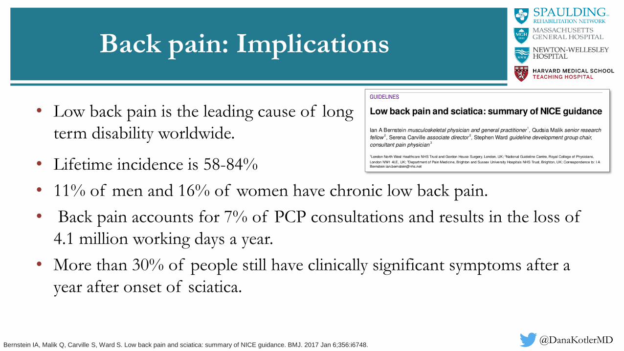
Back pain: Implications
• It’s annoying
• It’s getting in the way of recreational activity
• It’s getting in the way of necessary daily activity
• Other systems are not functioning
• Psychosocial ramifications
• Economic cost
Image: Dr. Mike Evans
@DanaKotlerMD
Back pain: Implications
• Low back pain is the leading cause of long
term disability worldwide.
• Lifetime incidence is 58-84%
• 11% of men and 16% of women have chronic low back pain.
• Back pain accounts for 7% of PCP consultations and results in the loss of
4.1 million working days a year.
• More than 30% of people still have clinically significant symptoms after a
year after onset of sciatica.
Bernstein IA, Malik Q, Carville S, Ward S. Low back pain and sciatica: summary of NICE guidance. BMJ. 2017 Jan 6;356:i6748.
@DanaKotlerMD
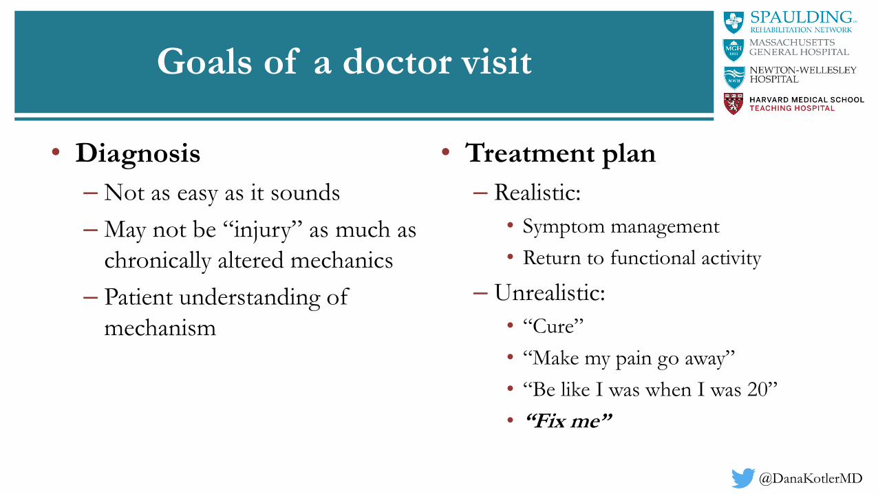
Goals of a doctor visit
• Diagnosis
– Not as easy as it sounds
– May not be “injury” as much as
chronically altered mechanics
– Patient understanding of
mechanism
• Treatment plan
– Realistic:
• Symptom management
• Return to functional activity
– Unrealistic:
• “Cure”
• “Make my pain go away”
• “Be like I was when I was 20”
• “Fix me”
@DanaKotlerMD
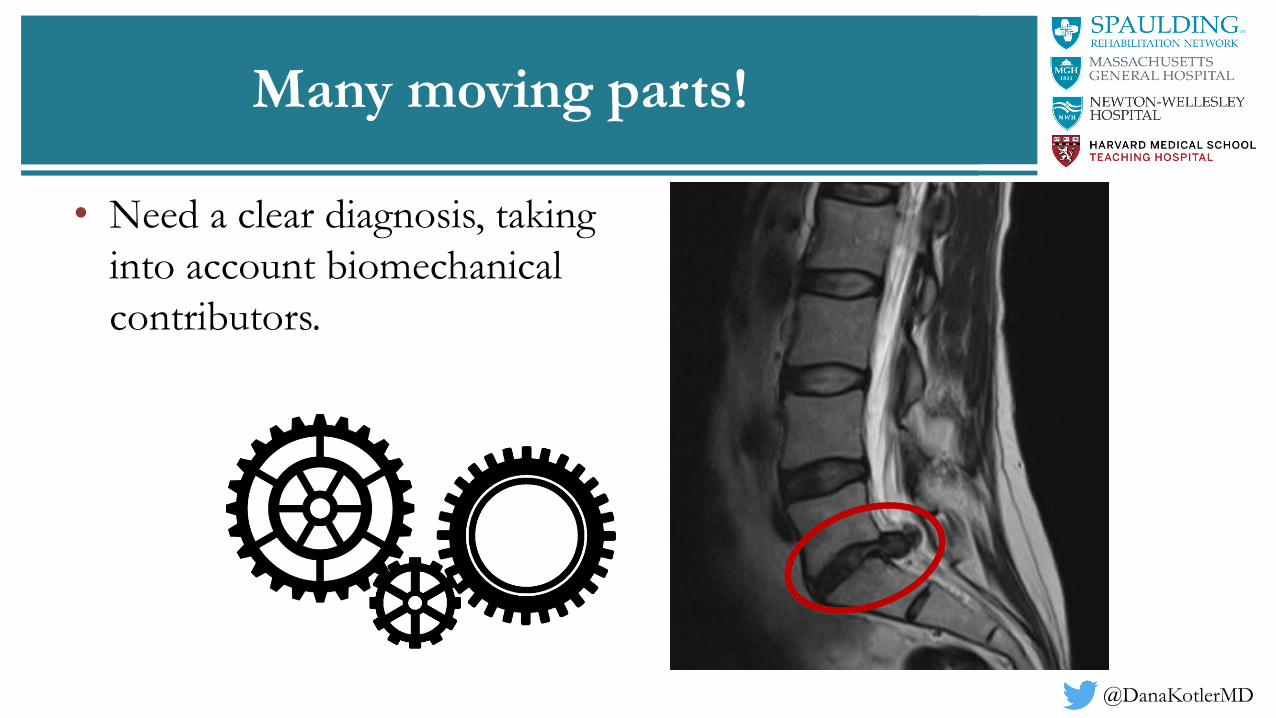
Many moving parts!
• Need a clear diagnosis, taking
into account biomechanical
contributors.

@DanaKotlerMD
DIAGNOSIS
• Main problem
• Underlying pathology, if known/suspected
• Superimposed issues
• Simple: Acute disc herniation
• Complex: Right L4 radiculopathy with underlying central and foraminal stenosis related to facet hypertrophy, ligamentum flavumthickening, with compensatory abnormalities in biomechanics resulting in intra-articular and extra-articular hip pain
@DanaKotlerMD

@DanaKotlerMD
Why you’re here!
• “…the clinical practice guidelines recommend history taking and physical examination to identify red flags, neurological testing to identify radicular syndrome, use of imaging if serious pathology is suspected (but discourage routine use), and assessment of psychosocial factors.”
• Non-specific → specific low back pain?
• Identify structural/mechanical factors
• Recommend specific treatment
Oliveira CB, Maher CG, Pinto RZ, Traeger AC, Lin CW, Chenot JF, van Tulder M, Koes BW. Clinical practice guidelines for the management of non-specific low back pain in primary care: an updated overview. European Spine Journal. 2018 Jul 3:1-3.

@DanaKotlerMD
#1 – Listen to the history
• Common themes:
– “I was putting on my pants”
– “I have to stop and sit down”
– “Every time I sneeze I think I’m going to die.”
– “I hate museums.”
• WHERE?
– Back pain?
– Back and butt pain?
– Back and leg pain?
– Butt pain or leg pain only?
• WHEN?
– Sitting, standing, walking, bending, lifting, coughing, sneezing, driving, exercise, going to the bathroom, brushing your teeth, having sex, only on weekends, only on work days…

@DanaKotlerMD
Key Questions
• Prior history of low back pain
• Prior treatment, successful?
• Duration
• Frequency
• Truly constant vs. episodic, positional, waxing/waning
• Exacerbating factors
• Alleviating factors
• Medical history
• Immunosuppression, cancer, procedures
• Psychosocial history
– Yellow flags
• Family history

@DanaKotlerMD
Key Questions: “Red Flags”
• Trauma• Intensity, duration,
frequency
• Systemic symptoms• Fever, chills
• Unexplained weight loss
• Fatigue
• Lumbar stiffness• Severe, 24/7
• Night pain
• Progressive
neurological symptoms
• Paresthesias, numbness
• Extremity weakness
• Gait dysfunction
• Bladder/bowel
dysfunction

@DanaKotlerMD
#2 - Do a GREAT physical exam
• Not just a straight leg raise!
• Evaluate biomechanical patterns.
• Functional testing
– Walking into the exam room
– Getting in and out of a chair
– Picking something off the floor
• Sport/activity-specific assessment
Images: NESN Celebrity Spotlight Series: Spaulding Cycling Medicine Program. https://www.youtube.com/watch?v=SkGAs4REbSE

@DanaKotlerMD
Physical Examination“Nobody has ever examined me before!”
• Inspection– Scoliosis, prominence of unilateral paraspinals
– Iliac crests even
– Scars
– Atrophy
• Palpation– TTP of spinous processes, lumbar paraspinals,
PSIS, gluteals
• Range of motion– Flexion, extension, rotation, lateral flexion
– Pain vs. limitation
• Special tests– Facet loading maneuver (extension + rotation)
– Straight leg raise
– Seated slump test
• Hip/pelvis examination– ROM (if restricted will shift demand to lumbar
spine)
– Provocative hip maneuvers
– SI joint maneuvers
• Neurologic examination– Lower extremity myotomes and dermatomes
• Functional testing– Gluteus medius testing
– Single-leg squat• Gives lots of good information about hips, knees,
feet, balance, strength
Video: https://www.youtube.com/watch?v=v2hX4qry5jY

@DanaKotlerMD
Lumbar Provocative Maneuvers
• Seated slump test
– Dural tension with slump and
knee extension
– Alleviated by neck extension
• Facet loading maneuver
– Extension with rotation stresses
ipsilateral facet joints
– May also cause pain from SI joint
(adjacent to lower lumbar facets)

@DanaKotlerMD
#3 – Use imaging appropriately
• Evidence confirming suspicion.
• Exclude zebras (mets, osteomyelitis, etc).
• “Why aren’t you better?”

@DanaKotlerMD
What is normal?
• Degenerative changes present in
– 37% of 20 year olds
– 96% of 80 year olds!
– Gray hair, wrinkles, etc.
Brinjikji W, Luetmer PH, Comstock B, Bresnahan BW, Chen LE, Deyo RA, Halabi S, Turner JA, Avins AL, James K, Wald JT. Systematic literature review of imaging features of spinal degeneration in asymptomatic populations. American Journal of Neuroradiology. 2014 Nov 27.
Brinjikji et al, 2014

@DanaKotlerMD
Wasserman Olympic Study
• 100 MRIs (out of 11,274 athletes) done during Rio games
• 52% of those showed moderate to severe spinal disease.
• Highest incidence:– Divers (67%, incidence 3/100).
– Weightlifters (67%, incidence 1.5/100).
• “A high number of the world’s premier athletes demonstrated moderate to severe spine disease… including moderate/severe degenerative disc changes with varying degrees of disc bulges and herniations.”
Wasserman MS, Guermazi A, Jarraya M, Engbretsen L, Abdalkader M, Roemer FW, Hayashi D, Crema MD, Mian AZ. Evaluation of spine MRIs in athletes participating in the Rio de Janeiro 2016 Summer Olympic Games. BMJ open sport & exercise medicine. 2018 Feb 1;4(1):e000335.

@DanaKotlerMD
Not so fast…
• Meta-analysis (2015)
• MR findings of – disc bulge
– disc degeneration
– extrusion, protrusion
– Modic 1 changes
– spondylolysis
• More prevalent in adults ≤50 years old with back paincompared with asymptomatic individuals.
Brinjikji W, Diehn FE, Jarvik JG, et al. MRI Findings of disc degeneration are more prevalent in adults with low back pain than in asymptomatic controls: a systematic review and meta-analysis. Am J Neuroradio 2015; 36: 2394–99. Brinjikji W, Luetmer PH, Comstock B, Bresnahan BW, Chen LE, Deyo RA, Halabi S, Turner JA, Avins AL, James K, Wald JT. Systematic literature review of imaging features of spinal degeneration in asymptomatic populations. American Journal of Neuroradiology. 2014 Nov 27.
Brinjikji, 2015

Diagnostic Imaging
• Interpretation of imaging is a subjective assessment

• 4 patients, all very similar complaints
• Very different looking spines

• You are not defined by pictures of
your insides
Left: 44 year old man with episodes of severe low back pain
Right: 56 year-old athletic woman with intermittent radiating pain to the right buttock and calf

@DanaKotlerMD
Natural Progression
Time →Jensen et al, Spine, 2006.Weinstein et al., Spine, 2008.

@DanaKotlerMD
SPORT Trial
• Spine Patient Outcomes Research Trial (SPORT)
• Herniated disc – Confirmed by imaging and leg symptoms persisting
for >6 weeks
• Surgery superior to non-operative treatment in relieving symptoms and improving function.
• “It is notable that the non-operative group improved significantly and this improvement persisted throughout the 4-year period.”
Weinstein JN, Lurie JD, Tosteson TD, Tosteson AN, Blood E, Abdu WA, Herkowitz H, Hilibrand A, Albert T, Fischgrund J. Surgical versus non-operative treatment for lumbar disc herniation: four-year results for the Spine Patient Outcomes Research Trial (SPORT). Spine. 2008 Dec 1;33(25):2789.

@DanaKotlerMD
Natural Progression – Imaging
• Followup MRIs after disc herniation
• 75% to 100% of broad-based protrusions, extrusions, and sequestrations improved
• Your body is capable of healing disc herniations.
• The larger the extrusion, the more likely it is to reabsorb.
Jensen TS, Albert HB, Soerensen JS, et al. Natural course of disc morphology in patients with sciatica: an MRI study using a standardized qualitative classification system. Spine (Phila Pa 1976) 2006;31(14): 1605–12.

@DanaKotlerMD
What’s in our toolbox?
Medications Movement Procedures Surgery
• Too tight? → Decompress
• Too mobile? → Fuse
• Anti-inflammatory
• Neuropathic agent
• Muscle relaxer
• Analgesic
• Physical Therapy
• Home exercise
• Exercise classes
Manual/Passive
• Chiropractic
• Massage
• ART, Graston
• Craniosacral
• Acupuncture
• Epidural steroid injection (disc,
nerve)
• Facet steroid injection
• Medial branch block,
radiofrequency lesioning
• Sacroiliac injectionLifestyle
• Education
• Reassurance
• Smoking
• Nutrition
• Weight management
• Mood
• Sleep
• Stress

@DanaKotlerMD
Analgesics and Anti-inflammatories
• Acetaminophen– Probable weak inhibitor of
prostaglandin synthesis
• Topical Lidocaine– Potential benefit– Lack of prospective, controlled
trials
• Tramadol– Mild opioid agonist– Inhibits reuptake of serotonin
and norepinephrine– ? Abuse potential, schedule IV
(8/14)
• NSAIDs– Mildly better than placebo in acute
and chronic low back pain for short term symptomatic relief
– Significant risks/side effects
• Oral steroids– Modest benefit
• Alternative anti-inflammatories– Omega-3 Fatty Acids: Fish Oil,
Flaxseed Oil• > 2.7g of EPA/DHA daily
– Turmeric 500 mg TID• Side effects• Increased bleeding risk (mild), dyspepsia
Enthoven et al. Cochrane Database of Systematic Reviews. 2016(2)Rasmussen‐Barr et al, Cochrane Database of Systematic Reviews. 2016(10).Roelofs et al Cochrane database of systematic reviews. 2008(1)..Goldberg H, et al. JAMA. 2015
Goldberg et al , Pain. 2007 May;129(1-2):210-23. Epub 2007Gimbel et al y. Am J Ther. 2005Kuptniratsaikul V et al. Clin Interv Aging. 2014 Lee et al Arch Med Res. 2012

@DanaKotlerMD
Neuropathic Pain agents and
Muscle Relaxants
• Muscle relaxants– Tizanidine
• Alpha-2 agonist, 2-4 mg up to TID
– Cyclobenzaprine• Related to TCA, 5 mg probably as effective as 10
mg with less side effects
• Most evidence
– Methocarbamol• Mechanism unknown
– Carisoprodol• Meprobamate – anxiolytic, schedule IV
• CNS depressant
– Benzodiazepines• Short or intermediate acting
• Schedule IV, potentially habit-forming
• Neuropathic pain agents– Gabapentin
• Inhibits voltage gated calcium channels (not actually GABA)
– Pregabalin• Structurally related to gabapentin, but
greater potency in pain and seizure disorders.
– Tricyclic antidepressants• Amitriptyline, nortriptyline
– Duloxetine• SNRI
• Neuropathic pain, fibromyalgia, and arthritis pain
Witenko C, Moorman-Li R, Motycka C, Duane K, Hincapie-Castillo J, Leonard P, Valaer C. Considerations for the appropriate use of skeletal muscle relaxants for the
management of acute low back pain. Pharmacy and therapeutics. 2014 Jun;39(6):427.

@DanaKotlerMD
GET MOVING
• Understand the progression, reassure
• Manage symptoms– Passive: Medications, Injections
– Active: Movement (positions, stretches, exercises)
• Return to functional activity– Movement is our only tool to change the
structure/mechanics of the body
– Correct imbalances/faults
– Improve quality of movement
– Prevent recurrence
• Understand limitations– Surgical referral when indicated
We seem to agree on avoiding bed rest

@DanaKotlerMD
Exercise
• Most clinical practice guidelines (10 out of 14; 71%) recommend exercise therapy for patients with chronic LBP.
• “Noteworthy, we identified great discrepancy in the type of exercise program (e.g., aquatic exercises, stretching, back schools, McKenzie exercise approach, yoga, and tai-chi) and mode of delivery(e.g., individually designed programs, supervised home exercise, and group exercise). Guidelines provided inconsistent recommendations on exercise therapy for acute LBP.”
Oliveira CB, Maher CG, Pinto RZ, Traeger AC, Lin CW, Chenot JF, van Tulder M, Koes BW. Clinical practice guidelines for the management of non-specific low back pain in primary care: an updated overview. European Spine Journal. 2018 Jul 3:1-3.

@DanaKotlerMD
Physical Therapy for Low Back Pain
• Patient Education
• Spinal manipulation, superficial heat
• Small-moderate evidence1
• Treatment exercises
• Evidence is conflicting2
• Post-treatment exercise program
• Moderate quality evidence of back pain prevention2
• Strengthening
• Flexibility
• Low impact aerobic exercises
• Postural exercises
1Chou R, Huffman LH; Nonpharmacologic therapies for acute and chronic low back pain: a review of the evidence for an American Pain Society/American College of Physicians clinical
practice guideline. Ann Intern Med. 2007 Oct 2;147(7):492-504. Review. 2Choi BK, Verbeek JH, Tam WW, Jiang JY. Exercises for prevention of recurrences of low-back pain. Cochrane Database Syst Rev. 2010 Jan 20;(1):CD006555.

@DanaKotlerMD
Exercise
• Cuenca-Martinez, 2018
– “chronic non-specific low back pain”
– 6 studies reviewed, 5 moderate quality and 1 low quality.
– Back School – ineffective
– McKenzie – ineffective
– OMT (3 studies) – effective short term
– Massages – effective short term
– Stretching/GPR – some effect but lowest quality study
• Nonspecific back pain
• Nonspecific research
• Nonspecific results
CUENCA-MARTÍNEZ F, CORTÉS-AMADOR S, ESPÍ-LÓPEZ GV. Effectiveness of classic physical therapy proposals for chronic non-specific low back pain: a literature review. Physical Therapy Research. 2018:E9937.

@DanaKotlerMD
Significance vs. Relevance
• Statistically significant vs. clinically relevant improvement.
• 42 RCTs encompassing 81 intervention comparisons.
• 60% (25 RCTs) were statistically significant
• Only 36% (15 RCTs) were both statistically and clinically significant.
• Most trials (38%) did not discuss the clinical relevance of treatment effects when results did not reach statistical significance.
• Among trials with non-statistically significant findings, 60% did not reach the planned sample size
Gianola S, Castellini G, Corbetta D, Moja L. Rehabilitation interventions in randomized controlled trials for low back pain: proof of statistical significance often is not relevant.
Health and quality of life outcomes. 2019 Dec;17(1):127. Published online 2019 Jul 22.

@DanaKotlerMD
Clinical Classification
• Identifying relevant subgroups of nonspecific low back pain patients may improve research efficiency and clinical outcomes. – Promising CDRs for: disc, SI joint, nerve
root, stenosis, spondylolisthesis
• Lack of consistency in description and diagnosis
• Few reliability/validity studies
• Few classification systems for back pain only 2017 meta-analysis: Clusters > single tests
Petersen T, Laslett M, Thorsen H, Manniche C, Ekdahl C, Jacobsen S. Diagnostic classification of non-specific low back pain. A new system integrating patho-anatomic and clinical categories. Physiotherapy Theory and Practice. 2003 Jan 1;19(4):213-37.Petersen T, Laslett M, Juhl C. Clinical classification in low back pain: best-evidence diagnostic rules based on systematic reviews. BMC musculoskeletal disorders. 2017 Dec;18(1):188.Stynes S, Konstantinou K, Dunn KM. Classification of patients with low back-related leg pain: a systematic review. BMC musculoskeletal disorders. 2016Dec;17(1):226.Haskins R, Osmotherly PG, Rivett DA. Diagnostic clinical prediction rules for specific subtypes of low back pain: a systematic review. journal of orthopaedic & sports physical therapy. 2015 Feb;45(2):61-76.

@DanaKotlerMD
McKenzie
• Mechanical Diagnosis and Treatment (MDT)
• Directional preference• Classification based on patterns of pain response noted during the assessment
• Centralization phenomenon
• “Generic McKenzie” vs. “Classification-based McKenzie.”
• “It is important to note that our results cannot be generalized to a classification-based McKenzie because trials reporting on generic McKenzie were included in the main analysis.”
Machado LA, De Souza MV, Ferreira PH, Ferreira ML. The McKenzie method for low back pain: a systematic review of the literature with a meta-analysis approach. Spine. 2006 Apr 20;31(9):E254-62.

@DanaKotlerMD
Rehab
• Correct imbalances
• Start with basic movements
• Make them harder
• Apply to functional activity

@DanaKotlerMD
Functional Movements
• Movements which existed before gyms were invented
• Movements people need to do every day to accomplish a task

@DanaKotlerMD
Functional Movements
• May be adapted or “scaled” for different
levels of ability.
• Every movement has progressions.
• Functional: Push-up is part of getting up
off the floor if you fall!

@DanaKotlerMD
CASES
@DanaKotlerMD
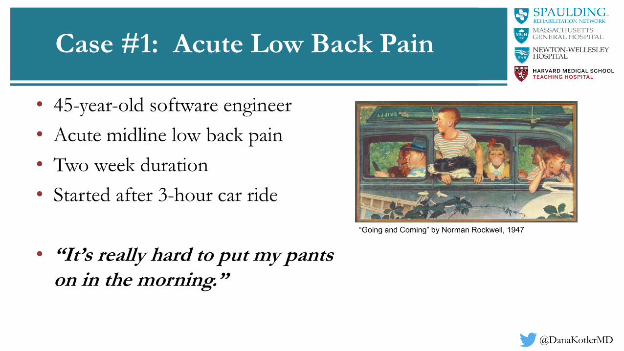
Case #1: Acute Low Back Pain
• 45-year-old software engineer
• Acute midline low back pain
• Two week duration
• Started after 3-hour car ride
• “It’s really hard to put my pants on in the morning.”
“Going and Coming” by Norman Rockwell, 1947

@DanaKotlerMD
Case #1: History
• Axial, no radiation
• Pain increases with:
– Sitting/forward bending
– Coughing/sneezing, road bumps, bowel movements
– Mornings are particularly bad
• PMH of similar self-limiting episodes, increasing in frequency, duration and intensity; otherwise healthy
• No neuro symptoms, bowel/bladder dysfunction
• No red or yellow flags

@DanaKotlerMD
Case #1: Physical Exam
• Inspection: May have side shift (lumbar listing)
• ROM: Very restricted, painful forward flexion, full nonpainful extension
• Minimal tenderness to palpation of lumbosacral spinous processes or lumbar paraspinals
• Seated slump test (+)
• Facet loading (-)
• Normal hip mobility and negative provocative maneuvers
• Normal neuro exam (strength, sensation, reflexes)
• Single leg squat with contralateral pelvic tilt and medial knee deviation bilaterally

@DanaKotlerMD
Case #1: Acute Low Back Pain
• How do we describe this?
– Acute, flexion-based,
axial low back pain
Deyo RA, Weinstein JN. Low back pain. N Engl J Med. 2001 Feb 1;344(5):363-70. Review.

@DanaKotlerMD
What is the most likely diagnosis?
• Acute, flexion-based, low back pain in a 40-year old.
1. Muscle strain
2. Discogenic pain
3. Lumbar compression fracture
4. Sacroiliac joint pain
5. I don’t know, it’s “nonspecific!”
• Severe flexion-based
acute low back pain
with associated
“lumbar shift” is
often disc related
– Annular tear
– Bulge/protrusion
– Discitis

@DanaKotlerMD
Disc pressures in different positions
• Measurement of intradiscal pressures in various positions (ouch!)
Nachemson AL. Towards a better understanding of low-back pain: a review of the mechanics of the lumbar disc. Rheumatology. 1975 Aug 1;14(3):129-43.
Nachemson AL, Morris JM. In vivo measurements of intradiscal pressure: discometry, a method for the determination of pressure in the lower lumbar discs. JBJS. 1964 Jul 1;46(5):1077-92.
Wilke HJ, Neef P, Caimi M, Hoogland T, Claes LE. New in vivo measurements of pressures in the intervertebral disc in daily life. Spine. 1999 Apr 15;24(8):755-62.

@DanaKotlerMD
Case #1: Acute Low Back Pain
• Workup:– X-rays
• Fractures
• Disc space narrowing or normal
• Transitional anatomy
– MRI• Neuro findings (weakness, reflex change)
• Red flags
• If not improving or if anatomic pathology suspected
– Discograms out of favor
– Labs • If underlying inflammatory etiology suspected
• ESR, CRP, CBC with diff

@DanaKotlerMD
Case #1: Management
• Medications– Anti-inflammatory of choice (short burst)
– Optimize sleep
• Physical Therapy– MDT/McKenzie
– Stabilization program
– Functional training
• Procedures– To reach discs/nerves → Epidural steroid
– No quality evidence for intradiscalprocedures
• Steroids, radiofrequency ablation, prolotherapy, platelet rich plasma, stem cells
• Education– Set appropriate expectations
– Avoid continuous sitting• Stand up every 20-30 minutes
– Standing desk or lumbar support
– Avoid excessive bending/lifting and improve mechanics
– Evaluate for directional preference
• Exercise– Encourage activity as tolerated
– Avoid flexion stretching
– Running? If tolerating.

@DanaKotlerMD
Spine does not stand alone
• Without muscular support, bony
structures buckle with very low
axial load (20 N or 4.5 lbs)1
• Requires stabilizing muscles of the
trunk and hip girdle
1Morris JM, Lucas DB, Bresler B. Role of the Trunk in Stability of the Spine. J Bone Joint Surg Am, 1961 Apr;43(3):327-351.
“THE CORE”
@DanaKotlerMD
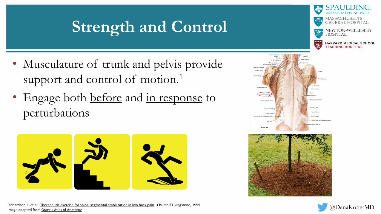
Strength and Control
• Musculature of trunk and pelvis provide
support and control of motion.1
• Engage both before and in response to
perturbations
Richardson, C et al. Therapeutic exercise for spinal segmental stabilization in low back pain. Churchill Livingstone, 1999.Image adapted from Grant’s Atlas of Anatomy

@DanaKotlerMD
“The Core” and
Functional Movement
• Perturbation– Trunk loading, limb movement
– TrA, OI, multifidi have been studied• “Abdominal hollowing” and abdominal bracing
– Increase intra-abdominal pressure, tension in thoracolumbar fascia, stiffening and stabilization
– TrA activates first • In unexpected and expected trunk loading.1
• Before the prime mover of the limb (in all directions).2
• Preprogrammed response by the CNS, in preparation for perturbation.2,3
– Dysfunction3 and reduced size4 in LBP patients3, improves with motor control exercise.4
– Proprioception and postural control altered in patients with chronic low back pain.5
• Specialized nerves in Z-joints & multifidi6,7
1Cresswell et al, 1994 3Hodges & Richardson,1996 5 Ebenbichler 2001 7 Bogduk 19972Hodges & Richardson,1997 4Ferreira & Hodges, 2010 6 Hides (in Richardson) 2004
@DanaKotlerMD
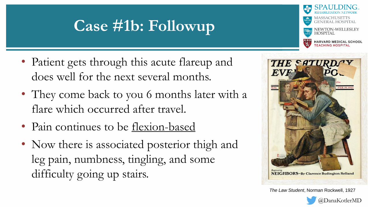
Case #1b: Followup
• Patient gets through this acute flareup and
does well for the next several months.
• They come back to you 6 months later with a
flare which occurred after travel.
• Pain continues to be flexion-based
• Now there is associated posterior thigh and
leg pain, numbness, tingling, and some
difficulty going up stairs.
The Law Student, Norman Rockwell, 1927

@DanaKotlerMD
Case #1b: Physical Exam
• Inspection: Side-shift away from the painful side
• ROM: Very restricted forward flexion with reproduction of left posterolateral leg pain
• TTP of bilateral lumbar paraspinals
• Seated slump test (+)
• Facet loading (-)
• Normal hip mobility and negative provocative maneuvers
• Reduced sensation in first web space and lateral foot
• Weakness of EHL on manual muscle testing
• Single-leg heel raises impaired on the left

@DanaKotlerMD
Case #1b: New diagnosis
• How do we describe this?
– Acute, flexion-based, _____________________
1. Lumbar spondylosis
2. Piriformis syndrome
3. Lumbar radiculopathy
4. Sacroiliac joint pain
5. I don’t know, it’s “nonspecific!”
• At this point imaging may
prove more helpful to find
the anatomic basis for the
radiculopathy
• Disc, osteophyte, other.

@DanaKotlerMD
Imaging: Radiculopathy
• Xray:
– Negative or degenerative changes
• MRI:
– Disc extrusion at L5-S1 with compression of the S1 nerve root
• There is no spinal cord in the lumbar spine!
• Sciatic nerve is not in the lumbar spine!
– May not always be this impressive!

@DanaKotlerMD
Electrodiagnostic testing
• When to do EMG/NCS?
• To discern:
– Mono vs. polyneuropathy (nerve pattern)
• i.e. leg pain and foot drop due to peroneal neuropathy
– Radiculopathy: Myotomal (nerve root pattern)
• i.e. leg pain and foot drop due to L4 radiculopathy
– Acute denervation changes vs. chronic reinnervation changes
• Prognostic value
Remember, you can hurt a lot and have a negative EMG/NCS!

@DanaKotlerMD
Radiculopathy: Management
• Ergonomics
• Exercise as tolerated
– Avoid excessive flexion if painful
– Standing exercise often OK
• Medications
– Anti-inflammatory of choice (short burst)
– Optimize sleep
• Physical Therapy
– MDT (often flexion-based)
• Injections
– To reach discs/nerves – epidural
• Surgical
– Discectomy
– Decompression

@DanaKotlerMD
Microdiscectomy for Disc Herniation

@DanaKotlerMD
Case #1c: Followup
• Patient gets through this flareup, strength returns, and does well for the next several years, using occasional medication and ongoing home PT exercise.
• They come back to you 5 years later after chronic, frequent exacerbations, and a flare which occurred after travel.
• Pain continues to be flexion-based
• Never fully remits
• Forward bending, brushing teeth, washing face have become more difficult.
• Continues to be active, but basic functional activity i.e. dressing becoming more difficult due to pain.

@DanaKotlerMD
Case #1c: Physical Exam
• Inspection: Neutral posture
• ROM: Painful but intact forward flexion
• Tender, taut bilateral lumbar paraspinals
• Seated slump test (+)
• Normal hip mobility and negative provocative maneuvers, but
tight hip flexors
• Normal neuro exam (strength, sensation, reflexes)

@DanaKotlerMD
Case #1b: New diagnosis
• How do we describe this?
– Chronic, flexion-based, axial back pain → ______________
1. Myofascial pain
2. Coccydynia
3. Sacroiliac joint pain
4. Degenerative disc disease
5. I don’t know, it’s “nonspecific!”
• At this point the
problem is no longer
pure disc pain, but now a
degenerative disc with
alteration in spinal
mechanics.

@DanaKotlerMD
Imaging: DDD
• Xray shows narrowing of disc space, endplate spurring
• MRI shows a desiccated, degenerative disc at L5-S1, may have endplate reactive marrow edema
• L4-5 also degenerative

@DanaKotlerMD
Case #1c: Management
• Medications
– Anti-inflammatory of choice (short
burst)
– Optimize sleep
• Physical Therapy
• Injections
– To reach discs/nerves – epidural
• Surgical interventions
– Lumbar fusion
• Anterior, transforaminal, posterior
• Ergonomics
– Avoid continuous sitting
– Frequent breaks
– Standing desk
– Use good body mechanics
• Exercise
– As tolerated
– Avoid flexion stretching
– Encourage strength/stability exercise

@DanaKotlerMD
Chronic Low Back Pain
Role of Surgical Intervention
• Systematic review by Phillips et al, Spine, 2013– Clinically meaningful improvement in pain and function after
lumbar fusion in selected patients with degenerative disc disease
• Lumbar fusion indications for axial lumbar pain (NASS)– Progressive deformity, pain with functional limitation
unresponsive to 1 year of conservative management
– Single level advanced disc degeneration, symptoms longer than 1 year and unresponsive to multi-disciplinary treatment, absence of significant active psychiatric disorder, no smoking for at least 3 months
Phillips FM, Slosar PJ, Youssef JA, Andersson G, Papatheofanis F. Lumbar spine fusion for chronic low back pain due to degenerative disc disease: a
systematic review. Spine (Phila Pa 1976). 2013 Apr 1;38(7):E409-22.

@DanaKotlerMD
Case #2
• 60-year-old financial advisor
• ~6 months of progressive bilateral lower back pain
• Worst with static standing
• “I hate museums.”

@DanaKotlerMD
Case #2: History
• Axial, radiates laterally and into buttocks
– No radiation beyond buttocks
• Pain increases with:
– Static standing
– Transitional movements
– Carrying objects
• Remits with:
– Sitting
– +/- Walking
• No neuro symptoms, bowel/bladder dysfunction
• No red or yellow flags
@DanaKotlerMD
Case #2: Physical Exam
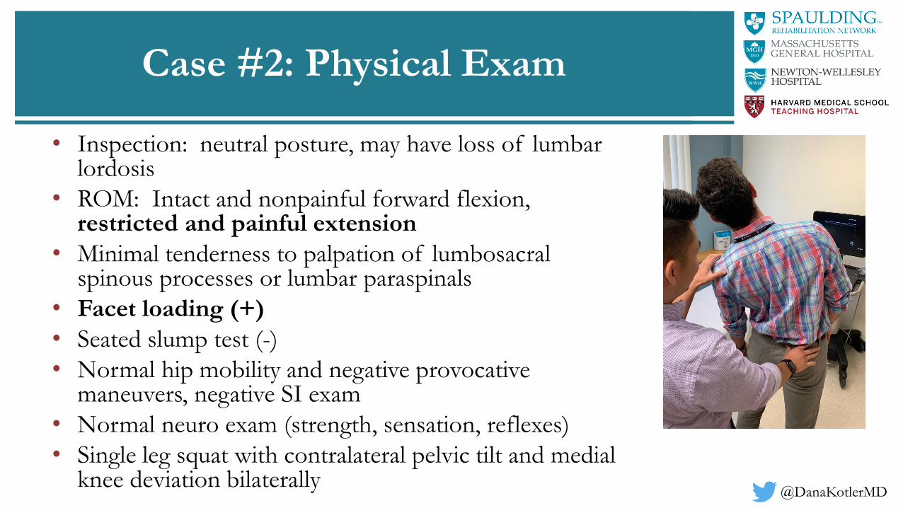
• Inspection: neutral posture, may have loss of lumbar lordosis
• ROM: Intact and nonpainful forward flexion, restricted and painful extension
• Minimal tenderness to palpation of lumbosacral spinous processes or lumbar paraspinals
• Facet loading (+)
• Seated slump test (-)
• Normal hip mobility and negative provocative maneuvers, negative SI exam
• Normal neuro exam (strength, sensation, reflexes)
• Single leg squat with contralateral pelvic tilt and medial knee deviation bilaterally

@DanaKotlerMD
Case #2: Impression
• How do we describe this?
– Chronic, extension-based, axial low back pain
1. Muscle strain
2. Discogenic pain
3. Degenerative disc disease
4. Facet-mediated pain
5. I don’t know, it’s “nonspecific!”
• People with facet pain
often have pain with
static positions,
particularly standing.
– Cocktail parties
– Museums
– Shopping

@DanaKotlerMD
Case #2: Imaging
• MRI shows facet joint arthritis, moderate L4/5 stenosis

@DanaKotlerMD
Facet-mediated pain: Management
• Medications
– Anti-inflammatory, analgesic
– Optimize sleep
• Physical Therapy
• Injections
– Facet joint injections
– Medial branch blocks
– Radiofrequency lesioning
of medial branch
• Ergonomics
– Avoid continuous
standing
• Exercise
– As tolerated
– Avoid extension/rotation
stretching

@DanaKotlerMD
Facet Joint Procedures
• Facetogenic (zygapophyseal joint) pain
– Limited evidence on intra-articular injections2
– Low to moderate quality evidence for
radiofrequency ablation1,2
• Short term pain relief
• Functional improvement
1Poetscher AW, Gentil AF, Lenza M, Ferretti M. Radiofrequency denervation for facet joint low back pain: a systematic review. Spine (Phila Pa 1976). 2014 Jun 15;39(14):E842-9.2Falco FJ et al. An update of the effectiveness of therapeutic lumbar facet joint interventions. Pain Physician. 2012 Nov-Dec;15(6):E909-53. Review.
@DanaKotlerMD
Case #3
• 75 year old woman with chronic and progressive bilateral buttock,
thigh, and leg pain, exacerbated by standing and walking.
• “I have to sit down and rest.”

@DanaKotlerMD
Case #3: History
• Bilateral buttock, thigh, and leg
pain
• Pain increases with:
– Walking
• Remits with:
– Sitting
• No neuro symptoms,
bowel/bladder dysfunction
• No red or yellow flags
– Incontinence, profound or
progressive weakness
@DanaKotlerMD
Case #3: Physical Exam
• Inspection: stooped posture, loss of lumbar lordosis
• ROM: Restricted in extension with reproduction of leg pain
• Minimal tenderness to palpation of lumbosacral spinous processes or lumbar paraspinals
• Normal hip mobility and negative provocative maneuvers, negative SI exam
• Neuro exam: may be normal or subtle myotomal weakness unilaterally or bilaterally
• Gait: Forward flexed

@DanaKotlerMD
Case #3: Impression
• How do we describe this?
– Chronic, extension-based, radicular pain/claudication
1. Lumbar spinal stenosis
2. Discogenic pain
3. Degenerative disc disease
4. Facet-mediated pain
5. I don’t know, it’s “nonspecific!”
• What is causing the stenosis and where is it?
• Central canal vs. foraminal stenosis
• Neurodegenerative and vascular disease also in differential

@DanaKotlerMD
Spinal Stenosis
• Symptoms
– Back, buttock, leg, or foot pain
– Numbness/tingling in leg/foot
– Weakness of the leg or foot
– Can be one-sided or both legs
@DanaKotlerMD
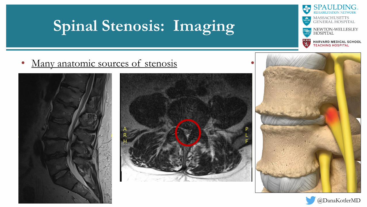
Spinal Stenosis: Imaging
• Many anatomic sources of stenosis • Common:
– Combination of
congenitally
narrowed canal, disc
bulge, and facet
hypertrophy
@DanaKotlerMD
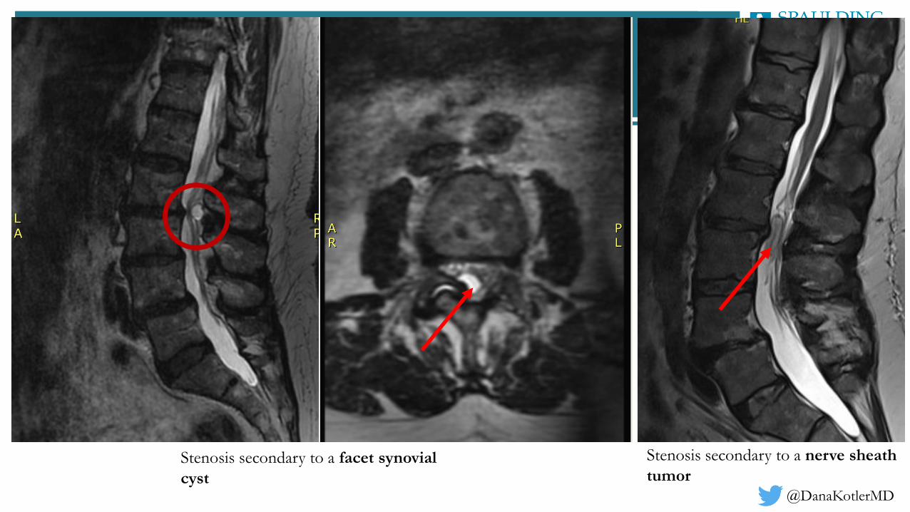
Stenosis secondary to a facet synovial
cyst
Stenosis secondary to a nerve sheath
tumor

@DanaKotlerMD• Stenosis related to spondylolisthesis and facet arthropathy
@DanaKotlerMD
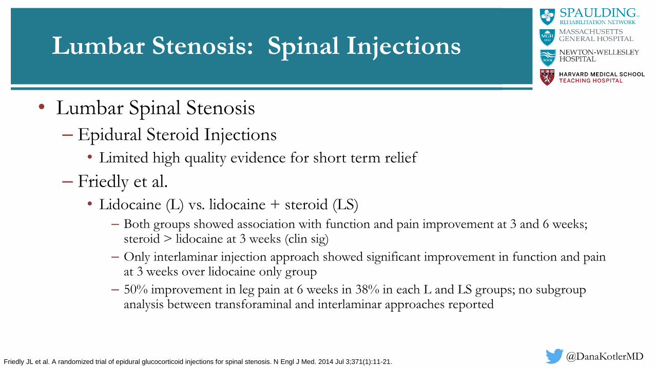
Lumbar Stenosis: Spinal Injections
• Lumbar Spinal Stenosis
– Epidural Steroid Injections
• Limited high quality evidence for short term relief
– Friedly et al.
• Lidocaine (L) vs. lidocaine + steroid (LS)– Both groups showed association with function and pain improvement at 3 and 6 weeks;
steroid > lidocaine at 3 weeks (clin sig)
– Only interlaminar injection approach showed significant improvement in function and pain at 3 weeks over lidocaine only group
– 50% improvement in leg pain at 6 weeks in 38% in each L and LS groups; no subgroup analysis between transforaminal and interlaminar approaches reported
Friedly JL et al. A randomized trial of epidural glucocorticoid injections for spinal stenosis. N Engl J Med. 2014 Jul 3;371(1):11-21.

@DanaKotlerMD
Lumbar Stenosis: Surgery
• Laminectomy: traditional gold standard for central stenosis
• *Advantage: complete wide decompression of neural structures with excellent visualization
• RISK: iatrogenic instability
• Most case series report > 85% good to excellent results after lumbar decompression
L3 Spinous process
Cauda Equina
Nerve Root
Facet
Disc Space
Verbiest HE. Results of surgical treatment of idiopathic developmental stenosis of the lumbar vertebral canal. A review of twenty-seven years' experience. The Journal of bone and joint surgery. British volume. 1977 May;59(2):181-8.Athiviraham A, Yen D. Is spinal stenosis better treated surgically or nonsurgically?. Clinical Orthopaedics and Related Research®. 2007 May 1;458:90-3.Malmivaara A, Slätis P, Heliövaara M, Sainio P, Kinnunen H, Kankare J, Dalin-Hirvonen N, Seitsalo S, Herno A, Kortekangas P, Niinimäki T. Surgical or nonoperative treatment for lumbar spinal stenosis?: a randomized controlled trial. Spine. 2007 Jan 1;32(1):1-8.

@DanaKotlerMD
Key Points
• Evaluation of back pain involves a detailed history, physical examination, and appropriate use of imaging.
• Diagnosis should be based on pattern of symptoms and then confirmed with appropriate imaging.
• Management of lumbar spine disorders is multifactorial, including medications, exercise, physical therapy, interventional injections, and sometimes surgery.
• Patient education on managing symptoms and preventing recurrence is essential.
@DanaKotlerMD
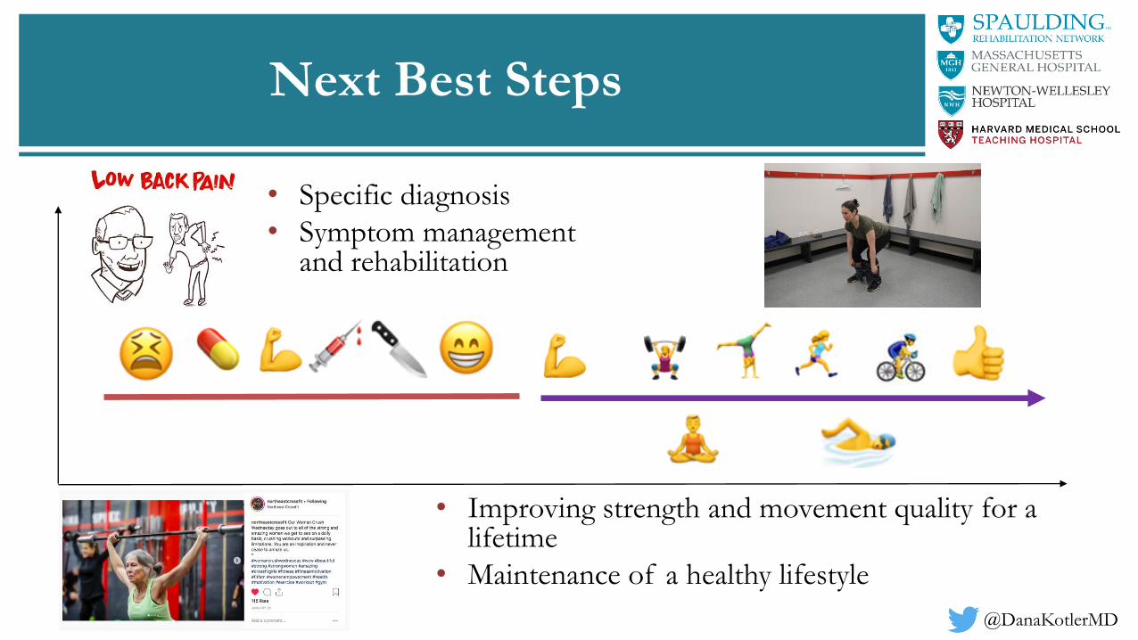
Next Best Steps
• Specific diagnosis
• Symptom management and rehabilitation
• Improving strength and movement quality for a lifetime
• Maintenance of a healthy lifestyle

@DanaKotlerMD
References
1. Bernstein IA, Malik Q, Carville S, Ward S. Low back pain and sciatica: summary of NICE guidance. BMJ. 2017 Jan 6;356:i6748.
2. Bogduk N: Clinical Anatomy of the Lumbar Spine and Sacrum. Edinburgh, Churchill Livingstone, 1997
3. Brinjikji W, Luetmer PH, Comstock B, Bresnahan BW, Chen LE, Deyo RA, Halabi S, Turner JA, Avins AL, James K, Wald JT. Systematic literature review of imaging features of spinal degeneration in asymptomatic populations. American Journal of Neuroradiology. 2014 Nov 27.
4. Brinjikji W, Diehn FE, Jarvik JG, et al. MRI Findings of disc degeneration are more prevalent in adults with low back pain than in asymptomatic controls: asystematic review and meta-analysis. Am J Neuroradio 2015; 36: 2394–99.
5. Choi BK, Verbeek JH, Tam WW, Jiang JY. Exercises for prevention of recurrences of low-back pain. Cochrane Database Syst Rev. 2010 Jan 20;(1):CD006555.
6. Chou R, Huffman LH; Nonpharmacologic therapies for acute and chronic low back pain: a review of the evidence for an American Pain Society/American College of Physicians clinical practice guideline. Ann Intern Med. 2007 Oct 2;147(7):492-504. Review.
7. Cresswell AG, Oddsson L, Thorstensson A. The influence of sudden perturbations on trunk muscle activity and intra-abdominal pressure while standing. Experimental Brain Research. 1994;98(2):336-41.
8. CUENCA-MARTÍNEZ F, CORTÉS-AMADOR S, ESPÍ-LÓPEZ GV. Effectiveness of classic physical therapy proposals for chronic non-specific low back pain: a literature review. Physical Therapy Research. 2018:E9937.
9. Deyo RA, Weinstein JN. Low back pain. N Engl J Med. 2001 Feb 1;344(5):363-70. Review.
10. Ebenbichler GR, Oddsson LI, Kollmitzer J, et al: Sensorymotor control of the lower back: Implications for rehabilitation. Med Sci Sports Exerc 2001;33:1889–98.
11. Enthoven WT, Roelofs PD, Deyo RA, van Tulder MW, Koes BW. Non‐steroidal anti‐inflammatory drugs for chronic low back pain. Cochrane Database of Systematic Reviews. 2016(2).
12. Falco FJ et al. An update of the effectiveness of therapeutic lumbar facet joint interventions. Pain Physician. 2012 Nov-Dec;15(6):E909-53. Review.
13. Ferreira PH, Ferreira ML, Maher CG, Refshauge K, Herbert RD, Hodges PW. Changes in recruitment of transversus abdominis correlate with disability in people with chronic low back pain. Br J Sports Med. 2010 Dec;44(16):1166-72.
14. Friedly JL et al. A randomized trial of epidural glucocorticoid injections forspinal stenosis. N Engl J Med. 2014 Jul 3;371(1):11-21.
15. Gianola S, Castellini G, Corbetta D, Moja L. Rehabilitation interventions in randomized controlled trials for low back pain: proof of statistical significance often is not relevant. Health and quality of life outcomes. 2019 Dec;17(1):127. Published online 2019 Jul 22.
@DanaKotlerMD
References
1. Gimbel J, Linn R, Hale M, Nicholson B. Lidocaine patch treatment in patients with low back pain: results of an open-label, nonrandomized pilot study. Am J Ther. 2005 Jul-Aug;12(4):311-9.
2. Goldberg H, et al. Oral steroids for acute radiculopathy due to a herniated lumbar disk: a randomized clinical trial. JAMA. 2015 May 19;313(19):1915-23.
3. Goldberg RJ, Katz J. A meta-analysis of the analgesic effects of omega-3 polyunsaturated fatty acid supplementation for inflammatory joint pain. Pain. 2007 May;129(1-2):210-23. Epub 2007 Mar 1.
4. Haskins R, Osmotherly PG, Rivett DA. Diagnostic clinical prediction rules for specific subtypes of low back pain: a systematic review. journal of orthopaedic & sports physical therapy. 2015 Feb;45(2):61-76.
5. Hides J: Paraspinal mechanism and support of the lumbar spine, in: Richardson C (ed): Therapeutic Exercise for Lumbopelvic Stabilization, ed 2. Edinburgh, Churchill Livingstone, 2004, pp 59–74.
6. Hodges PW, Richardson CA. Contraction of the abdominal muscles associated with movement of the lower limb. Physical Therapy. 1997 Feb;77(2):132-42; discussion 142-4.
7. Hodges PW, Richardson CA. Inefficient muscular stabilization of the lumbar spine associated with low back pain. A motor control evaluation of transversusabdominis. Spine. 1996 Nov 15;21(22):2640-50.
8. Jensen TS, Albert HB, Soerensen JS, et al. Natural course of disc morphology in patients with sciatica: an MRI study using a standardized qualitative classification system. Spine (Phila Pa 1976) 2006;31(14): 1605–12.
9. Kuptniratsaikul V et al. Efficacy and safety of Curcuma domestica extracts compared with ibuprofen in patients with knee osteoarthritis: a multicenter study. Clin Interv Aging. 2014 Mar 20;9:451-8. doi: 10.2147/CIA.S58535. eCollection 2014.
10. Lee YH, Bae SC, Song GG. Omega-3 Polyunsaturated Fatty Acids and the Treatment of Rheumatoid Arthritis: A Meta-analysis. Arch Med Res. 2012 Jul;43(5):356-62. doi: 10.1016/j.arcmed.2012.06.011. Epub 2012 Jul 24.
11. Machado LA, De Souza MV, Ferreira PH, Ferreira ML. The McKenzie method for low back pain: a systematic review of the literature with a meta-analysis approach. Spine. 2006 Apr 20;31(9):E254-62.
12. Mok NW, Brauer SG, Hodges PW: Hip strategy for balance control in quiet standing is reduced in people with low back pain. Spine 2004;29:E107–12.
13. Morris JM, Lucas DB, Bresler B. Role of the Trunk in Stability of the Spine. J Bone Joint Surg Am, 1961 Apr;43(3):327-351.
14. Nachemson AL, Morris JM. In vivo measurements of intradiscal pressure: discometry, a method for the determination of pressure in the lower lumbar discs. JBJS. 1964 Jul 1;46(5):1077-92.
15. Nachemson AL. Towards a better understanding of low-back pain: a review of the mechanics of the lumbar disc. Rheumatology. 1975 Aug 1;14(3):129-43.

@DanaKotlerMD
References
1. Oliveira CB, Maher CG, Pinto RZ, Traeger AC, Lin CW, Chenot JF, van Tulder M, Koes BW. Clinical practice guidelines for the management of non-specific low back pain in primary care: an updated overview. European Spine Journal. 2018 Jul 3:1-3.
2. O’Sullivan PB, Burnett A, Floyd AN, et al: Lumbar repositioning deficit in a specific low back pain population. Spine 2003;28:1074–9 -
3. Petersen T, Laslett M, Thorsen H, Manniche C, Ekdahl C, Jacobsen S. Diagnostic classification of non-specific low back pain. A new system integrating patho-anatomic and clinical categories. Physiotherapy Theory and Practice. 2003 Jan 1;19(4):213-37.
4. Petersen T, Laslett M, Juhl C. Clinical classification in low back pain: best-evidence diagnostic rules based on systematic reviews. BMC musculoskeletal disorders. 2017 Dec;18(1):188.
5. Phillips FM, Slosar PJ, Youssef JA, Andersson G, Papatheofanis F. Lumbar spine fusion for chronic low back pain due to degenerative disc disease: a systematic review. Spine (Phila Pa 1976). 2013 Apr 1;38(7):E409-22.
6. Poetscher AW, Gentil AF, Lenza M, Ferretti M. Radiofrequency denervation for facet joint low back pain: a systematic review. Spine (Phila Pa 1976). 2014 Jun15;39(14):E842-9.
7. Rasmussen‐Barr E, Held U, Grooten WJ, Roelofs PD, Koes BW, van Tulder MW, Wertli MM. Non‐steroidal anti‐inflammatory drugs for sciatica. Cochrane Database of Systematic Reviews. 2016(10).
8. Richardson, C et al. Therapeutic exercise for spinal segmental stabilization in low back pain. Churchill Livingstone, 1999.
9. Roelofs PD, Deyo RA, Koes BW, Scholten RJ, van Tulder MW. Non‐steroidal anti‐inflammatory drugs for low back pain. Cochrane database of systematic reviews. 2008(1).
10. Stynes S, Konstantinou K, Dunn KM. Classification of patients with low back-related leg pain: a systematic review. BMC musculoskeletal disorders. 2016Dec;17(1):226.
11. Wasserman MS, Guermazi A, Jarraya M, Engbretsen L, Abdalkader M, Roemer FW, Hayashi D, Crema MD, Mian AZ. Evaluation of spine MRIs in athletes participating in the Rio de Janeiro 2016 Summer Olympic Games. BMJ open sport & exercise medicine. 2018 Feb 1;4(1):e000335.
12. Weinstein JN, Lurie JD, Tosteson TD, Tosteson AN, Blood E, Abdu WA, Herkowitz H, Hilibrand A, Albert T, Fischgrund J. Surgical versus non-operative treatment for lumbar disc herniation: four-year results for the Spine Patient Outcomes Research Trial (SPORT). Spine. 2008 Dec 1;33(25):2789.
13. Wilder DG, Aleksiev AR, Magnusson ML, et al: Muscular response to sudden load: A tool to evaluate fatigue and rehabilitation. Spine 1996;21:2628–39.
14. Wilke HJ, Neef P, Caimi M, Hoogland T, Claes LE. New in vivo measurements of pressures in the intervertebral disc in daily life. Spine. 1999 Apr 15;24(8):755-62.