Embed Size (px)
Citation preview
Normal Intracardiac Pressures
Lancashire & South Cumbria Cardiac Network

Principle
• Pressures recorded from catheter tip
• Electrical transducer - wheatstone bridge
• mechanical to electrical waveform
• display - ECG, Intracardiac pressures, O2sat.

Purpose
• Measure intracardiac pressures• assess intracardiac blood flow• assess ventricular function• determine cardiac anatomy• assess valvular function• assess pulmonary and systemic circulatory
systems

Left Heart Catheterisation
• Aorta
• Left ventricle

Aortic pressure

• Systolic value - maximum pressure achieved by the left ventricle during systole
• Aorta is a strong, thick walled vessel -diastole the aortic pressure does not drop to zero but is maintained to a higher value
• This enables the pressure to be such that even at the peripheries, all cells are supplied with oxygen
Peak systole
• starts opening of the aortic valve• A sharp upstroke is seen on the pressure
tracing, which reflects ejection of blood from the left ventricle
• Upstroke is referred to as the ascending limb.

Anachrotic Notch
• During the first phase of ventricular systole (isovolumetric contraction), a presystolicrise may be seen - anachrotic notch
• Anachrotic notch - occurs before the opening of the aortic valve
Dichrotic notch
• With greater pressure in the aorta than the left ventricle, blood flow attempts to equalize by flowing backwards - results in closure of the aortic valve.
• dicrotic notch - this event marks the end of systole and the start of diastole

Diastolic pressure
• This value relates to the amount of recoil in the arterial system
• fast heart rate - shorter diastolic time, less time for run off into the more distal branches, leading to a higher diastolic pressure
• decline in pressure during diastole -descending limb

Pulse Pressure
• The difference between the systolic and diastolic pressure
• Factors that affect pulse pressure– changes in stroke volume– aortic regurgitation– changes in vascular compliance

Aortic Pressure
Peak measurement
Dichrotic notch
Anachrotic Notch
AORTA Normal systolic pressure = 120 mmHg (100 -140) Normal diastolic pressure = 70 mmHg (60 - 90)
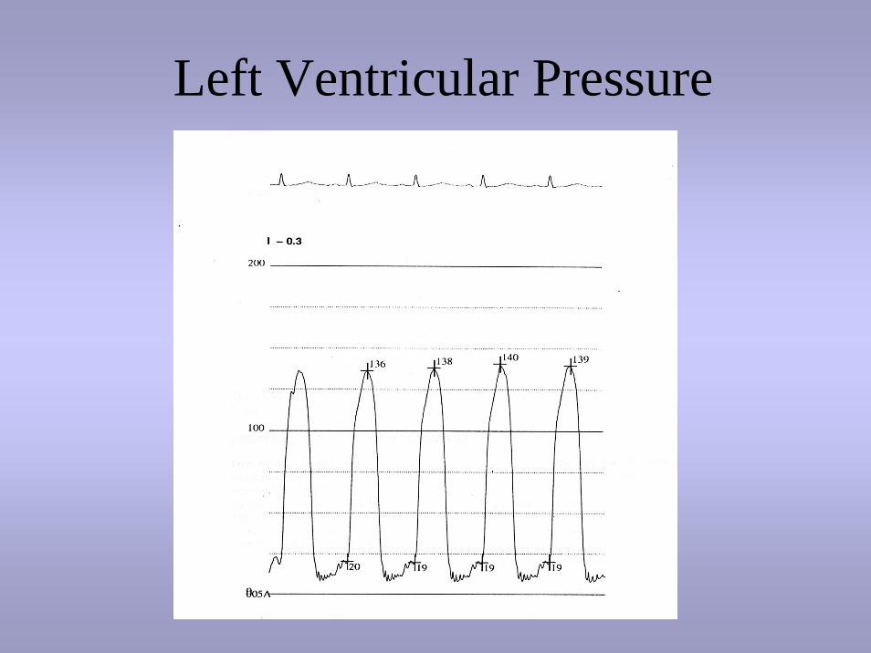
Left Ventricular Pressure

Ventricular systole
• Isovolumetric contraction (after closure of the mitral valve and before opening of the aortic valve) rapid rise in pressure until it exceeds that of the aortic pressure and the aortic valve opens
• Ejection phase (opening to the closing of the aortic valve) blood flows into the aorta until aortic valve closure

• Systolic ejection phase - QT interval on the ECG
• LV systolic pressure is measured at the peak pressure of the ejection phase

Isovolumetric contractionPeak measurement
LEFT VENTRICLE Normal systolic pressure = 120 mmHg (100-140)Normal diastolic pressure = 0-10(pre),
0-20 (post) mmHg

Ventricular Diastole
• relaxation of the ventricle (aortic valve closes and the mitral valve opens, allowing ventricular filling)
• Diastasis - later, slower period of filling when the left ventricle is nearly full. The pressure in the left ventricle is equal to the pressure in the left atrium. This filling continues until ejection occurs.

• Just before systole, is the point of the ventricular End Diastolic pressure measurement
• End Diastolic pressure can be measured on the R wave of the ECG, which will coincide just after the ‘a’ wave on the LV trace. This is called the post ‘a’ wave measurement of EDP.

End diastolic
R wave

Right heart catheterisation
• RA
• RV
• PA
• PCWP

Right Atrial Pressure

Electrical precedes mechanical• ‘a’ wave increase in pressure during atrial contraction
(PR interval- ECG)• ‘x’ descent the fall in pressure following the a wave
(represents atrial relaxation)• ‘c’ wave may occur as an interruption to the x descent
and represents the movement of the AV valve towards the atrium during valve closure. (RS-T junction- ECG)
• ‘v’ wave increase in pressure during ventricular systole, with bulging of the AV valve into the atrium. (T-P interval -ECG)
• ‘y’ descent the fall in pressure following the v wave (represents opening of the AV valve)

Right Atrial Pressure
RIGHT ATRIUM normal mean pressure < 5mmHg
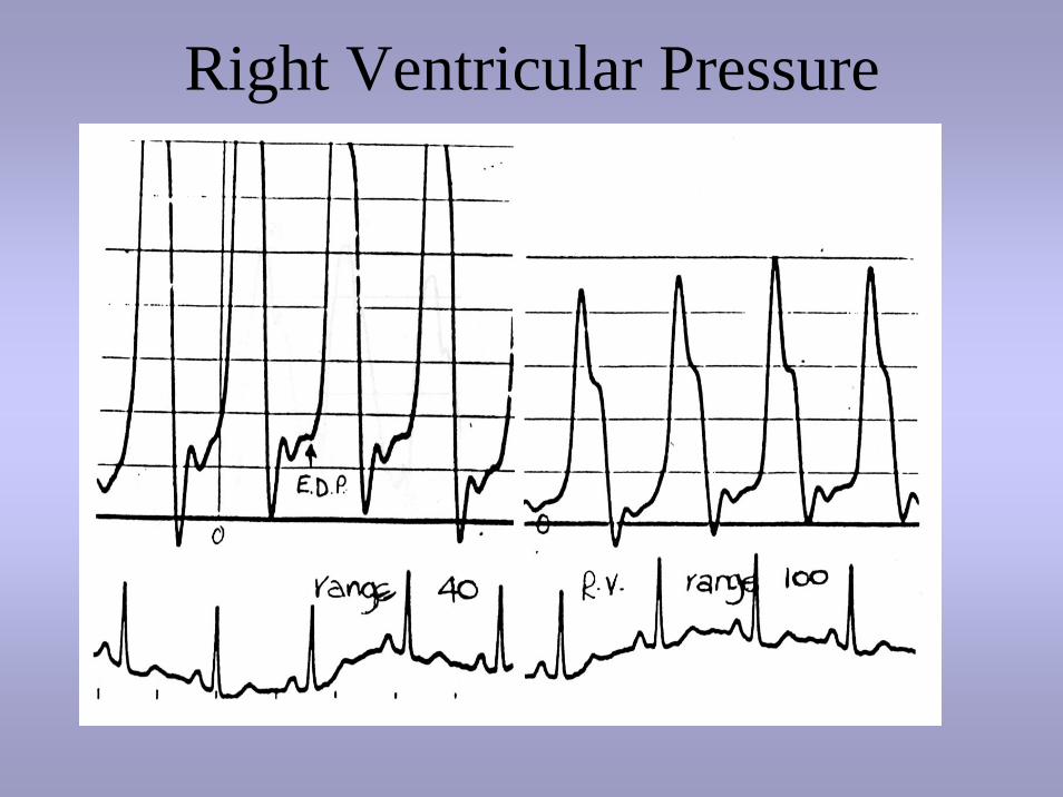
Right Ventricular Pressure

Ventricular systole
• Isovolumetric contraction (after closure of the tricuspid valve and before opening of the pulmonary valve) rapid rise in pressure until it exceeds that of the pulmonary artery pressure and the pulmonary valve opens
• Ejection phase (opening to the closing of the pulmonary valve) blood flows into the pulmonary artery until pulmonary valve closure

• Systolic ejection phase - QT interval on the ECG
• RV systolic pressure is measured at the peak pressure of the ejection phase

Right Ventricular Pressure
RIGHT VENTRICLE NORMAL SYSTOLIC PRESSURE < 25 MMHG NORMAL DIASTOLIC PRESSURE < 5 MMHG
Peak measurement

Ventricular Diastole
• relaxation of the ventricle (pulmonary valve closes and the tricuspid valve opens, allowing ventricular filling)
• Diastasis - later, slower period of filling when the right ventricle is nearly full. The pressure in the right ventricle is equal to the pressure in the right atrium. This filling continues until ejection occurs.

• Just before systole, is the point of the ventricular End Diastolic pressure measurement (QT timing - ECG)
• End Diastolic pressure can be measured on the R wave of the ECG, which will coincide just after the ‘a’ wave on the RV trace. This is called the post ‘a’ wave measurement of EDP.

RIGHT VENTRICLE NORMAL SYSTOLIC PRESSURE < 25 MMHG NORMAL DIASTOLIC PRESSURE < 5 MMHG

Pulmonary Artery Pressure

Pulmonary Artery Pressure
• Systolic phase - steep rise occurring during the Right Ventricular ejection after the opening of the pulmonary valve.
• This rise is followed by a general decrease in pressure whilst blood is being ejected from the right ventricle until closure of the pulmonary valve.
• Closure pulmonary valve - diachrotic notch

Pulmonary Artery Pressure
PULMONARY ARTERY normal systolic pressure < 25 mmHg normal diastolic pressure < 10 mmHg normal mean pressure < 15 mmhg
Dichrotic notch
Peak measurement

Pulmonary Capillary Wedge Pressure

Pulmonary Capillary Wedge Pressure
• difficult to catheterise left atrium retrogradely • assessment left atrial pressure - assumption that
the pressure in the pulmonary capillaries in the lungs are the same as that of the left atrium
• Placement of a catheter, ‘wedged’ into the pulmonary capillaries gives an ‘indirect’ left atrial pressure measurement.
• Direct left atrial measurement can be made via a PFO (patent foramen ovale) or using the transeptaltechnique incorporating the brockenboroughtechnique (puncture)
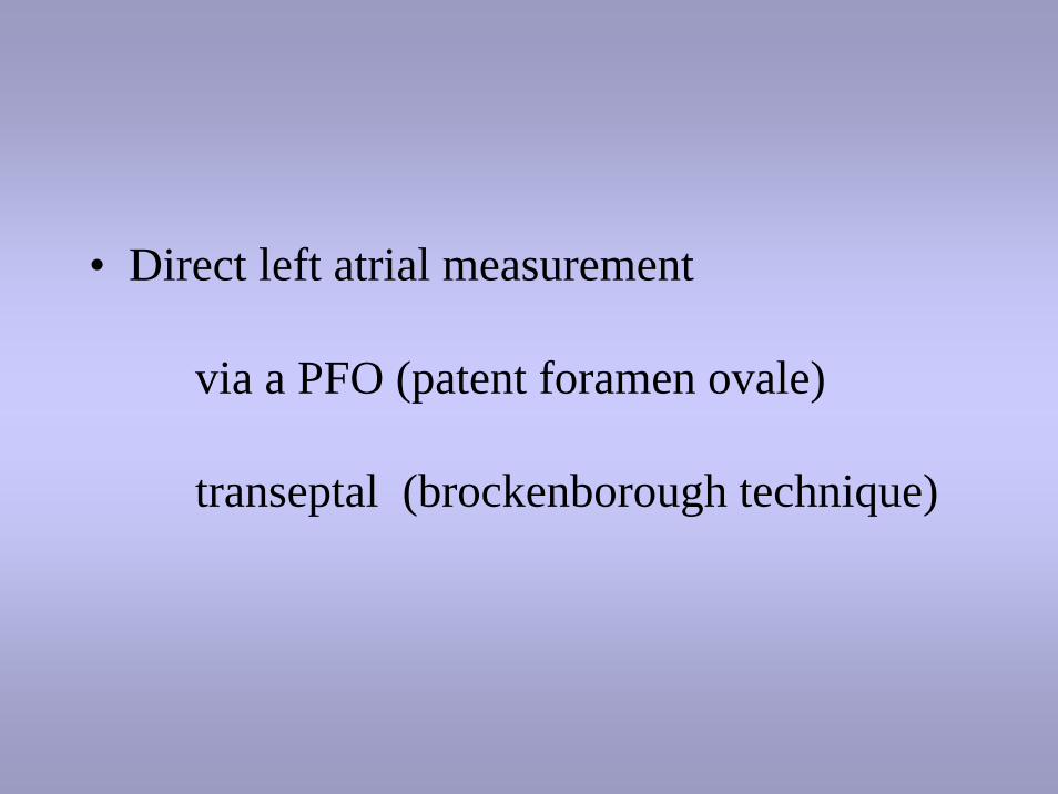
• Direct left atrial measurement
via a PFO (patent foramen ovale)
transeptal (brockenborough technique)

• Waveform is similar to RA - slightly higher• ‘a’ wave (P wave ECG) may be slightly
delayed due to retrograde transmission through pulmonary vasculature
• ‘c’ wave reflects closure of the AV valve at the start of ventricular systole. This small pressure change is often not seen in the PCW trace
• ‘v’ wave filling of the left atrium & bulging of the AV valve back into LA during ventricular systole (T wave - ECG)

PULMONARY CAPILLARY WEDGE normal mean pressure < 12mmHg

Summary
• LHC– Aortic pressure– LV Pressure
• RHC– RA Pressure– RV Pressure– PA Pressure– PCW Pressure
• Measure intracardiacpressures
• Assess intracardiac blood flow
• assess ventricular function• determine cardiac anatomy• assess valvular function• assess pulmonary and
systemic circulatory systems
In conjunction – screening, angiography, O2 saturation
& CO measurements