Embed Size (px)
Citation preview

Dr Etienne Leroy Terquem – Pr Pierre L’Her
SPI / ISP Soutien Pneumologique Internationa / International Support for Pulmonology
NORMAL
CHEST X RAY

THE CHEST RADIOGRAPHY (CXR)
• The CXR, is very usefull for all lung problems,
it extends the clinical examination and provides essential data for diagnosis
• Its technical realization, simple, uses X-rays
Principle : 1. The rays emitted by the X-ray tube through the chest
2. They are absorbed or mitigated to varying degrees by
the different structures crossings
3. The changes are recorded on X-ray film in a cassette
lead, then visualized (developing hand or in a machine
after opening the cassette in the darkroom)

Chest X ray
• Reproduce 1 volume (thorax) on 1 plan (film)
• On this flat reproduction, you must recognize normal anatomic structures of the thorax
• and find out what is abnormal
X Rays

Calcium Water Oil Air
Radiography of 4 identical tubes containing calcium, water, oil, air
One containing Ca stop X-rays => its image is white
Air is transparent to X-rays => its radiological picture is black
Fat and water have an intermediate absorption => white - gray
Opacity Clarity

The thorax is composed of:
• Bone (vertebrae, ribs, scapula…). The main component is calcium, which absorbs the x-ray considerably: the bone image is very opaque (white on the radiography)
• Blood and soft tissue (heart, mediastinum, vessels). The absorption of x-rays is less complete than bones: the image is less opaque (light grey)
• Fat tissue. the absorption of x-rays is lower: the image is dark grey
• Air (in lungs) which does not absorb the x-ray at all. The image of the lungs is black

calcium
Fluid / soft
tissu
Fat
air

The chest X ray
• 3 constants regulate its quality:
• the quantity of electrons which pass through the chest (milliampere)
• the speed of the electron (kilovoltage)
• the exposure time
• A chest X-ray of good quality requires :
• high voltage (120-140 kV) => Contrast
• a short time of exposure
• deep inspiration
• A long distance between X-ray tube focus and film
0.05 sec. Movement artefacts
Difference of density
levels of the different
organs and tissues
in the thorax

How to obtain a good quality chest X-ray
A long distance between X-ray tube focus and film
improves picture clarity and reduces geometric blur

• Good quality electrical power supply
• Effective & frequent maintenance of equipment
• Quality films and good storage conditions
• Good film processing (development, rinsing, fixing, washing, drying). If possible automatic developing machine
• Quality of the x-ray grid: the flat metallic plate with very narrow lead trips close to the film: increase in the image clarity and reduction of the scattered radiation from the patient
How to obtain a good quality chest X-ray
Other criteria

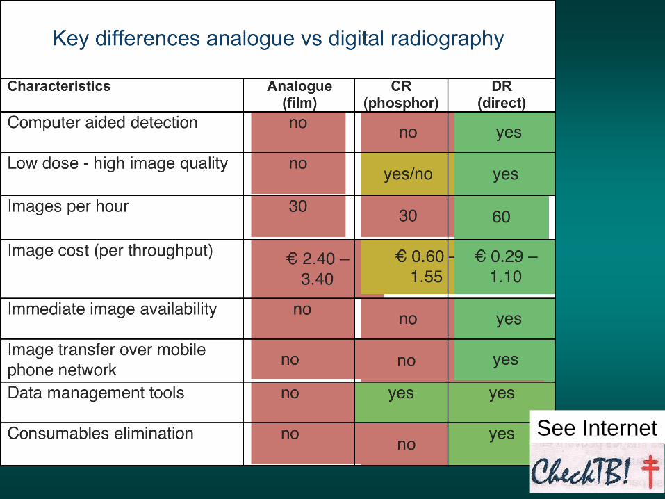
And digital radiography?
• The X-ray tube is identical
• the radiographic film is replaced by an X-ray receiver which
enables scanning in numeric
• The image display is done in grayscale high resolution on a
powerful computer
For computed radiography (CR) a reusable cassette
containing a luminescent screen memory ("phosphor plates")
introduced in a reader connected to the computer
For direct "digital“ Radiography (DR) direct scanning
receiver that transmits the image directly on the computer
*
*

And digital radiography?
• The X-ray tube is identical
• the radiographic film is replaced by an X-ray receiver which
enables scanning in numeric
• The image display is done in grayscale high resolution on a
powerful computer
*
X-Ray Tube
• Analog Rx Film - Chemical Development
• CR Reusable plate with radio-luminescent
memory screen + reader
• DR Electronic panel X-ray detector

Advantages : Quick and easy image processing
Adjusting the picture quality (Processing)
No films, No chemicals No darkroom
Data Storage
Transfer remote to an expert "telemedicine“
Digital Radiography
Disadvantages: Expensive initial investment (100 000 to 300 000 U.S.).
But prices decrease ..
Need for training for operators of Radiology
Need for maintenance

But prices decreases ,especially for DDR.

Clearly WHO recommands X-ray D DR and Xpert
See Internet
See Internet

Digital X ray system (Direct Digitalised System)
composed with:
• Electronic flat-panel X ray detector
• High resolution grayscale diagnostic display
• High performance computer
In summary
The top!,expansive
but prices decreases

comprises:
A flat panel electronic X-ray detector
Display high resolution grayscale
A high performance computer
Direct digital radiography (DR)

Transportable DR unit

Possible compromise with computed
radiography (CR system)
• Digital radiography using a Photo‐stimulable Phosphor
plate (PSP, also called Imaging Plate, IP) enclosed in a
cassette as a detector, instead of a film‐screen.
• The IP is then introduced into a CR Reader which reads
the film and converts the recorded signal into a digital
grey scale image
• Same avantages than digitalised system
• Lower cost than DR system, because can be used
with the existing X ray equipment.
Only a CR reader and CR cassette (with Imaging Plates)
need to be purchased

CENAT Phnom Penh Cambodge Dr Peou Satha
Computed Radiograpy (CR)
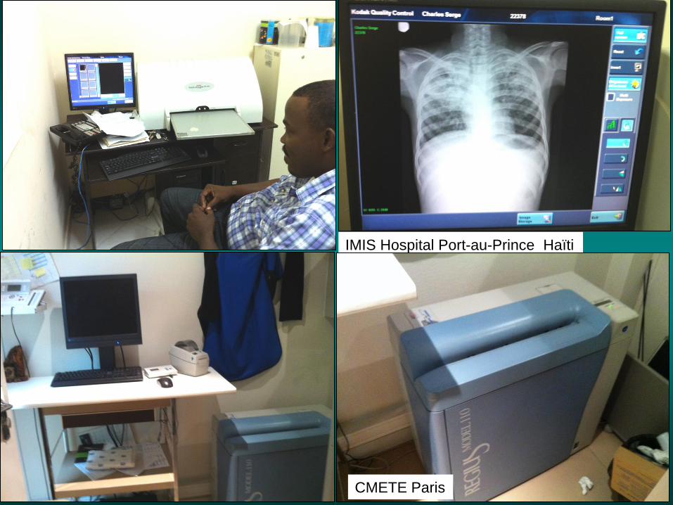
IMIS Hospital Port-au-Prince Haïti
CMETE Paris
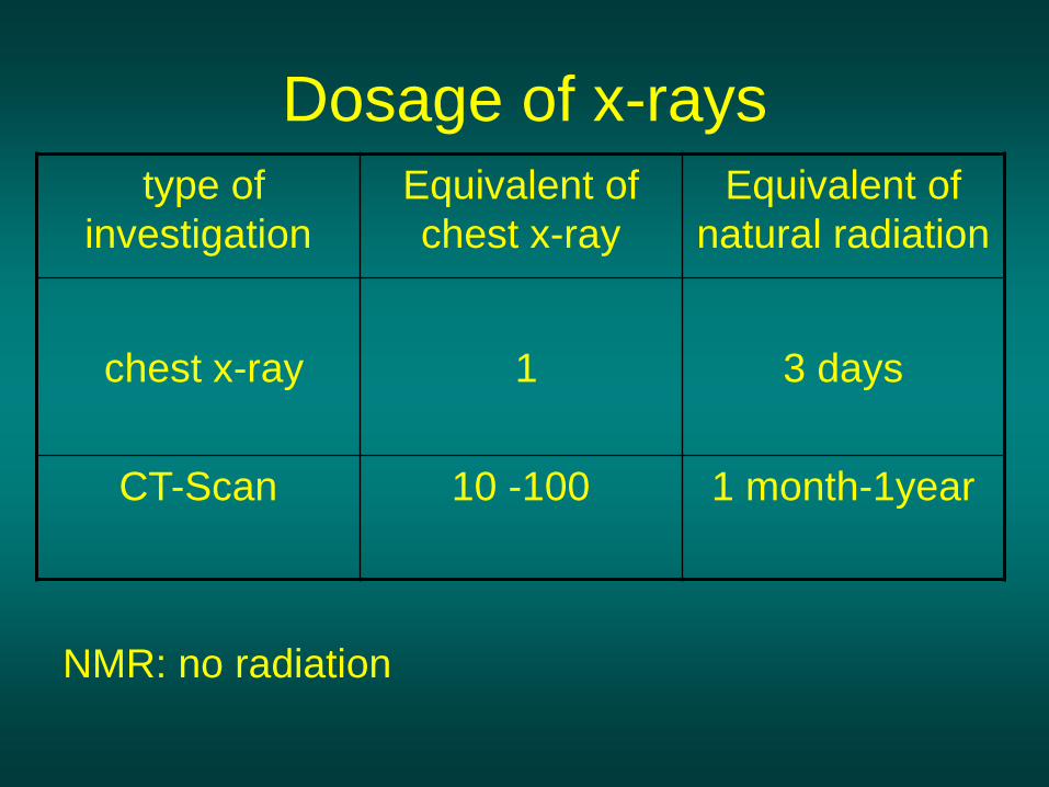
Dosage of x-rays
type of
investigation
Equivalent of
chest x-ray
Equivalent of
natural radiation
chest x-ray
1
3 days
CT-Scan 10 -100 1 month-1year
NMR: no radiation

Front view : Postero - anterior view
Lateral view
Other views :
Expiration Radiography
CXR in suppine position
Radiography in suppine and lateral position
Back view – Antero-posterior view
CXR - Basic radiographic views:

CXR - Quality Criteria
Deep inspiration
Adequate density / contrast
Good position of the patient (strictly front)
X-ray beam in postero-anterior incidence
(patient standing)

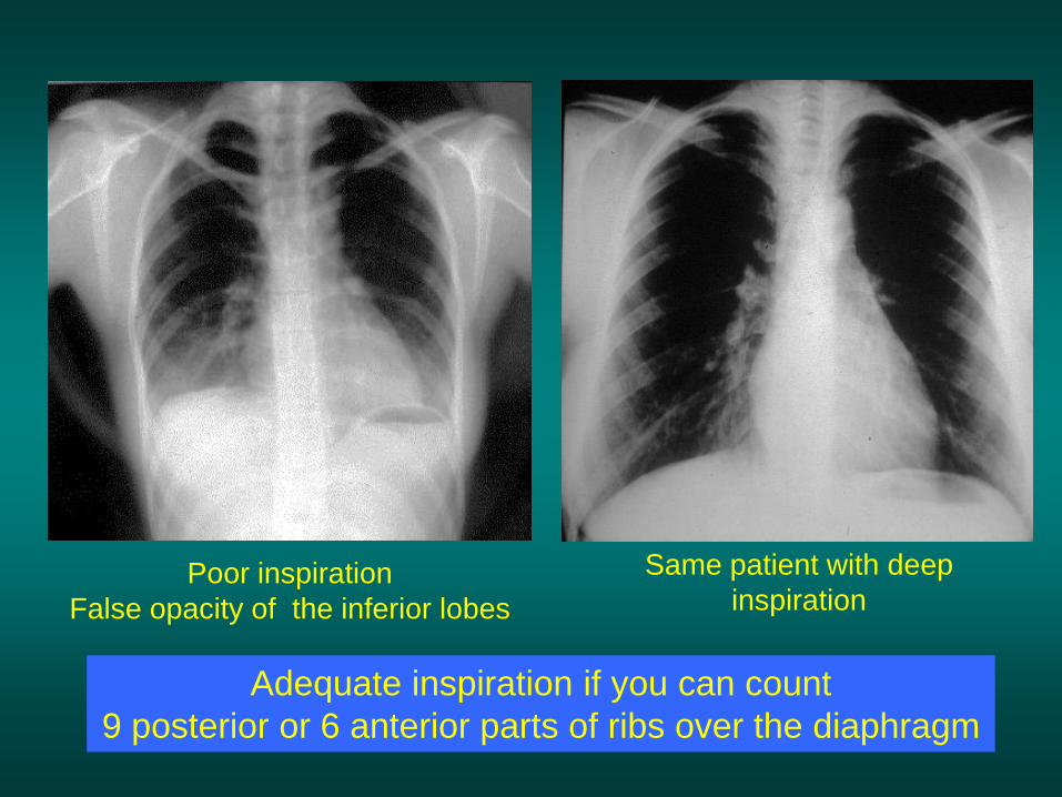
Adequate inspiration if you can count
9 posterior or 6 anterior parts of ribs over the diaphragm
Poor inspiration
False opacity of the inferior lobes
Same patient with deep
inspiration

9 posterior arches
or 6 anterior arches
above the diaphragm
1
2
3
4
5
6
7
8
9
1 2
3
4
5
6

False cardiomegaly
clavicles are high and horizontal
The x-ray beam is antero posterior
Same patient with correct
postero-anterior x-ray beam
incidence
AP versus PA view
The technician should specify the position of the patient if AP view
PA is the correct view

D1 D2
D1
D2
The heart outline is bigger on D2
( bird’s-eye view of the patient)
PA AP

Correct standing or sitting Postero Anterior
position for chest radiography

Standing or sitting position not always easy to obtain…

If the patient is in supine
position (too ill to stand up),
the cardiac outline and
Mediastinum is enlarged.
The scapula may be on the
lung field.
The chest x-ray has
poor quality for analysis

Patient in supine position Standing patient with
postero-anterior X ray beam
The technician should specify the patient position if supine

Light film Under penetration
No detail visible
in the mediastinum area.
Dark film no detail visible
in the lung area

Correct density
and good contrast:
- Pulmonary vessels
visible in the lungs, below
the diaphragm and behind
the heart
-Para-aortic line visible
- Vertebra visible behind
the mediastinum
© OFCP

Conditions for adequate
contrast / density
• Correct x-ray factor (Kv, Mas, exposure time)
• Good conditions of developing and good
quality of processing chemicals
• Correct temperature of developer
• Correct quality of film In case of digitalised or computerised system, imaging quality is adjusted
by computer processing and the 3 last conditions are no more needed.

Exact front view : the vertical line connecting the spinous
processes of thoracic vertebrae is in the middle of
the two sterno-clavicular joints

Front View l a o r a o
D1 D2 D3
D3> D1>D2

exact PA view left anterior oblique position
cardiac silhouette is smaller

right anterior oblique position
( false enlargment of cardiac silhouette)

Chest x-ray: to ensure top quality
• Deep inspiration
• Adequate contrast / density
• Correct position of the patient (exact front view)
• X-ray beam in postero anterior view (the patient is standing)


Normal chest radiography
and some pitfalls… ( trouble-shooting)

What do you read in
this triangle?
Courtesy Martina Martins, Charles Perrot and Nigel Howarth (Radiology depatment university hospital Geneva


Did you notice that « the » was
written twice?
Courtesy Martina Martins, Charles Perrot and Nigel Howarth (Radiology depatment univerity hospital Geneva
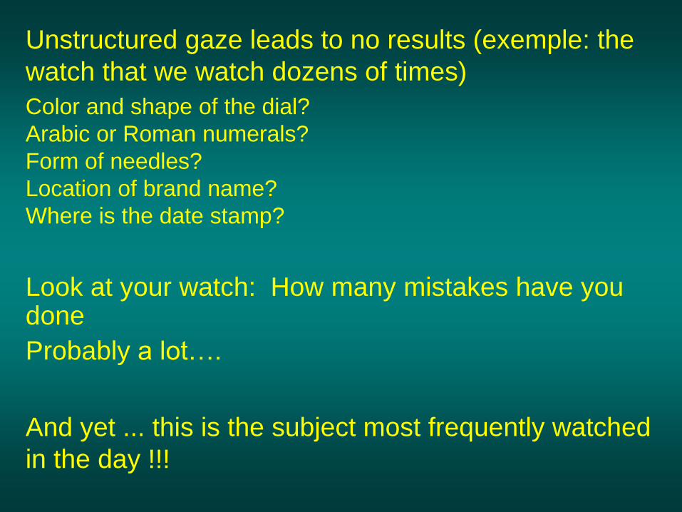
Unstructured gaze leads to no results (exemple: the
watch that we watch dozens of times)
Color and shape of the dial?
Arabic or Roman numerals?
Form of needles?
Location of brand name?
Where is the date stamp?
Look at your watch: How many mistakes have you done
Probably a lot….
And yet ... this is the subject most frequently watched
in the day !!!

To enjoy reading the chest radiograph,
the gase should be disciplined
To discipline the gase one must have
resort to a method of examination

Le syndrome de New York
Young man with
a right chest
pain and
shortness of
breath with
sudden onset

You had of
course noticed
the large right
pneumothorax

L’imagerie thoracique pour l’ECN
Pr C. H. Marquette- Pr B. Padovanni
Did you see that
he also had a
small left
pneumothorax?

Process for analysis of the chest
radiography: the check list
• Verification of name and date
• Clinical history and findings
• Verification of the factors for good quality
• Assess
- Thoracic wall and thoracic skeleton (« first circle »)
- Each lung field, one after the other (« 2nd circle »)
- Mediastinum (« third circle »)
Do not skip any item in the checklist !

First Circle:
Soft part of the chest wall
diaphragmatic areas
bony thorax

Second Circle:
Lung tissue
Reading from top to bottom
Comparing left and right side

Third circle:
Mediastinum:
The container (= edges)
The content (= behind the heart
Hilus areas

There are some areas that
need special attention,
because pathology in these
areas can easily be
overlooked:
Apical zones
Hilar zones
Retrocardial zone
Zone below the dome
of diaphragm
http://www.radiologyassistant.nl
The hidden area

© OFCP
Thoracic wall
And skeleton

Thoracic wall
Clavicles
External side of Sterno
cleido-mastoid muscle

Thoracic wall
The clavicles are
projected on the
level of the 3rd or 4th
posterior part of ribs
1
3
4

Minor malformation Cervical ribs

RUL TB infiltrate ?

trap picture: opacity of the superior part of right lung due to a hair braid
Be aware of foreign
body or artefacts
on the chest x-ray

Breast implant

Woman, cough but no fever, no woresening condition.
Diagnosis: Right inferior lobe pneumonia…

Breast prosthesis superposition!
(past history of breast cancer )

The retro-clavicular fields are always difficult to analyse,
because of bone superposition:
- Clavicles
- Anterior part of first rib
- Posterior part of third and fourth rib,
- Sterno-clavicular joint

There are 2 ways to correctly
analyse the retro-clavicular fields:
• Always compare right and left
• Ask for a chest x-ray with the patient’s back against the film (AP view, lordotic position)

RUL TB infiltrate
Always compare
left and right

Patient with fever, cough,
AFB sputum positive…

You have not CT scan. So use your eyes and
Compare right and left!
If any doubt, request AP/ lordotic chest X ray

Courtezy DR Nay Wunn Linn
NTP Myanmar
Special incidence for apex view

Normal chest x-ray ,
front close to the film
Normal AP chest x-ray,
back close to the film

Chest x-ray, front close to the film Chest x-ray, back close to the film
And clavicles out of the field
by raising hands

Fever and weight loss. No respiratory symptoms

Small infiltrate in the right apex well visible on PA view.
Notice the difference between right and left apex.

Chest x-ray back close to the film

Thoracic wall
physiological blur of
the inferior side of the
ribs
Rib view section

Is this CXR normal?

You must always “read“ a chest x-ray with methodology:
For the chest wall, you must look at every rib, one after the other
6th and 9th ribs
missing on this CXR

Chest wall
Top of the axillar hole
Big pectoral muscle

thoracic wall
Scapula

What is wrong with this chest x-ray?

Congenital clavicles agenesy
What is wrong with this chest x-ray?


Thoracic wall
Breast silhouette

Be careful with false opacities in the inferior lobes, consequences
of breast superposition.

Chest x-ray. Before and after right mastectomy

Carefull with the nipples…. (not only in women CXR)

Thoracic wall
Diaphragm
The right side is
usually higher than
the left side (3cm )


Component elements of lungs

On a normal chest x-ray, bronchi are not visible
except trachea and proximal part of main right and left bronchi (opacification with iodin hydrosoluble solution)

The grey part of this
diagrammis visible on the
CXR

Only trachea and proximal part of main left and right bronchi are visible on the chest X ray

…but pulmonary arteries are visible.

…but pulmonary arteries are visible.

Pulmonary arteries and bronchial tree are parallelic
( it is not the case for pulmonary veni )


Right view
Small fissura
Big fissura

Left view
Left fissura

Right superior lobe
pneumonia
minor
fissure
Large oblique
fissure posterior
part
minor
fissure


Right inferior pneumonia
Large oblique
fissure


Middle lobe pneumonia
Large fissure
(anterior part) minor
fissure
Small pleural
effusion


External segment of middle lobe pneumonia

External segment of middle lobe pneumonia

Left upper lobe pneumonia


Left inferior pneumonia
Left fissure


Component elements of
Mediatinum and hilus

RA RV LV
AO
PA
AO
RV
LA
LV
PA

Front view
SVC
RA
AO
PA
LV
AO
RV LA
LV
Lateral view
RA LA
LV
RV
PA
SVC: superor vena cava
AO : aortic vessel
PA: pulmonary artery
RA: right atrium
LV; left ventricle
LA: left atrium
RV: right ventricle


Right pulmonary
artery
Left pulmonary
artery


L A
L V
RSPV
RIPV
LPSV
The pulmonary vena are not physiologiccally visible

L A
L V
RSPV
RIPV
LPSV
The pulmonary vena are not physiologiccally visible

• X ray beam crossing thorax and mediastinum meets in some
places pleural thickness, producing images on the CXR like
lines defining mediastinum lines…
Mediastinum lines

1. Sub clavicle arterial line
2. Posterior mediastinum line
3. Brachio cepahalic vena line
4. para-azygos line
5. Anterior mediastinum line
6. Descending aortic line
7. Right and left paravertébral line
8. Inferior veina cava line
9. para-œsophageal line
10. para-trachéal line
Mediastinum lines
6 Too complicated
And not so usefull…

3 of them are really important.
Right paratracheal line
Para-aortic line
Aorto pulmonary line

Para tracheal line Aorto- pulmonary
line
Para aortic line

Mediastinum enlargment due to fat tissu

Mediastinum enlargment due to fat tissu

Be aware of false enlargment of mediastinum
if obesity, poor inspiration, oblique view or supine
position

Trap: false mediastinum enlargment in the
case of this older woman with cyphoscoliosis,
in supine position


Normal lateral view

Lateral view

AO
PA
RV
LA
LV

Front view
SVC
RA
AO
PA
LV
AO
RV LA
LV
Lateral view
RA LA
LV
RV
PA
SVC: superor vena cava AO : aortic vessel PA: pulmonary artery RA: right auricle LV; left ventricle LA: left auricle RV: right venticle

Ascending
Aorta
Superior
vena cava
area
Pulmonary
arteria
Right
ventricle
Descending
aorta
Heart and mediastinum vessels

Heart and
Mediastinum
vessels
Left
ventricle
Inferior
vena
cava

Mediastinum
vessels
Aortic arch

mediastinum
vessels
Descending aorta

Mediastinum
vessels
Right pulmonary artery
Left pulmonary artery

Lateral view is very useful for diagnosis of mediastinal adenopathies
normal CXR

Lateral view is very useful for diagnosis of mediastinal adenopathies

trachea
20 mm

Right superior
lobe bronchus
Left superior
lobe bronchus


Retro sternal clear space
Retro cardiac clear space
The «clear spaces»

The «clear spaces»
Retro tracheal space

enlargment of the clear spaces: Emphysema

Emphysema ( notice clear space enlargment
and flattened diaphragm )
Normal lateral view

The retro sternal space is filled: thymoma

The retro sternal space is filled: thymoma. Normal view on the right

Diaphragm
Right
diaphragm
Left
diaphragm

Summary normal CXR

Normal frontal chest view
Aortic arch (AA)
Descending aorta (DA)
Proximal left pulmonary
artery (PLPA)
Left interlobar pulmonary
artery (LPA)
Aortopulmonary window
(AW)
Left main bronchus (LB)
Left atrial appendage (LAA)
Left ventricle (LV)
Superior vena cava (SVC)
Right paratracheal stripe
(RPS)
Azygos vein (AV)
Aortic arch (AA)
Right interlobar pulmonary
artery (RIPA)
Right atrium (RA)
Trachea (T)
Right diaphragm (RD)
DA
LPA
RIPA
A W
LB
LV
SVC
RPTS
AV
RA
T
RD
LD
A A
SVC
T

Normal lateral chest view Posteriorly: vertebral bodies (V) and
intervertebral disc spaces (*)
Anteriorly: retrosternal clear space (RSCS)
Trachea (T)
Orifice of Right Upper Lobe bronchus
(RUL) appears as circular lucency
projecting over the continuation of the
tracheal air column
Left pulmonary artery (LPA)
Right pulmonary artery (RPA)
Left auricle (LA)
Left ventricle (LV)
Right ventricle (RV)
Aortic arch (AA)
Postero costo phrenic angle (PCPA)
Right diphragm (RD)
Left diaphragm (LD)
Inferior vena cava (IVC)
Scapula (Sc)
Retro cardiac clear space (RCCS)
T
V
*
RSCS
RUL
LPA
Sc
RPA
LA
LV
RV
AA
PCPA RD
LD
IVC
RCCS