Embed Size (px)
Citation preview
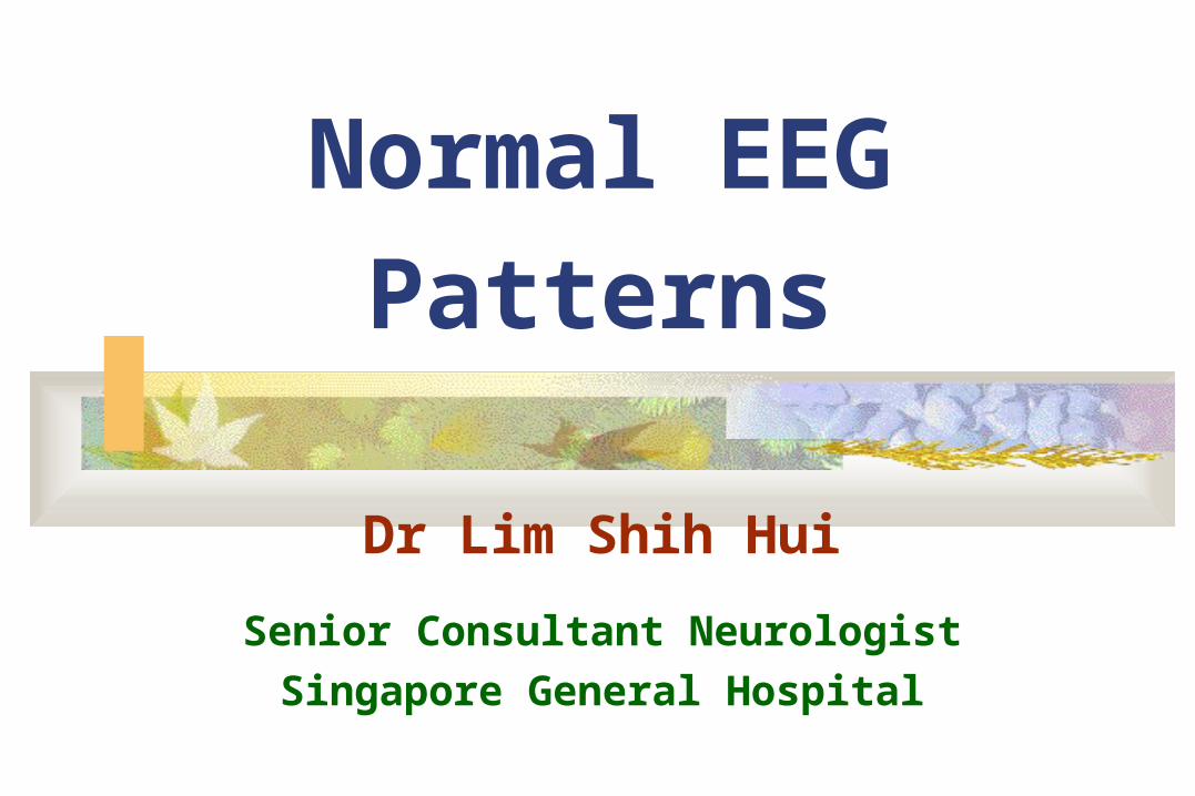
Normal EEG Patterns
Dr Lim Shih Hui
Senior Consultant Neurologist
Singapore General Hospital
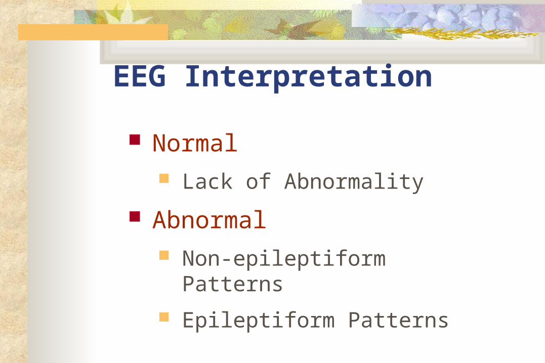
EEG Interpretation
Normal Lack of Abnormality
Abnormal Non-epileptiform Patterns
Epileptiform Patterns
Alpha Rhythm The starting point of analysing awake EEG 8-13 Hz activity occurring during wakefulness 20-60 mV, max over posterior head regions Present when eyes closed; blocked by eye opening or alerting
the patient 8 Hz is reached by 3 years of age and progressively increases in
a stepwise fashion until 9-12 Hz is reached by adolescence Very stable in an individual, rarely varying by more than 0.5 Hz. With drowsiness, alpha activity may decrease by 1-2 Hz A difference of greater than 1 Hz between the two hemispheres
is significant. 10% of adult have little or no alpha
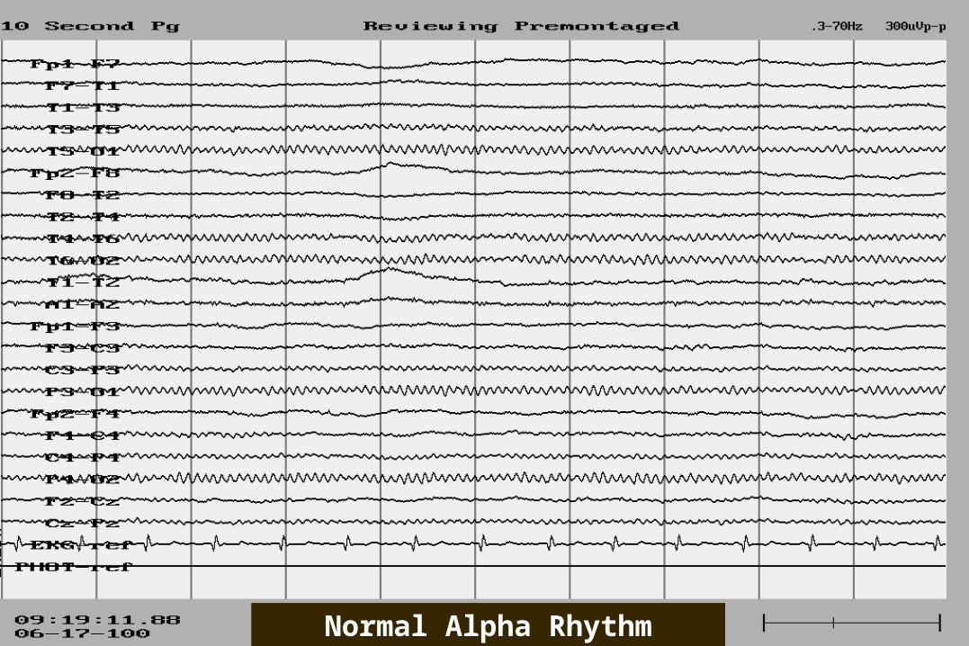
Normal Alpha Rhythm
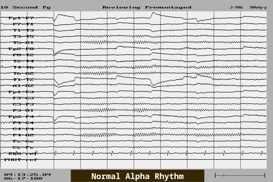
Normal Alpha Rhythm
Alpha Rhythm: Reactivity
Should attenuate bilaterally with eye opening alerting stimuli mental concentration
Some alpha may return when eyes remain open for more than a few seconds.
Failure of the alpha rhythm to attenuate on one side with either eye opening or mental alerting indicates an abnormality on the side that fails to attenuate
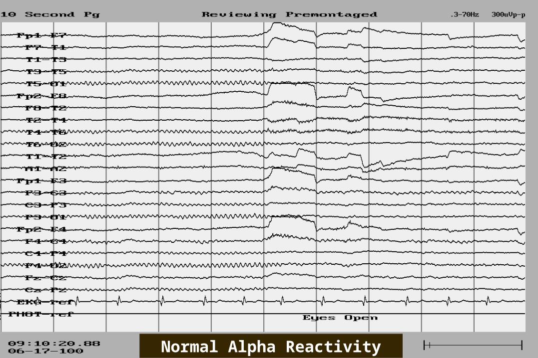
Normal Alpha Reactivity
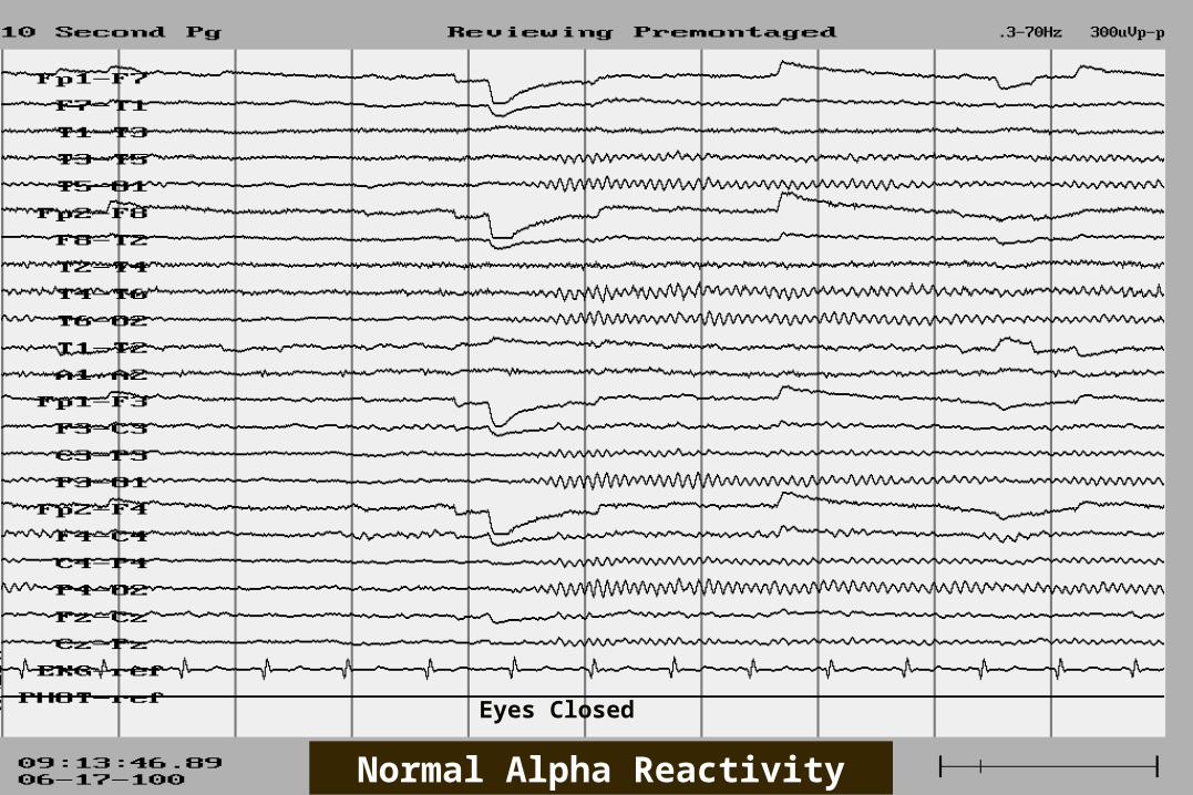
Normal Alpha Reactivity
Eyes Closed

Beta Activity
Frequency of over 13 Hz; if >30-35 Hz gamma activity or exceedingly fast activity by Gibbs.
Average voltage is 10-20 microvolts Two main types in adults: Often enhanced during drowsiness or when present
over a skull defect Should not be misinterpreted as a focus of
abnormal fast activity.

Beta Activity Frequency of over 13 Hz; if >30-35 Hz gamma activity or exceedingly
fast activity by Gibbs. Average voltage is 10-20 microvolts
Two main types in adults: The precentral type: predominantly over the anterior and
central regions; related to the functions of the sensorimotor cortex and reacts to movement or touch.
The generalized beta activity: induced or enhanced by drugs; may attain amplitude over 25 microvolts.
Often enhanced during drowsiness or when present over a skull defect Should not be misinterpreted as a focus of abnormal fast activity.
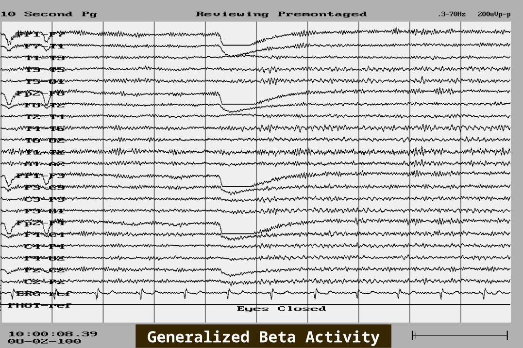
Generalized Beta Activity
Beta Activity
Frequency of over 13 Hz; if >30-35 Hz gamma activity or exceedingly fast activity by Gibbs.
Average voltage is 10-20 microvolts Two main types in adults: Often enhanced during drowsiness or when present
over a skull defect Should not be misinterpreted as a focus of
abnormal fast activity.
Theta Activity The term theta was coined by Gray Walter in 1944
when it was believed that this rhythm was related to the function of the thalamus.
Occurs as a normal rhythm during drowsiness In young children between age 4 months 8 years: predominance over the
fronto-central regions during drowsiness In adolescents: sinusoidal theta activity can occur over the anterior head
regions during drowsiness. In adults, theta components can occur diffusely or over the posterior head
regions during drowsiness. Single transient theta waveforms or mixed alpha-theta waves can be present
over the temporal regions in older adults.
Theta Activity
The term theta was coined by Gray Walter in 1944 when it was believed that this rhythm was related to the function of the thalamus.
Occurs as a normal rhythm during drowsiness
In young children between age 4 months 8 years: predominance over the fronto-central regions during drowsiness
In adolescents: sinusoidal theta activity can occur over the anterior head regions during drowsiness.
In adults, theta components can occur diffusely or over the posterior head regions during drowsiness.
Single transient theta waveforms or mixed alpha-theta waves can be present over the temporal regions in older adults.
Theta Activity
The term theta was coined by Gray Walter in 1944 when it was believed that this rhythm was related to the function of the thalamus.
Occurs as a normal rhythm during drowsiness In young children between age 4 months 8 years: predominance over
the fronto-central regions during drowsiness In adolescents: sinusoidal theta activity can occur over the anterior head
regions during drowsiness. In adults: theta components can occur diffusely or
over the posterior head regions during drowsiness. Single transient theta waveforms or mixed alpha-theta
waves can be present over the temporal regions in older adults.

Temporal Slowing Of The Elderly
Occur chiefly over the age of 60 years Confined to the temporal regions and are usually maximal anteriorly Occur more frequently on the left side Do not disrupt background activity Usually have a rounded morphologic appearance Voltage is usually less than 60-70 microvolts Attenuated by mental alerting and eye opening and increased by
drowsiness and hyperventilation Occur sporadically as single or double waves but not in longer
rhythmic trains Present for only a small portion of the tracing (up to 1%) of the
recording time when the patient is in a fully alert state
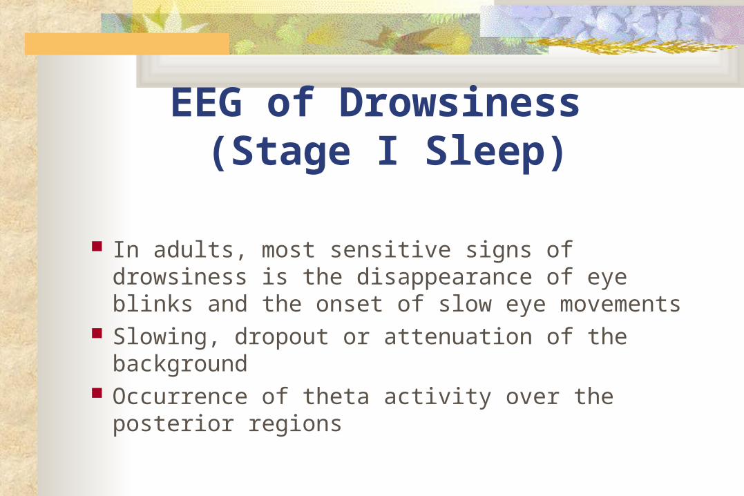
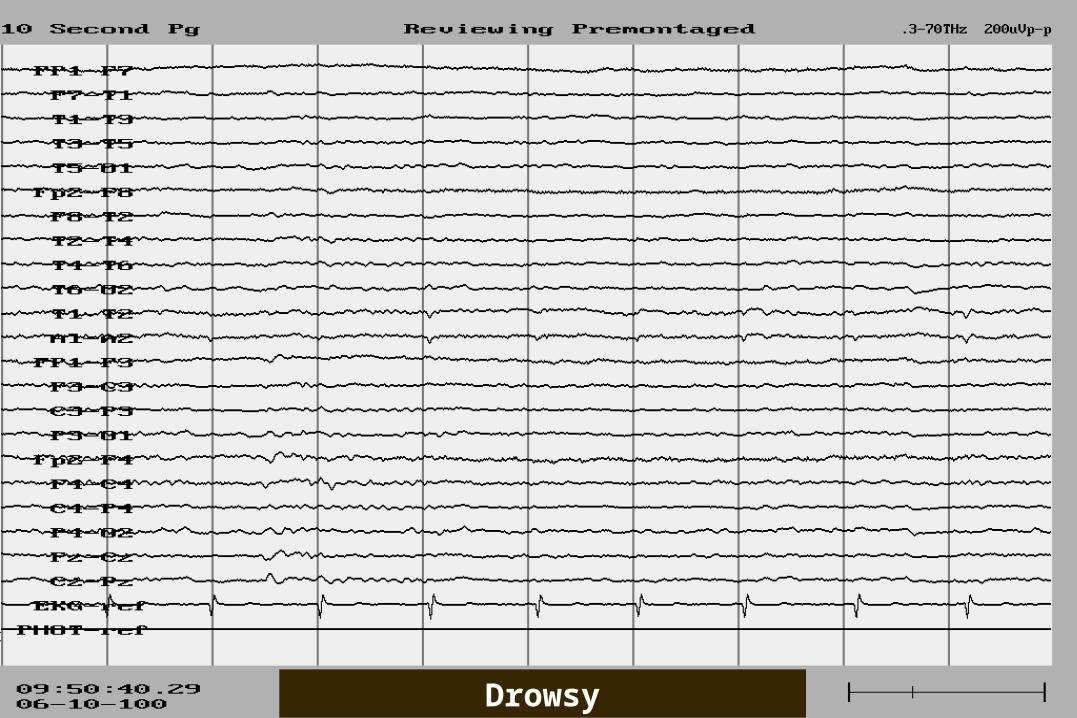
EEG of Drowsiness (Stage I Sleep)
In adults, most sensitive signs of drowsiness is the disappearance of eye blinks and the onset of slow eye movements
Slowing, dropout or attenuation of the background Occurrence of theta activity over the posterior
regions
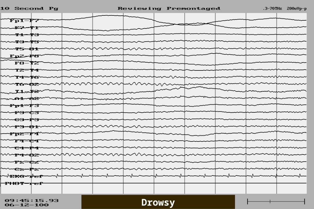
Drowsy

Drowsy
Drowsy
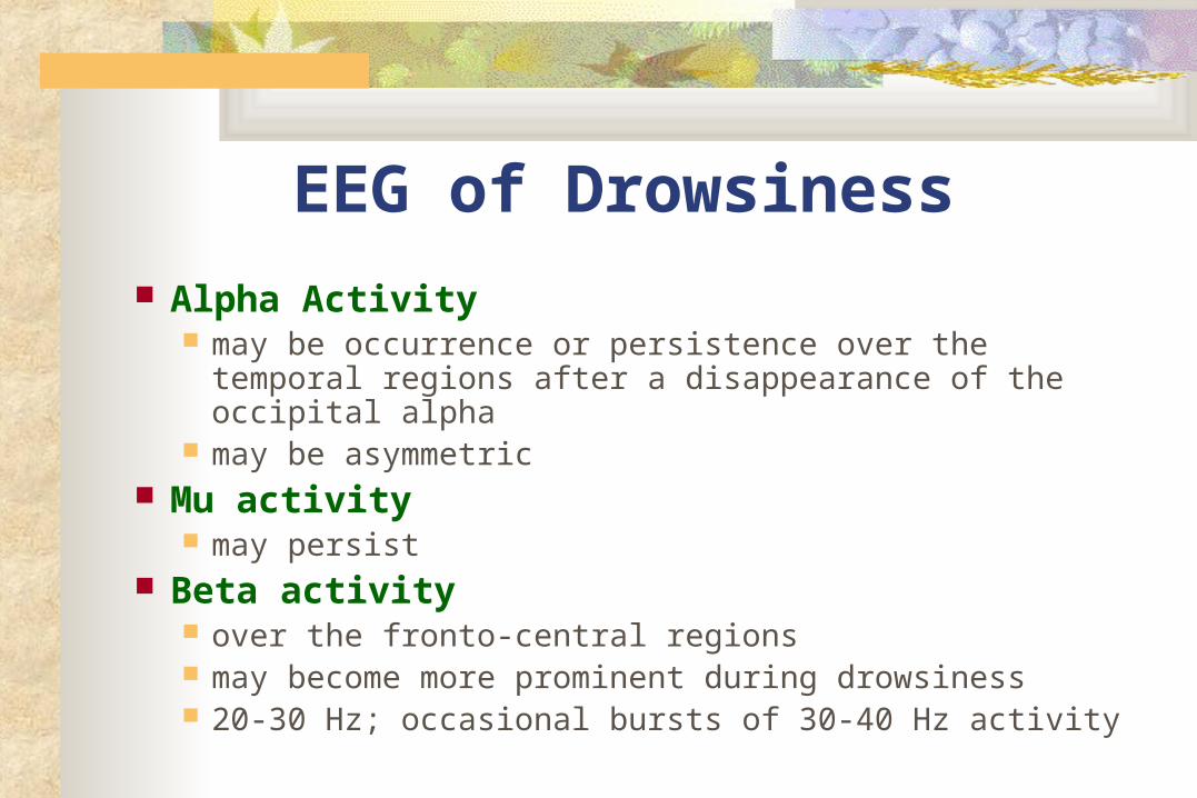
EEG of Drowsiness
Alpha Activity may be occurrence or persistence over the temporal
regions after a disappearance of the occipital alpha may be asymmetric
Mu activity may persist
Beta activity over the fronto-central regions may become more prominent during drowsiness 20-30 Hz; occasional bursts of 30-40 Hz activity
Other Activities During Stage I Sleep
Vertex Sharp Transients Positive Occipital Sharp
Transients of Sleep (POSTs)
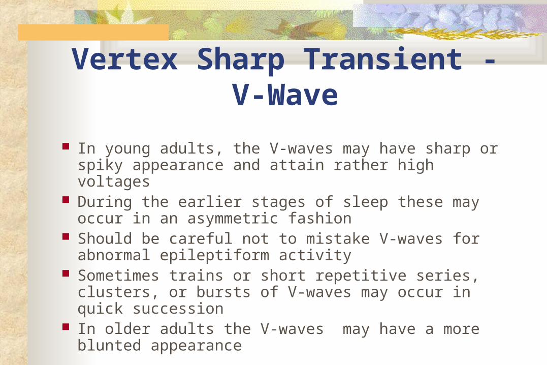
Vertex Sharp Transient -V-Wave
In young adults, the V-waves may have sharp or spiky appearance and attain rather high voltages
During the earlier stages of sleep these may occur in an asymmetric fashion
Should be careful not to mistake V-waves for abnormal epileptiform activity
Sometimes trains or short repetitive series, clusters, or bursts of V-waves may occur in quick succession
In older adults the V-waves may have a more blunted appearance
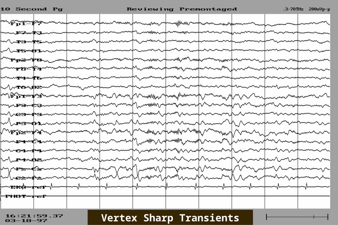
Vertex Sharp Transients
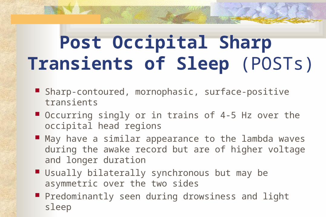
Post Occipital Sharp Transients of Sleep (POSTs)
Sharp-contoured, mornophasic, surface-positive transients Occurring singly or in trains of 4-5 Hz over the occipital head
regions May have a similar appearance to the lambda waves during
the awake record but are of higher voltage and longer duration
Usually bilaterally synchronous but may be asymmetric over the two sides
Predominantly seen during drowsiness and light sleep

POSTs
Stage II Sleep
Sleep Spindles K Complex

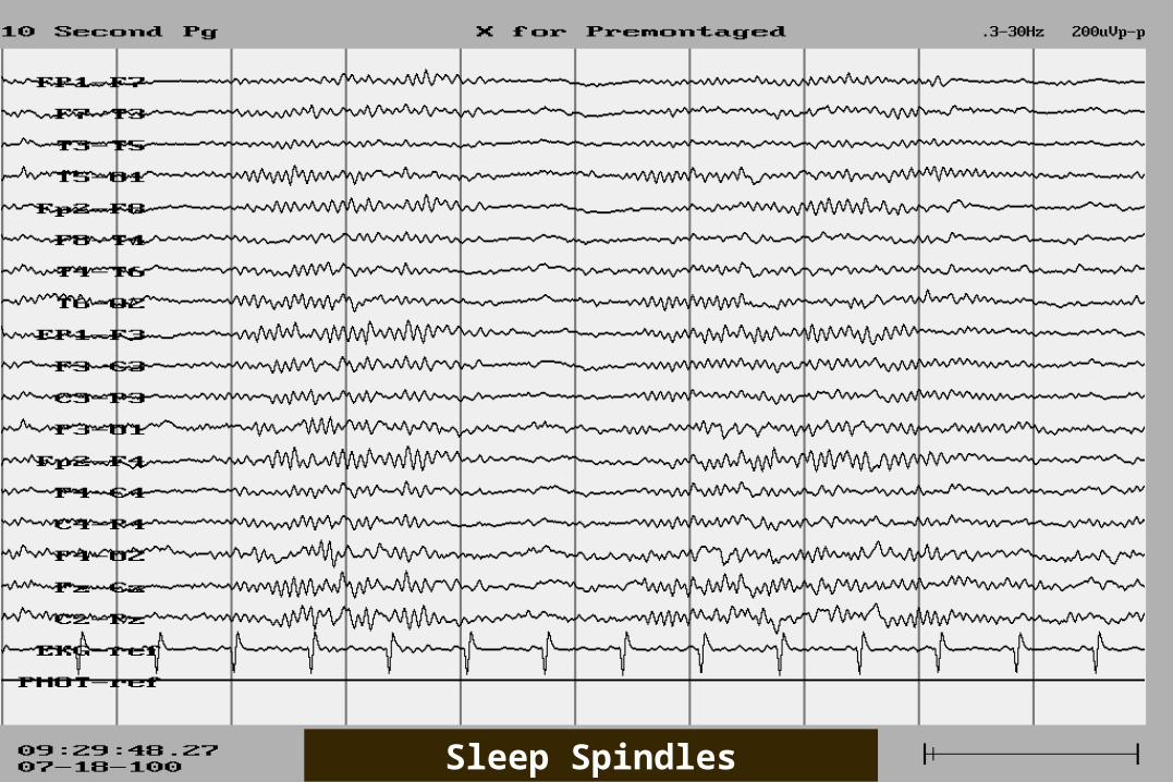
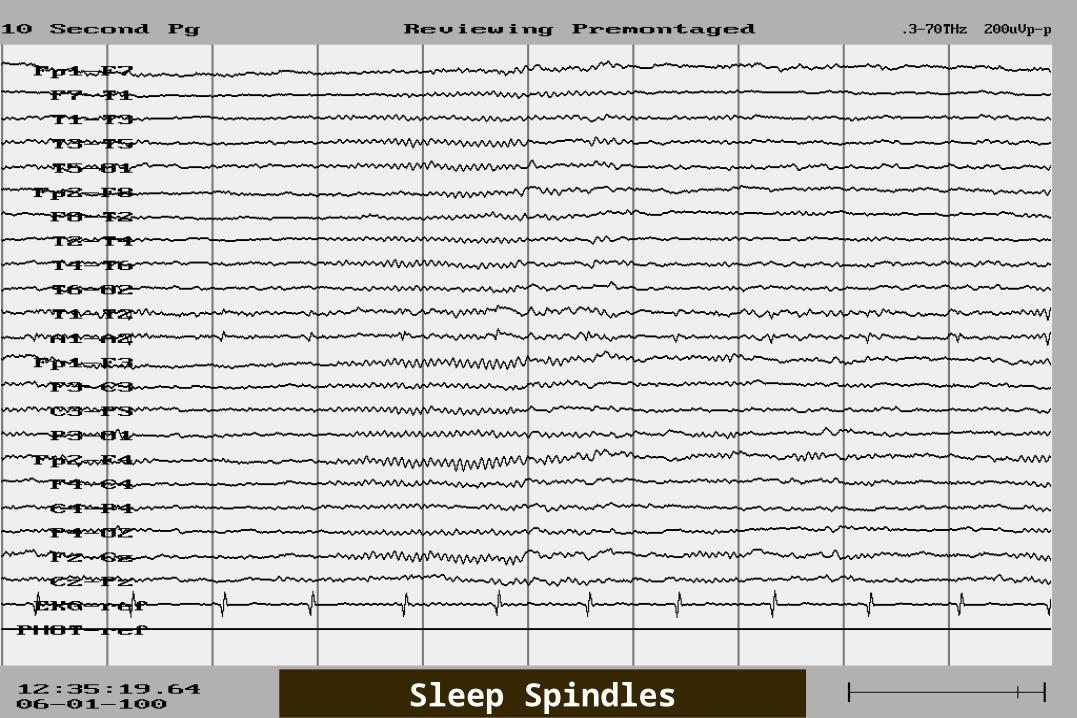
Sleep Spindles In adults, a frequency of 13-14 Hz occur in a symmetric and synchronous fashion over
the two hemispheres Usually these occur at intervals between 5-15
seconds, Spindle trains ranging from 0.5-1.5 seconds in
duration More prolonged trains or continuous spindle activity
may be seen in some patients on medication, particularly benzodiazepams

Sleep Spindles
Sleep Spindles
Sleep Spindles
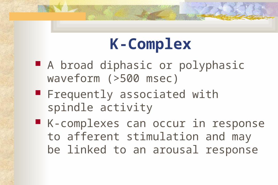
K-Complex A broad diphasic or polyphasic waveform
(>500 msec) Frequently associated with spindle activity K-complexes can occur in response to
afferent stimulation and may be linked to an arousal response

K-Complex

Hyperventilation
Often produces little change in the EEG in adult If there is a change, usually consists of generalized
slowing. either gradual or abrupt onset in theta or delta range may continue as series of rhythmic slow waves or consist
of repeated bursts of slow waves at irregular intervals Degree of response depends on the age, the vigor of
hyperventilation, blood sugar levels, and posture
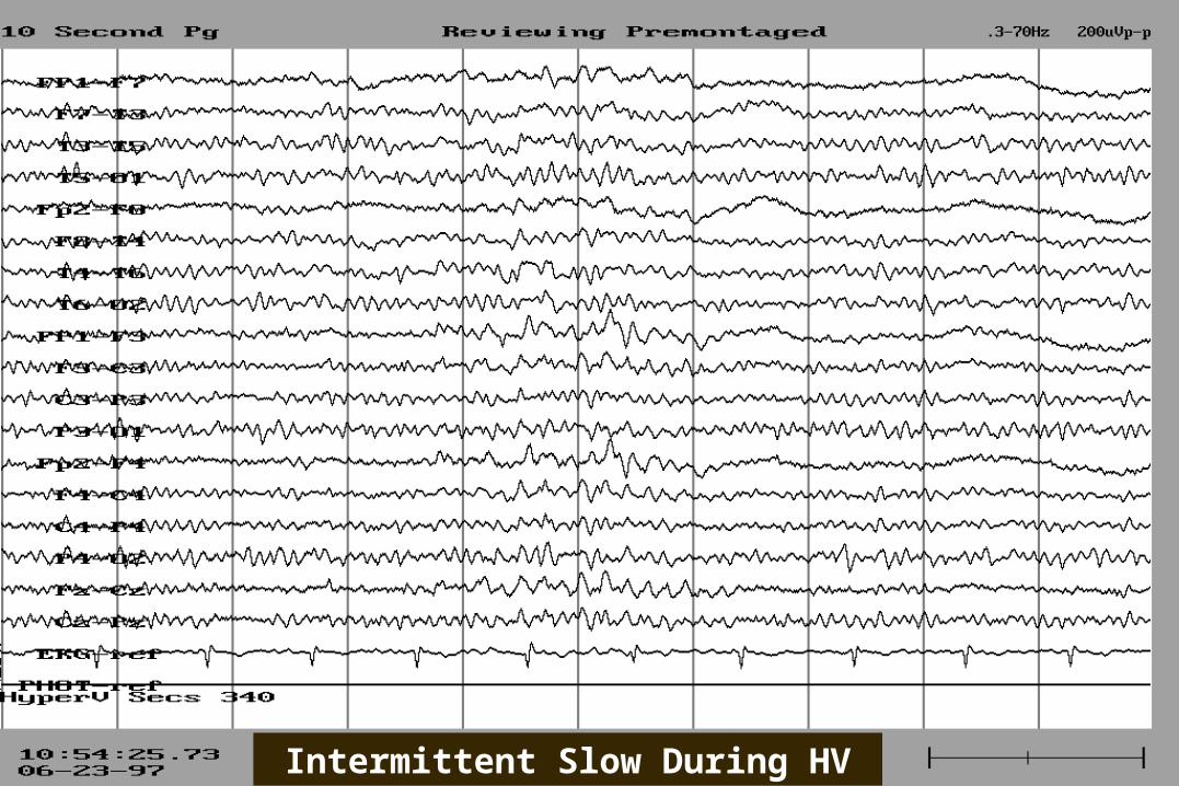
Intermittent Slow During HV
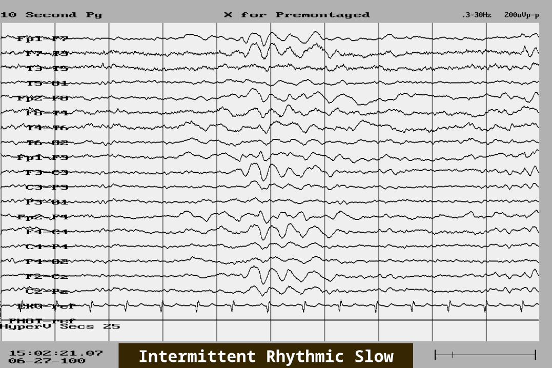
Intermittent Rhythmic Slow During HV

Persistence slowing following cessation of hyperventilation:
Check if patient is still continuing to hyperventilate or if patient is
hypoglycemic

Hyperventilation The findings accepted as unequivocal
evidence of abnormality: epileptiform discharges clear-cut focal or lateralized slowing or
asymmetry of activity
Contraindications: significant cardiac or cerebrovascular disease,
or respiratory dysfunction.
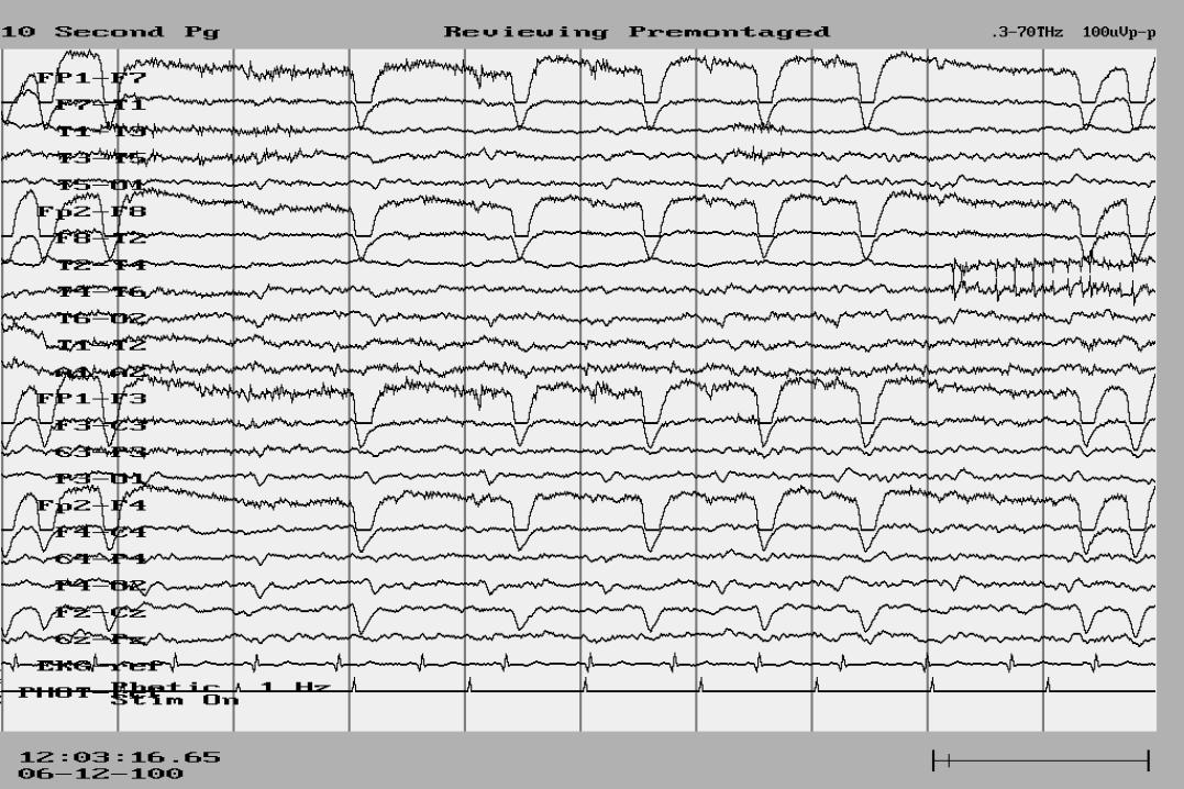
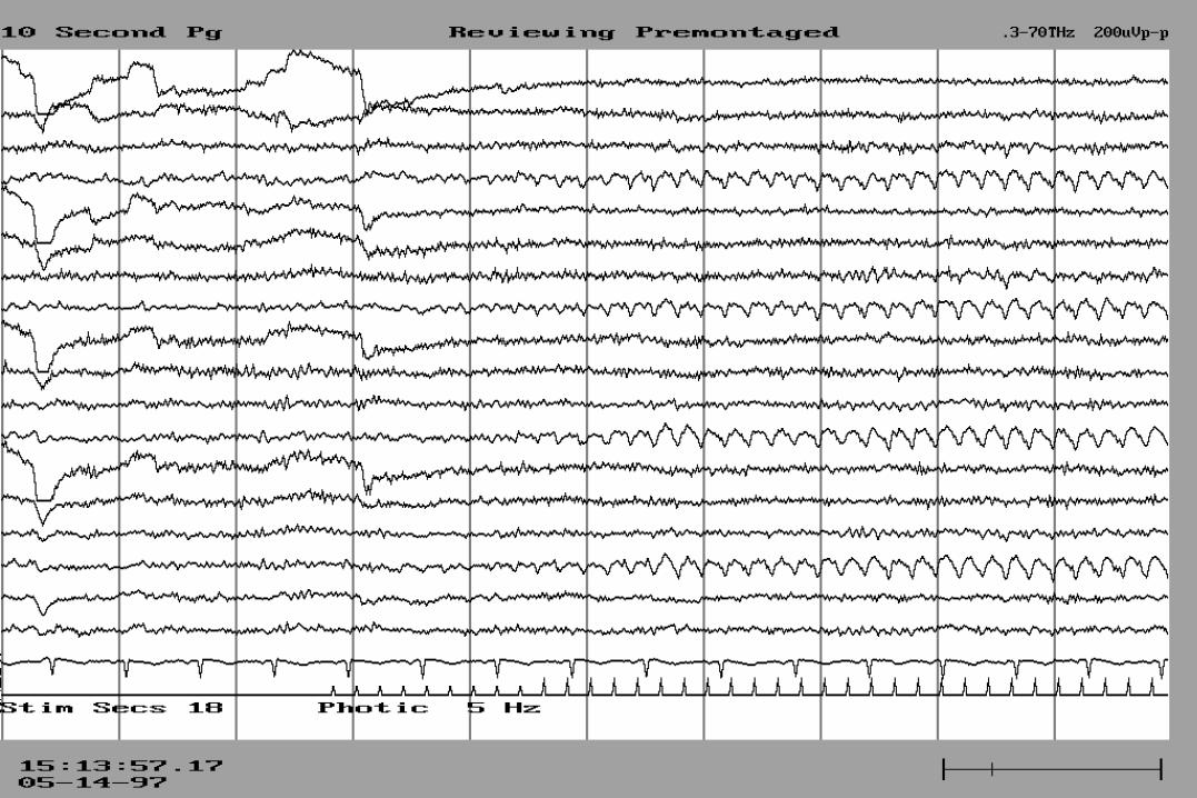
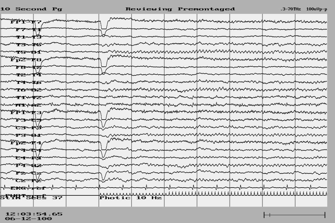
Photic Stimulation
Flash rate eliciting maximum driving response increases in rough parallel with age (Niedermeyer, 1982)
Driving response may normally have a notched appearance resembling a spike-wave discharge.
It can be distinguished from spike-waves by its time-locked appearance with the flash rate and its failure to persist after the stimulation stops.
Asymmetries of photic driving probably have less clinical value and can only be interpreted in association with other significant asymmetries
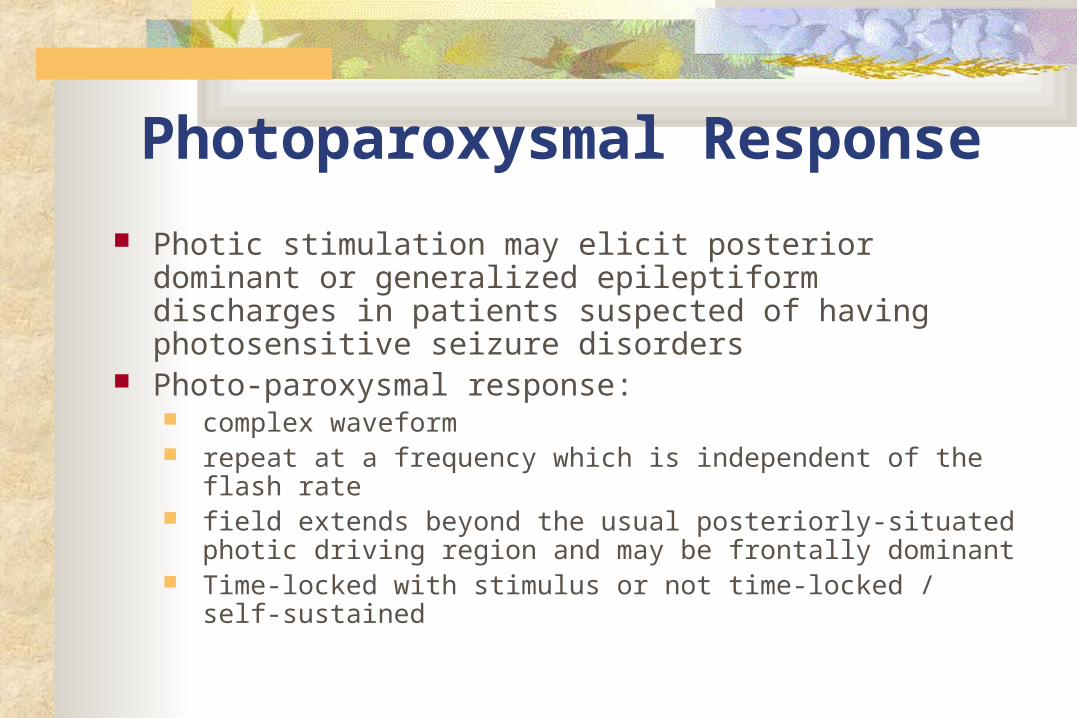
Photoparoxysmal Response
Photic stimulation may elicit posterior dominant or generalized epileptiform discharges in patients suspected of having photosensitive seizure disorders
Photo-paroxysmal response: complex waveform repeat at a frequency which is independent of the flash rate field extends beyond the usual posteriorly-situated photic
driving region and may be frontally dominant Time-locked with stimulus or not time-locked / self-sustained
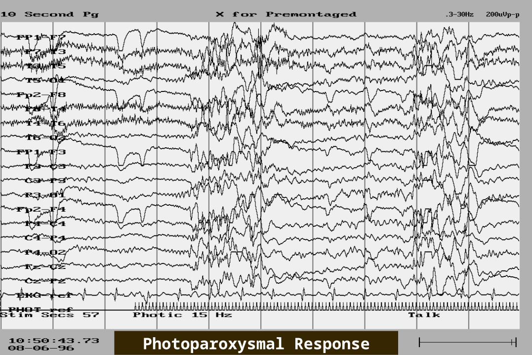
Photoparoxysmal Response
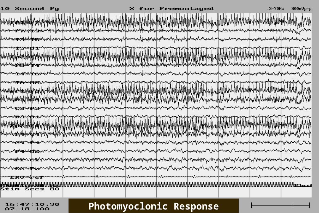
Photomyoclonic Response
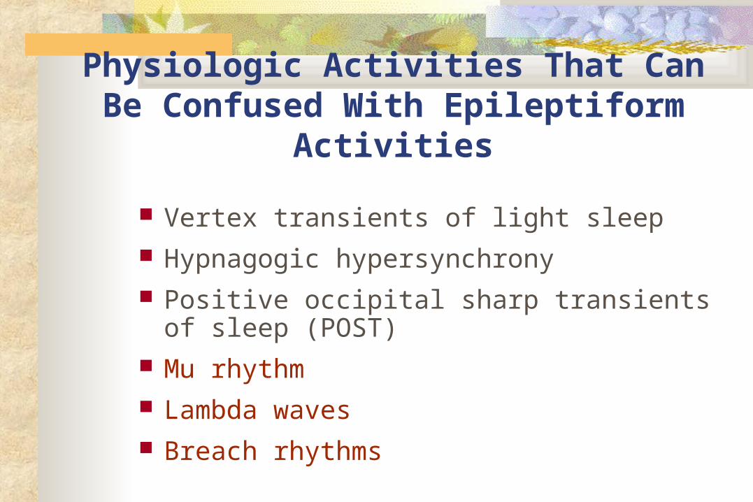
Physiologic Activities That Can Be Confused With Epileptiform Activities
Vertex transients of light sleep Hypnagogic hypersynchrony Positive occipital sharp transients of sleep
(POST) Mu rhythm Lambda waves Breach rhythms

Benign Variants Of Unknown Clinical Significance
Benign epileptiform transients of sleep (small sharp spikes)
6- and 14-Hz positive spikes Wicket spikes Psychomotor variants (rhythmic mid-temporal
theta discharge of drowsiness) Subclinical rhythmic EEG discharge of adults Phantom spike and wave