Embed Size (px)
DESCRIPTION
fetal anatomy-ultrasound
Citation preview
Normal fetal anatomy
Anatomy Scan• Indications
– Identify fetal abnormalities– Identify IUGR– Dating– Placental localisation
• Timing– 16-20 weeks– 22-24 weeks
• Machine– Good quality machine– TGC sliders– Record images– High quality curvelinear probe
• Problems– Incorrect dates– Poor views– Fetal position
Routine
• History and consent from patient– Previous child with abnormality– Family history of abnormality– Exposure to teratogens eg drugs, infections– Explain limitations
• Growth scan
• Placental localisation
• Anatomy

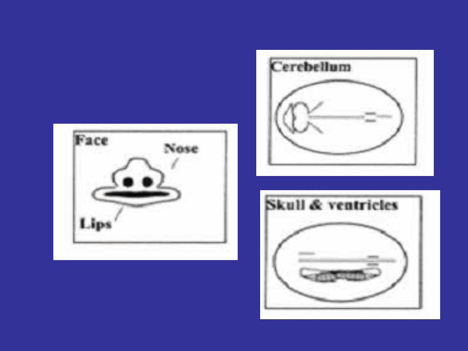
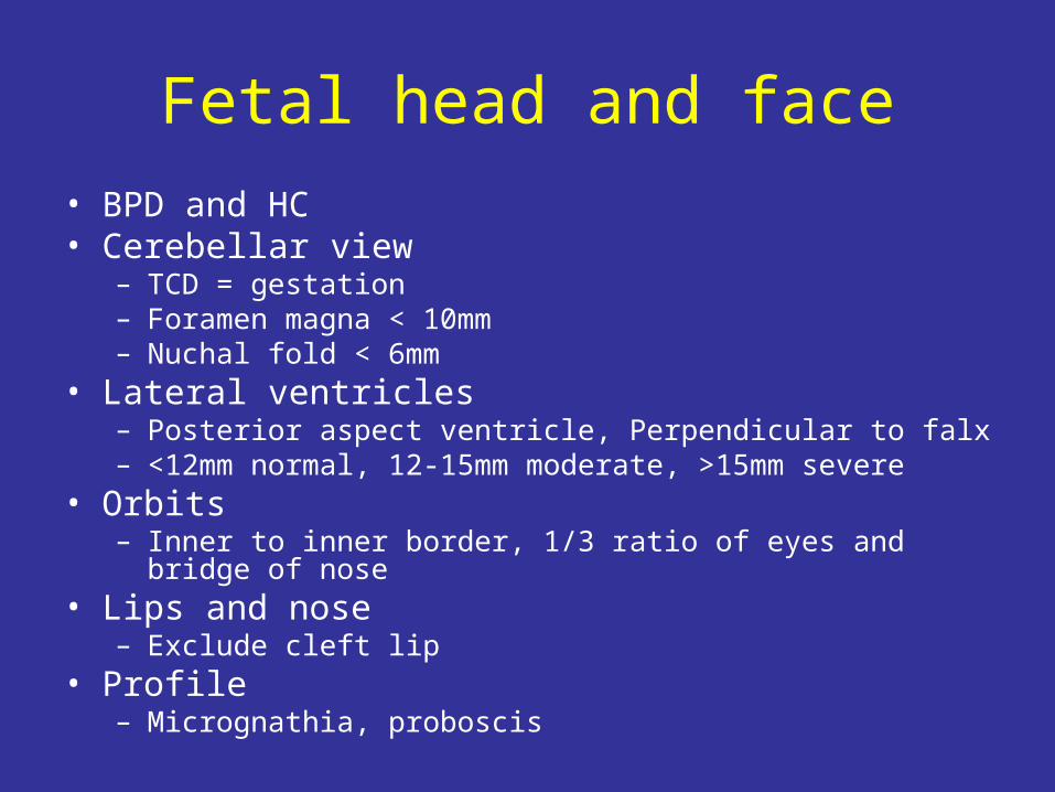
Fetal head and face
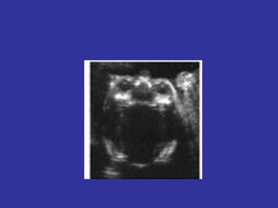
• BPD and HC• Cerebellar view
– TCD = gestation– Foramen magna < 10mm– Nuchal fold < 6mm
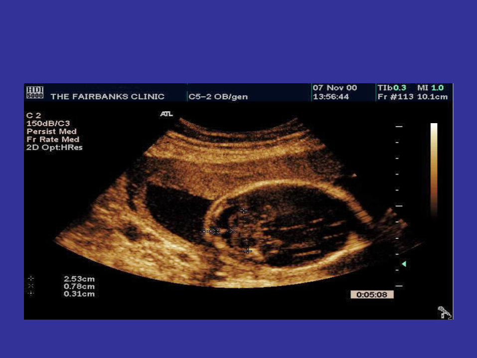
• Lateral ventricles– Posterior aspect ventricle, Perpendicular to falx– <12mm normal, 12-15mm moderate, >15mm severe
• Orbits– Inner to inner border, 1/3 ratio of eyes and bridge of nose
• Lips and nose – Exclude cleft lip
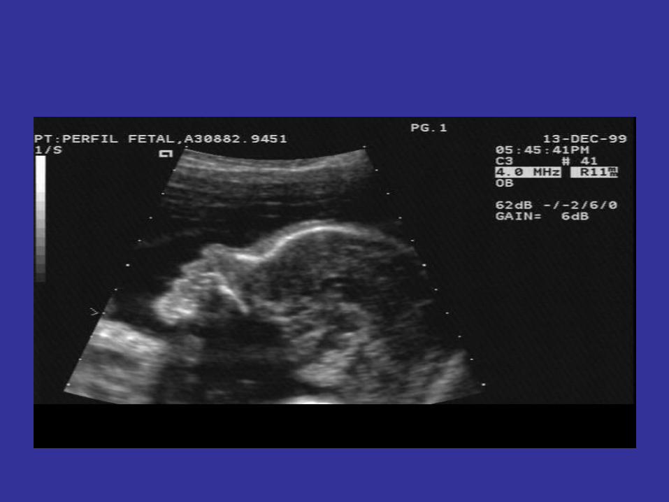
• Profile– Micrognathia, proboscis
Fetal head and face
• BPD and HC• Cerebellar view
– TCD = gestation– Foramen magna < 10mm– Nuchal fold < 6mm
• Lateral ventricles– Posterior aspect ventricle, Perpendicular to falx– <12mm normal, 12-15mm moderate, >15mm severe
• Orbits– Inner to inner border, 1/3 ratio of eyes and bridge of nose
• Lips and nose – Exclude cleft lip
• Profile– Micrognathia, proboscis
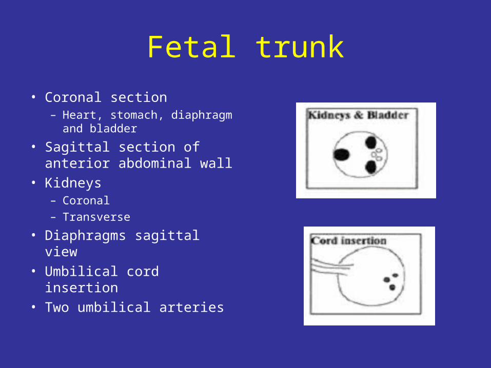
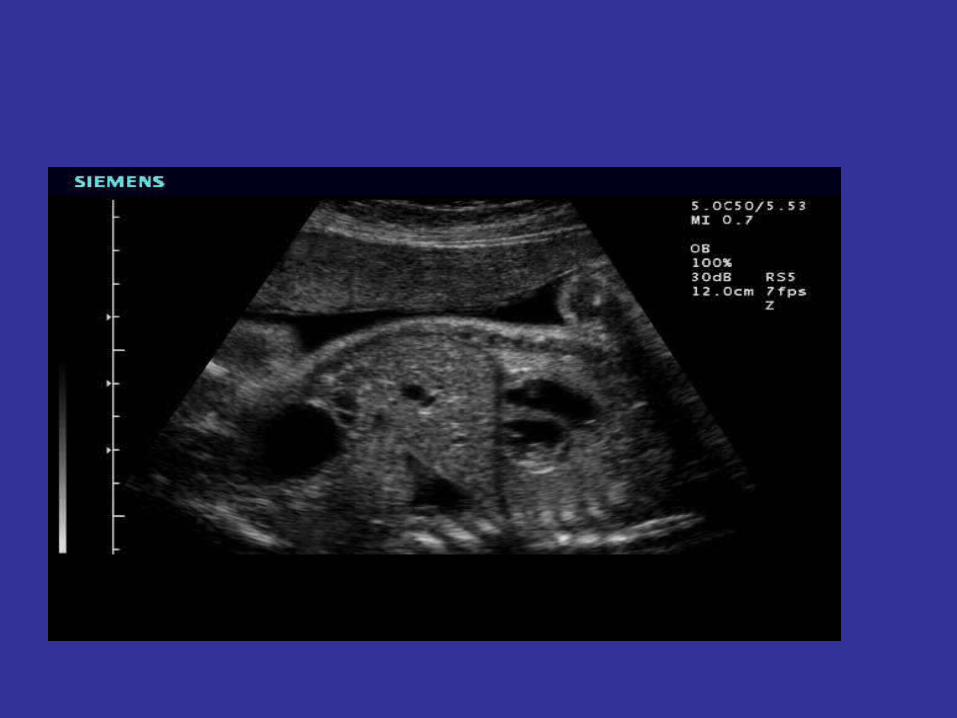
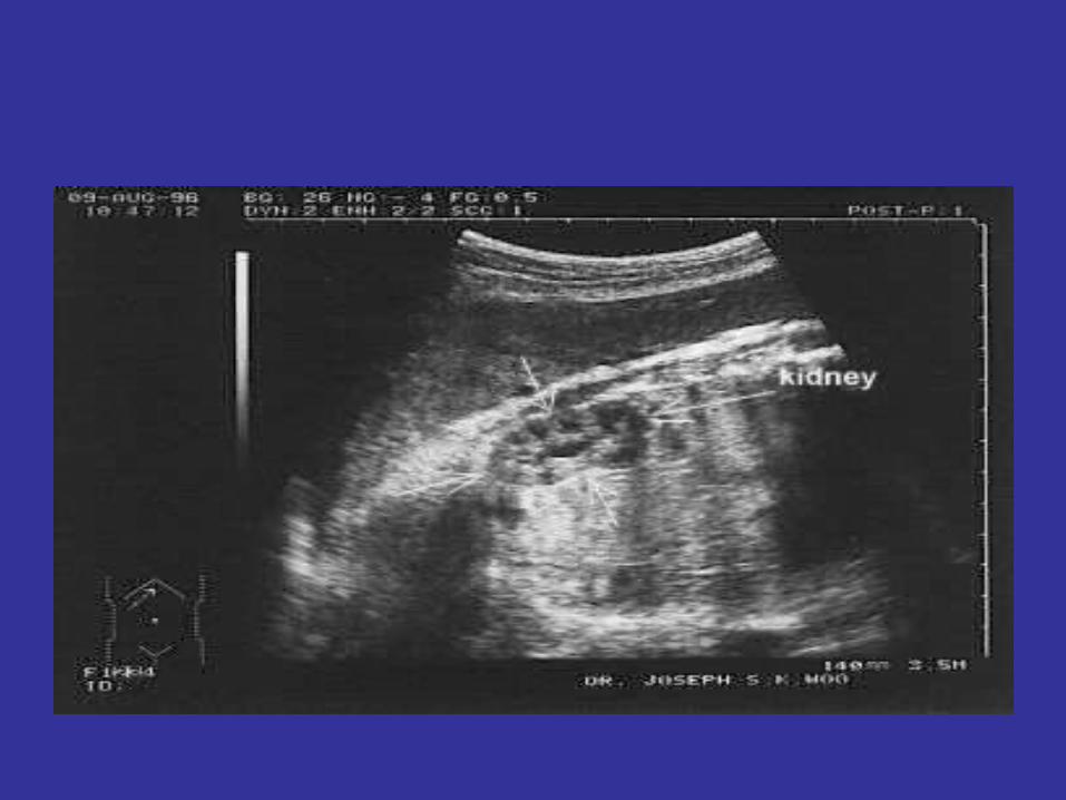
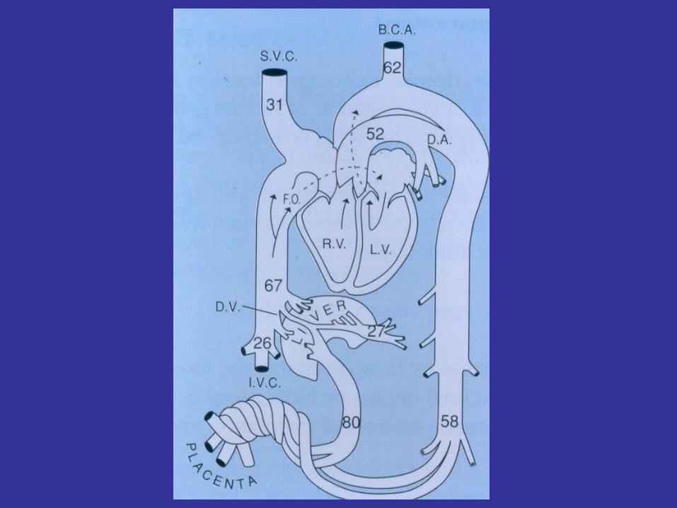
Fetal trunk
• Coronal section– Heart, stomach, diaphragm
and bladder
• Sagittal section of anterior abdominal wall
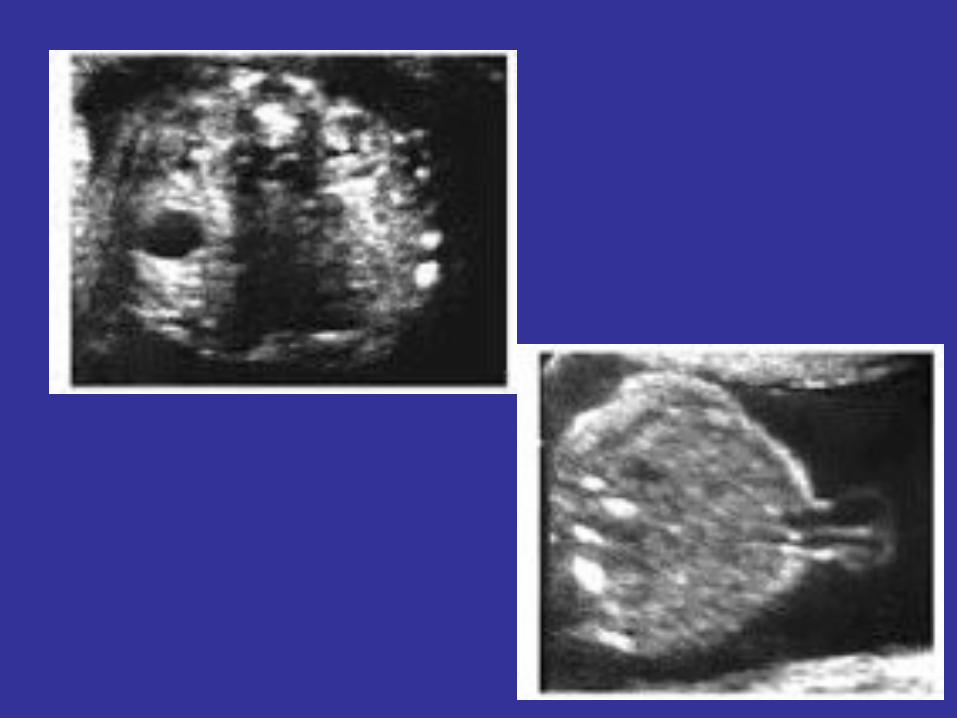
• Kidneys– Coronal– Transverse
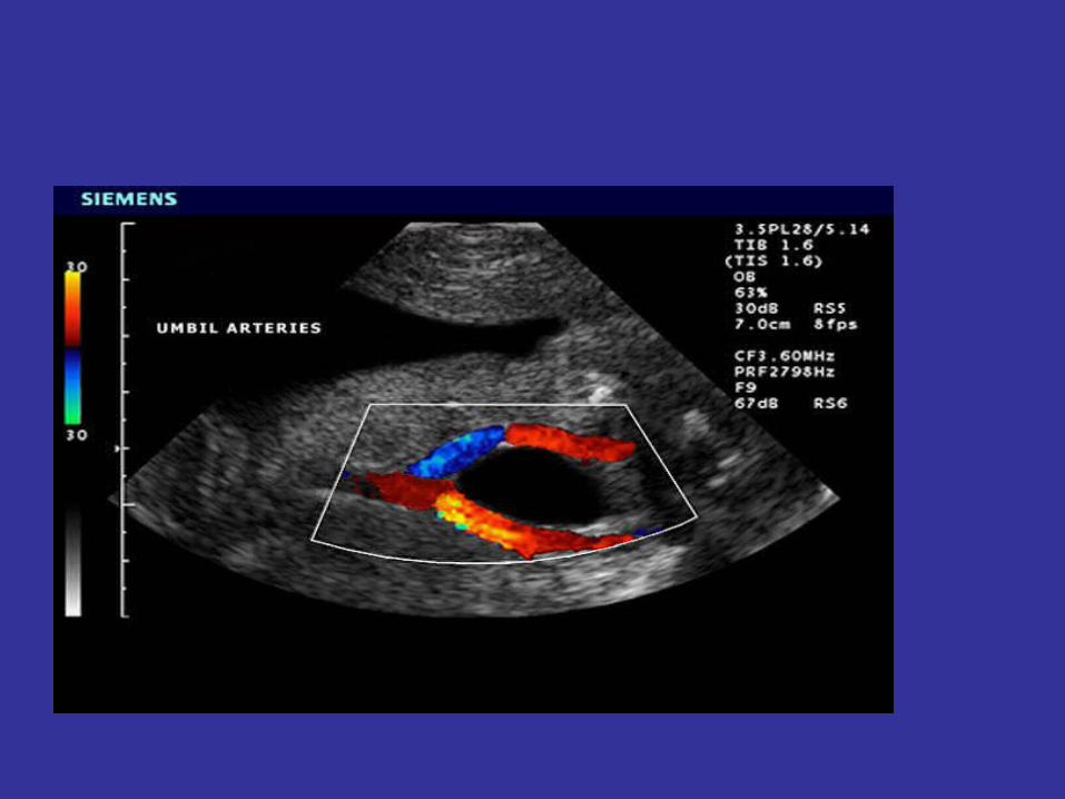
• Diaphragms sagittal view• Umbilical cord insertion• Two umbilical arteries

Fetal trunk
• Coronal section– Heart, stomach, diaphragm
and bladder
• Sagittal section of anterior abdominal wall
• Kidneys– Coronal– Transverse
• Diaphragms sagittal view• Umbilical cord insertion• Two umbilical arteries
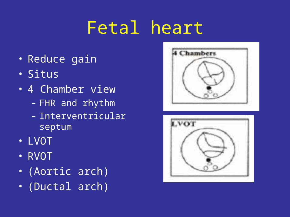
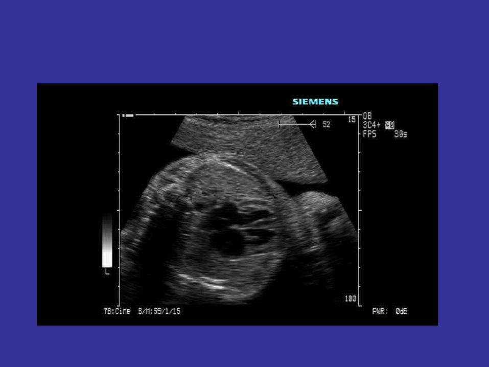
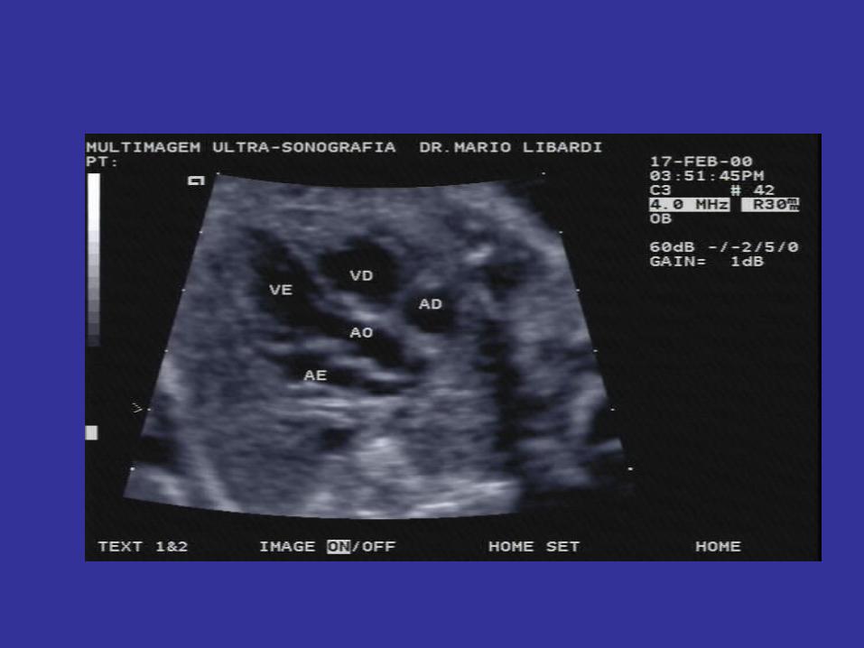
Fetal heart
• Reduce gain• Situs• 4 Chamber view
– FHR and rhythm– Interventricular septum
• LVOT• RVOT• (Aortic arch)• (Ductal arch)
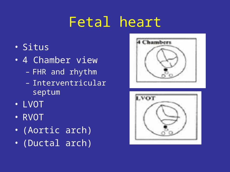
Fetal heart
• Situs• 4 Chamber view
– FHR and rhythm– Interventricular septum
• LVOT• RVOT• (Aortic arch)• (Ductal arch)
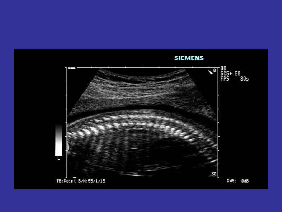
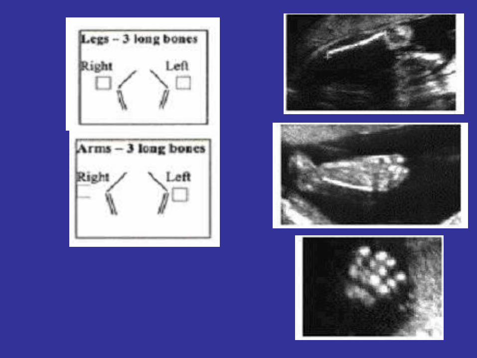
Fetal Spine and limbs
• Spine- reduce total gain• Sagittal with skinline in view• Coronal• Transverse- 3 ossification centres ‘tight’, not
splayed• Follow limb from trunk noting side• Full length all 12 bones• Exclude club foot• Feet- plantar view and count toes• Hands- open if possible and count fingers and
thumb
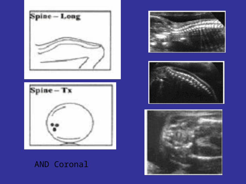
AND Coronal
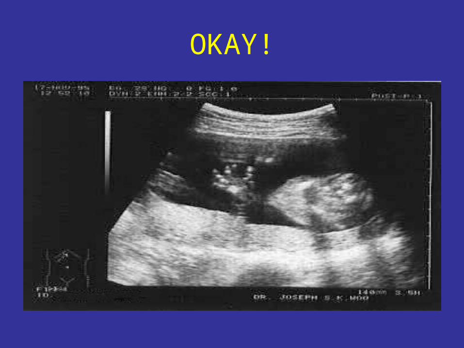
OKAY!


![Review Antenatal maternal anxiety and stress and the ... · information on normal fetal neurobehavioural development [26–28]. 2.1. Normal development of human fetal behaviour A](https://img.pdfslide.net/doc/110x75/5f180463600be842ce532d19/review-antenatal-maternal-anxiety-and-stress-and-the-information-on-normal-fetal.jpg)