Embed Size (px)
Citation preview
1/53
Order no. 173/2003 of16/10/2003
approving the Norms of Radiological Safety on Diagnostic and Interventional Radiology
Practices
Published in the Official Bulletin, Part I no. 924 of 23/12/2003
In accordance with the provisions of the:
- Law no. 111/1996 on safe deployment of nuclear activities, republished, with subsequent modifications and completions;
- Governmental Decision no. 746/2003 approving the internal rules of the Government working organizations;
- Governmental Urgency Ordinance no. 64/2003 establishing some measures on setting-up, organization, re-organisation or operation of some structures within the Government working organizations, of ministries, other specialized organization of the central public administration and of public institutions, with subsequent modifications;
CNCAN President issues the following order: Art. 1. – There are approved the Norms of Radiological Safety on Diagnostic and Interventional Radiology Practices, provided in the annex which is integralpart of the present order.
Art. 2. - The present order shall be published in the Romanian Official Bulletin, Part. I
Art. 3. - The norms provided under art. 1 shall enter into force at 1 January 2004.
Art. 4. – At the date of entering into force of the present norms, the art. 221 – 247 of the Republican Nuclear Safety Norms – The working regime with nuclear radiation sources, approved by Order no. 133/8.04.1976 of the President of the State Committee for Nuclear Energy, shall be repealed.
Art. 5. – The National Commission for Nuclear Activities Control, through the Division for application of radioactive sources, shall fulfil the provisions of the present order.
The President of the National Commission for Nuclear Activities Control Lucian Biro,
State Secretary Bucharest, 16 October 2003. No. 173.

2/53
Cap. I Scope and definitions
1.1 Scope
Art. 1 - (1) The scope of these norms is to establish the specific requirements for the practices of diagnostic and interventional radiology. (2) These norms detail and complete the basic requirements for radiological safety established in ―Radiological Safety Fundamental Norms‖, the requirements of ―Norms on radiation protection of individuals in medical ionizing radiation exposure‖ and the other norms provided in annex no. 1. (3) These norms cover all occupational, public, medical and potential exposure situations, including potential exposures (4) These norms establish the requirements of licensing and inspection issued by CNCAN (National Commission for Nuclear Activities Control) for the practices of diagnostic and interventional radiology.
1.2 Definitions
Art. 2 - (1) The definitions and terms used in these norms are defined in the Law no. 111/1996 with amendments, in the Annex no. 1 of ―Radiological Safety Fundamental Norms‖ and in the Annex no. 1 of ―Norms on radiation protection of individuals in medical ionizing radiation exposure‖. (2) In addition, the following definitions are used: a) Safety culture - The assembly of characteristics and attitudes of organizations and individuals which establishes that, as an overriding priority, protection and safety issues shall receive the attention warranted by their significance. b) Safety Assessment - A review of the aspects of design, operation and maintenance of a radiological installation which are relevant to the protection of persons or the safety of the source, including the analysis of the provisions for safety and protection established in the design, operation and maintenance of the radiological installation and the analysis of risks associated with normal conditions and accident situations. c) Standards dosimetry laboratory - A laboratory designated by CNCAN and authorised by Romanian Bureau of Legal Metrology, for the purpose of developing, maintaining or improving primary or secondary standards for radiation dosimetry.
Cap. II Application field
Art. 3 - (1) These norms apply to the practices of diagnostic and interventional radiology, which involve the risks associated with the exposure to ionizing radiation from the operation of radiological installations. (2) In the sense of these norms, the radiological installation means a medical device which emits X rays.
Cap. III Responsibilities
3.1 Managerial commitment and radiological safety policy statement
Art. 4 - (1) In every facility in which diagnostic and interventional radiology practices are in use, a safety culture shall be implemeneted and maintained in order to encourage an active and learning attitude to protection and safety and to discourage complacency. (2) To comply with this requirement, the licensee shall be committed to an effective protection and safety policy, particularly at managerial level and by clear demonstrable support for the persons with direct responsibility for radiation protection. (3) This commitment shall be expressed in a written policy statement that clearly assigns prime importance to protection and safety in the radiology services, while recognizing that the prime objective is the medical diagnostic, health and safety of the patients.

3/53
(4) This policy statement shall be made known to the medical personnel and shall be followed by establishing a radiation protection programme (RPP), which includes a quality assurance programme (QAP) and by fostering a safety culture in the hospital.
a) The aspects of a radiation protection programme are given in the Annex no. 2 b) The quality assurance programme (QAP) can be elaborated in compliance with ―WHO
Guidance document on QA in diagnostic radiology Efficacy and Radiation Safety in Interventional Radiology‖ WHO 2000, Geneva.
c) An example of quality assurance (QA) programme is given in Annex no. 3.
3.2 Organization and responsibilities
Art. 5 - (1) The main responsibility for the application of this regulation belongs to the legal person (registrants or licensees). (2) In radiology practice, the following persons shall have responsibilities for the application of radiation protection and safety regulations, by virtue of tasks involving decisionmaking, operation or handling of radiological installations:
a) radiological protection qualified expert b) the radiological safety responsible, c) medical physics expert and medical physicist; d) medical practitioners working in radiology (typically radiology specialists,
cardiologists, endoscopists, surgeons and other specialists performing interventions using x-ray, dentists);
e) other health professionals operating radiology equipment (e.g. radiographers or radiological technologists);
f) staff performing special tasks (e.g. type testing of equipment, quality control tests), service engineers;
g) suppliers; h) ethical review committees; i) any other category of staff involved in conducting diagnostic or interventional
radiology practices. Art. 6 - (1) All personnel involved in radiation protection and safety of radiological installations shall be adequately trained and qualified so that they understand their responsibilities and perform their duties with appropriate judgment and according to established procedures (2) The requirements for staff training are according to art. 7 and to annex no. 4 from ―Norms on radiation protection of individuals in medical ionizing radiation exposure‖. Art. 7 - (1) The licensee shall maintain the evidence for all personnel listed on art. 5 and 6, regarding relevant education and training to accomplish the responsibilities on radiation protection and safety. (2) For radiologists and other medical practitioners, medical physicists, radiation protection qualified experts, radiological safety responsible, radiology technologists, typical documentary evidence indicated in paragraph (1) shall consist in copies of documents which certify:
a) a degree relevant to the profession, issued by the competent education and examining authorities (Ministry of Education and Research,, Ministry of Health, etc.)
b) accreditation required to exercise the profession, granted by the relevant authority or other professional or academic organisations as required in Romania
c) a course on radiation protection approved by CNCAN and in compliance with the training requirements specified in ―Norms on radiation protection of individuals in medical ionizing radiation exposure‖.
d) Prior to work without supervision without supervision, on-the-job training supervised by radiation protection qualified expert.
(3) The maintenance (installation-construction, verification, service, repair, modification, dismantling, etc.) licensee for radiological installations shall have the documented evidence for the staff who accomplish the maintenance, which demonstrate the competence in maintenance activities. The evidence shall consist of:
a) certification by the manufacturer or his legal representative , of having completed a training programme on maintenance the type of authorized equipment;
b) a course on radiation protection according to the Norms on issuing practise permits of nuclear activities and designation of radiation protections qualified experts by CNCAN.
4/53
Art. 8 The licensee shall issue level 1 practise permits for all occupational exposed workers, who did not possess level 2 or level 3 practise permits issued by CNCAN. Art. 9 - (1) The licensee or registrant shall develop, implement and document a radiation protection programme commensurate with the nature and extent of the risks associated with radiology, under their responsibility and sufficient to ensure compliance with the requirements of the regulation. (2) This programme shall relate to all phases of the practice, from siting, construction, operation to decommissioning. (3) More, the licensee or registrant shall assure the necessary resources for effective implementation of this programme. Art. 10 - (1) The licensee or the registrant shall appoint, a radiation protection qualified expert, being in a legal contractual relationship, ,,or more experts, depending on the size of the radiology department. (2) The radiation protection qualified expert shall possess a level 3 practise permit, issued by CNCAN, for the field X-ray generators, practice Radiation Diagnostic. (3) The level 3 practise permit is requested and is issued according with ―Norms on issuing of practise permits of nuclear activities and designation of radiation protection qualified experts‖ Art. 11 - (1) The licensee or the registrant shall appoint in writing, a radiological safety responsible, for every controlled area. (2) The radiological safety responsible shall have sufficient authority regarding radiation protection regulations and license provisions. (3) The radiological safety responsible shall have a level 2 practise permit, issued by CNCAN, for the field of Radiation Diagnostic (RDG), specialty Röntgen Diagnostic (RTG), Pneumology (RTGF), Dental Röntgen Diagnostic (RTGD) or Interventional Radiology (RI),as appropriate,, or the field X-ray Generators, specialty maintenance (MRIVX) or Other Applications (AAX), by case. (4) The level 2 practise permit is requested and is issued according to ―Norms on issuing of practise permits of nuclear activities and designation of radiation protection qualified experts‖ Art. 12 The licensee or registrant shall develop, implement and document a quality assurance programme commensurate with the nature and extent of the risks associated with radiology practice, under their responsibility .. Art. 13 The licensee or the registrant shall ensure that the quality management (including quality control) in Diagnostic and Interventional Radiology such as quality control, clinic dosimetry and optimisation of patients’ protection shall be performed according to procedures approved by a medical physics qualified expert. Art. 14 In addition to the responsibilities established in the Annex no. 5 of ―Norms on issuing of practise permits of nuclear activities and designation of radiation protection qualified experts‖, the radiological protection qualified expert may be empowered by the licensee with the following responsibilities:
a) to approve the operational aspects of the radiation protection programme; b) to provide practical advice on implementation of local rules and procedures; c) to identify training needs and organize training activities; d) to systematically verify that tasks requiring personnel accreditation are performed only
by staff with the necessary accreditation; e) to identify deficiencies in the compliance with the radiation protection program and
report them to the registrant or licensee; f) to co-operate with CNCAN inspectors; g) to participate in purchasing radiological equipment, and in designing the radiological
facility;
Art. 15 - (1) The responsibilities of the radiological safety responsible are established in the Annex no. 4 of the ―Norms on issuing of practise permits of nuclear activities and designation of radiation protection qualified experts‖
5/53
(2) In addition to the responsibilities established in paragraph (1), the radiological safety responsible has the following responsibilities:
a) to participate in a continuing review of the radiology practice’s resources (including budget, equipment, and staffing), operations, policies and procedures;
b) to assure the implementation of these norms in the controlled areas and supervised areas
c) to supervise the deployment of the diagnostic and interventional radiology practices in compliance with the procedures and conditions from the licence;
d) to establish the operational aspects of radiation protection program; e) to elaborate and review periodically work procedures and local rules. f) to assure that the user manuals and user instructions for radiological installations are
known by operators; g) to assure the elaboration and the implementation of radiological emergency plan; h) to assure the periodical verification of radiological installations and dosimetric
apparatus; i) to conduct investigation in cases of exceeding investigation level, and in case of
incidents and accidents; j) to participate in purchasing of radiological installation, and in designing of radiological
facilities. Art. 16 The safety related responsibilities of the medical practitioner radiologist, are :
a) to ensure overall patient protection and safety; b) to justify diagnostic and interventional procedures using referral criteria, established
by specific regulations of the Ministry of Health; c) to provide consultation and clinical evaluation of patients; d) to establish optimized protocols for diagnostic and interventional procedures, in
consultation with the medical physicist; e) to control radiological techniques and protocols on a regular basis; f) to provide quality evaluation of radiology taking into account the results of patient
dose monitoring; g) to provide specific criteria to manage the examination of pregnant women, paediatric
patients, medico legal procedures, occupational health examinations and medical and biomedical research; and
h) to report the radiological incidents and accidents to the radiological safety responsible.
Art. 17 The responsibilities of the medical physicist are:
a) to develop requirements and specifications for the purchase of appropriate radiology equipment ensuring its radiation safety;
b) to plan the design requirements for siting,and construction of the radiological facility; c) to carry out or supervise acceptance testing, commissioning and quality control (QC),
of equipment; d) to establish patient dose assessment procedures; e) to supervise radiological installation construction and installation,maintenance,control
and repair; f) to supervise radiological installation inventory; g) to participate to the investigation and evaluation of radiological incidents and
accidents.
3.3 Quality assurance
6/53
Art. 18 (1) Quality assurance programme shall be established so that to lead to:
(a) adequate assurance that the specified requirements relating to radiation protection and
safety are satisfied;
(b) quality control mechanisms and procedures for reviewing and assessing the overall
effectiveness of radiology practice.
(2) The licensee and the management of Radiology Department shall provide the necessary
resources on personnel and budget to realize an effective quality assurance programme (QAP).
(3) The programme shall cover the entire process from the initial decision to adopt a particular
procedure through to the interpretation and recording of results and shall include a systematic
control methodology. (4) Continuous quality improvement shall be assured. This implies continuous improvement of the procedures of the use of radiological installations in diagnosis and interventional practices, improvement based on new information learned from their QAP and new techniques developed by the radiology community. (5) The review of QAP shall take into account the operational experience and lessons learned from accidents or near misses and shall help identify potential problems and correct deficiencies, and the review shall be used systematically, as part of the continuous quality improvement. (6) An example of a QAP is shown in the Annex no. 3. Art. 19 Quality assurance shall cover, as a minimum:
a) acceptance tests of radiological installation and commissioning; b) QC of radiological installation (hardware and software); c) operational procedures for radiological installation; d) selection of the correct procedure for the patient; e) appointment and patient information; f) clinical dosimetry; g) optimization of examination protocol; h) record keeping and report writing; i) training and continuing education of staff; j) clinical audit; and k) evaluation of general outcome of radiology service.
3.4 Human factors
Art. 20 - (1) The licensee or registrant shall establish the necessary provisions for reducing as far as practicable the contribution of human error to accidents and other events that could give rise to exposures (2) For this scope, all personnel responsible with radiation protection and safety shall be appropriately trained and qualified so that they understand their responsibilities and perform their duties with appropriate judgment and according to defined procedures
3.4.1 Staffing
Art. 21 - (1) The licensee or registrant shall appoint in writing all the professionals developing radiology, each one having an accreditation sufficient to ensure that all activities relevant to radiation protection and safety are carried out in accordance with Romanian regulations, with the radiation protection programme and with the conditions of licence. (2) The adequate number of persons shall be kept under review, especially as workload increases, or new techniques and new equipment are incorporated.
3.4.2 Education and training
Art. 22 - (1) All staff working with radiological installations in diagnostic and interventional radiology, shall have relevant qualifications and practical training in radiation protection. (2) Investment in radiological installations shall be accompanied by concomitant investment in training and authorization of staff involved in practices of diagnostic and interventional radiology.

7/53
(3) The licensee or registrant shall include in the application of the licence written proofs on qualifications in radiation protection of the medical practitioners, of the radiological protection qualified experts, of the radiological safety responsible, of the medical physicists. Art. 23 - (1) The registrant and licensee shall ensure that staff are aware of:
a) the conditions of the licence; b) operation of radiological installation;
c) instructions that shall be provided to patients and those helping the patients during exposures ;
d) institutional radiation protection policies and procedures; e) the local QAP and QC procedures; f) results of review and analysis of incidents and accidents that have occurred in the institution or elsewhere and needed preventive and corrective measures.
Art. 24- (1) The training of staff shall be completed before commencement of duties and shall be in compliance with the allocated responsibilities and job description. (2) The training shall be updated whenever significant changes in radiological installations, duties, regulations, the terms of the licence or radiation safety procedures occur. (3) The licensee or registrant shall provide means for continuing education and a programme of permanent professional development included in his staff policy. This policy shall improve the staff skills, maintain familiarity with current practices and foster a safety culture throughout the institution. Such training and development schemes shall be accomplished through informal meetings of the department, seminars, approved (accredited) continuing education programmes or other meetings. (4) The registrant and licensee shall prepare and keep a record of the initial and periodic instruction of personnel. These records shall be kept for at least five years after the diagnostic or interventional radiology practice ceases.
Cap. IV Licensing of practices
4.1 Licence
Art. 25 - (1) Any person set up according to the law who intend to use ionizing radiation sources in diagnostic and interventional radiology, shall notify this intention to CNCAN and shall apply for licence in the form of a licence or a registration, according to the Radiological Safety Norms - Licensing Procedures. (2) The medical radiological installations which shall be submitted to registration are listed in art. 11, e) and f) from Radiological Safety Norms - Licensing Procedures. Art. 26 - (1) The licence is granted if the adequate arrangements, endowment,staffing and the activity organization requirements are accomplished in compliance with the laws and norms from the annex no. 1. (2) Diagnostic and interventional radiology practices which are not subject to a registration are licensed for every phase, namely:
a) siting b) construction; c) operation d) modification
(3) In the case of arrangements are performed in the existing building, the siting phase and the construction phase can be joint. (4) For mobile radiological installation, the licensing of siting and construction phases is no more necessary. (5) For diagnostic and interventional radiology practices which use of radiological installations, it is not necessary the licensing of decommissioning phase or the licensing of partial or total cessation of the practice, being sufficient the dismantling of the X-ray installation by a body authorized by CNCAN for this activity of maintenance. (6) By exception from the provisions of paragraph (5) above, dismantling with destruction of the X-ray installation can be performed in compliance with licensee’s own procedures, who shall immediately notify CNCAN.
8/53
Art. 27 - (1) Any legal person shall submit to CNCAN for assessment the relevant information necessary to demonstrate the radiation protection and safety of the practice According to the law. An example of tehnical documentationthe information in support of a licence application is provided in Annex no. 4. (2) The legal person responsible shall include in the technical documentation for licence application:
a) proof on the qualifications in radiation protection of the medical practitioners who are to be so designated by name in the registration or licence; or
b) a statement stating that only medical practitioners with the qualifications in radiation protection specified in this regulation will be permitted to prescribe medical exposure by means of the authorized radiological installation.
Art. 28 The registrant and licensee shall apply for modification of the authorization according to art. 87 - art. 88 of the Radiological Safety Norms - Licensing Procedures, in the following situations:
a) the change of headquarters or other modifications in firm’s documents; b) the change of the responsible persons in radiological safety (radiation protection
qualified expert, the radiological safety responsible) c) the modifications of limits and conditions specified in licence; d) other modifications that could affect the sources of safety of radiological installations,
or radiation protection of personnel,, population or environment.
4.2 Renewal of licence or prelongation of validity Art. 29 The renewal of licence or prelongation of validity period is done according to art. 79-85 of the Radiological Safety Norms - Licensing Procedures.
4.3 Inspection Art. 30 - (1) The registrant or licensee shall permit inspection by special empowered inspectors of CNCAN according to the Law no. 111/1996 with subsequent modifications and completions, to verify the conformity with provisions of these norms. (2) An example of checklist for inspection of diagnostic and interventional radiology practices is provided in annex no.5.
4.4 Authorization of other practices related to radiology
Art. 31 - (1) According to the Law 111/1996 with subsequent modifications and completions , the following activities require a licence:
a) production, import-export, supply, leasing or transfer of radiological installations; b) maintenance of radiological installations (construction and installation, control,
service, repair,maintenance, modification,dismantling); (2) The individual dosimetric monitoring of professional exposed workers shall be conducted according to the Norms on individual dosimetey, by individual dosimetric bodies designated by CNCAN. (3) The licensee of a radiology practice shall contract for sevices involving the activities mentioned above only enterprises designatet/authorised by CNCAN.
Cap. V Safety of radiological installations
5.1 Safety of radiological installations
Art. 32 In medical exposures shall be used only radiological installations which:
1) possess Medical Device Certificate, issues by the Ministry of Health (MS), according to the Law no. 176/2000;
2) possess Radiological Safety Authorisation, issued by CNCAN, according to the Law no. 111/1996, with subsequent modifications and completions;
3) are tested periodically, at least once yearly, to verify the maintaining of nominal parameters.
9/53
Art. 33 - (1) In accordance with art. 59 of Radiological Safety Norms - Licensing Procedures, the technical documentation support to the application for Radiological Safety Licensing of a radiological installation, shall demonstrate that the radiological safety requirements for design and manufacture of installation are fulfilled. (2) The radiological safety requirements on design and manufacture of radiological installation,mentioned in annex no.6 shall be applied to installations purchased after the entry into force of these norms.
Art. 34 Registrants or licensees shall: a) take into account information provided by suppliers, identify possible equipment failures
and human errors that may result in unplanned medical exposures; b) take all reasonable measures to prevent failures and errors, including the selection of
suitably qualified personnel, the establishment of adequate procedures for the calibration, quality assurance and operation of diagnostic equipment, and the provision to personnel of initial appropriate training and periodical retraining of personnel, including protection and safety;
c) take all reasonable measures to minimize the consequences of failures and errors that may occur; and
d) develop appropriate contingency plans for responding to events that may occur, display plans prominently, and periodically conduct practice drills, as appropriate.
5.2 Design of radiological facilities
Art. 35 - (1) During designphase of the facility which use fixed radiological installations (X-ray rooms and other related rooms) shall assure needed measures to optimize protection and to limit the doses,, in the scope of achieving the radiological safety requirements shall be ensured (2) The facility design needs consideration to be given to classification of the areas within it, the type of work to be done and the X-ray systems intended to be used. (3) At designing of radiology facility the three factors relevant to dose reduction: time, distance and shielding shall be combined in the design. (4) Larger rooms are recommended to allow easy access for patients on a bed trolley and to reduce exposure of the staff and public and at the same time allow for patient positioning and easy movement during the procedure. Art. 36 Radiology facility will comprise, at least, as appopriate:
1. X-ray room designed for radiological installation
2. control room designed for control console, as appropriate
3. development room
4. undressing and waiting room for patients, as appropriate
5. room for images interpretation
6. medical consultation room
7. medical staff room
8. film archive and permanent records
9. cloakroom, sanitary group for staff and respectively for patients, by case. Art. 37 - (1) The surface of X-ray room shall correspond with the requirements of producer regarding the minimum surface needed for installation and mounting of respective radiological installation. (2) The mounting of radiological installation in a room smaller than that recommended by producer and the limitation of technical capabilities of installation because of insufficient surface of room, are not justified. Art. 38 Whenever the minimum dimension permitted for the surface of X-ray room is not specified in Radiological Safety Licence of that radiological installation, the minimum dimensions of x-ray room without the limitation of technical capabilities of installation shall be:
10/53
a) The rooms designed for radiological installation in diagnostic with one post will have a surface of minimum 20 m
2 and a square or rectangular form. The ratio between the
two dimensions will not be less than 2/3.
b) For the installations with 2 posts (radiography and fluoroscopy) in the same X-ray room, the surface of the room will not be less than 36 m
2. It is forbidden putting in this
space of furniture which is not strictly related to use of radiological installation.
c) In the case of installations with more posts or special installations, the space will be increased, as appropriate, taking into account the necessity of assurance of radiation protection of medical staff, of patients and of the other persons.
d) The X-ray room designed for an intra-oral dental radiological installation with the maximum voltage of 70 kV, will have a surface of at least 10,5 m
2. In case of
positioning of two intra-oral dental radiological installations the surface will be at least minimum 16 m
2, while the installations will operate only alternatively.
e) X-ray room designed for dental panoramic radiological installation with the maximum voltage of 90 kV will have a surface at least 16 m
2.
f) X-ray room designed for mammography radiological installation will have a surface at least 10,5 m
2.
g) X-ray room designed for bonedensitometry radiological installation with the maximum voltage of 80 kV will have a surface at least 16 m
2.
Art. 39 - (1) By rule, the positionin of radiological installation for diagnostic will be done in the centre of room. (2) The fuoroscopy radiological installation will be positioned with the axis of X-ray tube - image receptor, parallel with the short axis of x-ray room. (3) In case of fluoroscopy radiological installation, the minimum distance between focal spot and the nearest wall will be at least of 150 cm. (4) In case of fluoroscopy radiological installation with remote control, the requirement from paragraph (3) does’ not apply. Art. 40 (1) The mobile radiographic and fluoroscopic installations will be used as such. (2) The use of mobile radiological installations as stationary installation is forbidden. (3) The switch of exposure shall be connected to control console or to radiological installation by a wire of at least 3 meters long, to allow to the operator to move away from patient enough at the time of exposure. (4) It is forbidden the use of radiological installation without using of protective equipment against radiation, adequate for occupational exposed workers and for population. Art. 41 - (1) The design of the room shall be in such a way that the x-ray beam cannot be directed at any area which is not adequately shielded. (2) The X-ray room shall be designed so as to avoid the direct incidence of the X-ray beam on the access doors. Art. 42 The doors shall fulfil the requirements for a protective shield for scattered radiation and be shut when the X-ray beam is on. Art. 43 - (1) In case radiological installation is not provided with an audio communication system between control consol room and patient, this mandatory connection will be ensured by designing. (2) The provision of paragraph (1) does not apply when the starting of exposure is done from the same room. Art. 44 - (1) On X-ray room shall be a TV system or a viewing window to permit the operator to clearly observe the patient at all times during an x-ray procedure. (2) The viewing window shall fulfil the requirements of a protective shield for X-ray. Art. 45 (1) The sign ―Danger of ionising radiation‖ shall be posted o each entrance to X-ray room, according to the recommendation of International Standards Organization ISO no. 361 and according to art. 43 from NFSR. (2) The sign will be coloured on black and the background on yellow.

11/53
Art. 46 - (1) Labels containing the text ―controlled area‖ and information om source nature and associated risks shall be posted according to art. 43 from NFSR. (2) The dental intra oral radiological installations situated on stomatology cabinet are excepted for the provisions of art. 45 and art. 43 paragraph (1) Art. 47 A warning light shall be placed at the entrance to any room where fluoroscopy or CT equipment is in use. The light shall be illuminated continuouslywhen the x-ray beam being energized. Art. 48 (1) In rooms for fluoroscopy and interventional procedures, with staff close to the patients, ceiling mounted protective screens and table mounted leaded curtains shall be installed. (2) The medical staff shall wear adequate individual equipment for radiation protection, described on section 7.5 of these norms.
Considerations about radiation protection calculation Art. 49 - (1) Shielding barriers shall be calculated taking into account the attenuation provided. This is obtained by the ratio between the doses that would be received by the staff and public if shielding was not present and the doses that can be considered as optimized. (2) Doses that would be received without shielding are calculated by using tabulated workload values (mAmin per week for the different beam energy and filtration), tabulated ―use factors‖ for a given beam direction (fraction of the total amount of radiation emitted in that direction) and tabulated ―occupancy factors‖ (fraction of the total exposure which will actually affect individuals at a place, by virtue of the time permanence in that place). (3) For secondary barriers the ―use factor‖ is always 1. (4) Knowing the dose that would be received without shielding, the next step is to calculate the attenuation that is necessary to reduce this dose to a design level or to a level that can be considered ―optimized protection‖. (5) The calculation is simplified by using dose constraints as design levels, which restrict the optimization options. (6) Dose constraints also include consideration to exposure of individuals from more than one source of radiation. (7) The value of 20 μSv/hour, for the dose rate constraint on control consol of a radiological installation shall not be exceeded. Art. 50 - (1) X-ray room shall be designed so that dose rate does not exceed:
1. 15 mSv/year on work place of X-ray occupational exposed persons 2. 1 mSv/year on all areas where population might have access.
(2) Screens, other than walls of X-ray room, shall be designed so that dose rate doesn’t exceed 20 μSv/hour. Art. 51 - (1) To calculate the protective screens against X-ray radiation it methods and data from the following documents can be used..
a) NCRP Report 49 - Structural shielding design and evaluation for medical uses of x-rays and gamma-rays of energies up to 10 MeV. - National Council on Radiation Protection and Measurements, 1976.
b) DIN 6812 - Medical X-ray equipment up to 300 kV. Radiation protection rules for installation, 2002
c) or any other adequate standard recognized by CNCAN. (2) These documents provide tabulated values of workload, use and occupancy factors, scattering factors and attenuation values for the different beam qualities and scattered radiation. The tabulated values are conservative and overestimate the shielding. Art. 52 Typical conservative assumptions used in shielding design are:
a) attenuation by the patient and image receptor is usually not considered
b) workload, use and occupancy factors are overestimated
c) the conservative assumption that staff are always in the most exposed place of the room.
12/53
d) distances between personnel and X-ray source are assumed to be the minimum possible all the time
e) leakage radiation is assumed to be the maximum all the time
f) field size used for the calculation of scatter radiation is usually that maximum possible for the radiological facility.
g) the value of calculated air kerma (in mGy) is directly ―used‖ to compare with dose limits, constraints (mSv), which are given in terms of effective dose, without consideration that this value is substantially lower, given the dose distribution within the body for the beam qualities used in diagnostic and interventional radiology..
Art. 53 - (1) Shielding shall be calculated according to the principles of protection optimization (2) Dose constraints and dose limits shall be developed and used whilst considering that at given time other X-ray systems will be mounted in the same room and that the workload could be higher in the future. (3) The structure of the room shall provide adequate shielding for members of the staff involved in the x-ray procedure and persons in adjacent areas (staff, members of the public, patients or visitors). (4) If the existing structures do not provide sufficient shielding, then additional shielding shall be installed to create an intrinsically safe working environment. (5) For X rays of the energy used for diagnostic, it is in general, less expensive to design shielding conservatively with a view to avoiding expensive and inconvenient modifications to the room design in the future to accommodate changes in use or workload. Art. 54 The overall design of the facility including radiation protection calculations shall be performed by a qualified expert in radiological protection. Art. 55 Interventional radiology rooms require particular attention due to the generally much higher workload, and will most likely require a higher level of shielding.
5.3 Maintenance of radiological installations
5.3.1. General Requirements Art. 56 - (1) The registrant or licensee shall ensure that all maintenance operations: installation, assembling, verification, service, repair, dismantling/decommissioning, etc. of the radiological installations, are performed only.by a body authorized by CNCAN, according to the law. (2) The registrant or licensee shall keep the technical card of radiological installation for whole lifetime of installation, until the annulment. The technical card will contain records of performed operations of installation, mounting, repair, verification, service and all services performed until the annulment of installation. (3) Initial report, periodical reports and after every intervention on radiological installation of repair change of component, will be kept by licensee or registrant for inspections. Art. 57 - (1) The registrant or licensee shall ensure that adequate maintenance, preventive and corrective, and verification are performed as necessary to ensure that X-ray systems retain their design specification for image quality, radiation protection and safety for their life time.. (2) Daily, weekly and monthly verifications of radiological installation are performed according to the producer’s instructions by medical physicist, and, in case installation does’ not correspond, the authorized service body is appealed to. (3) All procedures used for verifications mentioned in paragraph (2) belong to QAP of user. (4) The verifications mentioned in paragraph (2) shall have records which will be kept for inspections for at least 5 years. Art. 58 - (1) All maintenance procedures (installation, assembling, verification, service, repair, dismantling/decommissioning, etc.) shall be included in the QAP of a body which is authorized for maintenance activity. (2) Servicing reports describing the findings and records of subsequent interventions shall be archived as part of the quality assurance programme.

13/53
(3) A qualified expert in radiological protection or medical physics shall participate and ensure that the equipment is in safe condition for clinical use after maintenance. (4) After every repair and periodical verification, not longer than one year, the handling licensee will issue a verification report that ensures that radiological installation retains its design specification.
5.3.1 Electrical and mechanical safety
Art. 59 - (1) The electrical and mechanical safety aspects of the radiological installation are an important part of the maintenance programme, and may have direct or indirect effects on radiation safety. (2) This work shall be performed by staff that belong to a body with manipulation licence issued by CNCAN, staff that are aware of the specification of the radiological installation and that have adequate certification granted by the producer of installation. (3) Electrical and mechanical maintenance of the radiological installation shall be included in the quality assurance programme. (4) Servicing reports on mechanical and electrical maintenance, as well as verification reports provided on art. 58, paragraph (4) shall be kept as part of quality assurance programme.
Cap. VI Justification, optimization and limitation of doses to individuals in diagnostic and interventional radiology practices
Art. 60- (1) The radiation protection requirements on justification, optimization and limitation of doses and doses constraints, that are formulated in chapter IV of NFSR shall apply on radiology practices taking into account the specifications mentioned below in this article. (2) Justification - All practices involving X-ray medical exposure, shall be justified, by weighing the diagnostic benefits it produces against the individual detriment that the exposure might cause, taking into account the benefits and risks of available alternative techniques, but involving no exposure to ionizing radiation. (3) Dose limitation
a) Dose limits does not apply to medical exposures of patients.
b) Dose limits formulated in Section II from NFSR, shall apply for occupational exposed workers, for pregnant workers, for apprentices and students and for persons from population.
(4) Radiation Protection Optimization
a) In diagnostic medical exposure, the protection optimization is realized by keeping the exposure of patients to the minimum necessary to achieve the required diagnostic objective.
b) Dose constraints are used for optimizing protection in the planning stage for each X-ray source.
c) When choosing dose constraints for the sources involved in a radiology facility, consideration needs to be given to the fact that medical staff often work in more than one facility. These constraints shall rely on realistic assumption..
Cap. VII Operational radiation protection
Art. 61 All radiation protection requirements that are formulated in chapter VI ―Operational Radiation Protection of Exposed Workers, Apprentices and Students‖ of NFSR shall apply on radiology practices.
7.1Responsibilities
Art. 62 The registrant or licensee, by the representative empowered to represent the legally set up person (director, manager, unique associate),, is responsible for the achievement of requirements on occupational exposure to ionising radiations.
7.2 Pregnant workers
14/53
Art. 63 - (1) As soon as, a female worker becomes aware that she is pregnant, she shall notify in writing the employer about that. (2) The licensee immediately will take all measures to ensure the protection of foetus at the same level of dose as required for members of the public. (3) The working conditions of pregnant worker shall ensure that effective dose to the child to be born will be as low as reasonably achievable, without exceeding 1 mSv during at least the remainder of the pregnancy.
7.3 Classification of areas
Art. 64 In a radiology laboratory, all rooms with mounted radiological installations (including X-ray tube assemblies and control console) and the areas where are used mobile radiological installations, are considered controlled areas. Art. 65 All the other neighbouring areas of controlled areas and the other areas of diagnostic and interventional radiology laboratory are considered public areas. In diagnostic and interventional radiology there are not supervised areas. Art. 66 - (1) Every room from radiology laboratory shall be used only according to its specific destination. (2) Doors of X-ray rooms shall be closed during X-ray procedures.
7.4 Local rules and supervision
Art. 67 - (1) Registrants or licensees, in consultation with the radiation protection qualified expert and the radiation safety officer, shall:
a) establish written local rules and procedures necessary to ensure adequate levels of protection and safety for workers and other persons;
b) include in the local rules and procedures the values of any relevant investigation level or authorized level, and the procedure to be followed in the event that any such value is exceeded;
c) make the local rules and procedures, the protective measures and safety provisions known to those workers to whom they apply and to other persons who may be affected by them;
d) ensure that any work involving occupational exposure to ionising radiation is adequately supervised and take all reasonable steps to ensure that the rules, procedures, protective measures and safety provisions are observed.
(2) Example of local rules for operational safety are provided in annex no. 7
7.5 Protective equipment
Art. 68 - (1) Registrants or licensees shall ensure that workers are provided with suitable and adequate individual protective equipment that meets the requirements of The Normative of granting and utilization of the individual protection equipment against ionizing radiation , RP 06/1997, published in Official Gazette no. 111bis on 04.06.1997. (2)Only the individual protective equipment, that is authorised according to the law, and for which CNCAN issued a Radiological Safety licence shall be used. . (3) The individual protective equipment that includes lead aprons, thyroid protectors, protective eye-wear, gloves, shall be in compliance with the technical specifications of producer and with the specific standards. (4) The necessary of these protective devices is established by the radiation protection qualified expert. Art. 69 Gloves are useful to protect the hands near to the beam, but they will be used with discernment, because they may produce the opposite effect during fluoroscopy with automatic brightness control (ABC) when the hands enter the area covered by the sensor of the ABC, because this would drive the exposure to higher levels for both the staff and the patient and would be ineffective in protecting the hands.
15/53
Art. 70 - (1) Registrants or licensees shall assure that:
a) the workers are adequately trained to the use of individual protective equipment;
b) only those persons ,who have medical advice to carry without problems the additional weight of the personal protective equipment,, will conduct activities which need its wearing.
c) all individual protective equipment is well maintained and is checked periodically, as apprporiate.
(2) An example of a list of protective clothing is given in annex no. 8. Art. 71 Additional protective devices are recommended to be used in fluoroscopy and interventional radiology rooms, which include:
a) Ceiling suspended protective screens for protecting eyes and thyroid while keeping visual contact with the patient.
b) Protective lead curtains mounted on the patient table. Art. 72 Over-couch tube geometry is not recommended for fluoroscopy because it involves a considerably higher radiation level at the operator position, by comparison with under-table geometry. If over-couch geometry is nonetheless used, protective lead curtains shall be used to reduce scatter radiation to staff. Art. 73 All staff from X-ray room for fluoroscopy, which is not staying behind a shielded control console, shall wear an individual protective lead apron. Art. 74 Registrants or licensees shall ensure that the patient and his helper are provided with suitable and adequate individual protective equipment, as necessary.
7.6 Individual monitoring and exposure assessment
Art. 75 - (1) Registrants or licensees shall ensure systematic individual monitoring for category A of exposed workers. (2) The monitoring shall be performed by an accredited dosimetry body. (3) The monitoring for category B of exposed workers, shall demonstrate the correct assessment of the workers in this category, after that this monitoring being no more necessary.. (4) In case of some practices, CNCAN may impose to ensure individual monitoring according to the requirements for category A and Bprofessional exposed workers. (5) The monitoring system for radiation exposure of occupational exposed workers is approved by CNCAN in the process of licensing of practice. (6) The requirements for individual dosimetry are formulated in the ―Norms on individual dosimetric monitoring‖ Art. 76 Other frequent users of radiological installations such as endoscopists, anaesthetists, cardiologists, surgeons etc., as well as ancillary workers, who frequently work in controlled areas, shall also be monitored. Art. 77 Individual external doses arisen from external exposure shall be determined by using individual monitoring devices such as thermoluminescent dosemeters, film badges or other devices that have Radiological Safety Licence issued by CNCAN. Art. 78 Each individual dosemeter shall be used only by the person to whom it was entrusted to.. Art. 79 - (1) The monitoring device shall be worn on the front of the upper torso of the body, between the shoulders and the waist. (2) The individual monitoring shall take place monthly. (3) The period between the dosemeters being received by the dosimetry service and return of the dose reports shall not exceed one month.
16/53
Art. 80 - (1) Because evaluation of dose is an essential part of the RPP, it is important that workers return dosemeters on time for processing.
(2) Delays in the evaluation of a dosemeter can result in the loss of the stored information, so that delayed return means disciplinary misbehaviour and shall be sanctioned by radiological safety officer.
(3) Registrants or Licensees shall analyse periodically the way in which the individual
dosimetry is performed.
Art. 81 - (1) When a lead apron is used, the dosemeter shall be worn under the apron and it will be shielded by the apron. (2) However, if the staff have a high workload and stand inside the X-ray room, radiological protection qualified expert shall decide additional dosimetry outside the apron (e.g. over the thyroid collar or on the shoulder, hands or fingers). (3) In case the dosemeter worn under is apron,, the effective dose would be underestimated iwhile in case the dosemeter is worn over the apron the effective dose is overestimated by one to two orders of magnitude. As long as the practice is consistent and clearly stated, each method is appropriate. Art. 82 For estimation of effective dose when wearing two dosemeters – one under and one outside the apron,, the following formula could be used: Effective dose (estimate) = 0.5HW + 0.025 HN Where HW is the dose at waist level under the apron and HN is the dose recorded by a dosemeter worn at neck level outside the apron. Art. 83 - (1) In some facilities and for some individuals with a low level of occupational exposure (medical practitioners in bonedensiometry, dentists), area dosemetry to estimate the level of dose per procedure can be an acceptable alternative. (2) Some radiological installation for dental radiography, or others, having a limited number of procedures per month could be spared from personal dosemeters for all staff involved, with the CNCAN agreement. (3) Individual exposure monitoring for all cases mentioned in para. (2) can be performed through area dosimetry or some other individual dose evaluation per procedure. Art. 84 - (1) IfIn case an individual dosemeter is lost, the radiological protection qualified expert shall perform a dose assessment and record this evaluation for the specific worker. (2) The loss of the dosemeter and the dose assessment shall be reported to CNCAN. (3) When an individual dosemeter was lost, the most reliable method for estimating an individual dose is to use his or her recent dose history. In those cases where the individual performs non-routine types of work, it may be better to use doses of co-workers who have performed the same work as a basis for the dose estimate.
7.7 Monitoring the workplace
Art. 85 - (1) The licensee or registrant shall ensure the radiological monitoring of workplaces. (2) The radiological monitoring of workplaces for controlled areas and public areas adjacent to controlled areas shall be done by dose rate measurements for external exposure, by indicating the quality of X-rays. (3) The radiological monitoring of workplaces shall be performed by own staff with its own equipment or by an external qualified licensee or registrant supervised by a radiological protection qualified expert. Art. 86 - (1) The licensee or registrant shall maintain the evidence of the results of X-ray field’s measurements for controlled areas and public areas adjacent to controlled areas, done for typical points where exposure is higher. (2) The evidence shall contain:
1. technical parameters of radiological installation 2. name of the measurement point; 3. dose rate for every point of measurement; 4. name of dosemeter used for measurements; data of its last calibration;
17/53
5. data of measurement. 6. reference levels and corrective actions in the case of exceeding of these levels; 7. Name, first name and qualification of person who has performed the measurements.
(3) The points of measurement are established and approved by CNCAN during licensing process. (4) The evidence of measurements is kept by the radiological safety responsible. (5) The periodicity of measurements is every tree months, by rule. In the case of dental, mammographic, bonedensiometry radiology, the measurements shall be done two times a year. After every repair or change of installation, the radiological measurements of work place will be performed,too.
Art. 87 The radiological monitoring of workplaces can be performed by using also film badges that have Radiological Safety Licence issued by CNCAN,( same type as dosemeters used for individual monitoring) or another adequate dosemeter,, placed during one month in the points with the highest dose rates, estimated or measured or on the most frequented places on controlled areas, or in their neighbourhood, by medical staff. Art. 88 - (1) All survey meters used for workplace monitoring shall be calibrated and this calibration shall be traceable to a standards dosimetry laboratory designated by CNCAN. (2) Initial monitoring shall be conducted immediately after the installation of new radiological installation and shall include both measurements of radiation leakage from installation provided at item 8 of annex no. 6 and area monitoring of useable space around X-ray rooms (of controlled areas). (3) All radiation monitors shall be calibrated, and their warning devices and operability shall be checked prior to each day of use.
7.8 Investigation levels for staff exposure
Art. 89 Registrants and licensees shall, in consultation with radiological protection qualified expert and radiological safety responsible.
a) include in the local rules and procedures the values of the established investigation level according to art. 91 or other authorized level, and
b) the procedure to be followed in the event that any such value is exceeded.
Art. 90 The investigation level is shall be used to provide a ―warning‖ on the need of reviewing procedures and performance, and in case something is not working as expected and shall lead to corrective actions if the doses received by the staff reach or exeed the investigation levels. Art. 91 - (1) Monthly values higher than or equal to 0.5 mSv (for the dosemeter worn under the lead apron) shall be investigated. (2) Monthly values higher than say 5 mSv for the dosemeter worn over the apron or in the hand or finger shall also be investigated with a view to optimization. (3) The licensee or registrant shall establish other investigation levels, but not higher than those mentioned above. Art. 92 The licensee shall conduct formal investigations, whenever:
a) an individual effective dose exceeds investigation levels;
b) any of the operational parameters related to protection or safety are out of the normal range established for operational conditions;
c) an equipment failure, severe accident or error has taken place, which causes, or has the potential to cause, a dose in excess of annual dose limits; and
d) any other event or unusual circumstance that causes, or has the potential to cause a dose in excess of the annual dose limits or the operational restrictions imposed on the installation (e.g., the significant change in workload or operating conditions of radiology equipment).
18/53
Art. 93 - (1) The investigations shall be initiated as soon as possible after having discovered the event. (2) After each investigation a written report shall be prepared and kept concerning the cause, determination or verification of any doses received, corrective actions taken, and instructions or recommendations to avoid recurrence.
7.9 Health surveillance of occupational exposed workers Art. 94 The licensee or registrant shall ensure the health surveillance of occupational exposed workers to ionising radiation, according to:
a) Health Minister Order no. 944 / 28 December 2001, for approval of Norms concerning medical surveillance of occupationally exposed workers to ionizing radiations, published in the Official Gazette, no. 34, on 18 January 2002
b) Health Minister Order no. 1032/20.12.2002 approving the amendments to the Norms on medical surveillance of occupational exposed workers to the ionizing radiation (approved by order no. 944/28.12.2001), published in the Official Gazette no. 15/13.01.2003.
Art. 95 - (1) The medical surveillance ensures the assessment of continuing fitness of occupational exposed workers for their work in an environment with ionising radiation.. (2) In case the worker is found ―unfit‖ he will be taken out of the ionizing radiation field, in compliance with art. 77 from NFSR. Art. 96 - (1) In case of an accidental exposure to high radiation doses of the order of magnitude of 0.2-0.5 Sv or higher, specific radiation-related medical investigations are necessary, their results being registered. (2) These levels of doses shall not expect to be encountered in diagnostic radiology. Doses at the level of deterministic effects shall not be reached .in interventional radiology.
7.10 Records
Art. 97 - (1) The licensee or registrant shall maintain exposure records according to the chapter ―Recording and reporting of the results of individual monitoring of occupational exposed workers‖ (art. 63-71) of NFSR and specified requirements from Norms on individual dosimetric monitoring. (2) More, the medical surveillance results for occupational exposed exposed to ionising radiationsshall be maintained and kept, in compliance with the Ministry of Health regulations, reports and records mentioned in art. 93 and 96. (3) The records of exposures mentioned on art. 114 and 116 shall be kept..
Cap. VIII Potential exposure and emergency
Art. 98 The radiation protection requirements on intervention in the case of emergency, which are stipulated on chapter, X from NFSR shall be applied in radiology. Art. 99 The registrant or licensee shall ensure that all reasonable steps are taken to reduce the probability and the magnitude of accidental or unintended doses to patients from radiological practices, economic and social factors being taken into account.
8.1 Safety Assessment in order to evaluate potential exposures
Art. 100 The registrant and licensee shall conduct a safety assessment applied to all stages of the siting and operation of the radiology facility. (2) The safety assessment shall include a systematic critical review to identify possible events leading to accidental exposure. (3) The safety assessment shall not only cover passed events, but it shall anticipate other events that have not previously been reported. (4) The safety assessment shall be documented and independently reviewed by a qualified expert, within the QAP. (5) Reviews of this assessment shall be performed as necessary whenever:
19/53
a) safety may be compromised as a result of modifications of the facilities or of the procedures;
b) operational experience or information on accidents or errors indicates that a review is necessary; or
c) significant changes to relevant guidelines or standards have been made. (6) The documents from paragraphs (1) - (5) shall be kept by the radiological safety responsible as part of the QAP.
8.2 Prevention of accidental exposure and mitigation of their consequences
Art. 101 The registrant or licensee shall incorporate within the RPP:
a) all taken measures to cope with identified events, and an evaluation of the safety systems
(including administrative and operational procedures, equipment and facility design); and
b) operational experience and lessons learned from accidents and errors. This information
shall be incorporated into the training, maintenance and QAP programmes.
Art. 102 The registrant or licensee shall make suitable arrangements to limit the consequences of any accident or incident that does occur and shall inform the CNCAN in 10 days of all events that lead to an accidental exposure.
8.3 Emergency plans
Art. 103 - (1) On the basis of the events identified by the safety assessment, the registrant and licensee shall prepare an emergency plan and procedures.
(2) The emergency plan shall be clear, concise and unambiguous and shall be posted visibly
in places where its need is anticipated.
Art. 104 An emergency plan shall, as a minimum, list the following:
a) predictable incidents and accidents and measures to deal with them;
b) intervention in case of natural disaster: fire, flood, earthquake, etc.;
c) the persons responsible for taking actions, with full contact details;
d) the individual responsibilities of personnel in emergency procedures for radiologists, medical physicists, technologists, etc.;
e) protective equipment and tools necessary to carry out the emergency plans;
f) training and periodic rehearsal;
g) recording and reporting system;
h) immediate measures to avoid unnecessary radiation doses to patients, staff and public. Art. 105 The emergency procedures shall contain in detail the way of accomplishment of intervention in emergency situation and in compliance with the approved emergency plan.
Cap. IX Medical exposure
Art. 106 The requirements for medical exposures are according to the Norms on protection of individuals against ionizing radiation in relation to medical exposures.
9.1 Responsibilities
Art. 107 Registrants or licensees shall ensure that:
a) medical practitioners be assigned the primary task and obligation of ensuring overall patient protection and safety in the prescription of, and during the delivery of, medical exposure;
b) no patient be administered a diagnostic medical exposure unless the exposure is prescribed by a medical practitioner;

20/53
c) medical personnel be available as needed having appropriate training to discharge assigned tasks in the conduct of the diagnostic procedure that the medical practitioner prescribes;
Art. 108 Medical practitioners shall promptly inform the radiological safety responsible of any deficiencies or needs with respect to protection and safety of patients and shall take such actions as may be appropriate to ensure the protection and safety of patients. Art. 109 The registrant or licensee shall ensure that all workers including medical practitioner, medical physicist, technologist:
a) follow all rules and procedures for the protection and safety of patients, as established by the registrant or licensee;
b) are competent in the operation and use of the equipment used in radiology, of the equipment for radiation detection and measurement, and of the safety systems and devices, commensurate with the significance of the workers’ functions and responsibilities; and
c) know their expected response in case of patient emergencies.
9.2 Justification Art. 110 - (1) Medical exposures shall be justified by weighing the diagnostic or therapeutic benefits they produce against the radiation detriment they might cause, taking into account the benefits and risks of available alternative techniques that do not involve medical exposure. (2) The medical practitioner shall consider the efficacy, benefits and risks of alternative diagnostic modalities, e.g. ultrasound or magnetic resonance imaging (MRI). Art. 111 - (1) Any radiological examination for occupational, legal or health insurance purposes undertaken without reference to clinical indications is not justified. (2) It is excepted from the provisions of paragraph (1) the case when it’s expected to be provided useful information on the health of the individual examined and the case when the specific type of examination is justified by those requesting it in consultation with relevant professional bodies. Art. 112 - (1) Mass screening of population groups involving medical exposure is deemed to be unjustified unless the expected advantages for the individuals examined or for the population as a whole are sufficient to compensate for the economic and social costs, including the radiation detriment. (2) Account shall be taken in justification of the potential of the screening procedure for detecting disease, the likelihood of effective treatment of cases detected and, for certain diseases, the advantages to the community from the control of the disease. Art. 113 The exposure of humans for medical research is deemed to be unjustified unless it is:
a) in accordance with the provisions of the Helsinki Declaration and follows the guidelines for its application prepared by Council for International Organizations of Medical Sciences (CIOMS) and WHO;
b) subject to the advice of an ethical review committee and to applicable regulations of the Ministry of Health
Art. 114 - (1) Some diagnostic examinations, particularly of children, can be performed better with the assistance of a helper or comforter, in case the patient is unable from paediatric point of view. (2) The helper shall wear the adequate protective clothes, will be exposed but the dose received will not exceed the dose constrain established in annex no. 3 of Norms on protection of individuals against ionizing radiation in relation to medical exposures. (3) The licensee or the practitioner shall consider the relative benefits and risks to the helper before any exposure of this kind.
21/53
(4) The exposures mentioned in para.(2) shall be registered. Art. 115 As children are at greater risk of incurring stochastic effects, paediatric examinations shall require special consideration in the justification process. Thus the benefit of some high dose examinations (e.g. computed tomography, etc.) shall be carefully weighed against the increased risk. Art. 116 - (1) The justification of examinations in pregnant women requires special consideration. (2) Due to the higher radiosensitivity of the foetus, the risk may be substantial, thus the licensee shall ascertain whether the female patient is pregnant before performing X-ray examination for diagnosis. (3) In these cases, the advice of a medical physics expert shall be required and a foetal dose and nominal foetal risk estimation shall be performed before deciding whether the examination shall be undertaken.
9.3 Optimization for Medical Exposures in Radiology Art. 117 - (1) The medical practitioners who prescribe or conduct radiological diagnostic examinations shall:
a) ensure that the appropriate equipment is used;
b) ensure that the exposure of patients be the minimum necessary to achieve the required diagnostic objective, taking into account norms of acceptable image quality established by appropriate professional bodies and relevant reference levels for medical exposure; and
c) take into account relevant information from previous examinations in order to avoid unnecessary additional examinations;
(2) The medical practitioner, the technologist or other imaging staff select the following parameters, as relevant, such that their combination produce the minimum patient exposure consistent with acceptable image quality and the clinical purpose of the examination, paying particular attention to this selection for paediatric radiology and interventional radiology:
a) the area to be examined, the number and size of views per examination (e.g. number of films or computed tomography slices) or the time per examination (e.g. fluoroscopic time);
b) the type of image receptor (e.g. high versus low speed screens);
c) the use of antiscatter grids;
d) proper collimation of the primary X ray beam to minimize the volume of patient tissue being irradiated and to improve image quality;
e) appropriate values of operational parameters (e.g. tube generating potential, current and time or their product);
f) appropriate image storage techniques in dynamic imaging (e.g. number of images per second); and
g) adequate image processing factors (e.g. developer temperature and image reconstruction algorithms);
(3) Medical practitioner is responsible for records of patients who are daily exposed and records of parameters used for examination in compliance with paragraph (2).
Art. 118 - (1) Portable and mobile radiological equipment shall be used only for examinations where it is impractical or not medically acceptable to transfer patients to a stationary radiological installation and only after proper attention has been given to the radiation protection measures required in its use; (2) Radiological examinations causing exposure of the abdomen or pelvis of women who are pregnant or likely to be pregnant shall be avoided unless there are strong clinical reasons for such examinations; (3) Any diagnostic examination of the abdomen or pelvis of women of reproductive capacity shall be planned to deliver the minimum dose to any embryo or foetus that might be present; and (4) Whenever feasible, shielding of radiosensitive organs such as the gonads, lens of the eye, breast and thyroid shall be provided as appropriate.
22/53
Art. 119 - (1) In radiographic rooms the operator shall always stand outside X-ray room, at the control console, where he will observe the patient at all times during the examination. (2) The patient shall be fully instructed as to his actions during a particular procedure, for example to avoid movement during the exposure. (3) Automatic exposure control (AEC) shall be incorporated in radiographic equipment, and shall be used, as appropriate. (4) If AEC is not available, technique charts for each x-ray unit including tube voltage (kVp), radiographic exposure (mAs), focus to skin distance, dimensions of the patient shall be used. (5) Protocols shall take into account the image receptor being used (for example film-screen sensitivity), use of a grid or air gap, AEC chamber, appropriate collimation and protection of radiosensitive organs. (6) No exposure shall be repeated unless the diagnostic value of the examination is compromised as assessed, where practicable, by the relevant medical practitioner. (7) No one other than the patient or his helper,as appropriate, shall be inside the X-ray room during X-ray procedure. (8) If a helper is needed, he or she shall be informed on the best position to stand (i.e. where scattered radiation levels are lowest) and shall wear protective clothing. (9) The provisions of paragraph (1) did not apply in the case of fluoroscopy, dental radiology, bonedensitometry, mobile radiological installations, as appropriate. Art. 120 - (1) It is forbidden to use Fluoroscopy as a substitute for radiography. (2) The source (x-ray target) to skin distance shall not be less than 45 cm in fluoroscopy. (3) Automatic exposure control (AEC) shall be selected in fluoroscopy. (4) In case protection of radiosensitive organs is used, and the protective shield obscures part of the image, the automatic exposure control shall be disabled to avoid high dose rates. (5) The image intensifier shall always be placed as close as possible to the exit surface of the patient as this reduces patient dose and improves image quality. (6) If the radiologist or other health professionals are required to be inside the X-ray room during the procedure, they shall be protected with protective aprons or other shields as appropriate and shall stand as far as possible from the patient (who is the main source of scattered radiation). (7) If the fluoroscopy system permits several beam orientations (e.g. C arm geometries), operators shall be aware that the level of scattered radiation is higher at the side of the patient closest to the X-ray tube.
9.3.1 Optimization by applying methods for dose reduction without loosing confidence on the
image information
Art. 121 - (1) Methods for dose reduction in diagnostic radiology and interventional procedures using X rays will be used. (2) Emphasis shall be given to assessing image information whenever methods for dose reduction are applied, so as to ensure that dose reduction is not detrimental to the diagnostic confidence. (3) Methods for dose reduction, that are described in ICRP 34 ―Protection of patient in diagnostic radiology‖, can be used.
9.3.1.1. General radiology
Sensitivity of image receptors for radiography Art. 122 - (1) The combination of film/intensifying screen shall be as sensitive possible, without compromising the image quality. (2) All combination of film/intensifying screen shall have Radiological Safety Licence (ASR), issued by CNCAN according to the Law no. 111/1996 with subsequent modifications and completions.
Image intensifier for fluoroscopy Art. 123 Patient doses in fluoroscopy may be significantly reduced by several methods:
a) the use of image intensifiers with a high conversion coefficient;
23/53
b) the uses of image memories in which the last television frame or frames (last-image hold) are displayed.
Art. 124 - (1) Magnification and high dose modes shall be only used when necessary as they can greatly increase the patient dose. (2) Television monitors shall be placed at suitable locations in the room and be visible at ambient light levels. Art. 125 An alarm shall alert the operator that a certain fluoroscopy time has elapsed. This is useful in minimising the use of fluoroscopy, and hence in minimising patient dose.
Beam quality (penetration) Art. 126 An X-ray beam of a higher mean energyshall be used, which is more penetrating than that with lower mean energy, because for the same dose at the image receptor, the entrance surface dose to the patient will be lower if an X-ray beam of a higher mean energy is used. Art. 127 At establishing of beam energy, the following beam parameters influencing the penetrating power of the beam will be taken into account:
a) Generator wave form: three phase or constant potential (or multi-pulse) generators will result in more X-ray photons of a higher energy for the same tube potential than single phase generators.
b) Filtration: adding filtration to an X-ray tube (usually in the form of aluminium filters) selectively removes low energy X-ray photons; these are otherwise more likely to be absorbed within the patient, and lead to increased patient dose.
c) Tube potential: by increasing the X-ray tube potential the mean energy of the X-ray photons increases and the patient dose decreases.
Art. 128 - (1) ,The fact, that the higher the mean energy, the lower the contrast of the image will be taken into account. (2) Image contrast is the main consideration when selecting the tube potential, which shall be as high as feasible consistent with sufficient image contrast for the diagnosis.
Anti-scatter grids Art. 129 - (1) Anti-scatter grids or other means shall be used to limit the degrading effect of scattered radiation on radiological images. (2) All methods of scattered radiation control (i.e. grids, air-gap or moving slit) increase patient dose for the same film density. (3) Scatter control devices shall only be used when necessary as, for example, a grid can increase patient doses by a factor of between 2 and 5. (4) Scatter control devices are not necessary when the irradiated mass is small and the amount of scattered radiation is acceptable.
Collimation Art. 130 - (1) Collimation shall reduce the amount of irradiated tissue to the minimum needed for the diagnosis. In addition, the exposure of tissues outside the beam, but close to it, increases steeply towards the field edge. (2) Collimation of beam is important and is tequired for certain sensitive organs, for which a good collimation may reduce doses by a factor of up to 100.
Gonads shielding Art. 131 For shielding the gonads special devices shall be used. (2) A gonad shield is an absorbing material (e.g. 1-2 mm lead equivalent rubber) placed between the X-ray tube and the gonads. (3) This shield shall be used whenever the gonads are in or immediately adjacent to the primary X-ray beam, provided it does not interfere with the areas of clinical interest to be imaged.
Focus skin distance
24/53
Art. 132 - (1) High focus skin distances will be used, because doses to tissues at beam entrance are greater at short focus skin distances for the same field size and dose at the plane of the image receptor. The X-ray beam area increases and the radiation intensity decreases with distance away from the X-ray tube focus according to the inverse square law. (2) A high focus skin distance improves the quality of image by reducing of penumbra.
Reducing attenuation between the patient and image receptor Art. 133 It is recommended that the patient’s couch, grid and cassette be manufactured from low attenuation materials, such as carbon fibre.
Examination technique Art. 134 - (1) In chest radiography the patient shall be positioned facing away from the X-ray tube to minimise breast dose and hematopoetical organs. (2) This is one of the reasons that mobile chest radiography in the ward shall be avoided.
Film processing Art. 135 - (1) Automatic film processing is recommended. Technical factors such as developer temperature, developing time and chemistry replenishment affect the quality of the films and are much more difficult to control in manual processing. (2) The developing process shall be conducted according to a written procedure, with acceptability criteria and permanent records. Art. 136 - (1) Manual-processing darkrooms shall be equipped with a timer and thermometer, and time/temperature development table. (2) The darkroom shall be adequate iluminated for the developing process. (3) A corresponding archiving and inventory of radiological films shall be ensured.
9.3.1.2 Computed tomography
Art. 137 - (1) In case the medical staff is required to be in the room during a CT examination, they shall wear protective clothing and be instructed as to where they are to stand to minimise scattered radiation dose. (2) This requirement specified on paragraph (1) especially applies during CT fluoroscopy procedures. Art. 138 The examination parameters such as scanned region, number of slices, slice thickness, slice spacing (or scan pitch), tube voltage (kVp) and tube current (mAs) shall be optimised and established in clinical protocols. Art. 139 - (1) Due to high doses which can be administered with CT installations, to the patients, especially with helical and multi-slice units, it a rigorous and comprehensive quality assurance programme shall be implemented and protocols for CT medical exposure shall be used in the scope to dose reduction to patient. (2) Patient dose reduction methods,described on ICRP no. 87., can be used.
9.3.1.3 Mammography
Art. 140 Mammography radiographers and radiologists shall be specially trained in mammography techniques, because the positioning of the patient is critical for the clinical outcome of the examination. Art. 141 - (1) Dedicated high sensitivity, high resolution mammography film-screen combinations or equivalent digital imaging systems shall be used to produce the image quality required at a low dose. (2) Aluminum filter shall not be used in mammography. Art. 142 A film processor designed for and dedicated to mammography processing shall be used.
25/53
Art. 143 Special viewing boxes (with high brightness and collimation), installed in a low ambient light level environment, shall be used. Art. 144 The operating factors of the equipment, such as target/filter combination, kVp, AEC detector position shall be chosen for each breast thickness and composition being examined. Art. 145 Breast compression shall be used to maximize image quality and minimize mean glandular dose. Art. 146 Automatic exposure control shall be used. Art. 147 A grid shall be used, except where a thin compressed breast thickness is being examined. Art. 148 All films taken in breast screening programmes shall be read independently by two radiologists.
9.3.1.4 Interventional Radiology
Art. 149 - (1) The users of interventional installations (interventional radiologists, cardiologists, urologists, etc.) shall have a specific training in radiation protection on the safe use of interventional radiography equipment. (2) Special attention will be payed to the radiation protection on interventional radiology which is mostly performed using fluoroscopy, because the exposure times can be much longer, and both the dose rate and cumulative dose can be much higher than in other fluoroscopic examination.
Art. 150 In compliance with the previews of publication ICRP 85, the following simple means shall be used to keep doses as low as possible, especially with a view to avoid radiation injuries from interventional procedures using X rays:
a) Keeping beam-on time to a minimum
b) The operator shall be aware that dose rate and dose increase faster in larger patients.
c) Using pulsed fluoroscopy and last image hold (LIH)
d) Keeping the X-ray tube as maximal distance from the patient and image intensifier as close as possible to the patient.
e) The grid shall be removed in case of patients of small size or when the image intensifier can be positioned close to patient.
f) When procedures are unexpectedly prolonged, consider options for positioning the patients or altering the x-ray field or other means to alter beam angulation so that the same area of skin is not continuously in the direct X-ray field, shall be considered.
g) The operator shall be aware that doses can vary as much as tenfold for the same fluoroscopy time, depending on patient size, location of the beam, beam angle, distance of the tube from the patient.
h) Use high-dose rates modes in fluoroscopy only during the minimum indispensable time necessary to the procedure.
Art. 151 The recommendations described in publication WHO/IAEA ―Practical aspects of radiation protection in interventional radiology‖, 1999, document containing also specific guides for different interventional radiology procedures can be used.
9.3.1.5 Paediatric radiology
Art. 152 Technologists shall have specific training in managing paediatric patients, in the appropriate radiographic techniques, and use of the immobilization devices. Art. 153 Wherever possible, dedicated paediatric x-ray systems shall be used for babies and

26/53
small children because they have special features such as special grids, beam quality (special filtration) and they also have the ability to use very short exposure times and thus to avoid a degradation of the image quality by patient movement. Art. 154 - (1) In case a conventional X-ray equipment for adults is to be used for babies and small children, the grid shall be removed where possible. (2) It will be used devices for immobilisation and fixation of patient Art. 155 The automatic exposure control for non-dedicated paediatric equipment shall be able to accommodate the different size and stature of children of a range of ages.
9.3.1.6 Dental Radiology
Art. 156 Intra-oral dental radiology shall be performed on dedicated equipment operating at tube potentials equal or above 50 kVp, preferably 70 kVp. Art. 157 - (1) The collimator shall provide a focus to skin distance of at least 20 cm and a field size no more than 6 cm in diameter at the collimator end. (2) It is preferably that field size shall be limited to the image receptor dimensions (4 x 5 cm). Art. 158 Only open-ended collimators shall be used. Art. 159 E-speed or faster film shall be used. The film shall be processed according to the manufacturer’s instructions. Art. 160 Panoramic dental radiography shall only be performed on dedicated x-ray equipment. (2) The vertical dimension of the x-ray beam in these devices shall not exceed the film width. Art. 161 Cephalometry shall be performed at a focus skin distance of at least 1m.
9.3.2 Calibration of Patient Dosimetry Equipment
Art 162 The registrant or licensee shall ensure that:
a) Equipment has Radiological Safety Authorization (ASR) issued by CNCAN.
b) Equipment used for patient dosimetry in radiology is calibrated and traceable to a standards dosimetry laboratory.
c) Measuring instruments used in quality control testing are calibrated and traceable to relevant national standards as appropriate.
d) Records of calibration measurements and associated calculations shall be maintained by medical physicist or by radiological safety responsible.
e) It is important that dosimetry and test equipment be calibrated at the beam qualities and dose and dose rate ranges used in the practice of radiology.
9.3.3 Clinical Dosimetry in Radiology: Patient Dose Assessment
Art. 163 - (1) Registrants or licensees shall ensure that in the radiological examinations also the representative values for typical sized adult patients of entrance surface doses, dose-area products, dose rates and exposure times, or organ doses be determined and documented. (2) Patient dose assessment is necessary for raising and maintaining awareness of doses, for comparing them at each individual facilities, with diagnostic reference levels, for applying methods of dose reductions, for assessment of population doses, for comparisons with other doses. (3) All records from paragraphs (1) and (2) shall be kept by medical physicist or by radiological safety responsible.
9.3.3.1 Patient Dose Assessment at Individual X ray Facilities
Art. 164 - (1) Patient dose assessment will be associated with the monitoring image

27/53
information, since doses alone are not meaningful without being sure that they correspond to images that provide the necessary confidence in the information to make the diagnosis. (2) This can be done by periodical assessments, for instance once a year, on a sample of typical patients, using, for comparisons, image quality criteria and diagnostic reference levels; in this way, it can be ensured that patient doses are sufficiently high to obtain the necessary diagnostic information but not substantially higher. (3) The written results of assessments from paragraph (2) are kept for inspections by radiological safety responsible.
Art. 165 - (1) In case of interventional procedures using X-rays, the frequency of establishing patient dose shall be higher. (2) Ideally cumulative doses to the most exposure areas would be determined on line for all patients, at least for those who are more likely to approach thresholds for deterministic effects, i.e., heavy patients with repeated procedures and complicated pathologies. (3) All records from paragraphs (1) and (2) shall be kept by radiological safety responsible.
9.3.3.2 Surveys of Patient Doses
Art. 166 Surveys of patient doses are carried out for various purposes and at various stages, such as for:
a) Comparing with diagnostic reference levels.
b) Comparing doses and dose distributions for the same type of examination, done with different exposure parameters or with different radiological installations, or in different x-ray rooms or different hospitals or different countries, or to monitor improvement by comparing before and after changes.
c) Comparing patient exposure among different types of examinations.
d) Assessing relative contributions to collective doses from various types of examinations or even comparing medical with non-medical radiation exposure.
e) Analysis of trends in the use of radiation for different types of examination due to change in frequencies and dose per examination or to the introduction of new techniques.
f) Establishment of new diagnostic reference levels at national level.
9.3.3.3 Quantities and Units for Patient Dose Assessment
Art. 167 The quantities to be used for patient dose assessment shall be easy to measure and an indicator from which patient exposure can be estimated, i.e., calculation of organs and tissue doses by using conversion factors provided by Monte Carlo codes applied on anatomical models. Art. 168 - (1) The Ministry of Health regulation on national protocol of patient dosimetry in compliance with international protocols shall be taken into account. (2) The registrant and licensee shall implement the national protocol of patient dosimetry by radiological safety responsible with consultation of radiation protection qualified expert and of medical physicist.
9.3.4 Quality assurance for medical exposures in radiology Art. 169 The registrant or licensee shall establish a comprehensive quality assurance programme for medical exposures with the participation of appropriate qualified experts, taking into account the internationally recognised principles.
Art. 170 Quality assurance programmes for medical exposures shall include:
a) measurements of the physical parameters of the radiatiological installations and imaging devices at the time of commissioning and periodically thereafter;
b) verification of the appropriate physical and clinical factors used in patient diagnosis; c) written records of relevant procedures and results;
28/53
d) verification of the appropriate calibration and conditions of operation of dosimetry and monitoring equipment.
e) corrective and preventive actions based on the results of the above mentioned components
Art. 171 - (1) A quality assurance programme shall not address only the equipment performance. The programme shall include image quality assessments, analysis of poor images and retakes,in order to find the causes of poor quality and to take necessary corrective actions as well as radiation doses controll. (2) The complete quality cycle shall follow a feedback mechanism for rectification of malfunction of equipment but also improving operator performance. Art. 172 - (1) After equipment installation, it is necessary to conduct acceptance tests, as part of the commissioning process and prior to the first clinical use. (2) Acceptance tests are performed to verify that the equipment conforms to technical specifications certified by the manufacturer. (3) Tests shall be performed by the staff of a body with licence for maintenance, issued by CNCAN and valid for that specific type of equipment in the presence of beneficiary representative and of a qualified expert in radiological protection or a medical physicis to decide on acceptance. (4) The set of tests to be used for machine acceptance shall include all parameters and situations intended for clinical use and establish the base line for constancy tests. (5) The procurement contract shall clearly establish responsibility of suppliers for resolving non-conformity identified during acceptance testing. (6) Quality control needs to be co-ordinated with maintenance programmes. (7) Verification tests shall be performed after any maintenance on the equipment that may affect its imaging and/or radiation characteristics, by a body authorized by CNCAN for maintenance of respective radiological installation.
9.4 Dose Reference Levels (Guidance Levels) in Diagnostic
Art. 173 Registrants or licensees shall ensure measurements of typical patient doses and take into consideration diagnostic reference levels (NRD), established in annex no. 2 of ―Norms on protection of individuals against ionizing radiation in relation to medical exposures‖, for use by medical practitioners in order to:
a) take corrective actions as necessary if doses fall substantially below the diagnostic
reference levels and the exposures do not provide useful diagnostic information and do
not yield the expected medical benefit to patients;
b) review the cases when doses exceed the guidance levels in order to ensure optimized
protection of patients and maintaining appropriate levels of good practice Art. 174 - (1) In addition to the diagnostic reference levels established in annex no. 2 of ―Norms on protection of individuals against ionizing radiation in relation to medical exposures‖, the following diagnostic reference levels for computed tomography for a typical adult patient (70 kg weight), are established: Examination: Multiple scan - average dose
*) (mGy)
Head 50 Lumbar spine 35 Abdomen 25 *)
Derived from measurement on the axis of rotation in water equivalent phantoms, 15 cm in length and 16 cm (head) and 30 cm (lumbar spine and abdomen) in diameter, using for example the method described in IEC 61223-2-6. (2) The diagnostic reference levels for computed tomography established in paragraph (1) will be use as a guide, until acquirement of other results of wide surveys internationally accepted. Art. 175 These diagnostic reference levels shall not be regarded as a guide for ensuring optimum performance in all cases, as they are appropriate only for typical adult patients (70 kg weight) and, therefore, in applying the values in practice, account shall be taken of body size and age.
29/53
9.5 Dose Constraints
Art. 176 An ethical review committee or other institutional body assigned with similar functions, according to the regulations of the Ministry of Health, shall specify dose constraints to be applied on a case by case basis in the optimization of protection for persons exposed for medical research purposes if such medical exposure does not produce direct benefit to the exposed individual. Art. 177 Registrants or licensees shall constrain the dose to any individual incurred knowingly while voluntarily helping (other than in their occupation) in the care, support or comfort of patients undergoing medical diagnosis, to a level not exceeding that specified in annex no. 3 of the ―Norms on protection of individuals against ionizing radiation in relation to medical exposures‖.
9.6 Investigation of Accidental Medical Exposure in Radiology
Art. 178 Registrants and licensees shall promptly investigate:
a) any diagnostic exposure substantially greater than intended or resulting in doses repeatedly and substantially exceeding the diagnostic reference levels established on art. 174 from these norms and on annex no. 2 of ―Norms on protection of individuals against ionizing radiation in relation to medical exposures‖;
b) any equipment failure, accident, error, mishap or other unusual occurrence with the potential for causing a patient exposure significantly different from that intended.
Art. 179 Registrants and licensees, with respect to any investigation described on art. 178, shall:
a) estimate the doses received and their distribution inside the patient;
b) indicate the corrective measures required to prevent recurrence of such an incident;
c) implement all the corrective and preventive measures that are under their own responsibility;
d) submit to the Ministry of Health and CNCAN, in 10 days, a written report which states the cause of the incident and includes the information specified in (a) to (c),
e) inform the patient and his or her doctor about the incident. Art. 180 Because accidental exposures are more likely to occur in interventional procedures, a special attention shall be paid to these procedures.
9.7 Records Art. 181 Registrants or licensees shall keep and make available to inspection bodies, as required, necessary information to allow retrospective dose assessment, including: the number of radiographic exposures, the number and duration of fluoroscopic examinations, and exposure of volunteers in medical research.
Cap. X Public exposure
10.1Responsibilities Art. 182 - (1) The registrant or licensee is responsible for controlling public exposure resulting from a radiology practice. (2) The registrant or licensee shall prevent unauthorized access at radiological installation and unauthorized use of this. (3) The access of members of the public in and near the radiological facility department shall be considered when designing of siting and construction of laboratory, taking into account the requirements of shielding of X-ray rooms.
10.2Control access of visitors
30/53
Art. 183 - (1) The access of visitors in X-ray room during operation of radiological installation is forbidden. (2) Persons allowed to stay in a controlled area are usually family members supporting patients, and will do so only if necessary .
10.3 Monitoring of public exposure Art. 184 The registrant or licensee shall, as appropriate:
establish and carry out a monitoring program sufficient to ensure that the legal requirements regarding public exposure to radiation sources are satisfied and to assess such exposure and
keep appropriate records of the results of the monitoring programs. Art. 185 The programme for monitoring public exposure from radiology shall include dose assessment in the areas surrounding radiology facilities, which are accessible to the members of the public.
Cap. XI Records and reports Art. 186 The registrant and licensee shall maintain:
1. up to day inventory of entrances, exits, circulation and use of radiological installations (art. 31, paragraph (10) and art. 132 from NFSR);
2. evidence of all occupational exposed personnel including relevant education and training on radiation protection and safety;
3. accountability list of occupational exposed personnel with responsibilities, including relevant education and training for achievement of radiation protection and safety;
4. records of individual monitoring of occupational exposed workers, in compliance with art 97. of these norms;
5. the medical surveillance results for occupational exposed to ionising radiations.
6. the results of X-ray field measurements for controlled areas and public areas adjacent to controlled areas, in compliance with art. 86 of these norms;
7. records of patient dosimetry;
8. necessary information to allow retrospective dose assessment, including: the number of radiographic exposures, the number and duration of fluoroscopic examinations, and exposure of volunteers in medical research and legal-medical, in compliance with art. 181 of these norms;
9. accountability of calibration and testing of dozimetric apparatus;
10. accountability of installation, assembling, initial and periodical verification, service, repair, dismantling/decommissioning, etc. of the radiological installations;
11. a copy of licensing application and technical documentation submitted to CNCAN, the licensee and practise permits;
12. evidence of exposures in case of incident and accident. Art. 187 The registrant or licensee shall report to CNCAN:
1. in compliance with the requirements of ―chapter II Conditions‖ from the licences issued by CNCAN;
2. every exceeding of dose limits of occupational exposed workers (art. 71 of NFSR);
3. every event that lead to an accidental exposure, in compliance with art. 102;
4. in 10 days, a written report about accidental medical exposure in compliance with art. 179 and 180;
5. every report according to the Norms on individual dosimetric monitoring.
Cap. XII Final and transitory provisions
31/53
Art. 188 In one year period from the entry into force of these norms, the registrants or licensees have duties to take all necessary measures to establish and implement a radiation protection programme including a quality assurance programme. Art. 189 Violation of the provisions under these norms shall entail disciplinary, administrative, contravention or penal sanctions, as applicable.
Abbreviations
ABC - Automatic Brightness Control AEC - Automatic Exposure Control CNCAN - National Commission for Nuclear Activities Control IAEA - International Atomic Energy Agency ICRP - International Commission on Radiological Protection IEC - International Electrotechnical Commission MS - Ministry of Health NCRP - National Council on Radiation Protection and Measurements NFSR - Radiological Safety Fundamental Norms NRD - Dose Reference Levels NSR-PA - Radiological Safety Norms - Licensing Procedures RPP - Radiation Protection Programme QAP - Quality Assurance Programme QC - Quality Control WHO - World Health Organization
Annex no. 1
Radiation protection legislation for radiology practice.
1. Law no. 111 / 10 October 1996, on the Safe Deployment of Nuclear Activities, - published in Official Gazette of Romania no. 267 on 29 October 1996; - republished in Official Gazette of Romania no. 78 on 18 February 1998
2. Law no. 384 / 10 July 2001, on the approval of Urgency Ordinance of Government no. 204/2000 modifying art. no. 8 of Law no. 111 / 10 October 1996, on the Safe Deployment of Nuclear Activities, published in Official Gazette of Romania, Part I, no. 400 on 20 July 2001
3. Law no. 193 on completion and modification of Law no. 111 / 10 October 1996, on the Safe Deployment of Nuclear Activities published in Official Gazette of Romania no. 343 on 20 May 2003.
4. The Normative of granting and utilization of ionizing radiation protection individual equipment, RP 06/1997, published in Official Gazette no. 111bis on 04.06.1997.
5. Norms on Designation of Notified Bodies for nuclear domain approved by Order no. 219/10 December 1999 of the President of CNCAN and published in Official Gazette no. 87 on 28 February 2000.
6. Radiological Safety Fundamental Norms / 24 January 2000 approved by Order no. 14 / 24 January 2000, of the President of CNCAN and published in Official Gazette no. 404 bis on 29 August 2000.
7. Norms of Radiological Safety on Operational Radiation Protection of outside Workers approved by the Order no. 353 / 20 August 2001 of the President of CNCAN and published in Official Gazette, Part I, no. 764 bis, on 30 November 2001.
8. Radiological Safety Norms - Licensing Procedures approved by Order no. 366 on 22 September 2001 of the President of CNCAN and published in Official Gazette, Part I no. 764 bis on 30 November 2001.
9. Norms on protection of individuals against ionizing radiation in relation to medical exposures, approved by Ministry of Health and CNCAN Common Order no. 285/79/2002 and published in Official Gazette, Part I, no. 446 bis on 25 June 2002
32/53
10. Norms on individual dosimetric monitoring approved by Order no. 180/05.09.2002 of the CNCAN President and published in Official Gazette, Part I, no. 769 bis on 22 October 2002.
11. Norms on issuing of exercising permits of nuclear activities and designation of radiation protection qualified experts approved by Order no. 202/15.10.2002 of the CNCAN President and published in Official Gazette, Part I, no. 936 bis on 20 December 2002.
12. Regulation on taxes and tariffs for licensing and control of nuclear activities, edition in force, approved by Order of the President of CNCAN and published in Official Gazette, Part I.
Annex no. 2
Aspects of radiation protection programme Radiation protection programme (included in diagnostic and interventional radiology procedures) will assure the following: 1. Compliance with administrative requirements: a) Data and type of authorization granted by the Regulatory Authority (siting-construction,
use) b) Specific conditions in chap. II Conditions from the licence, c) The provisions according to the inspection reports. 2. Security of radiological installations: a) The inventory of all x ray equipment and facilities. b) Name and position of the person designated for keeping the inventory. c) Indicate the means to prevent unauthorized access and use of the X-ray equipment. 3. Organization and responsibilities a) The radiation protection and safety programme is supported and signed by the individual
empowered to represent the legally set up person. b) The functions and responsibilities shall be well defined (For radiologists and other
clinicians using x rays, radiographers, medical physicist, maintenance engineers and radiological protection qualified expert).
c) These responsibilities are understood by the persons concerned. d) There are provisions to ensure that only qualified and accredited staff are designated the
responsibilities for using radiological installation. e) Describe the programme for education and continuous training of personnel. 4. Rules and procedures a) The procedure for purchasing radiological equipment b) The procedure for the operation of radiological installation c) The procedure for individual exposure monitoring of occupational exposed workers. d) The procedure for workplace monitoring. e) The procedure for radiological installation repairs and returns to use. 5. Protection of occupational exposure workers. a) The provisions to encourage pregnant workers to notify pregnancy and to adapt their
working conditions so as to ensure that the embryo or foetus is protected at the same broad level of protection as required for members of the public, without excluding the female worker from work.
5.1 Classification of areas
a) All rooms, in which radiological installations are placed,and installed shall be classified as controlled areas. b) The rooms where the mobile radiological installations are used during the time in which radiological work is being carried out, shall be classified as controlled areas. 5. 2 Local rules and supervision a) The procedures for ensuring adequate levels of protection and safety of workers b) Are these procedures, the protective measures and safety provisions known to those
33/53
workers to whom they apply and to other persons who may be affected by them? c) The person responsible for supervision to ensure observance of the procedures carried
out. d) The procedures shall include investigation levels. 5.3 Individual protective equipment a) The lead apron is available b) Other devices such as thyroid protective collar, protective eye-were and gloves with lead
are available for fluoroscopy. c) Other protective accessories for protection for interventional fluoroscopy are available,
such as ceiling suspended shielding. 5.4 Co-operation between licensees (in case the individual has two employers. a) There are provisions to exchange information between licencees on use of specific
exposure restrictions for workers, in specific situations. 5.5 Individual monitoring and exposure assessment a) There are arrangements to provide individual monitoring provided by an accredited and
authorized service in place. b) The staff members requiring individual monitoring are identified. c) Is the monthly monitoring period observed? 5.6 Monitoring of the workplace a) There are provisions for keeping the workplace under supervision and the monitoring at a
frequency that enables assessment in controlled areas and public areas. b) Contractual arrangement for workplace monitoring 5.7 Health surveillance of professional exposed workers a) There are arrangements in place for health surveillance according to the rules of the
Ministry of Health. b) Radiological safety responsible will be also adviser for pregnant women. c) The exposure and medical surveillance records are available. 6. Protection for medical exposure 6.1 Responsibilities a) The radiological procedure established with the responsibility well defined and established
for prescribing medical exposure and with a provision to prevent administration of a medical exposure without the prescription of a medical practitioner.
b) The overall responsibility for patient protection and safety is assigned to a medical practitioner.
c) There are established arrangements to ensure that the imaging and quality assurance requirements of the Standards are fulfilled with the advice of an own qualified expert or with outside experts on contractual arrangement.
6.2 Justification of medical exposure a) There is a formal procedure for the prescription and administration of medical exposure to
ensure that these are justified. b) There is a formal provision to justify research that involves application of radiation on
humans, according to the declaration of Helsinki. 6.3 Optimization: Equipment and testing consideration a) A programme for formal acceptance of equipment is in place. Describe the programme
with daily, weekly and monthly verifications of radiological installation or tests performed by maintenance - service licensee.
b) The acceptance is carried out in compliance with international or national standards for radiology equipment, such as IEC standards
6.4 Optimization: Operational considerations a) Provision for formal optimization (in diagnostic medical exposure, keeping the exposure of
patients to the minimum necessary to achieve the required diagnostic objective) 6.5 Optimization: Calibration a) The measurements in x-ray beams are made with an instrument that is traceable to a
standards dosemetry laboratory designed by CNCAN. 6.6 Optimization: Clinical dosemetry a) Provision to determine representative values for typical sized adult patients of entrance
doses, dose-area products, dose rates or organ doses. b) Evidence of registry of examined patients and of the helper and of the doses of these. 6.7 Optimization: Quality assurance

34/53
a) The quality assurance programme is established b) The programme is based on an accepted and proven protocol c) All tasks of the programme are assigned to trained persons 6.8 Investigation of accidental medical exposure a) The provision is in place to investigate and report: - any diagnostic exposure substantially greater than intended or resulting in doses
repeatedly and substantially exceeding the established guidance levels; and - any equipment failure, accident, error mishap or other unusual occurrence with potential
of causing a patient a significantly bigger exposure than the intended one. b) The provision to estimate the received doses and to indicate and implement the corrective
measures.
7. Protection of the public members a) Are the public members considered in the shielding design? b) Provisions concerning the control of visitors’ access. c) Provisions to forbid the public members’ access into controlled areas (X-ray rooms) in scope of avoiding potential exposure.
Annex no. 3
Example of quality assurance programme
1. Radiology has three major concerns: efficacy, quality of life and safety. A quality assurance programme shall be implemented in every facility for radiology. Only an integrated QAP approach to radiology, taking into account medical, physical and radiation safety aspects, can improve radiology so as to achieve an adequate image quality at the lowest reasonable doses to patients. QAP and the radiation protection programme implemented in a facility provide a good medical practice and the radiation protection of the staff, patients and public. Experience has shown that the frequency of accidental exposures in radiology departments is directly related to the absence or inadequacy of an established QAP in the department. This is an example of an outline of QAP, that registrants or licensees may use as guidance to develop their own programme adapted to that particular institution. 2. Quality assurance manual and procedures of system functions shall be elaborated according to the standard series: SR ISO 9000. 3. Specific procedures shall transpose Radiological Safety Fundamental Norms, Norms concerning the protection of individuals against ionizing radiation in case of medical exposures and these norms. 4. The following components merit special attention in a QAP:
No.: Component Factors affecting quality
1. Request Recording of patient history Justification of procedure Appropriateness of procedure Contra-indications Experience and competence of referring specialist
2. Scheduling Administrative routines Workload of the department
3. Patient care Patient identification Patient preparation Instructions and information provided to patient Waiting time
4. Patient examination/treatment
Quality of control of equipment Equipment performance and maintenance Data acquisition protocol
35/53
Optimization of the examination Reject analysis Clinical dosemetry Image quality evaluation Procedure manuals Training and experience of staff Staffing level and responsibilities
5. Report Equipment performance Processing protocols Training and experience of operators. Expertise of the radiology physician
6. Radiation protection Design of the facility Safe handling of x ray equipment Safety equipment Personal monitoring Health surveillance Workplace monitoring Local Rules Emergency procedures Training and experience of staff
5. Quality control procedures. Quality control (QC) procedures are major components of a QA programme. A manual with the CC procedures for the different X-ray systems and imaging chains shall be included in the QA programme. This manual shall contain protocols for performing the different tests with indication of: a) measuring instruments or other used tools; b) operational details; c) qualification level required for the staff performing the test; d) recommended frequency; and e) limiting values and tolerances in the results The following procedures shall be also included in the QA manual: a) Acceptance tests and commissioning b) Constancy tests (frequent and simple tests such as CT number for water) c) Status tests (full testing at longer periods, for example,annually) d) Calibration of test equipment used in quality control e) Follow up of any corrective action required as a result of the quality control tests
Annex no. 4
Technical documentation for application for authorization for radiology
1. This annex details the requirements on technical documentation for authorization of practice of radiology and use -operating of radiological installations (art. 62÷64 from Radiological Safety Norms - Licensing Procedures). 2. It will refer to previous submissions by date and application and authorization numbers. 3. In addition to the application for authorization elaborated according to art. 49 and art. 57 from Radiological Safety Norms - Licensing Procedures, the legal person who will be responsible for using the radiological installation radiology practice, shall send to CNCAN, by case:
- Technical documentation for construction - sit ing (complete section I, II and III for this annex)
- Technical documentation for use (complete section IV, V, VI and VII for this annex)
I-GENERAL INFORMATION I-1. Information about qualified experts: Radiological Protection Qualified Expert:
36/53
Name: Degree: level 3 practise permit,: Experience: Telephone number: Expertise: Radiology Specialist (or other medical doctor if authorized) Name: Degree: level 2 practise permit,: Experience: Telephone number: Expertise: Medical Physicist Name: Degree:
level 2 practise permit,: Experience: Telephone number: Radiographer Name: Degree:
level 1 practise permit,: Experience: Telephone number: I-2. Proposed date of installation and/or commissioning of facilities and equipment:
II X-RAY SYSTEMS AND ANCILLARY EQUIPMENT II-1. Specify the following (concerning the X-ray system and the room where it will be installed): X-ray system type (radiography, fluoroscopy, mammography, CT, interventional, dental, etc.): Name of manufacturer: Address: Model No. and Name: Country of manufacture: Year of manufacture: Radiological Safety Authorization (type approval): Year of production / year of achievement Image intensifier identification and main technical specifications: Indicate the TV monitors existing in the room (with their size and position) Describe the possible X-ray beam orientations: Indicate the maximum kV and mA Indicate the estimated weekly workload (or radiographic techniques and details of the examinations to be performed and the estimated number or procedures per week)

37/53
Indicate the primary and secondary barriers with their occupancy and use factors and the proposed shielding Indicate the position of the film storage and its shielding Indicate the protection tools (and their number) available in the X-ray room (aprons, thyroid protectors, gloves, protective eye-wear, ceiling suspended protective screens, etc.) Labels and "beam on" indication at the entrance door II-2. Standards Indicate to which IEC and ISO standards the radiological installations used for medical exposure conform: II-3. Servicing of equipment Identify the body legally authorized by CNCAN to perform service and maintenance on the equipment and their authorization number:
III RADIOLOGICAL FACILITIES (ARRANGEMENTS) III-1. Location of the radiological facility. 1. Describe the location of the radiological facilitiy including the neighbouring structures or rooms and activities.The following shall be shortly presented:
a) general description of the building in which the radiological facility is going to be arranged, indicating the number of building floors.
b) description of the radiological facility areas and rooms with their destination and their marking on the plan.
c) presentation of the activities which are developed in the neighbouring rooms of the radiological facility, placed both on the radiological facility floor and the floor from over and under it.
2. Attach the radiology laboratory drawing indicating: destination of each room, thickness of all shields (walls, doors, windows, etc), interconditionalities, screens warnings, ventilation and heating systems, water supply, sewage, lighting system. 3. Drawings, plans or architecture sketches shall be coted at scale, shall be readable and shall indicate all the elements necessary for the arrangement of equipment and protective shields, in compliance with the yechnical documentation and shall be signed by the designer. III-2. Dimensions of the radiological facility. Describe factors such as: - dimensions of the RX room and of the other rooms in the radiological facility; - describe the building materials, shielding, etc.
III-3. Safety assessments a) Taking into account the existing shielding of the RX beams, provide calculations of
the maximum dose rates expected in all areas outside the exposure room, which might be occupied and specify if additional screenings are necessary.
b) Enclose the brief calculation of necessary radiation protection shields, including the walls, if they have this destination, and indicating on plan their position related to the X-ray beams,
c) The specification of the functional circuit of staff and patients in compliance with these norms.
IV RADIATION PROTECTION AND SAFETY PROGRAMME
IV-1. Organisational structure I. The organisational chart and assignment of responsibilities related to radiation safety. II. Identify the authorised users, medical practitioners, medical physicist, radiological safety responsible and radiological protection qualified expert – their names, education, training and experience. IV-2. Workplace monitoring, area classification and individual monitoring
a) Describe your monitoring programme of the workplace
38/53
b) Describe the classification way of controlled and supervised areas as well as access control procedures into controlled areas.
c) Presentation of the individual monitoring system. Describe your policy for individual doses reviewing ,including the investigation levels as well as the corrective actions to be taken when these investigation levels are exceeded.
d) The name, address and data concerning the contract concluded with the individual dosimetry authorised body:
e) Dosemeters type and ASR: i) Film: ii) Thermo Luminescent Dosemeter (TLD): iii) Direct Reading Dosemeter (DRD): iv) other: IV-3. Local rules, quality assurance and supervision a) Describe your local rules and procedures with regard to: observance of the dose
restrictions and constraints, in accordance with the investigation levels, protective measures and safety provisions, providing with adequate supervision, providing of workers with information regarding health risks due to occupational exposure to ionising radiations, and training on emergency preparedness..
b) Provide copies of your quality assurance and radiation protection programmes. c) Describe your training programme, drawn up in such a way, that all appropriate
personnel be adequately trained, aware of the correct operating procedures and the way how their actions may affect safety.
d) Describe your policies with regard to pregnant occupationally exposed female workers (notification, adoption of working conditions to protect foetus/embryo) and the instructions you will provide them with.
e) Describe your programme on health surveillance of occupational exposed workers based on general principle of occupational health and mention the names of the physicians, who are appointed to survey the state of health of the ocupationaly exposed workers to radiations, respectively establish whether this are ―able of working in a radioactive environment‖ and whether this compatibility is continuously valid.
IV-4. System of Records, includes:
a) Personnel exposure • current records • prior work history
b) Area surveys (dose or dose rate) c) Instrument tests and calibrations d) Inventory of radiological installations including evidence registry for use, regular
service and tests of radiological installations. e) Audits and reviews of QA and radiation safety program f) Radiological incident or accident reports and records on their investigation. g) Maintenance and repair work h) Facility modifications i) Records of patients and doses of patients j) Records of helpers of patients and of the doses of helpers.
V MEDICAL EXPOSURE Describe the programme implemented to control medical exposure, including: V-1. Responsibilities for medical exposure
a) Describe your arrangements to ensure that patient examination will be prescribed only by medical practitioners.
b) Describe your arrangements to assure that the imaging and quality assurance requirements for radiology are fulfilled with the advice of a qualified expert in radio diagnostic physics.
39/53
c) Describe criteria and arrangements to ensure an adequate number of trained medical and paramedical personnel to discharge assigned tasks.
V-2. Justification of medical exposures
a) Describe your organisatory measures, set out in order to ensure that the benefits of the applied radiological imagistic methods outweigh the radiation detriment which it may cause, taking into account the benefits and risks of available alternative techniques that do not involve the medical exposure.
b) Confirm that exposure of humans for medical research, if performed, will always be in accordance with the Helsinki Declaration and will always follow the guidelines of application prepared by the Council of the International Organizations of Medical Sciences and the World Health Organisation and the specific regulations of Ministry of Health, respectively.
c) Confirm that each exposure of humans for medical research, if performed, is subjected to the notification of an Ethical Review Committee according to the specific regulations of the Ministry of Health.
V-3. Optimisation of patient protection Describe your organisatory measures, set out in order to ensure that the radiological installations agree with the requirements:
a) of the applicable standards of the International Electrotechnical Commission (IEC) and of the ISO or the equivalent Romanian standards (whether imported into or manufactured in Romania);
b) performance specifications and operating and maintenance instructions, including protection and safety instructions, will be provided in Romanian language;
c) where practicable, the operating terminology or its abbreviations will be displayed on the operating console, with explanations in Romanian language.
V-4. Quality assurance in medical exposures Describe your quality assurance programme according to the chapter 9.3.4 of these norms. V-5. Investigation of accidental medical exposures Describe the procedures established in order to promptly investigate any radiation incident. V-6. Dose constraints to persons helping the patient by providing him with the assistance and comfort necessary to the development of the radiological procedure. Describe your procedures in order to ensure that the dose received by any patient helper be as low as reasonably, taking the economical and social factors into account.
VI ORGANIZATION AND OPERATING REGULATIONS OF THE RADIOLOGY LABORATORY
VI-1 Describe the organisation and operating regulations of the radiology laboratory which comprise the responsibilities, the allocated duties including the sanctions which apply in case of non-observance of these norms provisions.
VII INTERVENTION PLAN IN CASE OF EMERGENCY VII-1 Describe your intervention plan elaborated according to chapter VIII of these norms.
Annex no. 5
Checklist for inspection of diagnostic radiological practices and interventional radiology.
I. IDENTIFYING INFORMATIONS

40/53
I-1. I-2. I-3. I-4.
Name of the institution: Social headquarters Address of radiological facility: Telephone/facsimile/e-mail:
I-5. Authorization number:
I-6. Name and qualification of the radiation protection responsible: Name: Degree: Level 2 Practise Permit, , issued by CNCAN: Experience:
I-7. I-8. I-9.
Name and qualifications of radiation protection qualified expert: Name: Degree: Level 3 Practise Permit, , issued by CNCAN: Experience: Name and qualification of medical physicist: Name: Degree: Level 2 Practise Permit, , issued by CNCAN: Experience: Name and qualification of physician- diagnostic radiology Name: Degree: Level 2 Practise Permit, , issued by CNCAN: Experience:
I-10. Name, first name and function of the person empowered to represent the legally set up person:
II. VERIFICATION OF SAFETY II-1. Radiological Installation
Type of X-ray equipment/type approval
Manufacturer: Manufacture data: Purchasing data:
Model no: Number of X-ray tubes
Maximal voltage
Maximal current
Exposure time per week
Workload per week
Describe any difference between the radiological installation in use and that approved by the regulatory authority (CNCAN) and any exceeding of the the parameters considered in the original radiological safety assessment (i.e. higher energy or higher weekly output). _______________________________________________________________________________________________________________________________________________________________________________________________________________________________________________________________________________________________________

41/53
II-2. Shielding against radiation
Describe any difference or modification from the radioprotection arrangements approved by the regulatory authority (CNCAN) and/or considered in the safety assessment (for example: shielding design, building materials, etc.):
a) Was the radiological safety assessment performed by a qualified expert prior to any modification?
Yes No
b) Are the thickness and type of shielding appropriate for the types and intensity of the radiation produced by X-ray devices?
Yes No
c) Is the room area, where the radiological installation is set,adequate? Yes No
d) Has the operator an adequate protection? Yes No
e) Are the appropriate accessories available? (Mobile protective shield/Lead rubber apron/Lead rubber gloves/Lead rubber flaps/Red goggles?/Fluoroscopic chair/Gondola shield) Note number of series or identification, their no. of ASR,
Yes No
II-3. Verifications of radiological safety corelated with the radiological installation
project
a) Radiology
i) Light beam diaphragm available: Yes No
ii) Diaphragm opening symmetrical: Yes No
. iii) Grid movement satisfactory: Yes No
. iv) Chest stands lead backing satisfactory? Yes No
v) Diaphragm/Cone available: Yes No
b) Fluoroscopy
i) Fluoroscopic screen brightness satisfactory? Yes No
ii) Tube to screen alignment satisfactory? Yes No
iii) Beam confinement to screen at maximum field size and table to screen distance satisfactory?
Yes No
iv) Shutter movements satisfactory? Yes No
v) Foot switch Provided? Used?
Yes Yes
No No
vi) Diaphragm control knobs shielded? Yes No
vii) Red light provided inside the room? Yes No
viii) Room darkening (lighting) adequate? Yes No
II-4. Warning systems:
a) Exposure signals and posted explanations (for example:. illuminated alarm signals, written signs, posters)
provided? working?
Yes Yes
No No
b) Warning notices (In local language) provided? working? legible? local language?
Yes Yes Yes Yes
No No No No
II-5. Working of installations in radiological safety conditions —
management requirements.
a) Is management of the institution (of the licensee) aquainted with the authorization (or of the certificate of registration) issued by the regulatory authority (CNCAN),and of the technical limits and conditions imposed in the authorisation?
Yes No
b) Does management provide sufficient staffing adequate for each necessary function?
Yes No
c) Does management provide the radiological radiation protection qualified expert and the radiation protection responsible authority in order to stop the unsafe working of the radiological installation ?
Yes No
42/53
d) Does management provide adequate resources for personnel training (time and money)?
Yes No
e) Does management provide adequate radiological installations? Yes No
f) Does management provide periodical radioprotection programme reviews and recommendations?
Yes No
i) Date of the last programme review: ii) Status of corrective measure fulfilment:
II-6. Safely working of installations — technical requirements
a) Does the radiation protection responsible (RPR) have adequate knowledge and Level 2Practise Permit, , issued by CNCAN ?
Yes No
b) Does the RPR collaborate with available qualified experts? (radiological protection authorised expert, medical physicist, specialist/primary physician of diagnostic radiology).
Yes No
c) Is the RPR aquainted with the requirements of the regulatory authority and the provisions of the certificate of authorization?
Yes No
d) Does the RPR have sufficient time and resources to fulfil his tasks (for example: is he not too busy with other assignments)?
Yes No
e) Does the RPR maintain the professional knowledge of the workers’ using radiation sources at an adequate level?
Yes No
f) Does the RPR conduct initial and periodic training of the workers using radiation sources?
Yes No
g) Does the RPR keep adequate records to demonstrate the protection provided to the workers and public persons?
Yes No
h) Adequate service Yes No
i) Periodical verifications of the radiological installations Yes No
II-7. Quality assurance programme (PAC)
a) Is there any written quality assurance programme? Procedures? Performed?
Yes Yes/
No No
b) Is maintenance and repair work (set-up – mounting, maintenance, service, repair, etc) in accordance with manufacturer's recommendations/requirements?
Scheduled? Performed?
Yes Yes/
No No
c) Are there any maintenance procedures (set-up – mounting, maintenance, verification, service, repair, etc.)?
Developed? Followed?
Yes Yes/
No No
d) Are there any records of the maintenance operations carried out by a body authorised by CNCAN ?
Yes No
e) Are there any periodical verifications carried out by the user ?
Yes No
f) Is the technical card of the radiological installation completed and up date ?
Yes No
II-8. Investigation of incidents/accidents
a) Was there any incident or accident? Yes No
b) If so, were incident and accident investigation reports drawn up? Yes No
c) Were the safety assessments reviewed, were the assessments made on the basis of the lessons learned from other accidents or incidents occurred at similar facilities?
Yes No
d) Is there any evidence of the received doses ? Yes No
e) Periodical medical control and co-operation with authorised physician Yes No
III VERIFICATION OF WORKERS PROTECTION
III-1. Classification of areas
a) Are controlled areas demarcated? Yes No
43/53
b) Are the sign of radiation danger and indications concerning the sort of area, the sources nature and the risks they imply (accord, to item 43 let.c and to annex no.5 from NSFR) posted at the access points of the controlled area?
provided? legible? drawn up in Romanian language?
Yes Yes
Yes
No No
No
III-2. Local rules and supervision
a) Are there written rules? Yes No
b) Do rules include investigation levels and authorised levels and the procedures to be followed when a level is exceeded?
Yes No
c) Are all workers (including nurses attending the patient) instructed in implementing the procedures?
Yes No
d) Is there an adequate supervision of the workers to ensure that rules, procedures, protective measures and safety provisions are observed?
Yes No
III-3. Monitoring
a) Does the licensee ensure individual dosimetry for the occupationally exposed personnel with an accredited body ?
Yes No
b) Are the individual dosemeters:
i) properly worn? Yes No
ii) technically verified? Yes No
iii) changed with the required frequency? Yes No
c) Are the exposures of the exposed personnel within the allowed limits? Yes No
d) Are the instruments of work environment monitoring :
i) adequate? Yes No
ii) calibrated? Yes No
iii) operational? Yes No iv) verified before use? Yes No
e) Do the results of the work environment monitoring show that the exposure room shielding is adequate and the dose rates around the room meet the authorised levels?
Yes No
Independent measurements recorded during the inspection:
Type/model no. of the dosimetric measuring instrument survey, series………….. no………:
Date of last calibration:
Do the inspector's results of the dosimetric measurements agree with the results of the licensee?
Yes No
IV. VERIFICATION OF PUBLIC PROTECTION
IV-1. Control of visitors
a) Is the access of visitors permitted to the controlled area? Yes No
b) Are the visitors entering the controlled area provided with adequate information ?
Yes No
c) Is the access to the controlled areas adequately controlled. Are there appropriate posters?
provided? legible? In Romanian language?
Yes Yes
Yes
No No
No
IV-2. Sources of exposure
a) Are the shielding and other protective measures optimised for restricting Yes No
44/53
public persons exposure to the external sources of X-ray?
b) Are the floor plans and the mounting of equipment appropriate taking the public areas adjacent to the radiological installation into account?
Yes No
IV-3. Monitoring of public persons exposure
a) Are routine periodical measurements of the exposure rates in the public areas adjacent to the radiological installation carried out by the staff or by the expert qualified in radiological protection?
Yes No
b) Independent measurements recorded during the inspection:
Type/model no. of the dosimetric measuring instrument:
Date of last calibration:
Do the inspector's results of the independent dosimetric measurements agree with the routine results of the licensee?
Yes No
Do the inspection results show that shielding is adequate and that dose rates outside the controlled and supervised areas are in accordance with the authorised radiation levels?
Yes No
V-EMERGENCY PREPAREDNESS
V-1. Intervention plan in case of emergency
a) Is there a written plan? Yes No
b) Is the plan periodically reviewed and updated? Yes No
c) Are the lessons learned from the experience and accidents at similar facilities taken into account by the plan?
Yes No
d) Have the workers been trained in the implementation of the plan? Yes No
VI MEDICAL EXPOSURE
VI-1. Responsibilities
a) No patient is subjected to X-ray exposure unless exposure is prescribed by a practitioner physician?
procedures?
followed?
Yes Yes
No No
b) Is there a sufficient number of medical and auxiliary personnel, properly prepared for carrying out their assigned responsibilities (tasks)?
Yes No
c) Are the quality assurance requirements of radiological imaging fulfilled with the advice of a radiological physics expert?
Yes No
VI-2.
Justification
a) Are diagnostic and interventional medical exposures justified by taking into account the benefits and risks of alternate techniques that do not involve medical exposure?
Yes No
b) Are there any procedures ensuring that exposure of humans for medical research is in accordance with the Helsinki Declaration and follows the guidelines of application prepared by the Council for International Organizations of Medical Sciences and the World Health Organization, the regulations of the Ministry of Health (MS) respectively?
Yes No
c) Is each case of human exposure for medical research subjected to the agreement of an Ethical Committee?
Yes No
45/53
d) Are the standards for radiological examinations from the mass screening programmes of large groups of persons or for the exposure under medical surveillance of the occupationally exposed persons, for legal exposure, or of health insurances available and observed?
Yes No
VI-3.
Optimisation
a) Do newly acquired radiological installations have the Registry Certificate issued by the Ministry of Health and the Radiological Safety Authorization issued by CNCAN?
Yes No
b) Are the technical specifications, operating and maintenance instructions available for the users in Romanian language and in compliance with the relevant IEC or ISO standards with regard to the"accompanying documents"?
Yes No
c) Are the operating terminology or its abbreviations displayed at the operating consoles in a language accessible to the users (Romanian, English or French)?
Yes No
d) Is there any Service contract with an unit authorized by CNCAN? Yes No
VI-4.
Operational considerations
a) Does the practitioner physician ensure that the appropriate radiological installation is used, that the exposure of the patient is the necessary one in order to achieve the diagnostic objective, and take into account the relevant information from the previous examinations so as to avoid unnecessary additional exposure?
Yes No
b) Does the practitioner physician, the technologists or other imaging staff select the parameters so that their combination leads to a minimum, consistent exposure of the patient with an acceptable image quality and the clinical purpose of the examination?
Yes No
c) Are radiological procedures causing exposure of the abdomen or pelvis of women who are pregnant avoided unless there are strong clinical reasons for such examinations?
Yes No
d) Are the radiological procedures causing exposure of the abdomen or pelvis of pregnant women planned so as to deliver a minimum dose to any embryo or foetus?
Yes No
VI-5.
Calibration of the dosemeter for the patient
a) Is the calibration of the dosemeter used for medical exposure traceable to a standard dosimetry laboratory (authorised by the Romanian Bureau of Legal Metrology an designatet by CNCAN)?
Yes No
b) Are calibrations carried out at commissioning, after any repair that could affect dosimetry and at periodical intervals?
Yes No
VI-6
Clinical dosimetry
Are the representative values of the diagnostic reference levels: entrance surface dose, dose-area product, dose rates and exposure times or organ doses for standard patients determined and documented?
Yes No
VI-7.
Quality assurance
a) Does the medical quality assurance program include:
i) measurements and verification of physical parameters at the time of commissioning and periodically thereafter?
procedures? followed?
Yes Yes
No No
ii) are there any written records of the relevant procedures and results? Yes No
iii) verification of the appropriate calibration, of the conditions of dosimetry operation and of the monitoring conditions of the work environment?
procedures? followed?
Yes Yes
No No
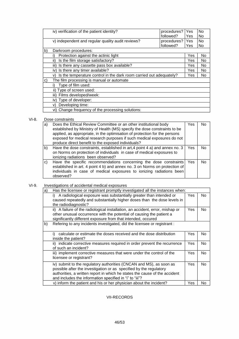
46/53
iv) verification of the patient identity? procedures? followed?
Yes Yes
No No
v) independent and regular quality audit reviews? procedures? followed?
Yes Yes
No No
b) Darkroom procedures:
i) Protection against the actinic light Yes No
ii) Is the film storage satisfactory? Yes No
iii) Is there any cassette pass box available? Yes No
iv) Is there any timer available? Yes No
v) Is the temperature control in the dark room carried out adequately? Yes No
c) The film processing is manual or automate
i) Type of film used:
ii) Type of screen used:
iii) Films developed/week:
iv) Type of developer:
v) Developing time:
vi) Change frequency of the processing solutions:
VI-8.
Dose constraints
a) Does the Ethical Review Committee or an other institutional body established by Ministry of Health (MS) specify the dose constraints to be applied, as appropriate, in the optimisation of protection for the persons exposed for medical research purposes if such medical exposures do not produce direct benefit to the exposed individuals?
Yes No
b) Have the dose constraints, established in art,4 point 4 a) and annex no. 3 on Norms on protection of individuals in case of medical exposures to ionizing radiations been observed?
Yes No
c) Have the specific recommendations concerning the dose constraints established in art. 4 point 4 b) and annex no. 3 on Norms on protection of individuals in case of medical exposures to ionizing radiations been observed?
Yes No
VI-9.
Investigations of accidental medical exposures
a) Has the licensee or registrant promptly investigated all the instances when:
i) A radiological exposure was substantially greater than intended or caused repeatedly and substantially higher doses than the dose levels in the radiodiagnostic?
Yes No
ii) A failure of the radiological installation, an accident, error, mishap or other unusual occurrence with the potential of causing the patient a significantly different exposure from that intended, occured
Yes No
b) Refering to any incidents investigated, did the licensee or registrant :
i) calculate or estimate the doses received and the dose distribution inside the patient?
Yes No
ii) indicate corrective measures required in order prevent the recurrence of such an incident?
Yes No
iii) implement corrective measures that were under the control of the licensee or registrant?
Yes No
iv) submit to the regulatory authorities (CNCAN and MS), as soon as possible after the investigation or as specified by the regulatory authorities, a written report in which he states the cause of the accident and includes the information specified in ―i‖ to ―iii‖?
Yes No
v) inform the patient and his or her physician about the incident? Yes No
VII-RECORDS
47/53
Note: Records will be required as part of the assessments and verifications specified in the prior sections of the list. This list is a reminder of the type of records that may be helpful. The appearance of the list at the end of the checklist does not mean that records review is the last thing to be done: preferably, an inspection may begin with an entrance interview and a visit to the facility followed by an initial review of some of the records listed below.
a) Copy of authorization or of registrance certificate b) System of records management c) Dosimetry, current and in thepast d) Area surveillance e) Instrument tests and calibrations f) Audits and reviews of radiation and safety programme g) Incident and accident investigation reports h) Verification, maintenance and repair work i) Modifications and arrangement of the facility j) Providing of Personnel training , initial and continuous k) Medical surveillance of the occupationally exposed persons l) Clinical dosimetry records
Annex no. 6
Radiological Safety Requirements for design and manufacture of radiological installations
1. Radiological installations used in medical exposure shall be designed and manufactured so that:
a) failure of a single component of the system be promptly detectable so that any unplanned exposure of patients or staff be minimized; and
b) the incidence of human error in the delivery of an unplanned medical exposure be minimized.
c) to permit the keeping of the medical exposures to a level as low as possible, which can be reasonably achieved consistent with obtaining the adequate diagnostic information;
d) operational parameters for the radiation generators, so that generating tube potential, filtration, focal spot position, source-image receptor distance, field size indication and either tube current and time or their product, be clearly and accurately indicated;
e) radiographic equipment be provided with devices that automatically terminate the irradiation after a presented time, a value of the current-time product or a dose (automatic exposure control)value;
f) fluoroscopic equipment be provided with devices for measuring and possibly record of the patient dose.
g) radiological installation for fluoroscopy be provided with a device that energises the X-ray tube only when continuously depressed (such as a 'dead man's switch') and equipped with indicators of the elapsed time and with monitors of the dose-surface product.
2. Other requirements with regard to the radiological installations used in medical exposures are:
a) whether imported into or manufactured in Romania, the equipment shall conform to the applicable standards of the International Electrotechnical Commission (IEC) and the ISO or to other equivalent Romanian standards;
b) performance specifications and operating and maintenance instructions, including protection and safety instructions,shall be provided in Romanian language and in compliance with the relevant IEC or ISO standards with regard to the ―accompanying documents‖, and this information shall be translated into Romanian language;
c) the operating terminology or its abbreviations shall be displayed on the operating consoles in Romanian or a language accessible to the user;
d) radiation beam control mechanisms shall be provided, including devices that indicate clearly and in a fail-safe manner whether there is an X-ray beam or not.

48/53
e) the exposure shall be limited to the area being examined by using collimating devices aligned with the radiation beam;
f) the radiation field shall be as uniform as possible; g) exposure rates outside the patient examination area, due to the leakage or scattering
radiation shall be kept at an as low as reasonably achievable level and in compliance with IEC standards and with these norms.
3. Radiological installations and their accessories shall be certified as complying with the relevant International Electrotechnical Commission (IEC), ISO or equivalent national standards .The compliance with IEC, ISO or applicable Romanian standards, is demonstrated with:
a) the written declaration of producer in accordance with the standards; b) the results of type tests of producer or of a body recognised by CNCAN;
4. The type classification of the tests according IEC standards is:
a) type tests, carried out by the manufacturer or by a body recognized by CNCAN for a type of radiological installation and which shall not carried out for each individual radiological installation.
b) site tests , carried out where each individual radiological installation is mounted. 5. The IEC teasts distinguishes three grades of tests:
a) Grade A: this grade refers to the analysis of the radiological installation design based upon an IEC safety requirement, which results in a written statement included in the technical description, with regard to the working principles or constructional means by which the IEC requirement is fulfilled.
b) Grade B: visual inspection or functional test or measurement. For this test grade the relevant IEC standards specify a procedure,. The test shall be performed according to this IEC procedure. Grade B tests may include fault situations, and yhey can achieved uninvadable without interference with the construction or circuitry of the radiological installation.
c) Grace C: functional test or measurement, which may involve interference with the construction or circuitry of the equipment, and shall be performed by, or under direct supervision of the manufacturer or a person authorised by the manufacturer.
6. A couple of IEC standards applicable to radiology are:
a) IEC-60336 (1993-07). X-ray tube assemblies for medical diagnosis. Characteristics of focal spots.
b) IEC- 62220-1 (2003-10) Medical electrical equipment- Characteristics of digital X-ray imaging devices - Part 1: Determination of detective quantum efficiency
c) IEC-60522 (1999-02). Determination of the permanent filtration of an X-ray tube assembly.
d) IEC-60601-1-3 (1994). Part 1: General requirements for safety. 3. Collateral standard: General requirements for radiation protection in diagnostic X-ray equipment.
e) IEC-60601-2-7 (1998-02). Part 2-7: Particular requirements for the safety of high-voltage generators of diagnostic X-ray generators.
f) IEC-60601-2-28 (1993-03). Part 2: Particular requirements for the safety of X-ray source assemblies and X-ray tube assemblies for medical diagnosis.
g) IEC-60601-2-32 (1994). Part 2: Particular requirements for the safety of associated equipment of X-ray equipment.
h) IEC 60788 (1984-01) Medical radiology - Terminology i) IEC standards series 61223 are related with evaluation and routine testing in medical
imaging departments (general aspects, constancy checks and acceptance tests). j) Series 61262 cover medical electrical equipment, characteristics of electro-optical X-
ray image intensifiers k) Series 61331 cover protective devices against diagnostic medical X radiation.
7. The acceptance protocol of the radiological installation shall include reference to the relevant safety tests described in the IEC standards. The relevant safety tests described in the IEC standards shall be specified in the purchasing conditions.
49/53
8. Leakage radiation The X-ray source assembly, which comprises the X ray tube, the housing and the collimator, shall not produce a leakage radiation that exceeds 1 mGy in one hour at 1 m.. This value can be averaged over an area not exceeding 100 cm
2.
9. Beam filtration a) The inherent filtration of every X-ray assembly shall be clearly and permanently marked
on the housing. The total filtration includes the inherent filtration, any added supplementary filtration and filtration offered by the attenuating material that permanently intercepts the beam, for example, the mirror of a light beam collimator. For conventional radiology the total filtration of the beam shall be the equivalent of at least 2.5 mm of aluminium, of which 1.5 mm shall be the permanent filtration.
b) The special radiological installations used, for example, for mammography, computed tomography, dental radiology and interventional procedures, require specific values of filtration, according to the applicable IEC standards, mentioned at piont no. 6.
10. Beam size a) The radiological installations shall always have a device for restricting the radiation field
size to the area of interest. This device shall be in form of adjustable diaphragm or collimator or, for specific examinations such as mammography and dental radiography, in form of fixed collimator.
b) For the graphic radiological installation, excepting the dental one there shall be a light beam to indicate the position and extent of the radiation beam, visible during normal lighting conditions.
c) In case of fluoroscopy, the radiological installation shall be provided with a device, preferably an automatic one, to confine the X-ray beam to the surface dimension of the image receptor, whatever the distance from the X-ray tube to the image receptor. Manual collimation shall be possible in addition to automatic collimation.
11. Image receptors a) All fluoroscopy units shall have an image intensifier (or equivalent technology). b) With regard to radiography, the rare earth intensifying screens are recommended -
gadolinium , which have a high X-ray absorption efficiency and higher light output, so that the required diagnostic information with a substantially lower radiation dose is obtained.
c) The radiographic film shall be adequate, compatible with the intensifying screen and shall assure a minimum X-ray dose at the patient exposure, with adequate image quality.
d) All X-ray detection systems: image intensifier, film screen cassette system, radiological films, etc. shall posses radiological safety licence (ASR).
12. Selection of materials for patient’s couch, film cassette etc. a) Attenuation of the X-ray beam between the patient and the image receptor shall be
minimized by using suitable materials for the patient table top (in case of X-rayv over couch tubes), for the film cassettes and the antiscatter grid.
b) this attenuation shall be known and guaranteed by producer. 13. Signals and marking a) The X-ray systems shall indicate at the control panel all the important technical
parameters relevant to image quality and patient dose. The tube voltage (kV), tube current (mA) and exposure time (or mAs) are the minimum parameters to be displayed during radiographic exposure.
b) Additional information about the selection of the automatic exposure device as well as the sensor area selected to terminate the exposure shall be available at the control console of the operator. It is recommended that data about the radiation field size and the distance from focus to patient skin be displayed. .
c) Instantaneous values of the tube voltage (kV), tube current (mA) and accumulated fluoroscopy time shall be available at the control console. The degree of magnification – the active area of the image intensifier, the different fluoroscopy modes, if they exist, shall be clearly shown to the operator.
50/53
d) If the fluoroscopy unit is able of operating at high dose-rates,a separate visual or audible warning shall be available to the operator.
14. Exposure switches a) Exposure switches on all radiological installations of diagnostic, except computed
tomography scanners, shall be arranged so that an exposure can only take place when the switch is continuously pressed and terminate if the switch is no longer pressed (except in case exposure did not previously finish by other means, for example, at the end of the set exposure time in radiography or by the automatic exposure control).
15. Control of exposure duration a) In case of radiography, exposure shall be terminated automatically after a pre-set time, an
electrical charge (mAs) or amount of radiation. b) In addition to these means of exposure termination can be provided some other means
too. c) For fluoroscopy, the release of an exposure switch shall be regarded as the normal
means of termination. d) Additional means of termination which operate automatically when a predetermined time
of maximum 10 minutes has elapsed, shall be provided e) An audible warning shall be activated 30 seconds before, to enable the operator to reset
the device if the exposure needs to be prolonged. f) It is recommended that radiological fluoroscopy systems incorporate a ―last image hold‖
mode, which shall ensure that the last acquired image is as long displayed as required.
16. Exposure measurement a) Only radiological installations provided with monitoring devices of patient exposure (dose
- area product and others) shall be used in fluoroscopy. b) In interventional procedures, these monitoring devices of patient exposure shall be also
used to detect equipment malfunction or progressive degradation of radiological installation.
17. Scattered radiation for fixed fluoroscopy a) All patient tables and stands used for fluoroscopy shall be provided with adequate
protection against scattered radiation. 18. Other requirements a) Practices of dental radiology, mammography, CT and interventional radiology require a dedicated radiological installation. b) Radiological installation for paediatric radiology also requires some specific conditions (for example, a power generator which permits very short exposure times, small focal spot sizes and adequate accessories for smaller patient dimensions). c) Film processors (development devices), viewing boxes, TV monitors, etc., are all important parts of the imaging chain and, consequently,they shall be selected, purchased and maintained at the same level required for the radiological facilities (service contract).. d) Digital radiological X-ray facilities necessitate additional requirements concerning the quality of high resolution monitors, of computers/networks, processing software, radiological information system (RIS), picture archiving and communication systems (PACS) and their connecting capabilities to the hospital information system (HIS).
Annex no. 7
Local rules examples for operational safety.
1. The following local rules can be used in a radiology facility. They shall be regarded as basic and simple rules only, and may be added to or modified according to the local circumstances or the CNCAN and MS requirements. Local rules shall be written in an easily understandable form and displayed prominently in working areas. They are part of the authorization documentation for the use of the radiological installation. 2. Patient and Public Protection
51/53
a) Lead protective equipments, either as half aprons or gonad shields, shall be used to cover the pelvic regions. This applies to every possible examination. The patient’s surface facing the primary beam shall be protected. The shielding obviously.applies only to the examinations that will not be affected by this.
b) Infants and children presenting for examinations of the hip shall have the first series without any protection and all progressive examinations with shielding.
c) No person accompanying the patient shall be in the controlled area... d) Any person who exceptionally helps patient assistance,shall wear a lead apron during the
exposure and avoid the primary beam. If this person has his/her hands near the primary beam, they shall then be provided with lead protection gloves, in order to wear them during the patient exposure.
e) Parents of children, who need assistance during examination, shall be encouraged to assist their children during examination. They shall be provided with adequate protection according to paragraph d) and clear instructions.
f) No person but the patient, except the persons from paragraph d) and e), shall be inside the X-ray exposure room during the patient exposure.
3. Pregnancy and X-ray examinations a) Introduction. The following measures shall be taken in order to protect all patients with
reproductive capacity, especially for those who know they are pregnant or who think they might be. The protocol shall consist of identification sections of patients at risk and procedure for all females with reproductive capacity. The primary responsibility for the identification of patients at risk revolves upon the doctor who prescribes the medical exposure, while the radiology staff provides a secondary verification.
b) Identification of pregnant patient. Female patients of reproductive age shall be asked: "Are you pregnant or do you think you might be pregnant?" A positive answer to this question is expected from women who think they might be pregnant, who are trying to become pregnant and those who know they are pregnant. In case of doubt, the patient shall be considered to be pregnant. The answer to this question shall be recorded.
c) If the answer to this question is 'no', a caution shall be then adopted in the radiological procedures which involve exposure of the lower abdomen and pelvic regions of women of reproductive capacity, in order to ensure that the received dose is as low as practicable.
d) Procedure when patient is pregnant. If the patient is pregnant, or is suspected to be pregnant, then the case shall be referred to a radiologist in order to decide on whether the X-ray examination shall proceed. In general, only urgent examinations of the pelvis and lower abdomen shall be carried out during pregnancy and particular care for avoiding irradiation of the foetus shall be taken, as posibly practicable. All radiographic factors shall be noted, so that the dose absorbed by the foetus can be calculated and recorded by the medical physicist.
e) Procedure when patient is found to be pregnant after an X-ray examination. Occasionally a female patient will not know that she is pregnant at the time of an x-ray examination, and she will naturally be very anxious when she learns that she is pregnant,. In such cases, estimations of the dose absorbed by the foetus shall be performed by a medical physicist experienced in dosimetry. The patient can then be better warned about the risks. In most cases there is effectively no risk, as the exposure to X-ray occurred in the first 21 days after conception. In a few cases the foetus will be older and the absorbed dose may be considerable. Though, there are extremely few cases when the dose is sufficiemtly large to require patient warning of consider termination of the pregnancy.
f) ICRP Publication no 84. ―Pregnancy and Medical Radiation‖, 2000 is warning that ―Termination of pregnancy is an individual decision affected by many factors. Foetal doses below 100 mGy shall not be considered a reason for terminating a pregnancy. A foetal dose above this level can lead to a foetal damage, the magnitude and type of damage depending on dose and stage of pregnancy‖.
4. Staff Protection 4.1 Dosemeters for individual monitoring a) Individual dosemeters shall be red every month. b) Workers shall wear individual dosemeters throughout their work in controlled areas.
52/53
c) When the worker is wearing a protection apron, dosemeter shall be worn underneath the apron (or as specified).
d) The RPR shall inform the staff about the individual monitoring results. These results shall be posted on the staff notice board.
4.2 Safe Operation of radiological installation a) The lead protection glass is only sufficient to stop scattered radiation. The x-ray tube shall
never be pointed to this glass. b) Protective lead aprons with at least 0,5 mm lead equivalent, shall be worn by staff while
operating mobile radiological installation. 4.3 Patient Immobilisation a) Immobilising devices shall be used whenever possible to minimise doses to patient and
staff, or to patient helper. 4.4 Lead Apron Testing a) All lead aprons shall be stored on hangers when not being used. They shall never be
folded for storage. b) All aprons shall be tested at intervals of approximately 12 - 18 month for verifying integrity.
Each apron shall have a permanent individual identification. c) If damage to an apron is ascertained or suspected, it shall be immediately reported to
RPR, and shall not be used until tested and declared safe. d) Permanent records of nonconformities, of testing way and of their repairing shall be kept. 5. General requirements a) Any occupationally exposed female who is pregnant shall notify the licensee. b) Any worker, after resignation, transfer or activity cessation, may request from the
licensee a copy of the history of the work in environment with ionizing radiations, which will comprise received doses, medical surveillance records as occupational exposed person.
6. Examples of more detailed local rules for specific radiology procedures can be found in the WHO/IAEA ―Manual on radiation protection in hospitals and general practice. Diagnostic Radiology.‖
Annex no. 8
Example of protective clothing against X-ray
1. Protective clothing used in radiology are: a) gowns, aprons and thyroid protective collars, made of a material (such as vinyl) which
contains lead; b) removable shields for the patient couch, made of the same material c) gloves made of the same material d) glasses (spectacles) with lenses made of leaded glass or leaded plastic e) viewing windows (fixed or mobile) made of leaded glass or leaded plastic
2. Gowns, aprons, thyroid protective collars
a) These may be manufactured in various forms: a coat which is fixed at the front, a ―poncho‖ which is fixed at the sides, gowns which are either opened at the back or contain less lead at the back, or gowns which are in two parts – a top in the form of a coat, and a bottom which is fixed around the waist.
b) Protective aprons shall be equivalent to at least 0.25 mm lead if the x-ray equipment operates up to 100 kV and at least 0.35mm lead, if it operates above 100 kV. At conventional radioscopy, without image intensifier, the apron shall have minimum 0,5 mm lead equivalent.
c) In interventional radiology staff shall use 0.5 mm lead equivalent clothing because of the high levels of scattered radiation.
d) The chosen clothing style depends on the radiology practice for which they will be used. However,it is always better to shield as much as possible the largest area of the body.
53/53
e) In interventional radiology, the thyroid normally needs protection. Some gowns include a collar that covers the thyroid, but in most cases, a separate thyroid collar is required.
3. Patient bed shielding In interventional radiology, the scattered radiation levels can be highly reduced by attaching lead vinyl sheets (aprons) to the patient table. As the weight is carried by the bed, higher values of lead equivalence may be used. 4. Lead Gloves The gloves are made of lead vinyl and are heavy. They have 0, 25 mm lead values, being difficult to use. In fact, their use increases in some cases procedure time and thus dose. Gloves shall therefore only be used if appropriate. There are also lighter gloves, similar to surgical gloves; These shall be used carefully,as they contain little lead and are only effective at tube voltages that are lower than 60 kVp. 5. Glasses In some interventional radiology procedures, it is possible for the chrystaline lens of the operator’s eye to receive an annual dose which approaches or even exceeds the NFSR dose limit (150 mSv)., The eye protection with 0, 25 mm lead equivalent is essential in these cases. 6. Viewing Windows
a) Leaded glass or plastic viewing windows are usually used in shielding of controlled areas. They shall be manufactured with lead equivalence corresponding to the maximum tube voltage (kVp) at which this applies and the attenuation that have to ensure.
b) A moveable viewing window is very useful for interventional radiological installations,. These are usually mounted on the ceiling, and can be placed in such a way,that the operator views the patient through the window . This window with at least 0,5 mm lead equivalence, provides protection against the radiation scattered by patient for both the eyes and thyroid.
7. Quality control of protective equipment
a) All protective equipments shall be tested immediately after purchase and then periodically (at least once every 2 years). If equipment is not correctly stored (on a coat hanger, for instance), it might eventually crack, causing loss of shielding. The damage can not be seen by a visual inspection.
b) All X-ray protective equipments shall be tested periodically at a Laboratory designated by CNCAN. Faulty clothing shall be immediately replaced with other new ones. .


![NSR-MSxT2BLB SUGバックアップ編 F V03 - Elecom...1 2 セットアップガイド[バックアップ編] NSR-MS2T2BLB NSR-MS4T2BLB NSR-MS6T2BLB NSR-MS8T2BLB 3 ACLの保存と復元](https://img.pdfslide.net/doc/110x75/5e6d5b3e33c1d14c254dcc0e/nsr-msxt2blb-sugffffffc-f-v03-1-2-fffffffffffffc.jpg)


![QUESTION BANK · 16. Interventional radiology in cerebral aneurysms. [05] 17. Describe radiological role in intracranial aneurysms with special reference to management by interventional](https://img.pdfslide.net/doc/110x75/5f7d9c3945a6940fca54b21d/question-bank-16-interventional-radiology-in-cerebral-aneurysms-05-17-describe.jpg)