Embed Size (px)
Citation preview
Northumbria Research Link
Citation: Hicks, Kirsty, Onambele-Pearson, Gladys, Winwood, Keith and Morse, Christopher (2017) Muscle-Tendon Unit Properties during Eccentric Exercise Correlate with the Creatine Kinase Response. Frontiers in Physiology, 8. p. 657. ISSN 1664-042X
Published by: Frontiers
URL: https://doi.org/10.3389/fphys.2017.00657 <https://doi.org/10.3389/fphys.2017.00657>
This version was downloaded from Northumbria Research Link: http://nrl.northumbria.ac.uk/32259/
Northumbria University has developed Northumbria Research Link (NRL) to enable users to access the University’s research output. Copyright © and moral rights for items on NRL are retained by the individual author(s) and/or other copyright owners. Single copies of full items can be reproduced, displayed or performed, and given to third parties in any format or medium for personal research or study, educational, or not-for-profit purposes without prior permission or charge, provided the authors, title and full bibliographic details are given, as well as a hyperlink and/or URL to the original metadata page. The content must not be changed in any way. Full items must not be sold commercially in any format or medium without formal permission of the copyright holder. The full policy is available online: http://nrl.northumbria.ac.uk/pol i cies.html
This document may differ from the final, published version of the research and has been made available online in accordance with publisher policies. To read and/or cite from the published version of the research, please visit the publisher’s website (a subscription may be required.)
ORIGINAL RESEARCHpublished: 19 September 2017doi: 10.3389/fphys.2017.00657
Frontiers in Physiology | www.frontiersin.org 1 September 2017 | Volume 8 | Article 657
Edited by:
Marco Vincenzo Narici,
University of Nottingham,
United Kingdom
Reviewed by:
Gaël Guilhem,
Institut National du Sport, de
l’expertise et de la Performance,
France
David A. Jones,
University of Birmingham,
United Kingdom
*Correspondence:
Kirsty M. Hicks
Specialty section:
This article was submitted to
Exercise Physiology,
a section of the journal
Frontiers in Physiology
Received: 24 November 2016
Accepted: 17 August 2017
Published: 19 September 2017
Citation:
Hicks KM, Onambele-Pearson GL,
Winwood K and Morse CI (2017)
Muscle-Tendon Unit Properties during
Eccentric Exercise Correlate with the
Creatine Kinase Response.
Front. Physiol. 8:657.
doi: 10.3389/fphys.2017.00657
Muscle-Tendon Unit Propertiesduring Eccentric Exercise Correlatewith the Creatine Kinase ResponseKirsty M. Hicks 1, 2*, Gladys L. Onambele-Pearson 2, Keith Winwood 2, 3 and
Christopher I. Morse 2
1Department of Sport, Exercise & Rehabilitation, Northumbria University, Newcastle Upon Tyne, United Kingdom,2Department of Exercise and Sport Science, Health Exercise and Active Living Research Centre, Manchester Metropolitan
University, Crewe, United Kingdom, 3 School of Healthcare Science, Manchester Metropolitan University, Manchester,
United Kingdom
Aim: The aim of this paper was to determine whether; (1) patella tendon stiffness, (2)
the magnitude of vastus lateralis fascicle lengthening, and (3) eccentric torque correlate
with markers of exercise induced muscle damage.
Method: Combining dynamometry and ultrasonography, patella tendon properties and
vastus lateralis architectural properties were measured pre and during the first of six
sets of 12 maximal voluntary eccentric knee extensions. Maximal isometric torque loss
and creatine kinase activity were measured pre-damage (−48 h), 48, 96, and 168 h
post-damage as markers of exercise-induced muscle damage.
Results: A significant increase in creatine kinase (883 ± 667 UL) and a significant
reduction in maximal isometric torque loss (21%) was reported post-eccentric
contractions. Change in creatine kinase from pre to peak significantly correlated with
the relative change in vastus lateralis fascicle length during eccentric contractions
(r = 0.53, p = 0.02) and with eccentric torque (r = 0.50, p = 0.02). Additionally, creatine
kinase tended to correlate with estimated patella tendon lengthening during eccentric
contractions (p< 0.10). However, creatine kinase did not correlate with resting measures
of patella tendon properties or vastus lateralis properties. Similarly, torque loss did not
correlate with any patella tendon or vastus lateralis properties at rest or during eccentric
contractions.
Conclusion: The current study demonstrates that the extent of fascicle strain during
eccentric contractions correlates with the magnitude of the creatine kinase response.
Although at rest, there is no relationship between patella tendon properties and markers
of muscle damage; during eccentric contractions however, the patella tendon may play
a role in the creatine kinase response following EIMD.
Keywords: creatine kinase, exercise-induced muscle damage, fascicle strain, maximal isometric torque loss,
tendon stiffness
Abbreviations: ANOVA, Analysis of variance; CK, Creatine kinase; EMG, Electromyography; EIMD, Exercise-induced
muscle damage;MVEKE, Maximal voluntary eccentric knee extensions;MVCKE, Maximal voluntary isometric knee extension;
MVCKF, Maximal voluntary isometric knee flexions; OCP, Oral contraceptive pill; 1CKpeak, The increase from pre to peak
CK over the 168 h; VL, Vastus lateralis; VLACSA, Vastus lateralis anatomical cross-sectional area.
Hicks et al. MTU Properties Modulate CK
INTRODUCTION
Although it is well accepted that unaccustomed eccentric exerciseresults in functional and cytoskeletal impairments, referred toas exercise-induced muscle damage (EIMD), the mechanicaldeterminants, which govern the severity of EIMD still remainsunclear. Previously, the differences in EIMD have been attributedto fascicle strain (Lieber and Friden, 1993; Peñailillo et al.,2015; Guilhem et al., 2016), eccentric force (Warren et al., 1993;Chapman et al., 2008), the elastic properties of the tendon(although not confirmed experimentally) (Marginson et al.,2005; Guilhem et al., 2016) and, in women, circulating estrogenlevels (Carter et al., 2001). At present, these aforementioneddeterminants have predominately been identified using variousin situ and in vitro conditions, with limited studies investigatingthe determinants in vivo (Chapman et al., 2008; Peñailillo et al.,2013; Hoffman et al., 2014; Guilhem et al., 2016). Althoughintegral to the understanding of EIMD, in vitro experimentsdo not include the in series elastic components of the muscle,often involve single fibers and induce eccentric strains beyondthe physiological range (Butterfield, 2010). Further insight intothe mechanical processes resulting in EIMD in vivo is critical forhealth and sport practitioners to understand the metabolic andstructural response to eccentric exercise programs.
Recently, the tendon has been reported to play an importantrole during eccentric contractions in vivo and has been shownto reduce fascicle lengthening (Hicks et al., 2013; Hoffman et al.,2014) and mediate peak force, peak torque and fascicle velocityin situ (Roberts and Azizi, 2010). Therefore, in accordance withMorgan’s (1990) popping sarcomere theory, by attenuating thedegree of fascicle lengthening, the tendon may limit EIMD(Hoffman et al., 2014). Although previously, Hicks et al. (2016)reported no significant correlation between resting patella tendonstiffness and the CK response, the role of tendon stiffnesson functional markers of EIMD, such as torque loss, remainsunknown. Interestingly, Guilhem et al. (2016) proposed thata more compliant Achilles tendon resulted in shorter fasciclelengthening and subsequently less damage. A conclusion couldnot be drawn however, as their experimental design did notmanipulate tendon stiffness, and pooled male and femaleparticipants, the tendon properties of which are known to differsignificantly (Kubo et al., 2003; Onambélé et al., 2007; Hickset al., 2013). Therefore, to investigate whether tendon stiffnesscorrelates with markers of EIMD, a range of tendon stiffness’sneed to be investigated whilst controlling for confoundingvariables such as sex.
Following the recent insight into the interaction between thepatella tendon and fascicle lengthening during eccentric musclecontractions in vivo (Hicks et al., 2013), fascicle lengthening hasbeen investigated as a crucial determinant of EIMD (Hoffmanet al., 2014; Peñailillo et al., 2015; Guilhem et al., 2016). Themajority of these studies however, have been conducted usingsubmaximal, multi-joint exercise (Hoffman et al., 2014; Peñaililloet al., 2015). Multi-joint movements associated with submaximalexercise may have obscured the role of fascicle lengtheningon EIMD. During single joint, maximal eccentric contractionsof the plantar flexors in vivo, Guilhem et al. (2016) reported
that maximal fascicle length was correlated with torque lossand the delayed onset of muscle soreness post EIMD. Therelative contribution of fascicle lengthening to total muscle-tendon lengthening during eccentric contractions however, issignificantly lower in the plantar flexors (51%, Guilhem et al.,2016) compared to the VL (89%, Hicks et al., 2013). Therefore,to further our understanding of the relationship betweenfascicle lengthening and EIMD, a muscle group with a highercontribution of fascicle lengthening to total muscle-tendon unitlengthening needs investigating.
In addition to high strain (fascicle lengthening), theproduction of high torque is another characteristic of eccentriccontractions which has been investigated as a determinant ofEIMD (Lieber and Friden, 1993; Warren et al., 1993; Chapmanet al., 2008; Guilhem et al., 2016). Within the elbow flexors andplantar flexors both Chapman et al. (2008) and Guilhem et al.(2016) (respectively) reported no correlation between eccentrictorque and markers of EIMD. Interestingly, within the plantarflexors, Guilhem et al. (2016) reported a signficant correlationbetween negative work performed (fascicle lengthening ×
eccentric torque) and torque loss, thus providing tangibleevidence that eccentric torque may contribute to EIMD. Theaverage eccentric torque reported by Guilhem et al. (2016) (100N·m) however, is substantially lower than the average eccentrictorque reported within the VL (255 N·m, Hicks et al., 2016).Therefore, futher investigation into a larger muscle group isrequired to gain a greater insight into the potential determinantsof EIMD.
Although fascicle lengthening and eccentric torque have allbeen investigated as potential determinants of EIMD in muscle-tendon complexes (Hoffman et al., 2014; Peñailillo et al., 2015;Guilhem et al., 2016), the mechanical processes which predisposethe degree of EIMD, specifically within the VL, remains unclear.Furthermore, due to limited research, the relationship betweenpatella tendon properties and indirect markers of EIMD remainsunknown. Therefore, the aim of this paper was to determinewhether (1) patella tendon stiffness, (2) the amount of VLfascicle lengthening (strain) and (3) eccentric torque correlate withmarkers of EIMD.
MATERIALS AND METHOD
SubjectsSixteen males (21.1 ± 1.6 years of age, 72.0 ± 7.5 kg and176± 6 cm) signed written informed consent to participate inthis study. All participants self-reported as being recreationallyactive (undertaking no more than 1 h of “moderate” physicalactivity per week) and did not take part in any structuredresistance training. All procedures complied with the Declarationof Helsinki and ethical approval was obtained through the EthicsCommittee of Manchester Metropolitan University. Exclusioncriteria included any resistance training in the last 6 months,occupation or lifestyle that required regular heavy lifting orcarrying, any known muscle disorder, the use of dietarysupplements (i.e. vitamin E), and any musculoskeletal injuryin the last 3 months. All inclusion and exclusion criteria were
Frontiers in Physiology | www.frontiersin.org 2 September 2017 | Volume 8 | Article 657
Hicks et al. MTU Properties Modulate CK
determined via a questionnaire prior to inclusion within thisstudy.
Testing ProtocolOnce selected, participants were asked to visit the laboratoryon five different occasions over a period of 9 days. Althoughdifferent participants have been used, the design and severalof the measurement techniques within the current studyhave been reported previously (Hicks et al., 2013, 2016).The sessions were as follows: (1) pre-damage (48 h prior todamage) (2) damage and (3) 48 h, (4) 96 h, and (5) 168 hpost-damage. Pre-damage assessments consisted of mass andstature (anthropometric measures), 5–6 ml blood sample, patellatendon moment arm, isokinetic-dynamometer familiarization,morphological and mechanical measures of the patella tendon(tendon size and stiffness), VL anatomical cross-sectional area(VLACSA) and resting architecture, and two maximal isometricvoluntary knee extension torque (MVCKE) measurements at 60,65, 70, 75, 80 and 90◦. Participants performed two practiceMVCKE, at 60◦ and 70◦ during the familiarization session.Mass and stature were measured using digital scales (Secamodel 873, Seca, Germany) and a wall mounted stadiometer(Harpenden, Holtain Crymych, UK) respectively. The damagesession consisted of eccentric exercise during which the degreeof fascicle lengthening was measured using ultrasound, a 5–6 mLvenous blood sample and MVCKE torque measurements. 48, 96,and 168 h session consisted of 5–6 mL blood sample and MVCKE
torque measurements.All tests were carried out on the self-reported non-dominant
leg, which was defined as the leg that provided stabilityduring movements which require balance e.g., kicking a ball.Participants were seated, with a hip angle of 85◦, in anisokinetic-dynamometer (Cybex Norm, Cybex International, NY,USA). Participants were secured in a seated position usinginextensible straps around the hips and shoulders. The isokinetic-dynamometer axis of rotation was visually aligned with the kneejoint’s center of rotation. The isokinetic-dynamometer settings,including the anatomical zero, were recorded during pre-damageand replicated in the following sessions.
Vastus Lateralis AnatomicalCross-Sectional AreaUsing a real-time B-mode ultrasound (AU5 Harmonic, EsaoteBiomedica, Genoa, Italy) VLACSA was measured. To identify 50%of VL length, the participant laid supine with their leg fullyextended (knee angle 0◦), the proximal and distal insertions sitesof the VL were identified using an ultrasound probe (7.5MHzlinear array probe, 38mm wide). At 50% VL length, the medialand lateral border of the VL were identified using the ultrasound.Using a fabric tape measure, axial sections were marked usingecho absorptive markers every 30mm from the medial borderto the lateral border of the VL. Using the osseous surface asan alignment guide, the ultrasound probe was orientated in theaxial-plane, perpendicular to the VL muscle, and steadily movedover the echo-absorptive markers from the medial to the lateraledge of the VL. Minimal pressure was applied to the ultrasoundprobe to avoid compression of the muscle. The images were
recorded in real time at 25 frames per second (Adobe Premier proVersion 6, Adobe Systems Software, Ireland). Using capturingsoftware (Adobe Premier Elements, version 10), individualimages were acquired at each 30mm interval. Shadows cast bythe echo-absorptive markers allowed the images to be aligned bythe outline of the muscle, thus forming the entire VLACSA in asingle image (Adobe Photoshop Elements, version 10). Digitizingsoftware (ImageJ 1.45, National Institutes of Health, USA) wasused to measure VLACSA. This method of calculating VLACSA haspreviously been accepted as reliable and valid when comparedto MRI, with a reported interclass correlation between 0.998 and0.999 and a coefficient of variation of 2.1% (Reeves et al., 2004).
Patella Tendon Length andCross-Sectional AreaA real-time B-mode ultrasound (AU5 Harmonic, EsaoteBiomedica, Genoa, Italy) was used to measure patella tendoncross-sectional area and patella length at a fixed 90◦ knee angle.The distance between the apex of the patella and the tibialtuberosity, marked using sagittal ultrasound images, was takenas patella tendon length. With the ultrasound probe orientatedin the transverse plane images were captured at 25, 50, and75% of patella length to measure patella tendon cross-sectionalarea. Using image analysis software, the ultrasound images werelater analyzed offline (ImageJ 1.45, National Institutes of Health,USA). High reliability for measuring patella tendon length andpatella tendon cross-sectional area was reported within thecurrent study (CV 0.69 and 3.50% respectively).
Patella Tendon StiffnessThe method for measuring patella tendon stiffness has beendetailed previously (Hicks et al., 2013, 2016). In brief, theparticipants were seated in the isokinetic dynamometer, withthe knee angle fixed at 90◦, and were instructed to performa ramped, isometric MVCKE lasting ∼5–6 s. Ultrasoundimages of the patella tendon and ramped MVCKE torque weresynchronized using a 10-V square wave signal generator. RampedMVCKE torque was presented on a Macintosh G4 computer(Apple Computer, Cupertino, CA, USA), via an A/D converterand subsequently analyzed with the accompanying software(Acknowledge, Biopac Systems, Santa Barbara, CA). Patellatendon displacement was measured over two ramped MVCKE,once with the probe at the distal edge of the patella and thesecond with the probe over the tibial tuberosity (Onambéléet al., 2007). Total patella tendon displacement was calculated asdisplacement at the apex of the patella plus the displacement atthe tibial tuberosity (Onambélé et al., 2007). An echo-absorptivemarker was placed on the skin perpendicular to the patellatendon. The marker cast a shadow on the ultrasound imagedelineating the position of the skin and deep tissue. The shadowacts as a fixed reference point from which, the distance froman anatomical reference point at the start of the contractionto the end of the contraction can be measured as tendonelongation. Ultrasound images were captured, and total patellatendon displacement was measured at ∼10% intervals of theramped MVCKE torque output (Onambélé et al., 2007). Patellatendon forces were calculated as: (MVCKE torque + antagonist
Frontiers in Physiology | www.frontiersin.org 3 September 2017 | Volume 8 | Article 657
Hicks et al. MTU Properties Modulate CK
co-activation torque) / patella tendonmoment arm. Themethodsused to measure patella tendon moment arm and antagonist co-activation torque during MVCKE are described in detail below.
The force—patella tendon elongation curve stemming fromdata at every 10% of ramped MVCKE was then fitted witha second-order polynomial function forced through zero(Onambélé et al., 2007). The tangential slope at discreetsections of the curve, relative to MVCKE force, was computedby differentiating the curve at every 10% force intervals. Tostandardize the comparison of tendon stiffness and Young’smodulus at an absolute load, the slope of the tangential line,corresponding to the MVCKE force of the weakest participant(2,972 N·m), was computed for each subject.
Patella Tendon Moment ArmPatella tendon moment arm was measured at 90◦ (fullextension = 0◦) in the sagittal plane, from a dual-energy X-rayabsorptiometry scan (frame 23.3 × 13.7 cm, Hologic Discovery,Vertec Scientific Ltd, UK), and subsequently analyzed using aDICOM image assessment tool (OsiriX DICOM viewer, ver. 4.0,Pixemo, Switzerland). Patella tendon moment arm length wasdetermined as the perpendicular distance from the center of thepatella tendon to the tibio–femoral contact point. Dual-energy X-ray absorptiometry scans have been compared to MRI measures,demonstrating consistent reliability and validity against thisstandard (Erskine et al., 2014).
Antagonist Co-activation TorqueTo determine co-activation during the ramped MVCKE,electromyography (EMG) of the bicep femoris was measured.Ultrasound, in the axial plane, was used to confirm that theplacement of two biopolar electrodes (Ambu, Neuroline 720,Denmark) was in the mid-sagittal line at 25% of bicep femorismuscle length (distal end = 0%). To reduce skin impedancebelow 5,000 �, the skin was shaved, gently abraded and cleansedwith an alcohol wipe prior to electrode placement. The electrodeswere placed in a bipolar configuration with a constant inter-electrode distance of 20mm. A reference electrode (Ambu, BlueSensor, Denmark) was placed on the lateral tibial condyle. Theraw EMG signal was amplified (×2000) and filtered (throughlow and high band pass filters of 10 and 500Hz respectively),with a common mode rejection ratio of 110 dB (50 Hz) andsampling frequency of 2,000Hz. Participants performed twomaximal voluntary isometric knee flexions (MVCKF) at 90◦.The participants were instructed to perform the contractionsas rapidly and as forcefully as possible. The participants wereinstructed to relax once a 2 s plateau on the dynamometer screenhad been observed. Ramped MVCKE torque and bicep femorisEMG were recorded in real time and synchronized using a 10-V square wave signal generator. The root mean square of thebicep femoris EMG signal, was calculated 500 ms either side ofinstantaneous MVCKF peak torque. The baseline signal noise wascalculated as the root mean square over 1 s and removed fromthe measured electromyography during MVCKF and MVCKE. Atevery 10% of ramped MVCKE torque the root mean square of thebicep femoris EMG was taken over 250 ms. Therefore, using theaforementioned methods, knee flexor co-activation torque was
calculated as described by Onambélé et al. (2007);
KF Coactivation =
(
BFRMSMVCKE
BFRMSMVCKF
)
·MVCKF Torque
Where, BFRMS = root mean square of the bicep femoris EMG,MVCKE = maximal isometric voluntary knee extension torque,MVCKF =maximal isometric voluntary knee flexion torque.
It must be noted that several assumptions have been made forthe calculation of co-activation. Firstly, it has been assumed thatthe bicep femoris is representative of the entire hamstring musclegroup (Carolan and Cafarelli, 1992) and secondly, in accordancewith previous literature (Lippold, 1952), it is assumed thatthe relationship between bicep femoris electromyography andMVCKF torque is linear. Finally, inline with previous research(Onambélé et al., 2007), hamstring co-activation torque wascalculated solely from the bicep femoris, which due to notcalculating semitendinosus and semimembranosus co-activationtorque, may result in an under represented total hamstringco-activation.
Patella Tendon Stress/Strain RelationshipPatella tendon stress was calculated by dividing patella tendonforce (N) by patella tendon cross-sectional area (mm2). Patellatendon strain (%) was calculated as the ratio between total patellatendon displacement to patella tendon length.
Young’s ModulusYoung’s modulus was calculated by dividing patella tendonlength (mm) by patella tendon cross-sectional area (mm2), thenmultiplying the answer by patella tendon stiffness.
Maximal Isometric Voluntary KneeExtensor Torque MeasurementsAt six different knee angles (60, 65, 70, 75, 80, and 90◦ (fullextension = 0◦)) participants were instructed to perform twoMVCKE lasting∼2 s with 90 s rest between contractions. Torquewas presented, in real time, on a Macintosh G4 computer (AppleComputer, Cupertino, CA, USA), via an A/D converter (BiopacSystems, Santa Barbara, CA). Torque measurements were lateranalyzed offline with the accompanying software (Acknowledge,version 3.9.2). The highest torque produced at each angle wastaken as MVCKE peak torque. During pre-damage the angle atwhich the highest MVCKE torque was produced was recorded asoptimal knee angle. To calculate loss of MVCKE torque followingeccentric exercise, MVCKE were repeated at the knee angledetermined as optimal during pre-damage, 1-h post eccentricexercise (to reduce any fatigue effect) and 48, 96, and 168 h posteccentric exercise.
“Damaging” Eccentric ExercisePrior to eccentric exercise, a warm-up of four isokineticconcentric knee extensions and knee flexions were carried out,ensuring a progressive increase in effort (with the last contractionbeing maximal). For the eccentric exercise, the knee extensionrange of motion was set at 20–90◦ (0◦ = full extension).Participants performed 12 maximal eccentric voluntary kneeextensions (MVEKE) repetitions at 30
◦·s−1, for six sets. During
Frontiers in Physiology | www.frontiersin.org 4 September 2017 | Volume 8 | Article 657
Hicks et al. MTU Properties Modulate CK
the concentric phase the leg was passively returned to 20◦ atan angular velocity of 60◦s−1. Participants had to remain seatedduring their 2 min rest between each set. Verbal encouragementand visual feedback was continuously provided throughout theprotocol. MVEKE torque measurements were later analyzedoffline. For each set, peak MVEKE torque was determined as thehighest torque out of the 12 repetitions.
Change in Vastus Lateralis Fascicle Lengthduring the Eccentric ProtocolTo measure VL fascicle length during MVEKE, the ultrasoundprobe (7.5MHz linear array probe, 38 mm wide) was held inposition by the experimenter at 50% of VL muscle length in themid-sagittal plane of the non-dominant leg. To provide a visualreference point for the internal structures, an echo-absorptivemarker was fixed onto the skin at 50% of VL muscle length.When measuring fascicle lengthening from 20◦ to 90◦ knee angleduringMVEKE, pilot data reported the probe tomove a negligible0.02 ± 0.05 cm proximally, and therefore was not consideredfurther in calculations for the present study. VL fascicle lengthwas determined as the linear distance along the fascicle as it ranfrom the deep to the superficial aponeurosis. A hypo-allergenicultrasound gel (Parker, Park Laboratories Inc., Fairfield, UK) wasused to enhance acoustic coupling between the skin and theultrasound probe.
During the first set (out of six) of MVEKE contractions,ultrasound images were recorded onto a PC, in real time, at 25frames per second (Adobe Premier pro Version 6). Only thefirst set was analyzed due to Guilhem et al. (2016) reporting nosignificant effect of eccentric set number on fascicle lengthening.A 10-V square wave signal generator was used to synchronizethe ultrasound images with the torque acquisition. ThreeMVEKEcontractions were chosen at random from the first set of12 repetitions for architectural analysis. Using frame capturesoftware (Adobe Premier Elements, version 10) the ultrasoundimage corresponding to every 10◦, from 20◦ to 90◦ was acquiredfor offline analysis. Movement of the shadow casted by the echo-absorptive marker would act as an indicator that the probehad moved during the MVEKE therefore if any movement wasobserved, the contraction was discarded and another repetitionwas chosen for analysis.
Using digitizing software (ImageJ 1.45, National Institutes ofHealth, USA), VL fascicle length was analyzed offline at every10◦. Fascicle length was measured from the visible insertion ofthe fiber from the deep into the superficial aponeurosis (Reevesand Narici, 2003). Where the fascicle extended longer thanthe ultrasound image (frame width 3.50 cm and height 4.15cm), linear continuation of the fascicle and aponeurosis wasassumed. Within the VL, a 2–7% error is associated with thelinear extrapolation method when used to calculate VL fasciclelength (e.g., measured at 11.3 cm) at a knee angle of 120◦ (Finniet al., 2003). Furthermore, using a 40 and 38 mm probe widthrespectively, Guilhem et al. (2011) and Hicks et al. (2013) bothreport high reliability when measuring VL fascicle lengtheningduring MVEKE. In agreement with previous research, the currentstudy reported high reliability when measuring VL fascicle
lengthening during MVEKE (ICC 0.99 and CV 2.95%). In orderto reduce any error associated with the estimation of VL fasciclelength, an average of three fascicles across the image wastaken (Guilhem et al., 2011). Fascicle length during eccentriccontractions was measured at every 10◦ knee angle (range 20◦–90◦, 0◦ = full extension) throughout the MVEKE. Change infascicle length is presented as fascicle length at a knee angle of90◦ made relative to fascicle length measured at a knee angle of20◦; hereafter termed “relative fascicle lengthening” and reportedas a percentage change from starting length at 20◦.
Vastus Lateralis Total Muscle-tendon UnitExcursionIn order to estimate the total VL muscle-tendon unit elongation,the tendon excursion method was adopted (Spoor et al., 1990);whereby the patella tendon moment arm at 90◦ knee angle wasmultiplied by the change in knee angle (70◦, 1.22 rad) during theMVEKE.
Blood SamplesTo measure CK levels a 21-gauge needle was inserted into theantecubital vein of the forearm, and 5–6 mL of blood wasdrawn into a serum collection tube. The sample was allowedto clot whilst on crushed ice for 60 min and then centrifugedat 4,500 rpm at 0◦C for 10min. Using a 200–1,000µl pipette(Eppendorf, USA), the resulting serum sample was separatedinto three aliquots (∼500µl each) and stored in Eppendorf tubesat −20◦C until later CK analysis. Creatine kinase activity wasmeasured using colorimetry at 340 nm optical density (BioTekEL × 800 96 well Microplate Reader), with enzyme activitycalculations carried out using a generic software (Gen5, version2.0). Each sample was run in duplicate using an EnzyChromTMCKAssay Kit (BioAssay Systems, Hayward, CA, sensitivity 5 U/L,intra-assay variability <5%, data from the manufacturer). Anaverage of the two readings was taken as the enzyme activityat each experimental phase. Throughout this manuscript CKactivity is reported in two ways: absolute values and peak CKabove baseline values (i.e., the change from pre to peak CK overthe 168 h (1CKpeak)).
StatisticsThe statistical software package SPSS (v.19, Chicago, IL) forWindows and Microsoft Excel were used to run statisticalanalysis. To check for parametricity, the Levene’s and Shapiro-Wilk tests were used to assess the variance and normality of thedata respectively. A one way repeated measures ANOVA (time, 5levels) was used for CK andMVCKE torque loss. The greenhouse-Geisser correction factor was applied if the assumption ofsphericity was violated. If a significant main effect was reported,a pairwise comparison, with a bonferroni correction, was usedto identify which time point was significantly different topre-damage. A one way repeated measures ANOVA (sets, 6levels) was used to investigate MVEKE torque during the EIMDprotocol. T-tests were used to compare the increase from pre,to peak CK (1CKpeak) and pre, to to peak MVCKE torqueloss. Linear correlations (Pearson r) were used to determinewhether 1CKpeak or absolute and relative MVCKE torque loss,
Frontiers in Physiology | www.frontiersin.org 5 September 2017 | Volume 8 | Article 657
Hicks et al. MTU Properties Modulate CK
correlated with either muscle properties, tendon properties,fascicle lengthening or MVEKE torque. Data is presented as mean± standard deviation. Statistical significance was accepted atp < 0.05.
RESULTS
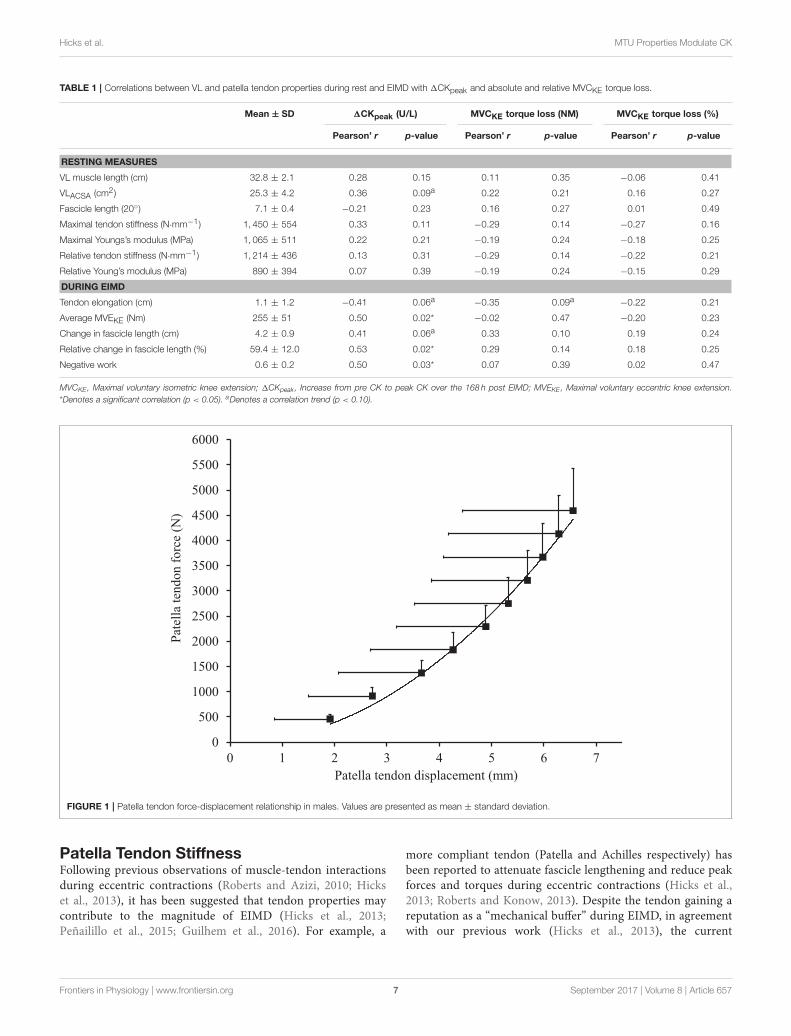
Pre-damage Vastus Lateralis and PatellaTendon PropertiesMuscle architecture and tendon properties, assessed at the “pre-damage phase,” are presented in Table 1. Resting patella tendonlength and patella tendon cross-sectional area was 57.8± 6.0mmand 78.1 ± 26.1mm2 respectively. Patella tendon moment armat 90◦’ knee angle was 4.33 ± 0.34 cm. To account for varyingmaximal MVCKE torque during ramped MVCKE (195.6 ± 36.8N·m), the patella tendon force (2,972 N) corresponding to thehighest MVCKE torque (127.2 N·m) of the weakest participantwas used to calculate standardized force level patella tendonstiffness (1,213 ± 436 N·mm−1, Figure 1) and Young’s modulus(1,030± 591 MPa).
Maximal Eccentric Voluntary KneeExtension Torque during the EccentricProtocolMVEKE torque was not significantly different throughout the sixsets (p = 0.868). During EIMD, average MVEKE peak torque(calculated over the six sets) was 255 ± 51 N·m. Peak MVEKEtorque was 97± 16% of “pre-damage” MVCKE torque.
Vastus Lateralis Fascicle Lengtheningduring Eccentric protocolA significant change in fascicle length from 20◦ knee angle(7.06± 0.43 cm) to 90◦ knee angle (11.3 ± 0.20 cm) was seen(4.20 ± 0.82 cm, p = 0.0004) during MVEKE. The change inVL fascicle length relative to fascicle length at knee angle of 20◦
was equivalent to a 59.4 ± 12.0% increase in fascicle lengtheningduring MVEKE.
Estimated Vastus Lateralis ExcursionBased on the tendon moment arm excursion, the estimatedincrease in VL muscle-tendon unit length from 20 to 90◦ kneeangle was 5.29± 0.41 cm during MVEKE.
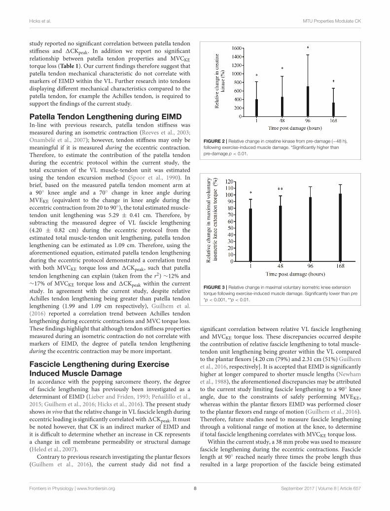
Creatine Kinase LevelsCreatine kinase significantly increased from pre-damage to 96 h(136± 114 U/L, 796± 723 U/L respectively, p= 0.014) but therewas no significant difference at 1 (430 ± 104 U/L, p = 0.167), 48(425 ± 82 U/L, p = 0.051) and 168 h (281 ± 58, p = 0.774) postEIMD.
Compared to pre-damage, relative CKwas significantly higherat every time point post EIMD (1 p = 0.004, 48 p = 0.004, 96p = 0.002 and 168 h p = 0.007, Figure 2). 1CKpeak (peak CKvalue—the pre CK values) was 883± 667 UL equating to an 885%increase in CK from pre-damage.
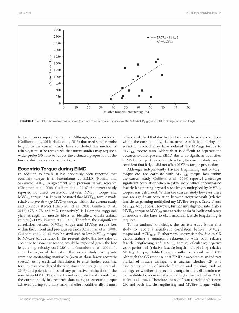
Maximal Isometric Voluntary KneeExtensor Torque LossMVCKE torque significantly decreased from pre-damage(264± 35 N·m), 1 h (209 ± 42 N·m, p = 0.0004) and 48 h(221.0 ± 48.4 N·m, p = 0.004) post EIMD, but had returnedto pre-damage by 96 (256 ± 14 N·m, p = 1.00) and 168 hpost damage (270 ± 13 N·m, p = 1.00). When made relativeto pre-damage, a significant reduction in MVCKE torqueloss remained 1 h (p = 0.0004) and 48 h (p = 0.005) postEIMD, but was not significantly different at any other timepoint (Figure 3). There was a significant rightward shiftin the optimal MVCKE knee angle, from pre-damage topost EIMD (mean, 77 ± 9◦, and 85 ± 7◦, respectively,p= 0.002).
Correlations between markers ofExercise-Induced Muscle DamageLinear correlations between the markers of muscle damage andVL and tendon patella tendon properties pre-damage and duringEIMD are presented in Table 1.
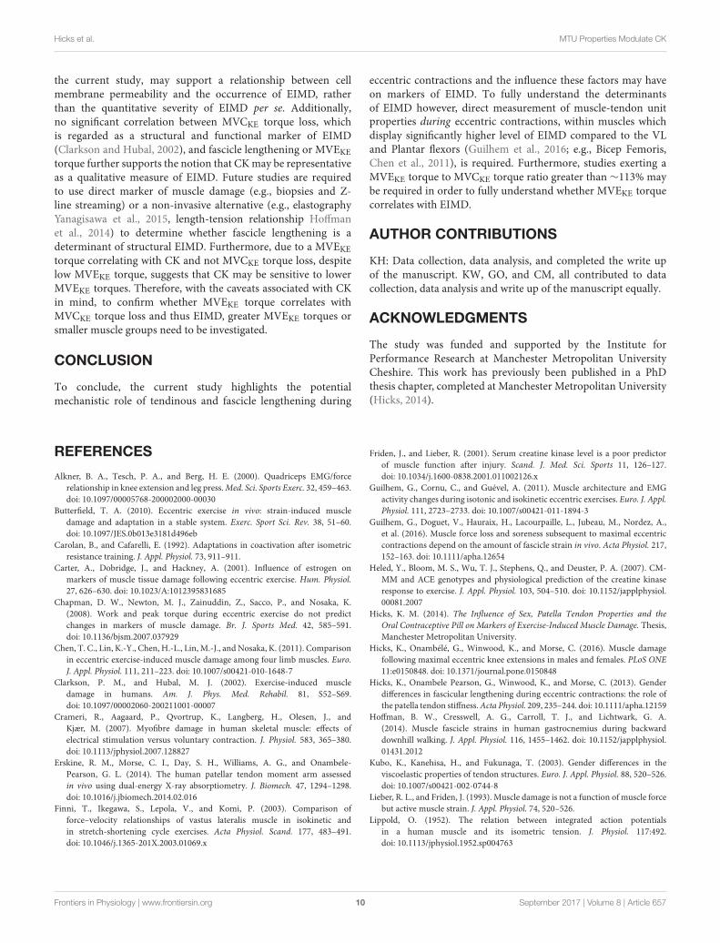
1CKpeak did not correlate with any resting patella tendon orVL properties (Table 1). During EIMD, 1CKpeak demonstrateda correlation trend with change in fascicle length, howeverwhen fascicle length was made relative to fascicle length at 20◦
knee angle a significant correlation was identified (Figure 4).Additionally, during EIMD, a correlation trend (p < 0.10) wasreported between 1CKpeak and estimated tendon lengthening.Finally, 1CKpeak significantly correlated with MVEKE torqueand negative work (relative fascicle lengthening multiplied myMVEKE torque).
MVCKE torque loss did not correlate with any restingpatella tendon or VL properties (Table 1). During EIMDMVCKE torque loss displayed a correlation trend with estimatedtendon elongation however, no significant correlation wasreported with any patella tendon or VL properties. Whenmade relative to pre-damage MVCKE torque loss did notcorrelate with any resting patella tendon or VL properties(Table 1).
DISCUSSION
The aim of the current study was to determine whether (1) patellatendon stiffness, (2) the amount of VL fascicle lengthening (strain),and (3) eccentric torque correlate with markers of EIMD. Thecurrent study reports three main findings; (1) During EIMD,VL relative fascicle lengthening, MVEKE torque and negativework correlated significantly with 1CKpeak, (2) Patella tendonproperties did not correlate with1CKpeak orMVCKE torque loss.(3) There was no significant correlations reported with MVCKE
torque loss. Within the current study, the VL was considereda surrogate of the quadriceps. Although there is currently nomeasure to quantify the individual muscle damage within thequadriceps, previous studies have reported VL to be a reliablesurrogate for predicting force output for the quadriceps (Alkneret al., 2000; Moreau et al., 2010).
Frontiers in Physiology | www.frontiersin.org 6 September 2017 | Volume 8 | Article 657
Hicks et al. MTU Properties Modulate CK
TABLE 1 | Correlations between VL and patella tendon properties during rest and EIMD with 1CKpeak and absolute and relative MVCKE torque loss.
Mean ± SD 1CKpeak (U/L) MVCKE torque loss (NM) MVCKE torque loss (%)
Pearson’ r p-value Pearson’ r p-value Pearson’ r p-value
RESTING MEASURES
VL muscle length (cm) 32.8 ± 2.1 0.28 0.15 0.11 0.35 −0.06 0.41
VLACSA (cm2) 25.3 ± 4.2 0.36 0.09a 0.22 0.21 0.16 0.27
Fascicle length (20◦) 7.1 ± 0.4 −0.21 0.23 0.16 0.27 0.01 0.49
Maximal tendon stiffness (N·mm−1) 1, 450 ± 554 0.33 0.11 −0.29 0.14 −0.27 0.16
Maximal Youngs’s modulus (MPa) 1, 065 ± 511 0.22 0.21 −0.19 0.24 −0.18 0.25
Relative tendon stiffness (N·mm−1 ) 1, 214 ± 436 0.13 0.31 −0.29 0.14 −0.22 0.21
Relative Young’s modulus (MPa) 890 ± 394 0.07 0.39 −0.19 0.24 −0.15 0.29
DURING EIMD
Tendon elongation (cm) 1.1 ± 1.2 −0.41 0.06a −0.35 0.09a −0.22 0.21
Average MVEKE (Nm) 255 ± 51 0.50 0.02* −0.02 0.47 −0.20 0.23
Change in fascicle length (cm) 4.2 ± 0.9 0.41 0.06a 0.33 0.10 0.19 0.24
Relative change in fascicle length (%) 59.4 ± 12.0 0.53 0.02* 0.29 0.14 0.18 0.25
Negative work 0.6 ± 0.2 0.50 0.03* 0.07 0.39 0.02 0.47
MVCKE , Maximal voluntary isometric knee extension; 1CKpeak , Increase from pre CK to peak CK over the 168 h post EIMD; MVEKE , Maximal voluntary eccentric knee extension.
*Denotes a significant correlation (p < 0.05). aDenotes a correlation trend (p < 0.10).
0
500
1000
1500
2000
2500
3000
3500
4000
4500
5000
5500
6000
0 1 2 3 4 5 6 7
Pat
ella
ten
do
n f
orc
e (N
)
Patella tendon displacement (mm)
FIGURE 1 | Patella tendon force-displacement relationship in males. Values are presented as mean ± standard deviation.
Patella Tendon StiffnessFollowing previous observations of muscle-tendon interactionsduring eccentric contractions (Roberts and Azizi, 2010; Hickset al., 2013), it has been suggested that tendon properties maycontribute to the magnitude of EIMD (Hicks et al., 2013;Peñailillo et al., 2015; Guilhem et al., 2016). For example, a
more compliant tendon (Patella and Achilles respectively) hasbeen reported to attenuate fascicle lengthening and reduce peakforces and torques during eccentric contractions (Hicks et al.,2013; Roberts and Konow, 2013). Despite the tendon gaining areputation as a “mechanical buffer” during EIMD, in agreementwith our previous work (Hicks et al., 2013), the current
Frontiers in Physiology | www.frontiersin.org 7 September 2017 | Volume 8 | Article 657
Hicks et al. MTU Properties Modulate CK
study reported no significant correlation between patella tendonstiffness and 1CKpeak. In addition we report no significantrelationship between patella tendon properties and MVCKE
torque loss (Table 1). Our current findings therefore suggest thatpatella tendon mechanical characteristic do not correlate withmarkers of EIMD within the VL. Further research into tendonsdisplaying different mechanical characteristics compared to thepatella tendon, for example the Achilles tendon, is required tosupport the findings of the current study.
Patella Tendon Lengthening during EIMDIn-line with previous research, patella tendon stiffness wasmeasured during an isometric contraction (Reeves et al., 2003;Onambélé et al., 2007); however, tendon stiffness may only bemeaningful if it is measured during the eccentric contraction.Therefore, to estimate the contribution of the patella tendonduring the eccentric protocol within the current study, thetotal excursion of the VL muscle-tendon unit was estimatedusing the tendon excursion method (Spoor et al., 1990). Inbrief, based on the measured patella tendon moment arm ata 90◦ knee angle and a 70◦ change in knee angle duringMVEKE (equivalent to the change in knee angle during theeccentric contraction from 20 to 90◦), the total estimated muscle-tendon unit lengthening was 5.29 ± 0.41 cm. Therefore, bysubtracting the measured degree of VL fascicle lengthening(4.20 ± 0.82 cm) during the eccentric protocol from theestimated total muscle-tendon unit lengthening, patella tendonlengthening can be estimated as 1.09 cm. Therefore, using theaforementioned equation, estimated patella tendon lengtheningduring the eccentric protocol demonstrated a correlation trendwith both MVCKE torque loss and 1CKpeak, such that patella
tendon lengthening can explain (taken from the r2) ∼12% and∼17% of MVCKE torque loss and 1CKpeak within the currentstudy. In agreement with the current study, despite relativeAchilles tendon lengthening being greater than patella tendonlengthening (1.99 and 1.09 cm respectively), Guilhem et al.(2016) reported a correlation trend between Achilles tendonlengthening during eccentric contractions and MVC torque loss.These findings highlight that although tendon stiffness propertiesmeasured during an isometric contraction do not correlate withmarkers of EIMD, the degree of patella tendon lengtheningduring the eccentric contraction may be more important.
Fascicle Lengthening during ExerciseInduced Muscle DamageIn accordance with the popping sarcomere theory, the degreeof fascicle lengthening has previously been investigated as adeterminant of EIMD (Lieber and Friden, 1993; Peñailillo et al.,2015; Guilhem et al., 2016; Hicks et al., 2016). The present studyshows in vivo that the relative change in VL fascicle length duringeccentric loading is significantly correlated with1CKpeak. It mustbe noted however, that CK is an indirect marker of EIMD andit is difficult to determine whether an increase in CK representsa change in cell membrane permeability or structural damage(Heled et al., 2007).
Contrary to previous research investigating the plantar flexors(Guilhem et al., 2016), the current study did not find a
FIGURE 2 | Relative change in creatine kinase from pre-damage (−48 h),
following exercise-induced muscle damage. *Significantly higher than
pre-damage p < 0.01.
FIGURE 3 | Relative change in maximal voluntary isometric knee extension
torque following exercise-induced muscle damage. Significantly lower than pre
*p < 0.001, **p < 0.01.
significant correlation between relative VL fascicle lengtheningand MVCKE torque loss. These discrepancies occurred despitethe contribution of relative fascicle lengthening to total muscle-tendon unit lengthening being greater within the VL comparedto the plantar flexors [4.20 cm (79%) and 2.31 cm (51%) Guilhemet al., 2016, respectively]. It is accepted that EIMD is significantlyhigher at longer compared to shorter muscle lengths (Newhamet al., 1988), the aforementioned discrepancies may be attributedto the current study limiting fascicle lengthening to a 90◦ kneeangle, due to the constraints of safely performing MVEKE,whereas within the plantar flexors EIMD was performed closerto the plantar flexors end range of motion (Guilhem et al., 2016).Therefore, future studies need to measure fascicle lengtheningthrough a volitional range of motion at the knee, to determineif total fascicle lengthening correlates with MVCKE torque loss.
Within the current study, a 38 mm probe was used to measurefascicle lengthening during the eccentric contractions. Fasciclelength at 90◦ reached nearly three times the probe length thusresulted in a large proportion of the fascicle being estimated
Frontiers in Physiology | www.frontiersin.org 8 September 2017 | Volume 8 | Article 657
Hicks et al. MTU Properties Modulate CK
y = 29.77x - 886.52
R² = 0.2855
0
250
500
750
1000
1250
1500
1750
2000
2250
2500
2750
0 10 20 30 40 50 60 70 80 90 100
Cre
atin
e kin
ase
(U/L
)
Relative fascicle lengthening (%)
FIGURE 4 | Correlation between creatine kinase (from pre to peak creatine kinase over the 168 h (1CKpeak )) and relative change in fascicle length.
by the linear extrapolation method. Although, previous research(Guilhem et al., 2011; Hicks et al., 2013) that used similar probelengths to the current study, have concluded this method asreliable, it must be recognized that future studies may require awider probe (50mm) to reduce the estimated proportion of thefascicle during eccentric contractions.
Eccentric Torque during EIMDIn addition to strain, it has previously been reported thateccentric torque is a determinant of EIMD (Nosaka andSakamoto, 2001). In agreement with previous in vivo research(Chapman et al., 2008; Guilhem et al., 2016) the current studyreported no direct correlation between MVEKE torque andMVCKE torque loss. It must be noted that MVEKE torque maderelative to pre-damage MVCKE torque within the current studyand previous studies (Chapman et al., 2008; Guilhem et al.,2016) (97, ∼77, and 94% respectively) is below the suggestedyield strength of muscle fibers as identified within animalstudies (>113%,Warren et al., 1993). Therefore, the insignificantcorrelation between MVEKE torque and MVCKE torque losswithin the current and previous research (Chapman et al., 2008;Guilhem et al., 2016) may be attributed to low MVEKE torqueto MVCKE torque ratio. In the present study, this low ratio ofeccentric to isometric torque, would be expected given the lowlengthening velocity used (30◦·s−1; Onambele et al., 2004). Itcould be suggested that within the current study participantswere not contracting maximally (even at these lower eccentricspeeds), using electrical stimulation to elicit higher eccentrictorques may have altered the recruitment pattern (Crameri et al.,2007) and potentially masked any protective mechanism of themuscle on EIMD. Therefore, by not using electrical stimulation,the current study has reported data using an eccentric torqueachieved during voluntary maximal effort. Additionally, it must
be acknowledged that due to short recovery between repetitionswithin the current study, the occurrence of fatigue during theeccentric protocol may have reduced the MVEKE torque toMVCKE torque ratio. Although it is difficult to separate theoccurrence of fatigue and EIMD, due to no significant reductionin MVEKE torque from set one to set six, the current study can beconfident that fatigue did not affect MVEKE torque production.
Although independently fascicle lengthening and MVEKEtorque did not correlate with MVCKE torque loss withinthe current study, Guilhem et al. (2016) reported a strongersignificant correlation when negative work, which encompassedfascicle lengthening beyond slack length multiplied by MVEKEtorque, was calculated. Within the current study however therewas no significant correlation between negative work (relativefascicle lengthening multiplied my MVEKE torque, Table 1) andMVCKE torque loss. However, further investigation into higherMVEKE torque toMVCKE torque ratios and a full volitional rangeof motion at the knee to elicit maximal fascicle lengthening isrequired.
To the authors’ knowledge, the current study is the firststudy to report a significant correlation between MVEKEtorque and 1CKpeak. Furthermore, unsurprisingly, due to CKdemonstrating a significant relationship with both relativefascicle lengthening and MVEKE torque, calculating negativework performed (relative fascicle length multiplied by relativeMVEKE torque, Table 1) significantly correlated with CK.Although the CK response post EIMD is accepted as an indirectmarker of muscle damage, it is unclear whether CK is atrue representation of muscle function and the magnitude ofdamage or whether it reflects a change in the cell membranespermeability to intramuscular proteins (Friden and Lieber, 2001;Heled et al., 2007). Therefore, the significant correlation betweenCK and both fascicle lengthening and MVEKE torque within
Frontiers in Physiology | www.frontiersin.org 9 September 2017 | Volume 8 | Article 657
Hicks et al. MTU Properties Modulate CK
the current study, may support a relationship between cellmembrane permeability and the occurrence of EIMD, ratherthan the quantitative severity of EIMD per se. Additionally,no significant correlation between MVCKE torque loss, whichis regarded as a structural and functional marker of EIMD(Clarkson and Hubal, 2002), and fascicle lengthening or MVEKEtorque further supports the notion that CKmay be representativeas a qualitative measure of EIMD. Future studies are requiredto use direct marker of muscle damage (e.g., biopsies and Z-line streaming) or a non-invasive alternative (e.g., elastographyYanagisawa et al., 2015, length-tension relationship Hoffmanet al., 2014) to determine whether fascicle lengthening is adeterminant of structural EIMD. Furthermore, due to a MVEKEtorque correlating with CK and not MVCKE torque loss, despitelow MVEKE torque, suggests that CK may be sensitive to lowerMVEKE torques. Therefore, with the caveats associated with CKin mind, to confirm whether MVEKE torque correlates withMVCKE torque loss and thus EIMD, greater MVEKE torques orsmaller muscle groups need to be investigated.
CONCLUSION
To conclude, the current study highlights the potentialmechanistic role of tendinous and fascicle lengthening during
eccentric contractions and the influence these factors may haveon markers of EIMD. To fully understand the determinantsof EIMD however, direct measurement of muscle-tendon unitproperties during eccentric contractions, within muscles whichdisplay significantly higher level of EIMD compared to the VLand Plantar flexors (Guilhem et al., 2016; e.g., Bicep Femoris,Chen et al., 2011), is required. Furthermore, studies exerting aMVEKE torque to MVCKE torque ratio greater than ∼113% maybe required in order to fully understand whether MVEKE torquecorrelates with EIMD.
AUTHOR CONTRIBUTIONS
KH: Data collection, data analysis, and completed the write upof the manuscript. KW, GO, and CM, all contributed to datacollection, data analysis and write up of the manuscript equally.
ACKNOWLEDGMENTS
The study was funded and supported by the Institute forPerformance Research at Manchester Metropolitan UniversityCheshire. This work has previously been published in a PhDthesis chapter, completed at Manchester Metropolitan University(Hicks, 2014).
REFERENCES
Alkner, B. A., Tesch, P. A., and Berg, H. E. (2000). Quadriceps EMG/force
relationship in knee extension and leg press.Med. Sci. Sports Exerc. 32, 459–463.
doi: 10.1097/00005768-200002000-00030
Butterfield, T. A. (2010). Eccentric exercise in vivo: strain-induced muscle
damage and adaptation in a stable system. Exerc. Sport Sci. Rev. 38, 51–60.
doi: 10.1097/JES.0b013e3181d496eb
Carolan, B., and Cafarelli, E. (1992). Adaptations in coactivation after isometric
resistance training. J. Appl. Physiol. 73, 911–911.
Carter, A., Dobridge, J., and Hackney, A. (2001). Influence of estrogen on
markers of muscle tissue damage following eccentric exercise. Hum. Physiol.
27, 626–630. doi: 10.1023/A:1012395831685
Chapman, D. W., Newton, M. J., Zainuddin, Z., Sacco, P., and Nosaka, K.
(2008). Work and peak torque during eccentric exercise do not predict
changes in markers of muscle damage. Br. J. Sports Med. 42, 585–591.
doi: 10.1136/bjsm.2007.037929
Chen, T. C., Lin, K.-Y., Chen, H.-L., Lin, M.-J., and Nosaka, K. (2011). Comparison
in eccentric exercise-induced muscle damage among four limb muscles. Euro.
J. Appl. Physiol. 111, 211–223. doi: 10.1007/s00421-010-1648-7
Clarkson, P. M., and Hubal, M. J. (2002). Exercise-induced muscle
damage in humans. Am. J. Phys. Med. Rehabil. 81, S52–S69.
doi: 10.1097/00002060-200211001-00007
Crameri, R., Aagaard, P., Qvortrup, K., Langberg, H., Olesen, J., and
Kjær, M. (2007). Myofibre damage in human skeletal muscle: effects of
electrical stimulation versus voluntary contraction. J. Physiol. 583, 365–380.
doi: 10.1113/jphysiol.2007.128827
Erskine, R. M., Morse, C. I., Day, S. H., Williams, A. G., and Onambele-
Pearson, G. L. (2014). The human patellar tendon moment arm assessed
in vivo using dual-energy X-ray absorptiometry. J. Biomech. 47, 1294–1298.
doi: 10.1016/j.jbiomech.2014.02.016
Finni, T., Ikegawa, S., Lepola, V., and Komi, P. (2003). Comparison of
force–velocity relationships of vastus lateralis muscle in isokinetic and
in stretch-shortening cycle exercises. Acta Physiol. Scand. 177, 483–491.
doi: 10.1046/j.1365-201X.2003.01069.x
Friden, J., and Lieber, R. (2001). Serum creatine kinase level is a poor predictor
of muscle function after injury. Scand. J. Med. Sci. Sports 11, 126–127.
doi: 10.1034/j.1600-0838.2001.011002126.x
Guilhem, G., Cornu, C., and Guével, A. (2011). Muscle architecture and EMG
activity changes during isotonic and isokinetic eccentric exercises. Euro. J. Appl.
Physiol. 111, 2723–2733. doi: 10.1007/s00421-011-1894-3
Guilhem, G., Doguet, V., Hauraix, H., Lacourpaille, L., Jubeau, M., Nordez, A.,
et al. (2016). Muscle force loss and soreness subsequent to maximal eccentric
contractions depend on the amount of fascicle strain in vivo. Acta Physiol. 217,
152–163. doi: 10.1111/apha.12654
Heled, Y., Bloom, M. S., Wu, T. J., Stephens, Q., and Deuster, P. A. (2007). CM-
MM and ACE genotypes and physiological prediction of the creatine kinase
response to exercise. J. Appl. Physiol. 103, 504–510. doi: 10.1152/japplphysiol.
00081.2007
Hicks, K. M. (2014). The Influence of Sex, Patella Tendon Properties and the
Oral Contraceptive Pill on Markers of Exercise-Induced Muscle Damage. Thesis,
Manchester Metropolitan University.
Hicks, K., Onambélé, G., Winwood, K., and Morse, C. (2016). Muscle damage
following maximal eccentric knee extensions in males and females. PLoS ONE
11:e0150848. doi: 10.1371/journal.pone.0150848
Hicks, K., Onambele Pearson, G., Winwood, K., and Morse, C. (2013). Gender
differences in fascicular lengthening during eccentric contractions: the role of
the patella tendon stiffness.Acta Physiol. 209, 235–244. doi: 10.1111/apha.12159
Hoffman, B. W., Cresswell, A. G., Carroll, T. J., and Lichtwark, G. A.
(2014). Muscle fascicle strains in human gastrocnemius during backward
downhill walking. J. Appl. Physiol. 116, 1455–1462. doi: 10.1152/japplphysiol.
01431.2012
Kubo, K., Kanehisa, H., and Fukunaga, T. (2003). Gender differences in the
viscoelastic properties of tendon structures. Euro. J. Appl. Physiol. 88, 520–526.
doi: 10.1007/s00421-002-0744-8
Lieber, R. L., and Friden, J. (1993). Muscle damage is not a function of muscle force
but active muscle strain. J. Appl. Physiol. 74, 520–526.
Lippold, O. (1952). The relation between integrated action potentials
in a human muscle and its isometric tension. J. Physiol. 117:492.
doi: 10.1113/jphysiol.1952.sp004763
Frontiers in Physiology | www.frontiersin.org 10 September 2017 | Volume 8 | Article 657
Hicks et al. MTU Properties Modulate CK
Marginson, V., Rowlands, A. V., Gleeson, N. P., and Eston, R. G. (2005).
Comparison of the symptoms of exercise-induced muscle damage after an
initial and repeated bout of plyometric exercise in men and boys. J. Appl.
Physiol. 99, 1174–1181. doi: 10.1152/japplphysiol.01193.2004
Moreau, N. G., Simpson, K. N., Teefey, S. A., and Damiano, D. L. (2010). Muscle
architecture predicts maximum strength and is related to activity levels in
cerebral palsy. Phys. Ther. 90, 1619–1630. doi: 10.2522/ptj.20090377
Morgan, D. (1990). New insights into the behavior of muscle during active
lengthening. Biophys. J. 57, 209–221. doi: 10.1016/S0006-3495(90)82524-8
Newham, D., Jones, D., Ghosh, G., and Aurora, P. (1988). Muscle fatigue and pain
after eccentric contractions at long and short length. Clin. Sci. 74, 553–557.
doi: 10.1042/cs0740553
Nosaka, K., and Sakamoto, K. (2001). Effect of elbow joint angle on the magnitude
of muscle damage to the elbow flexors. Med. Sci. Sports Exerc. 33, 22–29.
doi: 10.1097/00005768-200101000-00005
Onambélé, G. N. L., Burgess, K., and Pearson, S. J. (2007). Gender-specific in vivo
measurement of the structural andmechanical properties of the human patellar
tendon. J. Orthopaed. Res. 25, 1635–1642. doi: 10.1002/jor.20404
Onambele, G. N., Bruce, S. A., and Woledge, R. C. (2004). Effects of voluntary
activation level on force exerted by human adductor pollicis muscle during
rapid stretches. Pflüg. Archiv. 448, 457–461. doi: 10.1007/s00424-004-1265-6
Peñailillo, L., Blazevich, A., Numazawa, H., and Nosaka, K. (2013). Metabolic and
muscle damage profiles of concentric versus repeated eccentric cycling. Med.
Sci. Sports Exerc. 45, 1773. doi: 10.1249/MSS.0b013e31828f8a73
Peñailillo, L., Blazevich, A. J., and Nosaka, K. (2015). Muscle fascicle behavior
during eccentric cycling and its relation to muscle soreness. Med. Sci. Sports
Exerc. 708–717. doi: 10.1249/MSS.0000000000000473
Reeves, N. D., and Narici, M. V. (2003). Behavior of human muscle fascicles
during shortening and lengthening contractions in vivo. J. Appl. Physiol. 95,
1090–1096. doi: 10.1152/japplphysiol.01046.2002
Reeves, N. D.,Maganaris, C. N., andNarici, M. V. (2003). Effect of strength training
on human patella tendon mechanical properties of older individuals. J. Physiol.
548, 971–981. doi: 10.1113/jphysiol.2002.035576
Reeves, N. D., Maganaris, C. N., and Narici, M. V. (2004). Ultrasonographic
assessment of human skeletal muscle size. Euro. J. Appl. Physiol. 91, 116–118.
doi: 10.1007/s00421-003-0961-9
Roberts, T. J., and Azizi, E. (2010). The series-elastic shock absorber: tendons
attenuate muscle power during eccentric actions. J. Appl. Physiol. 109, 396–404.
doi: 10.1152/japplphysiol.01272.2009
Roberts, T. J., and Konow, N. (2013). How tendons buffer energy dissipation
by muscle. Exerc. Sport Sci. Rev. 41, 186–193. doi: 10.1097/JES.0b013e3182
a4e6d5
Spoor, C., Van Leeuwen, J., Meskers, C., Titulaer, A., and Huson, A.
(1990). Estimation of instantaneous moment arms of lower-leg
muscles. J. Biomechan. 23, 1247–1259. doi: 10.1016/0021-9290(90)
90382-D
Warren, G., Hayes, D., Lowe, D., and Armstrong, R. (1993). Mechanical
factors in the initiation of eccentric contraction-induced injury in rat
soleus muscle. J. Physiol. 464, 457–475. doi: 10.1113/jphysiol.1993.sp0
19645
Yanagisawa, O., Sakuma, J., Kawakami, Y., Suzuki, K., and Fukubayashi,
T. (2015). Effect of exercise-induced muscle damage on muscle hardness
evaluated by ultrasound real-time tissue elastography. SpringerPlus 4:308.
doi: 10.1186/s40064-015-1094-4
Conflict of Interest Statement: The authors declare that the research was
conducted in the absence of any commercial or financial relationships that could
be construed as a potential conflict of interest.
Copyright © 2017 Hicks, Onambele-Pearson, Winwood and Morse. This is an open-
access article distributed under the terms of the Creative Commons Attribution
License (CC BY). The use, distribution or reproduction in other forums is permitted,
provided the original author(s) or licensor are credited and that the original
publication in this journal is cited, in accordance with accepted academic practice.
No use, distribution or reproduction is permitted which does not comply with these
terms.
Frontiers in Physiology | www.frontiersin.org 11 September 2017 | Volume 8 | Article 657