Embed Size (px)
Citation preview

NOTES ON SOME INDO-PACIFIC UPOGEBIIDAE WITH DESCRIPTIONS OF FOUR NEW SPECIES (CRUSTACEA : THALASSINIDEA).
NGUYEN NGOC-HO
Ngoc-Ho, N. 1994 06 01: Notes on some Indo-Pacific Upogebiidae with descriptions of four new species (Crustacea: Thalassinidea). Memoirs of the Queensland Museum 35(1): 193-216. Brisbane. ISSN 0079-8835
Eleven species of Upogebiidae from New Guinea, New Caledonia, Kenya, Madagascar, Japan, Taiwan, Singapore and Vietnam have been studied. Four are new: Gebiacantha multispinosa, G. lifuensis, Upogebia sakaii and U. spinimanus. The seven additional species include: Gebiacantha laurentae Ngoc-Ho, Upogebia narutensis Sakai, U. pugnax de Man, U. savignyi (Strahl), U. wuhsienweni Yu, Wolffogebia inermis Sakai, W. phuketensis Sakai.\~\Crustacea, Thalassinidea, Upogebiidae, new species, Indo-Pacific, taxonomy.
Nguyen Ngoc-Ho, Museum national d'Histoire naturelle, Laboratoire de Zoologie-Arthropodes, 61 rue de Buffon, 75005 Paris, France; 10 February 1993.
This work provides further information about the rich upogebiid fauna of the Indo-Pacific (de Man,1927,1928; Poore & Gr i f f in , 1979; Sakai, 1982; Ngoc-Ho, 1990). Collections ex-amined come from: New Guinea (Gebiacantha laurentae Ngoc-Ho); New Caledonia (G. multi-spinosa sp.nov., G. lifuensis sp. nov., Upogebia pugnax de Man); Kenya ((/. savignyi (Strahl); Madagascar (U. spinimanus sp. nov.); Japan (U. sakaii sp. nov.) ; Singapore (Wolffogebia phuketensis Sakai); Taiwan (U. narutensis Sakai, U. wuhsienweni Yu); and Vietnam (W. inermis Sakai) The latter species was previously known only from the holotype which cannot be located. Its examination, together with that of W. phuketensis, adds generic features of Wolffogebia unmentioned by Sakai (1982).
New material of Upogebia savignyi confirms its synonymy with U. rhadames Nobili (Sakai, 1982). The first available adult female of U. pug-nax described in this work gives further informa-tion about this species. U. narutensis and U. wuhsienweni, both reported for the first time from Taiwan, are very similar to each other and also to Taiwanese U. edulis Ngoc-Ho & Chan, 1992.
Measurements given (mm) in the descriptions are: carapace length (cl.) = tip of the rostrum to the posterior border of the carapace; total length (tl.)= tip of the rostrum to the posterior border of the telson. Terminology of descriptions follows Ngoc-Ho (1981).
ABBREVIATIONS USED: BMNH, Bri t ish Museum (Natural History), London; MNHN, Museum National d'Histoire Naturelle, Paris; NNML, Nationaal Natuurhistorisch Museum, Leiden; NTOU, National Taiwan Ocean Univer-sity; QM, Queensland Museum, Brisbane; UMK,
Universitetets Zoologiske Museum, Kobenhavn; USNM, U.S. National Museum, Washington, D.C.; ZSM, Zoologische Staatssammlung, Munchen.
SYSTEMATICS
Family UPOGEBIIDAE Borradaile, 1903 Gebiacantha Ngoc-Ho, 1989
Gebiacantha laurentae Ngoc-Ho, 1989 (Fig. 1)
Gebiacantha laurentae Ngoc-Ho, 1989: 140, fig. 9.
MATERIAL EXAMINED Sek Harbour, New Guinea, St. 62, 37m, mud with a
little sand, W. Stephenson coll., 17.10.1969: 9 , cl. 7.5mm, tl. 18.5mm (QMW3317).
DISTRIBUTION Indonesia (Makassar Detroit) and now Papua
New Guinea. This record marks an easterly range extension.
REMARKS The specimen examined agrees well with the
female paratype of the species though it is slightly larger and has all spines comparatively more prominent. It also has 2 ventral spines on the 4th article of the antenna while the type has only one. The specimen is covered with mud and sediments although the area is dominated by soft corals and sponges (P. Davie, pers. comm.). Nothing is known of its ecology.

194 MEMOIRS OF THE QUEENSLAND MUSEUM
FIG 1. Gebiacantha laurentae Ngoc-Ho, female, QMW3317. A, anterior part of carapace, lateral view; B, first pereopod, mesial view. Scale line: 1mm.
Gebiacantha multispinosa sp. nov. (Fig. 2)
MATERIAL EXAMINED HOLOTYPE: New Caledonia: Loyalty Islands (Ouvea),
J.P. Menou coll., 16.11.1991, depth 6-10m: ovigerous 9 ,cl. 11.5mm, tl. 32.5mm (MNHNTh 1255). PARATYPE: same data as holotype, ovigerous 9 : cl. 10.5mm, tl. 28.5mm (MNHNTh 1256).
ETYMOLOGY Referring to the numerous spines present on the
body and pereopod 1.
DESCRIPTION Rostrum triangular, projecting far beyond eyes,
with 6-7 spiniform dorsal teeth on each lateral border; 3 large ventral spines. Fine and faint medio-dorsal groove (slightly dilated anteriorly) on rostrum and anterior part of gastric region, followed posteriorly by bare, unarmed, lon-gitudinal, low elevation. Lateral groove moderately broad, lateral ridge with 9-10 spiniform teeth or tubercles. Linea thalassinica distinct, extending to posterior border of carapace. Anterolateral border of carapace with 5-6 spinules. Anterolateral region of carapace (limited anteriorly and dorsally by anterolateral border and lateral ridge, posteriorly by linea

INDO-PACIFIC UPOGEBIIDAE 195
FIG. 2. Gebiacantha multispinosa sp. nov., holotype, ovig. female, MNHNThl255. A,B, anterior part of carapace, dorsal and lateral view; C, pereopod 1, external view; D, distal part of pereopod 1, mesial view; E,F, pereopods 2 and 3 respectively; G, telson and uropod. Scale line: 1mm.
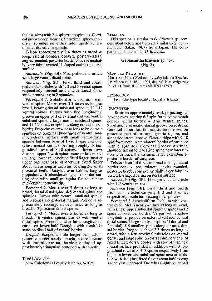
196 MEMOIRS OF THE QUEENSLAND MUSEUM
thalassinica) with 2-4 spines and spinules. Cervi-cal groove deep, bearing 5 proximal spines and 2 distal spinules on either side. Epistome ter-minates dorsally in spinule.
Telson approximately 1.4 times as broad as long, lateral borders convex, postero-lateral angles rounded, posterior border concave medial-ly, very faint inverted U-shaped carina on dorsal surface.
Antennule. (Fig. 2B). First peduncular article with large ventro-distal spine.
Antenna. (Fig. 2B). First, third and fourth peduncular articles with 1, 3 and 3 ventral spines respectively; second article with dorsal spine, scale terminating in 2 spinules.
Pereopod 1. Subcheliform. Ischium with ventral spine. Merus over 3.5 times as long as broad, bearing dorsal subdistal spine and 11-12 ventral spines. Carpus with fine longitudinal groove on upper part of external surface, ventral subdistal spine, 2 large mesial subdistal spines, and 11-13 spines or spinules along or near dorsal border. Propodus over twice as long as broad with spinules on proximal two-thirds of ventral mar-gin; external surface with small ventro-distal spinule between base of fixed finger and dac-tylus; mesial surface bearing roughly 6 lon-gitudinal rows of 8-10 spines, 4 lower rows distinct, upper 2 with spines more or less mixed up; large lower spine behind fixed finger, smaller upper one near base of dactylus; fixed finger about half as long as dactylus, bearing 2-3 small proximal teeth. Dactylus over half as long as propodus, with tubercles along upper border; cut-ting edge with small triangular flat tooth near mid-length; corneous tip.
Pereopod 2. Merus over 5 times as long as broad, dorsal distal spine, 4-5 ventral spines and spinules. Carpus with ventral subdistal spinule and 6 spines along dorsal margin. Propodus ap-proximately rectangular, over twice as long as broad, 1-2 proximal dorsal spines.
Pereopod 3. Merus over 5 times as long as broad, 5-6 ventral spines. Carpus with ventral distal spine. Propodus with faint longitudinal carina on lower half. Dactylus with comb-like setae on distal half of ventral border.
Uropod. Exopod a little longer than telson, posterior border nearly straight, not continuous with lateral external border; endopod ap-proximately triangular, protopod with spinule.
TYPE LOCALITY New Caledonia (Loyalty Islands), 6-10m.
REMARKS This species is similar to G. lifuensis sp. nov.
described below and both are similar to G. acan-thochela (Sakai, 1967) from Japan. The com-parison is made under G. lifuensis.
Gebiacantha lifuensis sp. nov. (Fig. 3)
MATERIAL EXAMINED H0L0TYPE:New Caledonia: Loyalty Islands (Ouvea),
J.P. Menou coll., 16.11.1991, depth 6-10m: ovigerous 9 , cl. 11.5mm, tl. 33mm (MNHNTh 1257).
ETYMOLOGY From the type locality, Loyalty Islands.
DESCRIPTION Rostrum approximately oval, projecting far
beyond eyes, bearing 6-8 spiniform teeth on each convex lateral border; 4 large ventral spines. Short and faint medio-dorsal groove on rostrum; rounded tubercles in longitudinal rows on posterior part of rostrum, gastric region, and alongside lateral groove. Gastric ridge with 9-10 spiniform teeth. Anterolateral border of carapace with 5 spinules. Cervical groove distinct, shoulder lateral to it bearing spine near intersec-tion with linea thalassinica, latter extending to posterior border of carapace.
Telson about 1.4 times as broad as long, lateral border convex, posterolateral angles rounded, posterior border concave medially, very faint in-verted U-shaped carina on dorsal surface.
Antennule (Fig. 3B). First peduncular article with 1-2 ventral spines.
Antenna (Fig. 3B). First, third and fourth peduncular articles carrying 1, 3 and 3 spines respectively; scale terminating in 2 spinules.
Pereopod 1. Subcheliform. Ischium with ven-tral spine. Merus nearly 4 times as long as broad, with single upper subdistal spine; 6 spines and 2 spinules on lower border. Carpus with shallow longitudinal groove on external surface; ventral distal spine; 3 large subdistal spines (1 dorsal and 2 mesial); 8-9 smaller spines along, or near, dor-sal border. Propodus about 2.5 times as long as broad, with a few proximal tubercles on ventral border and large spine on distal third near base of fixed finger; dorsal border with row of 9 spines; mesial surface provided in addition with 3 lon-gitudinal rows of 8,4, 3 spines respectively from upper to lower, and subdistal spine near articula-tion with dactylus; fixed finger about half as long as dactylus, unarmed. Dactylus slightly over half

INDO-PACIFIC UPOGEBIIDAE 197
FIG. 3. Gebiacantha lifuensis sp. nov., holotype, ovig. female, MNHNThl257. A,B, anterior part of carapace, dorsal and lateral view; C, pereopod 1, external view; D, distal part of pereopod 1, mesial view; E, telson and uropod. Scale line: 1mm.
as long as propodus with small tubercles on dorsal dopod approximately trapezoidal; protopod with border, cutting edge unarmed. spinule.
Uropod. Exopod a little longer than telson,
posterior border almost straight, meeting nearly T Y P E LOCALITY
at right angle with external lateral border; en- New Caledonia (Loyalty Islands), 6-10m.

198 M E M O I R S O F T H E Q U E E N S L A N D M U S E U M
T A B L E 1. Differentiat ing characters between G. acanthochela Sakai, G. multispinosa sp.nov. and G. lifuensis sp.nov.
G. acanthochella G. multispinosa G. lifuensis
Rostrum oval triangular oval
Infrarostral spines 2-3, small 3, large 4, large
Spines on antero-lateral region absent 2-4 absent of carapace
Spinules on atnero-lateral 7 6 5 border of carapace Spines on lateral shouler of 1 5 1 cervical groove
Peduncle of a2: -spinules on 2nd article 2-3 1 absent -ventral spines on 3rd article 2 3 3
Pereopod 1 propod: -ext. spines btn base of fixed absent 1 absent finger & dactyl -mesial spines 4 rows, 9-12 spines each 6 rows, 7-12 spines each 4 rows, 3-9 spines each -cutting edge of fixed finger narmed 2 teeth narmed
Pereopod 1 dactylus: -cutting edge -upper border
convex 3 proximal tubercles
convex tubercles along whole length
straight tubercles along whole length
Telson Slightly broader than long
1.5 times as broad as long 1.5 times as broad as long
REMARKS This species is closely related to G. multi-
spinosa captured at the same locality. It differs from the latter by: 1, shape of rostrum; 2, anterolateral region of carapace unarmed; 3, presence of single spine only on each lateral shoulder of cervical groove; 4, absence of small external distal spine on pereopod 1, between base of fixed finger and dactylus; 5, mesial surface of pereopod 1 with fewer spines; 6, fixed finger of pereopod 1 unarmed; 7, no tooth on cutting edge of pereopod 1 dactylus.
Both G. multispinosa and G. lifuensis are similar to G. acanthochela (Sakai) from Japan in the length and shape of their uropods, the slight median concavity of their telson, the numerous mesial spines on pereopod 1 propod. Their dif-ferences are listed in Table 1.
Upogebia Leach, 1814. Upogebia narutensis Sakai, 1986
(Figs 4; 5A-D)
Upogebia spinifrons (Haswel l , 1882): Sakai, 1984: 209 , f igs 1-3.
Upogebia narutensis Sakai, 1986: 25 , pi. 1.
MATERIAL EXAMINED HOLOTYPE: Naruto, Japan, M. Sh imoizumi col l . , date
unknown: 8 , cl. 2 6 m m , tl. 9 6 m m ( N N M L 3 6 7 7 7 ) . OTHER MATERIAL: Peng-Hu Island (West of Taiwan) ,
T - Y . Chan coll . , 1 October 1992: 1 8 , cl. 19 .5mm, tl. 6 0 m m ( M N H N T h 1258); 1 6 , cl. 19mm, tl. 5 9 m m , 1 9 , cl. 18mm, tl. 5 8 m m ( M N H N T h l 2 5 9 ) ; 1 8 , cl. 1 9 m m , t l . 5 8 m m , 1 9 , c l . 1 8 m m , t l . 5 7 m m ( M N H N T h 1260); 2 8 , cl. 1 8 m m and 19mm, tl. 5 2 m m and 5 8 m m ; 2 9 , cl. 1 7 m m and 17 .5mm, tl. 5 2 m m and 5 3 . 5 m m ( N T O U ) .
DESCRIPTION Rostrum egg-shaped, projecting far beyond
eye, with 1-3 small proximal tubercles on either lateral margin and the rest unarmed; 5 small ventral spines. Deep medio-dorsal groove on rostrum and anterior part of gastric region. Few small tubercles on gastric region most of them alongside moderately broad lateral groove. Lateral ridge divided by weak mid-dorsal notch: anterior half very setose dorsally with proximal tubercle and 2 spines at tip; posterior half with 3-4 spiniform or tuberculiform teeth. Anterolateral border of carapace bearing 4 spines. Linea thalas-sinica distinct. Cervical groove deep and con-tinuous, shoulders lateral to it armed with
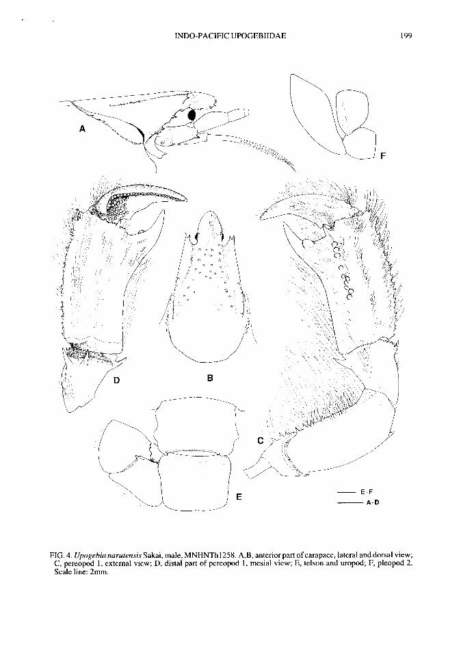
INDO-PACIFIC UPOGEBIIDAE 199
FIG. 4. Upogebia narutensis Sakai, male, MNHNThl258. A,B, anterior part of carapace, lateral and dorsal view; C, pereopod 1, external view; D, distal part of pereopod 1, mesial view; E, telson and uropod; F, pleopod 2. Scale line: 2mm.

200 MEMOIRS OF THE QUEENSLAND MUSEUM
FIG. 5. A-D, Upogebia narutensis Sakai. A,B, female; C,D, male, MNHNTh 1258, MNHNTh 1259; E-H, Upogebia wuhsienweni Sakai. E,G,H, male; F, female, MNHNThl265. A,F,G, pereopod 1, external view; B, H, distal part of pereopod 1, mesial view; C,D, pereopods 2 and 3 respectively; E, anterior part of carapace, lateral view. Scale line: 2mm.

INDO-PACIFIC UPOGEBIIDAE 201
spinules and tubercles. Arthrobranchs with a series of large lamellae on either side of rachis.
Telson approximately 1.3 times as broad as long, posterior margin slightly convex, very faint inverted U-shaped carina dorsally.
Antennule (Fig. 4A). Peduncle unarmed. Antenna (Fig. 4A). Third peduncular article
with small ventral subdistal spine, scale terminat-ing in blunt tip.
Mandible. Without mesio-anterior tooth. Maxillipeds. 1 and 3 both with epipod. Pereopod 1. Subcheliform, sexually dimor-
phic, much stouter in males. Ischium with 2-3 ventral spines. Merus with dorsal subdistal spine and 5-7 ventral spines. Carpus with large ventral spine; lower half of outer surface with lon-gitudinal crest of more or less conspicuous spinules terminating with largest one; upper half with fine longitudinal groove; carpal dorsal mar-gin carrying 5-6 spines or spinules, and large dorsal subdistal spine at tip; 3-4 dorsal subdistal spinules external to the latter and large distal spine on upper half of mesial surface.
In males (Fig. 4C,D), palm of propodus about 1.5 times as long as broad at mid-length, broader distally; row of 12-20 spinules on dorsal margin; outer surface with ventral longitudinal row of strong round tubercles on distal half and large acute distal spine near lower base of dactylus; mesial surface bearing spinules on distal border with large distal spine near lower base of dac-tylus; fixed finger distal, nearly two-thirds as long as dactylus, cutting edge bearing large round tooth on external surface. Dactylus two-thirds as long as propodus with 2 proximal tubercles on dorsal border and shallow longitudinal dorsal groove on external surface; mesial surface with curved tuberculate dorsal crest and proximal round tubercles underneath; cutting edge with large proximal tooth and corneous tip.
In females (Fig. 5A,B), palm of propodus ap-proximately 3.5 times as long as broad at mid-length and about as broad proximally as distally, unarmed except for row of 7 spinules on dorsal margin and external ventral distal spine between base of dactylus and fixed finger; fixed finger about one-fourth as long as dactylus, unarmed. Dactylus with corneous tip and shallow lon-gitudinal dorsal groove on external surface, cut-ting edge with 2 minute flat teeth.
Pereopod 2. Merus with dorsal distal spine and 4-5 ventral spines, the 2 proximal of which are large. Carpus bearing ventral and dorsal subdistal spine.
Pereopod 3. Merus carrying 3 ventral spines and 1 or 2 transversal proximal rows of short setae.
Males with genital openings on coxae of both P3 and P5. Females with genital opening on coxa of P3.
Pleopod2-5 (Fig. 4F). Endopod approximately quadrate with weak longitudinal carina.
Uropod. Latero-external and posterior margin of exopod, both slightly convex, meeting nearly at a right angle exteriorly. Latero-external margin of endopod with proximal shoulder terminating in large blunt tooth. Protopod with spinule hang-ing over base of endopod.
DISTRIBUTION Naruto (Japan), Taiwan.
REMARKS This material fits very well with the description
and figures given by Sakai (1984, 1986) and also with the holotype examined, except for the mesial proximal tubercles of PI dactylus that are more prominent and numerous.
The holotype of U. narutensis was once as-signed to U. spinifrons (Haswell) (Sakai, 1984). Differences between the two were given by Sakai (1986). U. narutensis is also very similar to U. edulis Ngoc-Ho & Chan, 1992 as well as to U. wuhsienweni Yu, 1931, all three reported from Taiwan. They all have the endopod of pleopods 2-5 in an unusual quadrate shape (Fig. 4F) which may exist in other species but have been over-looked by authors; it was not reported in the original description of U. edulis (Ngoc-Ho & Chan, 1992).
U. narutensis resembles U. edulis by the un-armed anterior part of the gastric ridges, the stout-ness of the male pereopod 1 and the large tooth on the cutting edge of the dactylus. It is similar to U. wuhsienweni in the shape of the rostrum, in the male pereopod 1 bearing a large round tooth on the cutting edge of the distal fixed finger. It differs from both U. edulis and U. wuhsienweni by the following features: 1, anterior half of the rostrum unarmed; 2, absence of a stridulating ridge on the propodus of the male pereopod 1; 3, latero-exter-nal margin of the uropod endopod with a large blunt tooth on the proximal shoulder.
In these three species of Upogebia, female pereopods 1 are hardly distinguishable. That of U. narutensis can be differentiated by the shape of its propod which is as broad distally as proximally while it is narrower distally in U. edulis and U. wuhsienweni.

202 MEMOIRS OF THE QUEENSLAND MUSEUM
Upogebia wuhsienweni Yu, 1931 (Fig. 5E-H)
Upogebia Wuhsienweni Yu, 1931: 89, fig. 2. Upogebia wuhsienweni: Liu, 1955: 68, ligs 7-12;
Ngoc-Ho & Chan, 1992: 38, fig. 4; not Sakai, 1993: 92, figs 1, 2 (= U. edulis Ngoc-Ho & Chan).
Upogebia (Upogebia) wuhsienweni: Sakai, 1982: 59 (in part, not figs l i d , 12f-g, 13g-h, pis G l -2 and material USNM59070, 59071, 59072, 59073 (= Upogebia edulis Ngoc-Ho & Chan).
MATERIAL EXAMINED How-Long, Northwestern Taiwan: 1 8 , cl. 17mm, tl.
52mm; 1 9 , cl. 19.5mm, tl. 60mm (MNHNTh 1265).
DESCRIPTION Rostrum rounded anteriorly, projecting beyond
eyes, bearing 5-6 spiniform teeth on each lateral border, ventral surface with 3 spines. Gastric ridge divided by weak mid-dorsal notch: anterior half with 5-7 round teeth and spine at tip; posterior half with 4-5 teeth. Anterolateral border of carapace bearing 6-7 spines or spinules. Cer-vical groove deep, shoulders lateral to it armed with spinules and tubercles. Epistome terminat-ing in 2 spinules. Arthrobranchs with large lamel-lae on either side of rachis.
Antennule (Fig.5E). Peduncle unarmed. Antenna (Fig. 5E). Third peduncular article
with small ventral subdistal spine, scale terminat-ing in blunt tooth.
Mandible. Without mesio-anterior tooth. Maxillipeds. 1-3 with epipod. Pereopod 1. Subcheliform. Basis with sharp
ventral spine. Ischium carrying 2-3 ventral spines. Dorsal subdistal spine and 6-7 ventral spines on merus. Carpus bearing longitudinal crest on external lower half with more or less conspicuous spinules and terminating in spine; large ventral distal spine, large dorsal distal spine along with 3-4 dorsal external spinules, large distal spine near middle of mesial surface; dorsal margin with 1-2 spines.
In males, propodus slightly broader distally than proximally with dorsal row of 8-9 spines and 8 spinules; external distal spines between base of dactylus and fixed finger; mesial surface bearing 2 proximal dorsal spines below dorsal row and slender elliptic stridulating ridge on vent ral distal half; fixed finger distal, about one-third as long as dactylus, carrying large rounded external tooth near middle of cutting edge. Dactylus with cor-neous tip , shallow longitudinal dorsal groove on external surface; longitudinal oblique carina on
mesial surface alongside fine corneous one and a few round proximal tubercles. In female, propodus narower distally than proximally with dorsal row of 9 spines, external distal spine be-tween base of dactylus and fixed finger; fixed finger short, hardly one-fourth length of dactylus, unarmed. Dactylus with corneous tip and shallow longitudinal dorsal groove on external surface.
DISTRIBUTION North China, Western Taiwan. This record
marks a southerly range extension.
REMARKS The present specimens from Taiwan agree
closely with the material of U. wuhsienweni from China examined previously (Ngoc-Ho & Chan, 1992) with the following exceptions: the male pereopod 1 is more slender, being about twice as long as broad at mid-length and the female pereopod 1 carries a distal external spine between the base of the dactylus and fixed finger, that is usually missing on Chinese specimens.
U. wuhsienweni differs from U. edulis Ngoc-Ho & Chan by many characters (Ngoc-Ho & Chan, 1992) especially by the anterior half of its gastric ridges armed with spines, and these are clearly shown on the original figure given by Yu (1931: fig. 11 A); in U. edulis, the same part of the gastric ridges is unarmed. All material assigned to U. wuhsienweni by Sakai (1982, 1993) but having an unarmed anterior half of the gastric ridges is likely to belong to U. edulis. Female pereopods 1 of U. wuhsienweni and U. edulis are hardly distinguishable except for a sharp spine on the basis in the former species that is replaced by a blunt tooth in the latter.
Upogebia pugnax de Man, 1905 (Fig. 6)
Upogebia (Upogebia) pugnax de Man, 1905: 600; de Man, 1928:66, fig. 8-8e, 8f; Sakai, 1982:52 (in part, not fig. 1 lb, pi E4, E6); not Sakai, 1984: 161 (= U. fallax de Man) and 1987: 302 (=Upogebia sakaii sp. nov.).
Upogebia pugnax: Ngoc-Ho, 1990: 987, fig. 7; 1991: 305, fig. 10.
MATERIAL EXAMINED New Caledonia: Loyalty Islands (Ouvea), J.P. Menou
coll., 18.11.1991,9-11 m: 1 ovigerous 9 ,cl . 10.5mm, tl. 31.5mm (MNHNTh 1261).

INDO-PACIFIC UPOGEBIIDAE 203
FIG. 6. Upogebia pugnax de Man., ovig. female, MNHNThl261. A, anterior part of carapace, dorsal view; B, pereopod 1, external view; C, distal part of pereopod 1, mesial view. Scale line: 1mm.
DESCRIPTION Rostrum about as long as broad at base, over-
reaching eyes, with 6 small teeth on either lateral border and slight median groove; proximal part of rostrum and anterior part of gastric region carrying round tubercles. Lateral groove moderately wide, lateral ridge with 10 teeth. Cer-vical groove deep, anterolateral border of carapace with spinule.
Pereopod 1. Subcheliform. Ischium with 2 ventral spines. Merus over 3 times as long as broad with dorsal subdistal spine and 8 ventral spines. Carpus with ventral spine and spine near middle of dorsal margin; external surface with dorsal distal spine and spinule, mesial surface with large dorsal distal spine and another near middle of distal margin. Propodus over 3 times as long as broad, carrying spine near proximal third
and large spine near middle of ventral margin, with smaller one beside the latter on external surface; upper border with 5 large spines; fixed finger approximately triangular, bearing small rounded teeth on cutting edge. Dactylus about two-third as long as propodus with corneous tip, upper border and cutting edge finely denticu-lated, faint longitudinal groove near upper border of external surface.
Genital openings on coxae of both pereopods 3 and 5.
Pleopod 1 present.
DISTRIBUTION Indonesia (Sumbawa), New Caledonia (St
Marie Island, Loyalty Islands) (significant southerly range extension).
204 MEMOIRS OF THE QUEENSLAND MUSEUM
REMARKS This is the first female reported for the species
and it agrees well with other described material (de Man, 1928; Ngoc-Ho, 1990, 1991). It con-firms that the two specimens from New Caledonia described by Ngoc-Ho (1991) are male.
With male and female adults now known, the following characteristics of U. pugnax can be noted: 1, Male pereopod 1 is dimorphic and can be "stout" or "slender", the latter type being very similar to that of the female. The same has been reported in U. edulis Ngoc-Ho & Chan (1992); 2, Males and females possess genital openings on coxae of both pereopods 3 and 5; only females possess pleopod 1; 3, The holotype (see de Man, 1928; Ngoc-Ho, 1990) is a young male of 18.5mm total length and has a "slender" type pereopod 1.
Upogebia sakaii sp. nov. (Fig. 7)
MATERIAL EXAMINED HOLOTYPE: Japan (Usa - Inoshiri, Kochi), K. Sakai
coll.,20.5.1990, coarse sand, tidal zone: 8 ,cl. 9.5mm, tl. 24.5mm (MNHNTh 1262). PARATYPES: 1 8 , cl. 10mm tl. 22mm, 1 9 , cl. 9mm
tl. 21.5mm (MNHNThl263); 3 8 , cl. 9-11mm, tl. 23-28mm; 4 9 , cl . 8 . 5 -9 .5mm, tl. 2 1 - 2 5 m m (MNHNTh 1264).
ETYMOLOGY For Dr. K. Sakai who collected and donated this
material.
DESCRIPTION Rostrum sub-triangular, c. 1.2 times as long as
broad at base, with 6 or 7 lateral spiniform teeth, slight longitudinal median groove; round tubercles on rostrum and anterior part of gastric region. Lateral groove moderately broad, lateral ridge with 9 or 10 teeth. Antero-lateral border of carapace with spinule. Cervical groove deep, linea thalassinica distinct, extending to posterior margin of carapace.
Telson slightly shorter than 6th abdominal seg-ment, lateral border convex at proximal third, posterior border concave medially and about 2/3 as broad as proximal; very faint inverted U-shaped carina on dorsal surface.
Arthrobranchs with one series of large tubular lamellae on either side of rachis.
Antennule (Fig. 7B). First peduncular article with large ventral subdistal spine
Antenna (Fig. 7B). Third peduncular article with large ventral subdistal spine; scale not demarcated from peduncle, terminating in spinule.
Mandible. With large antero-mesial tooth. Maxillipeds. 3 with small epipod. Pereopod 1. Subcheliform, sexually dimor-
phic, stouter in males. Ischium with ventral spine. Merus 2.5 times as, long as broad, bearing spine near distal quarter of dorsal margin, 7-10 ventral spines. Carpus with large ventral subdistal spine and spine near middle of dorsal margin; 3 dorsal distal (2 external, 1 mesial) spines and large spine near middle of mesial distal margin. Propodus over twice as long as broad, with 5 large dorsal spines and large ventral spine behind fixed finger; external surface with tubercles on lower third and one or 2 spinules near large ventral spine; mesial surface bearing 1-2 dorsal subdistal spines, near upper part of articulation with dactylus; fixed finger, broad and short, cutting edge with rounded teeth over proximal two-thirds. Dactylus about two-thirds length of propodus carrying faint longitudinal dorsal groove on external sur-face, cutting edge dentate on proximal half, with low triangular tooth near middle; tip corneous.
Female pereopod 1 with same spinulation as in males but more slender, merus and propodus about 3 and 2.5 times as long as broad respective-iy-
Pereopod 2. Merus with subdistal dorsal spine; 2 proximal spines on ventral border. Carpus bear-ing subdistal dorsal and subdistal ventral spine. Dactylus with faint longitudinal dorsal groove.
Pereopod 3. Merus with 3 ventral spines and a few tubercles on ventral margin. Subdistal ventral spine on carpus. Dactylus with comb-like setae on ventral margin.
Pereopod 4. Merus with 4-5 spiniform tubercles on ventral margin.
Males are provided with genital opening on coxae of pereopod 5, females have openings on coxae of both pereopods 3 and 5. Large coxal spine on pereopod 1, smaller ones on pereopods 2 and 3.
TYPE LOCALITY Japan (Usa - Inoshiri, Kochi).
REMARKS These specimens as well as others from Japan
have been previously assigned to U. pugnax de Man (Sakai, 1982, 1987). This is probably be-cause de Man (1928) stated that the holotype of the latter species was a female but it is actually a

INDO-PACIFIC UPOGEBIIDAE 205
FIG. 7. Upogebia sakaii sp. nov. A-C, F, G, holotype, male, MNHNThl262; D,E, female paratype, MNHNThl263. A,B, anterior part of carapace, dorsal and lateral view; C, telson and uropod; D,F, pereopod 1, external view; E,G, distal part of pereopod 1, mesial view. Scale line: 1mm.

206 MEMOIRS OF THE QUEENSLAND MUSEUM
male. In the present new species, the females are similar to the holotype of U. pugnax but males differ by many features.
Two males of U. pugnax were described by Ngoc-Ho (1991) and an ovigerous female reported earlier in the present paper. U. sakaii and U. pugnax are very similar in the shape of the rostrum and the morphology and spinulation of all cephalic appendages. They differ by the fol-lowing features: 1, epistome unarmed in U. sakaii but with a terminal spine in U. pugnax; 2, male pereopod 1 dimorphic in U. pugnax; 3, propod of male pereopod 1 distinctly broadened distally in U. sakaii; 4, in U. sakaii, the fixed finger of pereopod 1 is very short and the cutting edge level with the propod-dactylus articulation; in U. pug-nax, the fixed finger is longer and the cutting edge projects beyond the propod-dactylus articulation; 5, U. sakaii has the dactylus of pereopod 1 with upper border unarmed or rarely denticulated proximally and with a low triangular tooth near the middle of the cutting edge; U. pugnax has the upper border completely denticulated and the cutting edge smooth; 6, telson with proximal border about 1.5 times broader than distal in U. sakaii but approximately the same in U. pugnax; 7, exopod of uropod with posterior margin slight-ly convex and postero-lateral corner rounded in U. sakaii; posterior margin more or less straight and meeting nearly at right angle with lateral margin in U. pugnax.
Upogebia savignyi (Strahl, 1862). (Fig. 8)
Gebia sp. Savigny, 1817: pl.9, figs 3/2-2'. Calliadne savignii Strahl, 1862: 1064. Upogebia (Gebiopsis) rhadames Nobili, 1904: 235. Upogebia (Calliadne) savignyi: Nobili, 1906: 98; de
Man, 1927: 5, fig. 1; 1928: 47 (key). Upogebia (Calliadne) rhadames: Nobili, 1906: 100; de
Man, 1927: 6, pl.l , fig. 1; 1928: 47 (key); Sakai, 1975: 23, figs 6-8.
Upogebia (Upogebia) savignyi: Sakai, 1982: 14.; 1984: 154.
Upogebia (Upogebia) cargadensis: Sakai, 1982: 12 (in part, material from Kenya only, ZSM 1233/1 and ZSM 1233/2).
MATERIAL EXAMINED North Kenya Banks, "Dr. Fridtjor Nansen" Cruise N° 1, Stn. 04, 16.2.1975, 02°30'S-40°56'E, 77m, in "green sponges": 1 c? , cl. 12mm, tl. 30mm; 4 9 (1 ovigerous, 1 without abdomen), cl. 11.5-13mm, tl. 29-30mm (BMNH1993: 31.5); 10 6 , cl. 5-7.5mm; 2
9 , cl. 5.5-7mm (syntypes of U. rhadames Nobili, MNHNTh45).
DESCRIPTION Rostrum sub-triangular, as long as broad at
base, overreaching eye-stalk in male, shorter in female, with 6-8 small rounded tubercles on either lateral margin. Small round tubercles on rostrum and gastric region, with 23-25 on either gastric ridge; lateral groove long and narrow. Antero-lateral border of carapace unarmed; epis-tome rounded distally.
Telson sub-quadrate, c. 1.5 times as long as sixth abdominal segment, posterior border and lateral posterior angles rounded, faint and fine inverted U-shaped carina on dorsal surface.
Antennule and Antenna (Fig. 8C). With un-armed peduncle; antennal scale very small.
Maxillipeds. 1 (Fig. 8F), with exopod flattened distally bearing short and long setae, longer ex-ternally; 2 (Fig. 8G) with very small upright exopod; 3 without epipod.
Pereopod 1. Cheliform. Merus about 2.5 times as long as broad, 11-13 ventral spinules. Carpus unarmed; propodus unarmed except for 7-8 small teeth on proximal half of cutting edge of fixed finger. Dactylus slightly more than half length of propodus, bearing two dorsal proximal spiniform tubercles; mesial surface with longitudinal row of small round tubercles at mid-level and large, round proximal tooth near cutting edge.
Pereopod 2. Carpus with ventral subdistal spinule.
Uropod. Exopod slightly shorter than telson; posterior margin weakly convex; lateral angle rounded; proximal spine. Endopod trapezoidal. Basipod with posterior spine.
DISTRIBUTION Suez, Red Sea, Gulf of Aden, Persian Gulf,
Kenya.
REMARKS Examination of the present material and the
syntypes of U. rhadames Nobili (MNHNTh45) agree with Sakai (1982) that these two species are synonymous. Also belonging to U. savignyi are specimens from Kenya (ZSM 1233/1, 1233/2), assigned by Sakai first to U. rhadames (1975) and later to U. cargadensis Borradaile (Sakai, 1982 with selection of a neotype). This neotype selected from sample ZSM 1233/2 was refuted by Ngoc-Ho (1991) as not fitting Borradaile's (1910) original description.

INDO-PACIFIC UPOGEBIIDAE 207
FIG. 8. Upogebia savignyi (Strahl). A,C-E,H, male, tl. 30mm; B,F,G, female without abdomen, cl. 13mm, BMNH 1993:31.5; A-C, anterior part of carapace, dorsal and lateral view; D, pereopod 1, external view; E, distal part of pereopod 1, mesial view; F, maxilliped 1; G, maxilliped 2; H, telson and uropod. Scale line: 1mm.

208 MEMOIRS OF THE QUEENSLAND MUSEUM
There is some variation in U. savignyi: A, the triangular rostrum can be longer, equal, or shorter than the eye-stalk and is usually longer in males.; its tip can also be more or less pointed; B, in pereopod 1: ventral border of merus unarmed or with granules or denticles; carpus unarmed or with a ventral spinule; ventral border of propodus unarmed or with proximal denticles, dorsal bor-der (rarely) with a distal spinule; C, posterior border of telson more or less rounded.
Diagnosfic characters for the species are: 1, rostrum, gastric region and gastric ridges with numerous small tubercles; 2, linea thalassinica hardly visible posterior to cervical groove; 3, peduncle of both antennule and antenna unarmed; 4, maxilliped 1 without epipod, with exopod flat-tened distally, bearing setae of two lengths (Fig. 8F); maxilliped 2 with small upright epipod (Fig. 8G); maxilliped 3 without epipod; 5, Pereopod 1 cheliform; palm of propodus unarmed (with few exceptions), fixed finger with small teeth on proximal half of cutting edge; dactylus with a round mesial proximal tooth on cutting edge; 6, telson approximately quadrate, posterior border rounded; exopod of uropods with a proximal spine and basipod with a spine.
The morphology of the exopod of maxilliped 1 and the epipod of maxilliped 2 is unusual in the Upogebiidae. It has been reported in two other species: U. tractabilis (Hale) from Southern Australia (Ngoc-Ho, in press) and U. stenor-hynchus Ngoc-Ho, 1991 from New Caledonia. Differences between the latter species and U. savignyi were given (Ngoc-Ho, 1991). Com-parison of U. savignyi with U. tractabilis show certain similarities: 1, triangular shape of the rostrum; 2, peduncle of both antennule and anten-na unarmed; 3, proximal spinule on exopod of uropod and another on basipod. Distinguishing characters are: 1, rostrum and gastric ridges bear-ing tubercles in savignyi but spinules or spiniform tubercles in tractabilis; 2, pereopod 1: merus unarmed or with a few spinules in savignyi, with spines in tractabilis; carpus unarmed in savignyi, with a ventro-distal spine in tractabilis-, fixed finger cutting edge with small proximal teeth in savignyi, unarmed in tractabilis', dactylus with a proximal rounded tooth on cutting edge in savig-nyi but with 3-4 teeth medially in tractabilis; 3, telson with posterior border rounded in adults savignyi, with straight posterior border in adults tractabilis
Upogebia spinimanus sp. nov. (Fig. 9)
TYPE MATERIAL HOLOTYPE: Madagascar (Bombetoke Bay), Bastard
c o l l . ( n o date): 1 8 , c l . 5 . 5 m m , tl . 1 4 m m (MNHNTh790).
ETYMOLOGY Referring to the large dorsal spine on the palm
of pereopod 1.
DESCRIPTION Rostrum sub-oval, projecting far beyond eyes;
6 small rounded teeth on either lateral margin; faint longitudinal median groove. Rounded tubercles postero-dorsally on rostrum, and on gastric region alongside lateral groove; lateral groove moderately broad. Gastric ridge with 10-12 small spiniform tubercles. Antero-lateral bor-der of carapace with spinule. Cervical groove deep, bearing spine on either side near intersec-tion with linea thalassinica, the latter extending to posterior margin of carapace. Epistome ter-minates dorsally in minute spinule.
Telson slightly broader than long, lateral border convex, postero-lateral angles rounded, posterior border nearly straight; very faint and small in-verted U-shaped carina on dorsal surface.
Single pleurobranch on 5th thoracic segment in addition to arthrobranchs on maxilliped 3 and pereopods 1-4. Arthrobranchs with 2 tubular lamellae on either side of the rachis.
Antennule (Fig. 9B). First peduncular article with ventral distal spinule.
Antenna (Fig. 9B). Third peduncular article with ventral distal spinule; scale terminating in small flap extending to base of fourth article.
Maxillipeds. 1 -3 with epipod, that of maxilliped 1 very small.
Pereopod 1. Subcheliform. Ischium with ven-tral spine. Merus about 3 times as long as broad, bearing dorsal subdistal spine and 3-4 ventral spines. Carpus with ventral distal spine, fine lon-gitudinal groove on upper part of external sur-face; mesial surface with dorsal distal spine and spine near middle of distal margin. Propodus over 2.5 times as long as broad, carrying large spine near distal third of dorsal margin; fixed finger slender, about 1/3 as long as dactylus, cutting edge denticulated. Dactylus approximately 2/3 as long as propodus, with small corneous tip, un-armed.

INDO-PACIFIC UPOGEBIIDAE 209
FIG. 9. Upogebia spinimanus sp. nov., holotype, male, MNHNTh790. A,B, anterior part of carapace, dorsal and lateral view; C, pereopod 1, external view; D, distal part of pereopod 1, mesial view; E,F, pereopods 2 and 3 respectively; G, telson and uropod. Scale line: 1mm.
Pereopod 2. Merus with dorsal subdistal, and Pereopod 3. Merus with 2 spines on ventral
ventral proximal spine. Carpus bearing dorsal, m ^ g m . v r r t> ' Uropod. Exopod hardly longer than telson,
and ventral subdistal spine. posterior border and lateral external angle

210 MEMOIRS OF THE QUEENSLAND M U S E U M
rounded, with spinule proximally; endopod ap-proximately trapezoidal, protopod with spinule.
TYPE LOCALITY Madagascar (Bombetoke Bay).
REMARKS The possession of a pleurobranch on the 5th
thoracic segment places U. spinimanus sp. nov. within a special group of Upogebia which is dealt with in detail in another work (Ngoc-Ho, in press). Included are: U. africana (Ortmann, 1894); U. allobranchus Ngoc-Ho, 1991; U. capensis (Krauss, 1843); U. giralia Poore & Grif-fin, 1979; U. lenzrichtersi Sakai, 1982; U. stellata (Montagu, 1808).
U. spinimanus is most similar to U. lenzrichter-si also from Madagascar, and was compared with paratypes of the latter species in the Paris Museum (MNHNTh519, 520). The two have similar rostrums, telsons and uropods; both have a dorsal spine on the propodus of pereopod 1; both have coxal spines on pereopods 1-3. They can be separated by: 1, lateral shoulder of cervical groove with spine near intersection with linea thalassinica in U. spinimanus (spine absent in U. lenzrichtersi); 2, antennular and antennal peduncle with a ventral spine on first and third article in U. spinimanus (unarmed in U. lenzrich-tersi)', 3, male pereopod 1: U. spinimanus: merus with 3-4 ventral spines; propodus without dorsal carina behind dorsal spine, ventral margin un-armed; dactylus unarmed (U. lenzrichtersi: merus unarmed ventrally, or with tubercles; propodus with dorsal carina on proximal two-thirds and large mesial ventral spine near base of fixed finger; dactylus with dorsal tubercles and small round teeth on cuting edge); 4, pereopods 2 and 3 with 1-2 ventral spines in U. spinimanus but unarmed ventrally in U. lenzrichtersi.
WolfTogebia Sakai, 1982
REMARKS Wolffogebia Sakai, 1982 was established for 4
species: W. phuketensis Sakai, 1982 (type species); W. inermis Sakai, 1982; W. obtifrons Sakai, 1982; and Gebicula exigua Alcock, 1901. Sakai gave the following diagnosis: "Dorsal sur-face of anterior region with a median carina. Lateral frontal process of carapace developed. Lateral longitudinal groove definable. Antero-lateral margin of carapace armed or unarmed. First pereopod subchelate."
It is questionable whether Gebicula exigua (also the type species of Gebicula Alcock, 1901) really belongs to Wolffogebia. This species was considered by Sakai (1982) to be a senior synonym of Upogebia monoceros de Man but his action was thought doubtful by Ngoc-Ho (1989) who assigned U. monoceros to the genus Gebiacantha. The holotype (a female of 15mm in total length), and only existing specimen of Gebicula exigua, is deposited in the Indian Museum and unavailable for examination at present. The original figure in Alcock (1901) is in lateral view and the dorsal surface of the rostrum and anterior region of the carapace are not shown. It is impossible to confirm whether the specimen possesses the first three characters given by Sakai (1982) in the diagnosis of Wolffogebia. However, the figure shows the antero-lateral margin of the carapace bearing at least 2 spines, which is in contradiction with the type-species of Wolffogebia, W. phuketensis, which has this border unarmed (Sakai, 1982: fig. 18c). The two characters "unarmed antero-lateral border of the carapace" together with "pi sub-cheliform" displayed by W. phuketensis are un-common in the Upogebiidae. Until the holotype of Gebicula exigua can be examined, it is not possible to know whether this deep-sea species (captured at 485m depth) belongs to Wolffogebia. If it does, Wolffogebia would become a junior synonym of Gebicula. Wolffogebia and Gebicula are here provisionally retained as separated genera pending a future study of Gebicula exigua.
Characters given as diagnostic of Wolffogebia by Sakai (1982), are cited above. Those relating to the lateral frontal process of the carapace, the lateral longitudinal groove and the antero-lateral margin of the carapace are not precise enough to be useful as they belong to the great majority of upogebids, and roughly half of them actually possess a subchelate pereopod 1.
Wolffogebia species form a distinctive group within the Upogebiidae. Examination of the type species, as well as W. inermis reveals a number of morphological features which help to better define it. They are: absence of a median lon-gitudinal groove on rostrum; dorsal surface of anterior region with a slight median carina; antero-lateral border of carapace unarmed; arthrobranchs with a single series of large lamel-lae on either side of the rachis; maxilliped 1 with a large epipod; maxilliped 2 with exopod of one article, without flagellum; maxilliped 3 without epipod, exopod without flagellum; pereopod 1 subcheliform.

INDO-PACIFIC UPOGEBIIDAE 211
FIG. 10. Wolffogebia inermis Sakai. A-C, male, tl. 27mm, BMNH1993:30.2; D-F, female, tl. 27mm, MNHNTh 1279. A, anterior part of carapace, lateral view; B, pereopod 1, external view; C, distal part of pereopod 1, mesial view; D-F, maxillipeds 1, 2 and 3 respectively. Scale line: 1mm.

212 MEMOIRS OF THE QUEENSLAND MUSEUM
FIG. 11. Wolffogebia inermis Sakai. A,B, male, tl.21mm, MNHNTh 1279; C-E, female, tl.27mm, MNHNTh 1279. A,C, pereopod 1, external view; B,D, distal part of pereopod 1, mesial view; E, pereopod 2, external view. Scale line: 1mm.

INDO-PACIFIC UPOGEBIIDAE 213
In combinat ion with the characters "anterolateral border of carapace unarmed" and "pereopod 1 subcheliform", the morphology of maxillipeds, especially exopods without a flagel-lum in maxillipeds 2 and 3, is uncommon in the Upogebiidae.
Wolffogebia inermis Sakai, 1982 (Fig. 10)
Wolffogebia inermis Sakai, 1982: 81, figs 17c, 18g, 19a-b,pl.G6; 1993: 109, figs. 12-14.
MATERIAL EXAMINED Can-gio (Ho-chi-Minh city), Vietnam, Tran phi Hung
coll., 1.5.1993, mangrove area, in mud: 5 6 (4 juv.), cl. 4.5-7mm, tl. 14.5-22mm, 3 9 (2 ovig.), cl. 7.5-8mm, tl. 27-28mm (NMHNThl279); no locality data: 2 8 , cl. 8mm and 11.5mm, tl. 21.5mm and 27mm; 1 6 , 1 9 damaged, 1 male PI (BMNH1993:30.2).
DESCRIPTION Rostrum elongate, with rounded tip projecting
far beyond eye, setose but unarmed, as are gastric region and gastric ridge. Lateral groove narrow; antero-lateral border of carapace unarmed. Cer-vical groove moderately deep, linea thalassinica faint posterior to it. Epistome terminating in spinule. Arthrobranchs with single series of large lamellae on either side of rachis.
Antennule and Antenna (Fig. 10A). Both peduncles unarmed, antennal scale minute.
Maxillipeds 1 (Fig. 10D), with large epipod; 2 (Fig. 10E), with simple exopod, without flagel-lum; 3 (Fig. 10F), without epipod, exopod simple in large specimens, with short indifferentiated flagellum in juveniles.
Pereopod 1. Subcheliform, sexually dimor-phic. Ischium with ventral spine. Merus with dorsal subdistal, and ventral proximal spine. Car-pus bearing large ventral distal spine; external surface with faint longitudinal groove on upper half; mesial surface carrying dorsal distal spine and another near middle of distal border. Propodus in males over twice as long as broad at mid-length, more slender in females; mesial sur-face with fine longitudinal dorsal carina in adult males, smooth in juveniles and females, all with large dorsal subdistal spine; fixed finger subdis-tal, unarmed. Dactylus in adult males three-quarters as long as propodus, with corneous tip and denticles on dorsal margin, external surface with 0-6 round tubercles near cutting edge, mesial surface with longitudinal row of 2-6 teeth or tubercles on upper half; dactylus unarmed in
juveniles and females except for 1-3 small mesial tubercles.
Pereopod 2. Merus with dorsal subdistal and ventral proximal spine; carpus with dorsal sub-distal spine.
DISTRIBUTION Indonesia (Java, Mocara Tangerang), Vietnam
(Can-gio).
REMARKS Sakai (1982) stated that the holotype (S , 34mm
tl., from Java, Mocara, Tangerang) and sole specimen was deposited in the Zoologisch Museum-Universitat van Amsterdam, but there is no record of it there, nor in the Zoologisches Institut-Universitat Hamburg (D. Platvoet & G. Hartmann, pers. comm.).
The present material agrees well with the description and figures of the holotype especially in the absence of all spines or tubercles from the carapace. Pereopod 1 is slightly more slender in this material; in adult males, the dactylus bears denticles on the dorsal margin, external tubercles near the cutting edge, and a row of 2-6 teeth or tubercles on the mesial surface. The dactylus of PI on the holotype has both the dorsal margin and the cutting edge unarmed and only a single mesial tooth is present (Sakai, 1982: fig. 20E). Another variation concerns the right PI of a male from Vietnam of 22mm tl. which is larger than the left and armed with an external subdistal spine near the base of the fixed finger, and a mesial subdistal spine near the base of the dactylus (Fig. 11 A, B). These spines are not reported in the holotype and absent in the rest of the material examined.
Females studied also agree well with the specimen recently described by Sakai (1993: fig. 13b,c) (ovig. 9 , 30mm tl., from Darwin, Australia) except that in the Australian specimen, the exopods of both maxillipeds 2 and 3 are provided with a flagellum. It is questionable whether there was a mistake.
Wolffogebia phuketensis Sakai, 1982 (Fig. 12)
Wolffogebia phuketensis Sakai, 1982: 75, figs 17a, 18c-d, 20b.
MATERIAL EXAMINED HOLOTYPE: Phuket, Thailand, 6 , tl. 39m (UMK type
collection). OTHER MATERIAL: Northwestern Singapore, in
mangrove, in burrows of Thalassina anomala mound,

214 MEMOIRS OF THE QUEENSLAND MUSEUM
FIG. 12. Wolffogebia phuketensis Sakai, QMW14854. A-D, ovig. female, tl. 34mm; E, male; F, female tl. 25.5mm. A, anterior part of carapace, dorsal view; B, maxilliped 1; C, maxilliped 2; D, maxilliped 3; E,F, pereopod 1, mesial view. Scale line: 1mm.

INDO-PACIFIC UPOGEBIIDAE 215
P. Davie & P. Ng coll., 6.9.1987: 2 9 (1 ovig.), cl. 8.5mm and 10mm, tl. 25.5mm and 34mm; 1 8 without abdomen, cl. 13mm (QMW14854).
DESCRIPTION Rostrum low, triangular, about half as long as
wide at base in females, longer in male, projecting slightly beyond eyes, with 4 round teeth laterally; dorsal surface without median longitudinal groove, very setose. Gastric region setose lateral-ly with 8-9 round tubercles alongside either lateral groove; medially with a non setose area the anterior part of which slightly elevated in the shape of a weak carina pointing forwards. Lateral groove moderately deep; lateral ridge with small median notch, carrying 12-13 and 4-5 round tubercles on anterior and posterior half respec-tively. Anterolateral border of carapace unarmed. Cervical groove deep, linea thalasinica invisible posterior to it. Arthrobranchs with single series of large lamellae on either side of the rachis.
Antennule and Antenna. Peduncle unarmed. Mandible. Without antero-mesial tooth. Maxillipeds. 1 (Fig. 12B) with large epipod; 2
(Fig. 12C) with single article on exopod, without flagellum; 3 (Fig. 12D) without epipod, exopod without flagellum.
Pereopod 1. Subcheliform, stouter in male than in female. Merus with dorsal subdistal spine. Carpus with a ventral subdistal, and 2 large mesio-distal spines. Propodus about twice as long as broad at mid-length in male, over 3.5 times in female, palm unarmed, fixed finger with small teeth on proximal two-thirds of cutting edge. Dactylus with weak longitudinal mesial tubercu-late crest and a few tubercles in male, unarmed in female; cutting edge bearing 2-4 minute teeth.
Pereopod 2. Merus with dorsal subdistal and ventral proximal spine.
DISTRIBUTION Thailand (Phuket Island); Singapore.
REMARKS The specimens examined agree closely with
Sakai's description and figures and with the holotype of Wolffogebia phuketensis. The ex-opods of both maxillipeds 2 and 3 are clearly simple (Fig. 12C,D) TTie external view of male pereopod 1 was given by Sakai (1982), the mesial view of the same appendage in both male and female is provided (Fig. 12E,F).
ACKNOWLEDGEMENTS
The author wishes to thank the Natural History Museum, London (Paul Clark), the National Taiwan Ocean University (Tin-Yam Chan), The Nationaal Natuurhistorisch Museum, Leiden (Charles H.J.M. Fransen), the Queensland Museum, Brisbane (Peter Davie), the Univer-sitetets Zoologiske Museum, Kobenhavn (Dr Torben Wolff), the Zoologische Staatssam-mlung, Miinchen (Dr L. Tiefenbacher) for kindly making material available for examination, Peter Davie and Michele de Saint Laurent for their helpful comments on the manuscript.
LITERATURE CITED
BORRADAILE, L.A. 1910. Penaeidea, Stenopidea, and Reptantia from the western Indian Ocean. Transactions of the Linnean Society of London, Zoology 13 (2): 257-264.
LIU, J.Y. 1955. 'Economic shrimps and prawns of northern China'. (Marine Biological Institute, Academy of Sciences: Peking).
MAN, J.G. de. 1905. Diagnoses of new species of macrurous decapod Crustacea from the "Siboga Expedition". Rijdschrift der Nederlandsche Dierkundige Vereeniging 9 (2): 587-614.
1927. A contribution to the knowledge of twenty-one species of the genus Upogebia Leach. Capita Zoologica 2(5): 1-58.
1928. The Decapoda of the Siboga Expedition. Part VII. The Thalassinidae and Callianassidae col-lected by the Siboga-Expedition with some remarks on the Laomediidae. Siboga Expedition, Monograph 39(6): 1-187.
NGOC-HO, N. 1981. A taxonomic study of larvae of four thalassinid species (Decapoda,Thalas-sinidea) from the Gulf of Mexico. Bulletin of the British Museum of Natural History, Zoology 40 (5): 237-273.
1989. Sur le genre Gebiacantha gen. nov. avec la description de cinq especes nouvelles (Crustacea, Thalass inidea, Upogeb i idae ) . Bul le t in du Museum National d'Histoire Naturelle (Paris), Serie 4, 11A(1): 117-145.
1990. Nine Indo-Pacific species of Upogebia Leach (Crustacea : Thalassinidea : Upogebiidae). Jour-nal of Natural History 24: 965-985.
1991. Sur quelques Callianassidae et Upogebiidae de Nouvel le Caledonie (Crustacea, Thalas-sinidea). Pp. 281-311. In Richer de Forges, B. (ed.), 'Le benthos des fonds meubles des lagons de Nouvelle Caledonie'. Vol. 1. (Orstom Edi-tions: Paris).
IN PRESS. Some Callianassidae and Upogebiidae from Australia with descriptions of four new spccies Crustacea: Decapoda: Thalassinidea). Memoirs of the Museum of Victoria 54.

216 MEMOIRS OF THE QUEENSLAND MUSEUM
NGOC-HO, N. & CHAN, T-Y. 1992. Upogebia edulis, new species, a mud-shrimp (Crustacea : Thalas-sinidea : Upogebiidae) from Taiwan and Viet-nam, with a note on polymorphism in the male first pereiopod. Raffles Bulletin of Zoology 40(1): 33-43.
NOBILI, G. 1904. Diagnoses pr61iminaires de vingt-huit especes nouvelles de Stomatopodes et de Decapodes Macroures de la Mer Rouge. Bulletin du Museum d'Histoire Naturelle 5: 228-237.
1906. Faune carcinologique de la Mer Rouge, Decapodes et Stomatopodes. Annales des Scien-ces Naturelle (Zoologie) 4(1-3): 1-347.
SAKAI, K. 1975. Thalassinids of Kenya collected by Dr. A.J. Bruce. VeroffentlichungenderZoologis-chen Staatssamlung Miinchen 18: 1-44.
1982. Revision of Upogebiidae (Decapoda, Thalas-sinidea) in the Indo-west Pacific region. Re-searches on Crustacea, Special No. 1: 1-106.
1984. Some Upogebiidae (Crustacea, Decapoda) in the collection of the Rijksmuseum van Natuur-lijke Historie, Leiden. Zoologische Mededelin-gen 58(10): 149-162.
1984. A new record of Upogebia spinifrons (Has-well, 1882) (Decapoda, Thalassinidea) from Naruto , Japan, s h o w i n g poss ib l e her-maphroditism. Crustaceana 47(2): 209-214.
1986. On Upogebia narutensis, a new thalassinid (Decapoda, Crustacea) from Japan. Researches on Crustacea 15: 23-28.
1987. T w o new Thalass in idea (Crustacea: Decapoda) from Japan, with biogeographical dis-tribution of the Japanese Thalassinidea. Bulletin of Marine Science 41 (2): 296-308.
1993. On a collection of Upogebiidae (Crustacea, Thalassinidea) from the Northern Territory Museum, Australia, with the description of two new species. The Beagle 10(1): 87-113.
SAVIGNY, J. C. 1817. Crustaces. In 'Description de l'Egypte ou recueil des observations et des recherches qui ont ete faites en Egypte pendant l'expedition de l'armee franfaise, publie par les ordres de Sa Majeste l'Empereur Napoleon le Grand: Histoire naturelle, Crustaces'. 13 plates.
STRAHL, C. 1862. Uber einige neue von Hrn. F. Jagor eingesandte Thalassinen und die systematische Stellung dieser Familie. Monatsberichte der Deutschen Akademie der Wissenschaften zu Berlin 1861: 1055-1072.
YU, S.C. 1931. On some species of shrimp-shaped Anomura from north China. Bulletin of the Fan Memorial Institute of Biology 2(6): 85-97.


![[Gokigenyou] Seasons Black and Wite v.1 C.01](https://img.pdfslide.net/doc/110x75/577cd0761a28ab9e78924fac/gokigenyou-seasons-black-and-wite-v1-c01.jpg)