Embed Size (px)
Citation preview

Notice 1
Under the Copyright Act 1968, this thesis must be used only under the normal
conditions of scholarly fair dealing. In particular no results or conclusions should be
extracted from it, nor should it be copied or closely paraphrased in whole or in part
without the written consent of the author. Proper written acknowledgement should be
made for any assistance obtained from this thesis.
Notice 2
I certify that I have made all reasonable efforts to secure copyright permissions for
third-party content included in this thesis and have not knowingly added copyright
content to my work without the owner's permission
Influence of the extracellular matrix on the
in vitro differentiation of mouse embryonic
stem cells into neurons
A thesis submitted for the degree of
Doctor of Philosophy
Pankaj Gulati (M. Pharmacy)
Department of Pharmaceutical Biology and Pharmacology
Faculty of Pharmacy and Pharmaceutical Sciences
Monash University
Parkville, Victoria
Australia
2014
ii
Abstract:
Stem cell maintenance and differentiation are regulated by local inductive cues; one
potential source of inductive cues is the extracellular matrix. The fundamental
hypothesis of the thesis was that differentiation of embryonic stem cells can be
manipulated by substrates onto which cells are plated thus, our first experimental
chapter explores the impact of the extracellular matrix proteins, laminin, fibronectin and
type IV collagen on neural induction of E14Tg2a mouse embryonic stem cells (mESCs)
plated as adherent monolayers. In-cell Western assays were carried out to determine
coating efficiency of selected matrix proteins. Cell viability and proliferation was
estimated using the MTT assay. In addition, the effect of matrix proteins on neurite
outgrowth of mESCs-derived neurons was evaluated by counting the number of primary
neurites (using βIII tubulin labelling) and neural progenitor (nestin). Total neurite
length per neuron was quantified on neurons derived from the differentiated mESCs.
Immunocytochemistry against tyrosine hydroxylase and βIII tubulin was used to
observe the progression of neuronal maturation of cells plated onto matrix proteins. The
MTT assay was used to determine the effect of each matrix on adhesion of mESCs, 24
hours after replating. The results showed that laminin and fibronectin were associated
with increase in cell adhesion and they also enhanced the proliferation rate of mESCs
when compared to type IV collagen. Laminin significantly increased the number of βIII
tubulin positive cells 96 hours after re-plating, but failed to result in a larger numbers of
positive catecholaminergic neurons.
iii
Second experimental chapter focused on effect of the immobilization of growth factors
[sonic hedgehog (Shh), fibroblast growth factor 8b (FGF8) and fibroblast growth factor
basic (FGF2)] on the generation of midbrain dopaminergic neurons from mESCs. A
combination of matrix proteins components was produced by incubating laminin,
fibronectin, type IV collagen and heparan sulphate in a ratio of 1:1:1:0.1. This mixture
was used to immobilize growth factors within matrix proteins via non-covalent
interactions. In-cell Western assays were carried out to identify stable immobilization of
growth factors onto the matrix proteins. The effect of matrix proteins with and without
growth factors on neural differentiation was investigated by determining the expression
of the AMPR gene (encoding -lactamase) in Lmx1a-AMP mESCs.
Immunocytochemistry was used to observe the progression of neuronal maturation
under different conditions. FGF2 and FGF8b were successfully immobilized on a
combination of selected matrix proteins but the immobilized factors failed to
demonstrate any significant effect on the proportion of cells expressing Lmx1a. This is
likely to be explained by the unexpected high proportion of Lmx1a expressing cells
produced even in the absence of specific growth factors.
In the last experimental chapter the focus was on investigation of the signaling
pathways that regulate differentiations. New tools to promote the differentiation of stem
cells into particular cell types can be generated by identifying specific cues in the
microenvironments and deciphering how neighbouring cells and the extracellular matrix
control developmental fate. It is well established that complex interactions between
soluble and extracellular matrix molecules regulate intracellular signaling and
differentiation.
iv
Many of these signaling events involve one or more kinases including; focal adhesion
kinase (FAK), phosphatidylinositol 3-kinase (PI3K), protein kinase B (PKB/Akt,
hereafter called Akt), and mitogen-activated protein kinases (MAPK) – such as those
also known as Extracellular regulated kinases (ERKs). However, the relevance of a
given signaling molecule in mediating the pro-survival signaling induced by
extracellular matrix appears to be cell–type specific. Therefore, it is desirable to
determine which of these intracellular signaling pathways might be involved in the
effects of extracellular matrix on survival and differentiation of mouse embryonic stem
cells. This chapter explores the role of small molecule inhibitors of FAK, PI3K, Akt and
MAPK-ERK on adhesion and proliferation of cells produced by neural differentiation
of embryonic stem cells. The focus of the chapter is on early events in neural induction
making use of Sox1-eGFP reporter ESCs. This cell line expresses the eGFP reporter
under control of the Sox1 promoter, one allele expressing eGFP and the other Sox1. The
work showed that plating onto laminin (but not gelatin or poly-D-lysine) activated
PI3K-Akt and MAPK-ERK survival signaling pathways. Plating of cells on laminin in
the presence of CHIR99021, or in combination with PF562271, resulted in significant
conversion of viable embryonic stem cells to Sox1+ neural stem/progenitor cells,
indicating the role of laminin in the generation and survival of neurons.

v
Acknowledgements
On this gracious occasion, first of all, I bow to the Almighty without whose blessings
this work could have never been blossomed and completed.
I would like to express my deepest gratitude to my supervisors, Dr. John M Haynes and
Professor Colin W Pouton for their valuable advice, guidance, encouragement, and
support for me to achieve my best throughout this project.
I would like to extend my most sincere thanks to Professor Colin W Pouton for giving
me an opportunity to undertake a PhD in his Department and above all his immense
faith on me which had always encouraged me in this endeavour and to achieve my best
in this project. I would like to take this opportunity to thank all the staff in the
Department of Pharmaceutical Biology at the Faculty of Pharmacy and Pharmaceutical
Science for their support. I am grateful to Adrian who always promptly took care of my
orders and that Jian and Rebecca made it a pleasure to pick them up as soon as they had
arrived.
I am deeply indebted to Monash University for the Monash Graduate Scholarship
(MGS) and Monash International Postgraduate Research Scholarship (MIPRS) which
enabled me to carry out this research project.
I would also like to thank Dr. Warren Raye for his advice, assistance and support,
especially in relation to my understanding of mouse embryonic stem cells.

vi
Special thanks goes out to my fellow Stem cell group members and other colleagues at
Monash; Angela, Ben, Brad, Brigham, Cameron, Christian, Colin Su, Dipesh, Durgesh,
Lousie, Victoria, Stewart, Simer, Teshome and Wendy for their help and cheerful
moments.
I would like to give huge thanks to my parents, my in laws and all other family
members for making this doctorate possible.
As far as my life outside university was concerned I have been blessed with good
friends. A big thank you to Anita ji, Mandavi ji, Anjula, Swarna, Raji, Melissa, Shaku,
Nainesh, Ganesh, Usha, Vishal, Kiran, Vivek, Kanika, Snehal, Rohan, Vibhuti and
Rahul for sharing lovely relationship with me.
My daughter Ryka Pankaj, a great gift from God also deserves a special mention. I lack
the words to express my gratitude for her patience and cooperation while I worked on
my thesis and above all for her ability to endure our many days without being together.
Finally, words fall insufficient to express my heartfelt thanks to my wife, Dr. Vandana
Gulati. She is the real source of inspiration, strength and motivation behind this
achievement. Her consistent moral support and encouragement throughout the toughest
phase of my life drove me to reach to the completion of PhD. Without her support and
sacrifice this tough journey of PhD would have never been possible.

vii
Declaration
I hereby declare that the material presented for examination in this thesis has been
carried out solely by the candidate under the supervision of Dr. John M Haynes and
Professor Colin W Pouton. This thesis contains no material which has been accepted for
the award of any other degree at Monash or another university.
Signature of Candidate: .....................................................................
Date: ........................................................................
viii
Conference Presentations
Pankaj Gulati, Colin W Pouton and John M Haynes. Examining the Role of
Various Extracellular Matrices upon the differentiation of Mouse Embryonic
Stem Cells into Neurons. 5th
The Australian Health and Medical Research
Congress, Melbourne, Australia 2010 (Poster).
Pankaj Gulati, Colin W Pouton and John M Haynes. Examining the Role of
Various Extracellular Matrices upon the differentiation of Mouse Embryonic
Stem Cells into Neurons. 3rd
International Congress on Stem Cells and Tissue
Formation, Dresden Germany, 2010 (Poster).
Pankaj Gulati, Warren Raye, Colin W Pouton and John M Haynes. Examining
the Role of Various Extracellular Matrices upon the differentiation of Mouse
Embryonic Stem Cells into Neurons. ISSCR 7th
Annual Meeting, Barcelona,
Spain, 2009 (Poster).
Pankaj Gulati, Warren Raye, Colin W Pouton and John M Haynes. Examining
the Role of Various Extracellular Matrices upon the differentiation of Mouse
Embryonic Stem Cells into Neurons. Monash Institute of Pharmaceutical 3rd
Annual Symposium, Melbourne, Australia, 2009 (Poster).
Pankaj Gulati, Warren Raye, Colin W Pouton and John M Haynes. Examining
the Role of Various Extracellular Matrices upon the differentiation of Mouse
Embryonic Stem Cells into Neurons. Cancer Research Council 2nd
Annual
Symposium, Melbourne, Australia, 2009 (Poster).

ix
Pankaj Gulati, Warren Raye, Colin W Pouton and John M Haynes. The effect of
substrate (matrix) composition on the differentiation of neural stem cells into
dopaminergic neurons. Australian Neuroscience Society inc. 28th
Annual
Meeting, Hobart, Australia, 2008 (Poster).
x
Abbreviations
% Percent
°C Degree Celsius
μ Micro
h hour(s)
ATP Adenosine triphosphate
BBB Blood brain barrier
BSA Bovine serum albumin
CHO Chinese hamster ovary cells
CNS Central nervous system
DA Dopamine
DAPI 4’, 6-Diamidine-2-Phenylindole
DMEM Dulbecco’s Modified Eagle Medium
DMSO Dimethylsulfoxide
DNA Deoxyribonucleic acid
DRG Dorsal root ganglion
EDTA Ethylenediaminetetraacetic acid
EGF Epidermal growth factor
ERK1/2 Extracellular-regulated kinases 1 and 2
ES Embryonic stem cells
FACS Fluorescence activated cell sorting
FAK Focal adhesion kinase
FAT Focal adhesion terminal
FCS Fetal calf serum
FGF2 basic Fibroblast growth factor
xi
FGF8b Fibroblast growth factor 8b
FSC Forward scatter
GAPDH Glyceraldehyde-3-phosphate dehydrogenase
GFP Green fluorescent protein
GDNF Glial-derived neurotrophic factor
GSK 3β Glycogen synthase kinase
HSPG Heparan sulphate proteoglycan
iPS Induced pluripotent stem cells
JNK Jun N-terminal kinases
LIF Leukaemia inhibitory factor
MAPK Mitogen activated protein kinases
mDA Midbrain dopaminergic neurons
MDCK Madin-darby caninie kidney epithelial cells
mESCs Mouse embryonic stem cells
NC Neural crest
NE Norepinephrinergic
NGF Nerve growth factor
NSCs Neural stem cells
NSPC Neural stem precursor cell
PBS Phosphate buffered saline
PDL Poly-D-lysine
PI3K Phosphoinositide 3-kinases
PKB/Akt Protein kinase B
PLL Poly-L-lysine
PNS Peripheral nervous system
SAPK Stress activated protein kinases

xii
SEM Standard error of the mean
SGZ Subgranular zone
SSC Side scatter
SVZ Subventricular zone
VEGF Vascular endothelial growth factor

xiii
Contents
Abstract: ....................................................................................................................... ii
Acknowledgements ...................................................................................................... v
Declaration ................................................................................................................. vii
Conference Presentations .......................................................................................... viii
Abbreviations ............................................................................................................... x
Contents .................................................................................................................... xiii
Index of Figures ....................................................................................................... xvii
Index of Tables........................................................................................................... xx
Chapter 1 General Introduction ..................................................... 1
1.1. Stem cells .............................................................................................................. 2
1.1.1. Definition ....................................................................................................... 2
1.1.2. Establishment of embryonic stem cell lines ................................................... 3
1.1.3. Neural induction and application of stem cells .............................................. 4
1.2. Extracellular matrix ............................................................................................... 6
1.2.1. Collagens ........................................................................................................ 7
1.2.2. Glycoproteins ................................................................................................. 9
1.2.3. Glycosaminoglycans and proteoglycans ........................................................ 9
1.3. Integrins .............................................................................................................. 10
1.4. Structural overview of selected matrix proteins ................................................. 14
1.4.1. Type IV Collagen ......................................................................................... 14
1.4.2. Fibronectin ................................................................................................... 17
1.4.3. Laminin ........................................................................................................ 18
1.5. Role of extracellular matrix proteins in neurogenesis ........................................ 21
1.6. Experimental outline ........................................................................................... 36
1.6.1. Experimental study 1 (Chapter 2: Matrix proteins and their influence on
neural induction): ................................................................................................... 36
1.6.2. Experimental study 2 (Chapter 3: Immobilization of growth factors onto
surface coated with extracellular matrix components): ......................................... 37
1.6.3. Experimental study 3 (Chapter 4: Identifying the signalling pathways
through which mESCs attach and differentiated on matrix proteins found in basal
lamina): .................................................................................................................. 37
xiv
Chapter 2 Matrix proteins and their influence on neural induction
...................................................................................................... 39
2.1. Introduction ......................................................................................................... 40
2.1.1. Midbrain differentiation ............................................................................... 41
2.1.2. Growth factors.............................................................................................. 41
2.1.3. Chapter aims ................................................................................................ 43
2.2. Materials and Methods ........................................................................................ 44
2.2.1. Maintenance of mESCs ................................................................................ 44
2.2.2. Neural differentiation ................................................................................... 45
2.2.3. In-cell Western assay ................................................................................... 46
2.2.4. Microculture Tetrazolium (MTT) Assay ..................................................... 47
2.2.5. Immunocytochemistry ................................................................................. 48
2.2.6. Imaging and counting of βIII tubulin, Nestin and tyrosine hydroxylase
immunopositive cells ............................................................................................. 49
2.2.7. Quantification of neurite outgrowth ............................................................. 49
2.3.8. Statistical analysis ........................................................................................ 50
2.3. Results ................................................................................................................. 51
2.3.1. Coating evaluation ....................................................................................... 51
2.3.2. Morphological appearance of the mESCs during initial neural induction on
different matrix proteins......................................................................................... 53
2.3.3. Microculture Tetrazolium (MTT) Assay ..................................................... 56
2.3.4. Neural progenitor expansion and differentiation into neurons .................... 58
2.3.5. Quantification of tyrosine hydroxylase positive cells .................................. 63
2.5. Discussion ........................................................................................................... 66
2.6. Conclusion .......................................................................................................... 70
Chapter 3 Immobilization of growth factors onto surfaces coated
with extracellular matrix components ......................................... 71
3.1. Introduction ......................................................................................................... 72
3.1.1. The transcription factor, Lmx1a ................................................................... 72
3.1.2. Lmx1a is sufficient and required for midbrain dopaminergic neurons (mDA)
development in vivo ............................................................................................... 73
3.1.3. Chapter aims ................................................................................................ 74
3.2. Materials and Methods ........................................................................................ 76
3.2.1. Substrata preparation.................................................................................... 76
3.2.2. In-cell Western assay ................................................................................... 77
xv
3.2.3. Lmx1a-β-lactamase reporter mESCs ............................................................ 78
3.2.4. Effect of combined matrix proteins sequestered with growth factors on
differentiation of Lmx1a reporter cells................................................................... 78
3.2.5. Influence of individual matrix proteins on Lmx1a reporter cells ................. 79
3.2.6. β-lactamase staining of live cells ................................................................. 80
3.2.7. Statistical analysis ........................................................................................ 83
3.3. Results ................................................................................................................. 84
3.3.1. Growth factor immobilization studies.......................................................... 84
3.3.2. Immobilization efficiency ............................................................................ 88
3.3.3. Determination of physical stability of surfaces with immobilized FGF2 and
FGF8b .................................................................................................................... 90
3.3.4. Flow cytometry analysis of reporter gene expression during differentiation
on combinations of matrix proteins sequestered with growth factors ................... 91
3.3.5. Quantification of tyrosine hydroxylase positive cells .................................. 91
3.3.6. Influence of individual matrix proteins on Lmx1a+ cells ............................ 98
3.4. Discussion ......................................................................................................... 100
3.5. Conclusion ........................................................................................................ 105
Chapter 4 Identifying the signaling pathways through which
mESCs attach and differentiate on matrix proteins found in basal
lamina ......................................................................................... 106
4.1. Introduction ....................................................................................................... 107
4.1.1. Extracellular matrix and integrins .............................................................. 107
4.1.2. The role of extracellular matrix in cell survival ......................................... 108
4.1.3. Focal adhesion kinase (FAK) ..................................................................... 110
4.1.4. The role of FAK in spreading and survival of cells ................................... 111
4.1.5. Phosphatidylinositol 3-kinase (PI3K) ........................................................ 112
4.1.6. Protein kinase B (PKB) .............................................................................. 113
4.1.7. MAP kinase extracellular-regulated kinases 1 and 2 (ERK1/2) ................ 114
4.1.8. Glycogen synthase kinase 3 (GSK3) ......................................................... 115
4.1.9. Chapter aims .............................................................................................. 116
4.2. Materials and Methods ...................................................................................... 117
4.2.1. Generation of ‘ground state’ Sox1-eGFP reporter mESCs ........................ 117
4.2.2. Inhibitors selected for study ....................................................................... 118
4.2.3. Neural induction ......................................................................................... 122
4.2.4. Trypan blue staining................................................................................... 123
xvi
4.2.5. Flow cytometry .......................................................................................... 123
4.2.5. Statistical analysis ...................................................................................... 123
4.3. Results: .............................................................................................................. 124
4.3.1. Effect of laminin on cell survival ............................................................... 124
4.3.2. Role of the PI3K-Akt pathway in cell survival .......................................... 130
4.3.3. Role of the MAP kinase ERK pathway on cell survival ............................ 136
4.3.4. GSK3 and neurogenesis ............................................................................. 138
4.4. Discussion ......................................................................................................... 140
4.4.1. Ground state pluripotent stem cells and survival signaling from extracellular
matrix ................................................................................................................... 140
4.4.2. PI3K-Akt pathway in cell survival ............................................................. 141
4.4.3. Activation of the MAP kinase-ERK pathway ............................................ 143
4.4.4. Activation of GSK3β by matrix proteins ................................................... 145
4.5. Conclusion ........................................................................................................ 146
Chapter 5 Thesis summary and future directions ...................... 147
5.1. Introduction ....................................................................................................... 148
5.2. Influence of extracellular matrix proteins on neurogenesis .............................. 149
5.3. Growth factor immobilization ........................................................................... 151
5.4. Cell matrix interactions ..................................................................................... 155
5.5 Conclusion ......................................................................................................... 159
References .................................................................................. 160
xvii
Index of Figures
Figure 1.1: Structure of an integrin cell-surface matrix receptor……………………... 12
Figure 1.2: Structure of Type IV collagen……………………………………………. 15
Figure 1.3: Structures of (A) Fibronectin and (B) Laminin……………………………16
Figure 2.1: Diagrammatic overview of neural differentiation …………………………46
Figure 2.2: In-cell Western to evaluate coating procedure.…………………………… 52
Figure 2.3: Photomicrographs of mESCs after 24 hours.…………………………….. 54
Figure 2.4: Photomicrographs of mESCs after 96 hours.…………………………….. 55
Figure 2.5: Effects fibronectin, laminin and type IV collagen effects on cell adhesion.
………………………………………………………………………………………….57
Figure 2.6: Effects of type IV collagen, fibronectin and laminin on proliferation of cells
derived from mESCs.…………………………............................................................. 57
Figure 2.7: Expansion of mESCs-derived nestin positive neural progenitors.……….. 59
Figure 2.8: Neuronal cells derived from mESCs.…………………………………….. 60
Figure 2.9: Bar plots summarizing the effect of selected matrix proteins.……………. 61
Figure 2.10: Bar plots summarizing the effect of different substrates on total neurite
length per neuron.……………………………………………………………………... 62
Figure 2.11: Neuronal cells derived from differentiated mESCs.…………………….. 64
Figure 2.12: βIII tubulin (A) and tyrosine hydroxylase (B).………………………….. 65
Figure 3.1: In-cell Western to evaluate cross reactivity of antibodies.……………….. 85
Figure 3.2: In-cell Western to evaluate immobilization.……………………………… 86
Figure 3.3: Bar plots summarizing integrated intensity.……………………………… 87
Figure 3.4: In-cell Western to evaluate immobilization efficiency.………………….. 89
Figure 3.5: In-cell Western was performed on Day 0, 1, 4 and 6 to determine
stability………………………………………………………………………………… 90
xviii
Figure 3.6: Representative FACS data showing Lmx1a+ population differentiated under
three different conditions from day 8, 12 and 16.……………………………………. 94
Figure 3.7: Bar plots summarizing the effect of three different conditions.………….. 95
Figure 3.8: Immunofluorescence images showing representative fields.……………... 96
Figure 3.9: Bar plots summarizing the effect of three different conditions tested.……97
Figure 3.10: Bar plots summarizing the effect of selected matrix proteins on Lmx1a+
population.……………………………………………………………………………_ 99
Figure 3.11: Schematic diagram of immobilization of growth factors.……………... 101
Figure 4.1: Schematic representation of signaling pathways that may be involved in cell
survival, proliferation and differentiation of mESCs.……………………………….. 109
Figure 4.2: Structure of FAK ……...............................................................................111
Figure 4.3: Chemical structures of five kinases inhibitors.………………………….. 121
Figure 4.4: Flow chart of the experimental.…………………………………………. 122
Figure 4.5: The effect of selected substrates on Sox1+ (Fraction viable cells) quantified
from day 1-6 of neural induction.……………………………………………………. 127
Figure 4.6: The effect of selected substrates in presence of PF562271 on Sox1+
(Fraction viable cells) quantified from day 1-6 of neural induction.………………... 128
Figure 4.8: Sox1+ (Fraction viable cells) determined on day 4 of neural induction to
decipher the effect of different substrates in presence of PF562271.………………... 129
Figure 4.9: Sox1+ (Fraction viable cells) determined on day 4 of neural induction to
decipher the effect of different substrates in presence of 64850-29-7.……………… 131
Figure 4.10: Sox1+ (Fraction viable cells) determined on day 4 of neural induction to
decipher the effect of different substrates in presence of 64850-29-7 and PF562271.. 131
Figure 4.11: Sox1+ (Fraction viable cells) determined on day 4 of neural induction to
decipher the effect of different substrates in presence of 612847-09-3.…………….. 133
xix
Figure 4.12: Sox1+ (Fraction viable cells) determined on day 4 of neural induction to
decipher the effect of different substrates in presence of 612847-09-3 and PF56227..133
Figure 4.13: Sox1+ (Fraction viable cells) determined on day 4 of neural induction to
decipher the effect of different substrates in presence of 612847-09-3 and 648450-29-
7………………………………………………………………………………………. 135
Figure 4.14: Sox1+ (Fraction viable cells) determined on day 4 of neural induction to
decipher the effect of different substrates in presence of 612847-09-3, 648450-29-7 and
PF562271.……………………………………………………………………………. 135
Figure 4.15: Sox1+ (Fraction viable cells) determined on day 4 of neural induction to
decipher the effect of different substrates in presence of PD0325901………………. 137
Figure 4.16: Sox1+ (Fraction viable cells) determined on day 4 of neural induction to
decipher the effect of different substrates in presence of PD0325901 and PF562271..137
Figure 4.17: Sox1+ (Fraction viable cells) determined on day 4 of neural induction to
decipher the effect of different substrates in presence of CHIR99021………………139
Figure 4.18: Sox1+ (Fraction viable cells) determined on day 4 of neural induction to
decipher the effect of different substrates in presence of CHIR99021 and PF562271. 139

xx
Index of Tables
Table 1.1: Shows various types of collagen as they belong to the major collagen
families. ............................................................................................................................. 8
Table 1.2: Nomenclature of laminin. .............................................................................. 20
Table 4.1: Overview of the kinases used in the study ................................................... 120

1
Chapter 1 General Introduction
2
1.1. Stem cells
1.1.1. Definition
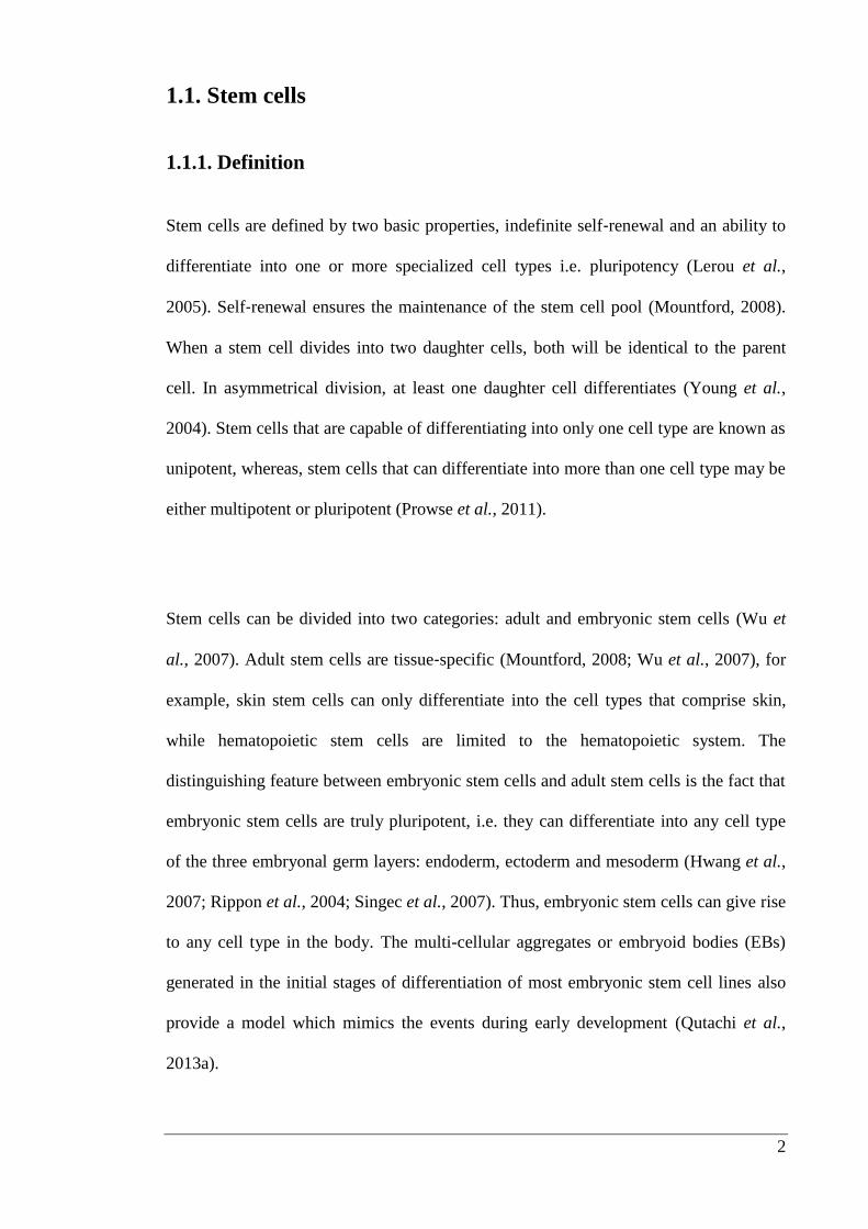
Stem cells are defined by two basic properties, indefinite self‐renewal and an ability to
differentiate into one or more specialized cell types i.e. pluripotency (Lerou et al.,
2005). Self‐renewal ensures the maintenance of the stem cell pool (Mountford, 2008).
When a stem cell divides into two daughter cells, both will be identical to the parent
cell. In asymmetrical division, at least one daughter cell differentiates (Young et al.,
2004). Stem cells that are capable of differentiating into only one cell type are known as
unipotent, whereas, stem cells that can differentiate into more than one cell type may be
either multipotent or pluripotent (Prowse et al., 2011).
Stem cells can be divided into two categories: adult and embryonic stem cells (Wu et
al., 2007). Adult stem cells are tissue‐specific (Mountford, 2008; Wu et al., 2007), for
example, skin stem cells can only differentiate into the cell types that comprise skin,
while hematopoietic stem cells are limited to the hematopoietic system. The
distinguishing feature between embryonic stem cells and adult stem cells is the fact that
embryonic stem cells are truly pluripotent, i.e. they can differentiate into any cell type
of the three embryonal germ layers: endoderm, ectoderm and mesoderm (Hwang et al.,
2007; Rippon et al., 2004; Singec et al., 2007). Thus, embryonic stem cells can give rise
to any cell type in the body. The multi-cellular aggregates or embryoid bodies (EBs)
generated in the initial stages of differentiation of most embryonic stem cell lines also
provide a model which mimics the events during early development (Qutachi et al.,
2013a).
3
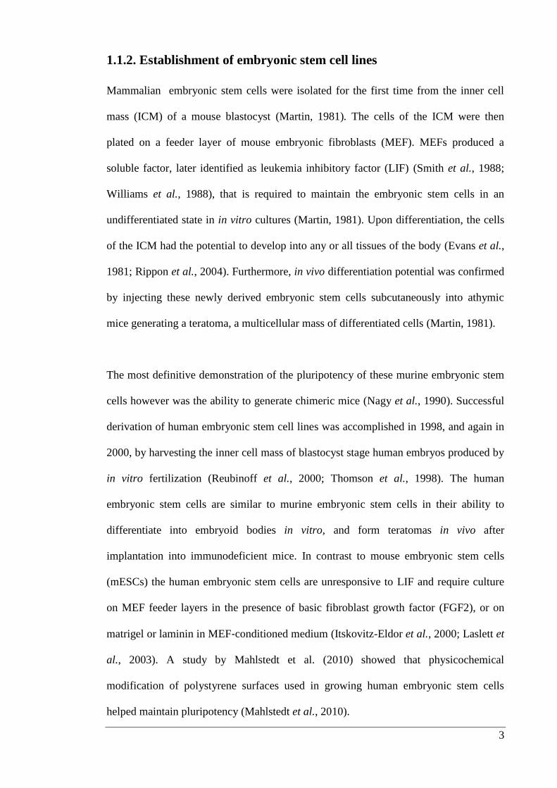
1.1.2. Establishment of embryonic stem cell lines
Mammalian embryonic stem cells were isolated for the first time from the inner cell
mass (ICM) of a mouse blastocyst (Martin, 1981). The cells of the ICM were then
plated on a feeder layer of mouse embryonic fibroblasts (MEF). MEFs produced a
soluble factor, later identified as leukemia inhibitory factor (LIF) (Smith et al., 1988;
Williams et al., 1988), that is required to maintain the embryonic stem cells in an
undifferentiated state in in vitro cultures (Martin, 1981). Upon differentiation, the cells
of the ICM had the potential to develop into any or all tissues of the body (Evans et al.,
1981; Rippon et al., 2004). Furthermore, in vivo differentiation potential was confirmed
by injecting these newly derived embryonic stem cells subcutaneously into athymic
mice generating a teratoma, a multicellular mass of differentiated cells (Martin, 1981).
The most definitive demonstration of the pluripotency of these murine embryonic stem
cells however was the ability to generate chimeric mice (Nagy et al., 1990). Successful
derivation of human embryonic stem cell lines was accomplished in 1998, and again in
2000, by harvesting the inner cell mass of blastocyst stage human embryos produced by
in vitro fertilization (Reubinoff et al., 2000; Thomson et al., 1998). The human
embryonic stem cells are similar to murine embryonic stem cells in their ability to
differentiate into embryoid bodies in vitro, and form teratomas in vivo after
implantation into immunodeficient mice. In contrast to mouse embryonic stem cells
(mESCs) the human embryonic stem cells are unresponsive to LIF and require culture
on MEF feeder layers in the presence of basic fibroblast growth factor (FGF2), or on
matrigel or laminin in MEF‐conditioned medium (Itskovitz-Eldor et al., 2000; Laslett et
al., 2003). A study by Mahlstedt et al. (2010) showed that physicochemical
modification of polystyrene surfaces used in growing human embryonic stem cells
helped maintain pluripotency (Mahlstedt et al., 2010).
4
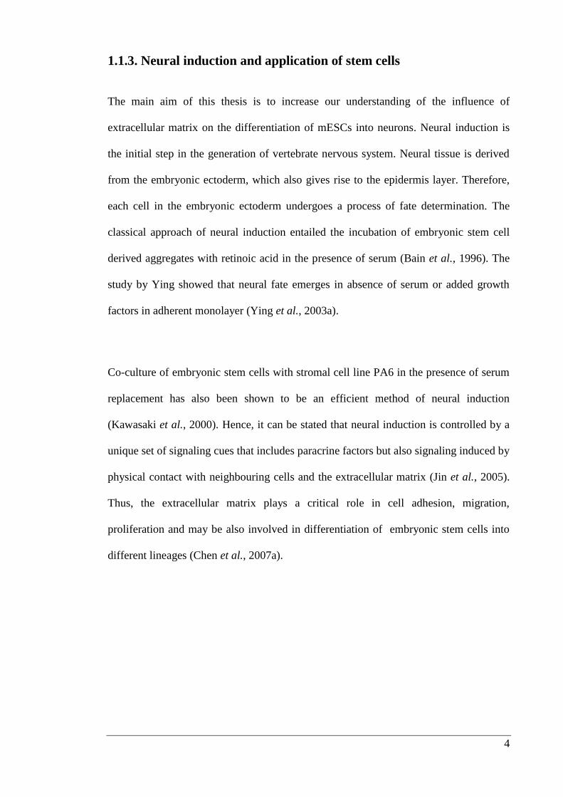
1.1.3. Neural induction and application of stem cells
The main aim of this thesis is to increase our understanding of the influence of
extracellular matrix on the differentiation of mESCs into neurons. Neural induction is
the initial step in the generation of vertebrate nervous system. Neural tissue is derived
from the embryonic ectoderm, which also gives rise to the epidermis layer. Therefore,
each cell in the embryonic ectoderm undergoes a process of fate determination. The
classical approach of neural induction entailed the incubation of embryonic stem cell
derived aggregates with retinoic acid in the presence of serum (Bain et al., 1996). The
study by Ying showed that neural fate emerges in absence of serum or added growth
factors in adherent monolayer (Ying et al., 2003a).
Co-culture of embryonic stem cells with stromal cell line PA6 in the presence of serum
replacement has also been shown to be an efficient method of neural induction
(Kawasaki et al., 2000). Hence, it can be stated that neural induction is controlled by a
unique set of signaling cues that includes paracrine factors but also signaling induced by
physical contact with neighbouring cells and the extracellular matrix (Jin et al., 2005).
Thus, the extracellular matrix plays a critical role in cell adhesion, migration,
proliferation and may be also involved in differentiation of embryonic stem cells into
different lineages (Chen et al., 2007a).
5
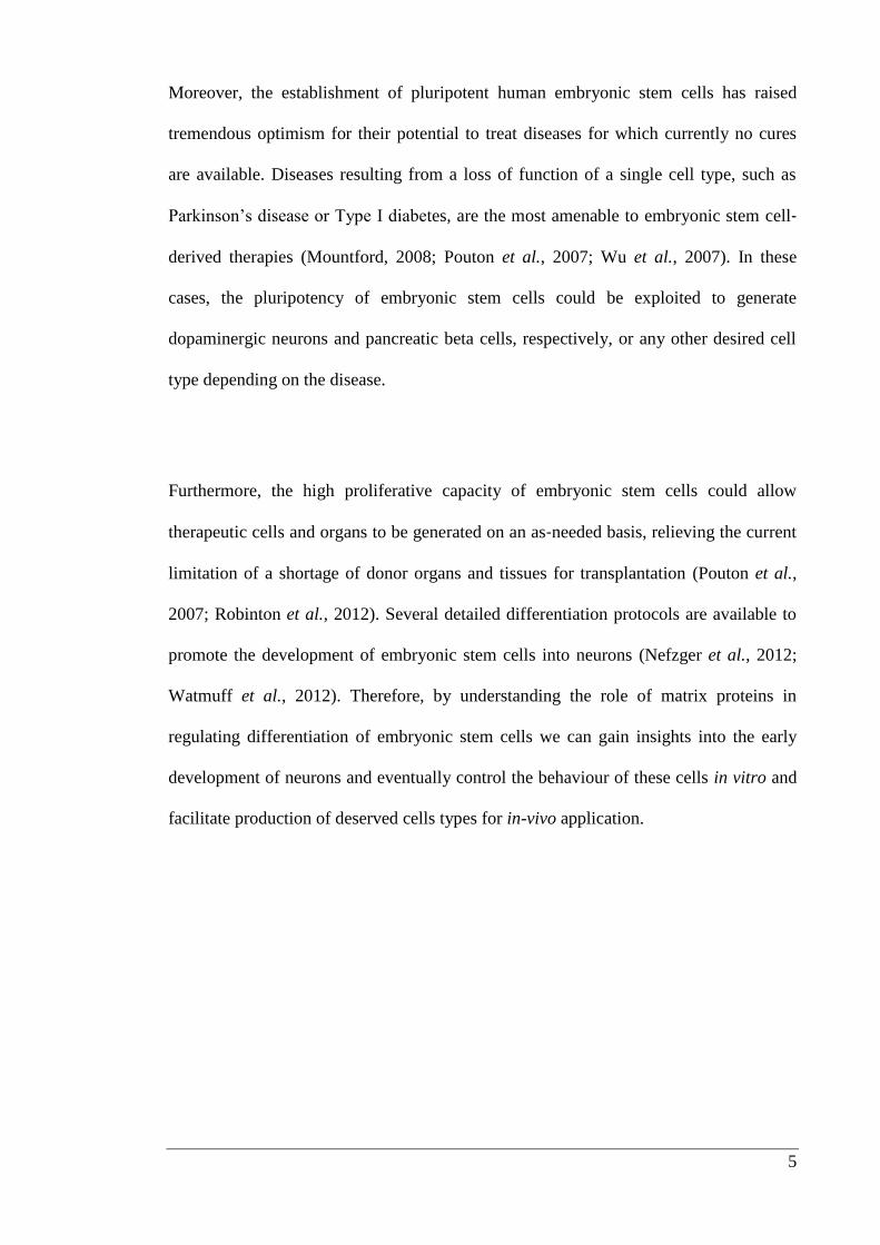
Moreover, the establishment of pluripotent human embryonic stem cells has raised
tremendous optimism for their potential to treat diseases for which currently no cures
are available. Diseases resulting from a loss of function of a single cell type, such as
Parkinson’s disease or Type I diabetes, are the most amenable to embryonic stem cell‐
derived therapies (Mountford, 2008; Pouton et al., 2007; Wu et al., 2007). In these
cases, the pluripotency of embryonic stem cells could be exploited to generate
dopaminergic neurons and pancreatic beta cells, respectively, or any other desired cell
type depending on the disease.
Furthermore, the high proliferative capacity of embryonic stem cells could allow
therapeutic cells and organs to be generated on an as‐needed basis, relieving the current
limitation of a shortage of donor organs and tissues for transplantation (Pouton et al.,
2007; Robinton et al., 2012). Several detailed differentiation protocols are available to
promote the development of embryonic stem cells into neurons (Nefzger et al., 2012;
Watmuff et al., 2012). Therefore, by understanding the role of matrix proteins in
regulating differentiation of embryonic stem cells we can gain insights into the early
development of neurons and eventually control the behaviour of these cells in vitro and
facilitate production of deserved cells types for in-vivo application.
6
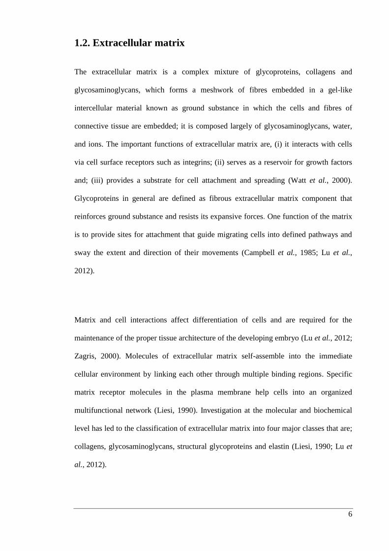
1.2. Extracellular matrix
The extracellular matrix is a complex mixture of glycoproteins, collagens and
glycosaminoglycans, which forms a meshwork of fibres embedded in a gel-like
intercellular material known as ground substance in which the cells and fibres of
connective tissue are embedded; it is composed largely of glycosaminoglycans, water,
and ions. The important functions of extracellular matrix are, (i) it interacts with cells
via cell surface receptors such as integrins; (ii) serves as a reservoir for growth factors
and; (iii) provides a substrate for cell attachment and spreading (Watt et al., 2000).
Glycoproteins in general are defined as fibrous extracellular matrix component that
reinforces ground substance and resists its expansive forces. One function of the matrix
is to provide sites for attachment that guide migrating cells into defined pathways and
sway the extent and direction of their movements (Campbell et al., 1985; Lu et al.,
2012).
Matrix and cell interactions affect differentiation of cells and are required for the
maintenance of the proper tissue architecture of the developing embryo (Lu et al., 2012;
Zagris, 2000). Molecules of extracellular matrix self-assemble into the immediate
cellular environment by linking each other through multiple binding regions. Specific
matrix receptor molecules in the plasma membrane help cells into an organized
multifunctional network (Liesi, 1990). Investigation at the molecular and biochemical
level has led to the classification of extracellular matrix into four major classes that are;
collagens, glycosaminoglycans, structural glycoproteins and elastin (Liesi, 1990; Lu et
al., 2012).

7
1.2.1. Collagens
Collagen is one of the most abundant proteins in the animal kingdom, representing
approximately one third of all proteins in tissues (van der Rest et al., 1991). The
collagen family is a highly diverse group of proteins as shown in Table 1.1 (Gelse et al.,
2003; Olsen, 1995; Prockop et al., 1995). The most abundant collagens form
extracellular fibrils or network-like structures, but the others fulfil a variety of
biological functions such as cell proliferation, migration and apoptosis (Gelse et al.,
2003). Structure of type IV collagen is discussed in detail in section 1.4.1..

8
Table 1.1: Shows various types of collagen as they belong to the major collagen
families.
Types Tissue distribution
Fibril-forming collagens
Type I, II, III, V and XI
Bone, dermis, tendons, ligaments,
cartilage and skin
Basement membrane collagens
Type IV
Basement membranes
Microfibrillar collagens
Type VI
Widespread: dermis, cartilage, placenta,
lungs and vessel wall
Anchoring fibrils
Type VII
Skin, dermal– epidermal junctions; oral
mucosa and cervix
Hexagonal network forming collagens
Type VIII and X
Endothelial cells, descemet’s membrane
and hypertrophic cartilage
Fibril-associated collagens
Type IX, XII, XIV, XX and XXI
Blood vessel wall, cartilage, vitreous
humor, cornea, dermis, tendon, vessel
wall, placenta, lungs and liver
Transmembrane domain
Type XIII and XVII
Epidermis, hair follicle, endomysium,
intestine, chondrocytes, lungs and liver
Multiplexins
Type XV, XVI and XVIII
Smooth muscle cells, kidney, pancreas
amnion and keratinocytes
9
1.2.2. Glycoproteins
Laminin, fibronectin, tenascin and entactin are glycoproteins, which are involved in cell
and tissue adhesion processes at specific developmental stages. These are multi-domain
molecules which interact with one another, with other extracellular matrix molecules
and with cell surfaces and are thought to be responsible for arranging the collagen,
proteoglycan and cells into an ordered structure (Aumailley et al., 2005; Bernardes et
al., 2009; Orsini et al., 2012; Yurchenco et al., 1993). The structures of fibronectin and
laminin are discussed in detail in 1.4.2. and 1.4.3..
1.2.3. Glycosaminoglycans and proteoglycans
Glycosaminoglycans such as hyaluronate, chondroitin sulphate, dermatan sulphate,
keratan sulphate, heparan sulphate are components of extracellular matrix which have
been implicated in the control of cell proliferation, migration, differentiation and
maintenance of morphogenetic structures. They occur as large polymers of repeating
disaccharides and are covalently linked to protein to form the proteoglycans. Heparan
sulphate is ubiquitously present on the cell surface and in extracellular matrix including
basement membrane. It is synthesized as an alternating copolymer of hexuronic acid
and glucosamine (Whitelock et al., 2005). Heparan sulphate proteoglycans (HSPGs)
consist of heparan sulphate chains that interact with a variety of proteins such as
heparin-binding growth and differentiation factors, morphogens, extracellular matrix
components, protease inhibitors, protease, lipoprotein lipase and various pathogens
(Habuchi et al., 2000; Iozzo, 1998).
10
1.3. Integrins
Integrins are transmembrane proteins that mediate interaction between the cell and
extracellular matrix. Many matrix proteins in vertebrates are recognized by multiple
integrins: for example, at least 8 integrins bind fibronectin, and at least 5 bind laminin.
About 20 integrin heterodimers, made from one of 9 types of β subunits and one of 14
types of α subunits, have been identified (Powell et al., 1997; Watt, 2002). The
interaction of cells with extracellular matrix proteins is crucial for activation of various
biological processes, including cell adhesion, spreading, proliferation, differentiation,
apoptosis, gene induction, embryogenesis and wound healing (Figure 1.1 A) (Giancotti
et al., 1999; Hynes, 2002; Juliano, 2002; Lee et al., 2004). Interaction of extracellular
matrix proteins with cells is mediated by transmembrane receptors, of which the
integrins constitute the most important class (Bosman et al., 2003; Boudreau et al.,
1999b; Gumbiner, 1996).
These heterodimers transmit extracellular signals when in contact with matrix proteins,
by way of one or more potential signaling cascades involving focal adhesion kinase
(FAK), phosphatidylinositol 3-kinase (PI3K), nuclear factor (NF)-kB, and mitogen-
activated protein kinases (MAPK). However, we still cannot explain how these signals
facilitate adhesion and proliferation (Figure 1.1 B and C) (Cho et al., 2000; Daley et al.,
2008; Frost et al., 1999).
11
The complexity of the extracellular surroundings can be appreciated by examining the
temporal expression of patterns of extracellular matrix components and their
corresponding cell surface receptors in the developing central and peripheral nervous
systems. The expression of extracellular matrix proteins increases as neural progenitors
differentiate, migrate and their neuronal axons elongate, but expression begins to taper
off towards the end of the development program (Flaim et al., 2005).
Cell-cell or cell-extracellular matrix interactions play a crucial role in controlling stem
cell differentiation, neural development, axon outgrowth and synapse formation (Cooke
et al., 2010; Lathia et al., 2007). The extracellular matrix proteins have not been
extensively investigated for their influence on differentiation of mESCs into neurons,
whereas, their effect has been explored in detail on cells extracted from mouse and
chick brain. These experiments have shown that extracellular matrix provides an
external factor in guiding and encouraging conversion of cells into neurons (Lein et al.,
1996, Kowtha et al., 1998). The hypothesis is that different extracellular matrix proteins
help in controlling the differentiation of cells through their numerous domains,
carbohydrate moieties and isoform specific affinities for cell integrins (Kowtha et al.,
1998; Obremski et al., 1995; Westermann et al., 1989).
12
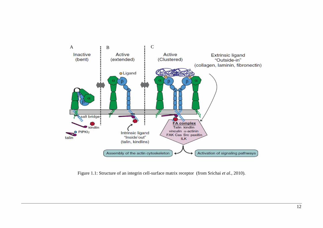
Figure 1.1: Structure of an integrin cell-surface matrix receptor (from Srichai et al., 2010).
A B C

13
The binding of an integrin to its ligand is dependent on extracellular divalent cations
(for example Ca2+
and Mg2+
), which bind to the integrins α subunit. The integrin then
binds with its ligand in the extracellular matrix which initiates the intracellular end of
the β subunit to bind to talin and α-actin (Figure 1.1 A). Intracellular attachment
proteins then aggregate at the cytoplasmic end of the integrin, enabling the integrin to
link with the cell’s actin filaments. Cells connect to the extracellular matrix at focal
adhesions, forming tight connections between the cell membrane and the extracellular
matrix as well as the cell membrane and the cytoskeleton (Figure 1.1 B). Focal
adhesions are assembled following the aggregation of signaling molecules and actin-
anchoring proteins. These proteins create the structural link, which enables stress fibers
to connect to the membrane and the integrins (Figure 1.1 C)
14
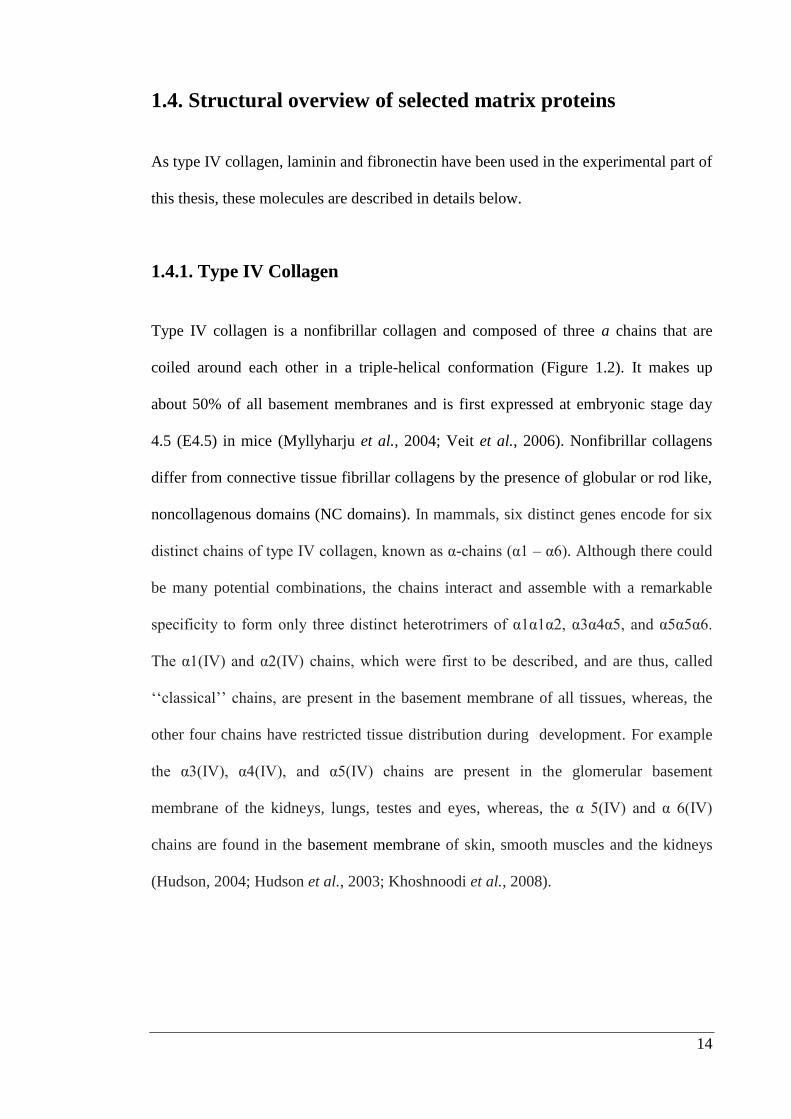
1.4. Structural overview of selected matrix proteins
As type IV collagen, laminin and fibronectin have been used in the experimental part of
this thesis, these molecules are described in details below.
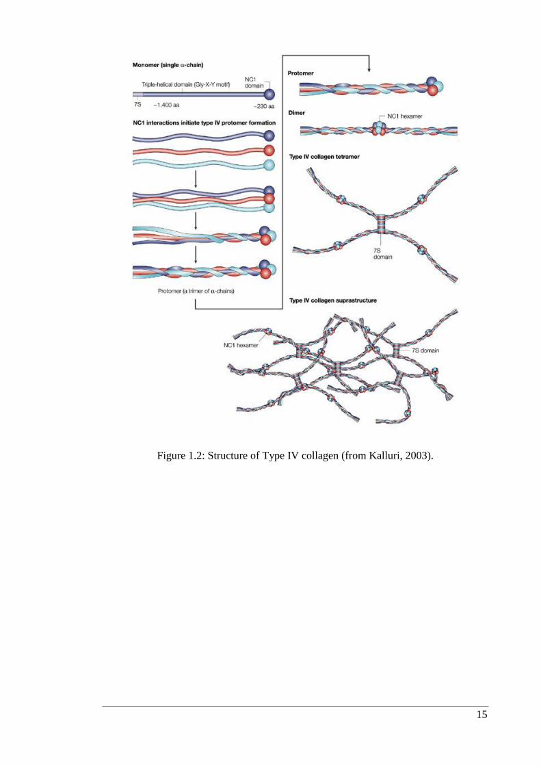
1.4.1. Type IV Collagen
Type IV collagen is a nonfibrillar collagen and composed of three a chains that are
coiled around each other in a triple-helical conformation (Figure 1.2). It makes up
about 50% of all basement membranes and is first expressed at embryonic stage day
4.5 (E4.5) in mice (Myllyharju et al., 2004; Veit et al., 2006). Nonfibrillar collagens
differ from connective tissue fibrillar collagens by the presence of globular or rod like,
noncollagenous domains (NC domains). In mammals, six distinct genes encode for six
distinct chains of type IV collagen, known as α-chains (α1 – α6). Although there could
be many potential combinations, the chains interact and assemble with a remarkable
specificity to form only three distinct heterotrimers of α1α1α2, α3α4α5, and α5α5α6.
The α1(IV) and α2(IV) chains, which were first to be described, and are thus, called
‘‘classical’’ chains, are present in the basement membrane of all tissues, whereas, the
other four chains have restricted tissue distribution during development. For example
the α3(IV), α4(IV), and α5(IV) chains are present in the glomerular basement
membrane of the kidneys, lungs, testes and eyes, whereas, the α 5(IV) and α 6(IV)
chains are found in the basement membrane of skin, smooth muscles and the kidneys
(Hudson, 2004; Hudson et al., 2003; Khoshnoodi et al., 2008).
15
Figure 1.2: Structure of Type IV collagen (from Kalluri, 2003).
16
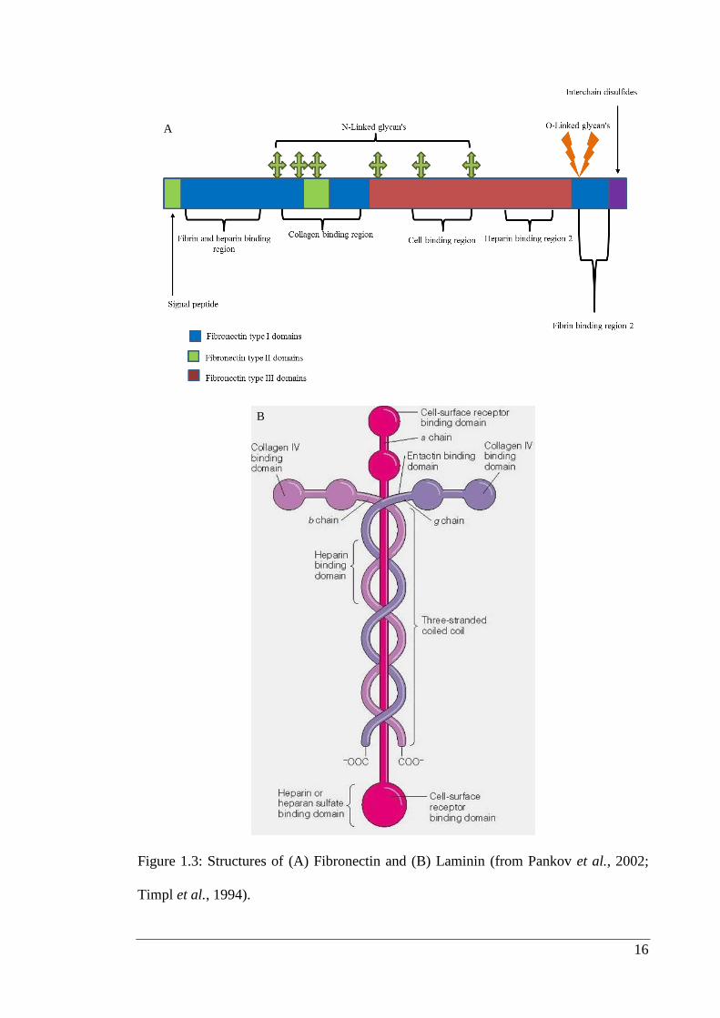
Figure 1.3: Structures of (A) Fibronectin and (B) Laminin (from Pankov et al., 2002;
Timpl et al., 1994).
B
A
17
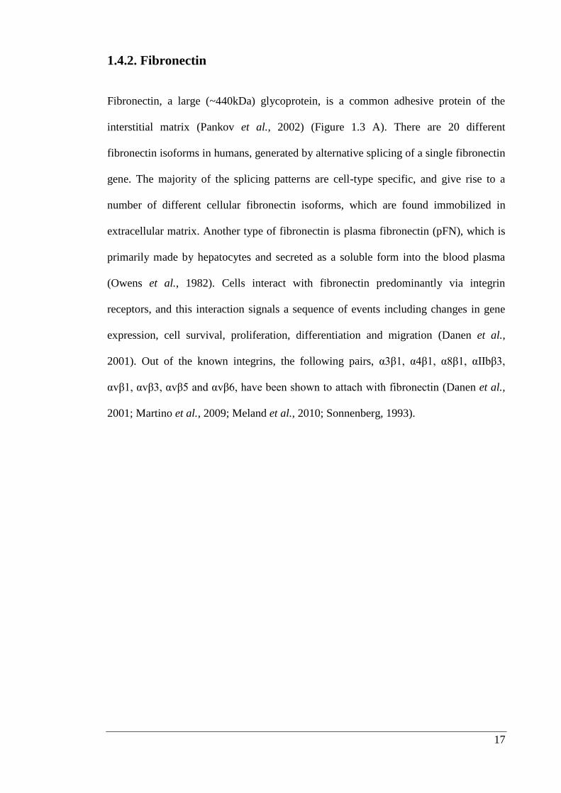
1.4.2. Fibronectin
Fibronectin, a large (~440kDa) glycoprotein, is a common adhesive protein of the
interstitial matrix (Pankov et al., 2002) (Figure 1.3 A). There are 20 different
fibronectin isoforms in humans, generated by alternative splicing of a single fibronectin
gene. The majority of the splicing patterns are cell-type specific, and give rise to a
number of different cellular fibronectin isoforms, which are found immobilized in
extracellular matrix. Another type of fibronectin is plasma fibronectin (pFN), which is
primarily made by hepatocytes and secreted as a soluble form into the blood plasma
(Owens et al., 1982). Cells interact with fibronectin predominantly via integrin
receptors, and this interaction signals a sequence of events including changes in gene
expression, cell survival, proliferation, differentiation and migration (Danen et al.,
2001). Out of the known integrins, the following pairs, α3β1, α4β1, α8β1, αΙΙbβ3,
αvβ1, αvβ3, αvβ5 and αvβ6, have been shown to attach with fibronectin (Danen et al.,
2001; Martino et al., 2009; Meland et al., 2010; Sonnenberg, 1993).
18
1.4.3. Laminin
Laminin is one of the major components of basement membrane that has a molecular
weight of Mr - 850,000. It is a multidomained, cross-shaped glycoprotein that is
structured in meshwork of basement membranes (Figure 1.3 B), such as epithelial
lining, adjacent blood vessels, nerves and underlying pial sheaths of the brain. It has
been reported to occur in sites other than basement membranes during early stages of
development and is localized to specific types of neurons in the central nervous system
(CNS) during both embryonic and adult stages (Zagris, 2000).
Laminin protein is composed of three different polypeptide chains, termed alpha, beta
and gamma. At present, 5 alpha, 3 beta, and 3 gamma chains are known for mouse and
human laminin (Yurchenco et al., 1993). All chains are glycosylated and few chains
have been shown to have glycosaminoglycan side chains. The first laminin isoform was
discovered 26 years ago and additional isoforms were simultaneously being identified
under different nomenclatures (Timpl et al., 1994). 18 years ago, a unifying
nomenclature for the trimers was introduced. It found widespread acceptance because it
was rational and allowed simplified transfer of information (Burgeson et al., 1994).
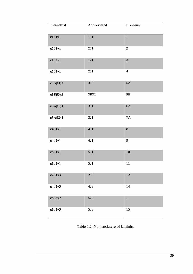
A more simplified trimer nomenclature was proposed using the numbers associated
with each subunit. Both names are provided in Table 1.2 (Aumailley et al., 2005). In
the nomenclature presented, a simple change makes it easier to identify the laminin
isoforms and name newly identified molecules. In the past, laminin trimers were
designated as laminin 1 to 15 in their order of discovery, with no direct relationship to
chain numbers (Table 1.2). Each trimer is composed of three genetically distinct chains
termed α, β, and γ. According to the previous nomenclature a trimer could either be
19
identified by the Arabic numeral (e.g. 10), or by its chains (e.g. α5β1γ1) (Burgeson et
al., 1994).
Laminin trimers were named exclusively on the basis of chain composition, either
according to Table 1.2, or without Greek letters; for example 211 for α2β1γ1, can
equally be used with no loss of information therefore, 211 is more informative than
laminin 4 and obviates the need to memorize the chain composition (Aumailley et al.,
2005). In the developing CNS, the presence of laminin has been observed in regions
where tracts are growing. Laminin is not only associated with the basement membrane
of developing Pia Mater and blood vessels, but is also found in small, punctuate
particles in the extracellular matrix where tract formation occurs. It also plays a role in
the migration of neural crest cells into the bowel and stimulates their early
differentiation into neurons (Luckenbill-Edds, 1997).
20
Standard Abbreviated Previous
α1β1γ1 111 1
α2β1γ1 211 2
α1β2γ1 121 3
α2β2γ1 221 4
α3Aβ3γ2 332 5A
α3Bβ3γ2 3B32 5B
α3Aβ1γ1 311 6A
α3Aβ2γ1 321 7A
α4β1γ1 411 8
α4β2γ1 421 9
α5β1γ1 511 10
α5β2γ1 521 11
α2β1γ3 213 12
α4β2γ3 423 14
α5β2γ2 522 -
α5β2γ3 523 15
Table 1.2: Nomenclature of laminin.
21
1.5. Role of extracellular matrix proteins in neurogenesis
Neurogenesis is the process of generation and differentiation of neurons from neural
stem cells. The mammalian CNS is generated from neural stem cells residing in the
deepest layer of the neural tube facing the ventricular lumen This area is called the
ventricular zone. Neural stem cells are defined as cells that exhibit the capacity to
differentiate into one of three cell types of the CNS (namely neurons, astrocytes and
oligodendrocytes) and also have the capacity to generate sufficient numbers of cells to
form an adult brain by self-renewal. Neurogenesis occurs predominantly during
embryonic stages is completed before birth in most parts of the mammalian CNS
(Hantaz-Ambroise et al., 1987; McKay, 1997).
The initial research was done on investigating the effect of extracellular matrix proteins
like laminin, fibronectin and collagens to purified neurons obtained from chick embryo.
Plastic substrata coated with fibronectin and laminin were tested by culturing
dissociated neurons from embryonic chick dorsal root sympathetic ganglia (peripheral
neurons), spinal cord and retina (CNS neurons). Laminin was able to attach and induce
extension of neurites from both central and peripheral neurons, whereas, fibronectin
only had these effects on peripheral neurons (Hall et al., 2008).
Neurite length, number of neurites initiated, and extent of neurite branching on
fibronectin and laminin coated surfaces were evaluated and compared with similar
measurements of neuronal responses to poly-L-lysine-treated plastic. Poly-L-lysine
provided an adhesive surface for neurite elongation, but fibronectin and laminin
appeared to promote more rapid neurite elongation, indicating that glycoproteins play
an important role during specific developmental stages (Rogers et al., 1983).
22
Carbonetto and Cochard (Carbonetto et al., 1987) took neurons from embryonic chick
sympathetic ganglia and dorsal root ganglia (DRG) to evaluate whether extracellular
matrix adhesive proteins like fibronectin, collagens (types I, III, IV) and laminin are
important determinants of nerve regeneration. Their findings showed that neurons grew
poorly on substrata containing glycosaminoglycans and in culture medium lacking
nerve growth factor. DRG neurons extended nerve fibers only on laminin and not on
fibronectin, collagen or polylysine. The neuron-like rat pheochromocytoma cell line,
(PC12) was utilised for studying interaction with extracellular matrix components
including laminin, type IV collagen and fibronectin. A cell attachment assay showed
that PC12 cells adhere readily to laminin or type IV collagen but poorly to fibronectin
(Tomaselli et al., 1987).
DRG neurons of purified chick embryo were used to study cooperative actions of
extracellular matrix proteins laminin and fibronectin in the presence of nerve growth
factor. Only 20% of cells survived in presence of nerve growth factors whereas, in
presence of laminin or fibronectin survival increased to 80% and was accompanied by
extensive neurite outgrowth. The same authors reported that increased levels of
potassium in the presence of laminin helped in survival and neurite outgrowth,
suggesting cooperative action between matrix and nerve growth factor (Millaruelo et
al., 1988).
23
Neuronal precursor cells present in chick DRG were excised to study conditions
required for initial differentiation and long-term survival. Cells isolated at embryonic
day 6 (E6) failed to attach when plated on surfaces coated with polyornithine alone or
with bovine serum albumin (BSA). When the same cells plated onto surfaces coated
with a combination of polyornithine with laminin or fibronectin, attachment as well as
development of neurons was observed (Ernsberger et al., 1988).
As a variety of intrinsic and environmental factors are known to direct growth of axons
to their peripheral targets, localization of a variety of extracellular matrix molecules
within the chick trigeminal mesenchyme has been studied using indirect
immunofluorescence. The study suggested that laminin is implicated in the guidance of
trigeminal peripheral axons and it might be produced in localized patches by peripheral
nervous system (PNS) components (Moody et al., 1989). Mesenchymal cells of the
chick tail bud were used to evaluate the role of different extracellular matrix proteins
on differentiation. Griffith and Sanders showed that laminin and laminin-containing
substrata (Matrigel) were found to promote the differentiation of neural crest
derivatives (neurons and melanocytes) and neuroepithelial cells; type I collagen
promoted both myogenesis and chondrogenesis; while type IV collagen promoted
myogenesis only (Griffith et al., 1991).
24
Sarthy (1993) investigated whether type IV collagen, which is associated with laminin
in basement membrane, is expressed by neuronal and non-neural cells during retinal
development. Immunostaining of type IV collagen showed it was expressed at
embryonic day 12 (E12) in the lens, embryonic (hyaloid) blood vessels and internal
limiting membrane (ILM) of the retina. At embryonic day 17 (E17), immunostaining
was reduced in the ILM, whereas, the lens and hyaloid were strongly stained and type
IV collagen was barely detected in the ILM of postnatal retinas. These workers
concluded that high levels of type IV collagens were present during the early
development when most of the axonal growth occured (Sarthy, 1993).
Therefore, it can be summarized from the above findings that matrix proteins not only
serve as attachment molecules but also helped in survival and development of neurons
isolated from chick embryo and out of tested matrix proteins laminin turned out to be
best as compared to fibronectin, type IV collagen and chemically defined substrates
like polyornthine and polylysine. Studies in early 1990 shifted with specific focus on
laminin extracted from plant and animal sources. The following findings also supported
the fact that laminin presence was most beneficial as compared to other extracellular
matrix proteins.
25
Lectin concanavalin A and laminin-like substrates extracted respectively from plants
and leech CNS. The extracted substrates were used to evaluate their effect on Retzius
neurons extracted from the leech. Neurons showed broad flat growth cones and thick
bundles of process on lectin concanavalin A whereas, on laminin-like substrate, fine
straight processes with numerous branches were observed thus, suggesting that nature
of the growth substrate can also influence morphology of neurons (Ross et al., 1988).
Dean et al. (1990) studied the role of carbohydrate moieties of laminin in cell migration
and neurite elongation. To test the hypothesis, lectins were used to block carbohydrate
moieties of laminin. Wheat germ agglutinin or Griffonia simplicifolia agglutinin
blocked the binding of the neuron-like rat PC12 onto plates coated with laminin. When
concanavalin A was used, cell binding was not affected but neurite outgrowth was
prevented. Non-glycosylated and glycosylated laminins were purified by
immunoaffinity from tunicamycin treated cultures of a mouse embryonal carcinoma
derived cell line. When PC12 cells were plated on surfaces coated with purified non-
glycosylated laminin, no effect was noted on binding of cells but neurite outgrowth was
impaired when compared to glycosylated laminin. Thus, it can be inferred that once
cells bound to laminin the carbohydrate residues of that glycoprotein must be available
to enable the cells to spread or to extend neurite processes (Dean et al., 1990).
26
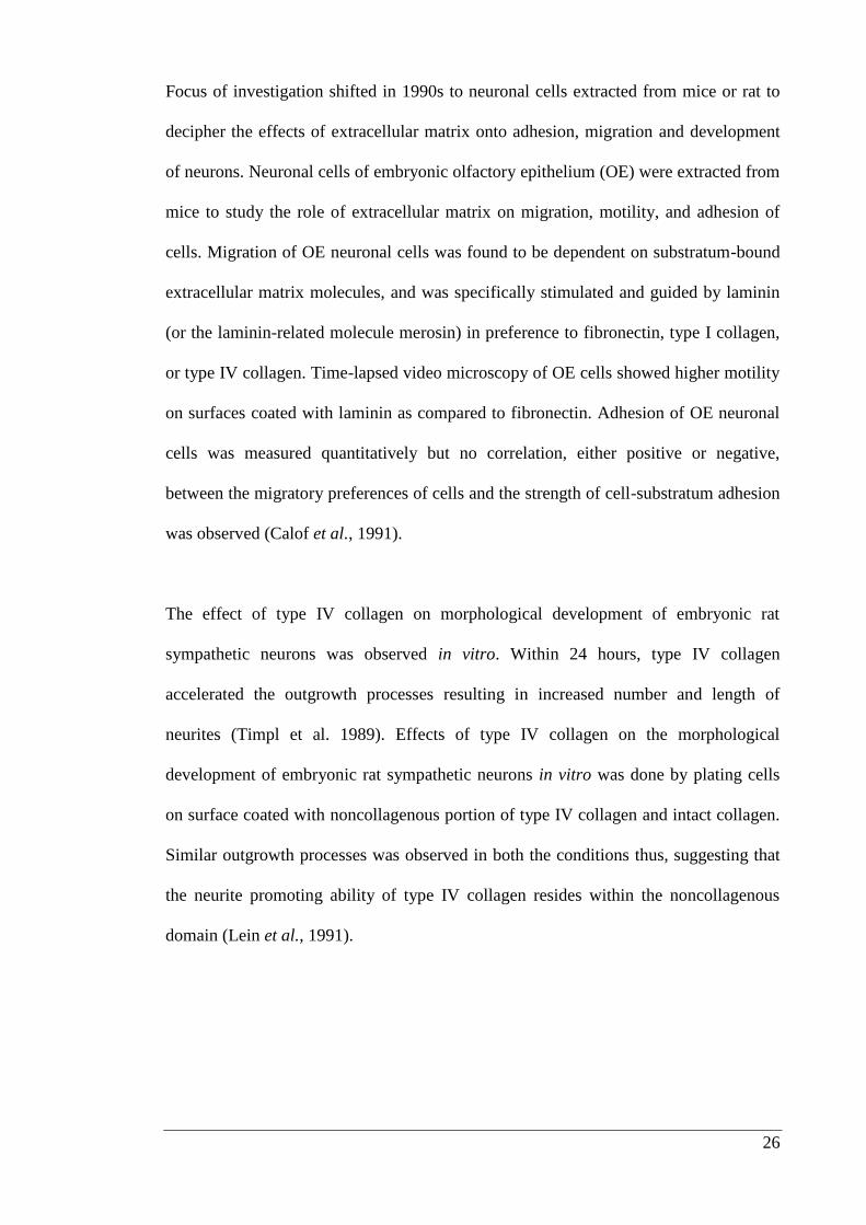
Focus of investigation shifted in 1990s to neuronal cells extracted from mice or rat to
decipher the effects of extracellular matrix onto adhesion, migration and development
of neurons. Neuronal cells of embryonic olfactory epithelium (OE) were extracted from
mice to study the role of extracellular matrix on migration, motility, and adhesion of
cells. Migration of OE neuronal cells was found to be dependent on substratum-bound
extracellular matrix molecules, and was specifically stimulated and guided by laminin
(or the laminin-related molecule merosin) in preference to fibronectin, type I collagen,
or type IV collagen. Time-lapsed video microscopy of OE cells showed higher motility
on surfaces coated with laminin as compared to fibronectin. Adhesion of OE neuronal
cells was measured quantitatively but no correlation, either positive or negative,
between the migratory preferences of cells and the strength of cell-substratum adhesion
was observed (Calof et al., 1991).
The effect of type IV collagen on morphological development of embryonic rat
sympathetic neurons was observed in vitro. Within 24 hours, type IV collagen
accelerated the outgrowth processes resulting in increased number and length of
neurites (Timpl et al. 1989). Effects of type IV collagen on the morphological
development of embryonic rat sympathetic neurons in vitro was done by plating cells
on surface coated with noncollagenous portion of type IV collagen and intact collagen.
Similar outgrowth processes was observed in both the conditions thus, suggesting that
the neurite promoting ability of type IV collagen resides within the noncollagenous
domain (Lein et al., 1991).
27
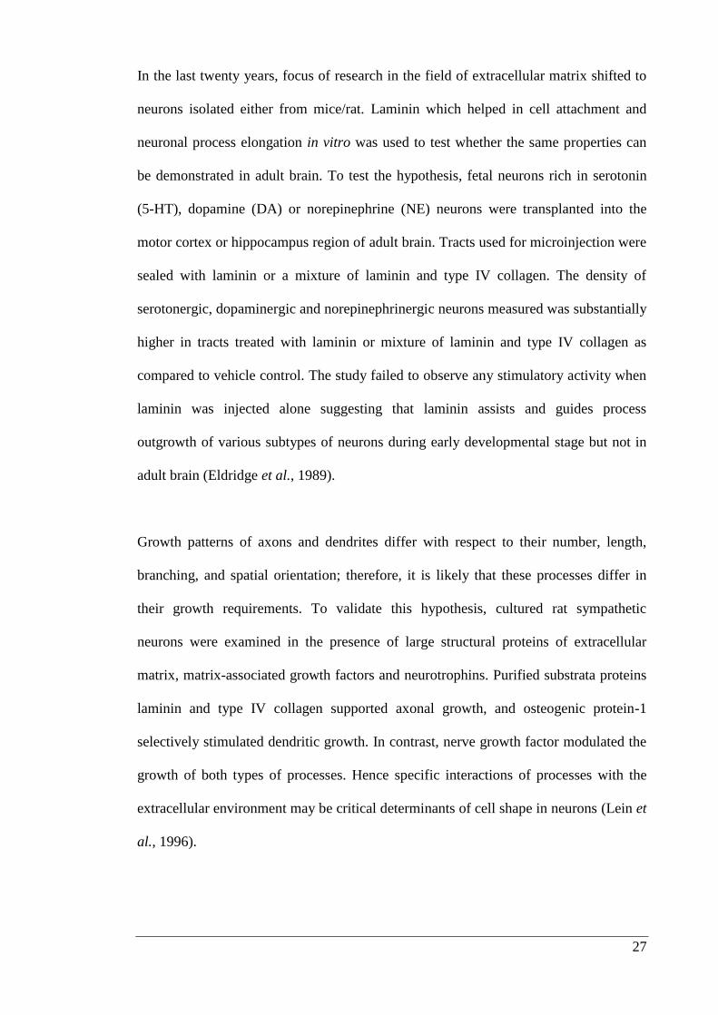
In the last twenty years, focus of research in the field of extracellular matrix shifted to
neurons isolated either from mice/rat. Laminin which helped in cell attachment and
neuronal process elongation in vitro was used to test whether the same properties can
be demonstrated in adult brain. To test the hypothesis, fetal neurons rich in serotonin
(5-HT), dopamine (DA) or norepinephrine (NE) neurons were transplanted into the
motor cortex or hippocampus region of adult brain. Tracts used for microinjection were
sealed with laminin or a mixture of laminin and type IV collagen. The density of
serotonergic, dopaminergic and norepinephrinergic neurons measured was substantially
higher in tracts treated with laminin or mixture of laminin and type IV collagen as
compared to vehicle control. The study failed to observe any stimulatory activity when
laminin was injected alone suggesting that laminin assists and guides process
outgrowth of various subtypes of neurons during early developmental stage but not in
adult brain (Eldridge et al., 1989).
Growth patterns of axons and dendrites differ with respect to their number, length,
branching, and spatial orientation; therefore, it is likely that these processes differ in
their growth requirements. To validate this hypothesis, cultured rat sympathetic
neurons were examined in the presence of large structural proteins of extracellular
matrix, matrix-associated growth factors and neurotrophins. Purified substrata proteins
laminin and type IV collagen supported axonal growth, and osteogenic protein-1
selectively stimulated dendritic growth. In contrast, nerve growth factor modulated the
growth of both types of processes. Hence specific interactions of processes with the
extracellular environment may be critical determinants of cell shape in neurons (Lein et
al., 1996).
28
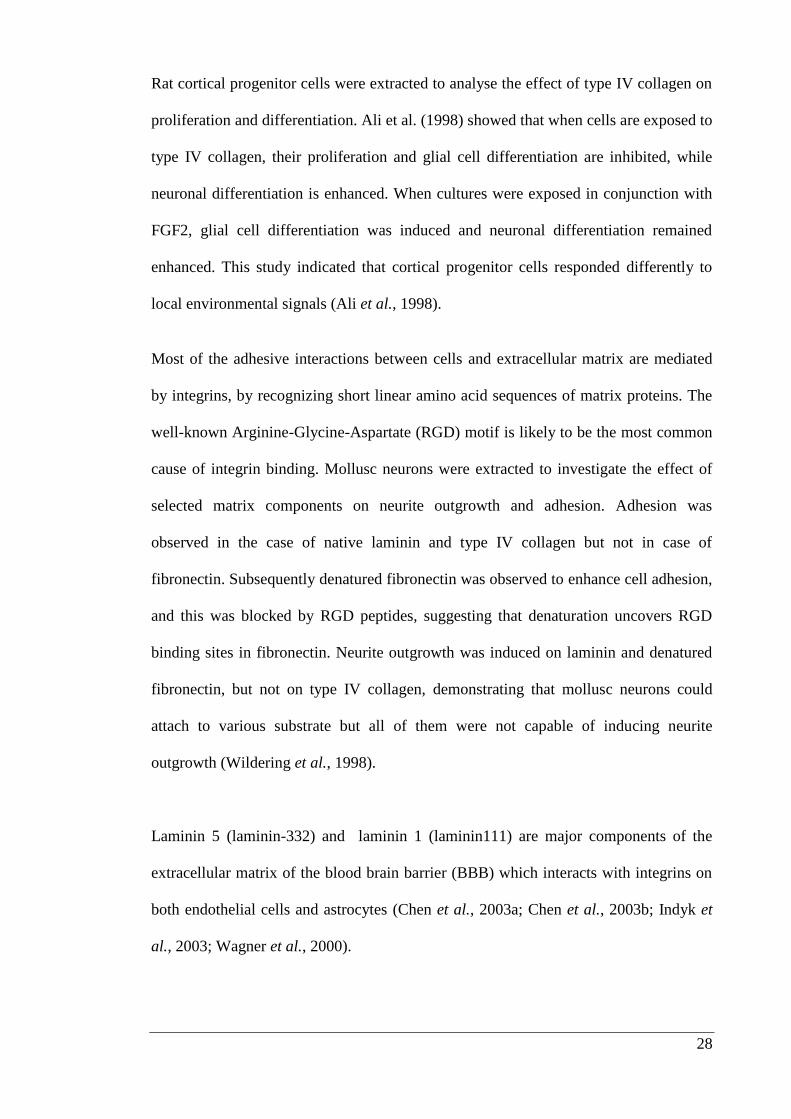
Rat cortical progenitor cells were extracted to analyse the effect of type IV collagen on
proliferation and differentiation. Ali et al. (1998) showed that when cells are exposed to
type IV collagen, their proliferation and glial cell differentiation are inhibited, while
neuronal differentiation is enhanced. When cultures were exposed in conjunction with
FGF2, glial cell differentiation was induced and neuronal differentiation remained
enhanced. This study indicated that cortical progenitor cells responded differently to
local environmental signals (Ali et al., 1998).
Most of the adhesive interactions between cells and extracellular matrix are mediated
by integrins, by recognizing short linear amino acid sequences of matrix proteins. The
well-known Arginine-Glycine-Aspartate (RGD) motif is likely to be the most common
cause of integrin binding. Mollusc neurons were extracted to investigate the effect of
selected matrix components on neurite outgrowth and adhesion. Adhesion was
observed in the case of native laminin and type IV collagen but not in case of
fibronectin. Subsequently denatured fibronectin was observed to enhance cell adhesion,
and this was blocked by RGD peptides, suggesting that denaturation uncovers RGD
binding sites in fibronectin. Neurite outgrowth was induced on laminin and denatured
fibronectin, but not on type IV collagen, demonstrating that mollusc neurons could
attach to various substrate but all of them were not capable of inducing neurite
outgrowth (Wildering et al., 1998).
Laminin 5 (laminin-332) and laminin 1 (laminin111) are major components of the
extracellular matrix of the blood brain barrier (BBB) which interacts with integrins on
both endothelial cells and astrocytes (Chen et al., 2003a; Chen et al., 2003b; Indyk et
al., 2003; Wagner et al., 2000).
29
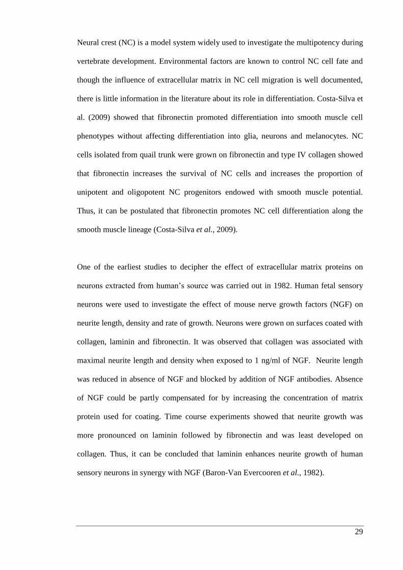
Neural crest (NC) is a model system widely used to investigate the multipotency during
vertebrate development. Environmental factors are known to control NC cell fate and
though the influence of extracellular matrix in NC cell migration is well documented,
there is little information in the literature about its role in differentiation. Costa-Silva et
al. (2009) showed that fibronectin promoted differentiation into smooth muscle cell
phenotypes without affecting differentiation into glia, neurons and melanocytes. NC
cells isolated from quail trunk were grown on fibronectin and type IV collagen showed
that fibronectin increases the survival of NC cells and increases the proportion of
unipotent and oligopotent NC progenitors endowed with smooth muscle potential.
Thus, it can be postulated that fibronectin promotes NC cell differentiation along the
smooth muscle lineage (Costa-Silva et al., 2009).
One of the earliest studies to decipher the effect of extracellular matrix proteins on
neurons extracted from human’s source was carried out in 1982. Human fetal sensory
neurons were used to investigate the effect of mouse nerve growth factors (NGF) on
neurite length, density and rate of growth. Neurons were grown on surfaces coated with
collagen, laminin and fibronectin. It was observed that collagen was associated with
maximal neurite length and density when exposed to 1 ng/ml of NGF. Neurite length
was reduced in absence of NGF and blocked by addition of NGF antibodies. Absence
of NGF could be partly compensated for by increasing the concentration of matrix
protein used for coating. Time course experiments showed that neurite growth was
more pronounced on laminin followed by fibronectin and was least developed on
collagen. Thus, it can be concluded that laminin enhances neurite growth of human
sensory neurons in synergy with NGF (Baron-Van Evercooren et al., 1982).
30
A study by Hirose et al. (1993) was reported to understand the effect of extracellular
matrix proteins on embryonic stem cells derived from mouse. Matrix proteins such as
fibronectin, laminin, heparan sulphate proteoglycan, type I collagen, type IV collagen,
and type VIII collagen were investigated for their neurite outgrowth potential by in
vitro assay of cholinergic neuronal cell lines and primary cultured neurons from
embryonic mouse brain. Findings showed that all the matrix proteins had high neurite
promoting activity but collagens only showed high neurite-promoting activity in
neurons from prenatal, not postnatal mouse brain. These workers postulated that
collagens only contribute to neurite extension of CNS neurons in the developing brain
(Hirose et al., 1993).
Rat embryos were used for extraction of DRG cells, which were seeded onto plates
coated with poly-L-lysine, laminin, poly-L-lysine combined with laminin, or type I
collagen. Phase-contrast microscopy was used to monitor cell survival and neurite
outgrowth. The results showed that DRG neurons grew slowly with high survival rate
on poly-L-lysine combined with laminin. However, those cells grown on type I
collagen were clustered with thicker and longer neurites showing their growth pattern
was influenced by presence or absence of particular substrata. It was concluded that
surfaces coated with poly-L-lysine and laminin were the best option for study of single
neurons (Guo et al., 2004).
31
Kohno et al. (2005) investigated the effect of laminin on organelle transport and its
relationship to neurite growth using dissociated mouse DRG neurons. A time-lapse
study demonstrated that many small-diameter branches were formed after the addition
of laminin. Addition of laminin resulted in a decrease in organelle movement in the
neurite shaft and growth cone which resulted in slow growth cone advancement. This
suggests that laminin inhibits the elongation of primary neurites, but promotes
branching and elongation of branches (Kohno et al., 2005).
In a study by Chen et al. (2007a) human skin fibroblast cells exposed to ascorbic acid
showed increased attachment to plastic due to enhanced collagen synthesis. In contrast,
inhibition of collagen synthesis by cis-hydroxyproline decreases the rate of attachment
of fibroblasts to the plastic surface and increased rate of their detachment by trypsin,
thus, showing that collagen helps in adhesion of cells onto at least one type of plastic
surface (Chen et al., 2007a).
There is considerable interest in the treatment of both peripheral and CNS disorders
with neural prosthetic implants, for this form of treatment to be successful effective
integration on implantation will be required. Research has been carried out in past few
years to understand the effect of extracellular matrix proteins such as fibronectin,
laminin and type IV collagen on adhesion and differentiation of mESCs. Embryonic
mouse neural stem cells (NSCs) isolated from E14 mice, were grown on plates coated
with laminin in presence of epidermal growth factor (EGF) and FGF2 in neurobasal
media (Balasubramaniyan et al., 2004).
32
After 7 days of in vitro cell cultures, 20% of the NSCs developed into morphologically
and biochemically fully mature neurons, with extensive dendrites and multiple synaptic
contacts. When these neurons were tested by electrophysiology experiments to assess
maturity on day 22, none of the neurons had developed the characteristics of a
functional neuron. These findings indicate that differentiation assessed by
morphological appearance does not correlate with electrophysiological maturation of
mouse NSCs into neurons. The processes of neurogenesis and subsequent maturation
may be regulated differently (Balasubramaniyan et al., 2004).
Extracellular matrix and cell-adhesive proteins like gelatin, type IV collagen, laminin
and polylysine, were immobilized on a polystyrene surface. A layer-by-layer technique
was used based on hydrophobic and electrostatic interactions between oppositely
charged macromolecules. The mESC line D3 was tested for adhesion and growth. The
cells grew best on surfaces coated with gelatin and type IV collagen assemblies. Effect
on growth of non-differentiated or differentiated cells was evaluated but no significant
differences were observed (Brynda et al., 2005).
Goetz et al. (2006) showed how environmental interactions alone can modify the
development of neurogenic precursor cells. They evaluated the influence of natural and
synthetic proteins including laminin, fibronectin, gelatin and poly-L-ornithine on
embryonic stem cell-neurogenesis. They found highest densities of neural precursors
when cells were grown in combination of laminin and poly-L-ornithine and reported
that more non-neural phenotypes were present on gelatin (a mixture of water-soluble
collagen components) and fibronectin (Goetz et al., 2006b).
33
Embryonic stem cell transplantation represents a potential means for the treatment of
degenerative diseases like Parkinson’s disease (Robinton et al., 2012; Watmuff et al.,
2012). A critical aspect of stem cell transplantation is to have appropriate embryonic
stem cell migration from the site of insertion to the mid brain. Surfaces coated with or
without type I collagen, type IV collagen, matrigel, fibronectin or laminin were tested
for generation of embryoid bodies from mESC lines. Among the matrix proteins tested,
type IV collagen showed maximum migration enhancing effect. In addition, pre-
treatment of undifferentiated or differentiated mESCs with type IV collagen resulted in
improved engraftment and growth after transplantation into the subcutaneous tissue of
nude mice (Li et al., 2010).
The influence of the above selected matrix proteins can be summarised as fibronectin
is crucial for embryogenesis (George et al., 1993) and is involved in the developing
nervous system (Reichardt et al., 1991). Fibronectin promotes both the survival and
migration of neural crest cells and migration of neurons to different cortical regions
(Henderson et al., 1997; Testaz et al., 2001). Neurite extension and synapse formation
in the developing brain is also known to be promoted by fibronectin (Einheber et al.,
1996; Sheppard et al., 1995). Laminin plays an important role in neural development
(Reichardt et al., 1991). Laminin 1 is known to promote cell survival and migration in
the developing nervous system (Perris et al., 2000) and both laminin 1 and 2 promote
neurite outgrowth (Colognato et al., 2000).
34
In the mature brain laminin 10 is found in the hippocampus and protects hippocampal
neurons from excitotoxic lesion. Indyk et al. (2003) reported that laminin 10 (laminin-
511) found in the mature brain hippocampus region protects hippocampal neurons from
excitotoxic lesions, and laminin α2 subunit is found in close association with dendritic
spines in the hippocampus, and may be involved in synaptogenesis (Indyk et al., 2003).
Laminin 5 is a major component of the extracellular matrix of the blood brain barrier,
where it can be found in the basement membrane of the blood vessels where it interacts
with integrins on both endothelial cells and astrocytes (Hagg et al., 1997; Wagner et
al., 2000). Type IV collagen plays crucial role in development and regeneration of the
PNS (Timpl et al., 1984).
Type IV collagen is reported to be synthesized by glial cells and is present in both fibre
tracts and ganglia of the PNS and it is one of important components of basement
membrane where it provides mechanical stability (Hirose et al., 1993; Olsen, 1995;
Tryggvason et al., 1993). Studies of extracellular environment from different tissues
and organisms have led to the identification of common signaling molecules including
growth factors, cell-cell signaling molecules, adhesion molecules and extracellular
matrix molecules. Combinations of these are likely to provide strategies to maintain
stem cell populations and enhance differentiation of cells into subtypes of neurons. In
addition, the extracellular matrix proteins influence the outgrowth of neuronal process.
The role of extracellular matrix proteins has been studied mainly in the PNS where they
have been shown to promote the migration of neural crest derivatives, the outgrowth
and guidance of neurites and the induction of proteins involved in neurotransmitter
metabolism (Frade et al., 1996; Luckenbill-Edds et al., 1995).
35
The ability of embryonic stem cells to generate neural cell types in vitro offers a
powerful tool to study how the cell-extracellular matrix interactions regulate neural
stem cell specification and lineage choice (Mruthyunjaya et al., 2010; Timpl et al.,
1994; Uemura et al., 2010). Findings have shown that the local microenvironment or
stem cell niche, regulates self-renewal and fate of stem cells in developing tissues or
organs (Fuchs et al., 2000; Ma et al., 2008). Within the niche, the extracellular matrix,
plays a critical role in regulating stem cell differentiation into different lineages, as well
as in cell migration and proliferation (Chen et al., 2007a). Therefore, based on the
above information the decision was made to investigate the influence of type IV
collagen, fibronectin and laminin on differentiation of mESCs.
36
1.6. Experimental outline
The experimental work of this thesis was divided into three chapters to understand the
influence of extracellular matrix protein on differentiation of mESCs.
1.6.1. Experimental study 1 (Chapter 2: Matrix proteins and their
influence on neural induction):
Extracellular matrix proteins as described in (Section 1.2) have history of biological
use, mainly to enhance adhesion of cells to plastic surfaces. They are now being
reported to play roles in proliferation and differentiation of stem cells (Divya et al.,
2007; Ma et al., 2008). It is generally understood that stem cells prefer to grow on a
biological rather than a synthetic surface thus, chapter 2 examines in vitro the influence
of extracellular matrix proteins on differentiation of mESCs into neurons.
Therefore, for this study, the surface of the tissue culture plates was coated with
extracellular matrix proteins and seeded with mESCs to understand their effects on cell
viability, proliferation and differentiation. Preliminary experiments determined the
concentration of selected matrix proteins for optimal coverage of the culture plate
surface. Immunolabeling was then carried out with βIII tubulin and nestin to
understand the effects of matrix proteins on neural induction and to determine if
differentiation of mESCs in to particular neuronal subtypes could be favoured by
particular components of the extracellular matrix.
37
1.6.2. Experimental study 2 (Chapter 3: Immobilization of growth
factors onto surface coated with extracellular matrix components):
Lmx1a is a key intrinsic determinant of midbrain dopaminergic neurons, expressed in
response to the ventralizing activity of sonic hedgehog (Shh) (Andersson et al., 2006).
FGF8b and FGF2 have also been reported to play crucial roles in generation of
midbrain dopaminergic neurons (Lee et al., 2000; Raye et al., 2007). Significantly,
when growth factors are present in immobilized form it reduces their biodegradation,
increasing their availability (Masters, 2011). Working on these principles decided to
investigate whether, the bioactivity of growth factors like FGF2, FGF8b and Shh could
be increased if they were immobilized onto combinations of selected matrix proteins,
thereby increasing differentiation of Lmx1a-eGFP mESCs into midbrain dopaminergic
neurons.
1.6.3. Experimental study 3 (Chapter 4: Identifying the signalling
pathways through which mESCs attach and differentiated on matrix
proteins found in basal lamina):
The attachment and binding of the extracellular matrix proteins to integrins initiates
intracellular signaling (Figure 1.1), which is essential for key functions, including,
migration, differentiation and proliferation. Integrins modulate cellular functions by
enabling cell-cell connections and cell-extracellular matrix connections. FAK has been
implicated to play a crucial role in activating the downstream signaling pathway such
as extracellular regulated kinase 1/2 (ERK1/2) MAPK (Forrest et al., 2009) and PI3K-
protein kinase B (PKB/Akt, hereafter called Akt) pathway (Parsons, 2003) (discussed
in sections 4.1.3.-4.1.8.).
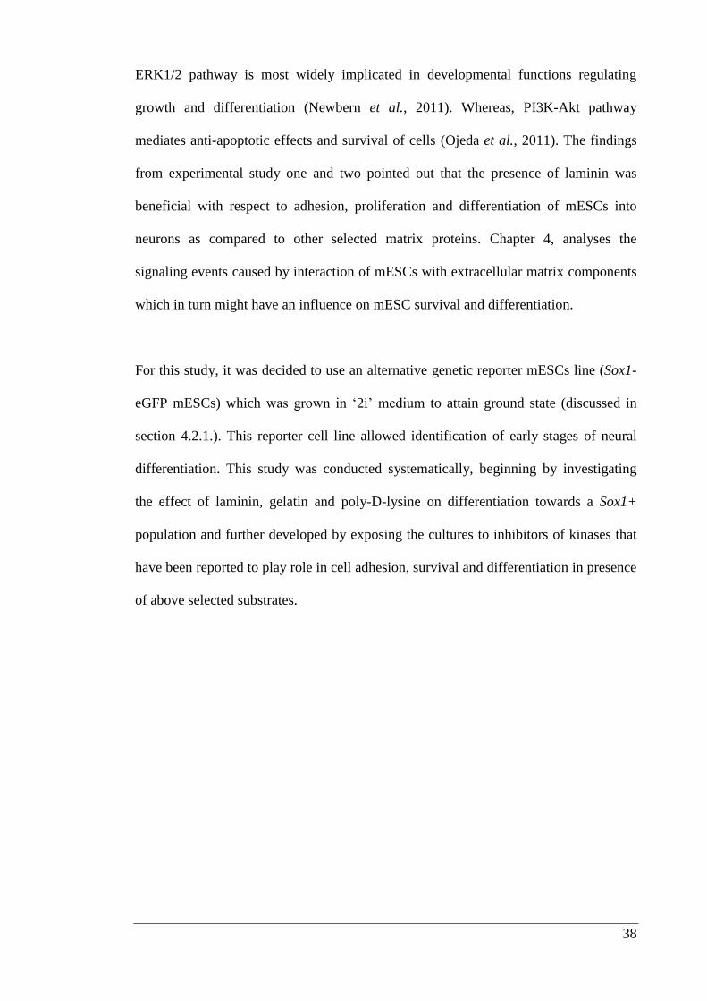
38
ERK1/2 pathway is most widely implicated in developmental functions regulating
growth and differentiation (Newbern et al., 2011). Whereas, PI3K-Akt pathway
mediates anti-apoptotic effects and survival of cells (Ojeda et al., 2011). The findings
from experimental study one and two pointed out that the presence of laminin was
beneficial with respect to adhesion, proliferation and differentiation of mESCs into
neurons as compared to other selected matrix proteins. Chapter 4, analyses the
signaling events caused by interaction of mESCs with extracellular matrix components
which in turn might have an influence on mESC survival and differentiation.
For this study, it was decided to use an alternative genetic reporter mESCs line (Sox1-
eGFP mESCs) which was grown in ‘2i’ medium to attain ground state (discussed in
section 4.2.1.). This reporter cell line allowed identification of early stages of neural
differentiation. This study was conducted systematically, beginning by investigating
the effect of laminin, gelatin and poly-D-lysine on differentiation towards a Sox1+
population and further developed by exposing the cultures to inhibitors of kinases that
have been reported to play role in cell adhesion, survival and differentiation in presence
of above selected substrates.

39
Chapter 2 Matrix proteins and their influence on
neural induction
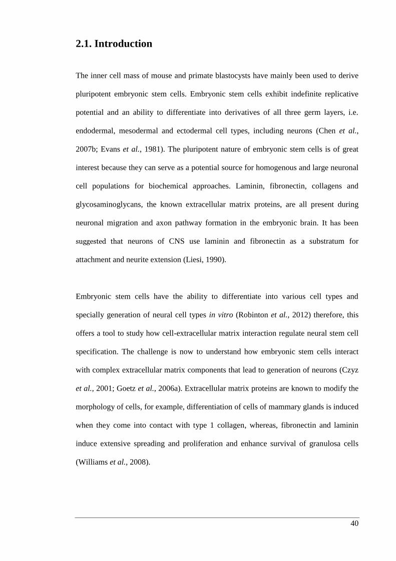
40
2.1. Introduction
The inner cell mass of mouse and primate blastocysts have mainly been used to derive
pluripotent embryonic stem cells. Embryonic stem cells exhibit indefinite replicative
potential and an ability to differentiate into derivatives of all three germ layers, i.e.
endodermal, mesodermal and ectodermal cell types, including neurons (Chen et al.,
2007b; Evans et al., 1981). The pluripotent nature of embryonic stem cells is of great
interest because they can serve as a potential source for homogenous and large neuronal
cell populations for biochemical approaches. Laminin, fibronectin, collagens and
glycosaminoglycans, the known extracellular matrix proteins, are all present during
neuronal migration and axon pathway formation in the embryonic brain. It has been
suggested that neurons of CNS use laminin and fibronectin as a substratum for
attachment and neurite extension (Liesi, 1990).
Embryonic stem cells have the ability to differentiate into various cell types and
specially generation of neural cell types in vitro (Robinton et al., 2012) therefore, this
offers a tool to study how cell-extracellular matrix interaction regulate neural stem cell
specification. The challenge is now to understand how embryonic stem cells interact
with complex extracellular matrix components that lead to generation of neurons (Czyz
et al., 2001; Goetz et al., 2006a). Extracellular matrix proteins are known to modify the
morphology of cells, for example, differentiation of cells of mammary glands is induced
when they come into contact with type 1 collagen, whereas, fibronectin and laminin
induce extensive spreading and proliferation and enhance survival of granulosa cells
(Williams et al., 2008).
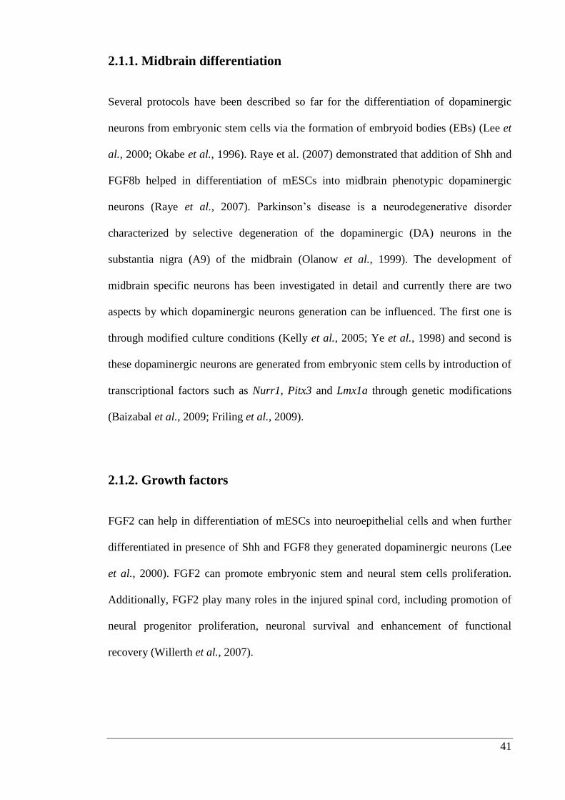
41
2.1.1. Midbrain differentiation
Several protocols have been described so far for the differentiation of dopaminergic
neurons from embryonic stem cells via the formation of embryoid bodies (EBs) (Lee et
al., 2000; Okabe et al., 1996). Raye et al. (2007) demonstrated that addition of Shh and
FGF8b helped in differentiation of mESCs into midbrain phenotypic dopaminergic
neurons (Raye et al., 2007). Parkinson’s disease is a neurodegenerative disorder
characterized by selective degeneration of the dopaminergic (DA) neurons in the
substantia nigra (A9) of the midbrain (Olanow et al., 1999). The development of
midbrain specific neurons has been investigated in detail and currently there are two
aspects by which dopaminergic neurons generation can be influenced. The first one is
through modified culture conditions (Kelly et al., 2005; Ye et al., 1998) and second is
these dopaminergic neurons are generated from embryonic stem cells by introduction of
transcriptional factors such as Nurr1, Pitx3 and Lmx1a through genetic modifications
(Baizabal et al., 2009; Friling et al., 2009).
2.1.2. Growth factors
FGF2 can help in differentiation of mESCs into neuroepithelial cells and when further
differentiated in presence of Shh and FGF8 they generated dopaminergic neurons (Lee
et al., 2000). FGF2 can promote embryonic stem and neural stem cells proliferation.
Additionally, FGF2 play many roles in the injured spinal cord, including promotion of
neural progenitor proliferation, neuronal survival and enhancement of functional
recovery (Willerth et al., 2007).
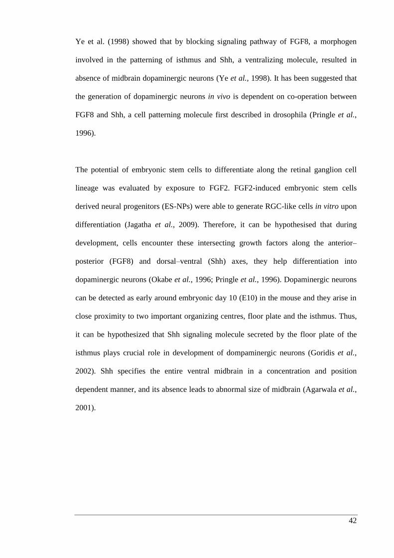
42
Ye et al. (1998) showed that by blocking signaling pathway of FGF8, a morphogen
involved in the patterning of isthmus and Shh, a ventralizing molecule, resulted in
absence of midbrain dopaminergic neurons (Ye et al., 1998). It has been suggested that
the generation of dopaminergic neurons in vivo is dependent on co-operation between
FGF8 and Shh, a cell patterning molecule first described in drosophila (Pringle et al.,
1996).
The potential of embryonic stem cells to differentiate along the retinal ganglion cell
lineage was evaluated by exposure to FGF2. FGF2-induced embryonic stem cells
derived neural progenitors (ES-NPs) were able to generate RGC-like cells in vitro upon
differentiation (Jagatha et al., 2009). Therefore, it can be hypothesised that during
development, cells encounter these intersecting growth factors along the anterior–
posterior (FGF8) and dorsal–ventral (Shh) axes, they help differentiation into
dopaminergic neurons (Okabe et al., 1996; Pringle et al., 1996). Dopaminergic neurons
can be detected as early around embryonic day 10 (E10) in the mouse and they arise in
close proximity to two important organizing centres, floor plate and the isthmus. Thus,
it can be hypothesized that Shh signaling molecule secreted by the floor plate of the
isthmus plays crucial role in development of dompaminergic neurons (Goridis et al.,
2002). Shh specifies the entire ventral midbrain in a concentration and position
dependent manner, and its absence leads to abnormal size of midbrain (Agarwala et al.,
2001).
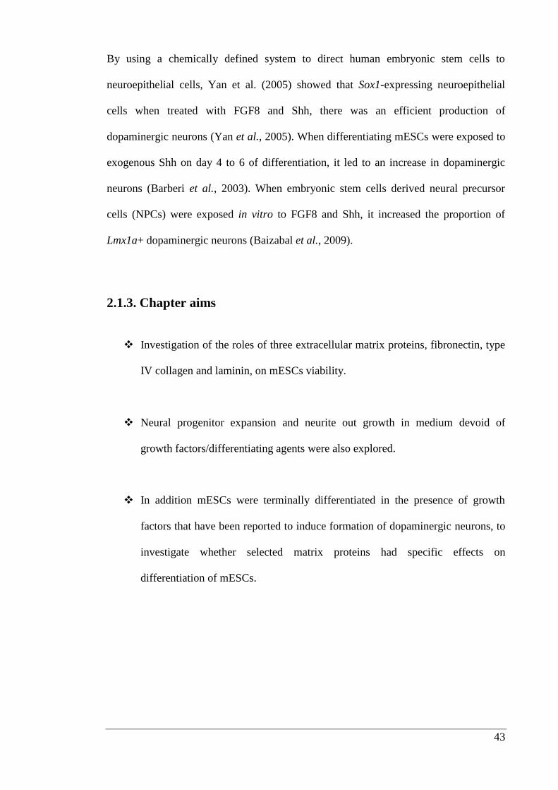
43
By using a chemically defined system to direct human embryonic stem cells to
neuroepithelial cells, Yan et al. (2005) showed that Sox1-expressing neuroepithelial
cells when treated with FGF8 and Shh, there was an efficient production of
dopaminergic neurons (Yan et al., 2005). When differentiating mESCs were exposed to
exogenous Shh on day 4 to 6 of differentiation, it led to an increase in dopaminergic
neurons (Barberi et al., 2003). When embryonic stem cells derived neural precursor
cells (NPCs) were exposed in vitro to FGF8 and Shh, it increased the proportion of
Lmx1a+ dopaminergic neurons (Baizabal et al., 2009).
2.1.3. Chapter aims
Investigation of the roles of three extracellular matrix proteins, fibronectin, type
IV collagen and laminin, on mESCs viability.
Neural progenitor expansion and neurite out growth in medium devoid of
growth factors/differentiating agents were also explored.
In addition mESCs were terminally differentiated in the presence of growth
factors that have been reported to induce formation of dopaminergic neurons, to
investigate whether selected matrix proteins had specific effects on
differentiation of mESCs.
44
2.2. Materials and Methods
2.2.1. Maintenance of mESCs
E14Tg2a, a sub clone of the E14 line derived from the inbred mouse strain 129/Ola was
used. E14Tg2a cells were procured from the American cell culture type (ATCC) and
had undergone 8 passages. The cell line was routinely cultured in Dulbecco's Modified
Eagle's Medium (Invitrogen, Australia) supplemented with 10% embryonic stem cell
qualified fetal calf serum (Invitrogen, Australia), 2 mM L-glutamine (Invitrogen,
Australia), 0.1 mM β-mercaptoethanol (Sigma-Aldrich, Australia) and 103 Units/ml
murine LIF (Millipore, Massachusetts) at 37°C with 5% CO2. Medium was changed
every 48 hours until cultures approached 75-80% confluence and were subsequently
passaged. Flasks, dishes and wells were pre-incubated with 0.1% gelatin solution in 1X
phosphate buffered saline (1X PBS) with final concentration of 2.7 mM potassium
chloride, 137 mM sodium chloride, 10 mM sodium phosphate and 1.76 mM potassium
phosphate (for at least 15 min on hot plate set at 37oC prior to use, to coat the surface of
plasticware with a gelatin film.
For passaging, medium was aspirated and cells were washed once with sterile 1X PBS.
Accutase (Sigma-Aldrich, Australia) was added at 1 ml per 7.5 cm2 surface area to give
a thin film over the cells and the cultures were incubated for 5 min at 37°C. Culture
medium containing serum was added to stop the enzymatic action. Cells were pelleted
in a conical tube (200g for 5 minutes). After re-suspension in fresh culture medium
(with supplements) mESCs were counted using a haemocytometer and seeded at 2 x 10⁴
cells/cm2 onto plates (surface coated with 0.1% v/v gelatin).
45
For long term storage, embryonic stem cells were frozen down in cryo-vials (Corning,
Australia) in a mixture of 50% fetal calf serum, 40% culture medium and 10% dimethyl
sulfoxide (DMSO) (Sigma, Australia) in an isopropanol filled “Mr Frosty” (Nalgene
labware, New York) for at least 90 minutes (at -80°C) and then transferred to liquid
nitrogen.
2.2.2. Neural differentiation
For neural induction an adherent monolayer approach was used (Figure 2.1). mESCs
were seeded on gelatin-coated tissue culture plates/flask at 4.5 x 10³ cells/cm
2 in normal
mESCs maintenance medium. 24 hours later, cells were washed once with 1X PBS and
switched to serum free N2B27 medium for 5 days. N2B27 medium is a 1:1 mixture of
Neurobasal medium supplemented with N2: DMEM/F12 medium supplemented with
B27, insulin and bovine serum albumin V. On day 5, adherent cells were dissociated to
produce a single cell suspension with Accutase solution (as described under
“Maintenance of mESCs” in section 2.2.1) and seeded at 5x10³
cells/cm2 onto surfaces
coated with selected matrix proteins including laminin (5 µg/cm2, Invitrogen,
Australia), fibronectin (5 µg/cm2, BD Bioscience, Australia) and type IV collagen (1
µg/cm2, BD Bioscience, Australia) in N2B27 supplemented with morphogens 20 ng/ml
FGF2 (R&D Systems, USA), 20 ng/ml heparin sulphate (Sigma-Aldrich, Australia),
100 ng/ml FGF8b (R&D Systems, USA) and 500 ng/ml Shh (R&D Systems, USA).
After 3 days (on day 8), the medium was replenished with fresh N2B27 supplemented
with morphogens. On day 6, after replating (day 11), cells were terminally
differentiated in N2B27 medium supplemented with 10 ng/ml glial-derived
neurotrophic factor (GDNF) (R&D Systems, USA) and 200 μM ascorbic acid (Sigma-
Aldrich, Australia) for protection of neurons and terminal differentiation. During this
phase the medium was changed every other day until day 21.
46
Figure 2.1: Diagrammatic overview of neural differentiation
2.2.3. In-cell Western assay
As no information was available on the mass of each matrix protein required to attain
uniform coating of individual well, a simple experiment on coating efficiency was
performed. The assay was carried out by coating wells of ninety-six well plates with
selected matrix proteins at 1, 5 and 10 µg/cm2. Each experiment was performed in
quadruplicate, including negative controls which were not exposed to primary
antibodies. Proteins coated surfaces were fixed by incubation with solution of 4%
paraformaldehyde in 1X PBS (w/v) for 30 minutes at room temperature. For
permeabilization, 100 µl of 0.1% TritonX-100 (Sigma-Aldrich, Australia) in 1X PBS
was added to each well for 30 minutes. Wells coated with matrix proteins were blocked
with 100 µl of blocking buffer (Li-cor Biotechnology, USA) for 90 minutes at room
temperature. Mouse anti-laminin (Sigma-Aldrich, Australia), rabbit anti-fibronectin
(Millipore, Australia) and mouse anti-type IV collagen (Abcam, UK) were diluted
1:200 in blocking buffer and incubated at 4ºC with volume of 50 µl per well.
47
Unbound primary antibody was removed by washing wells (5 times) with 200 µl 0.1%
Tween20 (Sigma-Aldrich, Australia) in 1X PBS with shaking. The secondary antibody,
IRDye 800CW donkey anti-mouse and IRDye 680LT donkey anti-rabbit (Li-cor
Biotechnology, USA) were diluted 1:1000 in blocking buffer. Unbound secondary
antibody was removed by washing each well (5 minutes) with 0.1% Tween20 (Sigma-
Aldrich, Australia) in 1X PBS (with gentle shaking) 5 times. Wells were scanned for
emission at 700 nm and 800 nm with the Odyssey Infrared Imaging system (ODY-
18090) (Li-cor Biotechnology, USA). Values from the 700 nm and 800 nm channel
corresponded to the amount of primary antibody bound to the wells. Control wells not
exposed to primary antibodies was used for background compensation. The background
fluorescence values at 700 nm and 800 nm were averaged and subtracted from the
respective values of wells incubated with primary antibodies. Coating efficiency was
estimated by comparing the integrated fluorescence intensity of individual wells
measured using the Odyssey.
2.2.4. Microculture Tetrazolium (MTT) Assay
The CellTiter 96® AQueous One Solution (Promega, Australia) was used to determine
the effects of different selected matrix protein concentrations on cell viablity and
proliferation. The MTS tetrazolium compound (Owen’s reagent) was reduced
enzymatically by cells into a soluble coloured formazan product. This conversion is
accomplished by NADPH or NADH-dependent reductase enzymes in metabolically
active cells. Ninety-six well plates were coated with selected matrix proteins (in
triplicate) at 1, 5 and 10 µg/cm2.
48
mESCs were seeded at a density of 5000 cells/well in presence of serum free N2B27
medium. A time course experiment was carried out and numbers of viable cells were
measured every 24 hours up to 96 hours after seeding onto assay plates. 20 μl of
CellTiter 96® AQueous One Solution Reagent was dispensed into each well of the
ninety-six well assay plate and incubated for four hours at 37oC. Absorbance was read
at 490 nm using a plate reader. Wells coated with selected concentrations of matrix in
presence of N2B27, but without mESCs served as negative control. Values at
wavelength 490 nm absorbance values were averaged and subtracted with absorbance
of negative control. The absorbance value/fraction of gelatin (Y axis) was plotted versus
concentration of the selected matrix proteins (X axis).
2.2.5. Immunocytochemistry
All incubation steps took place at room temperature unless stated otherwise. For fixing,
cells were incubated for 30 minutes in 4% paraformaldehyde. Subsequently, cells were
incubated with 1X PBS containing 0.5% TritonX-100 (Sigma-Aldrich, Australia) for
another 30 minutes. Non-specific antibody binding was blocked with 5% (w/v) bovine
serum albumin (BSA) (Sigma-Aldrich, Australia) in 1X PBS for one hour at room
temperature, this was followed by overnight exposure (at 4°C) with primary antibodies
either mouse-anti-β III tubulin (1:1000) (Sigma-Aldrich, Australia), mouse anti-nestin
(1:100) (Millipore, Australia) and rabbit-anti-tyrosine hydroxylase (1:200) (Millipore,
Australia) diluted in 1X PBS (containing 1% BSA). After 12 hours of incubation cells
were washed 4 times with 1X PBS containing 1% BSA, for 5 minutes each, then
incubated with secondary antibodies: donkey anti-mouse Alexa Fluor 488 (1:1000)
(Invitrogen, Australia) and Texas Red®-X donkey anti-rabbit 595 (1:1000) (Invitrogen,
49
Australia) for 120 minutes. Finally, cells were washed 4 times with 1X PBS containing
1% BSA for 5 minutes each and then covered with 1X PBS for imaging.
2.2.6. Imaging and counting of βIII tubulin, Nestin and tyrosine
hydroxylase immunopositive cells
Images were taken with a CoolSNAPFX camera (Photometrics, Arizona) attached to an
Eclipse TE-2000E microscope (Nikon, Japan). To quantify the number of βIII tubulin,
Nestin and tyrosine hydroxylase positive cells, images were captured using a 10 times
objective. Four sites/well were analysed by imaging up and down, left and right hand
corners respectively of each well, in duplicate from three separate experiments. βIII
tubulin, nestin and tyrosine hydroxylase positive cells were then counted manually (see
Figure 2.11.A for typical field of view and counts).
2.2.7. Quantification of neurite outgrowth
Neurite outgrowth was analysed using the NIH ImageJ analysis software using a
NeuronJ plugin for the multi-platform free image-processing program (Meijering et al.,
2004). Neurite tracer processes pairs of neuronal and nuclear marker images to obtain
skeletons of neuronal extensions and masks of neuronal nuclei. Neurites were quantified
from images of neurons immunostained for βIII tubulin. A neurite was defined as a
process having longer than the width of one cell body terminating in a growth cone, and
neurites were recorded using trace function. Total neurite length was measured and
divided by the number of neurons observed in the field.
50
For these analyses, each captured image was identified as a sampling unit and four
sites/well were analysed by imaging up and down, left and right hand corners
respectively of each well, in duplicate, from three separate experiments (Figure 2.8. B
typical example of outgrowth).
2.3.8. Statistical analysis
All results are presented as mean ± standard error of the mean (SEM) of at least three
separate experiments. Statistical analyses were performed on raw data using PRISM
v5.00 (GraphPad Software, USA). Data were analysed by Student’s t test or one-way
analysis of variance or two-way analysis of variance (ANOVA) with post-hoc
Bonferroni's test. In all cases, P < 0.05 was considered to be significant.
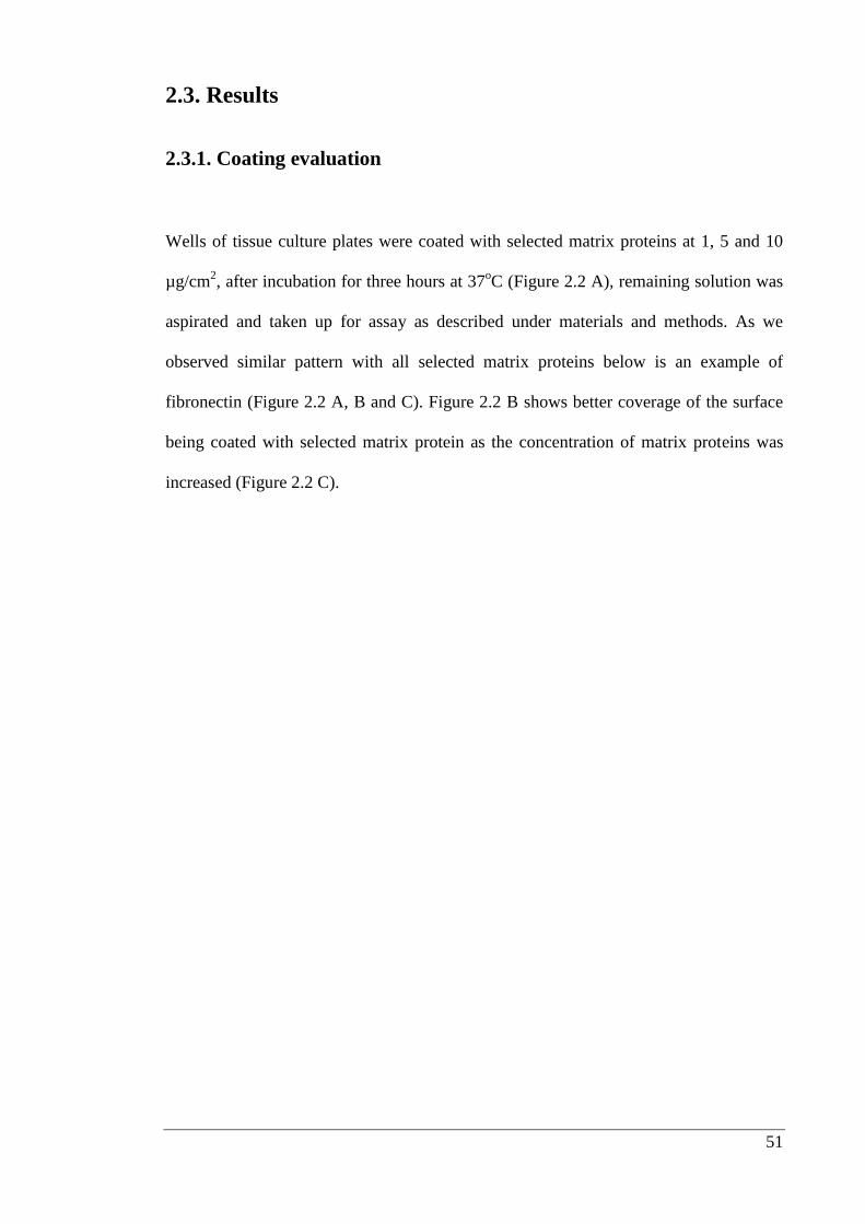
51
2.3. Results
2.3.1. Coating evaluation
Wells of tissue culture plates were coated with selected matrix proteins at 1, 5 and 10
µg/cm2, after incubation for three hours at 37
oC (Figure 2.2 A), remaining solution was
aspirated and taken up for assay as described under materials and methods. As we
observed similar pattern with all selected matrix proteins below is an example of
fibronectin (Figure 2.2 A, B and C). Figure 2.2 B shows better coverage of the surface
being coated with selected matrix protein as the concentration of matrix proteins was
increased (Figure 2.2 C).
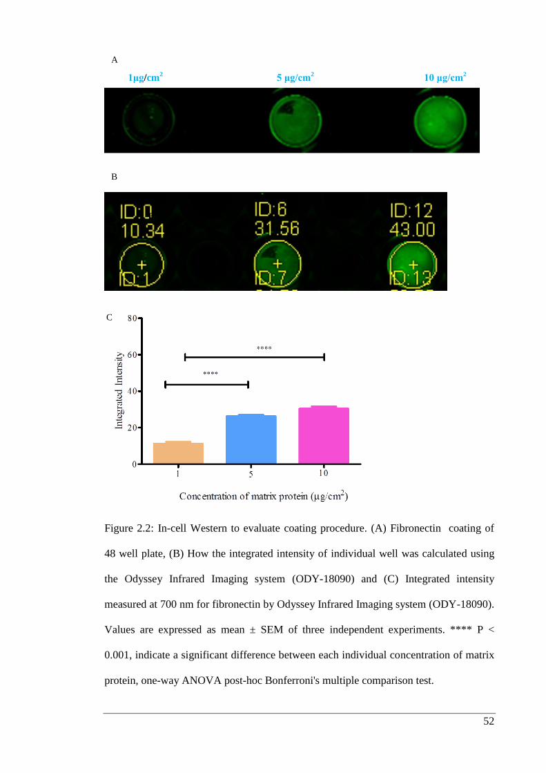
52
\\
Figure 2.2: In-cell Western to evaluate coating procedure. (A) Fibronectin coating of
48 well plate, (B) How the integrated intensity of individual well was calculated using
the Odyssey Infrared Imaging system (ODY-18090) and (C) Integrated intensity
measured at 700 nm for fibronectin by Odyssey Infrared Imaging system (ODY-18090).
Values are expressed as mean ± SEM of three independent experiments. **** P <
0.001, indicate a significant difference between each individual concentration of matrix
protein, one-way ANOVA post-hoc Bonferroni's multiple comparison test.
B
1μg/cm2 5 μg/cm
2 10 μg/cm
2
A
B
C
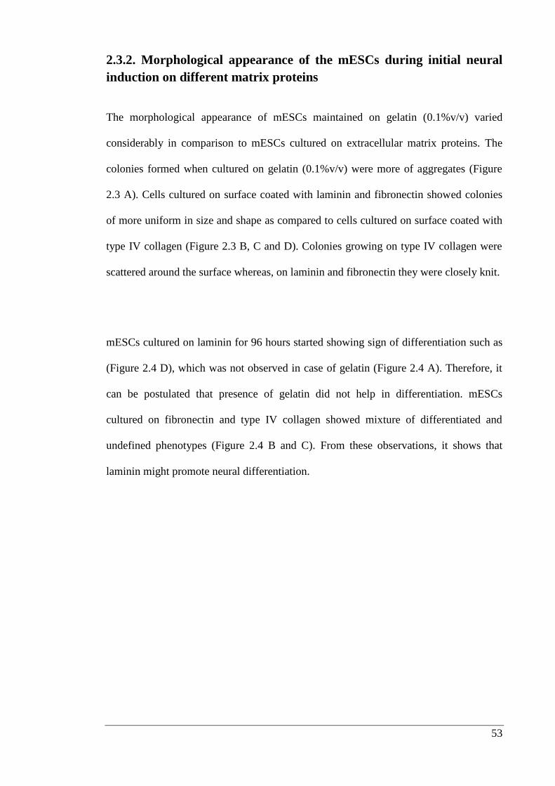
53
2.3.2. Morphological appearance of the mESCs during initial neural
induction on different matrix proteins
The morphological appearance of mESCs maintained on gelatin (0.1%v/v) varied
considerably in comparison to mESCs cultured on extracellular matrix proteins. The
colonies formed when cultured on gelatin (0.1%v/v) were more of aggregates (Figure
2.3 A). Cells cultured on surface coated with laminin and fibronectin showed colonies
of more uniform in size and shape as compared to cells cultured on surface coated with
type IV collagen (Figure 2.3 B, C and D). Colonies growing on type IV collagen were
scattered around the surface whereas, on laminin and fibronectin they were closely knit.
mESCs cultured on laminin for 96 hours started showing sign of differentiation such as
(Figure 2.4 D), which was not observed in case of gelatin (Figure 2.4 A). Therefore, it
can be postulated that presence of gelatin did not help in differentiation. mESCs
cultured on fibronectin and type IV collagen showed mixture of differentiated and
undefined phenotypes (Figure 2.4 B and C). From these observations, it shows that
laminin might promote neural differentiation.
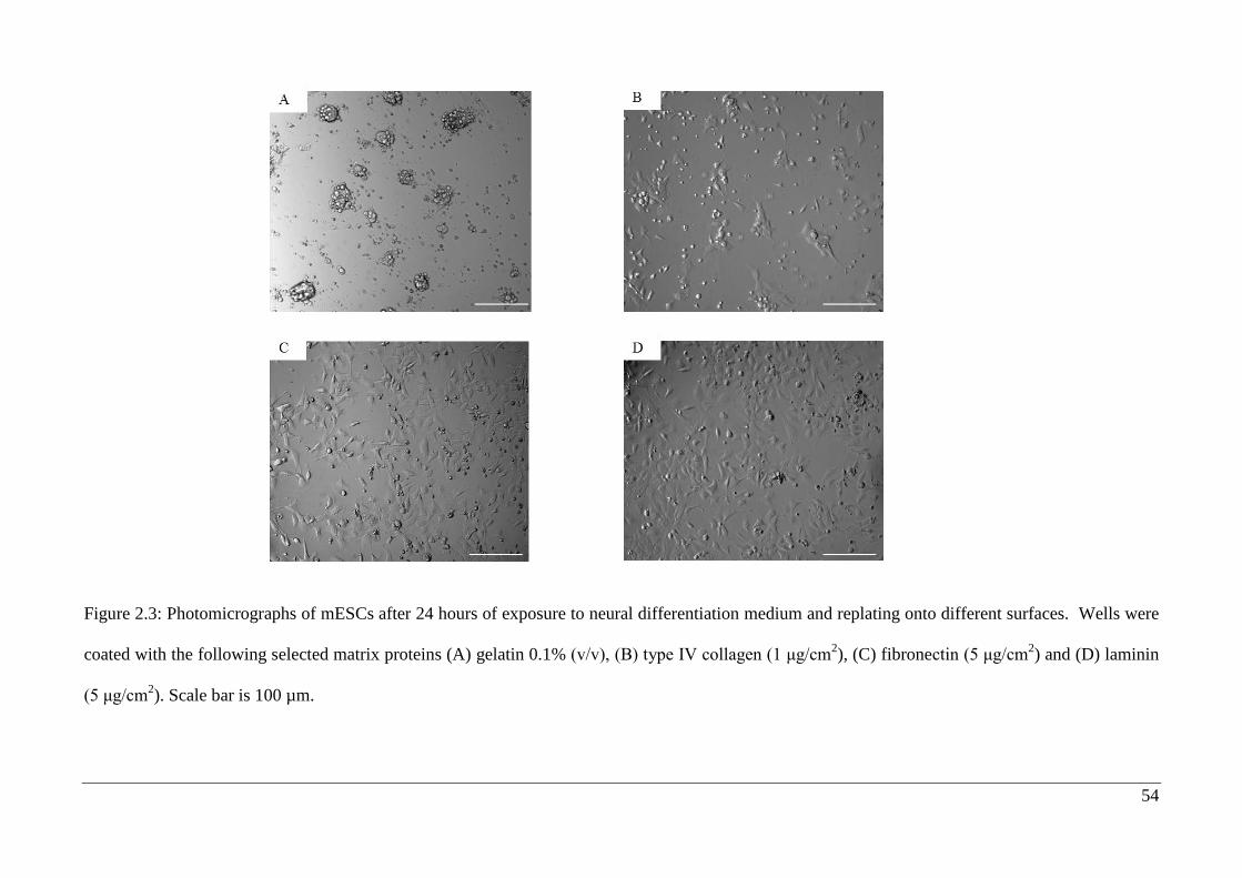
54
Figure 2.3: Photomicrographs of mESCs after 24 hours of exposure to neural differentiation medium and replating onto different surfaces. Wells were
coated with the following selected matrix proteins (A) gelatin 0.1% (v/v), (B) type IV collagen (1 μg/cm2), (C) fibronectin (5 μg/cm
2) and (D) laminin
(5 μg/cm2). Scale bar is 100 µm.
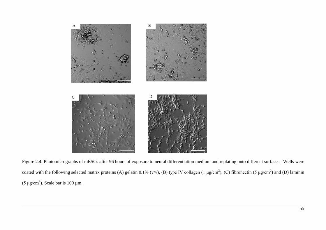
55
Figure 2.4: Photomicrographs of mESCs after 96 hours of exposure to neural differentiation medium and replating onto different surfaces. Wells were
coated with the following selected matrix proteins (A) gelatin 0.1% (v/v), (B) type IV collagen (1 μg/cm2), (C) fibronectin (5 μg/cm
2) and (D) laminin
(5 μg/cm2). Scale bar is 100 µm.
56
2.3.3. Microculture Tetrazolium (MTT) Assay
Cells responses to biological, chemical or mechanical changes in their intracellular or
extracellular environment can contribute to cell proliferation or death. Thus, MTT assay
was performed to show how different matrix proteins influence mESC viability and
proliferation. Photomicrographs taken 24 hours after replating onto surfaces coated
(Figure 2.3) with type IV collagen had few cells in contrast many cells were evident in
plates coated with laminin or fibronectin. The preference is still same after 96 hours
(Figure 2.4).
An MTT assay was carried out on mESCs 24 hours after replating. This assay showed
that viability in 96 plate wells coated with type IV collagen was not significantly
enhanced and a concentration-dependent effect on cell viability was not observed, as
seen in case of cells plated on surfaces coated with laminin or fibronectin (Figure 2.5).
Thus, based on the analysis 24 hours and 96 hours (Figure 2.6) after replating, the most
appropriate concentrations of matrix proteins were selected for further investigation,
namely 1 µg/cm2 for type IV collagen and 5 µg/cm
2 respectively for both laminin and
fibronectin. Since the major aim this thesis was to study the role of the three matrix
proteins on neural differentiation it was decided to normalise the data against mESCs
grown on gelatin. The reduction in cell viability after 24 hours of replating (Figure 2.6)
could be explained by either cell detachment/death or by changes in cell metabolic
activity over time (Hughes et al., 2012; Lawton et al., 2013; Li et al., 2012; Marcos-
Campos et al., 2012).
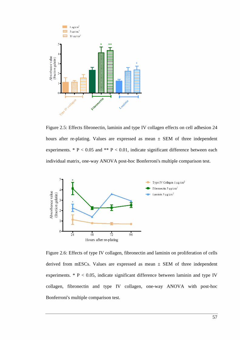
57
Figure 2.5: Effects fibronectin, laminin and type IV collagen effects on cell adhesion 24
hours after re-plating. Values are expressed as mean ± SEM of three independent
experiments. * P < 0.05 and ** P < 0.01, indicate significant difference between each
individual matrix, one-way ANOVA post-hoc Bonferroni's multiple comparison test.
Figure 2.6: Effects of type IV collagen, fibronectin and laminin on proliferation of cells
derived from mESCs. Values are expressed as mean ± SEM of three independent
experiments. * P < 0.05, indicate significant difference between laminin and type IV
collagen, fibronectin and type IV collagen, one-way ANOVA with post-hoc
Bonferroni's multiple comparison test.
58
2.3.4. Neural progenitor expansion and differentiation into neurons
To examine the effect of matrix on the adhesion and survival after replating of mESC-
derived neural progenitors, 5000 cells/well were seeded onto matrix coated wells and
after 24 hours, the number of nestin positive cells was quantified, from the images
captured from four corners of the well i.e. top and bottom left and right hand corners of
respectively of the well. Figure 2.7 A-C shows representative images indicating that
many of the cells were immunopositive for nestin and had a similar appearance on all
surfaces after 24 hours. A comparison of positive cell counts at this time showed that on
average the cell number was not significantly different after plating on the three
selected matrix proteins (Figure 2.7 D).
The effect of selected substrates on neurite outgrowth of mESC-derived neurons was
performed by quantifying number of primary neurites and total neurite length per
neuron. The mESCs-derived neurons were identified using βIII tubulin
immunocytochemistry (Figure 2.8 A and B). Generally, 24 hours after replating, the
proportion of cells immunopositive for βIII tubulin was less than 6% indicating that the
differentiation was predominantly at the progenitor stage. The numbers of βIII tubulin-
positive cells 24 hours after replating was independent of surface, but the number of
βIII tubulin-positive neurons increase from 11/view to 31/view on laminin after 96
hours whereas, it went from 10/view to 20/view, 8/view to 19/view respectively on
fibronectin and type IV collagen (Figure 2.8 C and D). Total neurite length was
calculated as described in section 2.2.7. Type IV collagen did show an increase in the
number of neurons but substantially increased neurite length as compared to fibronectin
and laminin (Figure 2.10 A and B). Therefore, laminin enhanced conversion of neuronal
progenitor positive cells into neurons (Figure 2.8) but failed to
stimulate neurite outgrowth.
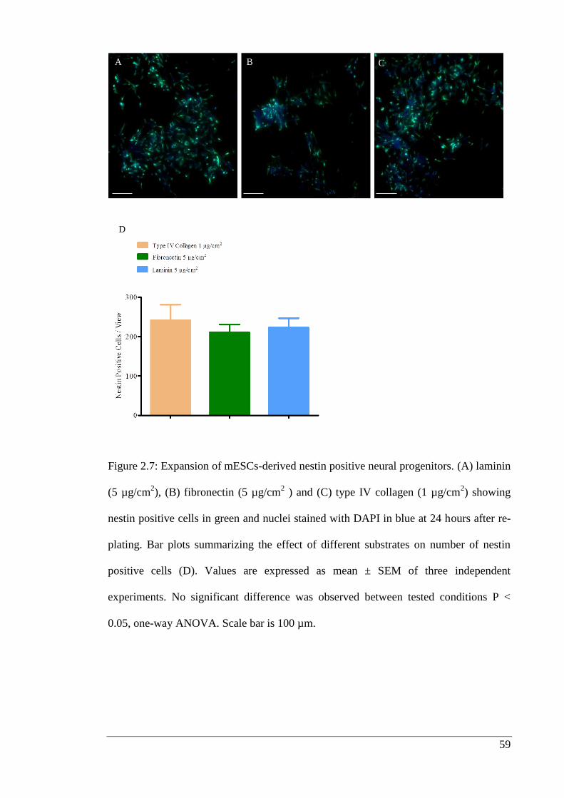
59
Figure 2.7: Expansion of mESCs-derived nestin positive neural progenitors. (A) laminin
(5 µg/cm2), (B) fibronectin (5 µg/cm
2 ) and (C) type IV collagen (1 µg/cm
2) showing
nestin positive cells in green and nuclei stained with DAPI in blue at 24 hours after re-
plating. Bar plots summarizing the effect of different substrates on number of nestin
positive cells (D). Values are expressed as mean ± SEM of three independent
experiments. No significant difference was observed between tested conditions P <
0.05, one-way ANOVA. Scale bar is 100 µm.
A B C
D
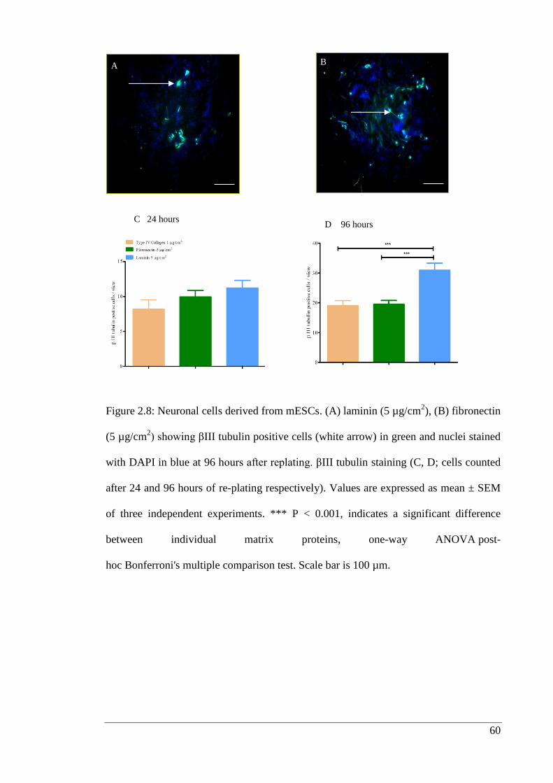
60
Figure 2.8: Neuronal cells derived from mESCs. (A) laminin (5 µg/cm2), (B) fibronectin
(5 µg/cm2) showing βIII tubulin positive cells (white arrow) in green and nuclei stained
with DAPI in blue at 96 hours after replating. βIII tubulin staining (C, D; cells counted
after 24 and 96 hours of re-plating respectively). Values are expressed as mean ± SEM
of three independent experiments. *** P < 0.001, indicates a significant difference
between individual matrix proteins, one-way ANOVA post-
hoc Bonferroni's multiple comparison test. Scale bar is 100 µm.
A B
C 24 hours D 96 hours
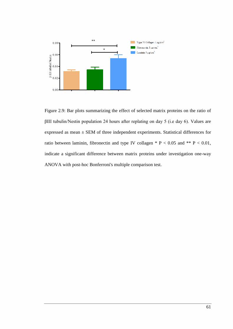
61
Figure 2.9: Bar plots summarizing the effect of selected matrix proteins on the ratio of
βIII tubulin/Nestin population 24 hours after replating on day 5 (i.e day 6). Values are
expressed as mean ± SEM of three independent experiments. Statistical differences for
ratio between laminin, fibronectin and type IV collagen * P < 0.05 and ** P < 0.01,
indicate a significant difference between matrix proteins under investigation one-way
ANOVA with post-hoc Bonferroni's multiple comparison test.
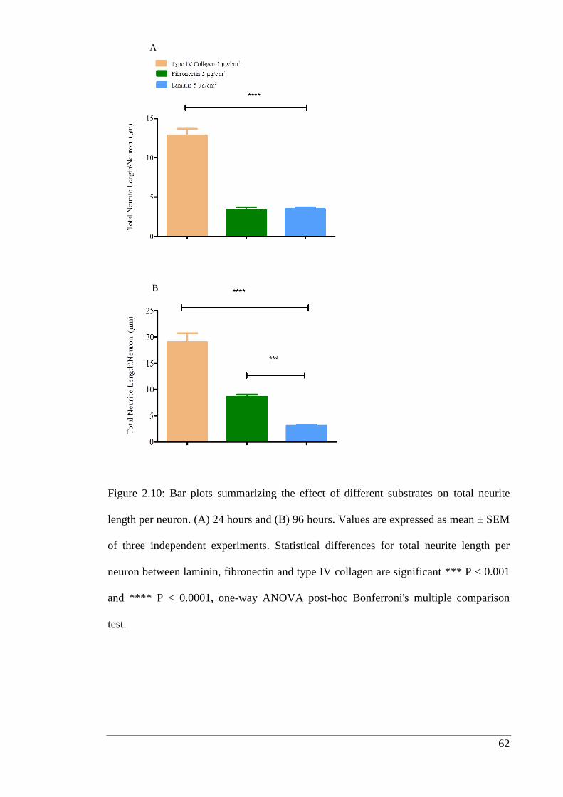
62
Figure 2.10: Bar plots summarizing the effect of different substrates on total neurite
length per neuron. (A) 24 hours and (B) 96 hours. Values are expressed as mean ± SEM
of three independent experiments. Statistical differences for total neurite length per
neuron between laminin, fibronectin and type IV collagen are significant *** P < 0.001
and **** P < 0.0001, one-way ANOVA post-hoc Bonferroni's multiple comparison
test.
A
B
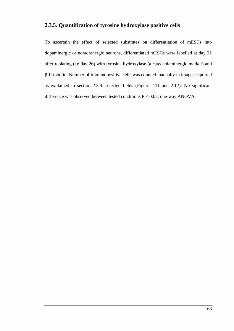
63
2.3.5. Quantification of tyrosine hydroxylase positive cells
To ascertain the effect of selected substrates on differentiation of mESCs into
dopaminergic or noradrenergic neurons, differentiated mESCs were labelled at day 21
after replating (i.e day 26) with tyrosine hydroxylase (a catecholaminergic marker) and
βIII tubulin. Number of immunopositive cells was counted manually in images captured
as explained in section 2.3.4. selected fields (Figure 2.11 and 2.12). No significant
difference was observed between tested conditions P < 0.05, one-way ANOVA.
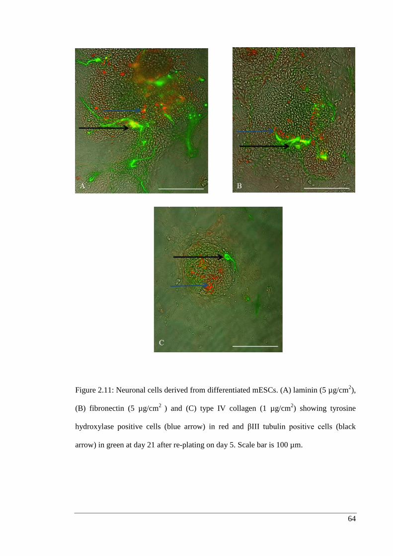
64
Figure 2.11: Neuronal cells derived from differentiated mESCs. (A) laminin (5 µg/cm2),
(B) fibronectin (5 µg/cm2
) and (C) type IV collagen (1 µg/cm2) showing tyrosine
hydroxylase positive cells (blue arrow) in red and βIII tubulin positive cells (black
arrow) in green at day 21 after re-plating on day 5. Scale bar is 100 µm.
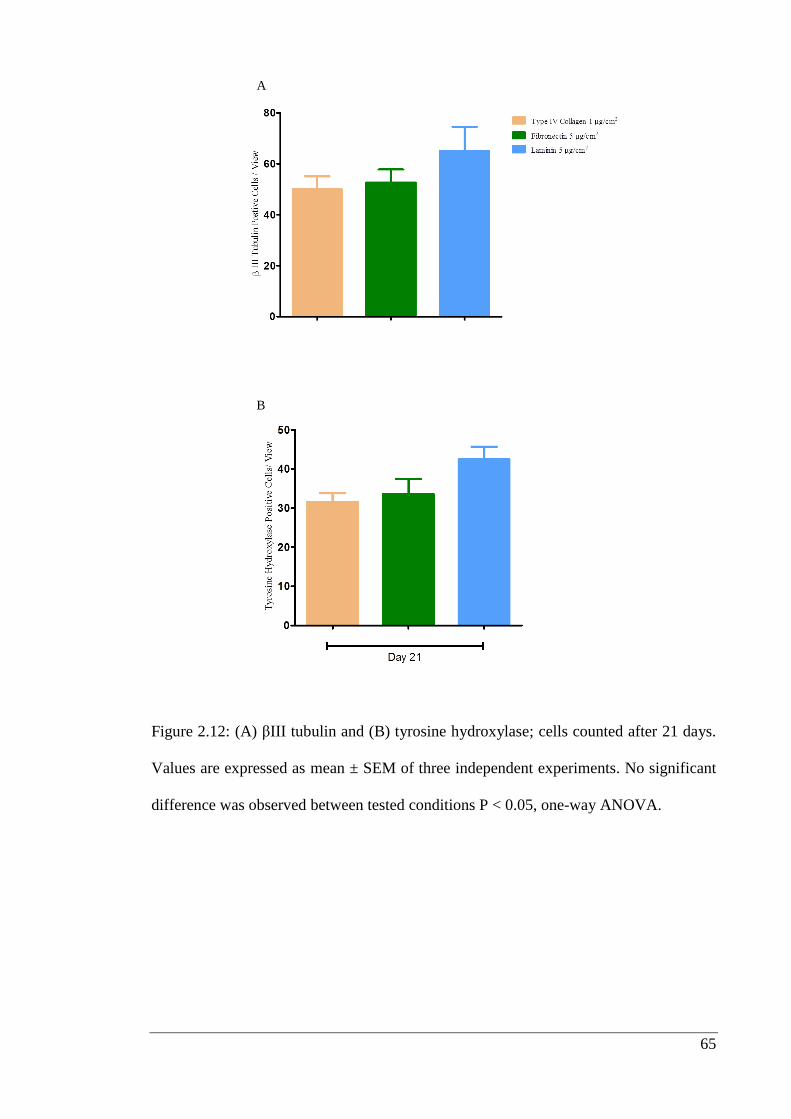
65
Figure 2.12: (A) βIII tubulin and (B) tyrosine hydroxylase; cells counted after 21 days.
Values are expressed as mean ± SEM of three independent experiments. No significant
difference was observed between tested conditions P < 0.05, one-way ANOVA.
A
B
66
2.5. Discussion
This study investigated the effects of three matrix proteins commonly found in the
basement membrane. mESCs attached and proliferated on surfaces coated with matrix
proteins after replating cells after 5 days of neural induction on gelatin in N2B27
medium. Given that survival of cells plated onto plastic plates is poor, it is clear that
matrix proteins play a crucial role in the attachment and proliferation of neural
progenitors. This is particularly important when cells are plated as monolayers and is
supported by previous studies (Gospodarowicz et al., 1980; Hall et al., 1987; Tomaselli
et al., 1987). Other published studies have shown that extracellular matrix proteins
including laminin and fibronectin stimulate adhesion and process outgrowth and these
studies support my observation that adhesion of mESC-derived neural stem cells was
promoted by laminin or fibronectin, rather than type IV collagen or gelatin (Majumdar
et al., 2003; Manthorpe et al., 1983; Rogers et al., 1983; Rogers et al., 1985).
The role of proteins like laminin, fibronectin, and type IV collagen has been extensively
studied in the PNS where they promote neurite outgrowth (Duband et al., 1982; Thiery
et al., 1982). In the CNS, extracellular matrix proteins and in particular laminin and
fibronectin are likely to play a role in neuronal differentiation, regeneration and neurite
elongation (Carri et al., 1988). This study evaluated the effect of matrix proteins on the
growth of neural progenitors by quantifying number of nestin positive cells 24 hours
after re-plating. Comparison of cell counts showed no significant expansion over the
first 24 hours on any of the matrix proteins.
67
When incidence of βIII tubulin positives was determined, laminin had the most
pronounced effect. The number of βIII tubulin positive cells doubled after 96 hours on
laminin after re-plating as compared to other matrix proteins under investigation.
Results from the above study showed that laminin is particularly effective and in line
with the information already in public domain.
Lein et al. (1991) reported that type IV collagen is synthesized by glial cells and is
present on ganglia in the peripheral nervous system, where it provides tensile strength
as a structural component of endoneural basement. When embryonic rat sympathetic
neurons were seeded onto surfaces coated with type IV collagen and laminin, it was
observed that type IV collagen but not laminin enhanced sympathetic neurite outgrowth.
This growth promoting activity was associated primarily with the NC1 domain. Tucker
and Erickson (1984) reported that collagen enhanced outgrowth of neural crest cells in
absence of fibronectin (Tucker et al., 1984).
Fibronectin is known to enhance neurite extension in neural crest cells (Sheppard et al.,
1995). There are several binding domains in fibronectin that may regulate this property.
This concept was tested by plating human neuroblastoma cells onto laminin, and after
overnight incubation, sizable percentages of cells showed extended neurites thus,
showing that laminin presence is helpful in neurite extension (Lewandowska et al.,
1989; Mugnai et al., 1988). Avian trunk neural crest cells bound to fibronectin which
facilitated rapid migration indicating that fibronectin is important for neural crest cell
adhesion and migration (Rovasio et al., 1983).
68
Extracellular matrix proteins are assumed to play a crucial role in migration of trunk
neural crest. To test this hypothesis, neural tubes from stage 12 chick embryos were
suspended within gelling solutions of either basement membrane or rat tail collagen.
Extensive crest migration was reported in basement membrane gel, as compared to rat
tail collagen. In basement membrane gels, the laminin cell-binding peptide (YIGSR)
completely inhibited migration of neural crest away from the neural tube, suggesting
that laminin is the migratory substratum. Indeed, laminin, as well as collagen and
fibronectin, are present in the embryonic extracellular matrix. Thus, it is possible that
other extracellular matrix proteins, in addition to or instead of fibronectin, may serve as
migratory substrata for neural crest in vivo (Bilozur et al., 1988).
In our study, quantification of the primary neurites and total neurite length per neuron
showed that laminin induced a significantly greater number of neurites as compared to
fibronectin and type IV collagen 96 hours post plating, but it was type IV collagen
which induced the most significant neurite elongation, thus suggesting that laminin
failed to enhance neurite elongation although it doubled the number of neurons. Both
laminin and fibronectin are synthesized in the CNS during development where they
influence neuronal migration and axon guidance (Smith et al., 1987). Liesi (1990)
showed that brain cells bound preferentially to glial laminin, indicating that laminin
plays crucial role in attachment of cells and neurite extension (Liesi, 1990). Laminin
and fibronectin induced pronounced neurite outgrowth of cultured human fetal sensory
neurons in presence of nerve growth factor. By increasing the amount of matrix, the
effect of withdrawal of nerve growth factor on neuron growth was reduced (Baron-Van
Evercooren et al., 1982).
69
Embryonal carcinoma PCC4uva cells have the ability to differentiate into neurons when
exposed to mixture of retinoic acid and dbcAMP. This in vitro model system was used
to study the effects of laminin on early neural differentiation. When compared to type
IV and I collagen or fibronectin, laminin was a more effective enhancer of neuronal
differentiation and neurite growth (Sweeney et al., 1990). These authors reported that
when cells were treated with antibodies against laminin, the laminin induced effects
were nullified. This supports the hypothesis that laminin plays a crucial role in
influencing early differentiation of cell populations (Sweeney et al., 1990).
Flanagan et al. (2006) plated human neural stem precursor cells (NSPCs) on different
substrates including poly-L-ornithine, fibronectin, laminin or Matrigel, to compare the
response of these cells with those of mouse NSPCs. These authors showed that laminin
enhanced migration of human and mouse NSPC-derived neurons, as well as their
migration, expansion, differentiation and neurite outgrowth. Laminin was more
effective than poly-L-ornithine, fibronectin or Matrigel (Flanagan et al., 2006).
Differentiation of human embryonic stem cells into neural progenitors and neurons was
reported to be stimulated by laminin, to a greater extent than fibronectin or type I
collagen. Ma et al. (2008) also observed that robust neuroectodermal precursors were
generated by neural rosettes on surfaces coated with laminin (Ma et al., 2008). In this
study it was observed that laminin stimulated neuronal differentiation but not neurite
outgrowth. Whereas, type IV collagen stimulated neurite outgrowth as compared to
laminin. This observation could be specific to the effects on neurons obtained from
mESCs.
70
Stem cells isolated from human embryos or foetal tissues have the potential to provide
neurons to ameliorate neural disorders, and foetal tissue has been used in attempts to
replace lost neurons in Parkinson’s disease. However, one major obstacle is that the
majority of cells do not differentiate into neurons when grafted into areas which are
outside the known areas of neurogenesis, namely the subgranular zone (SGZ) and
subventricular zone (SVZ) of the adult CNS and grafts do not exhibit constitutive
neurogenesis in vivo under normal conditions (Jiao et al., 2008). Priming foetal human
neural stem cells with laminin in vitro generated a pure population of neurons and
enhanced generation of cholinergic neurons in grafted cells (Tarasenko et al., 2004; Wu
et al., 2002). Working with these principles, it was explored whether priming with
selected matrix proteins, laminin, fibronectin or type IV collagen could enhance
development of dopaminergic neurons in vitro, but no significant difference between
the selected matrix proteins was observed.
2.6. Conclusion
I have demonstrated using a defined adherent culture system, that differentiation of
neurons from mESCs was significantly enhanced by laminin to a greater extent than
fibronectin or type IV collagen. Laminin stimulated neural progenitor generation,
expansion and differentiation into neurons, but did not enhance neurite outgrowth of
mESCs derived neurons. This finding is consistent with data showing that laminin is
crucial for neurogenesis. Thus, the interaction between stem cells and matrix proteins
appeared to be an early inductive signal to help regulate neural specification of mESCs.
This finding may smooth the progress of early mouse CNS development which could be
replicated for human CNS development for potential application in the management of
neurological diseases.

71
Chapter 3 Immobilization of growth factors onto
surfaces coated with extracellular matrix
components
72
3.1. Introduction
3.1.1. The transcription factor, Lmx1a
Transcriptional factors directing dopaminergic neurons maturation are not yet
completely understood but several key molecular events have been identified in
developing mouse midbrain. One of critical importance is the expression of the LIM
homeobox transcription factor (Lmx1a), which is essential for development of
dopaminergic neurons (Cai et al., 2009). In the past few years it has been shown that by
combining expression of Lmx1a and forkhead box protein A2 (Foxa2), dopaminergic
neurons could be differentiated from human fibroblasts (Andersson et al., 2006;
Pfisterer et al., 2011). Lmx1a targeted by siRNA in the chick embryo inhibited the
formation of mature dopaminergic neurons (Millonig et al., 2000). Friling et al. (2009)
also showed that increased expression of Lmx1a in mESCs provided a robust strategy
for efficient production and enrichment of dopaminergic neurons (Friling et al., 2009).
Lmx1a was discovered in the Dreher (dr) mouse, mutant with many developmental
defects, including cerebellum and dorsal spinal cord (Millonig et al., 2000). Positional
cloning of the gene responsible for the dr phenotype identified a 1.8kb cDNA clone
located on chromosome 1, which shared significant identity throughout the coding
region with the hamster Lmx1.1 (94% nucleotide identity, 98% amino-acid identity).
This gene was termed Lmx1a, the mouse homolog of the hamster Lmx1.1 (Millonig et
al., 2000). Follow-up studies showed that Lmx1a is widely expressed in the brain (Failli
et al., 2002).
73
3.1.2. Lmx1a is sufficient and required for midbrain dopaminergic
neurons (mDA) development in vivo
Over the past few years, there has been an explosion of research focusing on the
development of strategies to steer embryonic stem cells towards desired fates, with the
hope for an embryonic stem cells based replacement therapy for diseases such as
Parkinson disease (Barberi et al., 2003; Kawasaki et al., 2002). One potential advantage
of using embryonic stem cells derived midbrain dopaminergic neurons is the unlimited
supply of cells for transplantation, but the drawbacks with current differentiation
techniques are the efficiency and purity (Prowse et al., 2011).
Lmx1a is expressed both in neural progenitors and in post mitotic midbrain
dopaminergic neurons. However, the generation of ectopic midbrain dopaminergic
neurons is known to be limited to ventrolateral regions with the greatest frequency
occurring ventrally. It was shown by Lin et al. (2009) that Lmx1a alone is not sufficient
to induce ectopic dopaminergic neuron development in the lateral region of ventral
midbrain thus indicating that other factors are required to act in parallel to Lmx1a. One
such candidates is Foxa1/2, which cooperates with Lmx1a through a feed forward loop
during the induction of midbrain dopaminergic neurons (Lin et al., 2009).
By contrast, in dorsal regions, Lmx1a participates in a different developmental program
regulated by bone morphogenetic proteins (BMPs) to specify a functional roof plate
(Chizhikov et al., 2004). Therefore, the activity of Lmx1a is context dependent. In
addition, a mixture of cell types, including serotonin (5-HT) neurons and γ-
aminobutyric acid (GABA) neurons is often present in the differentiating embryonic
stem cell culture (Reubinoff et al., 2001). It has been reported that serotonin neurons
74
mediate dyskinetic side effects in Parkinson’s patients with neural transplants (Politis et
al., 2010). By contrast, GABAergic neurons send out long projections to their normal
targets and effect behavioural improvement (Thompson et al., 2008). Previous stem cell
differentiation protocols have relied on exogenous growth factors, such as Shh, FGF8
and Wnts. Numerous studies published from year 2000 to 2005 showed that
combination of Shh, FGF8 and FGF2 gave a high percentage of dopaminergic neurons
(Barberi et al., 2003; Buytaert-Hoefen et al., 2004; Kim et al., 2002). Thus, based on
the above studies it was decided to evaluate whether, Lmx1a could be induced by
growth factors such as Shh, FGF8b and FGF2 and matrix proteins.
3.1.3. Chapter aims
Strong binding to heparan is a characteristic feature of the fibroblast growth factors.
Complex formation protects fibroblast growth factors from heat and acid inactivation
and can also act as bioactive reservoirs for signaling factors (Gospodarowicz et al.,
1986; Vazin et al., 2010; Vlodavsky et al., 1990). Heparan sulphate one of
glycosaminoglycans occur naturally and can bind ionically to the amine groups on
growth factors via its sulphate and carboxylate side chains (Ashikari-Hada et al., 2004).
Heparan-containing delivery systems have been used to immobilize high affinity
heparan binding growth factors to protect them from degradation (Capila et al., 2002;
Edelman et al., 1992; Hou et al., 2010; Kreuger et al., 2006; Sakiyama-Elbert et al.,
2000b; Sakiyama-Elbert et al., 2000c; Skop et al., 2013; Vazin et al., 2010).

75
Thus, working on these principles I tried to explore whether heparan sulphate could
immobilize growth factors like FGF and Shh onto surfaces coated with laminin,
fibronectin and type IV collagen. Thus, I wanted to investigate
Immobilized growth factors were evaluated for physical stability and
availability by in-cell Western assay. The effectiveness of modified substrata
was investigated by determining the proportion of Lmx1a+ cells by flow
cytometry at various stages during in vitro differentiation.
Effects of individual matrix proteins on the proportion of Lmx1a+ expressing
cells.
76
3.2. Materials and Methods
The experimental procedures used in this chapter have already been explained in
Chapter 2, sections 2.2.1. - 2.2.7. unless detailed below.
3.2.1. Substrata preparation
Sterile round glass cover slips were placed into a 48 well plate. A mixture of laminin
(Invitrogen, Australia), fibronectin (Invitrogen, Australia), type IV collagen (Invitrogen,
Australia) and heparan sulphate (Sigma-Aldrich, Australia) in ratio of 1:1:1:0.1 along
with 100 ng Shh (Sigma-Aldrich, Australia), 50 ng FGF8b (R&D Systems, USA) and
10 ng FGF2 (R&D Systems, USA) were incubated at 37oC for 3 hours. After 3 hours
incubation, remaining mixture was aspirated carefully. The rationale behind this
strategy was to generate a basement membrane-like substrata, comprises of laminin,
fibronectin, type IV collagen and heparan sulphate (Laurie et al., 1982), to which
addition of biologically relevant growth factors, might result in an improved
microenvironment to support neural differentiation. It has been reported that a mixture
of laminin, type IV collagen and heparan sulphate, incubated at 35oC in a ratio of
1:1:0.1, generates a basement membrane like substrata (Grant et al., 1989). To this we
decided to add fibronectin on its reported role in differentiation of embryonic stem cells
into the glia cells and neurons (Liu et al., 2010).
77
3.2.2. In-cell Western assay
Every experiment was performed in quadruplicate, including negative controls which
were not exposed to primary antibodies. Growth factors immobilized onto matrix were
fixed by incubation with 4% paraformaldehyde in 1X PBS for 30 minutes at room
temperature. For permeabilization, 200 µl of 0.1% TritonX-100 (Sigma-Aldrich,
Australia) in 1X PBS was added to each well for 30 minutes. Surface coated wells were
blocked with 200 µl of blocking buffer (Li-cor Biotechnology, USA) for 90 minutes at
room temperature with gentle shaking. Mouse anti-FGF2 (Sigma-Aldrich, Australia),
goat anti-FGF8b (R&D Systems, USA) and goat anti-Shh (R&D Systems, USA) were
diluted 1:100, 1: 400 and 1: 200 respectively in blocking buffer and incubated overnight
at 4ºC in a volume of 100 µl per well.
Unbound primary antibody was removed by washing wells (5 times) with 200 µl 0.1%
Tween20 (Sigma-Aldrich, Australia) in 1X PBS. Wells were then exposed to secondary
antibody; IRDye 800CW donkey anti-mouse and anti-goat (Li-cor Biotechnology,
USA) were diluted 1:1000 in blocking buffer for two hours at room temperature.
Unbound secondary antibodies were removed by washing each well (5 minutes) with
0.1% Tween20 (Sigma-Aldrich, Australia) in 1X PBS (by gentle shaking 5 times).
Each individual well of 48 well plate coated with and without growth factors were
scanned for emission at 800 nm with the Odyssey Infrared Imaging system (ODY-
18090, Li-cor Biotechnology, USA).
78
Values from the 800 nm channel corresponded to the amount of primary antibody
bound to the wells. Control wells not exposed to primary antibodies were used for
background compensation. The obtained values at 800 nm were averaged and
subtracted from the respective values of wells incubated with primary antibodies and
normalization dye. Graphs were plotted using the integrated intensity measurements for
each well.
3.2.3. Lmx1a-β-lactamase reporter mESCs
An Lmx1a-AMP-IRES-eGFP-FneoF targeting vector was constructed for homologous
recombination, designed such that, the AMP-IRES-eGFP-FneoF construct replaced
exon 1 of the Lmx1a gene. This targeting vector was introduced into mouse E14Tg2a
ESCs by electroporation with a GenePulser XCellTM instrument (BioRad, USA) and
cells targeted at one allele were isolated by Dr Stewart Fabb (Monash Institute of
Pharmaceutical Sciences).
3.2.4. Effect of combined matrix proteins sequestered with growth
factors on differentiation of Lmx1a reporter cells
The mESCs reporter cell line Lmx1a-eGFP was differentiated under three different
conditions and Lmx1a reporter gene expression was quantified on days 8, 12, 16 and 20
of differentiation.
Condition 1: Cells were plated onto combined matrix proteins in N2B27 medium
supplemented with 500 ng/ml Shh, 10 ng/ml recombinant FGF2 and 100 ng/ml FGF8b
for a total of 6 days with medium replenished on day 3.

79
Condition 2: 100 ng/well Shh, 10 ng/well FGF2 and 50 ng/well FGF8b were pre-
immobilized onto the same combination of matrix proteins prior to plating. Cells were
incubated for 6 days in N2B27 medium, which was replenished without growth factors
on day 3 of differentiation.
Condition 3: 10 ng/well FGF2 and 50 ng/well FGF8b were pre-immobilized onto the
combination of matrix proteins prior to plating. Cells were incubated for 6 days with
medium replenished without growth factors on day 3.
Cells were terminally differentiated after incubation by each of the above three
conditions by exposing them to N2B27 medium supplemented with 10 ng/ml glial-
derived neurotrophic factor (GDNF) (R&D Systems, USA) and 200 μM ascorbic acid
(Sigma-Aldrich, Australia) from day 6 onwards for final differentiation. During the
terminal differentiation the medium was changed every other day until day 21.
3.2.5. Influence of individual matrix proteins on Lmx1a reporter cells
The embryonic stem cell reporter cell line was seeded at 5x10³
cells/cm2
onto surfaces
coated with 0.1 % v/v gelatin (Sigma-Aldrich, Australia), 0.1 mg/ml poly-D-lysine
(PDL, Sigma-Aldrich, Australia), laminin (5 µg/cm2, Invitrogen, Australia), fibronectin
(5 µg/cm2, BD Bioscience, Australia) or type IV collagen (1 µg/cm
2, BD Bioscience,
Australia) in N2B27 medium without any growth factors. Lmx1a promoter-driven
expression was quantified by flow cytometry (FACS Canto II) on days 3, 5, 7 and 9 of
neural induction to investigate the effect of individual matrix proteins.
80
3.2.6. β-lactamase staining of live cells
For quantitation using flow cytometry, the LiveBLAzer™ FRET – B/G Loading Kit
(Invitrogen, Australia) was used to produce fluorescence in AMP+ cells. Cells were
trypsinized and pelleted. Approximately 0.5 million cells were resuspended in 0.5 ml of
1X PBS and 100 µl of 6X Live Blazer substrate loading solution. -lactamase activity
was detected by incubating the cell suspension with CCF2-AM in dark conditions for
two hours. The cells were exposed to light at 406 nm. The substrate emits around 530
nm (green) however, if beta-lactamase reporter gene expression is activated it leads to
cleavage of the substrate, thereby disrupting the fluorescence resonance energy transfer
(FRET) and resulting in emission at 460 nm (blue) following excitation at 406 nm.
After incubation cells were pelleted, resuspended in 2 ml of 1X PBS, pelleted a second
time, resuspended in 2 ml of 1X PBS and cleared of clumps with a 70 µm strainer (BD
Biosciences, Australia). As a negative control, wild type mESCs were used having been
submitted to treatment with the LiveBlazer substrate over the same time period as
sample cells. Flow cytometry was conducted by setting gates such that 99.0-99.5%
precent of wild type cells were registered as Lmx1a-AMP negative. For each analysis
10,000 events were counted.
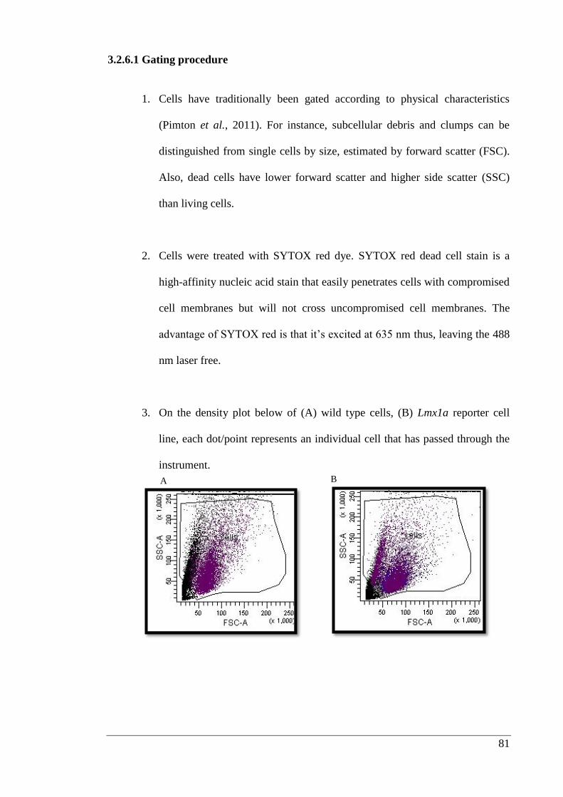
81
3.2.6.1 Gating procedure
1. Cells have traditionally been gated according to physical characteristics
(Pimton et al., 2011). For instance, subcellular debris and clumps can be
distinguished from single cells by size, estimated by forward scatter (FSC).
Also, dead cells have lower forward scatter and higher side scatter (SSC)
than living cells.
2. Cells were treated with SYTOX red dye. SYTOX red dead cell stain is a
high-affinity nucleic acid stain that easily penetrates cells with compromised
cell membranes but will not cross uncompromised cell membranes. The
advantage of SYTOX red is that it’s excited at 635 nm thus, leaving the 488
nm laser free.
3. On the density plot below of (A) wild type cells, (B) Lmx1a reporter cell
line, each dot/point represents an individual cell that has passed through the
instrument.
A B
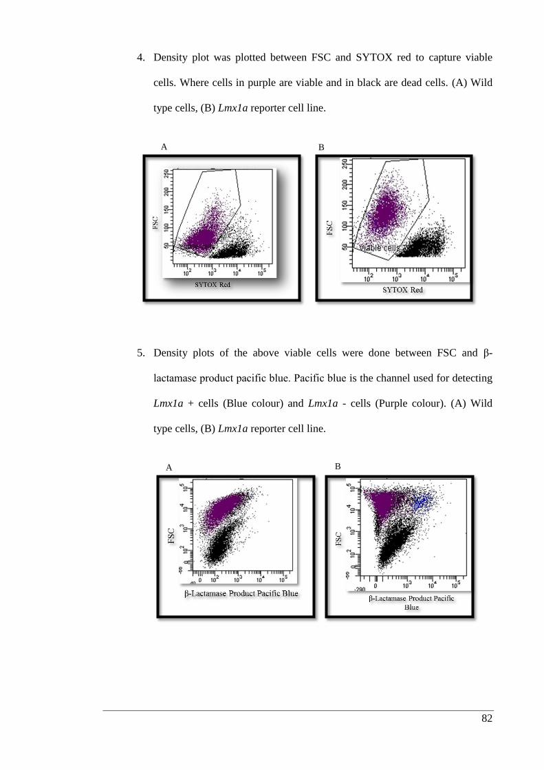
82
4. Density plot was plotted between FSC and SYTOX red to capture viable
cells. Where cells in purple are viable and in black are dead cells. (A) Wild
type cells, (B) Lmx1a reporter cell line.
5. Density plots of the above viable cells were done between FSC and β-
lactamase product pacific blue. Pacific blue is the channel used for detecting
Lmx1a + cells (Blue colour) and Lmx1a - cells (Purple colour). (A) Wild
type cells, (B) Lmx1a reporter cell line.
A B
A B
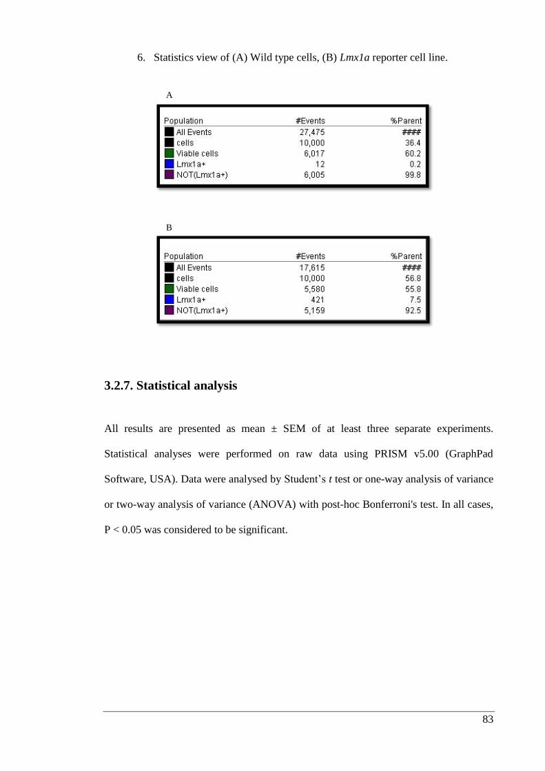
83
6. Statistics view of (A) Wild type cells, (B) Lmx1a reporter cell line.
3.2.7. Statistical analysis
All results are presented as mean ± SEM of at least three separate experiments.
Statistical analyses were performed on raw data using PRISM v5.00 (GraphPad
Software, USA). Data were analysed by Student’s t test or one-way analysis of variance
or two-way analysis of variance (ANOVA) with post-hoc Bonferroni's test. In all cases,
P < 0.05 was considered to be significant.
B
A
84
3.3. Results
3.3.1. Growth factor immobilization studies
With reference to previous literature, three different growth factors were selected for
study based on their ability to promote embryonic stem cell differentiation and survival
(Cohen et al., 2010). The initial objective was to confirm whether the selected
antibodies reacted with their expected targets. Figure 3.1 indicates that the selected
antibodies for FGF8b and Shh were specific in nature and showed no cross reactivity
whereas, FGF2 antibody (a polyclonal antibody) showed some cross reactivity with
FGF8b and Shh.
The next challenge was to determine the concentration of growth factors to be selected.
Coating experiments using solutions of growth factors in ascending concentrations were
carried out. For FGF2 the concentrations were 10, 25 and 50 ng/well, for FGF8b 50,
100 and 200 ng/well and for Shh 50, 100 and 200 ng/well were used (Figure 3.2). The
bar graphs (Figure 3.3) show concentration-dependent coating for FGF8b and Shh, but
not FGF2. Therefore, it was decided to use the following concentrations of growth
factors for further experiments FGF2 10 ng/well, FGF8b 50 ng/well and Shh 100
ng/well.
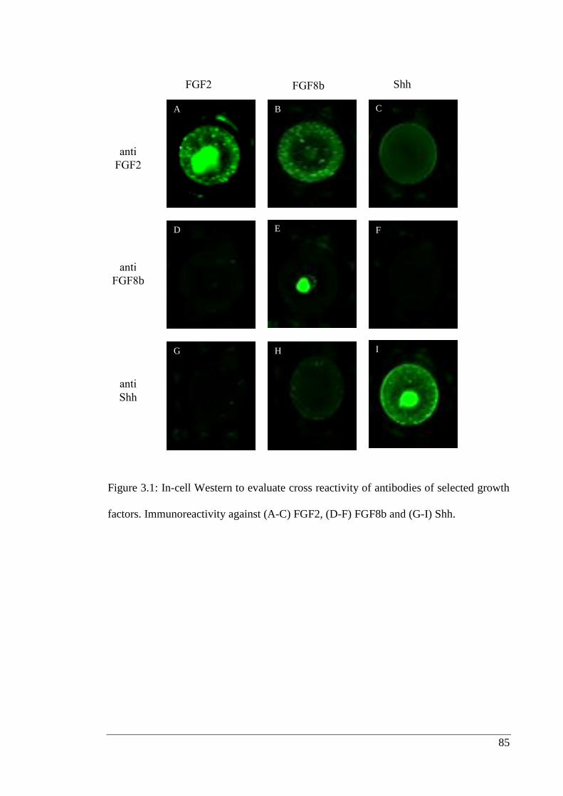
85
Figure 3.1: In-cell Western to evaluate cross reactivity of antibodies of selected growth
factors. Immunoreactivity against (A-C) FGF2, (D-F) FGF8b and (G-I) Shh.
FGF2 FGF8b Shh
anti
FGF2
anti
FGF8b
anti
Shh
A B C
D E F
G H I
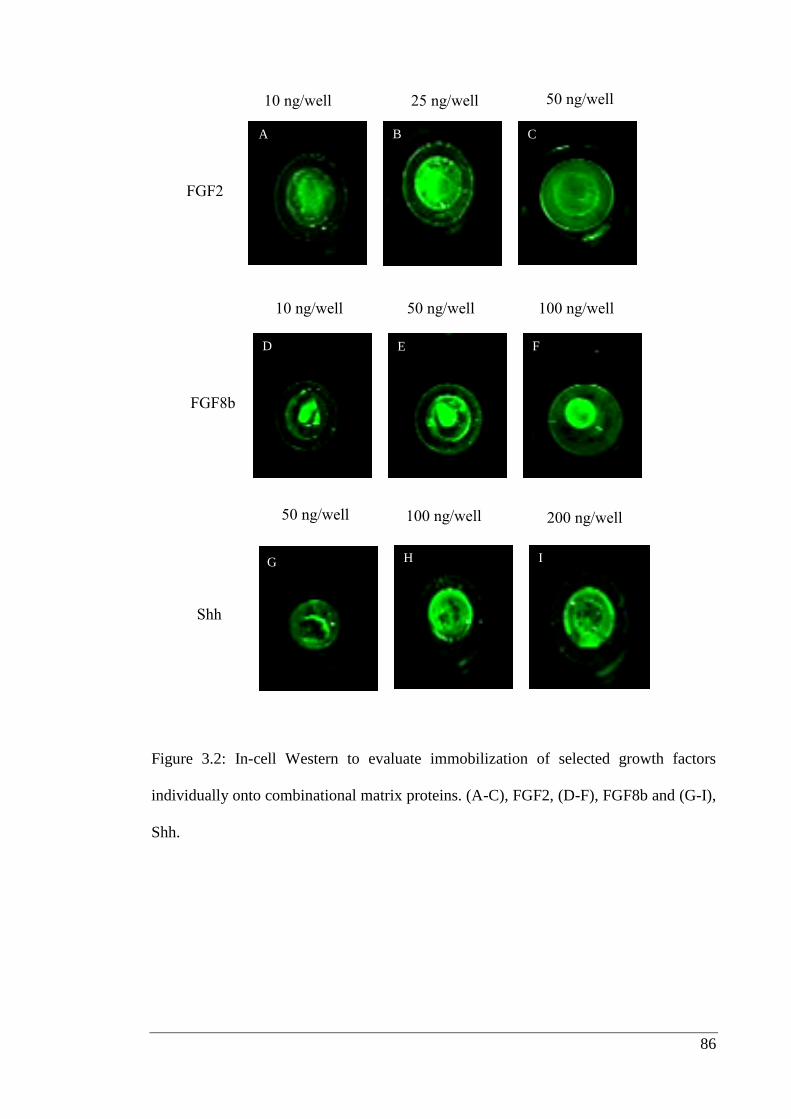
86
Figure 3.2: In-cell Western to evaluate immobilization of selected growth factors
individually onto combinational matrix proteins. (A-C), FGF2, (D-F), FGF8b and (G-I),
Shh.
G H I
10 ng/well 25 ng/well 50 ng/well
10 ng/well 50 ng/well 100 ng/well
50 ng/well 100 ng/well 200 ng/well
FGF2
Shh
FGF8b
D E F
A B C
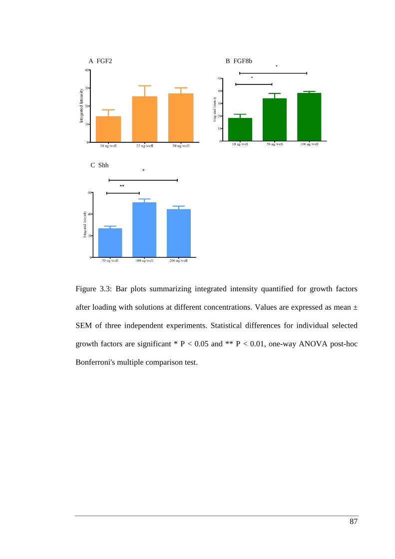
87
Figure 3.3: Bar plots summarizing integrated intensity quantified for growth factors
after loading with solutions at different concentrations. Values are expressed as mean ±
SEM of three independent experiments. Statistical differences for individual selected
growth factors are significant * P < 0.05 and ** P < 0.01, one-way ANOVA post-hoc
Bonferroni's multiple comparison test.
A FGF2 B FGF8b
C Shh
88
3.3.2. Immobilization efficiency
The next challenge was to determine the increase or decrease in availability of growth
factors after their immobilization as compared to traditional method of external addition
(i.e. in the culture medium) during differentiation. FGF2 and FGF8b were added to the
culture medium at 10 and 100 ng/well respectively. Bar graphs (Figure 3.4 G and H),
show that the amount of growth factors available to cells at the surface of the substrate
at the starting point of differentiation step was greater in case of pre-immobilized
factors compared to external addition. In practice, this represented a reduction in the
mass of FGF8b needed when pre-immobilization was used.
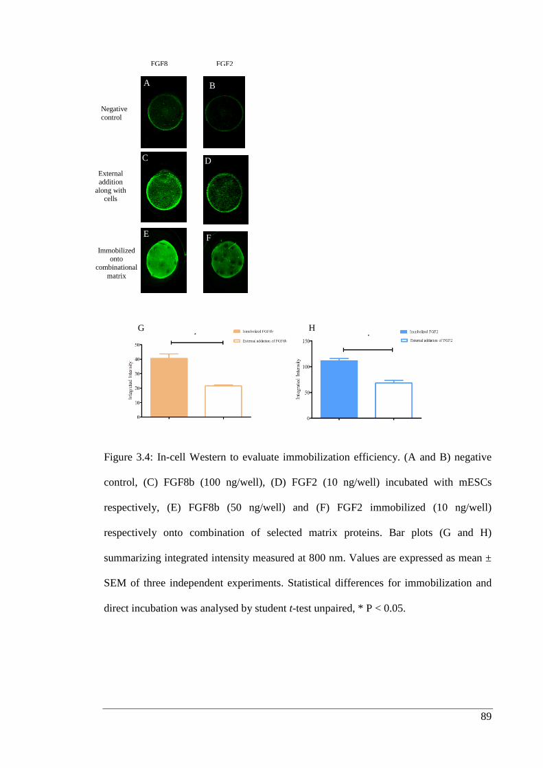
89
Figure 3.4: In-cell Western to evaluate immobilization efficiency. (A and B) negative
control, (C) FGF8b (100 ng/well), (D) FGF2 (10 ng/well) incubated with mESCs
respectively, (E) FGF8b (50 ng/well) and (F) FGF2 immobilized (10 ng/well)
respectively onto combination of selected matrix proteins. Bar plots (G and H)
summarizing integrated intensity measured at 800 nm. Values are expressed as mean ±
SEM of three independent experiments. Statistical differences for immobilization and
direct incubation was analysed by student t-test unpaired, * P < 0.05.
A
C
E
B
D
F
FGF8
b
FGF2
Negative
control
External
addition
along with cells
Immobilized onto
combinational
matrix
G H
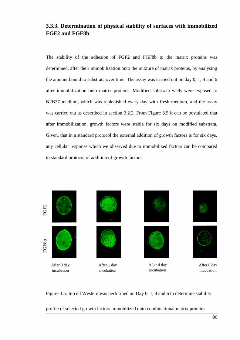
90
3.3.3. Determination of physical stability of surfaces with immobilized
FGF2 and FGF8b
The stability of the adhesion of FGF2 and FGF8b to the matrix proteins was
determined, after their immobilization onto the mixture of matrix proteins, by analysing
the amount bound to substrata over time. The assay was carried out on day 0, 1, 4 and 6
after immobilization onto matrix proteins. Modified substrata wells were exposed to
N2B27 medium, which was replenished every day with fresh medium, and the assay
was carried out as described in section 3.2.2. From Figure 3.5 it can be postulated that
after immobilization, growth factors were stable for six days on modified substrata.
Given, that in a standard protocol the external addition of growth factors is for six days,
any cellular response which we observed due to immobilized factors can be compared
to standard protocol of addition of growth factors.
Figure 3.5: In-cell Western was performed on Day 0, 1, 4 and 6 to determine stability
profile of selected growth factors immobilized onto combinational matrix proteins.
After 0 day
incubation
After 1 day
incubation
After 4 day
incubation
After 6 day
incubation
FG
F8b
FG
F2
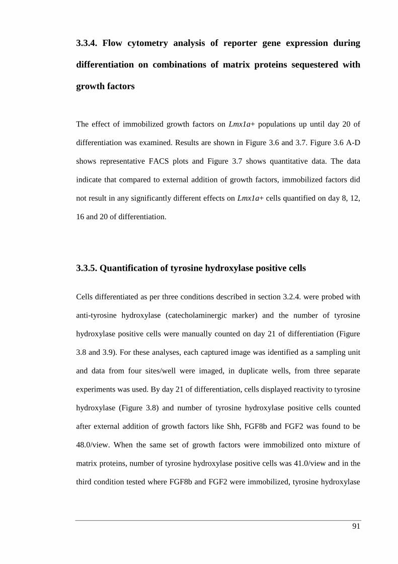
91
3.3.4. Flow cytometry analysis of reporter gene expression during
differentiation on combinations of matrix proteins sequestered with
growth factors
The effect of immobilized growth factors on Lmx1a+ populations up until day 20 of
differentiation was examined. Results are shown in Figure 3.6 and 3.7. Figure 3.6 A-D
shows representative FACS plots and Figure 3.7 shows quantitative data. The data
indicate that compared to external addition of growth factors, immobilized factors did
not result in any significantly different effects on Lmx1a+ cells quantified on day 8, 12,
16 and 20 of differentiation.
3.3.5. Quantification of tyrosine hydroxylase positive cells
Cells differentiated as per three conditions described in section 3.2.4. were probed with
anti-tyrosine hydroxylase (catecholaminergic marker) and the number of tyrosine
hydroxylase positive cells were manually counted on day 21 of differentiation (Figure
3.8 and 3.9). For these analyses, each captured image was identified as a sampling unit
and data from four sites/well were imaged, in duplicate wells, from three separate
experiments was used. By day 21 of differentiation, cells displayed reactivity to tyrosine
hydroxylase (Figure 3.8) and number of tyrosine hydroxylase positive cells counted
after external addition of growth factors like Shh, FGF8b and FGF2 was found to be
48.0/view. When the same set of growth factors were immobilized onto mixture of
matrix proteins, number of tyrosine hydroxylase positive cells was 41.0/view and in the
third condition tested where FGF8b and FGF2 were immobilized, tyrosine hydroxylase
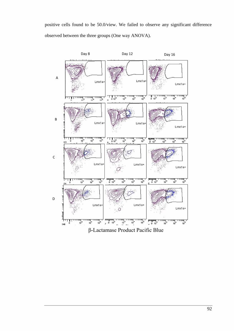
92
positive cells found to be 50.0/view. We failed to observe any significant difference
observed between the three groups (One way ANOVA).
Day 8 Day 12 Day 16
A
B
C
D
β-Lactamase Product Pacific Blue
93
β-Lactamase Product Pacific Blue
Day 8 Day 12 Day 16
A
B
C
D
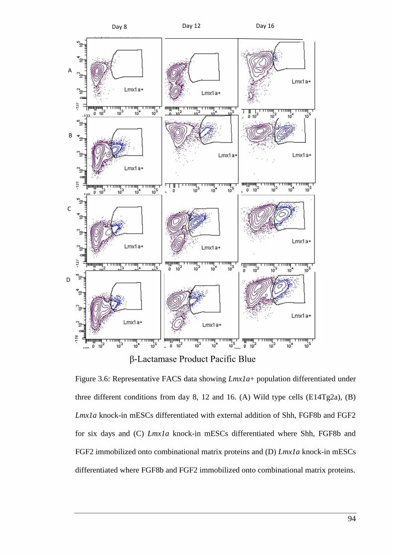
94
Figure 3.6: Representative FACS data showing Lmx1a+ population differentiated under
three different conditions from day 8, 12 and 16. (A) Wild type cells (E14Tg2a), (B)
Lmx1a knock-in mESCs differentiated with external addition of Shh, FGF8b and FGF2
for six days and (C) Lmx1a knock-in mESCs differentiated where Shh, FGF8b and
FGF2 immobilized onto combinational matrix proteins and (D) Lmx1a knock-in mESCs
differentiated where FGF8b and FGF2 immobilized onto combinational matrix proteins.
A
B
C
D
Day 8 Day 12 Day 16
β-Lactamase Product Pacific Blue
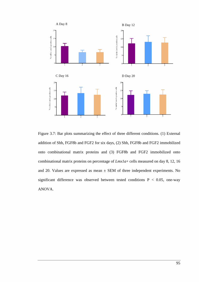
95
Figure 3.7: Bar plots summarizing the effect of three different conditions. (1) External
addition of Shh, FGF8b and FGF2 for six days, (2) Shh, FGF8b and FGF2 immobilized
onto combinational matrix proteins and (3) FGF8b and FGF2 immobilized onto
combinational matrix proteins on percentage of Lmx1a+ cells measured on day 8, 12, 16
and 20. Values are expressed as mean ± SEM of three independent experiments. No
significant difference was observed between tested conditions P < 0.05, one-way
ANOVA.
A Day 8 B Day 12
C Day 16 D Day 20

96
Figure 3.8: Immunofluorescence images showing representative fields of tyrosine
hydroxylase (white arrow) positive cells (red), βIII tubulin positive cells (green) and
nuclei stained with DAPI (blue) differentiated using one of three different conditions.
(1) External addition of Shh, FGF8b and FGF2 for six days, (2) Shh, FGF8b and FGF2
immobilized onto combinational selected matrix proteins and (3) FGF8b and FGF2
immobilized onto combinational selected matrix proteins for 21 days. Scale bar is 50
µm
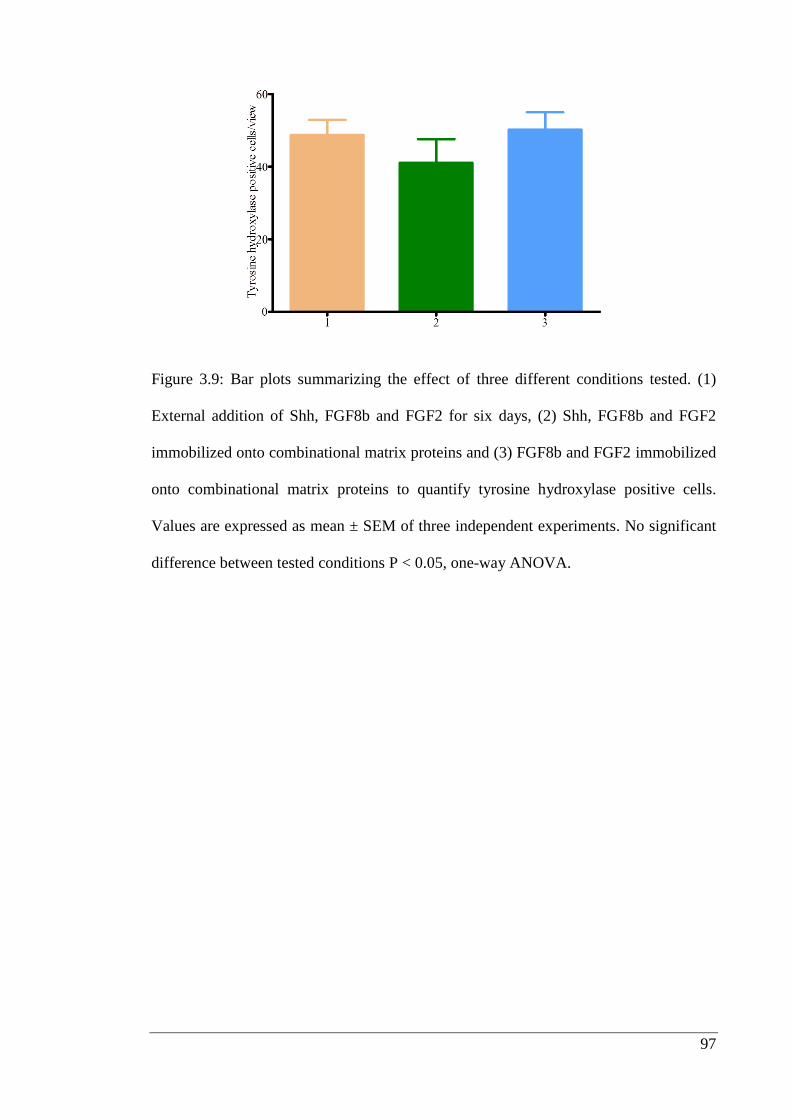
97
Figure 3.9: Bar plots summarizing the effect of three different conditions tested. (1)
External addition of Shh, FGF8b and FGF2 for six days, (2) Shh, FGF8b and FGF2
immobilized onto combinational matrix proteins and (3) FGF8b and FGF2 immobilized
onto combinational matrix proteins to quantify tyrosine hydroxylase positive cells.
Values are expressed as mean ± SEM of three independent experiments. No significant
difference between tested conditions P < 0.05, one-way ANOVA.
98
3.3.6. Influence of individual matrix proteins on Lmx1a+ cells
The percentage of Lmx1a+ cells was quantified on day 3, 5, 7 and 9 of neural induction
on surfaces coated with gelatin (0.1% v/v), PDL (0.1 mg/ml), laminin (5 µg/cm2),
fibronectin (5 µg/cm2) or type IV collagen (1 µg/cm
2). Figure 3.11 (A-D) shows that
laminin enhanced the Lmx1a+ population on day 5 when compared to fibronectin, type
IV collagen or gelatin, but this effect was not observed on days 3, 7 or 9 of neural
induction. Therefore, it can be inferred that the presence or absence of individual matrix
proteins have no direct impact on the proportion of Lmx1a+ cells; however it was
observed that it is necessary to have a suitable substrate for stable cell attachment.
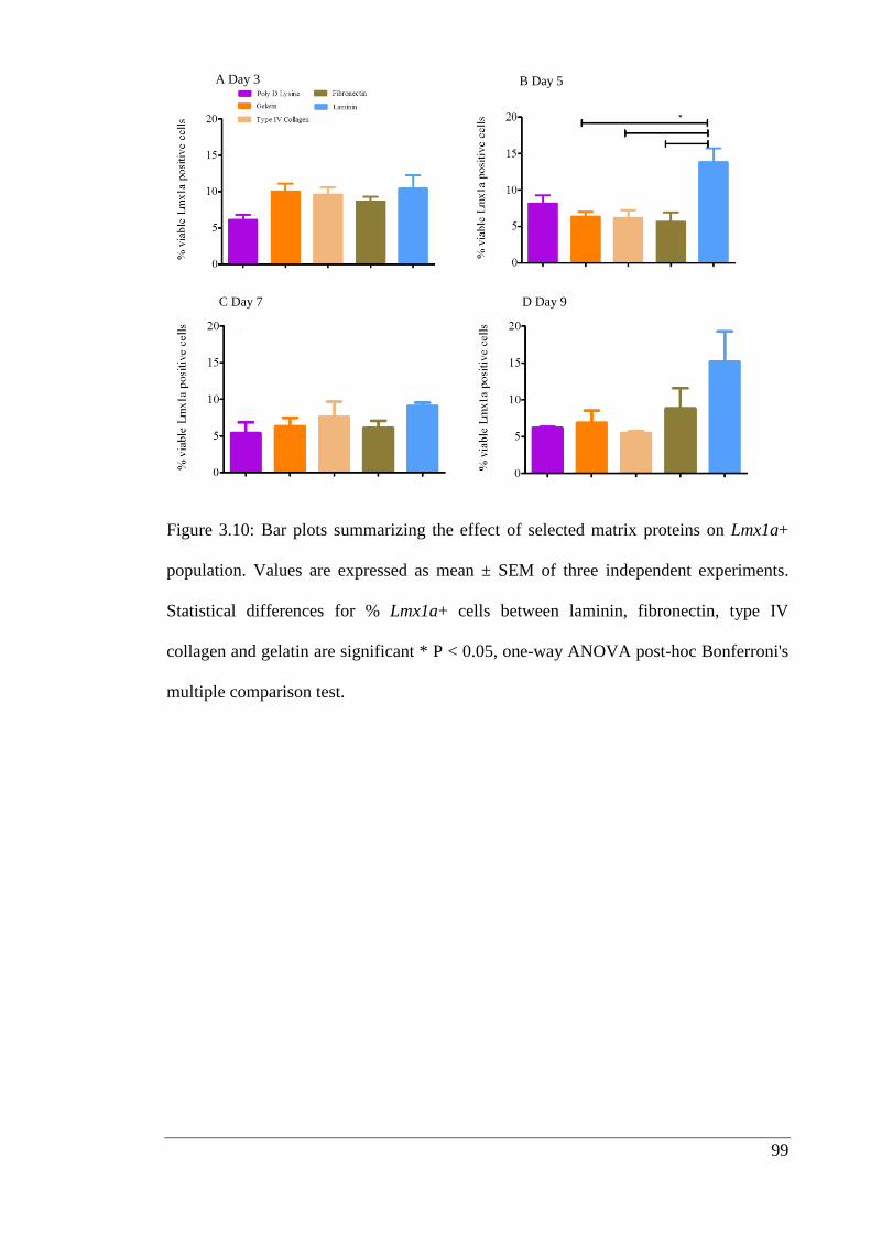
99
Figure 3.10: Bar plots summarizing the effect of selected matrix proteins on Lmx1a+
population. Values are expressed as mean ± SEM of three independent experiments.
Statistical differences for % Lmx1a+ cells between laminin, fibronectin, type IV
collagen and gelatin are significant * P < 0.05, one-way ANOVA post-hoc Bonferroni's
multiple comparison test.
A Day 3 B Day 5
C Day 7 D Day 9
100
3.4. Discussion
Growth factors are one of the critical factors regulating a wide variety of cellular
functions, including proliferation, migration and differentiation (Masters, 2011). A
significant issue with respect to availability of growth factors to cells during
induction/differentiation stages is that growth factors are prone to rapid degradation in
physiological conditions or inactivation due to enzymatic action (Masters, 2011). The
current strategy of delivering growth factors to the site of action through conjugated
/three dimensional biomaterial system requires expertise and significant funding
(Masters, 2011).
An alternative strategy is to use either covalent or non-covalent immobilization methods
to display growth factors on either chemically defined polymers or matrix proteins.
These strategies can prolong growth factor availability and reduce the mass of growth
factor required. Furthermore, this strategy has a major advantage which allows soluble
and matrix bound factors to perform the natural functions they perform in the in vivo
environment (Driessen et al., 2003; Ferrara, 2010). Making use of this principle, the
present study explored whether growth factors including FGF2, reported to have roles
in renewal and proliferation (Levenstein et al., 2006). FGF8b and Shh, which have been
shown to induce differentiation of neurons towards midbrain dopaminergic phenotypes
(Ye et al., 1998), could be immobilized onto combinations of selected matrix proteins
including laminin, fibronectin, type IV collagen and heparan sulphate.
It was decided to exploit the natural binding sites of matrix proteins facilitate binding of
selected growth factors. For example, the α chains of laminin contain binding sites for
heparan sulphate (Hozumi et al., 2009; Kouzi-Koliakos et al., 1989) and bind to type IV
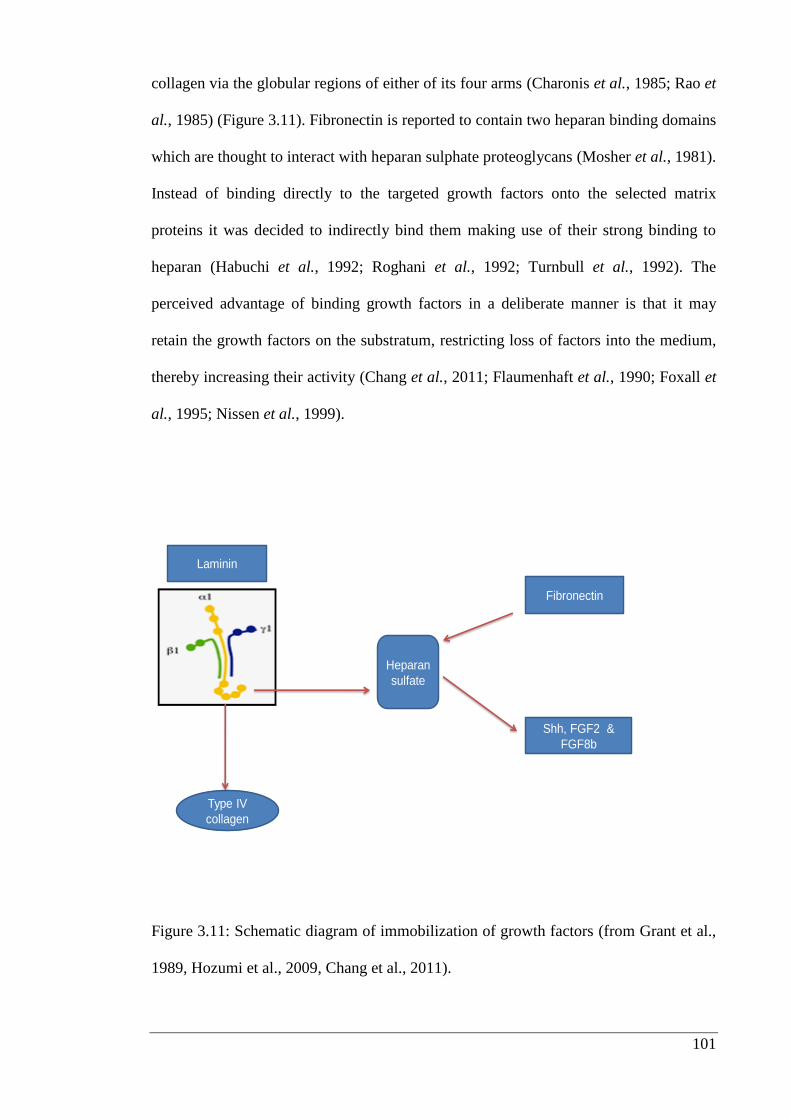
101
collagen via the globular regions of either of its four arms (Charonis et al., 1985; Rao et
al., 1985) (Figure 3.11). Fibronectin is reported to contain two heparan binding domains
which are thought to interact with heparan sulphate proteoglycans (Mosher et al., 1981).
Instead of binding directly to the targeted growth factors onto the selected matrix
proteins it was decided to indirectly bind them making use of their strong binding to
heparan (Habuchi et al., 1992; Roghani et al., 1992; Turnbull et al., 1992). The
perceived advantage of binding growth factors in a deliberate manner is that it may
retain the growth factors on the substratum, restricting loss of factors into the medium,
thereby increasing their activity (Chang et al., 2011; Flaumenhaft et al., 1990; Foxall et
al., 1995; Nissen et al., 1999).
Heparan
sulfate
Type IV
collagen
Fibronectin
Shh, FGF2 &
FGF8b
Laminin
Figure 3.11: Schematic diagram of immobilization of growth factors (from Grant et al.,
1989, Hozumi et al., 2009, Chang et al., 2011).
102
Grant et al. (1989) showed that when laminin, type IV collagen and heparan sulphate
proteoglycan are incubated in ratio of 1:1:0.1, this combination yields basement
membrane like structure (Grant et al., 1989). Working on the above principles, it was
possible to immobilize the three growth factors, Shh, FGF8b and FGF2. It was also
possible to show that the surfaces were more immunoreactive to relevant antibodies
when FGF8b and FGF2 were pre-immobilized onto the surface (Figure 3.5). This is
consistent with previous research which showed that immobilization of nerve growth
factors increases their availability (Kapur et al., 2003).
Similarly, heparin coated alginate gels were used to deliver FGF2 (Tanihara et al.,
2001). Vascular endothelial growth factor (VEGF) and FGF2 have also been delivered
by utilising either collagen gels or cross-linking poly (lactide-co-glycolide) (PLG) with
heparin (Jeon et al., 2006; Steffens et al., 2004). Although this type of strategy has been
used in the above mentioned papers, there are surprisingly few studies of this type,
making use of intact heparin/heparan as an integral component of the growth factor
delivery vehicle. The use of this strategy to enhance neural differentiation of embryonic
stem cells has not been described previously.
103
After showing that growth factors could be immobilized onto matrix, the next objective
was to explore whether this promising platform could be used to improve control of
stem cell differentiation. There are some interesting reports with other systems that
relate to the current study. In vivo LIF is present in both diffusible and matrix bound
forms, and is known to activate pathways that support the self-renewal of mESCs
(Rathjen et al., 1990; Williams et al., 1988). Interestingly immobilization of LIF has
been used to improve embryonic stem cells propagation by Alberti et al. (2008). These
authors were able to support pluripotency of mESCs for two weeks without extra
addition of LIF (Alberti et al., 2008). Differentiation has been studied using
immobilized VEGF. This protein plays a critical role in regulating angiogenesis and
endothelial cell function. It has also been postulated that signaling by VEGF is a rate
limiting step in initiating angiogenesis (Ferrara et al., 2003).
As the half-life of VEGF is very short and is capable of inducing undesirable side
effects, such as supporting tumour growth, controlled delivery of VEGF has been
investigated by immobilizing VEGF on modified collagens (Chiu et al., 2010; He et al.,
2011). The presentation of VEGF in an immobilized state also has physiological
importance, as living tissue contains both matrix-bound and soluble forms of VEGF.
Moreover, immobilized VEGF has been found to successfully stimulate the
proliferation of endothelial cells (Shen et al., 2008; Zisch et al., 2001). Rahman et al.
(2010) also showed that by immobilizing VEGF into an agarose hydrogel they were
able to induce differentiation of mESCs into blood progenitor cells. The technique was
more effective than exposure of mESCs to soluble VEGF (Rahman et al., 2010)
104
NGF which influences the proliferation, differentiation, survival and death of neuronal
cells (Heese et al., 2007) has been coupled onto different matrix proteins to examine
whether its availability could be improved. β-NGF was successfully delivered by
immobilizing onto a heparin-containing fibrin based system, and was able to enhance
neurite extension of cells derived from dorsal root ganglia (Sakiyama-Elbert et al.,
2000a). A fibrin based delivery system was developed which was able to immobilize
growth factors based on its ability to interact with heparin binding sites within the
system. Its efficiency was evaluated by measuring delivery of NGF on peripheral nerve
regeneration in a rat having sciatic nerve defect. The result showed that bound NGF was
able to enhance peripheral nerve regeneration (Lee et al., 2003).
Results of this study from section 3.3.4., 3.3.5. and 3.3.6. showed that individual matrix
proteins tested were consistently better than controls but there was no statistically
significant increase in expression of either Lmx1a or tyrosine hydroxylase when
embryonic stem cells were differentiated on matrix proteins pre-incubated with FGF8,
FGF2 and Shh. This was a surprising result in the light of the published studies
described (Section 3.1.2.) showed importance of growth factors like Shh/FGF8 and
Lmx1a gene plays a crucial role in generation of dopaminergic neurons. However, it can
be argued that research groups have also shown that growth factors like Shh/FGF8 are
not very effective at directly dopaminergic induction (Gaspard et al., 2008; Nefzger et
al., 2012). Moreover, it has been shown Lmx1a can be expressed by forebrain cells
(Nefzger et al., 2012) and this might mask any induction of midbrain dopaminergic
neurons. It has been reported that the yield of tyrosine hydroxylase positive neurons can
be enhanced by increasing neural induction time of embryonic stem cell-derived neural
progenitors with growth factors like FGF8b and Shh (Koch et al., 2009b; Zeng et al.,
105
2011). These are the probable reasons why our strategy of studying the effect of
immobilized growth factors failed to show any positive effects. When the individual
matrix proteins were evaluated against one another, to investigate which was the best
substrate for supporting neural development, only laminin showed a significant effect
on the proportion of cells that were Lmx1a+ population. This difference was significant
only on day 5 (Figure 3.10 B). The observed advantage of using laminin is in line with
the reported findings that laminin is known to assist and guide process outgrowth of
various subtypes of neurons during early development stage as compared to other
selected matrix proteins including fibronectin and type IV collagen (Eccleston et al.,
1985; Eldridge et al., 1989; Hantaz-Ambroise et al., 1987).
3.5. Conclusion
The present study has shown that stable and bioactive concentrations of FGF2, FGF8b
and Shh can be immobilized onto combinations of matrix proteins. Though there is
gradual dissociation and loss of the growth factors over a 5-7 day period, pre-
immobilization might be expected to be a more efficient means of presenting growth
factors to the embryonic stem cells. By utilising this strategy we were able to reduce the
mass required to half in case of FGF8b. The methodology of immobilization described
above is quite simple, and it has the potential to be used for many other growth factors
type to test their stepwise control of signaling during distinct stages of stem cell neural
fate conversion.

106
Chapter 4 Identifying the signaling pathways
through which mESCs attach and differentiate
on matrix proteins found in basal lamina

107
4.1. Introduction
4.1.1. Extracellular matrix and integrins
In the past few years it has become clear that extracellular matrix is an extremely
dynamic structure that it is an essential regulator of embryonic biological functions
(Bosman et al., 2003). Earlier, extracellular matrix was viewed as passive and inert
scaffold which acted as a stabilising factor. Cell adhesion to matrix proteins is mainly
mediated by heterodimeric transmembrane receptors which belong to the integrin
family consisting α and β subunits (Hynes, 2002). Each subunit has a large extracellular
domain, a transmembrane domain and generally short cytoplasmic domains (Burridge et
al., 1988; Geiger et al., 2001). At least 16 different α and 8 different β subunits have
been identified, forming more than 20 integrins (Geiger et al., 1992; Powell et al., 1997;
Rosales et al., 1992). The extracellular domain binds extracellular matrix proteins with
a specificity that is determined by the pairing of the different α and β subunits. Some
integrins, such as α5β1, the “classic” fibronectin receptor, interact only with a single
extracellular matrix protein; however, more commonly, a given integrin like αVβ3 will
recognize several extracellular matrix proteins including thrombospondin, laminin and
fibrinogen (Bosman et al., 2003; Hynes, 2002; Vinogradova et al., 2000).
108
4.1.2. The role of extracellular matrix in cell survival
Numerous factors, including the enzyme cytochrome P450 2E1 (i.e., CYP2E1), small
proteins (i.e., cytokines) involved in cell communication and oxidative stress, can
trigger apoptosis. A particular form of apoptosis, associated with response to
inappropriate cell/extracellular matrix interactions, is termed ‘anoikis’. Anoikis is a
Greek word meaning “homelessness” (Douma et al., 2004; Frisch et al., 2001). Cell
anchorage is essential for the survival of most cell types (Thomas et al., 1999) and it
has been reported that proteins such as fibronectin or collagen suppresses apoptosis of
mammary epithelial cells (Boudreau et al., 1995), indicating that a given cell type may
require specific extracellular matrix proteins to promote cell survival (di Summa et al.,
2012; Lam et al., 2012; Wang et al., 1999).
Integrins are known to keep cells alive via adhesion that involves activation, by tyrosine
phosphorylation (Giancotti, 2000; Pullan et al., 1996), of FAK (Kornberg et al., 1992a;
Kornberg et al., 1992b). Phosphorylated FAK leads to activation of various downstream
signaling molecules including PI3K, protein kinase B (PKB/Akt) and mitogen activated
protein (MAP) kinases, also known as ERKs (Kornberg et al., 1992b). These kinases
have been implicated in integrin-mediated protection of cells against various apoptotic
stimuli and in control of gene expression (Craddock et al., 2001; Ivankovic-Dikic et al.,
2000; Parsons, 2003).
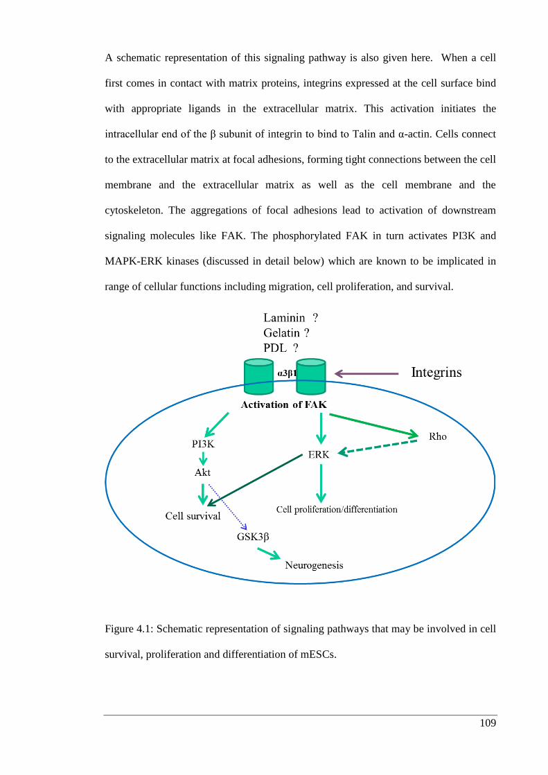
109
A schematic representation of this signaling pathway is also given here. When a cell
first comes in contact with matrix proteins, integrins expressed at the cell surface bind
with appropriate ligands in the extracellular matrix. This activation initiates the
intracellular end of the β subunit of integrin to bind to Talin and α-actin. Cells connect
to the extracellular matrix at focal adhesions, forming tight connections between the cell
membrane and the extracellular matrix as well as the cell membrane and the
cytoskeleton. The aggregations of focal adhesions lead to activation of downstream
signaling molecules like FAK. The phosphorylated FAK in turn activates PI3K and
MAPK-ERK kinases (discussed in detail below) which are known to be implicated in
range of cellular functions including migration, cell proliferation, and survival.
Figure 4.1: Schematic representation of signaling pathways that may be involved in cell
survival, proliferation and differentiation of mESCs.
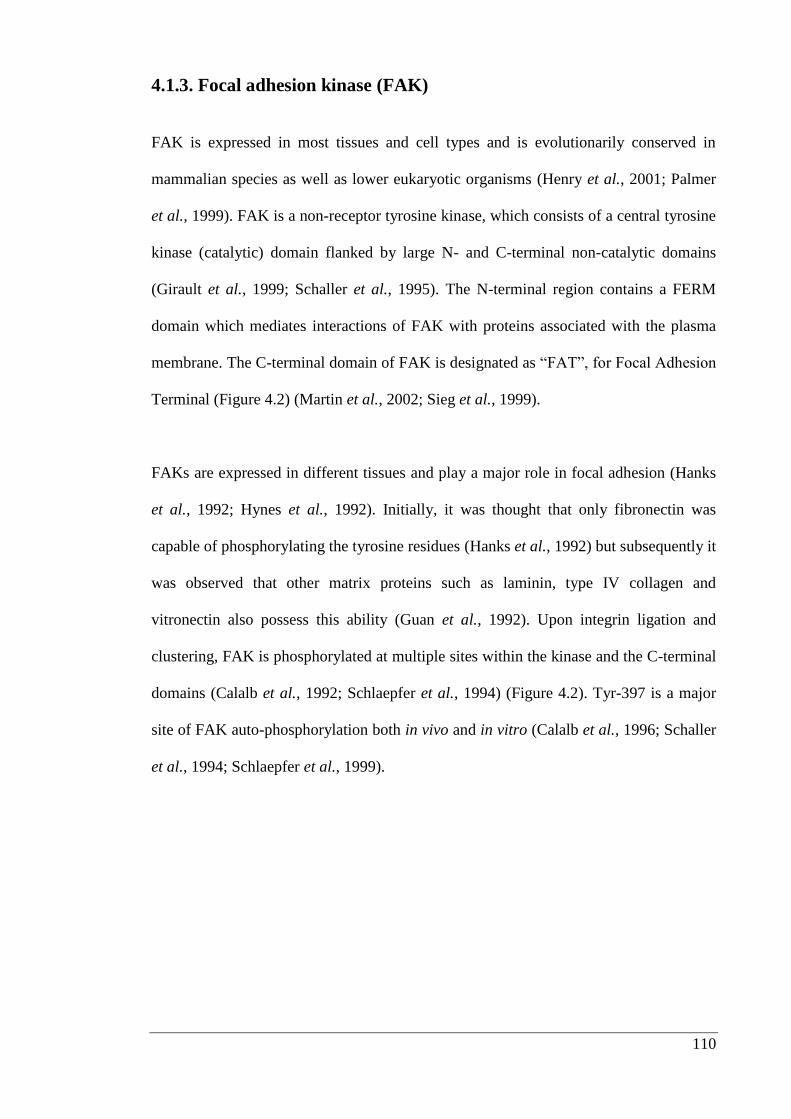
110
4.1.3. Focal adhesion kinase (FAK)
FAK is expressed in most tissues and cell types and is evolutionarily conserved in
mammalian species as well as lower eukaryotic organisms (Henry et al., 2001; Palmer
et al., 1999). FAK is a non-receptor tyrosine kinase, which consists of a central tyrosine
kinase (catalytic) domain flanked by large N- and C-terminal non-catalytic domains
(Girault et al., 1999; Schaller et al., 1995). The N-terminal region contains a FERM
domain which mediates interactions of FAK with proteins associated with the plasma
membrane. The C-terminal domain of FAK is designated as “FAT”, for Focal Adhesion
Terminal (Figure 4.2) (Martin et al., 2002; Sieg et al., 1999).
FAKs are expressed in different tissues and play a major role in focal adhesion (Hanks
et al., 1992; Hynes et al., 1992). Initially, it was thought that only fibronectin was
capable of phosphorylating the tyrosine residues (Hanks et al., 1992) but subsequently it
was observed that other matrix proteins such as laminin, type IV collagen and
vitronectin also possess this ability (Guan et al., 1992). Upon integrin ligation and
clustering, FAK is phosphorylated at multiple sites within the kinase and the C-terminal
domains (Calalb et al., 1992; Schlaepfer et al., 1994) (Figure 4.2). Tyr-397 is a major
site of FAK auto-phosphorylation both in vivo and in vitro (Calalb et al., 1996; Schaller
et al., 1994; Schlaepfer et al., 1999).
111
Figure 4.2: Linear structure of FAK showing its major domains, motifs and site of
tyrosine phosphorylation (from Lal et al., 2009).
The important tyrosine phosphorylation sites of FAK are shown above. The N-terminal
domain has the Tyr-397 autophosphorylation site. Phosphorylated Tyr-397 is reported
to interact with Src and PI3K proteins. The kinase domain has the Tyr-576/577 which
plays a critical role in FAK activity. The C-terminal domain has two phosphorylation
sites Tyr-861 and Tyr-925. Integrin association occurs through binding of paxillin and
talin at the FAT region in C-terminal domain.
4.1.4. The role of FAK in spreading and survival of cells
FAK plays a crucial role in regulating cell adhesion, survival and proliferation (Ilic et
al., 1995a; Owens et al., 1995). Fibroblasts derived from FAK knock-out mice migrate
and spread slowly onto surfaces coated with different matrix proteins (Ilic et al., 1995a).
An opposite effect was observed when FAK was overexpressed in Chinese hamster
ovary (CHO) cells (Cary et al., 1996).
112
Reconstitution of FAK-deficient cells with wild type FAK restores cell migration (Sieg
et al., 1999). FAK-deficient cells exhibited a decrease in directional guidance as they
did not respond to external forces whereas, FAK expressing cells responded by
reorienting their movement and forming prominent focal adhesions (Wang et al., 2001).
FAK plays a critical role in suppressing anoikis and apoptosis caused by diverse stimuli.
It has been shown by mutating FAK Tyr-397 that phosphorylation of Tyr-397 plays a
critical role in the cell survival function of FAK (Chan et al., 1999; Frisch et al., 1996;
Sonoda et al., 2000). Thus, it can be hypothesised that downstream signaling molecules
such as PI3K, Akt and ERK might be involved in regulation of cell migration and
survival.
4.1.5. Phosphatidylinositol 3-kinase (PI3K)
PI3Ks were first discovered over 25 years ago (Vanhaesebroeck et al., 2005). They
belong to a family of lipid kinases which are divided into three subgroups Class I, II and
III based on their sequence homologies and substrate specificities (Arcaro et al., 2000).
Class I PI3K is the best characterized subgroup and is activated in response
to growth factor and cytokine signaling. Class I PI3Ks comprise p110
catalytic subunit and a regulatory adapter subunit: p85. The p85 subunit includes an
SH3 domain and two SH2 domains, which act as mediators between p85 and tyrosine
phosphorylated sequences, such as those on phosphorylated FAK proteins (Cantley,
2002; Escobedo et al., 1991).
113
Activation of a range of downstream signaling cascades are facilitated primarily by
phosphoinositide products of PI3K activity, i.e. PI (3, 4, 5) P3 (phosphatidylinositol 3,
4, 5-trisphosphate). Overall, PI3Ks have been implicated in a wide array of
physiological processes such as proliferation, development, growth and migration
(Cantley, 2002).
4.1.6. Protein kinase B (PKB)
PKB commonly known as Akt, was identified in 1991 (Fayard et al., 2005). It is
thought to be a product of cellular homologue of v-Akt oncogene; so far three isoforms
have been identified in mammalian cells: PKBα/Akt1, PKBβ/Akt2 and PKBγ/Akt3
(Fayard et al., 2005). Even though they are encoded by distinct genes present on
different chromosomes, they share 80% of their amino acid sequence and similar
domain structures (Kumar et al., 2005). Akt isoforms are known to play major role in
signaling effects like cell survival, metabolism and proliferation (Manning et al., 2007;
Yang et al., 2003).
PI3K, a key regulator of fundamental cellular responses, promotes the phosphorylation
of an array of protein kinases including Akt. Both PI3K and Akt are activated by
numerous growth factors and insulin. Akt binds to second messenger of PI3K through
its PH domain (Thomas et al., 2002). The interaction between the two molecules does
not directly activate Akt; instead it is believed to induce a conformation change in Akt
which promotes phosphorylation at specific sites, particularly Threonine 308 and Serine
473, by phosphoinositide-dependent kinase 1 and 2 (PDK1 and PDK2), respectively
(Hodgkinson et al., 2002; Manning et al., 2007).
114
4.1.7. MAP kinase extracellular-regulated kinases 1 and 2 (ERK1/2)
The MAP kinases comprise a large family of signal-regulated serine/threonine kinases.
Three main MAP kinases cascades have been characterized: the ERK1/2 pathways, the
Jun N-terminal kinases/stress-activated protein kinases (JNK/SAPK) cascade and the
p38 pathway (Pearson et al., 2001). The MAP kinase ERK pathway can be activated by
many different stimuli, including growth factors, cytokines and extracellular matrix.
This pathway has been implicated in a wide range of cellular functions like migration,
cell proliferation and survival (Peyssonnaux et al., 2001; Roux et al., 2004). In general,
ERK activation is considered to be a pathway leading to increased cell survival and
protection against several different pro-apoptotic stimuli, including death receptor-
mediated activation and survival factor removal (Wada et al., 2004). MEK1 deficient
fibroblasts migrate more slowly than wild-type cells on a fibronectin substrate, but
behave normally on a collagen substrate. These findings are consistent with a
requirement for the ERK pathway for stimulation of migration by extracellular matrix
and cell survival (Giroux et al., 1999).
However, ERK1 disruption causes minimal phenotypic change, and has no effect on
cell survival (Pagès et al., 1999), but this may be explained by the ability of ERK2 to
compensate for the loss of ERK1. However, ERK activity has also been reported to
promote apoptosis in neuronal cells (Lu et al., 2006). It has been proposed that the
duration and/or amplitude of ERK signals may affect decisions related to cell fate (Lu et
al., 2006; Zugasti et al., 2001). In addition, it has been reported that the protective
effects of ERK2 against distinct apoptogenic stimuli are dependent on its cellular
localization (Ajenjo et al., 2004).
115
4.1.8. Glycogen synthase kinase 3 (GSK3)
GSK3, was identified in 1980 by Embi et al. (Embi et al., 1980). GSK3s are
serine/threonine kinases that were initially identified as key regulatory enzymes in
glucose metabolism (Woodgett et al., 1984). GSK3α and GSK3β are two known
isoforms, encoded by separate genes but they share 95% homology in the kinase
domain (Cohen et al., 2004; Wada, 2009). Seventeen years ago, GSK3 was shown to be
involved in the Wnt-β-catenin signaling pathway (Dominguez et al., 1995; He et al.,
1995; Pierce et al., 1995). Subsequently, β-catenin was identified as a substrate of
GSK3. GSK3-mediated phosphorylation triggers β-catenin destabilization (Yost et al.,
1996), marking phosphorylated β-catenin for degradation, and reducing the activity of
β-catenin in the cell nucleus. Thus, these findings established a central role for GSK3 in
Wnt-β-catenin signaling. Wnt proteins are now known to play crucial role in neural
development (Ding et al., 2000), neurogenesis, neuronal migration, neuronal
polarization and axon growth and guidance (Cohen et al., 2004; Hur et al., 2010; Wada,
2009), thus one would expect small molecule GSK3 inhibitors to influence these
processes.
116
4.1.9. Chapter aims
Cell adhesion molecules play an important role in holding the stem cells within their
specific niche and thereby allowing cell-cell and cell-extracellular matrix interactions.
These interactions as are essential to prevent ‘anoikis’, a form of apoptosis induced by
the lack of sufficient cell-extracellular matrix contact. Currently, tissue engineering
strategies typically involve the development of extracellular matrix scaffolds that
promote cell survival and differentiation (Arnaoutova et al., 2012; Kollmer et al., 2012;
Zhang et al., 2012a).
Sox1 is a specific marker for neuroectoderm in vivo (Wood et al., 1999) and has been
used to identify induction of neural differentiation (Aubert et al., 2003; Ying et al.,
2003b). Thus, working on this principle I decided to use mESCs expressing Sox1, which
is the marker for early neural development to investigate the following:
If selected matrix proteins like laminin improve cell survival as compared to
gelatin (0.1%v/v) and PDL?
If CHIR99021 (a small molecule GSK3β inhibitor) enhances neurogenesis or
not?
Whether laminin is able to activate signaling pathways indicated in cell
survival?
117
4.2. Materials and Methods
The experimental procedures used in this chapter have already been explained in
Chapter 2, sections 2.2.1.-2.2.7. and Chapter 3, sections 3.2.1.-3.2.6. unless detailed
below.
4.2.1. Generation of ‘ground state’ Sox1-eGFP reporter mESCs
Sox1-eGFP reporter mESCs were described by Austin Smith’s research group (Silva et
al., 2008; Ying et al., 2008) and were obtained as a gift. Smith and colleagues (2008)
showed that iPS cells could be derived more efficiently with the help of 2i medium. 2i
is a mixture of the following inhibitors; CHIR99021/GSK3 inhibitor (3 µM) and
PD0325091/MAPK/ERK inhibitor (1 µM) (EMD Calbiochem, USA) (Silva et al.,
2008). Working with ground state pluripotency cells offer the distinct advantage that
molecular dissection of signaling pathways involved in cell survival, such as those
provided by interaction with matrix proteins, can be studied with more confidence as
the starting point is more reproducible (Silva et al., 2008).
Thus, working on these principles, I decided to treat Sox1 knock in mESCs with 2i and
LIF for four passages before using them for experiment to understand the effect of
matrix proteins on cell survival and differentiation. Sox1-eGFP cells were maintained in
medium containing 2i and 103 Units/ml murine LIF (Millipore, Massachusetts) for four
passages and taken up for assay (Silva et al., 2008; Ying et al., 2008).
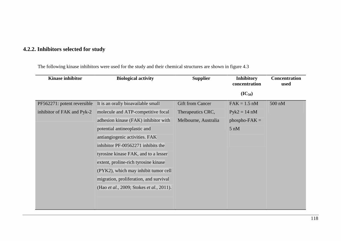
118
4.2.2. Inhibitors selected for study
The following kinase inhibitors were used for the study and their chemical structures are shown in figure 4.3
Kinase inhibitor Biological activity Supplier Inhibitory
concentration
(IC50)
Concentration
used
PF562271: potent reversible
inhibitor of FAK and Pyk-2
It is an orally bioavailable small
molecule and ATP-competitive focal
adhesion kinase (FAK) inhibitor with
potential antineoplastic and
antiangiogenic activities. FAK
inhibitor PF-00562271 inhibits the
tyrosine kinase FAK, and to a lesser
extent, proline-rich tyrosine kinase
(PYK2), which may inhibit tumor cell
migration, proliferation, and survival
(Hao et al., 2009; Stokes et al., 2011).
Gift from Cancer
Therapeutics CRC,
Melbourne, Australia
FAK = 1.5 nM
Pyk2 = 14 nM
phospho-FAK =
5 nM
500 nM
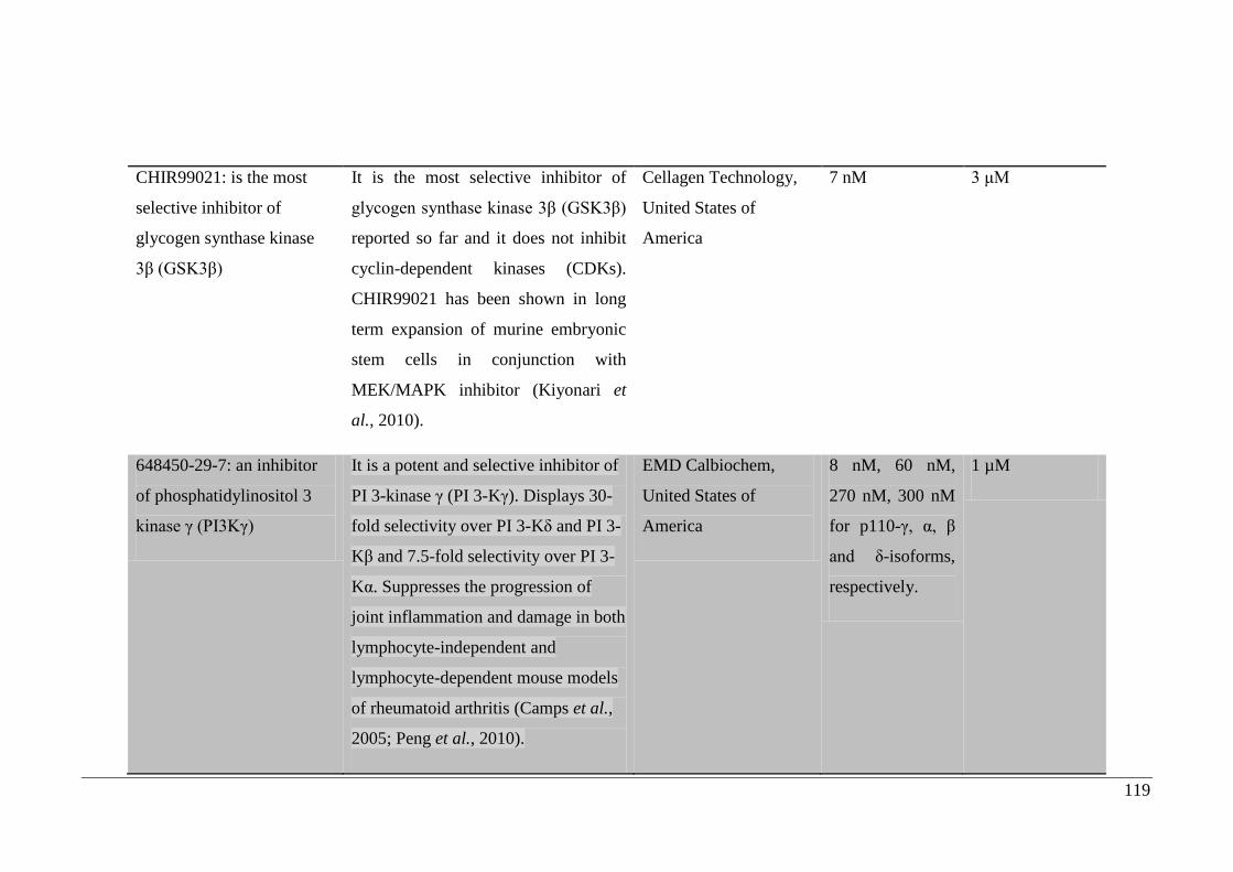
119
CHIR99021: is the most
selective inhibitor of
glycogen synthase kinase
3β (GSK3β)
It is the most selective inhibitor of
glycogen synthase kinase 3β (GSK3β)
reported so far and it does not inhibit
cyclin-dependent kinases (CDKs).
CHIR99021 has been shown in long
term expansion of murine embryonic
stem cells in conjunction with
MEK/MAPK inhibitor (Kiyonari et
al., 2010).
Cellagen Technology,
United States of
America
7 nM 3 μM
648450-29-7: an inhibitor
of phosphatidylinositol 3
kinase γ (PI3Kγ)
It is a potent and selective inhibitor of
PI 3-kinase γ (PI 3-Kγ). Displays 30-
fold selectivity over PI 3-Kδ and PI 3-
Kβ and 7.5-fold selectivity over PI 3-
Kα. Suppresses the progression of
joint inflammation and damage in both
lymphocyte-independent and
lymphocyte-dependent mouse models
of rheumatoid arthritis (Camps et al.,
2005; Peng et al., 2010).
EMD Calbiochem,
United States of
America
8 nM, 60 nM,
270 nM, 300 nM
for p110-γ, α, β
and δ-isoforms,
respectively.
1 µM
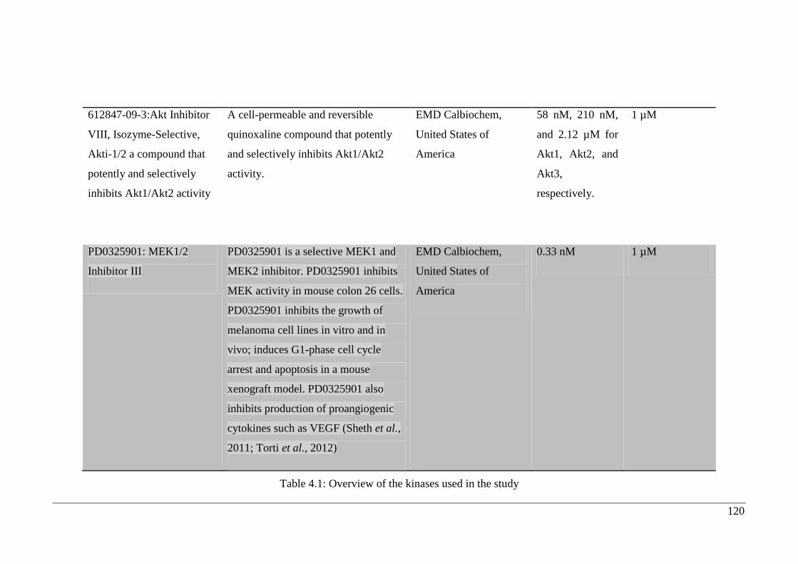
120
612847-09-3:Akt Inhibitor
VIII, Isozyme-Selective,
Akti-1/2 a compound that
potently and selectively
inhibits Akt1/Akt2 activity
A cell-permeable and reversible
quinoxaline compound that potently
and selectively inhibits Akt1/Akt2
activity.
EMD Calbiochem,
United States of
America
58 nM, 210 nM,
and 2.12 µM for
Akt1, Akt2, and
Akt3,
respectively.
1 µM
PD0325901: MEK1/2
Inhibitor III
PD0325901 is a selective MEK1 and
MEK2 inhibitor. PD0325901 inhibits
MEK activity in mouse colon 26 cells.
PD0325901 inhibits the growth of
melanoma cell lines in vitro and in
vivo; induces G1-phase cell cycle
arrest and apoptosis in a mouse
xenograft model. PD0325901 also
inhibits production of proangiogenic
cytokines such as VEGF (Sheth et al.,
2011; Torti et al., 2012)
EMD Calbiochem,
United States of
America
0.33 nM 1 µM
Table 4.1: Overview of the kinases used in the study
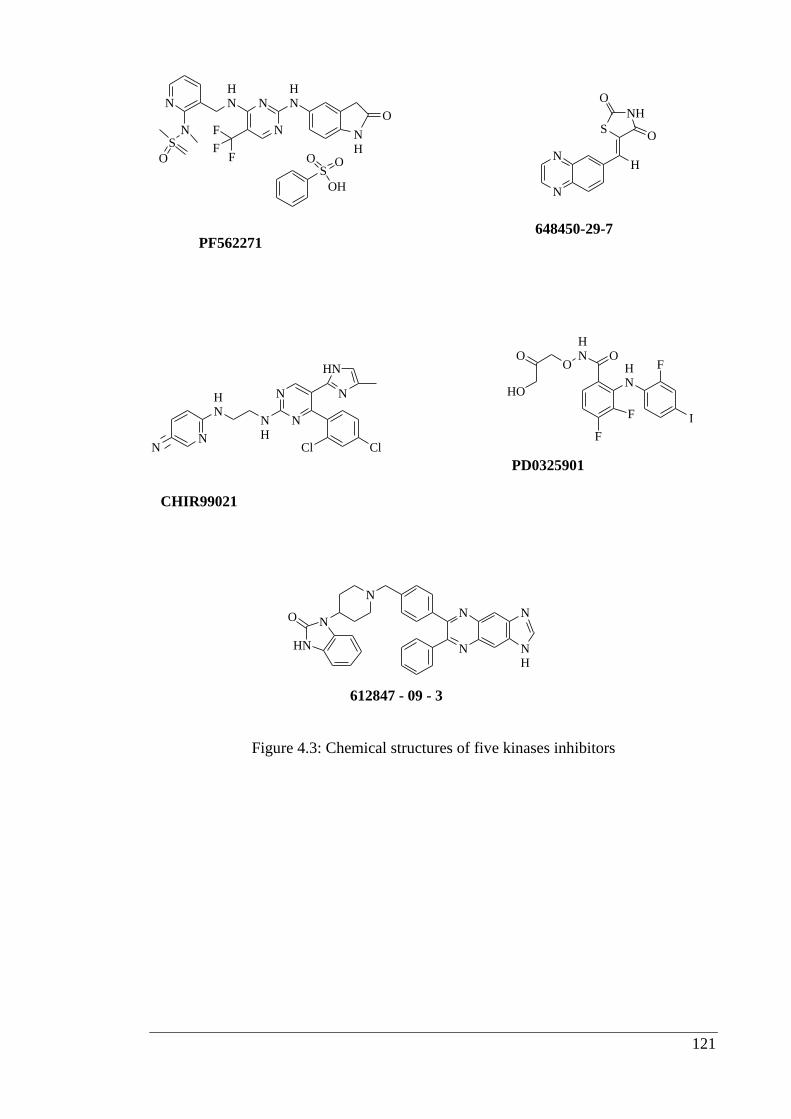
121
Figure 4.3: Chemical structures of five kinases inhibitors
N
N
HN
N
N
S
O
HN
F
FF
NH
O
SO O
OH
PF562271
N
NH
S
NH
O
O
648450-29-7
HN
NH
N
HN
NN
N
N
Cl Cl
CHIR99021
HN
F
F I
F
HN O
O
HO
O
PD0325901
HN
NO
N
N
N N
NH
612847 - 09 - 3
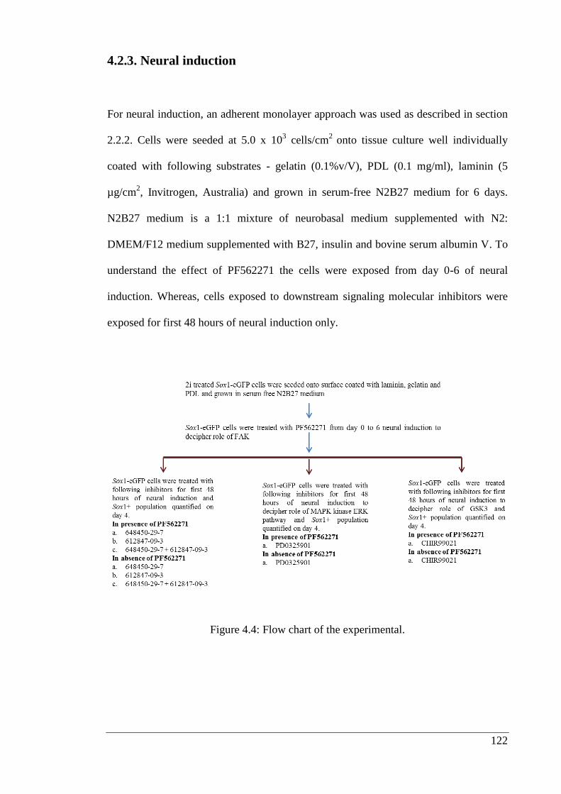
122
4.2.3. Neural induction
For neural induction, an adherent monolayer approach was used as described in section
2.2.2. Cells were seeded at 5.0 x 10³ cells/cm
2 onto tissue culture well individually
coated with following substrates - gelatin (0.1%v/V), PDL (0.1 mg/ml), laminin (5
µg/cm2, Invitrogen, Australia) and grown in serum-free N2B27 medium for 6 days.
N2B27 medium is a 1:1 mixture of neurobasal medium supplemented with N2:
DMEM/F12 medium supplemented with B27, insulin and bovine serum albumin V. To
understand the effect of PF562271 the cells were exposed from day 0-6 of neural
induction. Whereas, cells exposed to downstream signaling molecular inhibitors were
exposed for first 48 hours of neural induction only.
Figure 4.4: Flow chart of the experimental.
123
4.2.4. Trypan blue staining
Trypan blue is a membrane impermeable dye used to discriminate between viable and
non-viable cells. Cell aliquots were mixed with trypan blue dye by preparing a 1:1
dilution of the cell suspension using a 0.4% trypan blue solution in 1X PBS. Non-viable
cells appear blue whereas viable cells will be unstained under microscope.
4.2.5. Flow cytometry
Single cell suspension analysis was performed with a FACS Canto II analyser (BD
Science, Australia). Before analysis, cell suspensions were cleared of clumps by passing
through a 70 µm strainer (BD Biosciences, Australia) and SYTOX red dye was added to
identify dead cells. The mechanism of action of SYTOX red dye has been explained on
page number 82. For the gating of GFP positive cell populations, wild type cells were
submitted to comparable differentiation conditions and used as negative controls. Gates
were set so that 99.0-99.5% of wild type registered as GFP negative. For each analysis
10,000 events were counted.
4.2.5. Statistical analysis
All results are presented as mean ± SEM of at least three separate experiments.
Statistical analyses were performed on raw data using PRISM v5.00 (GraphPad
Software, USA). Data were analysed by Student’s t test or one-way analysis of variance
or two-way analysis of variance (ANOVA) with post-hoc Bonferroni's test. In all cases,
P < 0.05 was considered to be significant.
124
4.3. Results:
4.3.1. Effect of laminin on cell survival
In chapter 2, it was shown that by using adherent culture system, differentiation of
neurons from mESCs was significantly enhanced by laminin as compared to fibronectin
or type IV collagen. Based on these findings laminin was used as the relevant substrate
to identify signaling pathways implicated in survival and differentiation of mESCs.
Gelatin and PDL were used as control substrates.
In the initial experiments, when the analysis was carried out on day 4 of neural
induction, PDL appeared to be able to significantly enhance the percentage of Sox1+
cells in the population as compared to gelatin and laminin. On day 6, there appeared to
be more Sox1+ cells in the gelatin-coated plates (Figure. 4.5 A). There were no obvious
explanations for these observations. However, there were concerns that there were
Sox1-GFP+ cells present on days 1, 2 and 3, very early in the differentiation (Figure 4.5
A). This indicated that the embryonic stem cells used in my study may not have been
fully pluripotent. It has been reported previously that embryonic stem cells grown in
LIF and serum may become partially differentiated.
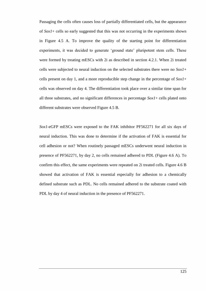
125
Passaging the cells often causes loss of partially differentiated cells, but the appearance
of Sox1+ cells so early suggested that this was not occurring in the experiments shown
in Figure 4.5 A. To improve the quality of the starting point for differentiation
experiments, it was decided to generate ‘ground state’ pluripotent stem cells. These
were formed by treating mESCs with 2i as described in section 4.2.1. When 2i treated
cells were subjected to neural induction on the selected substrates there were no Sox1+
cells present on day 1, and a more reproducible step change in the percentage of Sox1+
cells was observed on day 4. The differentiation took place over a similar time span for
all three substrates, and no significant differences in percentage Sox1+ cells plated onto
different substrates were observed Figure 4.5 B.
Sox1-eGFP mESCs were exposed to the FAK inhibitor PF562271 for all six days of
neural induction. This was done to determine if the activation of FAK is essential for
cell adhesion or not? When routinely passaged mESCs underwent neural induction in
presence of PF562271, by day 2, no cells remained adhered to PDL (Figure 4.6 A). To
confirm this effect, the same experiments were repeated on 2i treated cells. Figure 4.6 B
showed that activation of FAK is essential especially for adhesion to a chemically
defined substrate such as PDL. No cells remained adhered to the substrate coated with
PDL by day 4 of neural induction in the presence of PF562271.
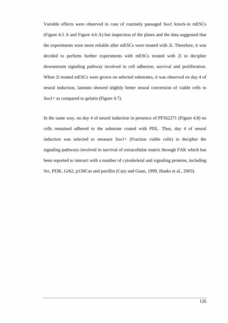
126
Variable effects were observed in case of routinely passaged Sox1 knock-in mESCs
(Figure 4.5 A and Figure 4.6 A) but inspection of the plates and the data suggested that
the experiments were more reliable after mESCs were treated with 2i. Therefore, it was
decided to perform further experiments with mESCs treated with 2i to decipher
downstream signaling pathway involved in cell adhesion, survival and proliferation.
When 2i treated mESCs were grown on selected substrates, it was observed on day 4 of
neural induction, laminin showed slightly better neural conversion of viable cells to
Sox1+ as compared to gelatin (Figure 4.7).
In the same way, on day 4 of neural induction in presence of PF562271 (Figure 4.8) no
cells remained adhered to the substrate coated with PDL. Thus, day 4 of neural
induction was selected to measure Sox1+ (Fraction viable cells) to decipher the
signaling pathways involved in survival of extracellular matrix through FAK which has
been reported to interact with a number of cytoskeletal and signaling proteins, including
Src, PI3K, Grb2, p130Cas and paxillin (Cary and Guan, 1999, Hanks et al., 2003).

127
Figure 4.5: The effect of selected substrates on Sox1+ (Fraction viable cells) quantified
from day 1-6 of neural induction. (A) Routinely passaged Sox1 knock-in mESCs and
(B) 2i treated Sox1 knock-in mESCs. Values are expressed as mean ± SEM of 3
independent experiments. Statistical differences between substrates tested individually
are significant * P < 0.05 and **** P < 0.0001, two-way ANOVA post-hoc
Bonferroni's multiple comparison test.
A
B
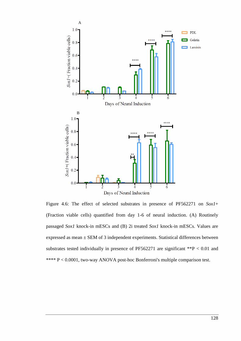
128
Figure 4.6: The effect of selected substrates in presence of PF562271 on Sox1+
(Fraction viable cells) quantified from day 1-6 of neural induction. (A) Routinely
passaged Sox1 knock-in mESCs and (B) 2i treated Sox1 knock-in mESCs. Values are
expressed as mean ± SEM of 3 independent experiments. Statistical differences between
substrates tested individually in presence of PF562271 are significant **P < 0.01 and
**** P < 0.0001, two-way ANOVA post-hoc Bonferroni's multiple comparison test.
A
B
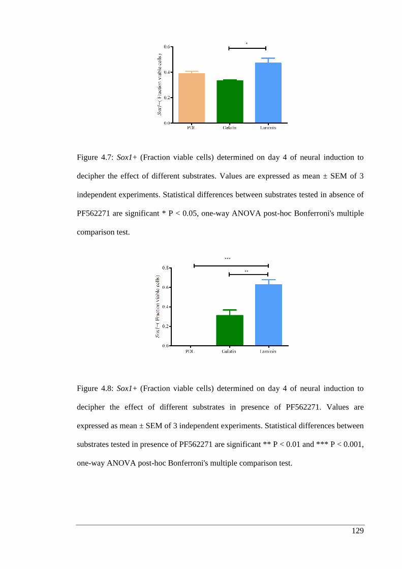
129
Figure 4.7: Sox1+ (Fraction viable cells) determined on day 4 of neural induction to
decipher the effect of different substrates. Values are expressed as mean ± SEM of 3
independent experiments. Statistical differences between substrates tested in absence of
PF562271 are significant * P < 0.05, one-way ANOVA post-hoc Bonferroni's multiple
comparison test.
Figure 4.8: Sox1+ (Fraction viable cells) determined on day 4 of neural induction to
decipher the effect of different substrates in presence of PF562271. Values are
expressed as mean ± SEM of 3 independent experiments. Statistical differences between
substrates tested in presence of PF562271 are significant ** P < 0.01 and *** P < 0.001,
one-way ANOVA post-hoc Bonferroni's multiple comparison test.
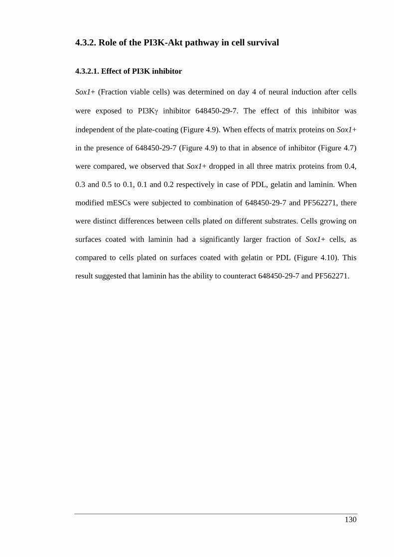
130
4.3.2. Role of the PI3K-Akt pathway in cell survival
4.3.2.1. Effect of PI3K inhibitor
Sox1+ (Fraction viable cells) was determined on day 4 of neural induction after cells
were exposed to PI3K inhibitor 648450-29-7. The effect of this inhibitor was
independent of the plate-coating (Figure 4.9). When effects of matrix proteins on Sox1+
in the presence of 648450-29-7 (Figure 4.9) to that in absence of inhibitor (Figure 4.7)
were compared, we observed that Sox1+ dropped in all three matrix proteins from 0.4,
0.3 and 0.5 to 0.1, 0.1 and 0.2 respectively in case of PDL, gelatin and laminin. When
modified mESCs were subjected to combination of 648450-29-7 and PF562271, there
were distinct differences between cells plated on different substrates. Cells growing on
surfaces coated with laminin had a significantly larger fraction of Sox1+ cells, as
compared to cells plated on surfaces coated with gelatin or PDL (Figure 4.10). This
result suggested that laminin has the ability to counteract 648450-29-7 and PF562271.
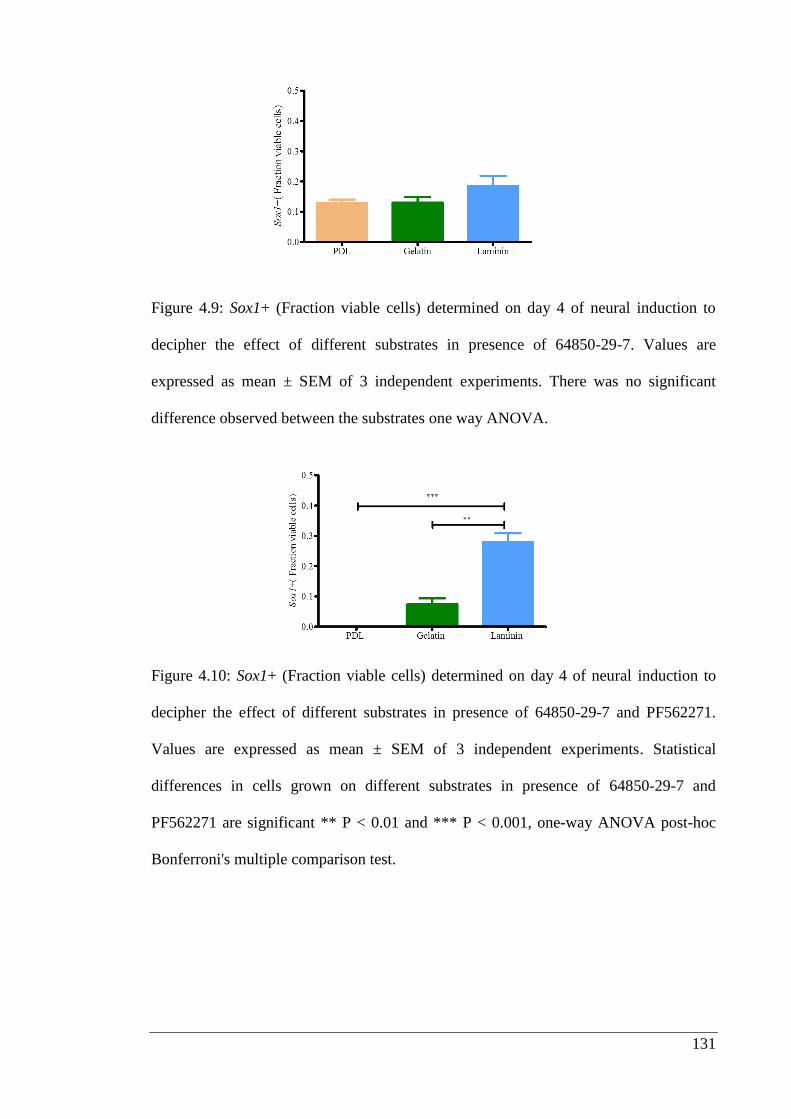
131
Figure 4.9: Sox1+ (Fraction viable cells) determined on day 4 of neural induction to
decipher the effect of different substrates in presence of 64850-29-7. Values are
expressed as mean ± SEM of 3 independent experiments. There was no significant
difference observed between the substrates one way ANOVA.
Figure 4.10: Sox1+ (Fraction viable cells) determined on day 4 of neural induction to
decipher the effect of different substrates in presence of 64850-29-7 and PF562271.
Values are expressed as mean ± SEM of 3 independent experiments. Statistical
differences in cells grown on different substrates in presence of 64850-29-7 and
PF562271 are significant ** P < 0.01 and *** P < 0.001, one-way ANOVA post-hoc
Bonferroni's multiple comparison test.
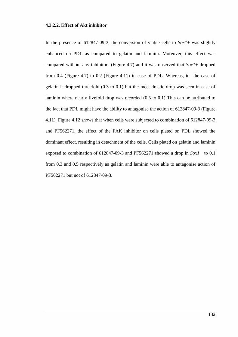
132
4.3.2.2. Effect of Akt inhibitor
In the presence of 612847-09-3, the conversion of viable cells to Sox1+ was slightly
enhanced on PDL as compared to gelatin and laminin. Moreover, this effect was
compared without any inhibitors (Figure 4.7) and it was observed that Sox1+ dropped
from 0.4 (Figure 4.7) to 0.2 (Figure 4.11) in case of PDL. Whereas, in the case of
gelatin it dropped threefold (0.3 to 0.1) but the most drastic drop was seen in case of
laminin where nearly fivefold drop was recorded (0.5 to 0.1) This can be attributed to
the fact that PDL might have the ability to antagonise the action of 612847-09-3 (Figure
4.11). Figure 4.12 shows that when cells were subjected to combination of 612847-09-3
and PF562271, the effect of the FAK inhibitor on cells plated on PDL showed the
dominant effect, resulting in detachment of the cells. Cells plated on gelatin and laminin
exposed to combination of 612847-09-3 and PF562271 showed a drop in Sox1+ to 0.1
from 0.3 and 0.5 respectively as gelatin and laminin were able to antagonise action of
PF562271 but not of 612847-09-3.
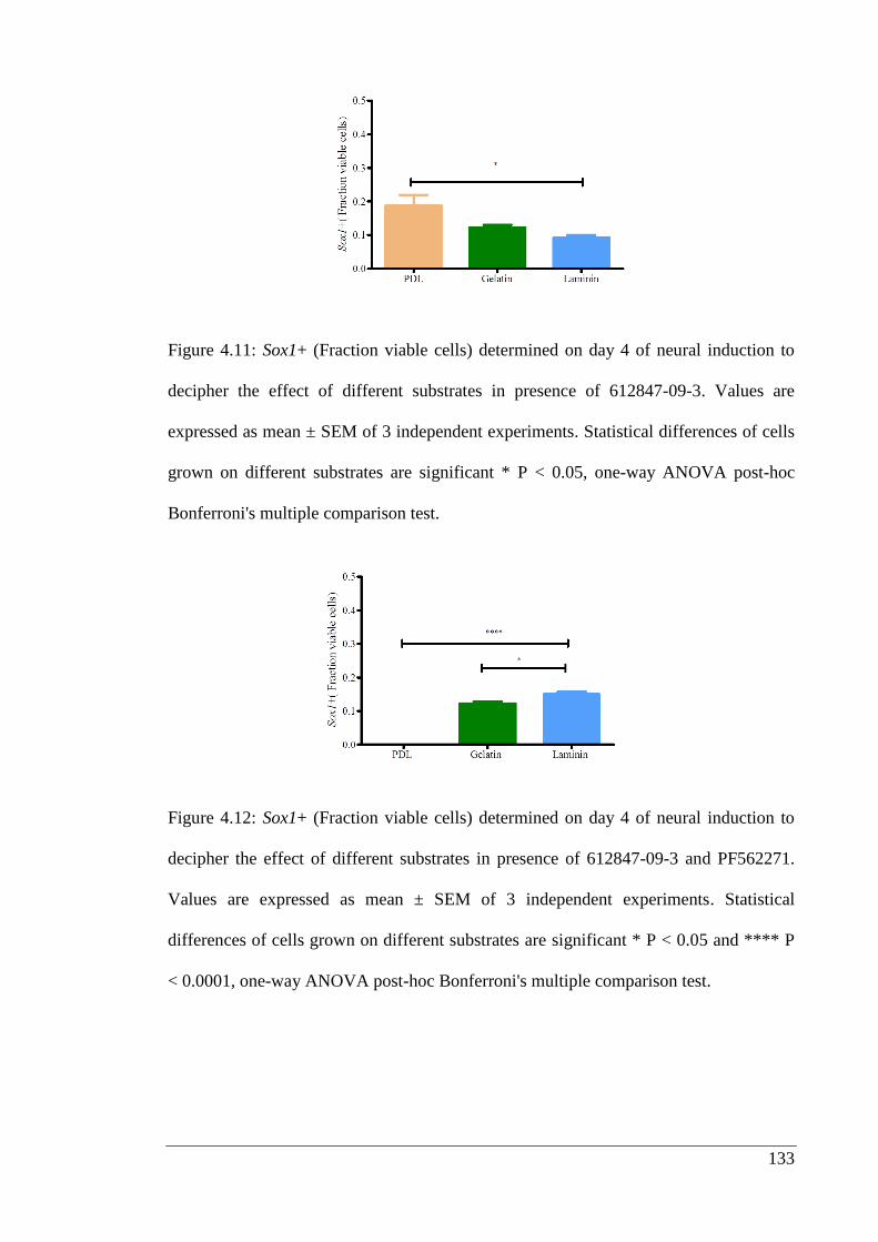
133
Figure 4.11: Sox1+ (Fraction viable cells) determined on day 4 of neural induction to
decipher the effect of different substrates in presence of 612847-09-3. Values are
expressed as mean ± SEM of 3 independent experiments. Statistical differences of cells
grown on different substrates are significant * P < 0.05, one-way ANOVA post-hoc
Bonferroni's multiple comparison test.
Figure 4.12: Sox1+ (Fraction viable cells) determined on day 4 of neural induction to
decipher the effect of different substrates in presence of 612847-09-3 and PF562271.
Values are expressed as mean ± SEM of 3 independent experiments. Statistical
differences of cells grown on different substrates are significant * P < 0.05 and **** P
< 0.0001, one-way ANOVA post-hoc Bonferroni's multiple comparison test.
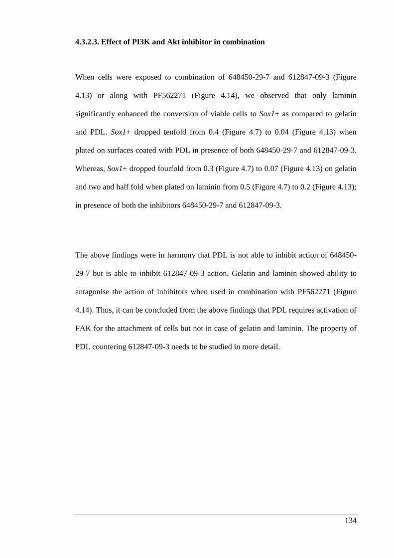
134
4.3.2.3. Effect of PI3K and Akt inhibitor in combination
When cells were exposed to combination of 648450-29-7 and 612847-09-3 (Figure
4.13) or along with PF562271 (Figure 4.14), we observed that only laminin
significantly enhanced the conversion of viable cells to Sox1+ as compared to gelatin
and PDL. Sox1+ dropped tenfold from 0.4 (Figure 4.7) to 0.04 (Figure 4.13) when
plated on surfaces coated with PDL in presence of both 648450-29-7 and 612847-09-3.
Whereas, Sox1+ dropped fourfold from 0.3 (Figure 4.7) to 0.07 (Figure 4.13) on gelatin
and two and half fold when plated on laminin from 0.5 (Figure 4.7) to 0.2 (Figure 4.13);
in presence of both the inhibitors 648450-29-7 and 612847-09-3.
The above findings were in harmony that PDL is not able to inhibit action of 648450-
29-7 but is able to inhibit 612847-09-3 action. Gelatin and laminin showed ability to
antagonise the action of inhibitors when used in combination with PF562271 (Figure
4.14). Thus, it can be concluded from the above findings that PDL requires activation of
FAK for the attachment of cells but not in case of gelatin and laminin. The property of
PDL countering 612847-09-3 needs to be studied in more detail.
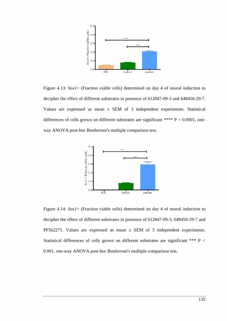
135
Figure 4.13: Sox1+ (Fraction viable cells) determined on day 4 of neural induction to
decipher the effect of different substrates in presence of 612847-09-3 and 648450-29-7.
Values are expressed as mean ± SEM of 3 independent experiments. Statistical
differences of cells grown on different substrates are significant **** P < 0.0001, one-
way ANOVA post-hoc Bonferroni's multiple comparison test.
Figure 4.14: Sox1+ (Fraction viable cells) determined on day 4 of neural induction to
decipher the effect of different substrates in presence of 612847-09-3, 648450-29-7 and
PF562271. Values are expressed as mean ± SEM of 3 independent experiments.
Statistical differences of cells grown on different substrates are significant *** P <
0.001, one-way ANOVA post-hoc Bonferroni's multiple comparison test.
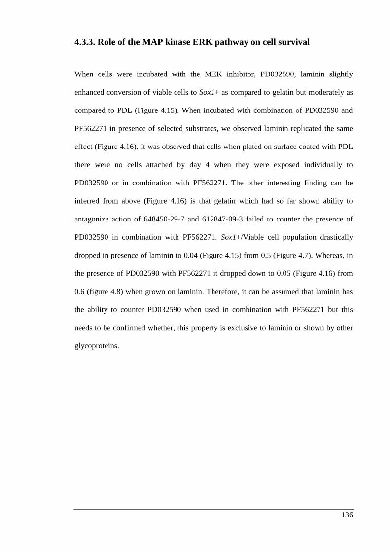
136
4.3.3. Role of the MAP kinase ERK pathway on cell survival
When cells were incubated with the MEK inhibitor, PD032590, laminin slightly
enhanced conversion of viable cells to Sox1+ as compared to gelatin but moderately as
compared to PDL (Figure 4.15). When incubated with combination of PD032590 and
PF562271 in presence of selected substrates, we observed laminin replicated the same
effect (Figure 4.16). It was observed that cells when plated on surface coated with PDL
there were no cells attached by day 4 when they were exposed individually to
PD032590 or in combination with PF562271. The other interesting finding can be
inferred from above (Figure 4.16) is that gelatin which had so far shown ability to
antagonize action of 648450-29-7 and 612847-09-3 failed to counter the presence of
PD032590 in combination with PF562271. Sox1+/Viable cell population drastically
dropped in presence of laminin to 0.04 (Figure 4.15) from 0.5 (Figure 4.7). Whereas, in
the presence of PD032590 with PF562271 it dropped down to 0.05 (Figure 4.16) from
0.6 (figure 4.8) when grown on laminin. Therefore, it can be assumed that laminin has
the ability to counter PD032590 when used in combination with PF562271 but this
needs to be confirmed whether, this property is exclusive to laminin or shown by other
glycoproteins.
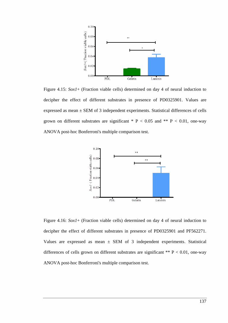
137
Figure 4.15: Sox1+ (Fraction viable cells) determined on day 4 of neural induction to
decipher the effect of different substrates in presence of PD0325901. Values are
expressed as mean ± SEM of 3 independent experiments. Statistical differences of cells
grown on different substrates are significant * P < 0.05 and ** P < 0.01, one-way
ANOVA post-hoc Bonferroni's multiple comparison test.
Figure 4.16: Sox1+ (Fraction viable cells) determined on day 4 of neural induction to
decipher the effect of different substrates in presence of PD0325901 and PF562271.
Values are expressed as mean ± SEM of 3 independent experiments. Statistical
differences of cells grown on different substrates are significant ** P < 0.01, one-way
ANOVA post-hoc Bonferroni's multiple comparison test.
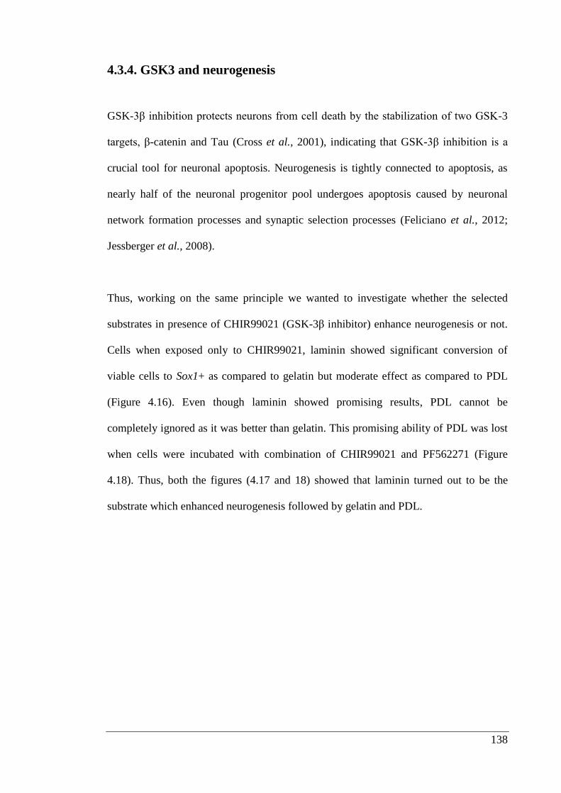
138
4.3.4. GSK3 and neurogenesis
GSK-3β inhibition protects neurons from cell death by the stabilization of two GSK-3
targets, β-catenin and Tau (Cross et al., 2001), indicating that GSK-3β inhibition is a
crucial tool for neuronal apoptosis. Neurogenesis is tightly connected to apoptosis, as
nearly half of the neuronal progenitor pool undergoes apoptosis caused by neuronal
network formation processes and synaptic selection processes (Feliciano et al., 2012;
Jessberger et al., 2008).
Thus, working on the same principle we wanted to investigate whether the selected
substrates in presence of CHIR99021 (GSK-3β inhibitor) enhance neurogenesis or not.
Cells when exposed only to CHIR99021, laminin showed significant conversion of
viable cells to Sox1+ as compared to gelatin but moderate effect as compared to PDL
(Figure 4.16). Even though laminin showed promising results, PDL cannot be
completely ignored as it was better than gelatin. This promising ability of PDL was lost
when cells were incubated with combination of CHIR99021 and PF562271 (Figure
4.18). Thus, both the figures (4.17 and 18) showed that laminin turned out to be the
substrate which enhanced neurogenesis followed by gelatin and PDL.
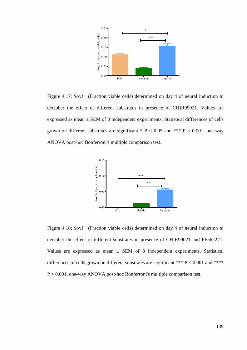
139
Figure 4.17: Sox1+ (Fraction viable cells) determined on day 4 of neural induction to
decipher the effect of different substrates in presence of CHIR99021. Values are
expressed as mean ± SEM of 3 independent experiments. Statistical differences of cells
grown on different substrates are significant * P < 0.05 and *** P < 0.001, one-way
ANOVA post-hoc Bonferroni's multiple comparison test.
Figure 4.18: Sox1+ (Fraction viable cells) determined on day 4 of neural induction to
decipher the effect of different substrates in presence of CHIR99021 and PF562271.
Values are expressed as mean ± SEM of 3 independent experiments. Statistical
differences of cells grown on different substrates are significant *** P < 0.001 and ****
P < 0.001, one-way ANOVA post-hoc Bonferroni's multiple comparison test.
140
4.4. Discussion
4.4.1. Ground state pluripotent stem cells and survival signaling from
extracellular matrix
Induced pluripotent stem cells (iPS) were first generated by Yamanaka and co-workers
group in 2006 by introducing specific genes Klf4, Sox2 and Oct3/4 into adult mouse
cells (Takahashi et al., 2006). They showed that iPS cells were reprogrammed to
resemble embryonic stem cells (Ebben et al., 2011). iPS technology is now commonly
used in stem cell laboratories and has reshaped our understanding of pluripotency.
Austin Smith and co-workers, identified small molecule kinase inhibitors that were able
to maintain embryonic stem cells, in a pluripotent state in the absence of LIF and serum
(Ebben et al., 2011; Silva et al., 2008). Growth of mESCs in this medium, now known
as 2i, contains inhibitors of MAPK signaling and GSK3. Embryonic stem cells grown
in 2i were described as reaching a ground state, which appears to be a more definitive
pluripotent state than mESCs grown in LIF and serum.
FAK is a 125 kD non-membrane bound protein tyrosine kinase that phosphorylates
upon activation of most integrins. The mechanism by which integrins activate FAK is
not known in detail but the process requires an intact action cytoskeleton since
cytochalasin D treatment prevented FAK activation (Shattil et al., 1994). Studies, using
FAK-deficient cells have displayed decrease in motility when plated on surfaces coated
with fibronectin thus, indicating that FAK is necessary for cell adhesion and migration
(Ilic et al., 1995a).
141
Thus, it was interesting to explore what happens when Sox1 knock-in mESCs are
exposed to PF562271. It can be inferred from (Figure 4.6 B) that addition of PF562271
leads to blockage of attachment of cells on surfaces coated with PDL which was not
observed in case of laminin and gelatin. Therefore, it can be inferred from these
findings that activation of FAK is essential for growth on PDL. It can also be inferred
that matrix proteins like laminin and gelatin either have ability to counter presence of
PF562271 or they can activate Src which also has been implicated to play role in cell
adhesion (Graf et al., 2013; Kaplan et al., 1995; Livshits et al., 2012).
4.4.2. PI3K-Akt pathway in cell survival
It has reported that when cells are plated on surfaces coated with laminin or fibronectin,
FAK is activated (Chen and Guan, 1996, Suh and Han, 2010, Xu et al., 2010). This
involves phosphorylation of the Tyr-395 residue of FAK, through which it activates
PI3K, which in turn activates Akt. Cells adhering on surfaces coated with fibronectin
experience an accumulation of PI3K-generated phosphorylated lipids, resulting in
activation of Akt activity (Chen and Guan, 1996, Chen et al., 1996). Dystroglycan, cell
surface receptor for laminin when blocked causes disruption in the PI3K/Akt signaling
pathway. It has been reported that addition of a PI3K inhibitor leads to decreased
phosphorylation of Akt and its downstream effector GSK3, which in turn leads to
detachment of cells (Langenbach and Rando, 2002).
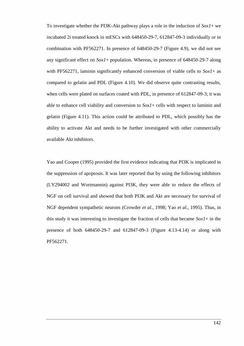
142
To investigate whether the PI3K-Akt pathway plays a role in the induction of Sox1+ we
incubated 2i treated knock in mESCs with 648450-29-7, 612847-09-3 individually or in
combination with PF562271. In presence of 648450-29-7 (Figure 4.9), we did not see
any significant effect on Sox1+ population. Whereas, in presence of 648450-29-7 along
with PF562271, laminin significantly enhanced conversion of viable cells to Sox1+ as
compared to gelatin and PDL (Figure 4.10). We did observe quite contrasting results,
when cells were plated on surfaces coated with PDL, in presence of 612847-09-3; it was
able to enhance cell viability and conversion to Sox1+ cells with respect to laminin and
gelatin (Figure 4.11). This action could be attributed to PDL, which possibly has the
ability to activate Akt and needs to be further investigated with other commercially
available Akt inhibitors.
Yao and Cooper (1995) provided the first evidence indicating that PI3K is implicated in
the suppression of apoptosis. It was later reported that by using the following inhibitors
(LY294002 and Wortmannin) against PI3K, they were able to reduce the effects of
NGF on cell survival and showed that both PI3K and Akt are necessary for survival of
NGF dependent sympathetic neurons (Crowder et al., 1998; Yao et al., 1995). Thus, in
this study it was interesting to investigate the fraction of cells that became Sox1+ in the
presence of both 648450-29-7 and 612847-09-3 (Figure 4.13-4.14) or along with
PF562271.
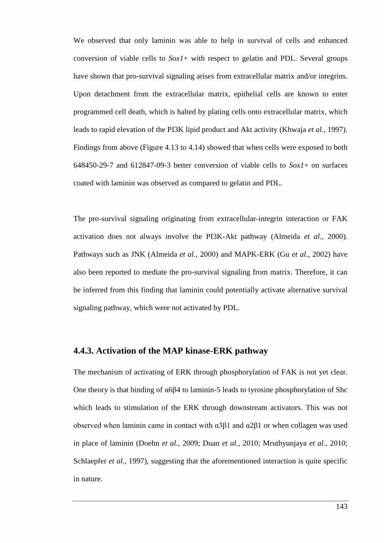
143
We observed that only laminin was able to help in survival of cells and enhanced
conversion of viable cells to Sox1+ with respect to gelatin and PDL. Several groups
have shown that pro-survival signaling arises from extracellular matrix and/or integrins.
Upon detachment from the extracellular matrix, epithelial cells are known to enter
programmed cell death, which is halted by plating cells onto extracellular matrix, which
leads to rapid elevation of the PI3K lipid product and Akt activity (Khwaja et al., 1997).
Findings from above (Figure 4.13 to 4.14) showed that when cells were exposed to both
648450-29-7 and 612847-09-3 better conversion of viable cells to Sox1+ on surfaces
coated with laminin was observed as compared to gelatin and PDL.
The pro-survival signaling originating from extracellular-integrin interaction or FAK
activation does not always involve the PI3K-Akt pathway (Almeida et al., 2000).
Pathways such as JNK (Almeida et al., 2000) and MAPK-ERK (Gu et al., 2002) have
also been reported to mediate the pro-survival signaling from matrix. Therefore, it can
be inferred from this finding that laminin could potentially activate alternative survival
signaling pathway, which were not activated by PDL.
4.4.3. Activation of the MAP kinase-ERK pathway
The mechanism of activating of ERK through phosphorylation of FAK is not yet clear.
One theory is that binding of α6β4 to laminin-5 leads to tyrosine phosphorylation of Shc
which leads to stimulation of the ERK through downstream activators. This was not
observed when laminin came in contact with α3β1 and α2β1 or when collagen was used
in place of laminin (Doehn et al., 2009; Duan et al., 2010; Mruthyunjaya et al., 2010;
Schlaepfer et al., 1997), suggesting that the aforementioned interaction is quite specific
in nature.
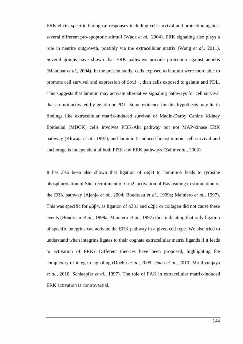
144
ERK elicits specific biological responses including cell survival and protection against
several different pro-apoptotic stimuli (Wada et al., 2004). ERK signaling also plays a
role in neurite outgrowth, possibly via the extracellular matrix (Wang et al., 2011).
Several groups have shown that ERK pathways provide protection against anoikis
(Manohar et al., 2004). In the present study, cells exposed to laminin were more able to
promote cell survival and expression of Sox1+, than cells exposed to gelatin and PDL.
This suggests that laminin may activate alternative signaling pathways for cell survival
that are not activated by gelatin or PDL. Some evidence for this hypothesis may lie in
findings like extracellular matrix-induced survival of Madin-Darby Canine Kidney
Epithelial (MDCK) cells involves PI3K-Akt pathway but not MAP-kinase ERK
pathway (Khwaja et al., 1997), and laminin 5 induced breast tumour cell survival and
anchorage is independent of both PI3K and ERK pathways (Zahir et al., 2003).
It has also been also shown that ligation of α6β4 to laminin-5 leads to tyrosine
phosphorylation of Shc, recruitment of Grb2, activation of Ras leading to stimulation of
the ERK pathway (Ajenjo et al., 2004; Boudreau et al., 1999a; Mainiero et al., 1997).
This was specific for α6β4, as ligation of α3β1 and α2β1 or collagen did not cause these
events (Boudreau et al., 1999a; Mainiero et al., 1997) thus indicating that only ligation
of specific integrins can activate the ERK pathway in a given cell type. We also tried to
understand when integrins ligates to their cognate extracellular matrix ligands if it leads
to activation of ERK? Different theories have been proposed, highlighting the
complexity of integrin signaling (Doehn et al., 2009; Duan et al., 2010; Mruthyunjaya
et al., 2010; Schlaepfer et al., 1997). The role of FAK in extracellular matrix-induced
ERK activation is controversial.
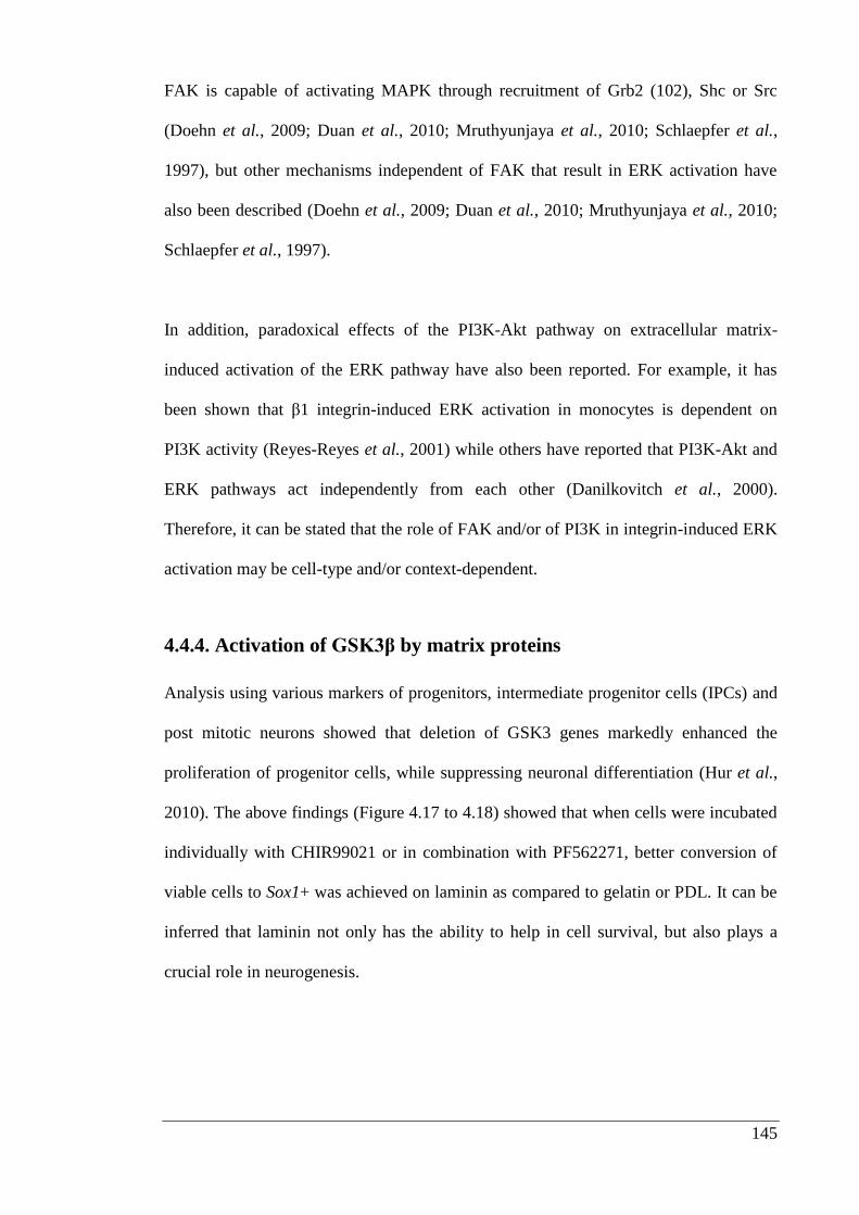
145
FAK is capable of activating MAPK through recruitment of Grb2 (102), Shc or Src
(Doehn et al., 2009; Duan et al., 2010; Mruthyunjaya et al., 2010; Schlaepfer et al.,
1997), but other mechanisms independent of FAK that result in ERK activation have
also been described (Doehn et al., 2009; Duan et al., 2010; Mruthyunjaya et al., 2010;
Schlaepfer et al., 1997).
In addition, paradoxical effects of the PI3K-Akt pathway on extracellular matrix-
induced activation of the ERK pathway have also been reported. For example, it has
been shown that β1 integrin-induced ERK activation in monocytes is dependent on
PI3K activity (Reyes-Reyes et al., 2001) while others have reported that PI3K-Akt and
ERK pathways act independently from each other (Danilkovitch et al., 2000).
Therefore, it can be stated that the role of FAK and/or of PI3K in integrin-induced ERK
activation may be cell-type and/or context-dependent.
4.4.4. Activation of GSK3β by matrix proteins
Analysis using various markers of progenitors, intermediate progenitor cells (IPCs) and
post mitotic neurons showed that deletion of GSK3 genes markedly enhanced the
proliferation of progenitor cells, while suppressing neuronal differentiation (Hur et al.,
2010). The above findings (Figure 4.17 to 4.18) showed that when cells were incubated
individually with CHIR99021 or in combination with PF562271, better conversion of
viable cells to Sox1+ was achieved on laminin as compared to gelatin or PDL. It can be
inferred that laminin not only has the ability to help in cell survival, but also plays a
crucial role in neurogenesis.
146
Numerous studies have demonstrated the importance of β-catenin-mediated Wnt
signaling in proliferation and fate determination during cerebral cortical development
(Gulacsi et al., 2008) and GSK3 activation leads to increase in phosphorylation of β-
catenin (Hur et al., 2010). Therefore, it needs to be further elucidated whether laminin
has the ability to activate β-catenin on its own.
4.5. Conclusion
This study has demonstrated survival of neural stem cells and induction of Sox1+ was
significantly enhanced by laminin as compared to gelatin or PDL. Laminin stimulated
cell survival and neurogenesis. This finding is consistent with data showing that laminin
and other matrix proteins have the ability to activate various downstream kinase
pathways involved in cell survival. Therefore, it can be summarized that laminin, has
the ability to activate both PI3K/Akt and ERK pathway to help in survival of cells. This
effect may be specific to cell type, but mESCs survived and differentiated towards
neural lineages more efficiently on laminin than gelatin or PDL. Thus, the interaction
between mESCs and key matrix proteins appeared to be an early inductive signal to
help regulate neural specification of mESCs. This finding which has not been shown by
any other group so far as per my information will help to gain better knowledge of both
the intracellular pathways involved in cell survival and factors regulating these
signaling pathways. These results are particularly relevant in providing an insight into
signaling pathway activation which leads to increase in survival of stem cells and could
be beneficial especially before transplantation of cells.

147
Chapter 5 Thesis summary and future directions
148
5.1. Introduction
The conclusion and recommendation outlined in this chapter refer to the outlines of the
study, described in Section 1.6. I acknowledge that although the discussion of results
was already detailed in the preceding chapters, a chapter to summarize the results and
discussion of the project is also needed.
This chapter provides a review of the work presented in this thesis and discusses its
implications. Immunocytochemistry, in-cell Western, quantification of primary neurite,
neural progenitor numbers, and total neurite length per neuron were undertaken to
examine the influence of matrix proteins on differentiation of mESCs into neurons.
These experiments were followed by In-cell Western to examine whether it was
possible to immobilize growth factors, including Shh, FGF2 and FGF8b, onto
combination of matrix proteins. It was of interest to examine whether immobilized
growth factors enhanced Lmx1a expression in mESCs. Lastly, a series of small
molecule enzyme inhibitors were used to investigate the signaling pathways involved in
the survival and neural induction of embryonic stem cells using a Sox1-eGFP mESC
reporter cell line.
149
5.2. Influence of extracellular matrix proteins on neurogenesis
In chapter 2, the effect of laminin, fibronectin and type IV collagen on the influence of
adhesion, proliferation and neuronal differentiation of mESCs were investigated. The
preliminary results confirmed that matrix proteins could be coated uniformly onto tissue
culture plates. As the concentration of proteins in the coating solution was increased,
there was an increase in surface area coated. The adhesive properties of the basement
membrane are usually ascribed to the glycoproteins. Laminin is the most abundant
glycoprotein in the basement membrane and it has structural as well as a regulatory
roles (Timpl et al., 1994; Yurchenco et al., 1993). The findings that laminin enhanced
stem cell adhesion and proliferation is consistent with the work of Eccleston et al.
(1985), who plated dissociated adult neurons onto surfaces coated with purified
glycoproteins of the extracellular matrix (Eccleston et al., 1985). There may, however,
be a difference in the thickness and distribution of the extracellular matrix coating on
the surface of well and this may have affected mESCs adhesion and proliferation.
Further studies are necessary to investigate by using technique like scanning electron
microscope. Detailed surface analysis by scanning electron microscope at the micron
level would help us in understanding surface characteristics of matrix proteins under
investigation such as whether these have a smooth or rough surface topography. It is
generally understood that cells prefer to grow on a rough surface, as the filopodia
require surface grooves to attach (Li et al., 2013). However, other research suggests on
the contrary. A study by Gordin et al. (2004) found that cell growth was better on a
smooth rather than rough surface when tested on the same base material (Gordin et al.,
2004).
150
Considering the studies in this area, it appears that the relationship between cell
attachment, growth and surface properties are dependent on the method of testing (in-
vivo or in-vitro), the cell type and the surface characteristics therefore, it would be
necessary to undertake specific cell studies to draw any conclusions on the ability of
cells to attach and grow on the surface of the extracellular matrix proteins.
Immunocytochemistry from chapter 2 show that none of the selected matrix proteins
enhanced the number of nestin positive or βIII tubulin positive cells 24 hours after
plating. Whereas, 96 hours after plating laminin increased the number of βIII tubulin
positive cells. Although, it is not completely clear from this study how and why laminin
enhanced cell number. These studies do indicate that laminin contributes to neural
induction and these effects can be seen after 96 hours of plating. To understand, the way
in which laminin stimulated the neural induction; further time lapse studies could be
done using immunofluorescent staining with different neural markers. It is also
necessary to determine if the same effect of laminin coating is observed on different cell
types.
It has been reported that laminin enhances the densities of serotonergic, dopaminergic
and norepinephrinergic neurons as compared to control, when used as a substrate for
transplantation of brain cells (Eldridge et al., 1989). Thus, working on the same
principle it was decided to test whether selected matrix proteins including laminin,
fibronectin and type IV collagen are able to enhance generation of dopaminergic
neurons from embryonic stem cells.
151
The results presented in chapter 2 sections 2.3.5. showed no significant differences. The
findings in the past decade have demonstrated that laminin enhances generation of
dopaminergic neurons from human neural stem cells (Ostenfeld et al., 2004; Yu et al.,
2007). Human neural stem cells responded to the priming effect of laminin and this
resulted in an increase in the percentage of cholinergic neurons (Tarasenko et al., 2004;
Wu et al., 2002). Therefore, the results obtained in our study suggest that there may be
differences in the effects of matrix proteins on embryonic stem cells and indicate the
need to carry out future studies using human embryonic stem cells.
5.3. Growth factor immobilization
Growth factors are proteins that transmit signals in order to regulate cellular activities
such as differentiation and proliferation (Babensee et al., 2000). They play critical roles
in cell regeneration (e.g. nerve regeneration) or repair of tissues (e.g. bone and
cartilage). Growth factor effects are concentration dependent (Babensee et al., 2000).
Growth factors such as FGF2, BMP and NGF, if presented in bioactive form, can also
influence the cells to produce and secrete other specific growth factors or cytokines,
which can often be part of either wound healing or specific events in development
(Babensee et al., 2000; Luginbuehl et al., 2004; Masters, 2011). Physical adsorption,
physical entrapment and covalent attachment are three common ways of attaching
growth factors onto surfaces (Masters, 2011; Wood et al., 2010).
152
Physical adsorption utilizes weak nonspecific intermolecular interactions such as
hydrogen bonding, hydrophobic interactions, Van der Waals forces, and valence
electron interactions (Vladkova, 2010; Zhang et al., 2012b). In vivo and in vitro heparin
has been shown to help in binding, preserving the stability and biological activity of the
growth factors (Arisaka et al., 2013; Lee et al., 2012a; Seo et al., 2012). Growth factors
have been incorporated into the scaffold matrix either by bulk encapsulation, specific or
nonspecific surface adsorption or by adding microspheres encapsulating them (Wood et
al., 2010). Thus, working on these principles tried to covalently immobilize growth
factors onto heparin and the results generated in chapter 3 showed that the growth
factors like FGF2 and FGF8b were successfully immobilized which also increased the
effectiveness of FGF8b and FGF2, as indicated in section 3.3.2 and 3.3.3..
Although these results show that physical adsorption is an effective way to immobilize
growth factors to the surface, coating only provides a transient modification of the
material surface (Vladkova, 2010). The inability to control the growth factor
conformation and orientation upon the adsorbing substrate, factor desorption (wash-
off), diffusion kinetics, and inaccessibility to large molecules on the material surfaces
are the drawbacks of this method (Laurencin et al., 2009; Lee et al., 2012b).
153
More refined approaches are available for the immobilization via chemical methods or
onto plasma-treated surfaces can be used to introduce active functional groups
(Laurencin et al., 2009). Plasma treatments under a wide range of reacting gases types
(ammonia, nitrogen, hydrogen, oxygen, and ozone) have been employed to introduce
various functional groups (e.g., carboxyl, hydroxyl, carbonyl, ether, peroxide, and
amine) onto the material surface (Vladkova, 2010). Chemically inert
polydimethylsiloxane (PDMS) was simultaneously mixed with acrylic acid (AA) and 2-
hydroxyethyl methacrylate (HEMA) employing so-called “two-step plasma treatment”,
followed by collagen immobilization to study the cellular responses to modified
surfaces. Such surface design significantly increased the number of the adhered and
proliferated cells (Almutairi et al., 2012; Vladkova, 2010; Zhou et al., 2012).
Yoon et al. (2006) also fabricated macroporous poly lactide-co-glycolide (PLGA)
scaffolds using blending mixture of PLGA and NH2-PEG-PLGA to generate surface
amine groups for heparin immobilization (Yoon et al., 2006). Interestingly heparin
immobilized microspheres also release bioactive FGF2 in a sustained manner and
exhibit pronounced angiogenic effect in an animal model (Kim et al., 2006).
Embryonic stem cells are pluripotent cells, which means they can be expanded
indefinitely, and if exposed to appropriate environmental conditions, can be
differentiated into target cell types like neurons and cardiomyocytes (Luginbuehl et al.,
2004). In the past few years, a considerable volume of research has been carried out to
utilise the potential of embryonic stem cells to help in the treatment of nerve injury,
however most findings failed to demonstrate a dramatic increase in functional recovery
(Johnson et al., 2010; Wood et al., 2010) following cell transplant.
154
One of the hypothesis for the failure of this approach is the cells were not exposed to an
appropriate microenvironment before transplant hence there has been a strong focus on
delivery of growth factors either through fibrin scaffolds, heparin alginate gels, cross-
linking with collagen, or by integrating poly (lactide-co-glycolide) as a scaffold
(Luginbuehl et al., 2004; Masters, 2011).
My attempts to provide growth factors in an immobilized form did not produce any
strong desired effects. Several mechanisms could explain this effect firstly; presence of
different extracellular matrix proteins could modulate the cellular response of the
immobilized growth factors. To illustrate this theory, a group in Japan tried using a
combinatorial approach the addition of growth factors and extracellular matrix proteins,
but they found that the cellular response to immobilized growth factors differed
depending upon the extracellular matrix coating present on the culture substrates. For
instance, the differentiation of neural stem cells to astrocytes was promoted on
fibronectin coated substrates modified with immobilized NGF, but inhibited when the
NGF was immobilized to laminin coated substrates (Nakajima et al., 2007; Saik et al.,
2011). Secondly, the temporal aspects of exposure may have been sub-optimal. The six
day exposure may have been too short. Evidence indicates that increasing the exposure
time during neural induction from 6 to 10 days alters the outcome substantially
(Colleoni et al., 2010; Koch et al., 2009a; Zeng et al., 2011).
155
In order to fabricate biomimetic materials that can promote long-term survival, a stable
immobilization of such bimolecular motives to the substrate surface is critical to
maintain the bioactivity and ultimately proper function (Kollmer et al., 2012; Rogers et
al., 2012; Skop et al., 2013). The development of a biomimetic microenvironment
conducive to cell adhesion, proliferation, differentiation and host tissue integration
requires a more in-depth investigation of the mechanisms of protein adsorption and
reorganization, as well as of the bio adhesion and cell/biomaterial and cell/extra cellular
matrix interactions, cell signaling and cell growth biology.
5.4. Cell matrix interactions
Several reports have shown that integrins activate several signaling pathways that
regulate pro-survival effects (Discher et al., 2009; Roberts et al., 2008). These signaling
pathways involve the, FAK, PI3K, Akt and the MAP kinase ERK. However, the
relevance of a given signaling molecule in mediating the pro-survival signaling induced
by extracellular matrix appears to be cell type specific. It was therefore necessary to
investigate which of these intracellular signaling pathways might be involved in the
effects of extracellular matrix on survival and differentiation of Sox1 eGFP mESCs.
In chapter 4, my initial findings indicated that the effects of selected substrates on the
Sox1+ population during days 1 to 6 of neural induction was that; PDL significantly
enhanced the number of cells expressing Sox1, as compared to gelatin and laminin, but
by day 6, there were more Sox1+ cells on gelatin surface. Subsequently, cells were
incubated with an inhibitor of FAK. This enzyme is known to play a major role in
adhesion (Hanks et al., 1992; Hynes et al., 1992).
156
In the presence of the FAK inhibitor, no cells remained attached to plates coated with
PDL, suggesting that activation of FAK is essential for cells to adhere onto surfaces
coated with PDL. We have observed that passaging of mESCs with LIF and serum over
time does not maintain cells in the pluripotent state and it’s supported by findings that
cells become more specialized and restricted in their developmental potential (Graf et
al., 2009; Murry et al., 2008; Sato et al., 2004).
Therefore, to make sure that initial findings obtained were due to blockage of the FAK
signaling pathway, I decided to generate ground state pluripotent stem cells as described
in section 4.2.1. 2i treated Sox1 eGFP mESCs were therefore, used to investigate the
signaling pathways involved in supportive effects of extracellular matrix proteins. I
showed that laminin and gelatin (which is a mixture of collagens) (Wu et al., 2002)
were able to offer protection from the FAK inhibitor PF562271, which was not seen in
case of PDL. This finding with PDL needs to be further investigated with other
chemically defined substrates like peptide acrylate and poly [2-
(methacryloyloxy) ethyl dimethyl-(3-sulfopropyl) ammonium hydroxide] (PMEDSAH),
which have been reported to sustain long term human embryonic stem cell growth in
several different culture medium (Villa-Diaz et al., 2010).
FAK is known to play a critical role in survival of cells by enhancing their adhesion
onto substrates (Ilic, 2006; Ilic et al., 1995b; Owens et al., 1995), particularly inactive
substrates. Natural matrix proteins may be able to activate alternative pathways through
which cells remained adherent. To investigate the roles of the PI3K-Akt signaling
module and the MAP kinase ERK pathway in the pro-survival effect of the matrix
proteins, these enzymes were blocked in turn using known pharmacological inhibitors.
157
Inhibition of the PI3K-Akt pathway showed that laminin enhanced conversion of Sox1+
cells as compared to gelatin and PDL. One unexpected and particularly interesting
result was the finding that when cells were incubated with 612847-09-3, conversion of
viable cells to Sox1+ cell was moderately enhanced on PDL as compared to gelatin and
laminin. The survival effect of gelatin and PDL was greatly abrogated when ERK
pathway was inhibited.
These findings suggested that laminin has the ability to activate both PI3K/Akt and
ERK pathway to help in survival of cells. This is consistent with the role of specialized
integrin-extracellular matrix ligand interactions regulate many cell functions, including
survival, proliferation, motility, morphology, and differentiation in a cell/tissue-specific
and time-dependent manner. α2β1 mediated cell attachment to type I collagen
stimulates the tyrosine phosphorylation of FAK and, subsequently, the activation of
ERK and MAPK that has been implicated in the control of osteoblast-specific gene
expression and matrix mineralization (Fu et al., 2013; Xu et al., 2012). Integrin α5β1 is
suggested to be involved in cell matrix adhesion (Sekine et al., 2012). Research groups
have demonstrated that by increasing micro scale roughness of substrates promoted
greater α5β1 binding and FAK phosphorylation in osteoblasts on fibronectin adsorbed
surfaces (Rechenmacher et al., 2013). Given the crucial and overlapping roles, multiple
integrins play in cell signaling and functional pathways it would be worthwhile to
explore whether β1 integrin was required for the responses shown by laminin in chapter
4.
158
The results in chapter 4 showed that laminin in presence of CHIR99021 enhanced
conversion of viable cells to Sox1+ with respect to gelatin and PDL. Blocking GSK3β
activity is analogous with activation of Wnt. Hence, the role of matrix proteins in
activating the Wnt pathway will provide us with new tools to help control stem cell
survival and differentiation (Munji et al., 2011). Migration of neural crest cells is an
intricate process that requires the delamination of cells from an epithelium and
movement into an extracellular matrix. It has been reported that the canonical Wnt
signaling pathway is needed for neural crest induction, while the non-canonical Wnt
pathway is required for neural crest migration (Munji et al., 2011).
By performing time-lapse analysis of cell movement and cell protrusion in neural crest
cultured on a laminin substrate, it would be possible to clarify whether the effects of
CHIR99021 are due to canonical or non-canonical Wnt signaling that controls neural
crest migration. This experiment lays the groundwork for more detailed studies
that could examine various matrix proteins, time points, and cell densities that may
contribute to the above effect from cells specifications. Although 2-D coatings have
been used in this study, an abundance of clinical applications rely on 3-D scaffolds for
nerve regeneration (Blumenthal et al., 2013; Khaing et al., 2012). Studies have
suggested that cells behave quite differently in 3-D environments than 2-D (Berneel et
al., 2012; Buttery et al., 2011; Lin et al., 2012; Qutachi et al., 2013b). Examination of
these substrate specific effects in 3-D physical environments would be an important
initial step towards application of the substrate specific strategy for nerve regenerative
purpose.
159
5.5 Conclusion
In summary, this research was undertaken to determine the potential effect of matrix
proteins for use in differentiation of stem cells into neurons. The results from this study
indicate that matrix proteins are important for differentiation, although further in vitro
testing is required to gain a more comprehensive understanding of the matrix effects on
differentiation of stem cells in neurons, and whether they have ability to enhance the
differentiation of specific subtypes of neurons. The effects of laminin need to be further
investigated with other matrix proteins like fibronectin, type IV collagen and
vitronectin. It can be concluded from this study that laminin which is most conducive
for generation of neurons and has the complex ability to promote cell survival either
through activation of PI3K-Akt or MAPK-ERK pathway.
160
References
Agarwala S, Sanders TA, Ragsdale CW (2001). Sonic hedgehog control of size and
shape in midbrain pattern formation. Science 291(5511): 2147-2150.
Ajenjo N, Cañón E, Sánchez-Pérez I, Matallanas D, León J, Perona R, et al. (2004).
Subcellular localization determines the protective effects of activated ERK2 against
distinct apoptogenic stimuli in myeloid leukemia cells. The Journal of Biological
Chemistry 279(31): 32813-32823.
Alberti K, Davey RE, Onishi K, George S, Salchert K, Seib FP, et al. (2008).
Functional immobilization of signaling proteins enables control of stem cell fate. Nature
Methods 5(7): 645-650.
Ali SA, Pappas IS, Parnavelas JG (1998). Collagen type IV promotes the differentiation
of neuronal progenitors and inhibits astroglial differentiation in cortical cell cultures.
Devlopment Brain Research 110(1): 31-38.
Almeida EA, Ilic D, Han Q, Hauck CR, Jin F, Kawakatsu H, et al. (2000). Matrix
survival signaling: from fibronectin via focal adhesion kinase to c-Jun NH(2)-terminal
kinase. The Journal of Cell Biology 149(3): 741-754.
Almutairi Z, Ren CL, Simon L (2012). Evaluation of polydimethylsiloxane (PDMS)
surface modification approaches for microfluidic applications. Colloids and Surfaces A:
Physicochemical and Engineering Aspects 415(0): 406-412.
Andersson E, Tryggvason U, Deng Q, Friling S, Alekseenko Z, Robert B, et al. (2006).
Identification of intrinsic determinants of midbrain dopamine neurons. Cell 124(2):
393-405.
Arcaro A, Zvelebil MJ, Wallasch C, Ullrich A, Waterfield MD, Domin J (2000). Class
II phosphoinositide 3-kinases are downstream targets of activated polypeptide growth
factor receptors. Molecular and Cellular Biology 20(11): 3817-3830.
Arisaka Y, Kobayashi J, Yamato M, Akiyama Y, Okano T (2013). Switching of cell
growth/detachment on heparin-functionalized thermoresponsive surface for rapid cell
sheet fabrication and manipulation. Biomaterials 34(17):4214-4222.
Arnaoutova I, George J, Kleinman HK, Benton G (2012). Basement membrane matrix
(BME) has multiple uses with stem cells. Stem Cell Reviews and Reports 8(1): 163-169.
161
Ashikari-Hada S, Habuchi H, Kariya Y, Itoh N, Reddi AH, Kimata K (2004).
Characterization of Growth Factor-binding Structures in Heparin/Heparan Sulfate Using
an Octasaccharide Library. Journal of Biological Chemistry 279(13): 12346-12354.
Aubert J, Stavridis MP, Tweedie S, O'Reilly M, Vierlinger K, Li M, et al. (2003).
Screening for mammalian neural genes via fluorescence-activated cell sorter
purification of neural precursors from Sox1-gfp knock-in mice. Proceedings of the
National Academy of Sciences of the United States of America 100 Suppl 1: 11836-
11841.
Aumailley M, Bruckner-Tuderman L, Carter WG, Deutzmann R, Edgar D, Ekblom P, et
al. (2005). A simplified laminin nomenclature. Matrix Biology 24(5): 326-332.
Babensee JE, McIntire LV, Mikos AG (2000). Growth factor delivery for tissue
engineering. Pharmaceutical Research 17(5): 497-504.
Bain G, Ray WJ, Yao M, Gottlieb DI (1996). Retinoic acid promotes neural and
represses mesodermal gene expression in mouse embryonic stem cells in culture.
Biochemical and Biophysical Research Communications 223(3): 691-694.
Baizabal JM, Covarrubias L (2009). The embryonic midbrain directs neuronal
specification of embryonic stem cells at early stages of differentiation. Developmental
Biology 325(1): 49-59.
Balasubramaniyan V, de Haas AH, Bakels R, Koper A, Boddeke HW, Copray JC
(2004). Functionally deficient neuronal differentiation of mouse embryonic neural stem
cells in vitro. Neuroscience Research 49(2): 261-265.
Barberi T, Klivenyi P, Calingasan NY (2003). Neural subtype specification of
fertilization and nuclear transfer embryonic stem cells and application in parkinsonian
mice. Nature Biotechnology 21: 1200.
Baron-Van Evercooren A, Kleinman HK, Ohno S, Marangos P, Schwartz JP, Dubois-
Dalcq ME (1982). Nerve growth factor, laminin, and fibronectin promote neurite
growth in human fetal sensory ganglia cultures. Journal of Neuroscience Research 8(2-
3): 179-193.
Bernardes GJL, Castagner B, Seeberger PH (2009). Combined approaches to the
synthesis and study of glycoproteins. ACS Chemical Biology 4(9): 703-713.
Berneel E, Desmet T, Declercq H, Dubruel P, Cornelissen M (2012). Double protein‐coated poly‐ε‐caprolactone scaffolds: Successful 2D to 3D transfer. Journal of
Biomedical Materials Research Part A 100(7): 1783-1791.
162
Bilozur ME, Hay ED (1988). Neural crest migration in 3D extracellular matrix utilizes
laminin, fibronectin, or collagen. Developmental Biology 125(1): 19-33.
Blumenthal J, Cohen-Matsliah SI, Levenberg S (2013). Olfactory bulb-derived cells
seeded on 3D scaffolds exhibit neurotrophic factor expression and pro-angiogenic
properties. Tissue Engineering Part A 19(19-20):2284-2291.
Bosman FT, Stamenkovic I (2003). Functional structure and composition of the
extracellular matrix. The Journal of Pathology 200(4): 423-428.
Boudreau N, Sympson CJ, Werb Z, Bissell MJ (1995). Suppression of ICE and
apoptosis in mammary epithelial cells by extracellular matrix. Science 267(5199): 891-
893.
Boudreau NJ, Jones PL (1999a). Extracellular matrix and integrin signalling: the shape
of things to come. Biochemical Journal 339(Pt 3): 481.
Boudreau NJ, Jones PL (1999b). Extracellular matrix and integrin signalling: the shape
of things to come. Biochemical Journal 339 ( Pt 3): 481-488.
Brynda E, Pachernik J, Houska M, Pientka Z, Dvorak P (2005). Surface immobilized
protein multilayers for cell seeding. Langmuir 21(17): 7877-7883.
Burgeson RE, Chiquet M, Deutzmann R, Ekblom P, Engel J, Kleinman H, et al. (1994).
A new nomenclature for the laminins. Matrix Biology 14(3): 209-211.
Burridge K, Fath K, Kelly T, Nuckolls G, Turner C (1988). Focal adhesions:
transmembrane junctions between the extracellular matrix and the cytoskeleton. Annual
Review of Cell Biology 4: 487-525.
Buttery L, Bielby R, Howard D, Shakesheff K (2011). Osteogenic Differentiation of
Embryonic Stem Cells in 2D and 3D Culture. In: Haycock JW (eds). 3D Cell Culture,
edn, Vol. 695: Humana Press: 281-308.
Buytaert-Hoefen KA, Alvarez E, Freed CR (2004). Generation of tyrosine hydroxylase
positive neurons from human embryonic stem cells after coculture with cellular
substrates and exposure to GDNF. Stem Cells 22(5): 669-674.
Cai J, Donaldson A, Yang M, German MS, Enikolopov G, Iacovitti L (2009). The role
of Lmx1a in the differentiation of human embryonic stem cells into midbrain dopamine
neurons in culture and after transplantation into a Parkinson's disease model. Stem Cells
27(1): 220-229.
163
Calalb MB, Fox DT, Hanks SK (1992). Molecular cloning and enzymatic analysis of
the rat homolog of "PhK-gamma T," an isoform of phosphorylase kinase catalytic
subunit. The Journal of Biological Chemistry 267(3): 1455-1463.
Calalb MB, Zhang X, Polte TR, Hanks SK (1996). Focal adhesion kinase tyrosine-861
is a major site of phosphorylation by Src. Biochemical and Biophysical Research
Communications 228(3): 662-668.
Calof AL, Lander AD (1991). Relationship between neuronal migration and cell-
substratum adhesion: laminin and merosin promote olfactory neuronal migration but are
anti-adhesive. The Journal of Cell Biology 115(3): 779-794.
Campbell A, Wicha MS, Long M (1985). Extracellular matrix promotes the growth and
differentiation of murine hematopoietic cells in vitro. Journal of Clinical Investigation
75(6): 2085.
Camps M, Ruckle T, Ji H, Ardissone V, Rintelen F, Shaw J, et al. (2005). Blockade of
PI3Kgamma suppresses joint inflammation and damage in mouse models of rheumatoid
arthritis. Nature medicine 11(9): 936-943.
Cantley LC (2002). The phosphoinositide 3-kinase pathway. Science 296(5573): 1655-
1657.
Capila I, Linhardt RJ (2002). Heparin-protein interactions. Angewandte chemie
international edition 41(3): 391-412.
Carbonetto S, Cochard P (1987). In vitro studies on the control of nerve fiber growth by
the extracellular matrix of the nervous system. Journal of Physiology - Paris 82(4):
258-270.
Carri NG, Perris R, Johansson S, Ebendal T (1988). Differential outgrowth of retinal
neurites on purified extracellular matrix molecules. Journal of Neuroscience Research
19(4): 428-439.
Cary LA, Chang JF, Guan JL (1996). Stimulation of cell migration by overexpression of
focal adhesion kinase and its association with Src and Fyn. Journal of Cell Science 109
( Pt 7): 1787-1794.
Chan PC, Lai JF, Cheng CH, Tang MJ, Chiu CC, Chen HC (1999). Suppression of
ultraviolet irradiation-induced apoptosis by overexpression of focal adhesion kinase in
Madin-Darby canine kidney cells. The Journal of Biological Chemistry 274(38): 26901-
26906.
164
Chang S-C, Mulloy B, Magee AI, Couchman JR (2011). Two distinct sites in sonic
hedgehog combine for heparan sulfate interactions and cell signaling functions. The
Journal of Biological Chemistry 286: 44391-44402.
Charonis AS, Tsilibary EC, Yurchenco PD, Furthmayr H (1985). Binding of laminin to
type IV collagen: a morphological study. The Journal of Cell Biology 100(6): 1848-
1853.
Chen SS, Fitzgerald W, Zimmerberg J, Kleinman HK, Margolis L (2007a). Cell-cell
and cell-extracellular matrix interactions regulate embryonic stem cell differentiation.
Stem Cells 25(3): 553-561.
Chen SS, Fitzgerald W, Zimmerberg J, Kleinman HK, Margolis L (2007b). Cell-cell
and cell-extracellular matrix interactions regulate Embryonic stem cell differentiation.
Stem Cells 25(3): 553-561.
Chen ZL, Indyk JA, Strickland S (2003a). The hippocampal laminin matrix is dynamic
and critical for neuronal survival. Molecular Biology of the Cell 14(7): 2665-2676.
Chen ZL, Strickland S (2003b). Laminin gamma1 is critical for Schwann cell
differentiation, axon myelination, and regeneration in the peripheral nerve. The Journal
of Cell Biology 163(4): 889-899.
Chiu LL, Radisic M (2010). Scaffolds with covalently immobilized VEGF and
Angiopoietin-1 for vascularization of engineered tissues. Biomaterials 31(2): 226-241.
Chizhikov VV, Millen KJ (2004). Control of roof plate formation by Lmx1a in the
developing spinal cord. Development 131(11): 2693-2705.
Cho SY, Klemke RL (2000). Extracellular-regulated kinase activation and CAS/Crk
coupling regulate cell migration and suppress apoptosis during invasion of the
extracellular matrix. The Journal of Cell Biology 149(1): 223-236.
Cohen MA, Itsykson P, Reubinoff BE (2010). The role of FGF-signaling in early neural
specification of human embryonic stem cells. Developmental Biology 340(2): 450-458.
Cohen P, Goedert M (2004). GSK3 inhibitors: development and therapeutic potential.
Nature Reviews Drug Discovery 3(6): 479-487.
165
Colleoni S, Galli C, Giannelli SG, Armentero M-T, Blandini F, Broccoli V, et al.
(2010). Long-term culture and differentiation of CNS precursors derived from anterior
human neural rosettes following exposure to ventralizing factors. Experimental Cell
Research 316(7): 1148-1158.
Colognato H, Yurchenco PD (2000). Form and function: the laminin family of
heterotrimers. Developmental Dynamics 218(2): 213-234.
Cooke MJ, Zahir T, Phillips SR, Shah DS, Athey D, Lakey JH, et al. (2010). Neural
differentiation regulated by biomimetic surfaces presenting motifs of extracellular
matrix proteins. Journal of Biomedical Materials Research Part A 93(3): 824-832.
Costa-Silva B, da Costa MC, Melo FR, Neves CM, Alvarez-Silva M, Calloni GW, et al.
(2009). Fibronectin promotes differentiation of neural crest progenitors endowed with
smooth muscle cell potential. Experimental Cell Research 315(6): 955-967.
Craddock BL, Hobbs J, Edmead CE, Welham MJ (2001). Phosphoinositide 3-kinase-
dependent regulation of interleukin-3-induced proliferation: involvement of mitogen-
activated protein kinases, SHP2 and Gab2. The Journal of Biological Chemistry
276(26): 24274-24283.
Cross DA, Culbert AA, Chalmers KA, Facci L, Skaper SD, Reith AD (2001). Selective
small-molecule inhibitors of glycogen synthase kinase-3 activity protect primary
neurones from death. Journal of Neurochemistry 77(1): 94-102.
Crowder RJ, Freeman RS (1998). Phosphatidylinositol 3-kinase and Akt protein kinase
are necessary and sufficient for the survival of nerve growth factor-dependent
sympathetic neurons. Journal of Neuroscience 18(8): 2933.
Czyz J, Wobus A (2001). Embryonic stem cell differentiation: the role of extracellular
factors. Differentiation 68(4-5): 167-174.
Daley WP, Peters SB, Larsen M (2008). Extracellular matrix dynamics in development
and regenerative medicine. Journal of Cell Science 121(Pt 3): 255-264.
Danen EH, Yamada KM (2001). Fibronectin, integrins, and growth control. Journal of
Cellular Physiology 189(1): 1-13.
Danilkovitch A, Donley S, Skeel A, Leonard EJ (2000). Two independent signaling
pathways mediate the antiapoptotic action of macrophage-stimulating protein on
epithelial cells. Molecular and Cellular Biology 20(6): 2218-2227.
166
Dean JW, 3rd, Chandrasekaran S, Tanzer ML (1990). A biological role of the
carbohydrate moieties of laminin. The Journal of Cell Biology 265(21): 12553-12562.
di Summa PG, Kalbermatten DF, Raffoul W, Terenghi G, Kingham PJ (2012).
Extracellular matrix molecules enhance the neurotrophic effect of Schwann cell-like
differentiated adipose-derived stem cells and increase cell survival under stress
conditions. Tissue Engineering Part A 19(3-4): 368-379.
Ding VW, Chen RH, McCormick F (2000). Differential regulation of glycogen synthase
kinase 3beta by insulin and Wnt signaling. The Journal of Biological Chemistry
275(42): 32475-32481.
Discher DE, Mooney DJ, Zandstra PW (2009). Growth factors, matrices, and forces
combine and control stem cells. Science 324(5935): 1673-1677.
Divya P, Sreerekha PR, Krishnan LK (2007). Growth factors upregulate deposition and
remodeling of ECM by endothelial cells cultured for tissue-engineering applications.
Biomolecular engineering 24(6): 593-602.
Doehn U, Hauge C, Frank SR, Jensen CJ, Duda K, Nielsen JV, et al. (2009). RSK is a
principal effector of the RAS-ERK pathway for eliciting a coordinate
promotile/invasive gene program and phenotype in epithelial cells. Molecular Cell
35(4): 511-522.
Dominguez I, Itoh K, Sokol SY (1995). Role of glycogen synthase kinase 3 beta as a
negative regulator of dorsoventral axis formation in Xenopus embryos. Proceedings of
the National Academy of Sciences of the United States of America 92(18): 8498-8502.
Douma S, Van Laar T, Zevenhoven J, Meuwissen R, Van Garderen E, Peeper DS
(2004). Suppression of anoikis and induction of metastasis by the neurotrophic receptor
TrkB. Nature 430(7003): 1034-1039.
Driessen RL, Johnston HM, Nilsson SK (2003). Membrane-bound stem cell factor is a
key regulator in the initial lodgment of stem cells within the endosteal marrow region.
Experimental Hematology 31(12): 1284-1291.
Duan SG, Cheng L, Li DJ, Zhu J, Xiong Y, Li XW, et al. (2010). The Role of MAPK-
ERK Pathway in 67-kDa Laminin Receptor-Induced FasL Expression in Human
Cholangiocarcinoma Cells. Digestive Diseases and Sciences 55(10): 2844-2852.
Duband JL, Thiery JP (1982). Distribution of fibronectin in the early phase of avian
cephalic neural crest cell migration. Developmental Biology 93(2): 308-323.
167
Ebben JD, Zorniak M, Clark PA, Kuo JS (2011). Introduction to induced pluripotent
stem cells: advancing the potential for personalized medicine. World Neurosurgery
76(3-4): 270-275.
Eccleston PA, Gunton DJ, Silberberg DH (1985). Requirements for brain cell
attachment, survival and growth in serum-free medium: effects of extracellular matrix,
epidermal growth factor and fibroblast growth factor. Developmental Neuroscience 7(5-
6): 308-322.
Edelman ER, Nugent MA, Smith LT, Karnovsky MJ (1992). Basic fibroblast growth
factor enhances the coupling of intimal hyperplasia and proliferation of vasa vasorum in
injured rat arteries. Journal of Clinical Investigation 89(2): 465-473.
Einheber S, Schnapp LM, Salzer JL, Cappiello ZB, Milner TA (1996). Regional and
ultrastructural distribution of the alpha 8 integrin subunit in developing and adult rat
brain suggests a role in synaptic function. Journal of Comparative Neurology 370(1):
105-134.
Eldridge CF, Bunge MB, Bunge RP (1989). Differentiation of axon-related Schwann
cells in vitro: II. Control of myelin formation by basal lamina. Journal of Neuroscience
9(2): 625-638.
Embi N, Rylatt DB, Cohen P (1980). Glycogen synthase kinase-3 from rabbit skeletal
muscle. Separation from cyclic-AMP-dependent protein kinase and phosphorylase
kinase. European Journal of Biochemistry / FEBS 107(2): 519-527.
Ernsberger U, Rohrer H (1988). Neuronal precursor cells in chick dorsal root ganglia:
differentiation and survival in vitro. Developmental Biology 126(2): 420-432.
Escobedo JA, Kaplan DR, Kavanaugh WM, Turck CW, Williams LT (1991). A
phosphatidylinositol-3 kinase binds to platelet-derived growth factor receptors through
a specific receptor sequence containing phosphotyrosine. Molecular and Cellular
Biology 11(2): 1125-1132.
Evans MJ, Kaufman MH (1981). Establishment in culture of pluripotential cells from
mouse embryos. Nature 292(5819): 154-156.
Failli V, Bachy I, Retaux S (2002). Expression of the LIM-homeodomain gene Lmx1a
(dreher) during development of the mouse nervous system. Mechanisms of development
118(1-2): 225-228.
Fayard E, Tintignac LA, Baudry A, Hemmings BA (2005). Protein kinase B/Akt at a
glance. Journal of Cell Science 118(Pt 24): 5675-5678.
168
Feliciano DM, Bordey A (2012). Newborn cortical neurons: only for neonates? Trends
in neurosciences 36(1): 51-61.
Ferrara N (2010). Binding to the extracellular matrix and proteolytic processing: two
key mechanisms regulating vascular endothelial growth factor action. Molecular
Biology of the Cell 21(5): 687-690.
Ferrara N, Gerber HP, LeCouter J (2003). The biology of VEGF and its receptors.
Nature medicine 9(6): 669-676.
Flaim CJ, Chien S, Bhatia SN (2005). An extracellular matrix microarray for probing
cellular differentiation. Nature Methods 2(2): 119-125.
Flanagan LA, Rebaza LM, Derzic S, Schwartz PH, Monuki ES (2006). Regulation of
human neural precursor cells by laminin and integrins. Journal of Neuroscience
Research 83(5): 845-856.
Flaumenhaft R, Moscatelli D, Rifkin DB (1990). Heparin and heparan sulfate increase
the radius of diffusion and action of basic fibroblast growth factor. The Journal of Cell
Biology 111(4): 1651-1659.
Forrest AD, Beggs HE, Reichardt LF, Dupree JL, Colello RJ, Fuss B (2009). Focal
adhesion kinase (FAK): A regulator of CNS myelination. Journal of Neuroscience
Research 87(15): 3456-3464.
Foxall C, Holme KR, Liang W, Wei Z (1995). An enzyme-linked immunosorbent assay
using biotinylated heparan sulfate to evaluate the interactions of heparin-like molecules
and basic fibroblast growth factor. Analytical Biochemistry 231(2): 366-373.
Frade JM, Martinez-Morales JR, Rodriguez-Tebar A (1996). Laminin-1 selectively
stimulates neuron generation from cultured retinal neuroepithelial cells. Experimental
Cell Research 222(1): 140-149.
Friling S, Andersson E, Thompson LH, Jonsson ME, Hebsgaard JB, Nanou E, et al.
(2009). Efficient production of mesencephalic dopamine neurons by Lmx1a expression
in embryonic stem cells. Proceedings of the National Academy of Sciences of the United
States of America 106(18): 7613-7618.
Frisch SM, Screaton RA (2001). Anoikis mechanisms. Current Opinion in Cell Biology
13(5): 555-562.
Frisch SM, Vuori K, Ruoslahti E, Chan-Hui PY (1996). Control of adhesion-dependent
cell survival by focal adhesion kinase. The Journal of Cell Biology 134(3): 793-799.
169
Frost EE, Buttery PC, Milner R, ffrench-Constant C (1999). Integrins mediate a
neuronal survival signal for oligodendrocytes. Current Biology 9(21): 1251-1254.
Fu H-L, Valiathan RR, Arkwright R, Sohail A, Mihai C, Kumarasiri M, et al. (2013).
Discoidin domain receptors: unique receptor tyrosine kinases in collagen-mediated
signaling. Journal of Biological Chemistry 288(11): 7430-7437.
Fuchs E, Segre JA (2000). Stem cells: a new lease on life. Cell 100(1): 143-155.
Gaspard N, Bouschet T, Hourez R, Dimidschstein J, Naeije G, van den Ameele J, et al.
(2008). An intrinsic mechanism of corticogenesis from embryonic stem cells. Nature
455(7211): 351-357.
Geiger B, Bershadsky A, Pankov R, Yamada KM (2001). Transmembrane crosstalk
between the extracellular matrix--cytoskeleton crosstalk. Nature Reviews. Molecular
Cell Biology 2(11): 793-805.
Geiger B, Salomon D, Takeichi M, Hynes RO (1992). A chimeric N-cadherin/beta 1-
integrin receptor which localizes to both cell-cell and cell-matrix adhesions. Journal of
Cell Science 103 ( Pt 4): 943-951.
Gelse K, Poschl E, Aigner T (2003). Collagens--structure, function, and biosynthesis.
Advanced Drug Delivery Reviews 55(12): 1531-1546.
George EL, Georges-Labouesse EN, Patel-King RS, Rayburn H, Hynes RO (1993).
Defects in mesoderm, neural tube and vascular development in mouse embryos lacking
fibronectin. Development 119(4): 1079-1091.
Giancotti FG (2000). Complexity and specificity of integrin signalling. Nature Cell
Biology 2(1): E13-14.
Giancotti FG, Ruoslahti E (1999). Integrin signaling. Science 285(5430): 1028-1032.
Girault JA, Labesse G, Mornon JP, Callebaut I (1999). The N-termini of FAK and JAKs
contain divergent band 4.1 domains. Trends in Biochemical Sciences 24(2): 54-57.
Giroux S, Tremblay M, Bernard D, Cardin-Girard JF, Aubry S, Larouche L, et al.
(1999). Embryonic death of Mek1-deficient mice reveals a role for this kinase in
angiogenesis in the labyrinthine region of the placenta. Current Biology 9(7): 369-372.
170
Goetz AK, Scheffler B, Chen H-X, Wang S, Suslov O, Xiang H, et al. (2006a).
Temporally restricted substrate interactions direct fate and specification of neural
precursors derived from embryonic stem cells. Proceedings of the National Academy of
Sciences of the United States of America 103(29): 11063-11068.
Goetz AK, Scheffler B, Chen HX, Wang S, Suslov O, Xiang H, et al. (2006b).
Temporally restricted substrate interactions direct fate and specification of neural
precursors derived from embryonic stem cells. Proceedings of the National Academy of
Sciences of the United States of America 103(29): 11063-11068.
Gordin DM, Gloriant T, Texier G, Thibon I, Ansel D, Duval JL, et al. (2004).
Development of a beta-type Ti-12Mo-5Ta alloy for biomedical applications:
cytocompatibility and metallurgical aspects. Journal of Materials Science 15(8): 885-
891.
Goridis C, Rohrer H (2002). Specification of catecholaminergic and serotonergic
neurons. Nature Reviews Neuroscience 3(7): 531-541.
Gospodarowicz D, Cheng J (1986). Heparin protects basic and acidic FGF from
inactivation. Journal of Cellular Physiology 128(3): 475-484.
Gospodarowicz D, Vlodavsky I, Savion N (1980). The extracellular matrix and the
control of proliferation of vascular endothelial and vascular smooth muscle cells.
Journal of Supramolecular Structure 13(3): 339-372.
Graf R, Barbero S, Keller N, Chen L, Uryu S, Schlaepfer D, et al. (2013). Src-inducible
association of CrkL with procaspase-8 promotes cell migration. Cell adhesion &
migration 7(4): 0--1.
Graf T, Enver T (2009). Forcing cells to change lineages. Nature 462(7273): 587-594.
Grant DS, Leblond CP, Kleinman HK, Inoue S, Hassell JR (1989). The incubation of
laminin, collagen IV, and heparan sulfate proteoglycan at 35 degrees C yields basement
membrane-like structures. The Journal of Cell Biology 108(4): 1567-1574.
Griffith CM, Sanders EJ (1991). Effects of extracellular matrix components on the
differentiation of chick embryo tail bud mesenchyme in culture. Differentiation 47(2):
61-68.
Gu J, Fujibayashi A, Yamada KM, Sekiguchi K (2002). Laminin-10/11 and fibronectin
differentially prevent apoptosis induced by serum removal via phosphatidylinositol 3-
kinase/Akt- and MEK1/ERK-dependent pathways. The Journal of Biological Chemistry
277(22): 19922-19928.
171
Guan JL, Shalloway D (1992). Regulation of focal adhesion-associated protein tyrosine
kinase by both cellular adhesion and oncogenic transformation. Nature 358(6388): 690-
692.
Gulacsi AA, Anderson SA (2008). Beta-catenin-mediated Wnt signaling regulates
neurogenesis in the ventral telencephalon. Nature Neuroscience 11(12): 1383-1391.
Gumbiner BM (1996). Cell adhesion: the molecular basis of tissue architecture and
morphogenesis. Cell 84(3): 345-357.
Guo L, Wang LQ, Song XQ, Wang XJ, Li CY (2004). Influence of substrates coated on
cultured dorsal root ganglion cells. Shi Yan Sheng Wu Xue Bao 37(3): 237-240.
Habuchi H, Suzuki S, Saito T, Tamura T, Harada T, Yoshida K, et al. (1992). Structure
of a heparan sulphate oligosaccharide that binds to basic fibroblast growth factor.
Biochemical Journal 285 ( Pt 3): 805-813.
Habuchi H, Tanaka M, Habuchi O, Yoshida K, Suzuki H, Ban K, et al. (2000). The
occurrence of three isoforms of heparan sulfate 6-O-sulfotransferase having different
specificities for hexuronic acid adjacent to the targeted N-sulfoglucosamine. The
Journal of Biological Chemistry 275(4): 2859-2868.
Hagg T, Portera-Cailliau C, Jucker M, Engvall E (1997). Laminins of the adult
mammalian CNS; laminin-alpha2 (merosin M-) chain immunoreactivity is associated
with neuronal processes. Brain Research 764(1-2): 17-27.
Hall DE, Neugebauer KM, Reichardt LF (1987). Embryonic neural retinal cell response
to extracellular matrix proteins: developmental changes and effects of the cell
substratum attachment antibody (CSAT). The Journal of Cell Biology 104(3): 623-634.
Hall PE, Lathia JD, Caldwell MA, Ffrench-Constant C (2008). Laminin enhances the
growth of human neural stem cells in defined culture media. BMC Neuroscience 9: 71.
Hanks SK, Calalb MB, Harper MC, Patel SK (1992). Focal adhesion protein-tyrosine
kinase phosphorylated in response to cell attachment to fibronectin. Proceedings of the
National Academy of Sciences of the United States of America 89(18): 8487-8491.
Hantaz-Ambroise D, Vigny M, Koenig J (1987). Heparan sulfate proteoglycan and
laminin mediate two different types of neurite outgrowth. Journal of Neuroscience 7(8):
2293-2304.
Hao H, Naomoto Y, Bao X, Watanabe N, Sakurama K, Noma K, et al. (2009). Focal
adhesion kinase as potential target for cancer therapy. Oncology reports 22(5): 973-979.
172
He Q, Zhao Y, Chen B, Xiao Z, Zhang J, Chen L, et al. (2011). Improved
cellularization and angiogenesis using collagen scaffolds chemically conjugated with
vascular endothelial growth factor. Acta Biomaterialia 7(3): 1084-1093.
He X, Saint-Jeannet JP, Woodgett JR, Varmus HE, Dawid IB (1995). Glycogen
synthase kinase-3 and dorsoventral patterning in Xenopus embryos. Nature 374(6523):
617-622.
Heese K, Low JW, Inoue N (2007). Nerve Growth Factor, Neural Stem Cells and
Alzheimer's Disease. Neurosignals 15(1): 1-12.
Henderson DJ, Copp AJ (1997). Role of the extracellular matrix in neural crest cell
migration. Journal of Anatomy 191 ( Pt 4): 507-515.
Henry CA, Crawford BD, Yan YL, Postlethwait J, Cooper MS, Hille MB (2001). Roles
for zebrafish focal adhesion kinase in notochord and somite morphogenesis.
Developmental Biology 240(2): 474-487.
Hirose H, Kitaguchi T, Tabira T (1993). Neurite promoting activity of collagens on
embryonic neurons: decreased effect at the postnatal stage. Tohoku Journal of
Experimental Medicine 170(4): 207-218.
Hodgkinson CP, Sale EM, Sale GJ (2002). Characterization of PDK2 activity against
protein kinase B gamma. Biochemistry 41(32): 10351-10359.
Hou Y-T, Ijima H, Matsumoto S, Kubo T, Takei T, Sakai S, et al. (2010). Effect of a
hepatocyte growth factor/heparin-immobilized collagen system on albumin synthesis
and spheroid formation by hepatocytes. Journal of Bioscience and Bioengineering
110(2): 208-216.
Hozumi K, Suzuki N, Uchiyama Y, Katagiri F, Kikkawa Y, Nomizu M (2009). Chain-
Specific Heparin-Binding Sequences in the Laminin α Chain LG45 Modules.
Biochemistry 48(23): 5375-5381.
Hudson BG (2004). The molecular basis of Goodpasture and Alport syndromes:
beacons for the discovery of the collagen IV family. Journal of the American Society of
Nephrology 15(10): 2514-2527.
Hudson BG, Tryggvason K, Sundaramoorthy M, Neilson EG (2003). Alport's
syndrome, Goodpasture's syndrome, and type IV collagen. The New England Journal of
Medicine 348(25): 2543-2556.
173
Hughes C, Radan L, Chang WY, Stanford WL, Betts DH, Postovit L-M, et al. (2012).
Mass Spectrometry–based Proteomic Analysis of the Matrix Microenvironment in
Pluripotent Stem Cell Culture. Molecular & Cellular Proteomics 11(12): 1924-1936.
Hur E-M, Zhou F-Q (2010). GSK3 signalling in neural development. Nature Reviews
Neuroscience 11(8): 539-551.
Hwang NS, Varghese S, Elisseeff J (2007). Cartilage tissue engineering: Directed
differentiation of embryonic stem cells in three-dimensional hydrogel culture. Methods
Molecular Biology 407: 351-373.
Hynes RO (2002). Integrins: bidirectional, allosteric signaling machines. Cell 110(6):
673-687.
Hynes RO, George EL, Georges EN, Guan JL, Rayburn H, Yang JT (1992). Toward a
genetic analysis of cell-matrix adhesion. Cold Spring Harbor Symposia on Quantitative
Biology 57: 249-258.
Ilic D (2006). Culture of human embryonic stem cells and the extracellular matrix
microenvironment. Regenerative Medicine 1(1): 95-101.
Ilic D, Furuta Y, Kanazawa S, Takeda N, Sobue K, Nakatsuji N, et al. (1995a).
Reduced cell motility and enhanced focal adhesion contact formation in cells from
FAK-deficient mice. Nature 377(6549): 539-544.
Ilic D, Furuta Y, Suda T, Atsumi T, Fujimoto J, Ikawa Y, et al. (1995b). Focal adhesion
kinase is not essential for in vitro and in vivo differentiation of ES cells. Biochemical
and Biophysical Research Communications 209(1): 300-309.
Indyk JA, Chen ZL, Tsirka SE, Strickland S (2003). Laminin chain expression suggests
that laminin-10 is a major isoform in the mouse hippocampus and is degraded by the
tissue plasminogen activator/plasmin protease cascade during excitotoxic injury.
Neuroscience 116(2): 359-371.
Iozzo RV (1998). Matrix proteoglycans: from molecular design to cellular function.
Annual Review of Biochemistry 67: 609-652.
Itskovitz-Eldor J, Schuldiner M, Karsenti D, Eden A, Yanuka O, Amit M, et al. (2000).
Differentiation of human embryonic stem cells into embryoid bodies compromising the
three embryonic germ layers. Molecular Medicine 6(2): 88-95.
174
Ivankovic-Dikic I, Gronroos E, Blaukat A, Barth B-U, Dikic I (2000). Pyk2 and FAK
regulate neurite outgrowth induced by growth factors and integrins. Nature Cell Biology
2(9): 574-581.
Jagatha B, Divya MS, Sanalkumar R, Indulekha CL, Vidyanand S, Divya TS, et al.
(2009). In vitro differentiation of retinal ganglion-like cells from embryonic stem cell
derived neural progenitors. Biochemical and Biophysical Research Communications
380(2): 230-235.
Jeon O, Kang SW, Lim HW, Hyung Chung J, Kim BS (2006). Long-term and zero-
order release of basic fibroblast growth factor from heparin-conjugated poly(L-lactide-
co-glycolide) nanospheres and fibrin gel. Biomaterials 27(8): 1598-1607.
Jessberger S, Aimone JB, Gage FH (2008). 4.41 - Neurogenesis. In: Editor-in-
Chief: John HB (ed). Learning and Memory: A Comprehensive Reference, edn.
Oxford: Academic Press: 839-858.
Jiao J, Chen DF (2008). Induction of neurogenesis in nonconventional neurogenic
regions of the adult central nervous system by niche astrocyte-produced signals. Stem
Cells 26(5): 1221-1230.
Jin G, Tan X, Tian M, Qin J, Zhu H, Huang Z, et al. (2005). The controlled
differentiation of human neural stem cells into TH-immunoreactive neurons in vitro.
Neuroscience Letters 386(2): 105-110.
Johnson PJ, Parker SR, Sakiyama-Elbert SE (2010). Fibrin-based tissue engineering
scaffolds enhance neural fiber sprouting and delay the accumulation of reactive
astrocytes at the lesion in a subacute model of spinal cord injury. Journal of Biomedical
Materials Research Part A 92(1): 152-163.
Juliano RL (2002). Signal transduction by cell adhesion receptors and the cytoskeleton:
functions of integrins, cadherins, selectins, and immunoglobulin-superfamily members.
Annual Review of Pharmacology and Toxicology 42: 283-323.
Kalluri R (2003). Basement membranes: structure, assembly and role in tumour
angiogenesis. Nature Reviews. Cancer 3(6): 422-433.
Kaplan KB, Swedlow JR, Morgan DO, Varmus HE (1995). c-Src enhances the
spreading of src-/- fibroblasts on fibronectin by a kinase-independent mechanism.
Genes and Development 9(12): 1505-1517.
175
Kapur TA, Shoichet MS (2003). Chemically-bound nerve growth factor for neural
tissue engineering applications. Journal of Biomaterials Science Polymer Edition 14(4):
383-394.
Kawasaki H, Mizuseki K, Nishikawa S, Kaneko S, Kuwana Y, Nakanishi S, et al.
(2000). Induction of midbrain dopaminergic neurons from ES cells by stromal cell-
derived inducing activity. Neuron 28(1): 31-40.
Kawasaki H, Mizuseki K, Sasai Y (2002). Selective neural induction from ES cells by
stromal cell-derived inducing activity and its potential therapeutic application in
Parkinson's disease. Methods in Molecular Biology 185: 217-227.
Kelly CM, Tyers P, Borg Mt, Svendsen CN, Dunnett SB, Rosser AE (2005). EGF and
FGF-2 responsiveness of rat and mouse neural precursors derived from the embryonic
CNS. Brain Research Bulletin 68(1-2): 83-94.
Khaing ZZ, Schmidt CE (2012). Advances in natural biomaterials for nerve tissue
repair. Neuroscience Letters 519(2): 103-114.
Khoshnoodi J, Pedchenko V, Hudson BG (2008). Mammalian collagen IV. Microscopy
Research and Technique 71(5): 357-370.
Khwaja A, Rodriguez-Viciana P, Wennstrom S, Warne PH, Downward J (1997). Matrix
adhesion and Ras transformation both activate a phosphoinositide 3-OH kinase and
protein kinase B/Akt cellular survival pathway. EMBO Journal 16(10): 2783-2793.
Kim HK, Chung HJ, Park TG (2006). Biodegradable polymeric microspheres with
"open/closed" pores for sustained release of human growth hormone. Journal of Control
Release 112(2): 167-174.
Kim JH, Auerbach JM, Rodriguez-Gomez JA, Velasco I, Gavin D, Lumelsky N, et al.
(2002). Dopamine neurons derived from embryonic stem cells function in an animal
model of Parkinson's disease. Nature 418(6893): 50-56.
Kiyonari H, Kaneko M, Abe S, Aizawa S (2010). Three inhibitors of FGF receptor,
ERK, and GSK3 establishes germline-competent embryonic stem cells of C57BL/6N
mouse strain with high efficiency and stability. Genesis 48(5): 317-327.
Koch P, Kokaia Z, Lindvall O, Brustle O (2009a). Emerging concepts in neural stem
cell research: autologous repair and cell-based disease modelling. Lancet Neurology
8(9): 819-829.
176
Koch P, Opitz T, Steinbeck JA, Ladewig J, Brustle O (2009b). A rosette-type, self-
renewing human ES cell-derived neural stem cell with potential for in vitro instruction
and synaptic integration. Proceedings of the National Academy of Sciences of the
United States of America 106(9): 3225-3230.
Kohno K-I, Kawakami T, Hiruma H (2005). Effects of soluble laminin on organelle
transport and neurite growth in cultured mouse dorsal root ganglion neurons: Difference
between primary neurites and branches. Journal of Cellular Physiology 205(2): 253-
261.
Kollmer M, Keskar V, Hauk TG, Collins JM, Russell B, Gemeinhart RA (2012). Stem
cell-derived extracellular matrix enables survival and multilineage differentiation within
superporous hydrogels. Biomacromolecules 13(4): 963-973.
Kornberg L, Earp HS, Parsons JT, Schaller M, Juliano RL (1992a). Cell adhesion or
integrin clustering increases phosphorylation of a focal adhesion-associated tyrosine
kinase. The Journal of Biological Chemistry 267(33): 23439-23442.
Kornberg L, Juliano RL (1992b). Signal transduction from the extracellular matrix: the
integrin-tyrosine kinase connection. Trends in Pharmacological Sciences 13(3): 93-95.
Kouzi-Koliakos K, Koliakos GG, Tsilibary EC, Furcht LT, Charonis AS (1989).
Mapping of three major heparin-binding sites on laminin and identification of a novel
heparin-binding site on the B1 chain. The Journal of Biological Chemistry 264(30):
17971-17978.
Kowtha VC, Bryant HJ, Pancrazio JJ, Stenger DA (1998). Influence of extracellular
matrix proteins on membrane potentials and excitability in NG108-15 cells.
Neuroscience Letters 246(1): 9-12.
Kreuger J, Spillmann D, Li JP, Lindahl U (2006). Interactions between heparan sulfate
and proteins: the concept of specificity. Journal of Cell Biology 174(3): 323-327.
Kumar CC, Madison V (2005). AKT crystal structure and AKT-specific inhibitors.
Oncogene 24(50): 7493-7501.
Lal H, Verma SK, Foster DM, Golden HB, Reneau JC, Watson LE, et al. (2009).
Integrins and proximal signaling mechanisms in cardiovascular disease. Frontiers in
Bioscience : A Journal and Virtual Library 14: 2307-2334.
Lam MT, Nauta A, Meyer NP, Wu JC, Longaker MT (2012). Effective Delivery of
Stem Cells Using an Extracellular Matrix Patch Results in Increased Cell Survival and
Proliferation and Reduced Scarring in Skin Wound Healing. Tissue Engineering Part A
19(5-6): 738-747.
177
Laslett AL, Filipczyk AA, Pera MF (2003). Characterization and culture of human
embryonic stem cells. Trends in Cardiovascular Medicine 13(7): 295-301.
Lathia JD, Rao MS, Mattson MP, Ffrench-Constant C (2007). The microenvironment of
the embryonic neural stem cell: lessons from adult niches? Developmental Dynamics
236(12): 3267-3282.
Laurencin CT, Kumbar SG, Nukavarapu SP (2009). Nanotechnology and orthopedics: a
personal perspective. Wiley Interdisciplinary Reviews: Nanomedicine and
Nanobiotechnology 1(1): 6-10.
Laurie GW, Leblond CP, Martin GR (1982). Localization of type IV collagen, laminin,
heparan sulfate proteoglycan, and fibronectin to the basal lamina of basement
membranes. The Journal of Cell Biology 95(1): 340-344.
Lawton BR, Sosa JA, Roman S, Krause DS (2013). Effect of a Matrigel Sandwich on
Endodermal Differentiation of Human Embryonic Stem Cells. Stem Cell Reviews and
Reports: 1-8.
Lee AC, Yu VM, Lowe JB, 3rd, Brenner MJ, Hunter DA, Mackinnon SE, et al. (2003).
Controlled release of nerve growth factor enhances sciatic nerve regeneration.
Experimental Neurology 184(1): 295-303.
Lee J, Yoo J, Atala A, Lee S (2012a). Controlled heparin conjugation on electrospun
poly (ε-caprolactone)/gelatin fibers for morphology-dependent protein delivery and
enhanced cellular affinity. Acta Biomaterialia 8(7): 2549-2558.
Lee J, Yoo JJ, Atala A, Lee SJ (2012b). The effect of controlled release of PDGF-BB
from heparin-conjugated electrospun PCL/gelatin scaffolds on cellular bioactivity and
infiltration. Biomaterials 33(28):6709-6720.
Lee JW, Juliano R (2004). Mitogenic signal transduction by integrin- and growth factor
receptor-mediated pathways. Molecular Cells 17(2): 188-202.
Lee SH, Lumelsky N, Studer L, Auerbach JM, McKay RD (2000). Efficient generation
of midbrain and hindbrain neurons from mouse embryonic stem cells. Nature
Biotechnology 18(6): 675-679.
Lein P, Guo X, Hedges AM, Rueger D, Johnson M, Higgins D (1996). The effects of
extracellular matrix and osteogenic protein-1 on the morphological differentiation of rat
sympathetic neurons. International Journal of Developmental Neuroscience 14(3): 203-
215.
178
Lein PJ, Higgins D, Turner DC, Flier LA, Terranova VP (1991). The NC1 domain of
type IV collagen promotes axonal growth in sympathetic neurons through interaction
with the alpha 1 beta 1 integrin. The Journal of Cell Biology 113(2): 417-428.
Lerou PH, Daley GQ (2005). Therapeutic potential of embryonic stem cells. Blood
reviews 19(6): 321-331.
Levenstein ME, Ludwig TE, Xu R-H, Llanas RA, VanDenHeuvel-Kramer K, Manning
D, et al. (2006). Basic Fibroblast Growth Factor Support of Human Embryonic Stem
Cell Self-Renewal. Stem Cells 24(3): 568-574.
Lewandowska K, Balachander N, Sukenik CN, Culp LA (1989). Modulation of
fibronectin adhesive functions for fibroblasts and neural cells by chemically derivatized
substrata. Journal of Cellular Physiology 141(2): 334-345.
Li HY, Liao CY, Lee KH, Chang HC, Chen YJ, Chao KC, et al. (2010). Collagen IV
significantly enhances migration and transplantation of embryonic stem cells:
involvement of alpha2beta1 integrin-mediated actin remodeling. Cell Transplantation
20(6):893-907.
Li Q, Cheung WH, Chow KL, Ellis-Behnke RG, Chau Y (2012). Factorial analysis of
adaptable properties of self-assembling peptide matrix on cellular proliferation and
neuronal differentiation of pluripotent embryonic carcinoma. Nanomedicine:
Nanotechnology, Biology and Medicine 8(5): 748-756.
Li Y, Tao P, Viswanath A, Benicewicz BC, Schadler LS (2013). Bimodal surface ligand
engineering: the key to tunable nanocomposites. Langmuir 29(4): 1211-1220.
Liesi P (1990). Extracellular matrix and neuronal movement. Cellular and Molecular
Life Sciences 46(9): 900-907.
Lin S, Mequanint K (2012). The role of Ras-ERK-IL-1β signaling pathway in
upregulation of elastin expression by human coronary artery smooth muscle cells
cultured in 3D scaffolds. Biomaterials 33(29):7047-7056.
Lin W, Metzakopian E, Mavromatakis YE, Gao N, Balaskas N, Sasaki H, et al. (2009).
Foxa1 and Foxa2 function both upstream of and cooperatively with Lmx1a and Lmx1b
in a feedforward loop promoting mesodiencephalic dopaminergic neuron development.
Developmental Biology 333(2): 386-396.
Liu J, Zhang Z, Gong A, Cao X, Qian L, Duan L, et al. (2010). Neuronal progenitor
cells seeded in fibrin gel differentiate into ChAT-positive neuron. In Vitro Cellular &
Developmental Biology - Animal 46(9): 738-745.
179
Livshits G, Kobielak A, Fuchs E (2012). Governing epidermal homeostasis by coupling
cell–cell adhesion to integrin and growth factor signaling, proliferation, and apoptosis.
Proceedings of the National Academy of Sciences 109(13): 4886-4891.
Lu P, Weaver VM, Werb Z (2012). The extracellular matrix: a dynamic niche in cancer
progression. The Journal of Cell Biology 196(4): 395-406.
Lu Z, Xu S (2006). ERK1/2 MAP kinases in cell survival and apoptosis. IUBMB Life
58(11): 621-631.
Luckenbill-Edds L (1997). Laminin and the mechanism of neuronal outgrowth. Brain
Research Reviews 23(1-2): 1-27.
Luckenbill-Edds L, Kaiser CA, Rodgers TR, Powell DD (1995). Localization of the 110
kDa receptor for laminin in brains of embryonic and postnatal mice. Cell and Tissue
Research 279(2): 371-377.
Luginbuehl V, Meinel L, Merkle HP, Gander B (2004). Localized delivery of growth
factors for bone repair. European Journal of Pharmaceutics and Biopharmaceutics
58(2): 197-208.
Ma W, Tavakoli T, Derby E, Serebryakova Y, Rao MS, Mattson MP (2008). Cell-
extracellular matrix interactions regulate neural differentiation of human embryonic
stem cells. BMC Developmental Biology 8: 90.
Mahlstedt MM, Anderson D, Sharp JS, McGilvray R, Munoz MD, Buttery LD, et al.
(2010). Maintenance of pluripotency in human embryonic stem cells cultured on a
synthetic substrate in conditioned medium. Biotechnology Bioengineering 105(1): 130-
140.
Mainiero F, Murgia C, Wary KK, Curatola AM, Pepe A, Blumemberg M, et al. (1997).
The coupling of α6β4 integrin to Ras–MAP kinase pathways mediated by Shc controls
keratinocyte proliferation. EMBO Journal 16(9): 2365-2375.
Majumdar MK, Keane-Moore M, Buyaner D, Hardy WB, Moorman MA, McIntosh
KR, et al. (2003). Characterization and functionality of cell surface molecules on
human mesenchymal stem cells. Journal of Biomedical Science 10(2): 228-241.
Manning BD, Cantley LC (2007). AKT/PKB signaling: navigating downstream. Cell
129(7): 1261-1274.
180
Manohar A, Shome SG, Lamar J, Stirling L, Iyer V, Pumiglia K, et al. (2004). Alpha 3
beta 1 integrin promotes keratinocyte cell survival through activation of a MEK/ERK
signaling pathway. Journal of Cell Science 117(Pt 18): 4043-4054.
Manthorpe M, Engvall E, Ruoslahti E, Longo FM, Davis GE, Varon S (1983). Laminin
promotes neuritic regeneration from cultured peripheral and central neurons. The
Journal of Cell Biology 97(6): 1882-1890.
Marcos-Campos I, Marolt D, Petridis P, Bhumiratana S, Schmidt D, Vunjak-Novakovic
G (2012). Bone scaffold architecture modulates the development of mineralized bone
matrix by human embryonic stem cells. Biomaterials 33(33):8329- 8342.
Martin GR (1981). Isolation of a pluripotent cell line from early mouse embryos
cultured in medium conditioned by teratocarcinoma stem cells. Proceedings of the
National Academy of Sciences 78(12): 7634-7638.
Martin KH, Boerner SA, Parsons JT (2002). Regulation of focal adhesion targeting and
inhibitory functions of the FAK related protein FRNK using a novel estrogen receptor
"switch". Cell Motility and the Cytoskeleton 51(2): 76-88.
Martino MM, Mochizuki M, Rothenfluh DA, Rempel SA, Hubbell JA, Barker TH
(2009). Controlling integrin specificity and stem cell differentiation in 2D and 3D
environments through regulation of fibronectin domain stability. Biomaterials 30(6):
1089-1097.
Masters KS (2011). Covalent growth factor immobilization strategies for tissue repair
and regeneration. Macromolecular Bioscience 11(9): 1149-1163.
McKay R (1997). Stem Cells in the Central Nervous System. Science 276(5309): 66-71.
Meijering E, Jacob M, Sarria JC, Steiner P, Hirling H, Unser M (2004). Design and
validation of a tool for neurite tracing and analysis in fluorescence microscopy images.
Cytometry A 58(2): 167-176.
Meland MN, Herndon ME, Stipp CS (2010). Expression of alpha5 integrin rescues
fibronectin responsiveness in NT2N CNS neuronal cells. Journal of Neuroscience
Research 88(1): 222-232.
Millaruelo AI, Nieto-Sampedro M, Cotman CW (1988). Cooperation between nerve
growth factor and laminin or fibronectin in promoting sensory neuron survival and
neurite outgrowth. Developmental Brain Research 38(2): 219-228.
181
Millonig JH, Millen KJ, Hatten ME (2000). The mouse Dreher gene Lmx1a controls
formation of the roof plate in the vertebrate CNS. Nature 403(6771): 764-769.
Moody SA, Quigg MS, Little CD (1989). Extracellular matrix components of the
peripheral pathway of chick trigeminal axons. Journal of Comparative Neurology
283(1): 38-53.
Mosher DF, Furcht LT (1981). Fibronectin: review of its structure and possible
functions. Journal of Investigative Dermatology 77(2): 175-180.
Mountford JC (2008). Human embryonic stem cells: origins, characteristics and
potential for regenerative therapy. Transfusion Medicine 18(1): 1-12.
Mruthyunjaya S, Manchanda R, Godbole R, Pujari R, Shiras A, Shastry P (2010).
Laminin-1 induces neurite outgrowth in human mesenchymal stem cells in
serum/differentiation factors-free conditions through activation of FAK-MEK/ERK
signaling pathways. Biochemical and Biophysical Research Communications 391(1):
43-48.
Mugnai G, Lewandowska K, Carnemolla B, Zardi L, Culp LA (1988). Modulation of
matrix adhesive responses of human neuroblastoma cells by neighboring sequences in
the fibronectins. The Journal of Cell Biology 106(3): 931-943.
Munji RN, Choe Y, Li G, Siegenthaler JA, Pleasure SJ (2011). Wnt Signaling
Regulates Neuronal Differentiation of Cortical Intermediate Progenitors. The Journal of
Neuroscience 31(5): 1676-1687.
Murry CE, Keller G (2008). Differentiation of embryonic stem cells to clinically
relevant populations: lessons from embryonic development. Cell 132(4): 661-680.
Myllyharju J, Kivirikko KI (2004). Collagens, modifying enzymes and their mutations
in humans, flies and worms. Trends in Genetics 20(1): 33-43.
Nagy A, Gocza E, Diaz EM, Prideaux VR, Ivanyi E, Markkula M, et al. (1990).
Embryonic stem cells alone are able to support fetal development in the mouse.
Development 110(3): 815-821.
Nakajima M, Ishimuro T, Kato K, Ko IK, Hirata I, Arima Y, et al. (2007).
Combinatorial protein display for the cell-based screening of biomaterials that direct
neural stem cell differentiation. Biomaterials 28(6): 1048-1060.
182
Nefzger CM, Su CT, Fabb SA, Hartley BJ, Beh SJ, Zeng WR, et al. (2012). Lmx1a
allows context-specific isolation of progenitors of GABAergic or dopaminergic neurons
during neural differentiation of embryonic stem cells. Stem Cells 30(7): 1349-1361.
Newbern JM, Li X, Shoemaker SE, Zhou J, Zhong J, Wu Y, et al. (2011). Specific
functions for ERK/MAPK signaling during PNS development. Neuron 69(1): 91-105.
Nissen NN, Shankar R, Gamelli RL, Singh A, DiPietro LA (1999). Heparin and heparan
sulphate protect basic fibroblast growth factor from non-enzymic glycosylation.
Biochemical Journal 338 ( Pt 3): 637-642.
Obremski VJ, Bunge MB (1995). Addition of purified basal lamina molecules enables
Schwann cell ensheathment of sympathetic neurites in culture. Developmental Biology
168(1): 124-137.
Ojeda L, Gao J, Hooten KG, Wang E, Thonhoff JR, Dunn TJ, et al. (2011). Critical
Role of PI3K/Akt/GSK3β in Motoneuron Specification from Human Neural Stem Cells
in Response to FGF2 and EGF. PLOS ONE 6(8): e23414.
Okabe S, Forsberg-Nilsson K, Spiro AC, Segal M, McKay RD (1996). Development of
neuronal precursor cells and functional postmitotic neurons from embryonic stem cells
in vitro. Mechanisms of development 59(1): 89-102.
Olanow CW, Tatton WG (1999). Etiology and pathogenesis of Parkinson's disease.
Annual Review of Neuroscience 22: 123-144.
Olsen BR (1995). New insights into the function of collagens from genetic analysis.
Current Opinion in Cell Biology 7(5): 720-727.
Orsini G, Ruggeri A, Mazzoni A, Nato F, Manzoli L, Putignano A, et al. (2012). A
review of the nature, role, and function of dentin non-collagenous proteins. Part 1:
proteoglycans and glycoproteins. Endodontic Topics 21(1): 1-18.
Ostenfeld T, Svendsen CN (2004). Requirement for Neurogenesis to Proceed through
the Division of Neuronal Progenitors following Differentiation of Epidermal Growth
Factor and Fibroblast Growth Factor-2–Responsive Human Neural Stem Cells. Stem
Cells 22(5): 798-811.
Owens LV, Xu L, Craven RJ, Dent GA, Weiner TM, Kornberg L, et al. (1995).
Overexpression of the focal adhesion kinase (p125FAK) in invasive human tumors.
Cancer Research 55(13): 2752-2755.
183
Owens MR, Cimino CD (1982). Synthesis of fibronectin by the isolated perfused rat
liver. Blood 59(6): 1305-1309.
Pagès G, Guérin S, Grall D, Bonino F, Smith A, Anjuere F, et al. (1999). Defective
thymocyte maturation in p44 MAP kinase (Erk 1) knockout mice. Science 286(5443):
1374.
Palmer RH, Fessler LI, Edeen PT, Madigan SJ, McKeown M, Hunter T (1999). DFak56
is a novel Drosophila melanogaster focal adhesion kinase. The Journal of Biological
Chemistry 274(50): 35621-35629.
Pankov R, Yamada KM (2002). Fibronectin at a glance. Journal of Cell Science
115(20): 3861-3863.
Parsons JT (2003). Focal adhesion kinase: the first ten years. Journal of Cell Science
116(8): 1409-1416.
Pearson LL, Castle BE, Kehry MR (2001). CD40-mediated signaling in monocytic
cells: up-regulation of tumor necrosis factor receptor-associated factor mRNAs and
activation of mitogen-activated protein kinase signaling pathways. International
Immunology 13(3): 273-283.
Peng XQ, Damarla M, Skirball J, Nonas S, Wang XY, Han EJ, et al. (2010). Protective
role of PI3-kinase/Akt/eNOS signaling in mechanical stress through inhibition of p38
mitogen-activated protein kinase in mouse lung. Acta pharmacologica Sinica 31(2):
175-183.
Perris R, Perissinotto D (2000). Role of the extracellular matrix during neural crest cell
migration. Mechanisms of development 95(1-2): 3-21.
Peyssonnaux C, Eychene A (2001). The Raf/MEK/ERK pathway: new concepts of
activation. Biology of the Cell 93(1-2): 53-62.
Pfisterer U, Wood J, Nihlberg K, Hallgren O, Bjermer L, Westergren-Thorsson G, et al.
(2011). Efficient induction of functional neurons from adult human fibroblasts. Cell
Cycle 10(19): 3311-3316.
Pierce SB, Kimelman D (1995). Regulation of Spemann organizer formation by the
intracellular kinase Xgsk-3. Development 121(3): 755-765.
Pimton P, Sarkar S, Sheth N, Perets A, Marcinkiewicz C, Lazarovici P, et al. (2011).
Fibronectin-mediated upregulation of alpha5beta1 integrin and cell adhesion during
differentiation of mouse embryonic stem cells. Cell Adhesion Migration 5(1): 73-82.
184
Politis M, Wu K, Loane C, Quinn NP, Brooks DJ, Rehncrona S, et al. (2010).
Serotonergic neurons mediate dyskinesia side effects in Parkinson's patients with neural
transplants. Science translational medicine 2(38): 38ra46.
Pouton CW, Haynes JM (2007). Embryonic stem cells as a source of models for drug
discovery. Nature Reviews Drug Discovery 6(8): 605-616.
Powell SK, Kleinman HK (1997). Neuronal laminins and their cellular receptors. The
International Journal of Biochemistry and Cell Biology 29(3): 401-414.
Pringle NP, Yu WP, Guthrie S, Roelink H, Lumsden A, Peterson AC, et al. (1996).
Determination of neuroepithelial cell fate: induction of the oligodendrocyte lineage by
ventral midline cells and sonic hedgehog. Developmental Biology 177(1): 30-42.
Prockop DJ, Kivirikko KI (1995). Collagens: molecular biology, diseases, and
potentials for therapy. Annual Review of Biochemistry 64: 403-434.
Prowse AB, Chong F, Gray PP, Munro TP (2011). Stem cell integrins: implications for
ex-vivo culture and cellular therapies. Stem Cell Research 6(1): 1-12.
Pullan S, Wilson J, Metcalfe A, Edwards GM, Goberdhan N, Tilly J, et al. (1996).
Requirement of basement membrane for the suppression of programmed cell death in
mammary epithelium. Journal of Cell Science 109 ( Pt 3): 631-642.
Qutachi O, Shakesheff KM, Buttery LD (2013a). Delivery of definable number of drug
or growth factor loaded poly(DL-lactic acid-co-glycolic acid) microparticles within
human embryonic stem cell derived aggregates. Journal of Controlled Release 168(1):
18-27.
Qutachi O, Shakesheff KM, Buttery LDK (2013b). Delivery of definable number of
drug or growth factor loaded poly(dl-lactic acid-co-glycolic acid) microparticles within
human embryonic stem cell derived aggregates. Journal of Controlled Release 168(1):
18-27.
Rahman N, Purpura KA, Wylie RG, Zandstra PW, Shoichet MS (2010). The use of
vascular endothelial growth factor functionalized agarose to guide pluripotent stem cell
aggregates toward blood progenitor cells. Biomaterials 31(32): 8262-8270.
Rao CN, Margulies IMK, Liotta LA (1985). Binding domain for laminin on type IV
collagen. Biochemical and Biophysical Research Communications 128(1): 45-52.
185
Rathjen PD, Toth S, Willis A, Heath JK, Smith AG (1990). Differentiation inhibiting
activity is produced in matrix-associated and diffusible forms that are generated by
alternate promoter usage. Cell 62(6): 1105-1114.
Raye WS, Tochon-Danguy N, Pouton CW, Haynes JM (2007). Heterogeneous
population of dopaminergic neurons derived from mouse embryonic stem cells:
preliminary phenotyping based on receptor expression and function. European Journal
of Neuroscience 25(7): 1961-1970.
Rechenmacher F, Neubauer S, Polleux J, Mas‐Moruno C, De Simone M, Cavalcanti‐Adam EA, et al. (2013). Functionalizing αvβ3‐or α5β1‐Selective Integrin Antagonists
for Surface Coating: A Method To Discriminate Integrin Subtypes In Vitro.
Angewandte Chemie International Edition 52(5): 1572-1575.
Reichardt LF, Tomaselli KJ (1991). Extracellular matrix molecules and their receptors:
functions in neural development. Annual Review of Neuroscience 14: 531-570.
Reubinoff BE, Itsykson P, Turetsky T, Pera MF, Reinhartz E, Itzik A, et al. (2001).
Neural progenitors from human embryonic stem cells. Nature Biotechnology 19(12):
1134-1140.
Reubinoff BE, Pera MF, Fong CY, Trounson A, Bongso A (2000). Embryonic stem cell
lines from human blastocysts: somatic differentiation in vitro. Nature Biotechnol 18(4):
399-404.
Reyes-Reyes M, Mora N, Zentella A, Rosales C (2001). Phosphatidylinositol 3-kinase
mediates integrin-dependent NF-(κ) B and MAPK activation through separate signaling
pathways. Journal of Cell Science 114(8): 1579-1589.
Rippon HJ, Bishop AE (2004). Embryonic stem cells. Cell Proliferation 37(1): 23-34.
Roberts JE, Nikolopoulos SN, Oktem O, Giancotti F, Oktay K (2008). Integrin Beta-4
Signaling Plays a Key Role in Mouse Embryogenesis. Reproductive Sciences 16(3):
286-293.
Robinton DA, Daley GQ (2012). The promise of induced pluripotent stem cells in
research and therapy. Nature 481(7381): 295-305.
Rogers CM, Woolley TS, Cruwys SC, Buttery LDK, Rose FRAJ, Shakesheff KM
(2012). Engineering an in-vitro model of rodent cartilage. Journal of Pharmacy and
Pharmacology 64(6): 821-831.
186
Rogers SL, Letourneau PC, Palm SL, McCarthy J, Furcht LT (1983). Neurite extension
by peripheral and central nervous system neurons in response to substratum-bound
fibronectin and laminin. Developmental Biology 98(1): 212-220.
Rogers SL, McCarthy JB, Palm SL, Furcht LT, Letourneau PC (1985). Neuron-specific
interactions with two neurite-promoting fragments of fibronectin. Journal of
Neuroscience 5(2): 369-378.
Roghani M, Moscatelli D (1992). Basic fibroblast growth factor is internalized through
both receptor-mediated and heparan sulfate-mediated mechanisms. The Journal of
Biological Chemistry 267(31): 22156-22162.
Rosales C, Gresham HD, Brown EJ (1992). Expression of the 50-kDa integrin-
associated protein on myeloid cells and erythrocytes. Journal of Immunology 149(8):
2759-2764.
Ross WN, Arechiga H, Nicholls JG (1988). Influence of substrate on the distribution of
calcium channels in identified leech neurons in culture. Proceedings of the National
Academy of Sciences of the United States of America 85(11): 4075-4078.
Roux PP, Blenis J (2004). ERK and p38 MAPK-activated protein kinases: a family of
protein kinases with diverse biological functions. Microbiology Molecular Biology
Review 68(2): 320-344.
Rovasio RA, Delouvee A, Yamada KM, Timpl R, Thiery JP (1983). Neural crest cell
migration: requirements for exogenous fibronectin and high cell density. The Journal of
Cell Biology 96(2): 462-473.
Saik JE, Gould DJ, Keswani AH, Dickinson ME, West JL (2011). Biomimetic
hydrogels with immobilized ephrinA1 for therapeutic angiogenesis. Biomacromolecules
12(7): 2715-2722.
Sakiyama-Elbert SE, Hubbell JA (2000a). Controlled release of nerve growth factor
from a heparin-containing fibrin-based cell ingrowth matrix. Journal of Controlled
Release 69(1): 149-158.
Sakiyama-Elbert SE, Hubbell JA (2000b). Controlled release of nerve growth factor
from a heparin-containing fibrin-based cell ingrowth matrix. Journal of Controlled
Release 69(1): 149-158.
Sakiyama-Elbert SE, Hubbell JA (2000c). Development of fibrin derivatives for
controlled release of heparin-binding growth factors. Journal of Controlled Release
65(3): 389-402.
187
Sarthy V (1993). Collagen IV mRNA expression during development of the mouse
retina: an in situ hybridization study. Investigative Ophthalmology and Visual Science
34(1): 145-152.
Sato N, Meijer L, Skaltsounis L, Greengard P, Brivanlou AH (2004). Maintenance of
pluripotency in human and mouse embryonic stem cells through activation of Wnt
signaling by a pharmacological GSK-3-specific inhibitor. Nature medicine 10(1): 55-
63.
Schaller MD, Hildebrand JD, Shannon JD, Fox JW, Vines RR, Parsons JT (1994).
Autophosphorylation of the focal adhesion kinase, pp125FAK, directs SH2-dependent
binding of pp60src. Molecular and Cellular Biology 14(3): 1680-1688.
Schaller MD, Otey CA, Hildebrand JD, Parsons JT (1995). Focal adhesion kinase and
paxillin bind to peptides mimicking beta integrin cytoplasmic domains. The Journal of
Cell Biology 130(5): 1181-1187.
Schlaepfer DD, Hanks SK, Hunter T, van der Geer P (1994). Integrin-mediated signal
transduction linked to Ras pathway by GRB2 binding to focal adhesion kinase. Nature
372(6508): 786-791.
Schlaepfer DD, Hauck CR, Sieg DJ (1999). Signaling through focal adhesion kinase.
Progress in Biophysics and Molecular Biology 71(3-4): 435-478.
Schlaepfer DD, Hunter T (1997). Focal adhesion kinase overexpression enhances ras-
dependent integrin signaling to ERK2/mitogen-activated protein kinase through
interactions with and activation of c-Src. The Journal of Biological Chemistry 272(20):
13189-13195.
Sekine K, Kawauchi T, Kubo K-i, Honda T, Herz J, Hattori M, et al. (2012). Reelin
Controls Neuronal Positioning by Promoting Cell-Matrix Adhesion via Inside-Out
Activation of Integrin α5β1. Neuron 76(2): 353-369.
Seo JH, Shin D-S, Mukundan P, Revzin A (2012). Attachment of hydrogel
microstructures and proteins to glass via thiol-terminated silanes. Colloids and Surfaces
B: Biointerfaces 98: 1-6.
Shattil SJ, Haimovich B, Cunningham M, Lipfert L, Parsons JT, Ginsberg MH, et al.
(1994). Tyrosine phosphorylation of pp125FAK in platelets requires coordinated
signaling through integrin and agonist receptors. The Journal of Biological Chemistry
269(20): 14738-14745.
188
Shen YH, Shoichet MS, Radisic M (2008). Vascular endothelial growth factor
immobilized in collagen scaffold promotes penetration and proliferation of endothelial
cells. Acta Biomaterialia 4(3): 477-489.
Sheppard AM, Brunstrom JE, Thornton TN, Gerfen RW, Broekelmann TJ, McDonald
JA, et al. (1995). Neuronal production of fibronectin in the cerebral cortex during
migration and layer formation is unique to specific cortical domains. Developmental
Biology 172(2): 504-518.
Sheth PR, Liu Y, Hesson T, Zhao J, Vilenchik L, Liu YH, et al. (2011). Fully activated
MEK1 exhibits compromised affinity for binding of allosteric inhibitors U0126 and
PD0325901. Biochemistry 50(37): 7964-7976.
Sieg DJ, Hauck CR, Schlaepfer DD (1999). Required role of focal adhesion kinase
(FAK) for integrin-stimulated cell migration. Journal of Cell Science 112 ( Pt 16):
2677-2691.
Silva J, Barrandon O, Nichols J, Kawaguchi J, Theunissen TW, Smith A (2008).
Promotion of Reprogramming to Ground State Pluripotency by Signal Inhibition. PLOS
Biology 6(10): e253.
Singec I, Jandial R, Crain A, Nikkhah G, Snyder EY (2007). The Leading Edge of Stem
Cell Therapeutics. Annual Review of Medicine 58(1): 313-328.
Skop NB, Calderon F, Levison SW, Gandhi CD, Cho CH (2013). Heparin crosslinked
chitosan microspheres for the delivery of neural stem cells and growth factors for
central nervous system repair. Acta Biomaterialia 9(6): 6834-6843.
Smith AG, Heath JK, Donaldson DD, Wong GG, Moreau J, Stahl M, et al. (1988).
Inhibition of pluripotential embryonic stem cell differentiation by purified polypeptides.
Nature 336(6200): 688-690.
Smith RA, Orr DJ (1987). The survival of adult mouse sensory neurons in vitro is
enhanced by natural and synthetic substrata, particularly fibronectin. Journal of
Neuroscience Research 17(3): 265-270.
Sonnenberg A (1993). Integrins and their ligands. Current Topics in Microbiology and
Immunology 184: 7-35.
Sonoda Y, Matsumoto Y, Funakoshi M, Yamamoto D, Hanks SK, Kasahara T (2000).
Anti-apoptotic role of focal adhesion kinase (FAK). Induction of inhibitor-of-apoptosis
proteins and apoptosis suppression by the overexpression of FAK in a human leukemic
cell line, HL-60. The Journal of Biological Chemistry 275(21): 16309-16315.
189
Srichai MB, Zent R (2010). Integrin Structure and Function. Cell-Extracellular Matrix
Interactions in Cancer: 19-41.
Steffens GC, Yao C, Prevel P, Markowicz M, Schenck P, Noah EM, et al. (2004).
Modulation of angiogenic potential of collagen matrices by covalent incorporation of
heparin and loading with vascular endothelial growth factor. Tissue Engineering 10(9-
10): 1502-1509.
Stokes JB, Adair SJ, Slack-Davis JK, Walters DM, Tilghman RW, Hershey ED, et al.
(2011). Inhibition of focal adhesion kinase by PF-562,271 inhibits the growth and
metastasis of pancreatic cancer concomitant with altering the tumor microenvironment.
Molecular Cancer Therapeutics 10(11): 2135-2145.
Sweeney TM, Ogle RC, Little CD (1990). Laminin potentiates differentiation of
PCC4uva embryonal carcinoma into neurons. Journal of Cell Science 97 ( Pt 1): 23-31.
Takahashi K, Yamanaka S (2006). Induction of Pluripotent Stem Cells from Mouse
Embryonic and Adult Fibroblast Cultures by Defined Factors. Cell 126(4): 663-676.
Tanihara M, Suzuki Y, Yamamoto E, Noguchi A, Mizushima Y (2001). Sustained
release of basic fibroblast growth factor and angiogenesis in a novel covalently
crosslinked gel of heparin and alginate. Journal of Biomedical Materials Research Part
A 56(2): 216-221.
Tarasenko YI, Yu Y, Jordan PM, Bottenstein J, Wu P (2004). Effect of growth factors
on proliferation and phenotypic differentiation of human fetal neural stem cells. Journal
of Neuroscience Research 78(5): 625-636.
Testaz S, Duband JL (2001). Central role of the alpha4beta1 integrin in the coordination
of avian truncal neural crest cell adhesion, migration, and survival. Developmental
Dynamics 222(2): 127-140.
Thiery JP, Duband JL, Rutishauser U, Edelman GM (1982). Cell adhesion molecules in
early chicken embryogenesis. Proceedings of the National Academy of Sciences of the
United States of America 79(21): 6737-6741.
Thomas CC, Deak M, Alessi DR, van Aalten DM (2002). High-resolution structure of
the pleckstrin homology domain of protein kinase b/akt bound to phosphatidylinositol
(3,4,5)-trisphosphate. Current Biology 12(14): 1256-1262.
Thomas FT, Contreras JL, Bilbao G, Ricordi C, Curiel D, Thomas JM (1999). Anoikis,
extracellular matrix, and apoptosis factors in isolated cell transplantation. Surgery
126(2): 299-304.
190
Thompson LH, Kirik D, Bjorklund A (2008). Non-dopaminergic neurons in ventral
mesencephalic transplants make widespread axonal connections in the host brain.
Experimental neurology 213(1): 220-228.
Thomson JA, Itskovitz-Eldor J, Shapiro SS, Waknitz MA, Swiergiel JJ, Marshall VS, et
al. (1998). Embryonic stem cell lines derived from human blastocysts. Science
282(5391): 1145-1147.
Timpl R, Brown JC (1994). The laminins. Matrix Biology 14(4): 275-281.
Timpl R, Fujiwara S, Dziadek M, Aumailley M, Weber S, Engel J (1984). Laminin,
proteoglycan, nidogen and collagen IV: structural models and molecular interactions.
Ciba Foundation Symposium 108: 25-43.
Tomaselli KJ, Damsky CH, Reichardt LF (1987). Interactions of a neuronal cell line
(PC12) with laminin, collagen IV, and fibronectin: identification of integrin-related
glycoproteins involved in attachment and process outgrowth. The Journal of Cell
Biology 105(5): 2347-2358.
Torti VR, Wojciechowicz D, Hu W, John-Baptiste A, Evering W, Troche G, et al.
(2012). Epithelial Tissue Hyperplasia Induced by the RAF Inhibitor PF-04880594 Is
Attenuated by a Clinically Well-Tolerated Dose of the MEK Inhibitor PD-0325901.
Molecular Cancer Therapeutics 11(10): 2274-2283.
Tryggvason K, Zhou J, Hostikka SL, Sariola H (1993). Type IV collagen in normal and
diseased glomerular basement membrane. Advances In Nephrology from the Necker
Hospital 22: 1-14.
Tucker RP, Erickson CA (1984). Morphology and behavior of quail neural crest cells in
artificial three-dimensional extracellular matrices. Developmental Biology 104(2): 390-
405.
Turnbull JE, Fernig DG, Ke Y, Wilkinson MC, Gallagher JT (1992). Identification of
the basic fibroblast growth factor binding sequence in fibroblast heparan sulfate. The
Journal of Biological Chemistry 267(15): 10337-10341.
Uemura M, Refaat MM, Shinoyama M, Hayashi H, Hashimoto N, Takahashi J (2010).
Matrigel supports survival and neuronal differentiation of grafted embryonic stem cell-
derived neural precursor cells. Journal of Neuroscience Research 88(3): 542-551.
van der Rest M, Garrone R (1991). Collagen family of proteins. The FASEB Journal
5(13): 2814-2823.
191
Vanhaesebroeck B, Ali K, Bilancio A, Geering B, Foukas LC (2005). Signalling by
PI3K isoforms: insights from gene-targeted mice. Trends Biochemistry Science 30(4):
194-204.
Vazin T, Schaffer DV (2010). Engineering strategies to emulate the stem cell niche.
Trends Biotechnology 28(3): 117-124.
Veit G, Kobbe B, Keene DR, Paulsson M, Koch M, Wagener R (2006). Collagen
XXVIII, a novel von Willebrand factor A domain-containing protein with many
imperfections in the collagenous domain. The Journal of Biological Chemistry 281(6):
3494-3504.
Villa-Diaz LG, Nandivada H, Ding J, Nogueira-de-Souza NC, Krebsbach PH, O'Shea
KS, et al. (2010). Synthetic polymer coatings for long-term growth of human
embryonic stem cells. Nature Biotechnology 28(6): 581-583.
Vinogradova O, Haas T, Plow EF, Qin J (2000). A structural basis for integrin
activation by the cytoplasmic tail of the alpha IIb-subunit. Proceedings of the National
Academy of Sciences of the United States of America 97(4): 1450-1455.
Vladkova TG (2010). Surface Engineered Polymeric Biomaterials with Improved
Biocontact Properties. International Journal of Polymer Science 2010:1-22.
Vlodavsky I, Korner G, Ishai-Michaeli R, Bashkin P, Bar-Shavit R, Fuks Z (1990).
Extracellular matrix-resident growth factors and enzymes: possible involvement in
tumor metastasis and angiogenesis. Cancer and Metastasis Reviews 9(3): 203-226.
Wada A (2009). GSK-3 inhibitors and insulin receptor signaling in health, disease, and
therapeutics. Frontiers in Bioscience : A Journal and Virtual Library 14: 1558-1570.
Wada T, Penninger JM (2004). Mitogen-activated protein kinases in apoptosis
regulation. Oncogene 23(16): 2838-2849.
Wagner S, Gardner H (2000). Modes of regulation of laminin-5 production by rat
astrocytes. Neuroscience Letters 284(1-2): 105-108.
Wang HB, Dembo M, Hanks SK, Wang Y (2001). Focal adhesion kinase is involved in
mechanosensing during fibroblast migration. Proceedings of the National Academy of
Sciences of the United States of America 98(20): 11295-11300.
Wang RN, Rosenberg L (1999). Maintenance of beta-cell function and survival
following islet isolation requires re-establishment of the islet-matrix relationship. The
Journal of Endocrinology 163(2): 181-190.
192
Wang Y, Yang F, Fu Y, Huang X, Wang W, Jiang X, et al. (2011). Spatial
Phosphoprotein Profiling Reveals a Compartmentalized Extracellular Signal-regulated
Kinase Switch Governing Neurite Growth and Retraction. The Journal of Biological
Chemistry 286(20): 18190-18201.
Watmuff B, Pouton CW, Haynes JM (2012). In vitro maturation of dopaminergic
neurons derived from mouse embryonic stem cells: implications for transplantation.
PLOS ONE 7(2): e31999.
Watt FM (2002). Role of integrins in regulating epidermal adhesion, growth and
differentiation. EMBO Journal 21(15): 3919-3926.
Watt FM, Hogan BL (2000). Out of Eden: stem cells and their niches. Science
287(5457): 1427-1430.
Westermann R, Mollenhauer J, Johannsen M, Unsicker K (1989). Laminin and other
basal lamina proteins with neurite promoting activity in medium conditioned by C6
glioma cells. International Journal of Developmental Neuroscience 7(2): 219-230.
Whitelock JM, Iozzo RV (2005). Heparan sulfate: a complex polymer charged with
biological activity. Chemical Reviews 105(7): 2745-2764.
Wildering WC, Hermann PM, Bulloch AG (1998). Neurite outgrowth, RGD-dependent,
and RGD-independent adhesion of identified molluscan motoneurons on selected
substrates. Journal of Neurobiology 35(1): 37-52.
Willerth SM, Faxel TE, Gottlieb DI, Sakiyama-Elbert SE (2007). The effects of soluble
growth factors on embryonic stem cell differentiation inside of fibrin scaffolds. Stem
Cells 25(9): 2235-2244.
Williams CM, Engler AJ, Slone RD, Galante LL, Schwarzbauer JE (2008). Fibronectin
expression modulates mammary epithelial cell proliferation during acinar
differentiation. Cancer Research 68(9): 3185-3192.
Williams RL, Hilton DJ, Pease S, Willson TA, Stewart CL, Gearing DP, et al. (1988).
Myeloid leukaemia inhibitory factor maintains the developmental potential of
embryonic stem cells. Nature 336(6200): 684-687.
Wood HB, Episkopou V (1999). Comparative expression of the mouse Sox1, Sox2 and
Sox3 genes from pre-gastrulation to early somite stages. Mechanisms of Development
86(1-2): 197-201.
193
Wood MD, Hunter D, Mackinnon SE, Sakiyama-Elbert SE (2010). Heparin-binding-
affinity-based delivery systems releasing nerve growth factor enhance sciatic nerve
regeneration. Journal of Biomaterials Science Polymer Edition 21(6): 771-787.
Woodgett JR, Cohen P (1984). Multisite phosphorylation of glycogen synthase.
Molecular basis for the substrate specificity of glycogen synthase kinase-3 and casein
kinase-II (glycogen synthase kinase-5). Biochimica et Biophysica Acta 788(3): 339-347.
Wu DC, Boyd AS, Wood KJ (2007). Embryonic stem cell transplantation: potential
applicability in cell replacement therapy and regenerative medicine. Frontiers in
Bioscience 12: 4525-4535.
Wu P, Tarasenko YI, Gu Y, Huang LY, Coggeshall RE, Yu Y (2002). Region-specific
generation of cholinergic neurons from fetal human neural stem cells grafted in adult
rat. Nature Neuroscience 5(12): 1271-1278.
Xu H, Bihan D, Chang F, Huang PH, Farndale RW, Leitinger B (2012). Discoidin
Domain Receptors Promote α1β1-and α2β1-Integrin Mediated Cell Adhesion to
Collagen by Enhancing Integrin Activation. PLOS ONE 7(12): e52209.
Yan Y, Yang D, Zarnowska ED, Du Z, Werbel B, Valliere C, et al. (2005). Directed
Differentiation of Dopaminergic Neuronal Subtypes from Human Embryonic Stem
Cells. Stem Cells 23(6): 781-790.
Yang ZZ, Tschopp O, Hemmings-Mieszczak M, Feng J, Brodbeck D, Perentes E, et al.
(2003). Protein kinase B alpha/Akt1 regulates placental development and fetal growth.
The Journal of Biological Chemistry 278(34): 32124-32131.
Yao R, Cooper GM (1995). Requirement for phosphatidylinositol-3 kinase in the
prevention of apoptosis by nerve growth factor. Science 267(5206): 2003.
Ye W, Shimamura K, Rubenstein JL, Hynes MA, Rosenthal A (1998). FGF and Shh
signals control dopaminergic and serotonergic cell fate in the anterior neural plate. Cell
93(5): 755-766.
Ying QL, Stavridis M, Griffiths D, Li M, Smith A (2003a). Conversion of embryonic
stem cells into neuroectodermal precursors in adherent monoculture. Nature
Biotechnology 21(2): 183-186.
Ying QL, Stavridis M, Griffiths D, Li M, Smith A (2003b). Conversion of embryonic
stem cells into neuroectodermal precursors in adherent monoculture. Nature
Biotechnology 21(2): 183-186.
194
Ying QL, Wray J, Nichols J, Batlle-Morera L, Doble B, Woodgett J, et al. (2008). The
ground state of embryonic stem cell self-renewal. Nature 453(7194): 519-523.
Yoon JJ, Chung HJ, Lee HJ, Park TG (2006). Heparin-immobilized biodegradable
scaffolds for local and sustained release of angiogenic growth factor.
Journal of Biomedical Materials Research Part A 79(4): 934-942.
Yost C, Torres M, Miller JR, Huang E, Kimelman D, Moon RT (1996). The axis-
inducing activity, stability, and subcellular distribution of beta-catenin is regulated in
Xenopus embryos by glycogen synthase kinase 3. Genes and Development 10(12):
1443-1454.
Young HE, Black AC, Jr. (2004). Adult stem cells. The Anatomical Record. Part A,
Discoveries in Molecular, Cellular, and Evolutionary biology 276(1): 75-102.
Yu Y, Gu S, Huang H, Wen T (2007). Combination of bFGF, heparin and laminin
induce the generation of dopaminergic neurons from rat neural stem cells both in vitro
and in vivo. Journal of the Neurological Sciences 255(1-2): 81-86.
Yurchenco PD, Sung U, Ward MD, Yamada Y, O'Rear JJ (1993). Recombinant laminin
G domain mediates myoblast adhesion and heparin binding. The Journal of Biological
Chemistry 268(11): 8356-8365.
Zagris N (2000). Extracellular matrix in development of the early embryo. Micron
32(4): 427-438.
Zahir N, Lakins JN, Russell A, Ming W, Chatterjee C, Rozenberg GI, et al. (2003).
Autocrine laminin-5 ligates alpha6beta4 integrin and activates RAC and NFkappaB to
mediate anchorage-independent survival of mammary tumors. The Journal of Cell
Biology 163(6): 1397-1407.
Zeng WR, Fabb SR, Haynes JM, Pouton CW (2011). Extended periods of neural
induction and propagation of embryonic stem cell-derived neural progenitors with EGF
and FGF2 enhances Lmx1a expression and neurogenic potential. Neurochemistry
International 59(3): 394-403.
Zhang J, Klos M, Wilson GF, Herman AM, Lian X, Raval KK, et al. (2012a).
Extracellular Matrix Promotes Highly Efficient Cardiac Differentiation of Human
Pluripotent Stem Cells: The Matrix Sandwich Method. Circulation Research 111(9):
1125-1136.
Zhang Q, He Q-F, Zhang T-H, Yu X-L, Liu Q, Deng F-l (2012b). Improvement in the
delivery system of bone morphogenetic protein-2: a new approach to promote bone
formation. Biomedical Materials 7(4): 045002.

195
Zhou J, Khodakov DA, Ellis AV, Voelcker NH (2012). Surface modification for
PDMS-based microfluidic devices. Electrophoresis 33(1): 89-104.
Zisch AH, Schenk U, Schense JC, Sakiyama-Elbert SE, Hubbell JA (2001). Covalently
conjugated VEGF--fibrin matrices for endothelialization. The Journal of Controlled
Release 72(1-3): 101-113.
Zugasti O, Rul W, Roux P, Peyssonnaux C, Eychene A, Franke TF, et al. (2001). Raf-
MEK-Erk cascade in anoikis is controlled by Rac1 and Cdc42 via Akt. Molecular and
Cellular Biology 21(19): 6706-6717.