Embed Size (px)
Citation preview
Vojnosanit Pregl 2014; 71(3): 285–292. VOJNOSANITETSKI PREGLED Strana 285
Correspondence to: Milan Blagojevi , University of Kragujevac, Faculty of Engineering, Sestre Janji 6, 34 000 Kragujevac, Serbia.E-mail: [email protected]
O R I G I N A L A R T I C L E UDC: 004::616.1-07-036DOI: 10.2298/VSP1403285B
A novel framework for fluid/structure interaction in rapid subject-specific simulations of blood flow in coronary artery bifurcations
Nova platforma za brzo simuliranje interakcije fluida i strukture pri strujanjukrvi kroz realne geometrije bifurkacija koronarne arterije
Milan Blagojevi *, Aleksandar Nikoli *, Miroslav Živkovi *,Milorad Živkovi †, Goran Stankovi †
* Faculty of Engineering, University of Kragujevac, Kragujevac, Serbia; †Faculty ofMedicine, University of Belgrade, Belgrade, Serbia
Abstract
Background/Aim. Practical difficulties, particularly longmodel development time, have limited the types and appli-cability of computational fluid dynamics simulations in nu-merical modeling of blood flow in serial manner. In thesesimulations, the most revealing flow parameters are the en-dothelial shear stress distribution and oscillatory shear in-dex. The aim of this study was analyze their role in the di-agnosis of the occurrence and prognosis of plaque devel-opment in coronary artery bifurcations. Methods. We de-veloped a novel modeling technique for rapid cardiovascularhemodynamic simulations taking into account interactionsbetween fluid domain (blood) and solid domain (arterywall). Two numerical models that represent the observedsubdomains of an arbitrary patient-specific coronary arterybifurcation were created using multi-slice computed tomog-raphy (MSCT) coronagraphy and ultrasound measurementsof blood velocity. Coronary flow using an in-house finiteelement solver PAK-FS was solved. Results. Overall be-havior of coronary artery bifurcation during one cardiac cycleis described by: velocity, pressure, endothelial shear stress, os-cillatory shear index, stress in arterial wall and nodal dis-placements. The places where (a) endothelial shear stress isless than 1.5, and (b) oscillatory shear index is very small(close or equal to 0) are prone to plaque genesis. Conclusion.Finite element simulation of uid–structure interaction wasused to investigate patient-specific ow dynamics and wallmechanics at coronary artery bifurcations. Simulation modelrevealed that lateral walls of the main branch and lateral wallsdistal to the carina are exposed to low endothelial shear stresswhich is a predilection site for development of atherosclero-sis. This conclusion is confirmed by the low values of oscil-latory shear index in those places.
Key words:models, theoretical; computer simulation; coronaryvessels; blood flow velocity; atherosclerosis; riskfactors.
Apstrakt
Uvod/Cilj. Prakti ne poteško e, posebno dugo vreme raz-voja modela, ograni avaju tipove i primenljivost ra unske di-namike fluida u numeri kom modeliranju strujanja krvi za ve ibroj bolesnika. U ovim simulacijama, parametri strujanja kojinajviše pokazuju su endotelijski smicajni napon i osciluju ismicajni indeks. Cilj rada bio je da se analizira njihova uloga udijagnozi i prognoziranju razvoja plaka na ra vama (bifurkaci-jama) koronarnih arterija. Metode. Razvili smo novu tehnikukompjuterskog modeliranja za potrebe brzih kardiovaskular-nih hemodinami kih simulacija uzimaju i u obzir interakcijeizme u domena fluida (krv) i domena strukture (arterijskog zi-da). Generisana su dva numeri ka modela koja predstavljajuposmatrane poddomene bifurkacije koronarne arterije slu ajnoizabranog bolesnika, koriš enjem multi-slajsne kompjuterizo-vane tomografije (MSCT) koronarografije i ultrazvu nog me-renja brzine strujanja krvi. Simulacija koronarnog strujanja jeizvršena koriš enjem sopstvenog softvera PAK-FS. Rezultati.Sveukupno ponašanje strujanja krvi u bifurkacijama koronar-nih arterija je opisano preko slede ih parametara: brzina, priti-sak, endotelijski smicajni napon, osciluju i smicajni indeks, na-pon na zidu arterije i vorna pomeranja. Mesta gde je (a) en-dotelijski smicajni napon manji od 1,5 i (b) osciluju i smicajniindeks veoma mali (blizak ili jednak 0) su sklona razvoju plaka.Zaklju ak. Simulacija interakcije fluida-strukture metodomkona nih elemenata je koriš ena da se ispitaju dinamika struja-nja krvi i mehani ke karakteristike zida bifurkacije koronarnearterije slu ajno izabranog bolesnika. U numeri kom modeluje otkriveno da lateralni zidovi glavne grane i lateralni zidovidistalno od karine imaju nizak endotelijski smicajni napon štoje preduslov za razvoj ateroskleroze na tim mestima. Ovaj za-klju ak je potvr en i niskim vrednostima oscilatornog smicaj-nog indeksa na istim mestima.
Klju ne re i:modeli, teorijski; simulacije, kompjuterske; koronarnikrvni sudovi; krv, brzina protoka; ateroskleroza; faktoririzika.
Strana 286 VOJNOSANITETSKI PREGLED Volumen 71, Broj 3
Blagojevi M, et al. Vojnosanit Pregl 2014; 71(3): 285–292.
Introduction
Bifurcation sites of human arteries are among the mostfrequent locations affected by atherosclerosis, being involvedin up to 20% of percutaneous interventions. Several studieson the distribution of atherosclerotic plaques in human arte-rial systems have shown that atherosclerosis occurs pre-dominantly at branching locations of the vascular tree wherethe arteries have relatively complex geometry that result indisturbed blood flow behavior 1–2. In these regions, complexhemodynamic conditions prevail and local flow disturbancesdictate the localization and progression of atheroma. Endo-thelial shear stress (ESS), as the main local flow-related fac-tor, was investigated for the first time on computational fluiddynamics models of carotid and coronary bifurcation 3 anddistal abdominal aortas 4 based on autopsy material. Thosestudies demonstrated that areas with low ESS correlate withatherosclerotic plaque development confirmed at autopsy 5.In addition, several in vivo experiments on animal modelsconfirmed proatherogenic effects of low ESS 6. Recentlyhuman in vivo studies of arterial models derived from multi-slice computed tomography (MSCT) or intravascular ultra-sound demonstrated the role of low ESS in the initiation andprogression of atherosclerosis and arterial remodeling 7. Ac-cording to ESS spatial distribution plaques are more preva-lent in low ESS areas, lateral walls of the main vessel andside branches, while they are less common in the flow di-vider or carina, which is characterized by high ESS 8. Recentbasic research studies have shed light on the precise path-ways by which low ESS leads to atherosclerosis 5.
Over the years, mathematical modeling has becomecomplementary to experimental testing in predicting thebiomechanical behavior and investigating clinical problems9, 10. Finite element analysis allows us to repeat numerical ex-periments changing some parameters, thus analyzing the effectand influence of a single component within the observed phe-nomenon 11, 12. While multiphysics problems, particularlyfluid–structure interaction (FSI) problems, are too complex tosolve analytically, computational simulations have become auseful tool in studying blood flow in the cardiovascular sys-tem. Coupled problems require software to analyze the fluiddomain (blood) and the solid domains (artery wall).
There are many reports in which the arterial wall is ob-served as rigid. Some papers report that wall deformationcaused by interaction between the blood flow and artery wallchanges the hemodynamic forces acting on the arterial wallcompared to the rigid wall simulation 13, 14. Rigid wall simu-lations overvalue the magnitude and the distribution of ESSon the arterial wall 14, 15.
The aim of this study was to present a new computa-tional framework for rapid modeling, considering a fluidstructure interaction, and calculation of ESS and oscillatoryshear index (OSI) of atherosclerotic plaque free coronary ar-tery bifurcation obtained from MSCT, as well as to prove ifspecific bifurcation segments (lateral walls of main vessel,side branch lateral walls) correlate with low ESS and discussthe influence of this local hemodynamics conditions for po-tential plaque formation.
Methods
Definition of ESS and flow characteristics
ESS is the component of stress coplanar with an endo-thelial surface caused by the friction of the flowing bloodand the arterial wall. ESS is expressed in units of force perunit area (1 N/m2 = 1 Pascal [Pa] =10 dyne/cm2) 16, 17.
In relatively straight segments of the artery, ESS is pul-satile and unidirectional and varies within a range of 1.5 to 3Pa (physiological ESS) throughout the cardiac cycle andyields a positive time-average 18, 19. In contrast, in arterieswith geometrically irregular regions, and disturbed laminarflow, pulsatile flow causes low and/or oscillatory ESS. LowESS may be unidirectional at any point, but has a periodi-cally fluctuating magnitude in a considerably low time-average (less than 1–1.5 Pa) 18–20. Low ESS usually appearsat the inner segments of curvatures and upstream of steno-sis 16. Oscillatory ESS is described by changes in both direc-tions (bidirectional) and magnitude between systole and di-astole, with a result of very low time-average, commonlyclose to 0 18, 19. Oscillatory ESS qualifies predominantlydownstream of stenosis, at the bifurcation lateral walls, andin the area of branch points 3, 19, 21–22.
The OSI is a dimensionless index that describes thevariation of ESS at the arterial wall during the cardiac cycletaking into account only the changes of ESS direction 23. Atthe places where ESS have extreme changes the value of thisindex is 0.5. As opposed, at the places where ESS have nochanges the value of this index is 0.
Numerical simulation was performed on one model ofplaque-free coronary artery bifurcation based on MSCTcoronary angiography.
Fluid-structure interaction governing equations
Fluid and structure occupy different subdomains, thustheir system of equations can be set-up individually withintheir subdomains. The governing equations for the fluid do-main are based on the Navier-Stokes equations and theequation of continuity 24. These equations can be expressedin the matrix form as:
f vv vp vM V K V K P F (1)
0TvpK V
(2)where fM is the mass matrix, vvK is the convective matrix,
vpK is the matrix of pressure gradient, vF is the vector of
external forces. The vectors V and V represent the accel-eration and velocity of fluid. A discrete system of equationsfor the dynamics of solids is:
sMU CU KU F (3)
where M is the constant mass matrix, C is the constantdamping matrix, K is the stiffness matrix which may de-pend on displacement, sF is the vector of external forces
Volumen 71, Broj 3 VOJNOSANITETSKI PREGLED Strana 287
Blagojevi M, et al. Vojnosanit Pregl 2014; 71(3): 285–292.
and , ,U U U are the vectors of acceleration, velocity anddisplacement, respectively. A detailed explanation of vari-ables in the equations 1–3 is available in 24–26.
Among others, we calculate the distribution of OSI andESS in the desired number of discrete time instants withinthe cardiac cycle. ESS was calculated based on the equation:
tt t
ijwall
vn
(4)
where ij are the components of endothelial shear stress,
is the dynamic viscosity, tv is the tangential velocity and nis the direction of a unit vector normal to the wall at the mo-ment t .
The OSI was calculated based on equation:
0
0
1
1 12 1
T
s
T
s
dtT
OSI
dtT
t
t
(5)
where st is the surface shear vector, defined as:
st t t n n (6)
and t is the shear vector, defined as:t n (7)
In the equation (7) is the stress tensor.
Simulation code
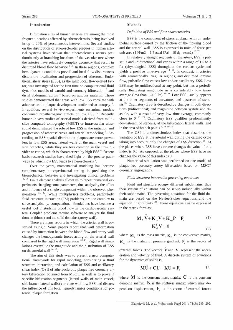
The two main approaches for the numerical simulationof fluid–structure interaction problems are: (a) monolithicapproach (strong coupling), and (b) partitioned approach(weak coupling). The monolithic approach involves the de-velopment of new software, while the partitioned approachuses the existing software to solve new classes of problems.The software PAK-FS is developed by coupling existingcomputational solid dynamics (CSD) software PAK-S andcomputational fluid dynamics (CFD) software PAK-F theusing partitioned approach 27. The algorithm of developedcode is shown in Figure 1. This algorithm partially separatesthe used solvers in space. As boundary conditions the CFDprogram uses the current geometry of the interface and thevelocity of the interface nodes. This information is obtainedfrom the CSD program for solving the solids. On the otherhand, loads from the fluid domain obtained using the CFDprogram is transmitted to CSD program.
Model generation
The finite element method requires the physical domainin which the problem is discretized. To date, the methodolo-gies to characterize local shear stress distribution and otherhemodynamic parameters in circulation have been used forresearch purposes only, because no tools are available to
provide this information in a routine manner. Artery bifurca-tions may have a very complicated configuration 28. Lately,the trend and need is patient-specific modeling 29, 30. It is verychallenging to generate quality hexahedral meshes for com-plicated structures 31. The hexahedral mesh quality has a sig-nificant role in the simulation by the finite elementmethod 32. This is very important in CFD, where numericalerrors become visible in the flow field. During generatingprocedures and developing software for the creation of thefinite element model the authors have sought to satisfy all thelisted constraints.
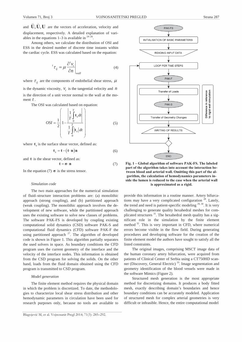
The original images, comprising MSCT image data ofthe human coronary artery bifurcation, were acquired frompatients of Clinical Center of Serbia using a CT750HD scan-ner (Discovery, General Electric) 33. Image segmentation andgeometry identification of the blood vessels were made inthe software Mimics (Figure 2).
Structured mesh generation is the most appropriatemethod for discretizing domains. It produces a body fittedmesh, exactly describing domain’s boundaries and henceboundary conditions can be accurately modeled. Applicationof structured mesh for complex arterial geometries is verydifficult or infeasible. Hence, the entire computational model
Fig. 1 – Global algorithm of software PAK-FS. The labeledpart of the algorithm takes into account the interaction be-tween blood and arterial wall. Omitting this part of the al-gorithm, the calculation of hemodynamics parameters in-
side the lumen is reduced to the case when the arterial wallis approximated as a rigid.
Strana 288 VOJNOSANITETSKI PREGLED Volumen 71, Broj 3
Blagojevi M, et al. Vojnosanit Pregl 2014; 71(3): 285–292.
Fig. 2 – Volumetric model obtained from the original mul-tislice computed tomography (MSCT) data. The left coro-
nary artery is marked in red. The modeled segment ofplaque free coronary artery bifurcation is marked with (a).
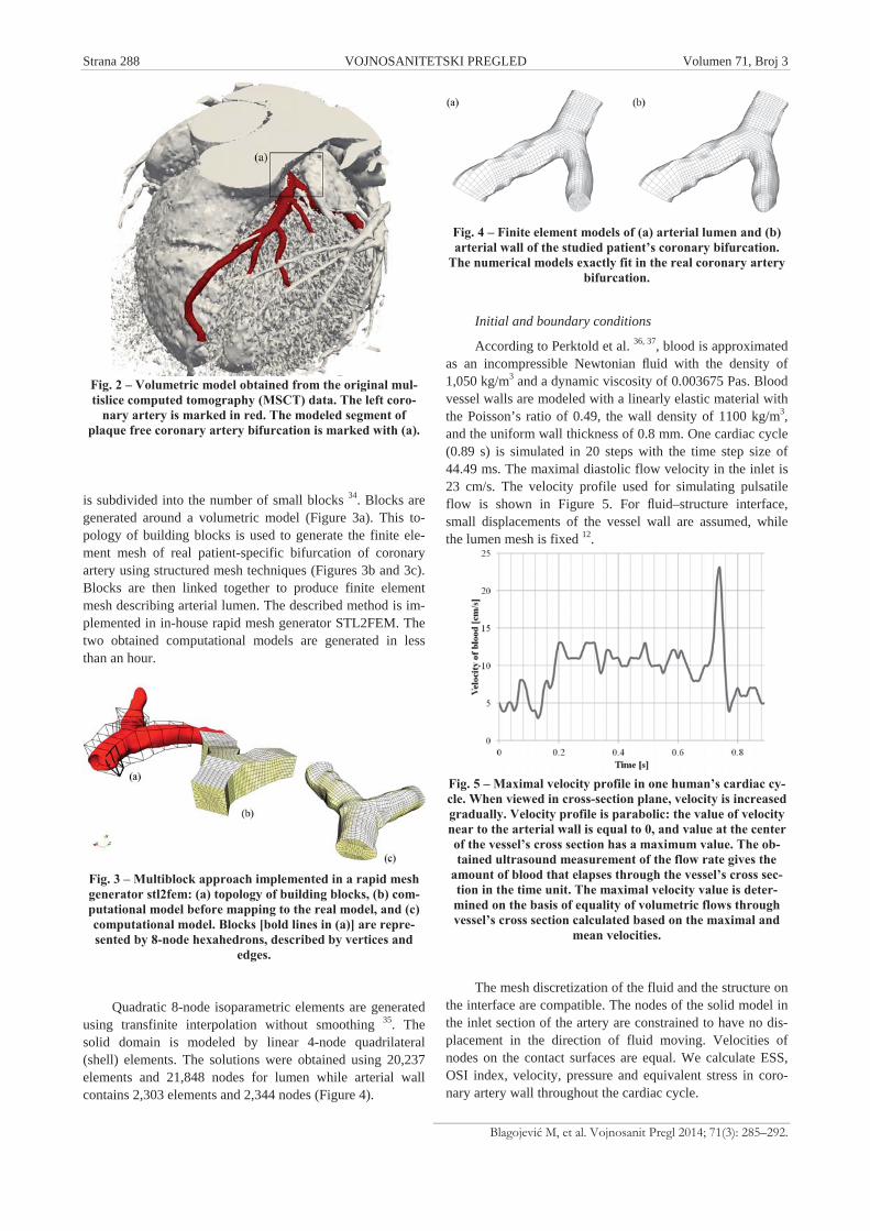
is subdivided into the number of small blocks 34. Blocks aregenerated around a volumetric model (Figure 3a). This to-pology of building blocks is used to generate the finite ele-ment mesh of real patient-specific bifurcation of coronaryartery using structured mesh techniques (Figures 3b and 3c).Blocks are then linked together to produce finite elementmesh describing arterial lumen. The described method is im-plemented in in-house rapid mesh generator STL2FEM. Thetwo obtained computational models are generated in lessthan an hour.
Fig. 3 – Multiblock approach implemented in a rapid meshgenerator stl2fem: (a) topology of building blocks, (b) com-putational model before mapping to the real model, and (c)computational model. Blocks [bold lines in (a)] are repre-sented by 8-node hexahedrons, described by vertices and
edges.
Quadratic 8-node isoparametric elements are generatedusing transfinite interpolation without smoothing 35. Thesolid domain is modeled by linear 4-node quadrilateral(shell) elements. The solutions were obtained using 20,237elements and 21,848 nodes for lumen while arterial wallcontains 2,303 elements and 2,344 nodes (Figure 4).
Fig. 4 – Finite element models of (a) arterial lumen and (b)arterial wall of the studied patient’s coronary bifurcation.
The numerical models exactly fit in the real coronary arterybifurcation.
Initial and boundary conditions
According to Perktold et al. 36, 37, blood is approximatedas an incompressible Newtonian fluid with the density of1,050 kg/m3 and a dynamic viscosity of 0.003675 Pas. Bloodvessel walls are modeled with a linearly elastic material withthe Poisson’s ratio of 0.49, the wall density of 1100 kg/m3,and the uniform wall thickness of 0.8 mm. One cardiac cycle(0.89 s) is simulated in 20 steps with the time step size of44.49 ms. The maximal diastolic flow velocity in the inlet is23 cm/s. The velocity profile used for simulating pulsatileflow is shown in Figure 5. For fluid–structure interface,small displacements of the vessel wall are assumed, whilethe lumen mesh is fixed 12.
Fig. 5 – Maximal velocity profile in one human’s cardiac cy-cle. When viewed in cross-section plane, velocity is increasedgradually. Velocity profile is parabolic: the value of velocitynear to the arterial wall is equal to 0, and value at the centerof the vessel’s cross section has a maximum value. The ob-tained ultrasound measurement of the flow rate gives the
amount of blood that elapses through the vessel’s cross sec-tion in the time unit. The maximal velocity value is deter-mined on the basis of equality of volumetric flows throughvessel’s cross section calculated based on the maximal and
mean velocities.
The mesh discretization of the fluid and the structure onthe interface are compatible. The nodes of the solid model inthe inlet section of the artery are constrained to have no dis-placement in the direction of fluid moving. Velocities ofnodes on the contact surfaces are equal. We calculate ESS,OSI index, velocity, pressure and equivalent stress in coro-nary artery wall throughout the cardiac cycle.
Volumen 71, Broj 3 VOJNOSANITETSKI PREGLED Strana 289
Blagojevi M, et al. Vojnosanit Pregl 2014; 71(3): 285–292.
Results
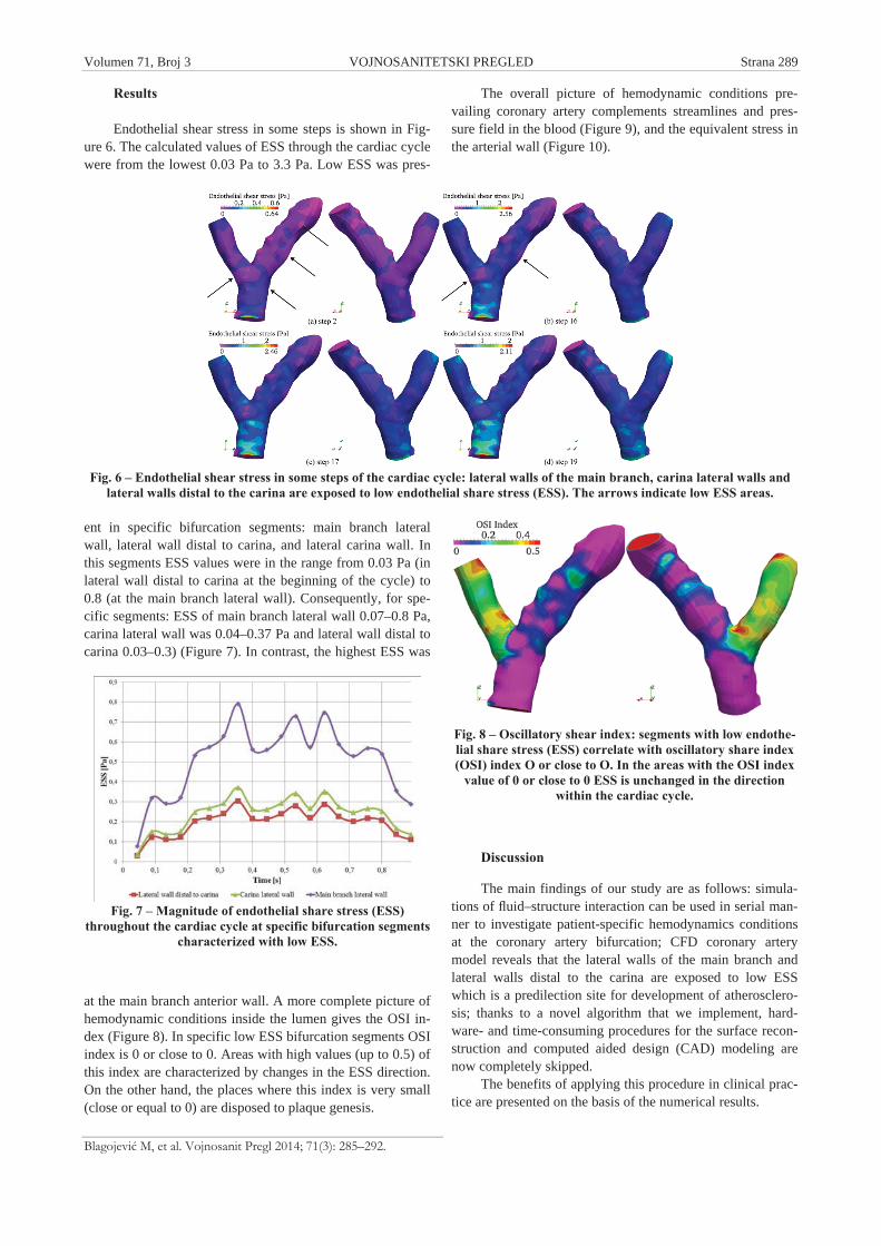
Endothelial shear stress in some steps is shown in Fig-ure 6. The calculated values of ESS through the cardiac cyclewere from the lowest 0.03 Pa to 3.3 Pa. Low ESS was pres-
ent in specific bifurcation segments: main branch lateralwall, lateral wall distal to carina, and lateral carina wall. Inthis segments ESS values were in the range from 0.03 Pa (inlateral wall distal to carina at the beginning of the cycle) to0.8 (at the main branch lateral wall). Consequently, for spe-cific segments: ESS of main branch lateral wall 0.07–0.8 Pa,carina lateral wall was 0.04–0.37 Pa and lateral wall distal tocarina 0.03–0.3) (Figure 7). In contrast, the highest ESS was
at the main branch anterior wall. A more complete picture ofhemodynamic conditions inside the lumen gives the OSI in-dex (Figure 8). In specific low ESS bifurcation segments OSIindex is 0 or close to 0. Areas with high values (up to 0.5) ofthis index are characterized by changes in the ESS direction.On the other hand, the places where this index is very small(close or equal to 0) are disposed to plaque genesis.
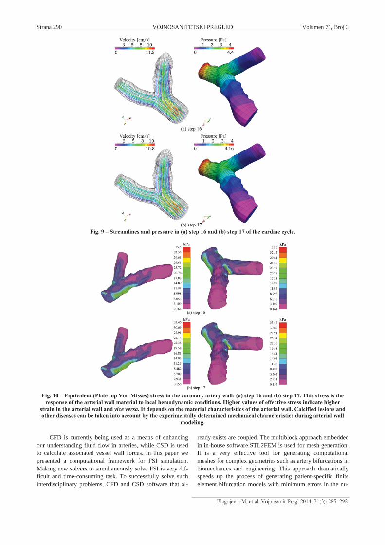
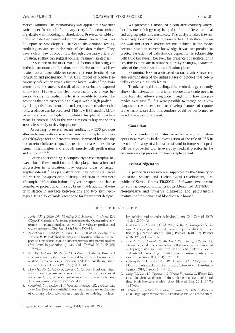
The overall picture of hemodynamic conditions pre-vailing coronary artery complements streamlines and pres-sure field in the blood (Figure 9), and the equivalent stress inthe arterial wall (Figure 10).
Fig. 8 – Oscillatory shear index: segments with low endothe-lial share stress (ESS) correlate with oscillatory share index(OSI) index O or close to O. In the areas with the OSI index
value of 0 or close to 0 ESS is unchanged in the directionwithin the cardiac cycle.
Discussion
The main findings of our study are as follows: simula-tions of fluid–structure interaction can be used in serial man-ner to investigate patient-specific hemodynamics conditionsat the coronary artery bifurcation; CFD coronary arterymodel reveals that the lateral walls of the main branch andlateral walls distal to the carina are exposed to low ESSwhich is a predilection site for development of atherosclero-sis; thanks to a novel algorithm that we implement, hard-ware- and time-consuming procedures for the surface recon-struction and computed aided design (CAD) modeling arenow completely skipped.
The benefits of applying this procedure in clinical prac-tice are presented on the basis of the numerical results.
Fig. 6 – Endothelial shear stress in some steps of the cardiac cycle: lateral walls of the main branch, carina lateral walls andlateral walls distal to the carina are exposed to low endothelial share stress (ESS). The arrows indicate low ESS areas.
Fig. 7 – Magnitude of endothelial share stress (ESS)throughout the cardiac cycle at specific bifurcation segments
characterized with low ESS.
Strana 290 VOJNOSANITETSKI PREGLED Volumen 71, Broj 3
Blagojevi M, et al. Vojnosanit Pregl 2014; 71(3): 285–292.
CFD is currently being used as a means of enhancingour understanding fluid flow in arteries, while CSD is usedto calculate associated vessel wall forces. In this paper wepresented a computational framework for FSI simulation.Making new solvers to simultaneously solve FSI is very dif-ficult and time-consuming task. To successfully solve suchinterdisciplinary problems, CFD and CSD software that al-
ready exists are coupled. The multiblock approach embeddedin in-house software STL2FEM is used for mesh generation.It is a very effective tool for generating computationalmeshes for complex geometries such as artery bifurcations inbiomechanics and engineering. This approach dramaticallyspeeds up the process of generating patient-specific finiteelement bifurcation models with minimum errors in the nu-
Fig. 9 – Streamlines and pressure in (a) step 16 and (b) step 17 of the cardiac cycle.
Fig. 10 – Equivalent (Plate top Von Misses) stress in the coronary artery wall: (a) step 16 and (b) step 17. This stress is theresponse of the arterial wall material to local hemodynamic conditions. Higher values of effective stress indicate higher
strain in the arterial wall and vice versa. It depends on the material characteristics of the arterial wall. Calcified lesions andother diseases can be taken into account by the experimentally determined mechanical characteristics during arterial wall
modeling.
Volumen 71, Broj 3 VOJNOSANITETSKI PREGLED Strana 291
Blagojevi M, et al. Vojnosanit Pregl 2014; 71(3): 285–292.
merical solution. The methodology was applied to a vascularpatient-specific model of coronary artery bifurcation includ-ing elastic wall modeling in simulations. Previous considera-tions indicate that developed computational frame gives use-ful inputs to cardiologists. Thanks to the obtained results,cardiologists are set in the role of decision makers. Theyhave a clear view of blood flow through a coronary artery bi-furcation, so they can suggest optimal treatment strategies.
ESS is one of the most essential factors influencing en-dothelial structure and function, and it is the main local flowrelated factor responsible for coronary atherosclerotic plaqueformation and progression 2, 5. A CFD model of plaque freecoronary bifurcation reveals that the lateral walls of the mainbranch, and the lateral walls distal to the carina are exposedto low ESS. Thanks to the clear picture of this parameter be-havior during the cardiac cycle, it is possible to predict thepositions that are suspectible to plaque with a high probabil-ity. Using this facts, formation and progression of atheroscle-rotic, a plaque can be predicted. This low ESS specific bifur-cation segment has higher probability for plaque develop-ment. In contrast ESS in the carina region is higher and thisarea is less likely to develop plaque.
According to several recent studies, low ESS promoteatherosclerosis with several mechanisms: through nitric ox-ide (NO)-dependent athero-protection, increased low-densitylipoprotein cholesterol uptake, sustain increase in oxidativestress, inflammation and smooth muscle cell proliferationand migration 5, 20.
Better understanding a complex dynamic interplay be-tween local flow conditions and the plaque formation andprogression in bifurcations may express areas of angio-graphic interest 8. Plaque distribution may provide a usefulinformation for appropriate technique selection in treatmentof complex bifurcation lesions. It gives the operator a chanceconsider to protection of the side branch with additional wireor to decide in advance between one and two stent tech-niques. It is also valuable knowledge for future stent designs.
We presented a model of plaque-free coronary artery,but this methodology may be applicable in different clinicaland angiographic circumstances. This analysis takes into ac-count only kinematic and dynamic effects. Calcifications onthe wall and other disorders are not included in the modelbecause based on current knowledge it was not possible topredict the extent of calcification deposition in relationshipwith fluid behavior. However, the presence of calcification ispossible to simulate in future studies by changing character-istics of the arterial wall at calcification place.
Examining ESS in a diseased coronary artery may en-able identification of the initial stages of plaques that poten-tially evolve a high-risk lesion.
Thanks to rapid modeling, this methodology not onlyallows characterization of arterial plaque at a single point intime but, also allows prognostic insight into how plaquesevolve over time 38. If it were possible to recognize in vivoplaques that were expected to develop features of ruptureprone lesions, specific interventions could be performed toavoid adverse cardiac event.
Conclusion
Rapid modeling of patient-specific artery bifurcationopens new avenues in the investigation of the role of ESS inthe natural history of atherosclerosis and in future we hope itwill be a powerful tool in everyday medical practice in thedecision making process for every single patient.
Acknowledgements
A part of this research was supported by the Ministry ofEducation, Science and Technological Development, Re-public of Serbia, Grants TR32036 – Software developmentfor solving coupled multiphysics problems and OI175082 –Non-invasive and invasive diagnostic and percutaneoustreatment of the stenosis of blood vessels branch.
R E F E R E N C E S
1. Zarins CK, Giddens DP, Bharadvaj BK, Sottiurai VS, Mabon RF,Glagov S. Carotid bifurcation atherosclerosis. Quantitative cor-relation of plaque localization with flow velocity profiles andwall shear stress. Circ Res 1983; 53(4): 502 14.
2. Nakazawa G, Yazdani SK, Finn AV, Vorpahl M, Kolodgie FD,Virmani R. Pathological findings at bifurcation lesions: the im-pact of flow distribution on atherosclerosis and arterial healingafter stent implantation. J Am Coll Cardiol 2010; 55(16):1679 87.
3. Ku DN, Giddens DP, Zarins CK, Glagov S. Pulsatile flow andatherosclerosis in the human carotid bifurcation. Positive cor-relation between plaque location and low oscillating shearstress. Arteriosclerosis 1985; 5(3): 293 302.
4. Moore JE, Xu C, Glagov S, Zarins CK, Ku DN. Fluid wall shearstress measurements in a model of the human abdominalaorta: oscillatory behavior and relationship to atherosclerosis.Atherosclerosis 1994; 110(2): 225 40.
5. Chatzizisis YS, Coskun AU, Jonas M, Edelman ER, Feldman CL,Stone PH. Role of endothelial shear stress in the natural historyof coronary atherosclerosis and vascular remodeling: molecu-
lar, cellular, and vascular behavior. J Am Coll Cardiol 2007;49(25): 2379 93.
6. Gambillara V, Chambaz C, Montorzi G, Roy S, Stergiopulos N, Si-lacci P. Plaque-prone hemodynamics impair endothelial func-tion in pig carotid arteries. Am J Physiol Heart Circ Physiol2006; 290(6): H2320 8.
7. Samady H, Eshtehardi P, McDaniel MC, Suo J, Dhawan SS,Maynard C, et al. Coronary artery wall shear stress is associatedwith progression and transformation of atherosclerotic plaqueand arterial remodeling in patients with coronary artery dis-ease. Circulation 2011; 124(7): 779 88.
8. Giannoglou GD, Antoniadis AP, Koskinas KC, Chatzizisis YS.Flow and atherosclerosis in coronary bifurcations. EuroInter-vention 2010; 6(Suppl J): J16 23.
9. Kung EO, Les AS, Figueroa AC, Medina F, Arcaute K, Wicker RB,et al. In vitro validation of finite element analysis of bloodflow in deformable models. Ann Biomed Eng 2011; 39(7):1947 60.
10. Stepanovi Ž, Živkovi M, Vulovi S, A imovi L, Risti B, Mati A,et al. High, open wedge tibial osteotomy: Finite element analy-
Strana 292 VOJNOSANITETSKI PREGLED Volumen 71, Broj 3
Blagojevi M, et al. Vojnosanit Pregl 2014; 71(3): 285–292.
sis of five internal fixation modalities. Vojnosanit Pregl 2011;68(10): 867 71. (Serbian)
11. Mari nas M, Kuzborska Z. Influence of load magnitude and du-ration on the relationship between human arterial blood pres-sure and flow rate. Acta Bioeng Biomech 2011; 13(2): 67 72.
12. Kim H, Vignon-Clementel I, Figueroa C, Jansen K, Taylor C. Devel-oping computational methods for three-dimensional finiteelement simulations of coronary blood flow. Finite Elem AnalDes 2010; 46(6): 514 25.
13. Torii R, Oshima M, Kobayashi T, Takagi K, Tezduyar TE. Com-puter modeling of cardiovascular fluid-structure interactionswith the deforming-spatial-domain/stabilized space-time for-mulation. Comput Methods Appl Mech Eng 2006; 195: 1885–95.
14. Bazilevs Y, Hsu MC, Zhang Y, Wang W, Liang X, Kvamsdal T, etal. A fully-coupled fluid-structure interaction simulation ofcerebral aneurysms. Comput Mech 2010; 46(1): 3 16.
15. Weydahl ES, Moore JE. Dynamic curvature strongly affects wallshear rates in a coronary artery bifurcation model. J Biomech2001; 34(9): 1189 96.
16. Nichols WW, Orourke MF. McDonalds blood flow in arteries:Theoretica, experimental and clinical principles. 5th ed. Lon-don: A Hodder Arnold Publicatio; 2005.
17. Slager CJ, Wentzel JJ, Gijsen FJ, Schuurbiers JC, Wal AC, Steen AF,et al. The role of shear stress in the generation of rupture-prone vulnerable plaques. Nat Clin Pract Cardiovasc Med2005; 2(8): 401 7.
18. Malek AM, Alper SL, Izumo S. Hemodynamic shear stress andits role in atherosclerosis. JAMA 1999; 282(21): 2035 42.
19. Gimbrone MA, Topper JN, Nagel T, Anderson KR, Garcia-CardeñaG. Endothelial dysfunction, hemodynamic forces, and athero-genesis. Ann N Y Acad Sci 2000; 902: 230 9; discussion9 40.
20. Stone PH, Coskun AU, Kinlay S, Clark ME, Sonka M, Wahle A,et al. Effect of endothelial shear stress on the progression ofcoronary artery disease, vascular remodeling, and in-stent re-stenosis in humans: in vivo 6-month follow-up study. Circula-tion 2003; 108(4): 438 44.
21. Ku DN. Blood flow in arteries. Annu Rev Fluid Mech 1997;29(1): 399 434.
22. Soulis JV, Giannoglou GD, Chatzizisis YS, Farmakis TM, Gian-nakoulas GA, Parcharidis GE, et al. Spatial and phasic oscillationof non-Newtonian wall shear stress in human left coronaryartery bifurcation: an insight to atherogenesis. Coron ArteryDis 2006; 17(4): 351 8.
23. Younis HF, Kaazempur-Mofrad MR, Chan RC, Isasi AG, HintonDP, Chau AH, et al. Hemodynamics and wall mechanics inhuman carotid bifurcation and its consequences for athero-genesis: investigation of inter-individual variation. BiomechModel Mechanobiol 2004; 3(1): 17 32.
24. Koji M, Filipovi N, Stojanovi B, Koji N. Computer Modeling inBioengineering: Theoretical background, examples and soft-ware. Chichester: John Wiley & Sons; 2008.
25. Bathe K. Finite element procedures in engineering analysis.Englewood Cliffs, NJ: Prentice Hall; 1996.
26. Kojic M, Slavkovic R, Zivkovic M, Grujovic N. The finite elementmethod: Linear analysis. Kragujevac: Faculty of MechanicalEngineering of Kragujevac; 1998. (Serbian)
27. Živkovi M. Department: Department for applied mechanicsand automatic control. Kragujevac: Faculty of Engineering,University of Kragujevac; 2004.
28. Zhao SZ, Ariff B, Long Q, Hughes AD, Thom SA, Stanton AV,Xu XY. Inter-individual variations in wall shear stress and me-chanical stress distributions at the carotid artery bifurcation ofhealthy humans. J Biomech 2002; 35(10): 1367 77.
29. Soulis JV, Farmakis TM, Giannoglou GD, Louridas GE. Wallshear stress in normal left coronary artery tree. J Biomech2006; 39(4): 742 9.
30. Nguyen KT, Clark CD, Chancellor TJ, Papavassiliou DV. Carotidgeometry effects on blood flow and on risk for vascular dis-ease. J Biomech 2008; 41(1): 11 9.
31. Goubergrits L, Affeld K, Fernandez-Brittoy J, Falcon L. Investiga-tion of geometry and atherosclerosis in the human carotid bi-furcations. J Mech Med Biol 2003; 3(1): 31 48.
32. Zhang YJ, Bajaj C. Adaptive and quality quadrilat-eral/hexahedral meshing from volumetric data. ComputMethods Appl Mech Engrg 2006; 195: 942 60.
33. de Santis G, de Beule M, van Canneyt K, Segers P, Verdonck P, Ver-hegghe B. Full-hexahedral structured meshing for image-basedcomputational vascular modeling. Med Eng Phys 2011; 33(10):1318 25.
34. Shirsat A, Gupta S, Shevare GR. Generation of multi-block to-pology for discretisation of three-dimensional domains. Com-put Graph 1999; 23(1): 45 57.
35. Yuan C, Yih N. A transfinite interpolation method of gridgeneration based on multipoints. J Sci Comp 1998; 13(1):105 14.
36. Perktold K, Resch M, Florian H. Pulsatile non-Newtonian flowcharacteristics in a three-dimensional human carotid bifurca-tion model. J Biomech Eng 1991; 113(4): 464 75.
37. Perktold K, Resch M, Peter RO. Three-dimensional numericalanalysis of pulsatile flow and wall shear stress in the carotidartery bifurcation. J Biomech 1991; 24(6): 409 20.
38. Stone PH, Feldman CL. In vivo assessment of the risk profileof evolving individual coronary plaques: a step closer. Circula-tion 2011; 124(7): 763 5.
Received on October 26, 2012.Revised on November 15, 2012.
Accepted on November 16, 2012.


![ДОКУМЕНТАЦИЈА ТЕХНИЧКОГ РЕШЕЊАis.fink.rs/podaci/Aleksandar_Nikolic/119/TR-71-2012.pdf · човека, скелетни систем итд.) [1-3]. Познато](https://img.pdfslide.net/doc/110x75/5ec4cc7c09d61b7a4d29f0c4/oe-isfinkrspodacialeksandarnikolic119tr-71-2012pdf.jpg)


![ANIMACIJA MEĐUDJELOVANJA KRUTIH TIJELA I FLUIDA · 2007. 10. 12. · simuliranje međudjelovanja između krutih tijela i fluida nazvanu Kruti fluidi [1]. Seminar počinje opisima](https://img.pdfslide.net/doc/110x75/5fe38337f94138153743a58b/animacija-meudjelovanja-krutih-tijela-i-fluida-2007-10-12-simuliranje-meudjelovanja.jpg)