Embed Size (px)
Citation preview

Novel Assays for Assessing Cardiotoxicity, Contractility, and
ECG-Like Aberrations in Stem Cell-Derived Cardiomyocytes
Greg Luerman, PhDChanTest Corporation
1

FDA CiPA Initiative
Sager PT, et al. 2014. Rechanneling the cardiac proarrhythmia safety paradigm: A meeting report from the Cardiac Safety Research Consortium. Am Heart J. 167:292-300. 2

Multi-Electrode Array (MEA) vs. xCELLigence: complementary, but unique technologies
MEA xCELLigence
Axion BiosystemsMaestro MEA
ACEA Biosciences xCELLigence Cardio 3

iPSC Advantage: Humanizing Safety Pharmacology
Human iPSC cardiomyocytes Derived from non-embryonic source
High purity (>99%) cardiomyocyte population
Normal cardiomyocyte physiology
- ventricular, atrial, and nodal cell types
- electrically coupled activity
- integrated ion channel function
Spontaneously beating
Sensitive to arrhythmogenic drugs
Viable in culture for months
4

MEA• 768 electrodes, • 16 electrodes/well• 48 well assay plate
Middleman• Power• Pre-processing • IO
Maestro • Recording• Heating • A/D
Multi-Well Maestro MEA System
Detects effects of test compounds on electrical activity in stem cell-derived human cardiomyocytes
• extracellular field potential (ECG related signal)
• beat prolongation• beat irregularities
Axion MEA system–monitors spontaneous electrical activity in cardiomyocytemonolayers

xCELLigence: Impedance Assay
6
xCELLigence RTCA Cardio (ACEA) continuously monitors electrical impedance in cardiomyocyte monolayers during spontaneous beating
Detects effects on cardiomyocytes in 96-well format
• contractility• beat rate• beat irregularities• cytotoxicity < 1 sec
Contraction
Relaxation
Analyzer Cardio Station

MEA - extracellular field potentialsxCELLigence - myocyte contractility
FPD
Na+ SpikeAmplitude
Beat Period
T-wave
MEA
7
QRS
MEA: Measures extracellular field potentials yielding ECG-like recordings. Na+ spike amplitude, field potential duration (FPD), beat period/rate in stem cell-derived human cardiomyocytes
xCELLigence
a
DMSO ouabain 10 nM
b
control ouabain, 11.8 hr
control ouabain 10 nM, 3.8 hr
control DMSO, 11.8 hr
control DMSO, 3.8 hr
0.02 CI
500 ms
xCELLigence: Measures electrical impedance changes resulting from myocyte contraction. Detects amplitude, rise and decay times, beat period/rate, cell index
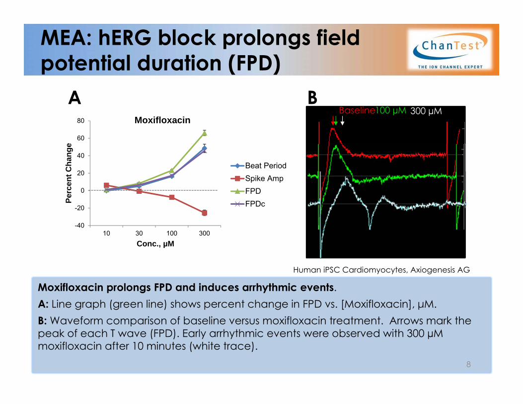
MEA: hERG block prolongs field potential duration (FPD)
Moxifloxacin prolongs FPD and induces arrhythmic events. A: Line graph (green line) shows percent change in FPD vs. [Moxifloxacin], μM. B: Waveform comparison of baseline versus moxifloxacin treatment. Arrows mark the peak of each T wave (FPD). Early arrhythmic events were observed with 300 µM moxifloxacin after 10 minutes (white trace).
A B300 µMBaseline100 µM
Human iPSC Cardiomyocytes, Axiogenesis AG
8
-40
-20
0
20
40
60
80
10 30 100 300
Perc
ent C
hang
e
Conc., µM
Moxifloxacin
Beat PeriodSpike AmpFPDFPDc
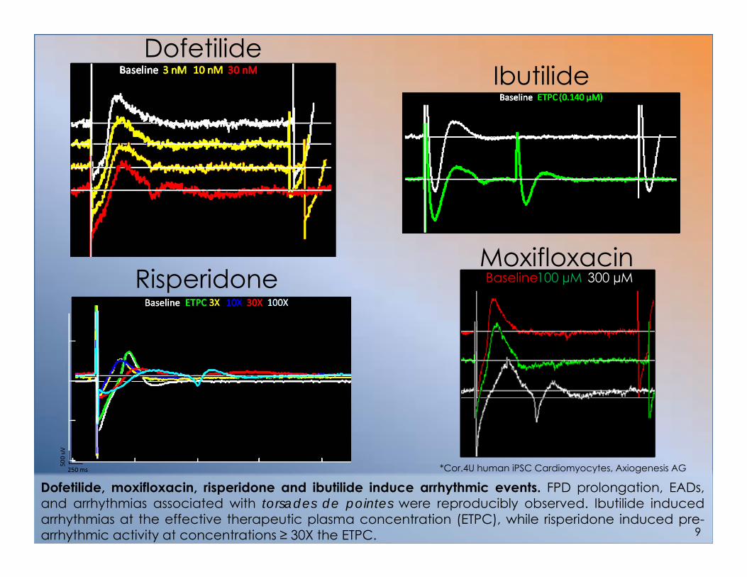
Dofetilide
Moxifloxacin300 µMBaseline100 µM
Dofetilide, moxifloxacin, risperidone and ibutilide induce arrhythmic events. FPD prolongation, EADs,and arrhythmias associated with torsades de pointes were reproducibly observed. Ibutilide inducedarrhythmias at the effective therapeutic plasma concentration (ETPC), while risperidone induced pre-arrhythmic activity at concentrations ≥ 30X the ETPC.
Ibutilide
Risperidone
*Cor.4U human iPSC Cardiomyocytes, Axiogenesis AG
9

MEA: Standard 5-concentration analysis
10
hIPSC cardiomyocytes recapitulate the multiple ion channel effects (MICE) model1
• hERG endpoint analysis is not sufficient to predict arrhythmias.
• Integrated ion channel effects must be evaluated for accurate arrhythmia prediction.
1Kramer, et al. 2013. MICE models: superior to hERG model in predicting TdP. Sci. Rep. 3:2100
*EADs expected at concentrations > 100X ETPC Cor.4U human iPSC Cardiomyocytes, Axiogenesis AG
EADs Observed at Test Concentrations Normalized to Effective Therapeutic Plasma Concentration (ETPC)
TDP-
Posi
tive
1X 3X 10X 30X 100X
IbutilideThioridazine
DroperidolRisperidone
TerodilineMoxifloxacin
Sunitinib*Amiodarone*Paroxetine
TDP-
Neg
ativ
e
VerapamilFALSE FALSE FALSE FALSE FALSEMetronidazole
TolterodineDonepezil
Arrhythmic Event at Concentration (x ETPC)
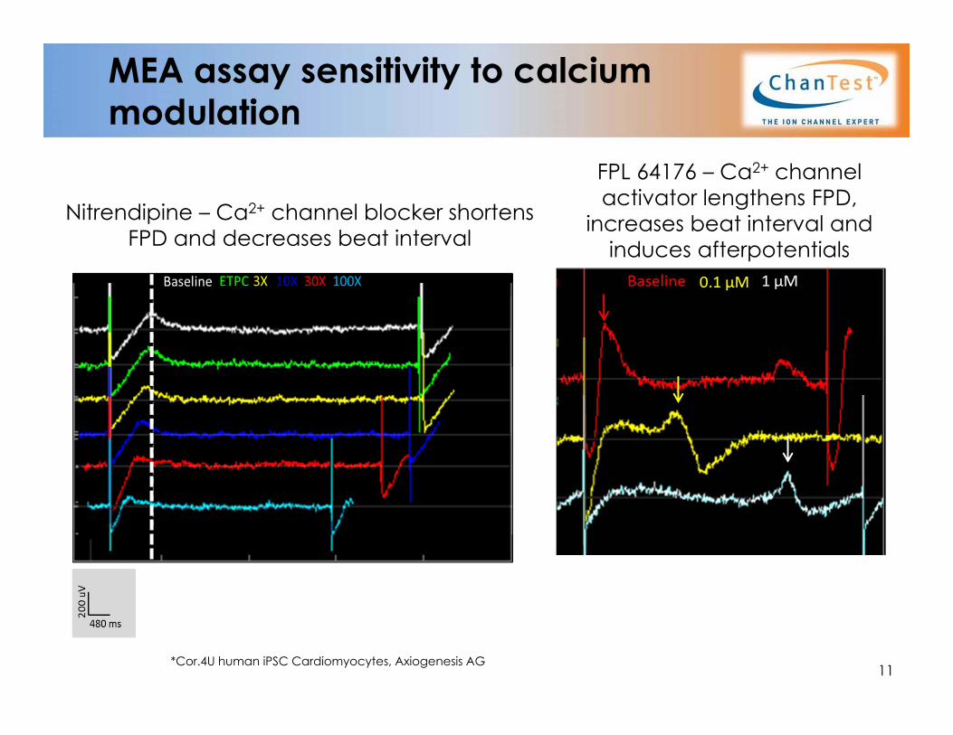
MEA assay sensitivity to calcium modulation
Nitrendipine – Ca2+ channel blocker shortens FPD and decreases beat interval
FPL 64176 – Ca2+ channel activator lengthens FPD,
increases beat interval and induces afterpotentials
*Cor.4U human iPSC Cardiomyocytes, Axiogenesis AG11

MEA detects Na+ channel block
Human iPSC Cardiomyocytes, Axiogenesis AG12
-100%
-80%
-60%
-40%
-20%
0%
20%
40%
0.1 µM 0.3 µM 1 µM 3 µM
Tetrodotoxin
Beat PeriodSpike Amp
FPDFPDc
% C
hang
e
[TTX]
Tetrodotoxin reduces the Na+ spikeLine graph (green line) shows percent change in spike amplitude vs. [Tetrodotoxin], μM.

MEA: Effect of Na+ channel block on Na+ spike amplitude
DMSO
Lidocaine
Tetrodotoxin
Ranolazine
Amiodarone
30 µM 30 µM 100 µM 100 µM 200 µM 200 µM
1 µM 1 µM 10 µM 10 µM 30 µM 30 µM
1 µM 1 µM 10 µM 10 µM 100 µM 100 µM
0.3 µM 0.3 µM 3 µM 3 µM 25 µM 25 µM
3.6 mV 0 mVKey =
Effect of Na+ channel blockers on Na+ amplitude. hIPSC cardiomyocytes were treated with test compounds at multiple concentrations in duplicate wells. The heat map shows that addition of Na+ channel blockers decrease the Na+ spike amplitude (peak QRS voltage) detected in each electrode/well. 13
iCell Cardiomyocytes, Cellular Dynamics

MEA Assay Data Reporting
Typical Report Figures Statistical summary of each compound Line graph summarizing change in
experimental parameters +/- Arrhythmic event Representative waveforms
*Cor.4U human iPSC Cardiomyocytes, Axiogenesis AG
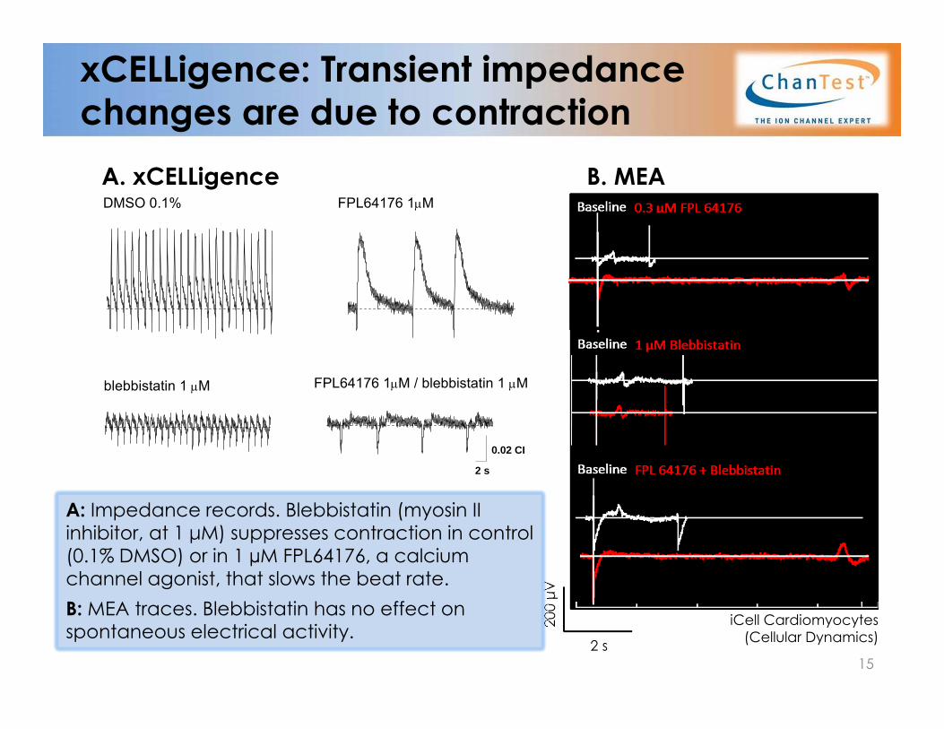
xCELLigence: Transient impedance changes are due to contraction
200 μV
2 s
DMSO 0.1% FPL64176 1M
A: Impedance records. Blebbistatin (myosin II inhibitor, at 1 μM) suppresses contraction in control (0.1% DMSO) or in 1 µM FPL64176, a calcium channel agonist, that slows the beat rate. B: MEA traces. Blebbistatin has no effect on spontaneous electrical activity.
A. xCELLigence B. MEA
iCell Cardiomyocytes(Cellular Dynamics)
15
blebbistatin 1 M FPL64176 1M / blebbistatin 1 M
0.02 CI
2 s
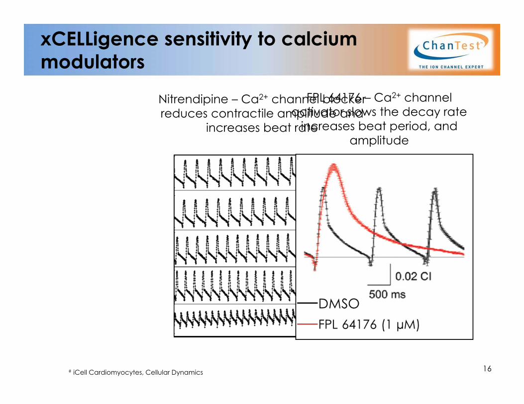
xCELLigence sensitivity to calcium modulators
# iCell Cardiomyocytes, Cellular Dynamics 16
Nitrendipine – Ca2+ channel blocker reduces contractile amplitude and
increases beat rate
FPL 64176 – Ca2+ channel activator slows the decay rate
increases beat period, and amplitude
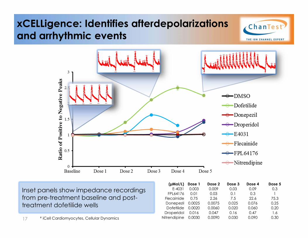
xCELLigence: Identifies afterdepolarizationsand arrhythmic events
Inset panels show impedance recordings from pre-treatment baseline and post-treatment dofetilide wells
17 # iCell Cardiomyocytes, Cellular Dynamics

xCELLigence: Subacute/chronic dosing to identify trafficking effects, cytotoxicity
Pentamidine prolongs the cardiac action potential by inhibiting hERG trafficking1 to the cell surface. “Fall Time” indirectly measures hERG channel activity. Loss of surface hERG expression prolongs the falling time.
Doxorubicin induces dose-dependent cytotoxicity (decline in cell index)
1Kuryshev YA et al. (2005) J. Pharmacol Exp. Ther. 312(1):316-23
Each data point represents the mean ± SEM for ≥ 3 wells per condition)
ΔCell Index for Doxorubicin
ΔFall Time for Pentamidine
Cor.4U human iPSC Cardiomyocytes, Axiogenesis AG 18

xCELLigence Data Reporting
Typical Report Figures Visual summary for each compound data
set (with statistics) Line graph summarizing changes in each
experimental parameter over time Single .xls summary file Acute to chronic dosing/time points Representative impedance traces
*Cor.4U human iPSC Cardiomyocytes, Axiogenesis AG
DM
SOE-
4031
19

xCELLigence Summary
* Protocols and concentrations are flexible. Our study directors will optimize your assay to fit your needs!† We have validated numerous endpoint-specific controls
MEA Summary
Human iPSC cardiomyocyte assays:protocols and reports
20
AssayPlating
Option# Test Cpds Typical Protocol Positive Controls End Points
xCELLigence 96 well 1 - 54 concentrations, n=4 wells/conc.
Positive & vehicle controls includedTurnaround time, 2 weeks
Customer-specifiede.g. Pentamidine,
E-4031
ΔCell index∆ Beat Rate∆ Amplitude∆ Fall Time
Assay Plating Option
# Test Cpds Typical Protocol Concentrations
Positive Controls
Typical End Points
48 well 1- 9MEA
5 concentrations , n=4 Sequential dosing
Positive & vehicle controlsTurnaround time, 3-4 weeks
2 log dosing recommended
e.g., ETPC, 3X, 10X, 30X, 100X
Serum-free conditions
Moxifloxacin(or customer-
specified)
Δ FPD ("QT")Δ Beat PeriodΔ Spike Amp ± Arrhythmia
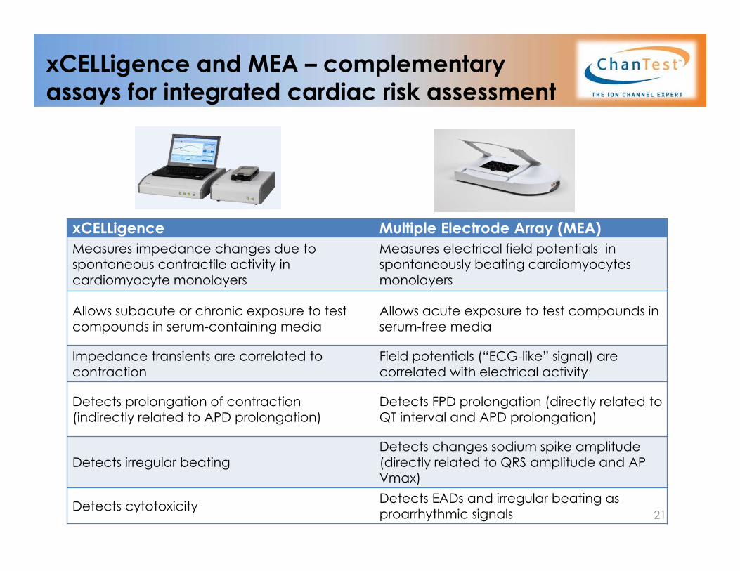
xCELLigence Multiple Electrode Array (MEA)Measures impedance changes due to spontaneous contractile activity in cardiomyocyte monolayers
Measures electrical field potentials in spontaneously beating cardiomyocytes monolayers
Allows subacute or chronic exposure to test compounds in serum-containing media
Allows acute exposure to test compounds in serum-free media
Impedance transients are correlated to contraction
Field potentials (“ECG-like” signal) are correlated with electrical activity
Detects prolongation of contraction (indirectly related to APD prolongation)
Detects FPD prolongation (directly related to QT interval and APD prolongation)
Detects irregular beatingDetects changes sodium spike amplitude (directly related to QRS amplitude and AP Vmax)
Detects cytotoxicity Detects EADs and irregular beating as proarrhythmic signals 21
xCELLigence and MEA – complementary assays for integrated cardiac risk assessment

Acknowledgements
ChanTest ScientistsGreg Luerman, PhD
Andrew Bruening-Wright, PhDCarlos Obejero-Paz, PhD
John DenkerJim Kramer, PhD
22

Contact Information
Headquarters:14656 Neo Parkway
Cleveland, Ohio 44128Email: [email protected]
www.ChanTest.com
877-828-1777 Toll Free216.332.1665 Tel216.332.1706 Fax
23