Embed Size (px)
Citation preview
Articles
Novel Benzodiazepine Photoaffinity Probe Stereoselectively Labels a Site Deep within theMembrane-Spanning Domain of the Cholecystokinin Receptor
Elizabeth M. Hadac,† Eric S. Dawson,‡ James W. Darrow,§ Elizabeth E. Sugg,| Terry P. Lybrand,‡ and Laurence J. Miller*,†
Department of Molecular Pharmacology and Experimental Therapeutics and Cancer Center, Mayo Clinic, Scottsdale, Arizona 85259,Department of Chemistry and Center for Structural Biology, Vanderbilt UniVersity, NashVille, Tennessee 37235-1822, Glaxo-SmithKlineResearch Laboratories, Research Triangle Park, North Carolina, and CGI Pharmaceuticals, Inc., Branford, Connecticut 06405
ReceiVed NoVember 17, 2004
An understanding of the molecular basis of drug action provides opportunities for refinement of drug propertiesand for development of more potent and selective molecules that act at the same biological target. In thiswork, we have identified the active enantiomers in racemic mixtures of structurally related benzophenonederivatives of 1,5-benzodiazepines, representing both antagonist and agonist ligands of the type Acholecystokinin receptor. The parent compounds of the 1,5-benzodiazepine CCK receptor photoaffinity ligandswere originally prepared in an effort to develop orally active drugs. The enantiomeric compounds reportedin this study selectively photoaffinity-labeled the CCK receptor, resulting in the identification of a site ofattachment for the photolabile moiety of the antagonist probe deep within the receptor’s membrane-spanningregion at Leu88, a residue within transmembrane segment two. In contrast, the agonist probe labeled a regionincluding extracellular loop one and a portion of transmembrane segment three. The antagonist covalentattachment site to the receptor served as a guide in the construction of theoretical three-dimensional molecularmodels for the antagonist-receptor complex. These models provided a means for visualization of physicallyplausible ligand-receptor interactions in the context of all currently available biological data that addresssmall molecule interactions with the CCK receptor. Our approach, featuring the use of novel photolabilecompounds targeting the membrane-spanning receptor domain to probe the binding site region, introducespowerful tools and a strategy for direct and selective investigation of nonpeptidyl ligand binding to peptidereceptors.
Introduction
Understanding of the molecular basis of drug action is ex-tremely important, contributing to refinement of the specificity,affinity, and potency of lead compounds, and even to the rationaldesign of new drugs. Insights into this can come from the com-plementary approaches of receptor mutagenesis and photoaf-finity labeling. Loss of function in a receptor mutant can beexplained either by direct or allosteric effects, while clean photo-affinity labeling of specific residues is direct and unambiguous,but can be quite difficult to achieve. Typical challenges includeidentification of a small molecule ligand that can accommodateboth a photolabile functional group and a radiolabel, whileretaining receptor binding affinity and biological activity.
We have previously reported the development of structurallyrelated 1,5-benzodiazepine ligands that act as agonists andantagonists at the type A cholecystokinin receptor.1 This is aguanine nucleotide-binding protein (G protein)-coupled receptorin the rhodopsin-â-adrenergic receptor family.2 It normally bindsand responds to a peptide hormone that is important in theregulation of gallbladder contraction, exocrine pancreatic secre-tion, gastric emptying, enteric transit, and satiety.3 We have beenable to incorporate photolabile benzophenone and diazirinemoieties into these nonpeptidyl CCKa receptor ligands.1 We
have also been successful in radiolabeling them with14C,without changing their agonist or antagonist properties and withmaintenance of adequate binding affinity.1 Despite the extremelylow specific radioactivity of these probes (0.05 Ci/mmol),representing less than one thirty-thousandth of the activity ofthe radioiodinated peptide probes we have previously used inaffinity labeling this receptor,4-7 we have been successful inusing these unique ligands to photoaffinity label the CCKreceptor.1
In this work, through the use of resolved enantiomeric CCK-Aligands, we have been able to extend the insights possible withthe racemic compounds that we initially described in a priorpublication.1 The previously reported racemic photolabile benzo-phenone derivatives of the 1,5-benzodiazepine agonist (mixtureof compounds5 and 6, Figure 1) and antagonist (mixture ofcompounds7 and8, Figure 1) were separated into their enantio-meric components, and each chiral compound was then assayedfor biological activity, binding affinity and CCK receptorlabeling efficiency. For the subsequent photoaffinity labelingwork, the racemic intermediate amines (shown in Scheme 1,1,2 and3, 4) were resolved into their respective enantiomers andconverted into14C analogues of each of the four separatedenantiomers (shown in Scheme 2,5, 6, 7, and8).
When the racemic mixture demonstrating agonist activity wasseparated into its components, only the putative (S)-enantiomer(6) displayed biological agonist activity, and it was also the* To whom correspondence should be addressed. Tel: (480) 301-6650.
Fax: (480) 301-6969. E-mail: [email protected].† Mayo Clinic.‡ Vanderbilt University.| Glaxo-SmithKline Research Laboratories.§ CGI Pharmaceuticals, Inc.
a Abbreviations used: CCK, cholecystokinin; Alexa488-CCK, Alexa488-Gly-[(Nle28,31)CCK-26-33]; CHO, Chinese hamster ovary; KRH, Krebs-Ringers-HEPES medium.
850 J. Med. Chem.2006,49, 850-863
10.1021/jm049072h CCC: $33.50 © 2006 American Chemical SocietyPublished on Web 01/07/2006
isomer that bound more tightly to the receptor. Therefore, thecorresponding (R)-enantiomer in this series (5) actually repre-sents a functional antagonist. Of interest, this (S)-enantiomer(6) was substantially less efficient in covalent labeling of theCCK receptor than was the (R)-enantiomer (5). In contrast, bothantagonist enantiomers (7 and8) had similar binding affinitiesfor the CCK receptor, but the proposed (S)-enantiomer (8) wassubstantially more efficient in covalent labeling of the receptorthan the corresponding (R)-enantiomer (7). Covalent attachmentsites for these structurally similar agonist and antagonist probeswere characterized, and the results indicate that agonist andantagonist ligands covalently labeled distinct regions within theCCK receptor. The site labeled by the antagonist was determineddefinitively with proteolytic peptide mapping, followed byEdman degradation sequencing of the labeled fragment. Theantagonist was covalently attached to Leu88 within the secondtransmembrane helical segment, clearly establishing a nonpep-tidyl antagonist binding site that is distinct from the extramem-branous receptor regions involved in binding the natural peptideagonist ligand.8,9 The nonpeptidyl agonist ligand covalentlylabeled a different portion of the receptor, believed to representthe cyanogen bromide fragment that includes the first extracel-lular loop and the upper portion of the third transmembranesegment. The receptor fragments covalently labeled by agonistand antagonist probes were distinct, but coincubation of thereceptor preparation with a competing concentration (1µM) ofthe endogenous peptide ligand, CCK, along with the benzodi-azepine probes, did not block photoaffinity labeling by eitherprobe. However, competitive coincubation with the nonpeptidylantagonist, devazepide (also referred to in the literature as
L-364,718 and MK329) (9), blocked photoaffinity labeling byboth agonist and antagonist probes in a concentration-dependentmanner. On the basis of these results, we propose that bothagonist and antagonist probes probably bind to receptor sitesthat are composed of partially overlapping regions within theCCK receptor membrane-spanning domains.
The data obtained from photoaffinity labeling studies withthese compounds were used to construct three-dimensionalmolecular models for ligand-receptor complexes. Small mol-ecule probes containing a fused benzodiazepine ring system havefar less conformational flexibility than peptide ligands. There-fore, they proved amenable to conformational search strategiesthat are impractical for peptide ligands. For example, methodswere applied that evaluated various conformations of the ligandswithin the context of the putative receptor binding site. Thepredominantly alpha-helical transmembrane domain where thenonpeptidyl ligands bind also possesses far fewer degrees ofconformational freedom than the flexible extracellular loopsshown to be important for peptide binding.8,9 Therefore, we wereable to generate plausible three-dimensional models for theseligand-receptor complexes using only the photoaffinity labelingresults, along with mutagenesis data from earlier studies for theclosely related antagonist, devazepide.
Results
Scheme 1 illustrates the strategy utilized for the preparationof the key racemic amine intermediates (1, 2 and 3, 4) to beused in the synthesis of the photolabile chiral probes (5, 6, 7,and8). The known bromoacetamides (12and13) were preparedin high yield from bromoacetyl bromide and the corresponding
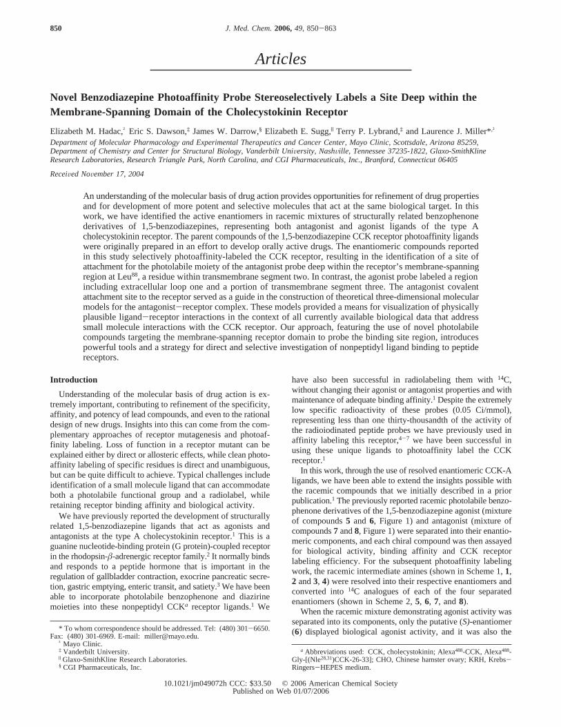
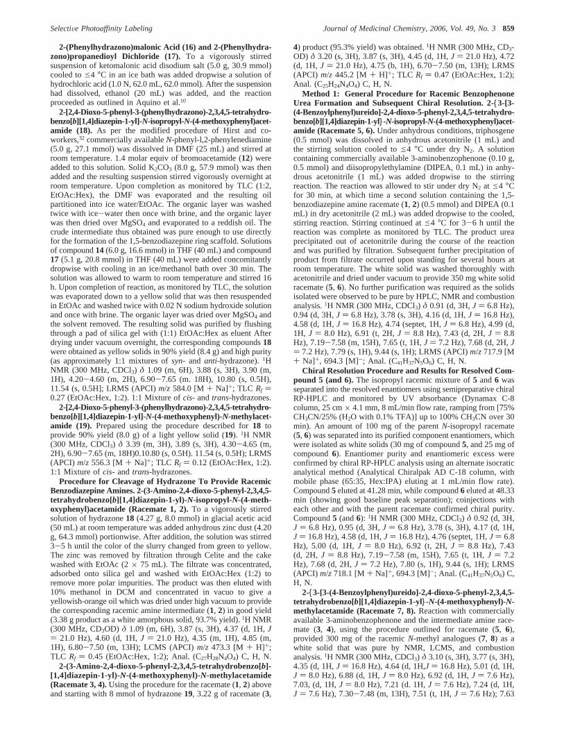
Figure 1. Chemical structures of nonpeptidyl CCK receptor ligands used in the current series of studies. The racemic mixture of compounds5 and6 having an agonist profile was separated into its components (5, (R)-form with no agonist activity and6, (S)-form with agonist activity), and theracemic mixture of compounds7 and8 having an antagonist profile was similarly separated into its components (7, (R)-form and8, (S)-form). Alsoillustrated is the structure of a related 1,4-benzodiazepine CCK receptor antagonist that was developed by Merck, initially called L-364,718 andsubsequently referred to as devazepide (9). This was used in pharmacological competition assays and molecular model-building studies.
SelectiVe Photoaffinity Labeling Journal of Medicinal Chemistry, 2006, Vol. 49, No. 3851

N-methyl or N-isopropyl secondary amine using the generalprocedure as described in Aquino et al.10 Subsequent alkylationof commercially availableN-phenylphenylenediamine with theappropriate bromoacetamide yielded the substituted phenylene-diamine intermediates (14 and 15). The known 2-(phenylhy-drazono-propanedioyldichloride (17) was prepared in two stepsfrom the commercially available ketomalonic acid using theprocedure outlined in Aquino et al.10 and condensed with thephenylenediamine intermediates (14 and 15) to afford thecorresponding hydrazones (18 and 19). Zinc reduction of thehydrazones yielded the racemic amines (1, 2 and3, 4).
With the amine racemates in hand, we approached the synthe-sis of the final resolved chiral photoaffinity labeling probes (5,6, 7, and 8) using two complementary routes, as shown inScheme 2. Both methods provide pure, separated photoaffinityligands with similar synthetic efficiency and enantiopurity.
As illustrated by the first method shown in Scheme 2, initiallyfor the bioactivity and binding experiments, the racemic amineintermediates were cleanly converted to the racemic benzophe-none ureas (5, 6 and7, 8) using a phosgene equivalent to couple
the amines with commercially available 3-aminobenzophenone.Subsequently, chiral chromatography resolved each of these tworacemic benzophenone-1,5-benzodiazepine urea pairs into thefinal four enantiomers (5, 6, 7, and 8) in high enantiomericexcess and complete baseline peak separation under chiral HPLCconditions.
For further exploration with modeling analysis, and in theabsence of being able to obtain adequate crystallographic dataon the resolved enantiomers, we provisionally assigned theabsolute configurations of our compounds based on comparisonto reported X-ray structures for highly analogous CCK ligands.Within the racemate originally displaying agonist activity(racemate5, 6), after enantiomer separation, the antagonistcompound5 was putatively assigned the (R)-configuration basedon its strong structural homology with devazepide (9), as wellas other benzodiazepine CCK antagonists having X-ray struc-tures reported in the literature.11-13 The only agonist ligand inthe set, compound6, was correspondingly assigned the (S)-configuration. Comparing, under the same stationary and mobilephase conditions (see Experimental Section), the relative chiral
Scheme 1a
a (a) C6H5NHNH2, EtOH, H2O, HCl; (b) PCl5, CCl4; (c) bromoacetyl bromide, DIPEA, DCM; (d)N-phenyl phenylenediamine, NaH, DMF; (e) THF,e4°C; (f) Zn dust, AcOH.
Scheme 2a
a (a) Phosgene, 3-aminobenzophenone, base,<4 °C; (b) chiral reverse-phase HPLC purification.
852 Journal of Medicinal Chemistry, 2006, Vol. 49, No. 3 Hadac et al.
HPLC retention times of these two compounds (41.3 forcompound5 and 48.3 min for compound6) with those observedfor the two antagonist enantiomers (49.9 min for7 and 82.2min for 8) allowed for the provisional assignment of compounds7 and8 to be (R) and (S), respectively.
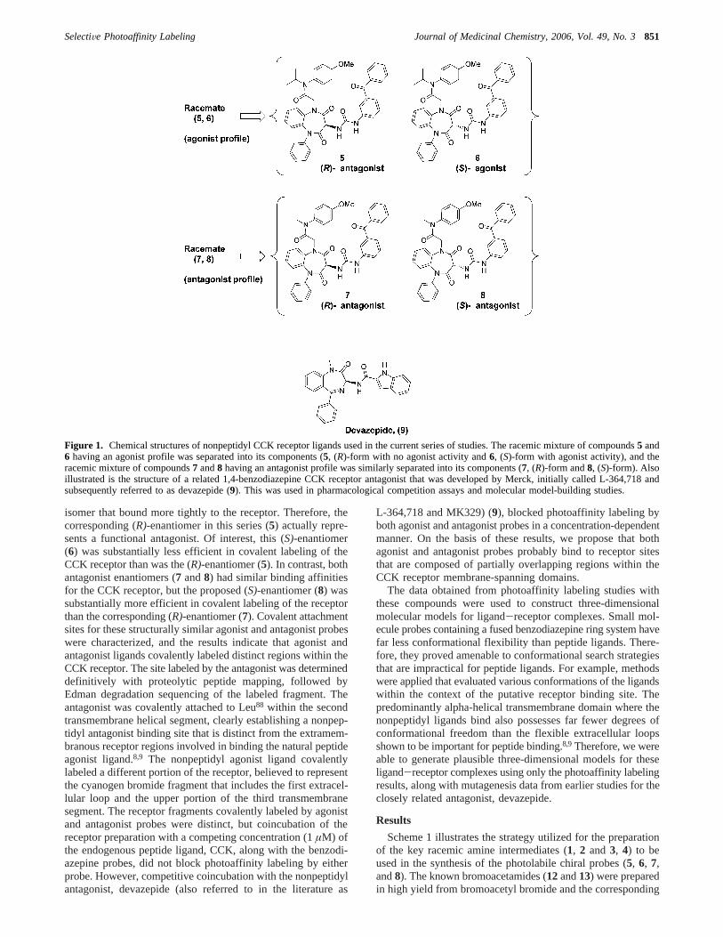
As can be seen in Figure 2, after separating the racemicmixture that demonstrated agonist activity, only the putative(S)-enantiomer (6) displayed intrinsic agonist activity (EC50 150( 36 nM). The corresponding (R)-enantiomer (5) showed noability to stimulate an increase in pancreatic acinar cell amylasesecretion in concentrations as high as 1µM. These two enan-tiomers also displayed distinct binding affinities, with compound6 having a higher affinity than compound5 (IC50s of 61( 17nM for compound6 and 710( 420 nM for compound5). Thedata for compound5 are consistent with the pharmacologicalprofile of a type A CCK receptor antagonist. Figure 2 also showsbinding data for the pair of compounds separated from theantagonist racemic mixture. Both compounds7 and8 had similarapparent binding affinities for the CCK receptor, with IC50s forinhibition of the binding of the CCK-like peptide radioligandof 490(55 nM for the proposed (S)-enantiomer (8) and 470(66 nM for the corresponding (R)-enantiomer (7).
To minimize the use of radioactivity during the synthesis andpurification of the chiral photoaffinity probes, we employed thesecond method shown in Scheme 2 whereby the racemic amineintermediates were separated into their respective enantiomersprior to phosgene coupling with the 3-aminobenzophenone. Thisapproach ultimately allowed facile incorporation of14C into themolecules during the last possible step, thus providing moreefficient handling and purification of radioactive compounds.
However, to validate the ultimate enantiopurity of thissynthetic methodology, each chiral amine was first cleanlyconverted into its respective chiral urea (5, 6, 7, and8) usingnonradioactive phosgene and 3-aminobenzophenone. The puri-fied compounds synthesized using this approach were comparedvia chiral HPLC analysis to the corresponding separated
enantiomeric final products previously obtained using method1, and each was determined to be>96.5% enantiomeric excess(ee). Using this methodology, the enantiopurity of the final ureassynthesized from the resolved amine intermediates closelymatched the enantiopurities of the respective intermediatesthemselves, indicating no significant reduction in enantiomericexcess during the final urea formation and purification. Furtherexperiments, in particular the treatment of the separated enan-tiomers under basic conditions (with TEA or DIPEA), causedrapid racemization and the appearance of both parent peaks inthe chiral HPLC traces, yet no degradation of compound to otherspecies by HPLC, NMR, or MS analysis. Method 2 was thenapplied directly to the synthesis of the14C incorporatedphotoaffinity probes (5, 6, 7, and 8), and the previouslysynthesized and purified nonradioactive, “cold” analogues werethen used as comparative standards during the synthesis andpurification of the radiolabeled versions.
Each of the four14C-tagged probes labeled the CCK receptor,as demonstrated by the covalent labeling of a band migratingatMr ) 85000-95000 (Figure 3). This band had a core proteinmigrating atMr ) 42000 (data not shown). These match theestablished electrophoretic migrations of this receptor glyco-protein and its deglycosylated core protein, as well as the broadnature of those bands resolved under these conditions, aspreviously reported.14 Each probe labeled the receptor withdistinct efficiencies, as reflected by the relative intensities ofthe bands on autoradiographs, as seen and quantified in Figure3. Compound8 (the proposed (S)-enantiomer antagonist)reproducibly generated the strongest receptor signal in experi-ments utilizing the same amounts of receptor-bearing membraneand similar amounts of probes having similar specific radioac-tivity.
Inhibition of affinity labeling of the CCK receptor by eachof the four probes was attempted with various concentrationsof CCK and devazepide (9). Of note, no inhibition was achievedfor any probe after competitive coincubation with concentrations
Figure 2. Binding and biological activity of CCK receptor ligands. Shown are curves representing the abilities of different concentrations of CCKreceptor ligands (the racemic mixture having agonist action and its enantiomeric components in the top left panel, and the racemic mixture havingantagonist action and its enantiomeric components in the bottom left panel) to compete for the saturable binding of125I-D-Tyr-Gly-[(Nle28,31)CCK-26-33]. 100% represents total saturable binding of this CCK-like radioligand in the absence of competitor and 0% represents the binding in thepresence of 1µM CCK, with this value always less than 10% of total bound radioactivity. Shown in the right panels are biological activity curves,representing the abilities of these compounds to stimulate amylase secretion from dispersed rat pancreatic acini. 100% represents the maximalresponse of these cells to CCK, occurring at 0.1 nM CCK. Shown are means( SEM of the values from three independent experiments.
SelectiVe Photoaffinity Labeling Journal of Medicinal Chemistry, 2006, Vol. 49, No. 3853
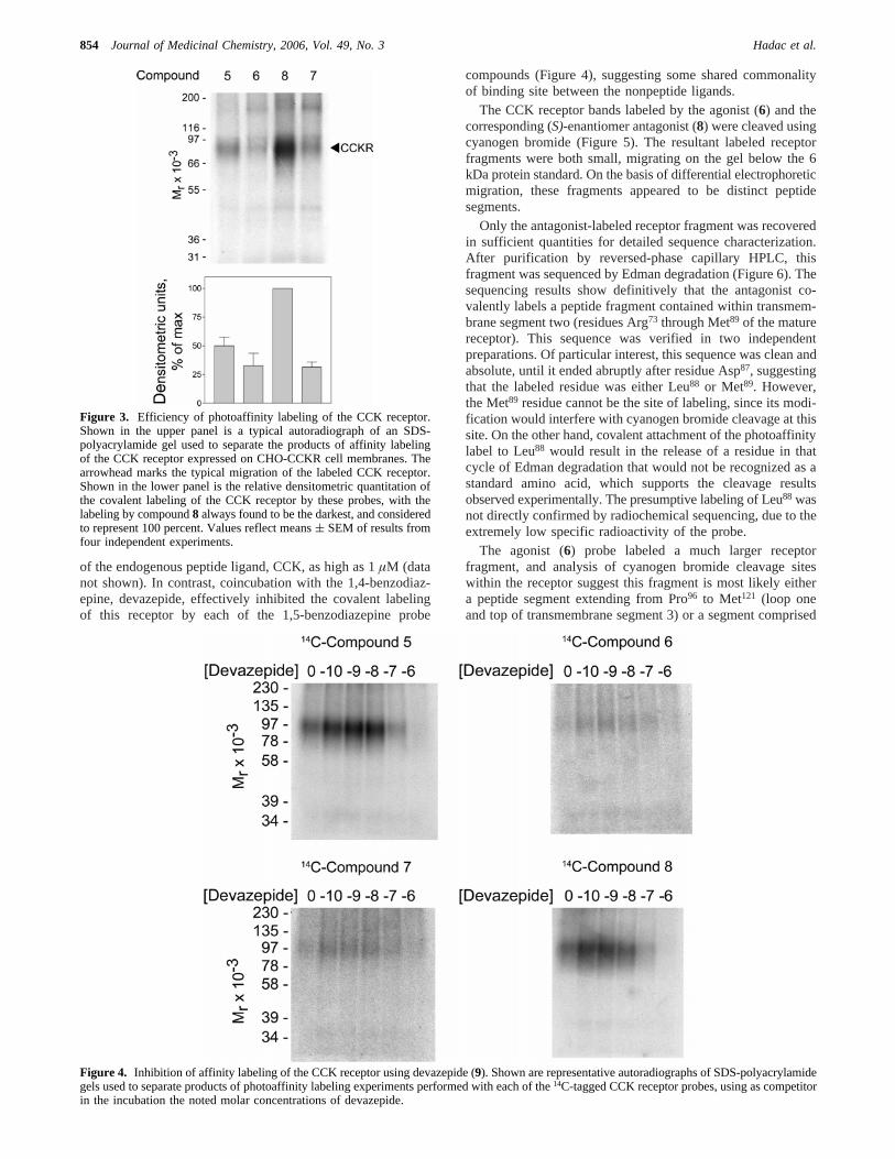
of the endogenous peptide ligand, CCK, as high as 1µM (datanot shown). In contrast, coincubation with the 1,4-benzodiaz-epine, devazepide, effectively inhibited the covalent labelingof this receptor by each of the 1,5-benzodiazepine probe
compounds (Figure 4), suggesting some shared commonalityof binding site between the nonpeptide ligands.
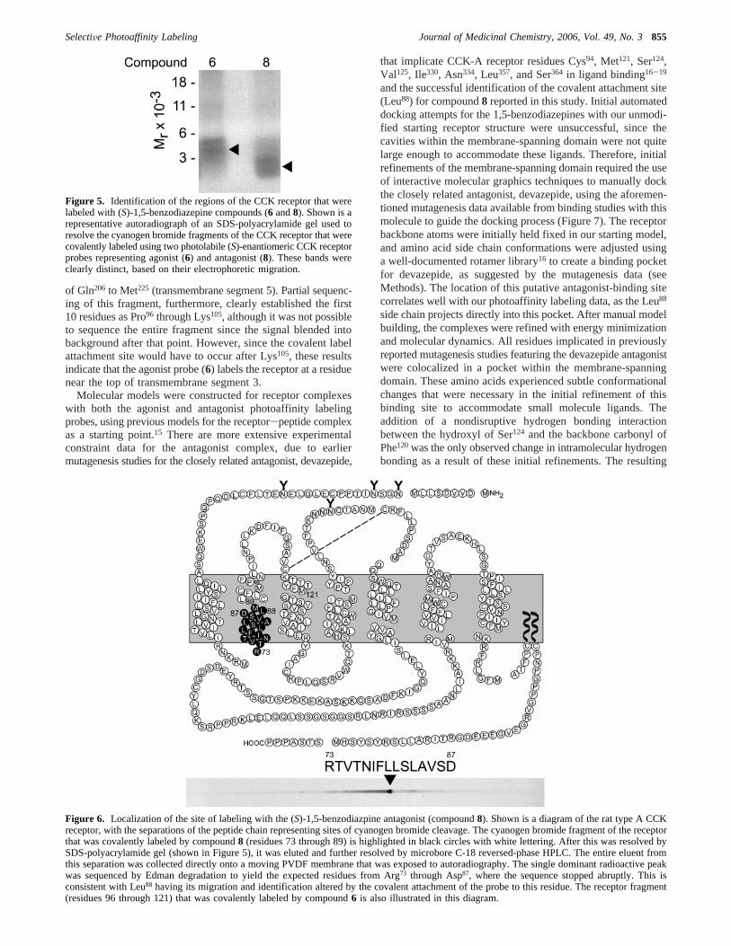
The CCK receptor bands labeled by the agonist (6) and thecorresponding (S)-enantiomer antagonist (8) were cleaved usingcyanogen bromide (Figure 5). The resultant labeled receptorfragments were both small, migrating on the gel below the 6kDa protein standard. On the basis of differential electrophoreticmigration, these fragments appeared to be distinct peptidesegments.
Only the antagonist-labeled receptor fragment was recoveredin sufficient quantities for detailed sequence characterization.After purification by reversed-phase capillary HPLC, thisfragment was sequenced by Edman degradation (Figure 6). Thesequencing results show definitively that the antagonist co-valently labels a peptide fragment contained within transmem-brane segment two (residues Arg73 through Met89 of the maturereceptor). This sequence was verified in two independentpreparations. Of particular interest, this sequence was clean andabsolute, until it ended abruptly after residue Asp87, suggestingthat the labeled residue was either Leu88 or Met89. However,the Met89 residue cannot be the site of labeling, since its modi-fication would interfere with cyanogen bromide cleavage at thissite. On the other hand, covalent attachment of the photoaffinitylabel to Leu88 would result in the release of a residue in thatcycle of Edman degradation that would not be recognized as astandard amino acid, which supports the cleavage resultsobserved experimentally. The presumptive labeling of Leu88 wasnot directly confirmed by radiochemical sequencing, due to theextremely low specific radioactivity of the probe.
The agonist (6) probe labeled a much larger receptorfragment, and analysis of cyanogen bromide cleavage siteswithin the receptor suggest this fragment is most likely eithera peptide segment extending from Pro96 to Met121 (loop oneand top of transmembrane segment 3) or a segment comprised
Figure 3. Efficiency of photoaffinity labeling of the CCK receptor.Shown in the upper panel is a typical autoradiograph of an SDS-polyacrylamide gel used to separate the products of affinity labelingof the CCK receptor expressed on CHO-CCKR cell membranes. Thearrowhead marks the typical migration of the labeled CCK receptor.Shown in the lower panel is the relative densitometric quantitation ofthe covalent labeling of the CCK receptor by these probes, with thelabeling by compound8 always found to be the darkest, and consideredto represent 100 percent. Values reflect means( SEM of results fromfour independent experiments.
Figure 4. Inhibition of affinity labeling of the CCK receptor using devazepide (9). Shown are representative autoradiographs of SDS-polyacrylamidegels used to separate products of photoaffinity labeling experiments performed with each of the14C-tagged CCK receptor probes, using as competitorin the incubation the noted molar concentrations of devazepide.
854 Journal of Medicinal Chemistry, 2006, Vol. 49, No. 3 Hadac et al.
of Gln206 to Met225 (transmembrane segment 5). Partial sequenc-ing of this fragment, furthermore, clearly established the first10 residues as Pro96 through Lys105, although it was not possibleto sequence the entire fragment since the signal blended intobackground after that point. However, since the covalent labelattachment site would have to occur after Lys105, these resultsindicate that the agonist probe (6) labels the receptor at a residuenear the top of transmembrane segment 3.
Molecular models were constructed for receptor complexeswith both the agonist and antagonist photoaffinity labelingprobes, using previous models for the receptor-peptide complexas a starting point.15 There are more extensive experimentalconstraint data for the antagonist complex, due to earliermutagenesis studies for the closely related antagonist, devazepide,
that implicate CCK-A receptor residues Cys94, Met121, Ser124,Val125, Ile330, Asn334, Leu357, and Ser364 in ligand binding16-19
and the successful identification of the covalent attachment site(Leu88) for compound8 reported in this study. Initial automateddocking attempts for the 1,5-benzodiazepines with our unmodi-fied starting receptor structure were unsuccessful, since thecavities within the membrane-spanning domain were not quitelarge enough to accommodate these ligands. Therefore, initialrefinements of the membrane-spanning domain required the useof interactive molecular graphics techniques to manually dockthe closely related antagonist, devazepide, using the aforemen-tioned mutagenesis data available from binding studies with thismolecule to guide the docking process (Figure 7). The receptorbackbone atoms were initially held fixed in our starting model,and amino acid side chain conformations were adjusted usinga well-documented rotamer library16 to create a binding pocketfor devazepide, as suggested by the mutagenesis data (seeMethods). The location of this putative antagonist-binding sitecorrelates well with our photoaffinity labeling data, as the Leu88
side chain projects directly into this pocket. After manual modelbuilding, the complexes were refined with energy minimizationand molecular dynamics. All residues implicated in previouslyreported mutagenesis studies featuring the devazepide antagonistwere colocalized in a pocket within the membrane-spanningdomain. These amino acids experienced subtle conformationalchanges that were necessary in the initial refinement of thisbinding site to accommodate small molecule ligands. Theaddition of a nondisruptive hydrogen bonding interactionbetween the hydroxyl of Ser124 and the backbone carbonyl ofPhe120was the only observed change in intramolecular hydrogenbonding as a result of these initial refinements. The resulting
Figure 5. Identification of the regions of the CCK receptor that werelabeled with (S)-1,5-benzodiazepine compounds (6 and8). Shown is arepresentative autoradiograph of an SDS-polyacrylamide gel used toresolve the cyanogen bromide fragments of the CCK receptor that werecovalently labeled using two photolabile (S)-enantiomeric CCK receptorprobes representing agonist (6) and antagonist (8). These bands wereclearly distinct, based on their electrophoretic migration.
Figure 6. Localization of the site of labeling with the (S)-1,5-benzodiazpine antagonist (compound8). Shown is a diagram of the rat type A CCKreceptor, with the separations of the peptide chain representing sites of cyanogen bromide cleavage. The cyanogen bromide fragment of the receptorthat was covalently labeled by compound8 (residues 73 through 89) is highlighted in black circles with white lettering. After this was resolved bySDS-polyacrylamide gel (shown in Figure 5), it was eluted and further resolved by microbore C-18 reversed-phase HPLC. The entire eluent fromthis separation was collected directly onto a moving PVDF membrane that was exposed to autoradiography. The single dominant radioactive peakwas sequenced by Edman degradation to yield the expected residues from Arg73 through Asp87, where the sequence stopped abruptly. This isconsistent with Leu88 having its migration and identification altered by the covalent attachment of the probe to this residue. The receptor fragment(residues 96 through 121) that was covalently labeled by compound6 is also illustrated in this diagram.
SelectiVe Photoaffinity Labeling Journal of Medicinal Chemistry, 2006, Vol. 49, No. 3855
receptor complex structure (Figure 7) differed from our previ-ously reported model15 by only 0.97 Å RMSD in the membrane-spanning region.
Automated docking calculations for the agonist compoundproduced many equally plausible docked conformers with highscores that were all consistent with labeling of a receptorfragment composed of portions of the third transmembrane helixand the first extracellular loop regions. The agonist models werenot refined further due to the lack of an identified site of covalentattachment for the agonist photoaffinity ligand. On the basis ofthe limited nature of the current data, we cannot rule out apossible attachment site for the agonist photoaffinity probe inthe first extracellular loop of the receptor, although the failureof coincubation with the peptide agonist to block photolabelingby the 1,5-benzodiazepine agonist probe would appear topreclude a direct interaction in the first extracellular loop.
The benzophenone photophore reacts with the receptorthrough a bond-insertion mechanism that targets electron-richC-H bonds such as those found in residues with tertiary centers
(isoleucine, leucine, and valine) or next to heteroatoms, forexample, peptide backbone CR-H bonds, glycine CR-H bonds,and the Cγ-H bonds of methionine.20 The 25 best-scoringcomplexes from both shape-based and energy-based DOCK 4.0scoring functions were examined and those that were consistentwith the antagonist photoaffinity labeling data were retained asstarting points for further consideration. None of the structuresgenerated by automated docking positioned the 1,5-benzodiaz-epine in perfect position to form a covalent bond with the targetreceptor residue, Leu88. However, one of the top-scoring dockedcomplexes did orient the ligand such that the benzophenonesubstituent was properly aligned for covalent insertion in theCγ-H bond of Leu88, although the interatomic separation wastoo long (10 Å) to be consistent with the benzophenone reactionmechanism. This complex was further refined with manualadjustments and restrained molecular dynamics simulations.
The refined structure displays a 3.5 Å separation betweenthe benzophenone carbonyl carbon and the reactive Cγ-H bondof Leu88 (Figure 8), quite consistent with the labeling results
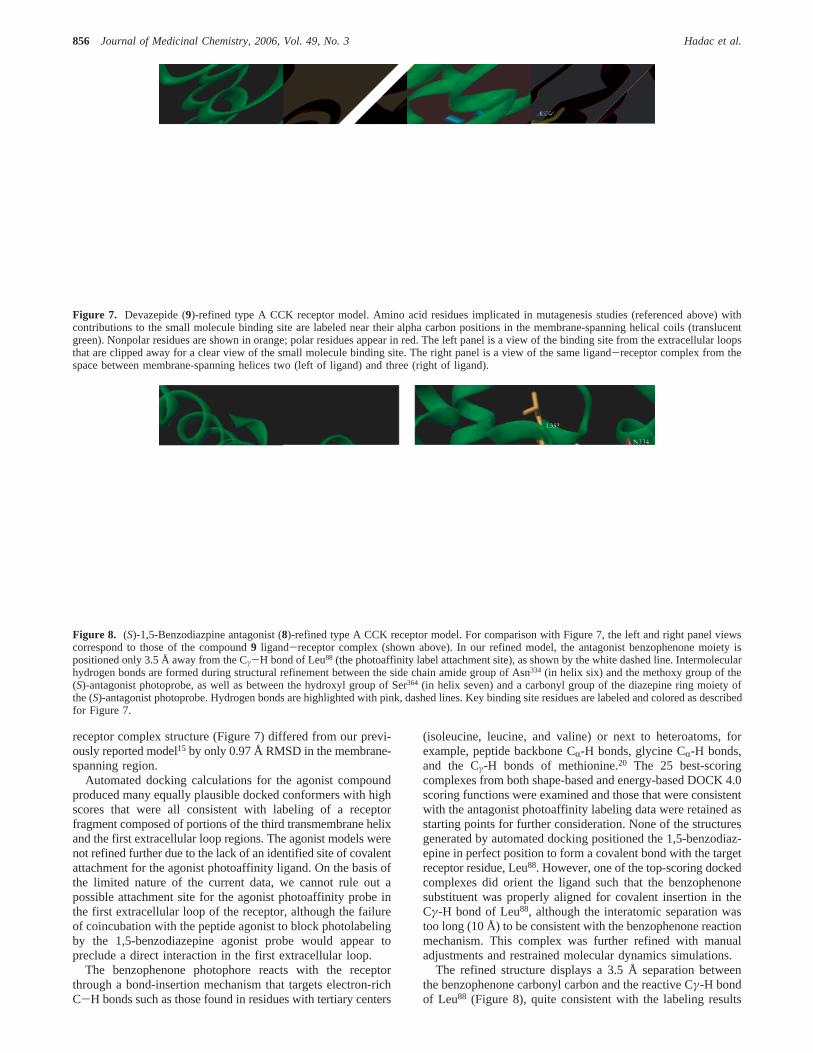
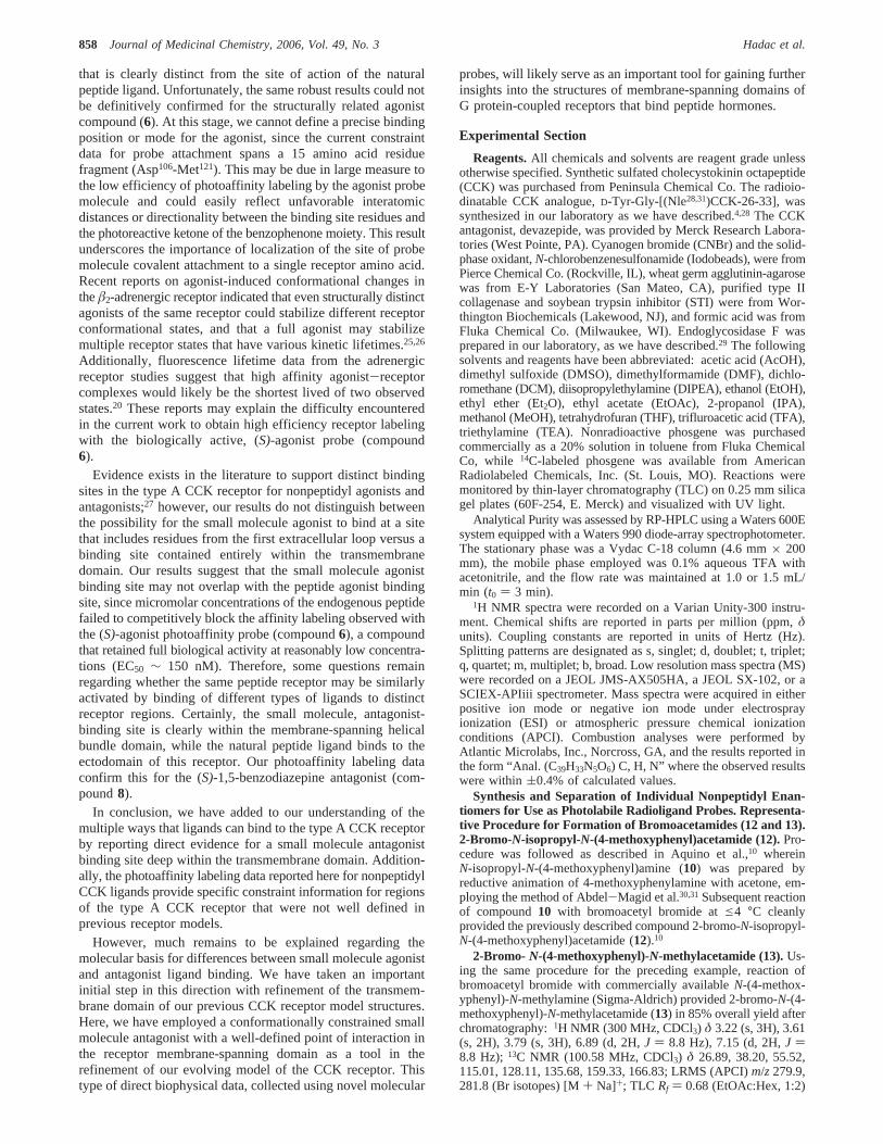
Figure 7. Devazepide (9)-refined type A CCK receptor model. Amino acid residues implicated in mutagenesis studies (referenced above) withcontributions to the small molecule binding site are labeled near their alpha carbon positions in the membrane-spanning helical coils (translucentgreen). Nonpolar residues are shown in orange; polar residues appear in red. The left panel is a view of the binding site from the extracellular loopsthat are clipped away for a clear view of the small molecule binding site. The right panel is a view of the same ligand-receptor complex from thespace between membrane-spanning helices two (left of ligand) and three (right of ligand).
Figure 8. (S)-1,5-Benzodiazpine antagonist (8)-refined type A CCK receptor model. For comparison with Figure 7, the left and right panel viewscorrespond to those of the compound9 ligand-receptor complex (shown above). In our refined model, the antagonist benzophenone moiety ispositioned only 3.5 Å away from the Cγ-H bond of Leu88 (the photoaffinity label attachment site), as shown by the white dashed line. Intermolecularhydrogen bonds are formed during structural refinement between the side chain amide group of Asn334 (in helix six) and the methoxy group of the(S)-antagonist photoprobe, as well as between the hydroxyl group of Ser364 (in helix seven) and a carbonyl group of the diazepine ring moiety ofthe (S)-antagonist photoprobe. Hydrogen bonds are highlighted with pink, dashed lines. Key binding site residues are labeled and colored as describedfor Figure 7.
856 Journal of Medicinal Chemistry, 2006, Vol. 49, No. 3 Hadac et al.

and the photochemistry involved. In addition, two intermolecularhydrogen bonds are formed in this docked complex with residuesAsn334and Ser364(Figure 8) that were both previously implicatedin a mutagenesis study by Smeets et al.21 Furthermore, favorablevan der Waals interactions are maintained with all abovereferenced residues from mutagenesis studies that were used inthe initial refinement of the membrane-spanning region of ourmodel using devazepide. The refined receptor complex structurefor the putative (S)-1,5-benzodiazepine antagonist probe (8)(Figure 8) differed from the starting structure (Figure 7) by 2.57Å RMSD in the membrane-spanning regions. This differencereflects some minor repacking of the model in this region. Theonly significant changes observed were a bending of the secondtransmembrane helix at Pro96 to form favorable packinginteractions with the (S)-1,5-benzodiazepine antagonist probe(8) and a disruption of one turn of the sixth transmembranehelix by the formation of a hydrogen bond between the hydroxylof Ser360 and the backbone carbonyl of Ile356. The dockedcomplex was subsequently refined at two different temperatures,20 K (not shown) and 300 K (Figure 8) and both of the featuresdetailed above were present in both of the resulting models,which differed by only 1.2 Å RMSD in their membrane-spanning regions. Therefore, it is likely that these features ofour refined model are mostly due to the presence of the larger(S)-antagonist photoprobe ligand (8) that is now docked into apreviously poorly refined region of the small molecule bindingsite. Of note, docking of the (R)-antagonist photoprobe ligand(7) using similar protocols (data not shown) did not result inthe formation of significant intermolecular hydrogen bonds withpolar residues or produce favorable van der Waals contacts witha majority of the binding site residues implicated by previousmutagenesis studies, as did the docking of the experimentallymore efficient (S)-antagonist photoprobe (8).
Discussion
G protein-coupled receptors are remarkable for the structuraldiversity of natural ligands that bind and activate thesemolecules, ranging from photons, odorants, and small biogenicamines to larger peptides, proteins, and even viral particles.22
The CCK receptor is a member of the rhodopsin/â-adrenergicreceptor family of G protein-coupled receptors.2 Its naturalligand is a small linear peptide hormone. A series of receptormutagenesis and photoaffinity labeling studies have localizedthe peptide-binding domain of this receptor to the extracellularsurface of the membrane, where the peptide ligand interacts withregions of the receptor amino terminus and loops.6,7,9,23We havepreviously generated and refined three-dimensional CCK pep-tide-receptor complex models that are consistent with allexisting structure-activity data and direct biophysical measure-ments, including photoaffinity labeling and fluorescence reso-nance energy transfer experiments.9,15
We have reported previously that the bovine rhodopsin crystalstructure24 is probably not an ideal template for homologymodeling of peptide receptors such as the type A CCK receptor,since the extracellular loops that comprise the peptide hormonebinding site probably adopt quite different conformations inrhodopsin and CCK.9 Therefore, we have used de novo model-ing techniques, together with constraint data from mutagenesisand biophysical experiments, to generate three-dimensionalmodels for peptide complexes with the CCK receptor.9 The newphotoaffinity labeling data reported here for nonpeptidyl CCKligands is especially useful, as these data provide specificconstraint information for regions of the type A CCK receptorthat were not well defined in previous models.
Many previous studies of small molecule complexes with typeA CCK receptor relied primarily on site-directed mutagenesisand other indirect structural probes to investigate ligand-receptor complexes.19 These data are quite useful, but interpreta-tion of results can be difficult, and it is generally not possibleto build unique structural models based on indirect data alone.Nonetheless, these data have provided important clues aboutthe location and nature of the small molecule binding site inthe type A CCK receptor. For example, residues His381 andVal354 in the rat type B CCK receptor align with type A receptorresidues Leu357 and Ile330. In type B CCK receptor mutantproteins, changes at these positions to the corresponding typeA residues shifted the pharmacological profile of devazepidesubstantially toward that of the type A CCK receptor.18,19
Previous alanine-scanning mutagenesis experiments impli-cated the polar residues Ser124, Asn334, and Ser364 in bindingto devazepide.21 Mutation of residues Cys94, Met121, and Val125
in the type A CCK receptor abolished binding for the non-peptidyl agonist, SR-146,131.17 All these residues impli-cated in previous mutagenesis studies are colocalized in a cavitywithin the membrane-spanning domain of our previous type ACCK receptor model. The key Leu88 residue covalently labeledby our antagonist probe is also present in this cavity, and ourinitial manual docking studies were guided and constrained bythese observations. Both the devazepide antagonist ligand9(Figure 7) and the 1,5-benzodiazepine antagonist photoaffinityprobe (Figure 8) form plausible interactions with these bindingpocket residues in our refined receptor complex models. Thesecurrent models are also fully compatible with all previouslyreported structure-activity and photoaffinity labeling data forthe type A CCK receptor. However, this current study providesthe first direct experimental evidence for a specific ligandcontact with a CCK receptor residue deep inside the transmem-brane domain.
The methodological difficulties and uniqueness of this effortshould be noted. Photoaffinity probes require the incorporationof both photolabile and indicator moieties. In peptide and proteinligands, it is often feasible to incorporate a photolabile aminoacid derivative and a site for radioiodination that can be utilizedin autoradiography. Indeed, a single radioiodine per probemolecule provides a specific radioactivity of 2000 Ci/mmol.Small drug candidates are often much less able to accommodateextraneous groups. We were fortunate that structure-activityconsiderations allowed the incorporation of a benzophenonemoiety to confer photolability, without having substantialnegative impact on either biological activity or binding affinity.However, for practical reasons, we were able to incorporate onlya single14C into this compound, yielding a specific radioactivityof 0.05 Ci/mmol. This resulted in a specific radioactivity thatwas 30000-fold lower than that of any of the peptide photoaf-finity labeling probes previously utilized to label this receptorand its subdomains and residues.4-7,9,23 Nonetheless, we werestill able to demonstrate specific photoaffinity labeling of thisreceptor with the benzodiazepine probes, and we were also ableto localize the site of covalent attachment for the antagonistprobe that labeled this receptor.
While it is well recognized that different G protein-coupledreceptors bind their natural ligands in different ways and indifferent domains, correlating with the distinct chemical char-acteristics of their ligands,22 our data present a clear, directdemonstration of ligand binding to two distinct domains withinthe same receptor. Our current study has directly establishedan intramembranous binding site with moderately high affinityfor the putative (S)-1,5-benzodiazpine antagonist (compound8)
SelectiVe Photoaffinity Labeling Journal of Medicinal Chemistry, 2006, Vol. 49, No. 3857
that is clearly distinct from the site of action of the naturalpeptide ligand. Unfortunately, the same robust results could notbe definitively confirmed for the structurally related agonistcompound (6). At this stage, we cannot define a precise bindingposition or mode for the agonist, since the current constraintdata for probe attachment spans a 15 amino acid residuefragment (Asp106-Met121). This may be due in large measure tothe low efficiency of photoaffinity labeling by the agonist probemolecule and could easily reflect unfavorable interatomicdistances or directionality between the binding site residues andthe photoreactive ketone of the benzophenone moiety. This resultunderscores the importance of localization of the site of probemolecule covalent attachment to a single receptor amino acid.Recent reports on agonist-induced conformational changes intheâ2-adrenergic receptor indicated that even structurally distinctagonists of the same receptor could stabilize different receptorconformational states, and that a full agonist may stabilizemultiple receptor states that have various kinetic lifetimes.25,26
Additionally, fluorescence lifetime data from the adrenergicreceptor studies suggest that high affinity agonist-receptorcomplexes would likely be the shortest lived of two observedstates.20 These reports may explain the difficulty encounteredin the current work to obtain high efficiency receptor labelingwith the biologically active, (S)-agonist probe (compound6).
Evidence exists in the literature to support distinct bindingsites in the type A CCK receptor for nonpeptidyl agonists andantagonists;27 however, our results do not distinguish betweenthe possibility for the small molecule agonist to bind at a sitethat includes residues from the first extracellular loop versus abinding site contained entirely within the transmembranedomain. Our results suggest that the small molecule agonistbinding site may not overlap with the peptide agonist bindingsite, since micromolar concentrations of the endogenous peptidefailed to competitively block the affinity labeling observed withthe (S)-agonist photoaffinity probe (compound6), a compoundthat retained full biological activity at reasonably low concentra-tions (EC50 ∼ 150 nM). Therefore, some questions remainregarding whether the same peptide receptor may be similarlyactivated by binding of different types of ligands to distinctreceptor regions. Certainly, the small molecule, antagonist-binding site is clearly within the membrane-spanning helicalbundle domain, while the natural peptide ligand binds to theectodomain of this receptor. Our photoaffinity labeling dataconfirm this for the (S)-1,5-benzodiazepine antagonist (com-pound8).
In conclusion, we have added to our understanding of themultiple ways that ligands can bind to the type A CCK receptorby reporting direct evidence for a small molecule antagonistbinding site deep within the transmembrane domain. Addition-ally, the photoaffinity labeling data reported here for nonpeptidylCCK ligands provide specific constraint information for regionsof the type A CCK receptor that were not well defined inprevious receptor models.
However, much remains to be explained regarding themolecular basis for differences between small molecule agonistand antagonist ligand binding. We have taken an importantinitial step in this direction with refinement of the transmem-brane domain of our previous CCK receptor model structures.Here, we have employed a conformationally constrained smallmolecule antagonist with a well-defined point of interaction inthe receptor membrane-spanning domain as a tool in therefinement of our evolving model of the CCK receptor. Thistype of direct biophysical data, collected using novel molecular
probes, will likely serve as an important tool for gaining furtherinsights into the structures of membrane-spanning domains ofG protein-coupled receptors that bind peptide hormones.
Experimental Section
Reagents.All chemicals and solvents are reagent grade unlessotherwise specified. Synthetic sulfated cholecystokinin octapeptide(CCK) was purchased from Peninsula Chemical Co. The radioio-dinatable CCK analogue,D-Tyr-Gly-[(Nle28,31)CCK-26-33], wassynthesized in our laboratory as we have described.4,28 The CCKantagonist, devazepide, was provided by Merck Research Labora-tories (West Pointe, PA). Cyanogen bromide (CNBr) and the solid-phase oxidant,N-chlorobenzenesulfonamide (Iodobeads), were fromPierce Chemical Co. (Rockville, IL), wheat germ agglutinin-agarosewas from E-Y Laboratories (San Mateo, CA), purified type IIcollagenase and soybean trypsin inhibitor (STI) were from Wor-thington Biochemicals (Lakewood, NJ), and formic acid was fromFluka Chemical Co. (Milwaukee, WI). Endoglycosidase F wasprepared in our laboratory, as we have described.29 The followingsolvents and reagents have been abbreviated: acetic acid (AcOH),dimethyl sulfoxide (DMSO), dimethylformamide (DMF), dichlo-romethane (DCM), diisopropylethylamine (DIPEA), ethanol (EtOH),ethyl ether (Et2O), ethyl acetate (EtOAc), 2-propanol (IPA),methanol (MeOH), tetrahydrofuran (THF), trifluroacetic acid (TFA),triethylamine (TEA). Nonradioactive phosgene was purchasedcommercially as a 20% solution in toluene from Fluka ChemicalCo, while 14C-labeled phosgene was available from AmericanRadiolabeled Chemicals, Inc. (St. Louis, MO). Reactions weremonitored by thin-layer chromatography (TLC) on 0.25 mm silicagel plates (60F-254, E. Merck) and visualized with UV light.
Analytical Purity was assessed by RP-HPLC using a Waters 600Esystem equipped with a Waters 990 diode-array spectrophotometer.The stationary phase was a Vydac C-18 column (4.6 mm× 200mm), the mobile phase employed was 0.1% aqueous TFA withacetonitrile, and the flow rate was maintained at 1.0 or 1.5 mL/min (t0 ) 3 min).
1H NMR spectra were recorded on a Varian Unity-300 instru-ment. Chemical shifts are reported in parts per million (ppm,δunits). Coupling constants are reported in units of Hertz (Hz).Splitting patterns are designated as s, singlet; d, doublet; t, triplet;q, quartet; m, multiplet; b, broad. Low resolution mass spectra (MS)were recorded on a JEOL JMS-AX505HA, a JEOL SX-102, or aSCIEX-APIiii spectrometer. Mass spectra were acquired in eitherpositive ion mode or negative ion mode under electrosprayionization (ESI) or atmospheric pressure chemical ionizationconditions (APCI). Combustion analyses were performed byAtlantic Microlabs, Inc., Norcross, GA, and the results reported inthe form “Anal. (C39H33N5O6) C, H, N” where the observed resultswere within(0.4% of calculated values.
Synthesis and Separation of Individual Nonpeptidyl Enan-tiomers for Use as Photolabile Radioligand Probes. Representa-tive Procedure for Formation of Bromoacetamides (12 and 13).2-Bromo-N-isopropyl-N-(4-methoxyphenyl)acetamide (12).Pro-cedure was followed as described in Aquino et al.,10 whereinN-isopropyl-N-(4-methoxyphenyl)amine (10) was prepared byreductive animation of 4-methoxyphenylamine with acetone, em-ploying the method of Abdel-Magid et al.30,31Subsequent reactionof compound10 with bromoacetyl bromide ate4 °C cleanlyprovided the previously described compound 2-bromo-N-isopropyl-N-(4-methoxyphenyl)acetamide (12).10
2-Bromo- N-(4-methoxyphenyl)-N-methylacetamide (13).Us-ing the same procedure for the preceding example, reaction ofbromoacetyl bromide with commercially availableN-(4-methox-yphenyl)-N-methylamine (Sigma-Aldrich) provided 2-bromo-N-(4-methoxyphenyl)-N-methylacetamide (13) in 85% overall yield afterchromatography:1H NMR (300 MHz, CDCl3) δ 3.22 (s, 3H), 3.61(s, 2H), 3.79 (s, 3H), 6.89 (d, 2H,J ) 8.8 Hz), 7.15 (d, 2H,J )8.8 Hz); 13C NMR (100.58 MHz, CDCl3) δ 26.89, 38.20, 55.52,115.01, 128.11, 135.68, 159.33, 166.83; LRMS (APCI)m/z 279.9,281.8 (Br isotopes) [M+ Na]+; TLC Rf ) 0.68 (EtOAc:Hex, 1:2)
858 Journal of Medicinal Chemistry, 2006, Vol. 49, No. 3 Hadac et al.
2-(Phenylhydrazono)malonic Acid (16) and 2-(Phenylhydra-zono)propanedioyl Dichloride (17). To a vigorously stirredsuspension of ketomalonic acid disodium salt (5.0 g, 30.9 mmol)cooled toe4 °C in an ice bath was added dropwise a solution ofhydrochloric acid (1.0 N, 62.0 mL, 62.0 mmol). After the suspensionhad dissolved, ethanol (20 mL) was added, and the reactionproceeded as outlined in Aquino et al.10
2-[2,4-Dioxo-5-phenyl-3-(phenylhydrazono)-2,3,4,5-tetrahydro-benzo[b][1,4]diazepin-1-yl]-N-isopropyl-N-(4-methoxyphenyl)acet-amide (18). As per the modified procedure of Hirst and co-workers,32 commercially availableN-phenyl-l,2-phenylenediamine(5.0 g, 27.1 mmol) was dissolved in DMF (25 mL) and stirred atroom temperature. 1.4 molar equiv of bromoacetamide (12) wereadded to this solution. Solid K2CO3 (8.0 g, 57.9 mmol) was thenadded and the resulting suspension stirred vigorously overnight atroom temperature. Upon completion as monitored by TLC (1:2,EtOAc:Hex), the DMF was evaporated and the resulting oilpartitioned into ice water/EtOAc. The organic layer was washedtwice with ice-water then once with brine, and the organic layerwas then dried over MgSO4 and evaporated to a reddish oil. Thecrude intermediate thus obtained was pure enough to use directlyfor the formation of the 1,5-benzodiazepine ring scaffold. Solutionsof compound14 (6.0 g, 16.6 mmol) in THF (40 mL) and compound17 (5.1 g, 20.8 mmol) in THF (40 mL) were added concomitantlydropwise with cooling in an ice/methanol bath over 30 min. Thesolution was allowed to warm to room temperature and stirred 16h. Upon completion of reaction, as monitored by TLC, the solutionwas evaporated down to a yellow solid that was then resuspendedin EtOAc and washed twice with 0.02 N sodium hydroxide solutionand once with brine. The organic layer was dried over MgSO4 andthe solvent removed. The resulting solid was purified by flushingthrough a pad of silica gel with (1:1) EtOAc:Hex as eluent Afterdrying under vacuum overnight, the corresponding compounds18were obtained as yellow solids in 90% yield (8.4 g) and high purity(as approximately 1:1 mixtures ofsyn- and anti-hydrazone).1HNMR (300 MHz, CDCl3) δ 1.09 (m, 6H), 3.88 (s, 3H), 3.90 (m,1H), 4.20-4.60 (m, 2H), 6.90-7.65 (m. 18H), 10.80 (s, 0.5H),11.54 (s, 0.5H]; LRMS (APCI)m/z 584.0 [M + Na]+; TLC Rf )0.27 (EtOAc:Hex, 1:2). 1:1 Mixture ofcis- andtrans-hydrazones.
2-[2,4-Dioxo-5-phenyl-3-(phenylhydrazono)-2,3,4,5-tetrahydro-benzo[b][1,4]diazepin-1-yl]-N-(4-methoxyphenyl)-N-methylacet-amide (19). Prepared using the procedure described for18 toprovide 90% yield (8.0 g) of a light yellow solid (19). 1H NMR(300 MHz, CDCl3) δ 3.39 (m, 3H), 3.89 (s, 3H), 4.30-4.65 (m,2H), 6.90-7.65 (m, 18H)0.10.80 (s, 0.5H). 11.54 (s, 0.5H); LRMS(APCI) m/z 556.3 [M + Na]+; TLC Rf ) 0.12 (EtOAc:Hex, 1:2).1:1 Mixture of cis- and trans-hydrazones.
Procedure for Cleavage of Hydrazone To Provide RacemicBenzodiazepine Amines. 2-(3-Amino-2,4-dioxo-5-phenyl-2,3,4,5-tetrahydrobenzo[b][1,4]diazepin-1-yl)-N-isopropyl-N-(4-meth-oxyphenyl)acetamide (Racemate 1, 2).To a vigorously stirredsolution of hydrazone18 (4.27 g, 8.0 mmol) in glacial acetic acid(50 mL) at room temperature was added anhydrous zinc dust (4.20g, 64.3 mmol) portionwise. After addition, the solution was stirred3-5 h until the color of the slurry changed from green to yellow.The zinc was removed by filtration through Celite and the cakewashed with EtOAc (2× 75 mL). The filtrate was concentrated,adsorbed onto silica gel and washed with EtOAc:Hex (1:2) toremove more polar impurities. The product was then eluted with10% methanol in DCM and concentrated in vacuo to give ayellowish-orange oil which was dried under high vacuum to providethe corresponding racemic amine intermediate (1, 2) in good yield(3.38 g product as a white amorphous solid, 93.7% yield).1H NMR(300 MHz, CD3OD) δ 1.09 (m, 6H), 3.87 (s, 3H), 4.37 (d, 1H,J) 21.0 Hz), 4.60 (d, 1H,J ) 21.0 Hz), 4.35 (m, 1H), 4.85 (m,1H), 6.80-7.50 (m, 13H); LCMS (APCI)m/z 473.3 [M + H]+;TLC Rf ) 0.45 (EtOAc:Hex, 1:2); Anal. (C27H28N4O4) C, H, N.
2-(3-Amino-2,4-dioxo-5-phenyl-2,3,4,5-tetrahydrobenzo[b]-[1,4]diazepin-1-yl)-N-(4-methoxyphenyl)-N-methylacetamide(Racemate 3, 4).Using the procedure for the racemate (1, 2) aboveand starting with 8 mmol of hydrazone19, 3.22 g of racemate (3,
4) product (95.3% yield) was obtained.1H NMR (300 MHz, CD3-OD) δ 3.20 (s, 3H), 3.87 (s, 3H), 4.45 (d, 1H,J ) 21.0 Hz), 4.72(d, 1H, J ) 21.0 Hz), 4.75 (b, 1H), 6.70-7.50 (m, 13H); LRMS(APCI) m/z 445.2 [M + H]+; TLC Rf ) 0.47 (EtOAc:Hex, 1:2);Anal. (C25H24N4O4) C, H, N.
Method 1: General Procedure for Racemic BenzophenoneUrea Formation and Subsequent Chiral Resolution. 2-{3-[3-(4-Benzoylphenyl)ureido]-2,4-dioxo-5-phenyl-2,3,4,5-tetrahydro-benzo[b][1,4]diazepin-1-yl}-N-isopropyl-N-(4-methoxyphenyl)acet-amide (Racemate 5, 6).Under anhydrous conditions, triphosgene(0.5 mmol) was dissolved in anhydrous acetonitrile (1 mL) andthe stirring solution cooled toe4 °C under dry N2. A solutioncontaining commercially available 3-aminobenzophenone (0.10 g,0.5 mmol) and diisopropylethylamine (DIPEA, 0.1 mL) in anhy-drous acetonitrile (1 mL) was added dropwise to the stirringreaction. The reaction was allowed to stir under dry N2 at e4 °Cfor 30 min, at which time a second solution containing the 1,5-benzodiazepine amine racemate (1, 2) (0.5 mmol) and DIPEA (0.1mL) in dry acetonitrile (2 mL) was added dropwise to the cooled,stirring reaction. Stirring continued ate4 °C for 3-6 h until thereaction was complete as monitored by TLC. The product ureaprecipitated out of acetonitrile during the course of the reactionand was purified by filtration. Subsequent further precipitation ofproduct from filtrate occurred upon standing for several hours atroom temperature. The white solid was washed thoroughly withacetonitrile and dried under vacuum to provide 350 mg white solidracemate (5, 6). No further purification was required as the solidsisolated were observed to be pure by HPLC, NMR and combustionanalysis.1H NMR (300 MHz, CDCl3) δ 0.91 (d, 3H,J ) 6.8 Hz),0.94 (d, 3H,J ) 6.8 Hz), 3.78 (s, 3H), 4.16 (d, 1H,J ) 16.8 Hz),4.58 (d, 1H,J ) 16.8 Hz), 4.74 (septet, 1H,J ) 6.8 Hz), 4.99 (d,1H, J ) 8.0 Hz), 6.91 (t, 2H,J ) 8.8 Hz), 7.43 (d, 2H,J ) 8.8Hz), 7.19-7.58 (m, 15H), 7.65 (t, 1H,J ) 7.2 Hz), 7.68 (d, 2H,J) 7.2 Hz), 7.79 (s, 1H), 9.44 (s, 1H); LRMS (APCI)m/z 717.9 [M+ Na]+, 694.3 [M]-; Anal. (C41H37N5O6) C, H, N.
Chiral Resolution Procedure and Results for Resolved Com-pound 5 (and 6).The isopropyl racemic mixture of5 and6 wasseparated into the resolved enantiomers using semipreparative chiralRP-HPLC and monitored by UV absorbance (Dynamax C-8column, 25 cm× 4.1 mm, 8 mL/min flow rate, ramping from [75%CH3CN/25% (H2O with 0.1% TFA)] up to 100% CH3CN over 30min). An amount of 100 mg of the parentN-isopropyl racemate(5, 6) was separated into its purified component enantiomers, whichwere isolated as white solids (30 mg of compound5, and 25 mg ofcompound6). Enantiomer purity and enantiomeric excess wereconfirmed by chiral RP-HPLC analysis using an alternate isocraticanalytical method (Analytical Chiralpak AD C-18 column, withmobile phase (65:35, Hex:IPA) eluting at 1 mL/min flow rate).Compound5 eluted at 41.28 min, while compound6 eluted at 48.33min (showing good baseline peak separation); coinjections witheach other and with the parent racemate confirmed chiral purity.Compound5 (and6): 1H NMR (300 MHz, CDCl3) δ 0.92 (d, 3H,J ) 6.8 Hz), 0.95 (d, 3H,J ) 6.8 Hz), 3.78 (s, 3H), 4.17 (d, 1H,J ) 16.8 Hz), 4.58 (d, 1H,J ) 16.8 Hz), 4.76 (septet, 1H,J ) 6.8Hz), 5.00 (d, 1H,J ) 8.0 Hz), 6.92 (t, 2H,J ) 8.8 Hz), 7.43(d, 2H, J ) 8.8 Hz), 7.19-7.58 (m, 15H), 7.65 (t, 1H,J ) 7.2Hz), 7.68 (d, 2H,J ) 7.2 Hz), 7.80 (s, 1H), 9.44 (s, 1H); LRMS(APCI) m/z 718.1 [M+ Na]+, 694.3 [M]-; Anal. (C41H37N5O6) C,H, N.
2-{3-[3-(4-Benzoylphenyl)ureido]-2,4-dioxo-5-phenyl-2,3,4,5-tetrahydrobenzo[b][1,4]diazepin-1-yl}-N-(4-methoxyphenyl)-N-methylacetamide (Racemate 7, 8).Reaction with commerciallyavailable 3-aminobenzophenone and the intermediate amine race-mate (3, 4), using the procedure outlined for racemate (5, 6),provided 300 mg of the racemicN-methyl analogues (7, 8) as awhite solid that was pure by NMR, LCMS, and combustionanalysis.1H NMR (300 MHz, CDCl3) δ 3.10 (s, 3H), 3.77 (s, 3H),4.35 (d, 1H,J ) 16.8 Hz), 4.64 (d, 1H,J ) 16.8 Hz), 5.01 (d, 1H,J ) 8.0 Hz), 6.88 (d, 1H,J ) 8.0 Hz), 6.92 (d, 1H,J ) 7.6 Hz),7.03, (d, 1H,J ) 8.0 Hz), 7.21 (d. 1H,J ) 7.6 Hz), 7.24 (d, 1H,J ) 7.6 Hz), 7.30-7.48 (m, 13H), 7.51 (t, 1H,J ) 7.6 Hz); 7.63
SelectiVe Photoaffinity Labeling Journal of Medicinal Chemistry, 2006, Vol. 49, No. 3859
(t, 1H, J ) 7.2 Hz), 7.68 (d, 2H,J ) 7.2 Hz), 7.79 (s, 1H), 9.43(s, 1H); LRMS (APCI)m/z 689.9 [M + Na]+, 666.7 [M]-; Anal.(C39H33N5O6) C, H, N.
Chiral Resolution Procedure and Results for ResolvedCompound 7 (and 8).Following the procedure for the resolvedenantiomers5 and6, 100 mg of the parentN-methyl racemate (7,8) was separated into its purified component enantiomers, whichwere isolated as white solids (35 mg of compound7, and 25 mg ofcompound8). Enantiomer purity and enantiomeric excess wereconfirmed by chiral RP-HPLC analysis using an alternate isocraticanalytical method (Analytical Chiralpak AD C-18 column, withmobile phase (65:35, Hex:IPA) eluting at 1 mL/min flow rate).Compound7 eluted at 49.94 min, while compound8 eluted at 82.17min (showing good baseline peak separation); coinjections witheach other and with the parent racemate confirmed chiral purity.Compound7 (and8): 1H NMR (300 MHz, CDCl3) δ 3.09 (s, 3H),3.76 (s, 3H), 4.37 (d, 1H,J ) 16.8 Hz), 4.67 (d, 1H,J ) 16.8 Hz),5.02 (d, 1H,J ) 8.0 Hz), 6.88 (d, 1H,J ) 8.0 Hz), 6.92 (d, 1H,J ) 7.6 Hz), 7.04, (d, 1H,J ) 8.0 Hz), 7.21 (d. 1H,J ) 7.6 Hz),7.24 (d, 1H,J ) 7.6 Hz), 7.30-7.48 (m, 13H), 7.50 (t, 1H,J )7.6 Hz); 7.63 (t, 1H,J ) 7.2 Hz), 7.69 (d, 2H,J ) 7.2 Hz), 7.79(s, 1H), 9.43 (s, 1H); LRMS (APCI)m/z 689.9 [M + Na]+, 666.7[M] -; Anal. (C39H33N5O6) C, H, N.
Method 2: General Procedure for Resolving the Enantio-meric Amine Intermediates Prior to Forming Chiral Ureas.Resolved (R)- and (S)-2-(3-Amino-2,4-dioxo-5-phenyl-2,3,4,5-tetrahydrobenzo[b][1,4]diazepin-1-yl)-N-isopropyl-N-(4-meth-oxyphenyl)acetamide (1 and 2).For use in the synthesis of14C-labeled(R)- and (S)-photoaffinity probes, the isopropyl racemicmixture of amines1 and2 were then separated into the resolvedenantiomers using semipreparative chiral RP-HPLC and monitoredby UV absorbance, showing good baseline peak separation (Dy-namax C-8 column, 25 cm× 4.1 mm, 8 mL/min flow rate, rampingfrom [40% CH3CN/60% (H2O with 0.1% TFA)] up to 100% CH3-CN over 30 min). Each enantiomer was collected in a flaskcontaining at least 1 equiv of acid (HCl) in order to preventepimerization of the purified enantiomers upon sample concentra-tion. Chiral HPLC purification of the original 500 mg of parentracemate yielded 202 mg of compound1 and 165 mg of compound2. Under standard achiral NMR conditions and LCMS conditions,both enantiomers of each pair gave identical spectra to each otheras well as to that of the parent racemate (1, 2) provided above.Enantiomer purity and enantiomeric excess were confirmed bychiral RP-HPLC analysis, showing good baseline peak separationusing an alternate isocratic analytical method (Analytical ChiralpakAD C-18 column, with mobile phase (3:1, Hex:IPA) eluting at 1mL/min flow rate). Compound1 eluted at 20.60 min, whilecompound2 eluted at 31.76 min; coinjections with the parentracemate confirmed chiral purity. Compound1 (and2) 1H NMR(300 MHz, CD3OD) δ 1.10 (m, 6H), 3.87 (s, 3H), 4.35 (d, 1H,J ) 21.0 Hz), 4.61 (d, 1H,J ) 21.0 Hz), 4.35 (m, 1H), 4.87(m, 1H), 6.80-7.50 (m, 13H); LCMS (APCI)m/z473.3 [M+ H]+;TLC Rf ) 0.45 (EtOAc:Hex, 1:2); Anal. (C27H28N4O4) C, H,N.
Resolved (R)- and (S)-2-(3-Amino-2,4-dioxo-5-phenyl-2,3,4,5-tetrahydrobenzo[b][1,4]diazepin-1-yl)-N-(4-methoxyphenyl)-N-methylacetamide (3 and 4).The methyl racemate (3, 4) wasseparated similarly into its respective enantiomeric amines3 and4using the procedure outlined for racemate (1, 2). Chiral HPLCpurification of the original 500 mg parent racemate yielded 185mg of (3) and 242 mg of (4). As with the isopropyl analogues1and2, under standard achiral NMR conditions and LCMS condi-tions, both enantiomers of each pair gave identical spectra to eachother as well as to that of the parent racemate (1, 2) provided above.Enantiomer purity and enantiomeric excess were confirmed bychiral RP-HPLC analysis using an alternate isocratic analyticalmethod (Analytical Chiralpak AD C-18 column, with mobile phase(3:1, Hex:IPA) eluting at 1 mL/min flow rate). Compound3 elutedat 36.44 min, while compound4 eluted at 42.03 min; coinjectionswith the parent racemate confirmed chiral purity. Compound3 (and4): δ 1H NMR (300 MHz, CD3OD) δ 3.15 (s, 3H), 3.82 (s, 3H),
4.41 (d, 1H,J ) 21.0 Hz), 4.69 (d, 1H,J ) 21.0 Hz), 4.75 (b,1H), 6.70-7.50 (m, 13H). LRMS (APCI)m/z 445.2 [M + H]+;TLC Rf ) 0.47 (EtOAc:Hex, 1:2); Anal. (C25H24N4O4) C, H, N.
Representative Procedure for Chiral Urea Formation. 2-{3-[3-(4-Benzoylphenyl)ureido]-2,4-dioxo-5-phenyl-2,3,4,5-tetra-hydrobenzo[b][1,4]diazepin-1-yl}-N-isopropyl-N-(4-methoxy-phenyl)acetamide (5). 3-Aminobenzophenone (0.016 g, 0.08mmol) was dissolved in anhydrous CH3CN (1 mL) and stirred underN2. DIPEA (0.010 g, 0.08 mmol, 14µL) was then added directlyinto the reaction solution, and the reaction solution was cooled toe4 °C in an ice-water bath. Phosgene (20% in toluene, 0.08 mmol,42.1 µL) was quickly syringed directly into the reaction solutionand the reaction stirred ate4 °C under N2 for 1 h. Dropwise thechiral amine hydrochloride salt of compound1 (0.038 g, 0.08 mmol)in 2 mL of dry CH3CN was added to the stirring reaction solution.After 2-3 min, 1 equiv of DIPEA (0.010 g, 0.08 mmol, 14µL)was added to thee4 °C solution while stirring under N2. After anadditional 2 min, the final 1 equiv of DIPEA (0.010 g, 0.08 mmol,14 µL) was added and the reaction allowed to stir ate4 °C underN2 for 2 h.
For each of the isopropyl compounds (5 and 6), the productprecipitated out of solution and was filtered and washed with dryacetonitrile and then dried to constant weight under vacuum.Analytical data for the isolated products matched the data obtainedfor the same materials synthesized via method 1.
For the methyl compounds (7 and8), the solvent was evaporatedusing a stream of dry N2, then the resulting oil was chromatographedon silica gel using (55% EtOAc/45% Hex) as eluent. The purifiedmaterial was evaporated to a white solid and then dried to constantweight under vacuum. As with the isopropyl compounds, theanalytical data for the isolated products matched the data obtainedfor the same materials synthesized via method 1 previously shown.In all four examples (5, 6, 7, and8), pure white solids were typicallyobtained in 80% yields. This procedure was used for subsequent14C synthesis of the enantiomers5, 6, 7, and8, merely substituting14C-labeled phosgene solution for the nonradioactive phosgene usedabove.
The enantiopurity of the final compounds was verified usingreverse phase HPLC (Analytical Chiralpak AD C-18 column,isocratic method with 1 mL/min flow rate, eluent) 65% hexane/35% IPA). Retention times for the isolated enantiomers obtainedby using method 2 are given in Table 1.
Enantiomer Racemazation Conditions.The purified enanti-omer (1, 2, 3, 4, 5, 6, 7, or 8) (0.1 g) was dissolved in DCM and1.5 equiv of TEA or DIPEA added at room temperature. Thereaction was allowed to stir at room temperature in open to air forseveral hours, and the conversion back to the parent racemate wasfollowed by chiral HPLC, using the conditions described previouslyfor the individual enantiomers. In all cases, conversion was allowedto proceed until approximately 1:1 ratio of stereoisomers wasachieved. The residual solvent and base were then removed invacuo, and the newly formed racemate analyzed for degradationproducts. In all cases, each newly formed racemate was clean ofimpurities and provided the identical analytical results to thoseobtained for the original parent racemate given above.
Receptor-Bearing Cell Line. The Chinese hamster ovary cellline stably expressing the type A rat CCK receptor (CHO-CCKR)that has previously been fully characterized was utilized as sourceof receptor for the biochemical and receptor domain-mappingstudies.14 This cell line was cultured in Ham’s F-12 mediumsupplemented with 5% fetal clone-2 (Hyclone Laboratories, Logan,UT) at 37°C in an environment of 5% CO2 in Costar tissue cultureplasticware. Cells were harvested mechanically and passaged twiceweekly.
Table 1
compound retention time, min purity, % enantiomeric excess, %
5 41.3 >99 >996 48.3 >99 >997 49.9 >99 97.08 82.2 >99 96.5
860 Journal of Medicinal Chemistry, 2006, Vol. 49, No. 3 Hadac et al.
Radioligand Binding Studies.Membranes were prepared fromthe CHO-CCKR cells, as we have previously described.14 Forbinding characterization, membranes (3-5 µg) were incubated witha constant amount of the CCK-like radioligand,125I-D-Tyr-Gly-[(Nle28,31)CCK-26-33] (2-5 pM), in the absence or presence ofvariable concentrations of unlabeled competing ligand. Incubationswere performed to attain steady state, achieved after incubationfor 3 h at 30°C in Krebs-Ringers-HEPES (KRH) medium (25mM HEPES, pH 7.4, 104 mM NaCl, 5 mM KCl, 1.2 mM MgSO4,2 mM CaCl2, 1 mM KH2PO4, 0.2% bovine serum albumin, 0.01%soybean trypsin inhibitor, and 1 mM phenylmethylsulfonyl fluoride).Bound and free radioligand were separated using a Skatron cellharvester (Molecular Devices, Sunnyvale, CA) with receptor-binding filtermats that had been presoaked with 0.2% bovine serumalbumin. Bound radioactivity was quantified in a gamma-spectrometer. Nonspecific binding was determined in the presenceof 1 µM CCK and represented less than 10% of total boundradioactivity.
Affinity Labeling Studies. For covalent labeling studies,substantially greater amounts of receptor-bearing membranes (100-200 µg) were incubated with 1-10 µM radioligand, representingthe 14C-labeled compounds described above, in the absence orpresence of competing unlabeled CCK receptor ligands in KRHmedium for 3 h at 30°C. Tubes were exposed to photolysis for 30min at 4°C in a Rayonet photochemical reactor equipped with 3,-500 Å lamps (Southern New England Ultraviolet, Hamden, CT).Membranes were then isolated by centrifugation, washed twice inKRH medium, and solubilized with 1% NP-40 in KRH for 18 h at4 °C with constant mixing.
Solubilized affinity-labeled receptor was diluted with an equalvolume of KRH medium and one-tenth volume of a 50% slurry ofWGA-agarose, with the incubation allowed to proceed for 24 h at4 °C with constant mixing. The WGA-agarose was pelleted andwashed with a solution containing 0.5 M NaCl and 0.1% NP-40,followed by a water wash. In preparation for electrophoresis,reducing sample buffer was added to the pelleted WGA-agarosebeads, and this was allowed to sit for at least 30 min at roomtemperature. Samples were applied to a 10% SDS-polyacrylamidegel for electrophoresis using the conditions described by Laemmli.33
At the completion of electrophoresis the receptor band was excisedfor further study or the gel was dried in preparation for autorad-iography. Dried gels were placed into film cassettes containingBioMax MS X-ray film and a BioMax TranScreen-LE (EastmanKodak Company, Rochester, NY). Cassettes were kept at-80 °Cfor 1-6 weeks before the film was developed.
Gel-purified affinity-labeled receptor was deglycosylated withendoglycosidase F and/or cleaved with cyanogen bromide usingconditions we have previously reported, with minor modifications.34
Gel regions containing receptor bands were excised, diluted withwater, and treated with dounce homogenization. Gel pellets wereremoved by centrifugation, and the supernatants containing thelabeled receptor or its fragments were lyophilized to dryness.Detergent was removed from the samples by precipitation with 85%ethanol at-20 °C for 18 h. Precipitates were pelleted, dried, andthen resuspended in 70% formic acid containing 2.5 mg of cyanogenbromide. Cleavage was allowed to proceed for 18 h under nitrogenat room temperature. Acid was then removed from the samples bythree cycles of water washes and vacuum centrifugation. Samplebuffer was added to tubes in preparation for electrophoresis on a10% NuPAGE gel (Invitrogen, Carlsbad, CA) with MES runningbuffer. A small portion of the sample was run in a separate lane soit could be dried and used for autoradiography to identify theposition of the cleavage fragment. The remaining portion of thegel was frozen, and, once the position of the labeled fragment ofinterest was identified, that region of the gel could be excised andthe sample eluted for further purification by HPLC.
HPLC Purification and Sequence Analysis.Prior to havingcomponents resolved by HLPC, the samples were applied to aDispo-Biodialyzer (The Nest Group, Inc., Southborough, MA) witha molecular weight cutoff of 1 kDa, and samples were dialyzedfor 24 h against 2 L of water. Capillary HPLC was performed using
a Perkin-Elmer/Brownlee reversed-phase C18 column, measuring0.5 × 150 mm with 5µm beads and a pore size of 300 Å. Thebuffer composition was 0.1% TFA as buffer A and 0.085% TFA/80% acetonitrile as buffer B. The sample was injected at 10% B,and the system was allowed to run for 20 min before beginning a230 min gradient to 90% B. The flow rate was 5µL/min, andsamples were collected directly on poly(vinylidene difluoride)(PVDF) membranes. The UV detector monitored absorbance at 210nm. The PVDF sample membrane was exposed to BioMax MSX-ray film and a BioMax TranScreen-LE. The major radioactivespot was localized and excised from the PVDF membrane and wasthen subjected to Edman degradation sequencing using an AppliedBiosystems automated instrument.
Biological Activity Studies. Rat pancreatic acini were freshlyprepared from 80 to 100 g male Sprague Dawley rats. All studieswere reviewed and approved by the Mayo Clinic Animal Care andUse Committee. Pancreatic tissue was harvested and placed in iced,oxygenated KRH medium. The pancreas was trimmed of fat andconnective tissue and injected with 600 units of purified collagenasediluted to 6 mL with KRH medium. The tissue was minced,oxygenated, and incubated at 37°C for 5 min in a shaking waterbath. Additional digestion took place with 10 min of shaking byhand before the acini were washed and filtered through 200µmnytex mesh. The health of the acini was assessed morphologically,and the cells were suspended in KRH medium for use.
One milliliter aliquots of cells were added to tubes containingvarious concentrations of secretagogues. The samples were incu-bated for 30 min at 37°C in a shaking water bath. The secretionassay was terminated by centrifugation of an aliquot of the cellsuspension over Nyosil oil (William F. Nye, Inc., New Bedford,MA). Amylase measurement utilized the Phadebas amylase reagent(Pharmacia Diagnostics, Uppsala, Sweden). Tablets were dissolvedin assay buffer (0.02 M NaH2PO4, 0.05 M NaCl, 0.02% NaN3, pH7.0). Two milliliters of Phadebas suspension was added to aliquotsof secretion samples in lysis buffer (0.01 M NaH2PO4, pH 7.8,containing 0.1% SDS and 0.1% bovine serum albumin). Sampleswere incubated at 37°C for 15 min before the incubation wasterminated by addition of 0.5 M NaOH. Samples were measuredwith a spectrophotometer at 620 nm, and values were expressed aspercentages of amylase release in response to maximal stimulationwith CCK (0.1 nM CCK).
Molecular Modeling. Our current model for the high affinityagonist-bound type A CCK receptor served as a starting point forthe construction of molecular models that incorporated the photo-affinity labeling data reported in this study.15 The receptor’s smallmolecule binding site was manually adjusted via interactivecomputer graphics techniques to accommodate small moleculeligands within the membrane-spanning domain. These adjustmentswere performed using the most probable side chain rotamerconformations from the backbone-dependent amino acid rotamerlibrary of Dunbrack and Karplus,16 without alteration of thepositions of protein backbone atoms. We targeted stable, low energystructure modifications by choosing only combinations of rotamerswith the three highest probabilities based on a statistical analysisof experimentally observed conformations in the above library ofside chain dihedral angles for each of the following amino acidside chains (Thr118, Ser124, Asn334, Arg337, Tyr339, Thr341, Ile356).We next performed manual docking studies for the nonpeptidylantagonist, devazepide,35 a 1,4-benzodiazepine that competitivelyblocks 1,5-benzodiazepine agonist and antagonist photoaffinitylabeling. Manual docking of devazepide (compound9) was guidedby mutagenesis data from previous ligand-binding studies describedabove.
Limited energy minimization and constrained molecular dynam-ics simulations (5 ps at 20 K) of the devazepide ligand-receptorcomplex were performed in vacuo with a distance dependentdielectric constant to relieve some minor steric and conformationalstrain introduced during initial ligand docking. In this initialrefinement of the membrane-spanning region, previously publishedconstraints were used to maintain the overall conformation of thereceptor starting structure.15 Snapshots were taken from the
SelectiVe Photoaffinity Labeling Journal of Medicinal Chemistry, 2006, Vol. 49, No. 3861
molecular dynamics trajectories; each molecular dynamics snapshotwas energy minimized, and then the devazepide antagonist ligand(9) was removed to generate target receptor structures for automatedligand docking calculations for the 1,5-benzodiazepines (5, 6, 7,and8).
The setup for DOCK 4.036 calculations involved the generationof a negative image of potential ligand binding sites in the receptorby filling the area within the confluence of membrane-spanninghelices with spheres. Boundaries for agonist and antagonist bindingsites were defined using photoaffinity-labeling data for the respec-tive ligands, and sphere generation was confined to bounding boxescentered at the receptor positions for agonist and antagonist covalentlabeling. Although the bounding boxes for the agonist andantagonist sites have significant overlap, the antagonist photoaf-finity-labeling data clearly precludes any receptor contacts forcompound8 in the first extracellular loop. Therefore, the boundingbox for the antagonist photoaffinity label was limited to themembrane-spanning receptor domain, while the bounding box (andcorresponding sphere set) for the agonist photoaffinity ligandcontained portions of the receptor membrane-spanning domain andthe extracellular loop regions.
Docking calculations were performed using low energy confor-mations for each compound. Geometry optimizations were per-formed using ab initio Hartree-Fock calculations with a 3-21G*basis set. Quantum mechanical electrostatic potentials were calcu-lated for each molecule using a 6-31G* basis set and then used tofit atom-centered point charges with the RESP module of AMBER7.37 Conformational searches in torsion angle space for all rotatablebonds of each ligand were conducted using the torsion-drive featurein DOCK 4.0.36 Most sp2-sp3 bonds were scanned at 60°increments, and all sp3-sp3 bonds were probed at-60°, +60°,and 180°. Additionally, energy minimization of the ligand conform-ers generated from this search were performed ‘on the fly’ in DOCKto facilitate the identification of low energy complexes for eachligand with the receptor model. Docked complexes were evaluatedaccording to the DOCK 4.0 shape-based and energy-based scoringfunctions.36 Representative receptor-ligand complex structuresgenerated with DOCK were then refined in vacuo with limited (500steps) energy minimization. For the receptor-(S)-antagonist (8)docked complex, a limited number of additional dihedral angleadjustments were performed (following the above outlined strategy)for the following protein side chains prior to energy refinementsto relieve unfavorable van der Waals contacts around the photo-affinity-labeling site (Leu88, Cys91, Cys94, Met121, Leu357, Ser364).Molecular dynamics simulations for the (S)-antagonist (8) complexwere performed in vacuo with a distance-dependent dielectricconstant at temperatures of 20 K and 300 K for 100 ps. Since ourmodel does not include membrane lipids or solvent, weak (1-10kcal/mol) harmonic constraints were gradually imposed for the 300K simulations over a 50 ps time frame to stabilize the conformationof the membrane-spanning helix bundle at the higher temperatures.An NMR-style energy restraint (5 kcal/mol) was also imposed onthe distance between the ketone carbonyl of the photolabilebenzophenone moiety and the Cγ-H bond of Leu88 to maintain aphysically realistic photoincorporation distance consistent with theexperimental data in this report.
All quantum mechanical calculations were performed withGaussian98.38 Model structures were evaluated for quality of sidechain packing, backbone geometry, and stereochemistry using theprograms QPACK39 and PROCHECK.40 Manual model buildingand rms deviation calculations were performed with the interactivegraphics program PSSHOW.41 All energy minimization and mo-lecular dynamics calculations were performed using the AMBER7.0 suite of programs.37 All nondatabase force field parameters forthe benzodiazepine ligands were developed by analogy to databasevalues in the parm96.dat parameter set of AMBER 7.0.37 MDDIS-PLAY was used for visual analysis of molecular dynamicstrajectories.42 Figure generation was performed using DINO version0.9.43
Statistical Analysis.All observations were repeated at least threetimes in independent experiments and are expressed as the means
( SEM. Binding and biological activity curves were analyzed andplotted using the nonlinear regression analysis routine in the Prismsoftware package (GraphPad Software, San Diego, CA). Bindingkinetics were determined by analysis with the LIGAND program.44
Acknowledgment. We acknowledge the expert contributionof Manon Villeneuve and Itzela Correa of Glaxo-SmithKlineResearch Laboratories, who performed the chiral separationsand the analysis and radiolabeling of the compounds used inthis study. We thank Dr. Maoqing Dong for his advice and helpwith preparing this manuscript. This work was supported bygrants from the National Institutes of Health (DK32878 to L.J.M.and NS33290 to T.P.L.), Glaxo-SmithKline, and the FitermanFoundation.
References(1) Darrow, J. W.; Hadac, E. M.; Miller, L. J.; Sugg, E. E. Structurally
similar small molecule photoaffinity CCK-A agonists and antagonistsas novel tools for directly probing 7TM receptor-ligand interactions.Bioorg. Med. Chem. Lett.1998, 8, 3127-3132.
(2) Ulrich, C. D.; Ferber, I.; Holicky, E.; Hadac, E.; Buell, G.; Miller,L. J. Molecular cloning and functional expression of the humangallbladder cholecystokinin A receptor.Biochem. Biophys. Res.Commun.1993, 193, 204-211.
(3) Mutt, V. Cholecystokinin: isolation, structure, and functions. InGastrointestinal hormones; Glass, G. B. J., Ed.; Raven Press: NewYork, 1980; pp 169-221.
(4) Powers, S. P.; Fourmy, D.; Gaisano, H.; Miller, L. J. Intrinsicphotoaffinity labeling probes for cholecystokinin (CCK)-gastrinfamily receptors D-Tyr-Gly-[Nle28,31,pNO2-Phe33)CCK-26-33).J.Biol. Chem.1988, 263, 5295-5300.
(5) Klueppelberg, U. G.; Gaisano, H. Y.; Powers, S. P.; Miller, L. J.Use of a nitrotryptophan-containing peptide for photoaffinity labelingthe pancreatic cholecystokinin receptor.Biochemistry1989, 28,3463-3468.
(6) Ji, Z.; Hadac, E. M.; Henne, R. M.; Patel, S. A.; Lybrand, T. P.;Miller, L. J. Direct identification of a distinct site of interactionbetween the carboxyl-terminal residue of cholecystokinin and thetype A cholecystokinin receptor using photoaffinity labeling.J. Biol.Chem.1997, 272, 24393-24401.
(7) Hadac, E. M.; Pinon, D. I.; Ji, Z.; Holicky, E. L.; Henne, R. M.;Lybrand, T. P.; Miller, L. J. Direct identification of a second distinctsite of contact between cholecystokinin and its receptor.J. Biol.Chem.1998, 273, 12988-12993.
(8) Ding, X. Q.; Miller, L. J. Characterization of the type A cholecys-tokinin receptor hormone-binding domain: use of contrasting andcomplementary methodologies [Review].Peptides2001, 22, 1223-1228.
(9) Ding, X. Q.; Pinon, D. I.; Furse, K. E.; Lybrand, T. P.; Miller, L. J.Refinement of the conformation of a critical region of charge-chargeinteraction between cholecystokinin and its receptor.Mol. Pharmacol.2002, 61, 1041-1052.
(10) Aquino, C. J.; Armour, D. R.; Berman, J. M.; Birkemo, L. S.; Carr,R. A. E.; Croom, D. K.; Dezube, M.; Dougherty, R. W., Jr.; Ervin,G. N.; Grizzle, M. K.; Head, J. E.; Hirst, G. C.; James, M. K.;Johnson, M. F.; Miller, L. J.; Queen, K. L.; Rimele, T. J.; Smith, D.N.; Sugg, E. E. Discovery of 1,5-benzodiazepines with peripheralcholecystokinin (CCK-A) receptor agonist activity .1. Optimizationof the agonist “Trigger”.J. Med. Chem.1996, 39, 562-569.
(11) Evans, B. E.; Rittle, K. E.; Bock, M. G.; DiPardo, R. M.; Freidinger,R. M.; Whitter, W. L.; Gould, N. P.; Lundell, G. F.; Homnick, C.F.; Veber, D. F.; Anderson, P. S.; Chang, R. S. L.; Lotti, V. J.; Cerino,D. J.; Chen, T. B.; King, P. J.; Kunkel, K. A.; Springer, J. P.;Hirshfield, J. Design of nonpeptidal ligands for a peptide receptor:cholecystokinin antagonists.J. Med. Chem.1987, 30, 1229-1239.
(12) Evans, B. E.; Rittle, K. E.; Bock, M. G.; DiPardo, R. M.; Freidinger,R. M.; Whitter, W. L.; Lundell, G. F.; Veber, D. F.; Anderson, P.S.; Chang, R. S.; Lotti, V. J.; Cerino, D. J.; Chen, T. B.; Kling, P. J.;Kunkel, K. A.; Springer, J. P.; Hirshfield, J. Methods for drugdiscovery: development of potent, selective, orally effective chole-cystokinin antagonists.J. Med. Chem.1988, 31, 2235-2246.
(13) Ursini, A.; Capelli, A. M.; Carr, R. A.; Cassara, P.; Corsi, M.;Curcuruto, O.; Curotto, G.; Dal, C. M.; Davalli, S.; Donati, D.;Feriani, A.; Finch, H.; Finizia, G.; Gaviraghi, G.; Marien, M.;Pentassuglia, G.; Polinelli, S.; Ratti, E.; Reggiani, A. M.; Tarzia,G.; Tedesco, G.; Tranquillini, M. E.; Trist, D. G.; van Amsterdam,F. T. Synthesis and SAR of new 5-phenyl-3-ureido-1,5-benzodiaz-epines as cholecystokinin-B receptor antagonists.J. Med. Chem.2000,43, 3596-3613.
862 Journal of Medicinal Chemistry, 2006, Vol. 49, No. 3 Hadac et al.

(14) Hadac, E. M.; Ghanekar, D. V.; Holicky, E. L.; Pinon, D. I.;Dougherty, R. W.; Miller, L. J. Relationship between native andrecombinant cholecystokinin receptors- role of differential glyco-sylation.Pancreas1996, 13, 130-139.
(15) Harikumar, K. G.; Pinon, D. I.; Wessels, W. S.; Dawson, E. S.;Lybrand, T. P.; Prendergast, F. G.; Miller, L. J. Mesurement ofintermolecular distances for the natural agonist peptide docked atthe cholecystokinin receptor expressed in situ using fluorescenceresonance energy transfer.Mol. Pharmacol.2004, 65, 28-35.
(16) Dunbrack, R. L., Jr.; Karplus, M. Backbone-dependent rotamer libraryfor proteins. Application to side-chain prediction.J. Mol. Biol.1993,230, 543-574.
(17) Escrieut, C.; Gigoux, V.; Archer, E.; Verrier, S.; Maigret, B.;Behrendt, R.; Moroder, L.; Bignon, E.; Silvente-Poirot, S.; Pradayrol,L.; Fourmy, D. The biologically crucial C terminus of cholecystokininand the non-peptide agonist SR-146,131 share a common bindingsite in the human CCK1 receptor- Evidence for a crucial role ofMet-121 in the activation process.J. Biol. Chem.2002, 277, 7546-7555.
(18) Jagerschmidt, A.; Guillaume-Rousselet, N.; Vikland, M. L.; Goudreau,N.; Maigret, B.; Roques, B. P. His381 of the rat CCKB receptor isessential for CCKB versus CCKA receptor antagonist selectivity.Eur.J. Pharmacol.1996, 296, 97-106.
(19) Kopin, A. S.; McBride, E. W.; Quinn, S. M.; Kolakowski, L. F., Jr.;Beinborn, M. The role of the cholecystokinin-B/gastrin receptortransmembrane domains in determining affinity for subtype-selectiveligands.J. Biol. Chem.1995, 270, 5019-5023.
(20) O’Neil, K. T.; Erickson-Viitanen, S.; DeGrado, W. F. Photolabelingof calmodulin with basic, amphiphilic alpha-helical peptides contain-ing p-benzoylphenylalanine.J. Biol. Chem.1989, 264, 14571-14578.
(21) Smeets, R. L.; IJzerman, A. P.; Hermsen, H. P.; Ophorst, O. J.; vanEmst-de Vries SE.; De; Pont, J. J.; Willems, P. H. Mutational analysisof the putative devazepide binding site of the CCK(A) receptor.Eur.J. Pharmacol.1997, 325, 93-99.
(22) Ji, T. H.; Grossmann, M.; Ji, I. G protein-coupled receptors. I.Diversity of receptor-ligand interactions.J. Biol. Chem.1998, 273,17299-17302.
(23) Hadac, E. M.; Ji, Z. S.; Pinon, D. I.; Henne, R. M.; Lybrand, T. P.;Miller, L. J. A peptide agonist acts by occupation of a monomeric Gprotein-coupled receptor: Dual sites of covalent attachment todomains near TM1 and TM7 of the same molecule make biologicallysignificant domain-swapped dimerization unlikely.J. Med. Chem.1999, 42, 2105-2111.
(24) Palczewski, K.; Kumasaka, T.; Hori, T.; Behnke, C. A.; Motoshima,H.; Fox, B. A.; Trong, I. L.; Teller, D. C.; Okada, T.; Stenkamp, R.E.; Yamamoto, M.; Miyano, M. Crystal structure of rhodopsin: AG protein-coupled receptor.Science2000, 289, 739-745.
(25) Ghanouni, P.; Steenhuis, J. J.; Farrens, D. L.; Kobilka, B. K. Agonist-induced conformational changes in the G-protein-coupling domainof the beta 2 adrenergic receptor.Proc. Natl. Acad. Sci. U.S.A.2001,98, 5997-6002.
(26) Ghanouni, P.; Gryczynski, Z.; Steenhuis, J. J.; Lee, T. W.; Farrens,D. L.; Lakowicz, J. R.; Kobilka, B. K. Functionally different agonistsinduce distinct conformations in the G protein coupling domain ofthe beta 2 adrenergic receptor.J. Biol. Chem.2001, 276, 24433-24436.
(27) Gouldson, P.; Legoux, P.; Carillon, C.; Delpech, B.; Le Fur, G.;Ferrara, P.; Shire, D. The agonist SR 146131 and the antagonist SR27897 occupy different sites on the human CCK1 receptor.Eur. J.Pharmacol.2000, 400, 185-194.
(28) Powers, S. P.; Pinon, D. I.; Miller, L. J. Use of N,O-bis-Fmoc-D-Tyr-ONSu for introduction of an oxidative iodination site intocholecystokinin family peptides.Int. J. Pept. Protein Res.1988, 31,429-434.
(29) Pearson, R. K.; Miller, L. J.; Hadac, E. M.; Powers, S. P. Analysisof the carbohydrate composition of the pancreatic plasmalemmal
glycoprotein affinity labeled by short probes for the cholecystokininreceptor.J. Biol. Chem.1987, 262, 13850-13856.
(30) Abdel-Magid, A. F.; Maryanoff, C. A.; Carson, K. G. Reductiveamination of aldehydes and ketones by using sodium triacetoxy-borohydride.Tetrahedron Lett.1990, 31, 5595-5598.
(31) Abdel-Magid, A. F.; Carson, K. G.; Harris, B. D.; Maryanoff, C.A.; Shah, R. D. Reductive amination of aldehydes andketones withsodium triacetoxyborohydride. Studies on direct and indirect reductiveamination procedures.J. Org. Chem.1996, 61, 3849-3862.
(32) Hirst, G. C.; Aquino, C.; Birkemo, L.; Croom, D. K.; Dezube, M.;Dougherty, R. W., Jr.; Ervin, G. N.; Grizzle, M. K.; Henke, B.; James,M. K.; Johnson, M. F.; Momtahen, T.; Queen, K. L.; Sherrill, R. G.;Szewczyk, J.; Willson, T. M.; Sugg, E. E. Discovery of 1,5-benzodiazepines with peripheral cholecystokinin (CCK-A) receptoragonist activity (II): Optimization of the C3 amino substituent.J.Med. Chem.1996, 39, 5236-5245.
(33) Laemmli, U. K. Cleavage of structural proteins during the assemblyof the head of bacteriophage T4.Nature1970, 227, 680-685.
(34) Dong, M.; Wang, Y.; Pinon, D. I.; Hadac, E. M.; Miller, L. J.Demonstration of a direct interaction between residue 22 in thecarboxyl-terminal half of secretin and the amino-terminal tail of thesecretin receptor using photoaffinity labeling.J. Biol. Chem.1999,274, 903-909.
(35) Evans, B. E.; Bock, M. G.; Rittle, K. E.; DiPardo, R. M.; Whitter,W. L.; Veber, D. F.; Anderson, P. S.; Freidinger, R. M. Design ofpotent, orally effective, nonpeptidal antagonists of the peptidehormone cholecystokinin.Proc. Natl. Acad. Sci. U.S.A.1986, 83,4918-4922.
(36) Ewing, T. J. A.; Kuntz, I. D. DOCK 4.0.J. Comput. Chem.1997,18, 1175-1189.
(37) Case, D. A.; Pearlman, D. A.; Caldwell, J. W.; Cheatham, T. E.;Wang, J.; Ross, W. S.; Simmerling, C.; Darden, T.; Merz, K. M.;Stanton, R. V.; Cheng, A.; Vincent, J. J.; Crowley, M.; Tsui, V.;Gohlke, H.; Radmer, R.; Duan, Y.; Pitera, J.; Massova, I.; Seibel,G. L.; Singh, U. C.; Weiner, P.; Kollman, P. A. AMBER 7.0.University of California, San Francisco, 2002.
(38) Frisch, M. J.; Trucks, G. W.; Schlegel, H. B.; Scuseria, G. E.; Robb,M. A.; Cheeseman, J. R.; Zakrzewski, V. G.; Montgomery, J. A.,Jr.; Stratmann, R. E.; Burant, J. C.; Dapprich, S.; Millam, J. M.;Daniels, A. D.; Kudin, K. N.; Strain, M. C.; Farkas, O.; Tomasi, J.;Barone, V.; Cossi, M.; Cammi, R.; Mennucci, B.; Pomelli, C.;Adamo, C.; Clifford, S.; Ochterski, J.; Petersson, G. A.; Ayala, P.Y.; Cui, Q.; Morokuma, K.; Malick, D. K.; Rabuck, A. D.;Raghavachari, K.; Foresman, J. B.; Cioslowski, J.; Ortiz, J. V.;Baboul, A. G.; Stefanov, B. B.; Liu, G.; Liashenko, A.; Piskorz, P.;Komaromi, I.; Gomperts, R.; Martin, R. L.; Fox, D. J.; Keith, D.;Al-Laham, M. A.; Peng, C. Y.; Nanayakkara, A.; Challacombe, M.;Gill, P. M. W.; Johnson, B.; Chen, W.; Wong, M. W.; Andres, J. L.;Gonzalez, C.; Head-Gordon, M.; Replogle, E. S.; Pople, J. A.Gaussian 98, ReVision A.9.Gaussian, Inc., Pittsburgh, PA, 1998.
(39) Gregoret, L. M.; Cohen, F. E. Novel method for the rapid evaluationof packing in protein structures.J. Mol. Biol. 1990, 211, 959-974.
(40) Laskowski, R. A.; MacArthur, M. W.; Moss, D. S.; Thornton, J. M.PROCHECK.J. Appl. Crystallogr.1993, 26, 283-291.
(41) Swanson, E. PSSHOW: Silicon Graphics 4D version.1995. Seattle,WA.
(42) Moth, C.; Callahan, T.; Swanson, E.; Lybrand, T. P. MDDISPLAYv.3.0.2002.
(43) DINO: Visualizing Structural Biology. Ansgar Philippsen. http:www. dino3d.org, 2003.
(44) Munson, P. J.; Rodbard, D. LIGAND: a versatile computerizedapproach for characterization of ligand-binding systems.Anal.Biochem.1980, 107, 220-239.
JM049072H
SelectiVe Photoaffinity Labeling Journal of Medicinal Chemistry, 2006, Vol. 49, No. 3863


![Lawrence Berkeley National Laboratory Title: Author: Bhat ...3H]Azidodantrolene photoaffinity... · Lawrence Berkeley National Laboratory Title: [3H]Azidodantrolene photoaffinity](https://img.pdfslide.net/doc/110x75/5e1fd0c77fb4f741772956eb/lawrence-berkeley-national-laboratory-title-author-bhat-3hazidodantrolene.jpg)