Embed Size (px)
Citation preview

Sensors 2012, 12, 8100-8111; doi:10.3390/s120608100
sensors ISSN 1424-8220
www.mdpi.com/journal/sensors
Article
Novel Biochip Platform for Nucleic Acid Analysis
Salvatore Pernagallo 1,†
, Giorgio Ventimiglia 2,*
,†, Claudia Cavalluzzo
1, Enrico Alessi
2,
Hugh Ilyine 1, Mark Bradley
3 and Juan J. Diaz-Mochon
1,*
1 DestiNA Genomics Ltd., West Mains Road, Edinburgh EH9 3JJ, UK;
E-Mails: [email protected] (S.P.);
[email protected] (C.C.); [email protected] (H.I.) 2
STMicroelectronics, ANALOG, MEMS and SENSORS Group, Healthcare BDU,
Stradale Primosole 50, Catania 95121, Italy; E-Mail: [email protected] 3
School of Chemistry, University of Edinburgh, West Mains Road, Edinburgh EH9 3JJ, UK;
E-Mail: [email protected]
† These authors contributed equally to this work.
* Authors to whom correspondence should be addressed; E-Mails: [email protected] (G.V.);
[email protected] (J.J.D.-M.); Tel.: +39-095-740-4344 (G.V.);
+44-0-131-650-4821 (J.J.D.-M.).
Received: 25 April 2012; in revised form: 21 May 2012 / Accepted: 31 May 2012 /
Published: 11 June 2012
Abstract: This manuscript describes the use of a novel biochip platform for the rapid
analysis/identification of nucleic acids, including DNA and microRNAs, with very high
specificity. This approach combines a unique dynamic chemistry approach for nucleic acid
testing and analysis developed by DestiNA Genomics with the STMicroelectronics
In-Check platform, which comprises two microfluidic optimized and independent PCR
reaction chambers, and a sequential microarray area for nucleic acid capture and
identification by fluorescence. With its compact bench-top “footprint” requiring only a
single technician to operate, the biochip system promises to transform and expand routine
clinical diagnostic testing and screening for genetic diseases, cancers, drug toxicology and
heart disease, as well as employment in the emerging companion diagnostics market.
OPEN ACCESS

Sensors 2012, 12 8101
Keywords: Lab-on-Chip (LoC); microarray; nucleic acid; dynamic chemistry; polymerase
chain reaction (PCR); peptide nucleic acid (PNA); microfluidic; single nucleotide
polymorphism (SNP); microRNA-122 (miRNA122); mengo virus (MGV); nucleic acid
test (NAT); point-of-care (POC)
1. Introduction
The development of rapid diagnostic device platforms is one of the major scientific aims in the
world of life-science research, drug discovery, medical diagnostics and biotechnology [1–7]. Several
point-of-care (POC) diagnostic platforms have been launched in the past few years with some
approved by the Food and Drug Administration (FDA) for in vitro diagnostic (IVD) applications [8–10].
Emerging nucleic acid test (NAT) technologies have allowed the development of applications for
genotyping [single nucleotide polymorphisms (SNPs) and indel identification], epigenetic studies,
array comparative genome hybridisation (aCGH), pre-natal screening, and microRNAs (miRNAs)
profiling to name just a few, providing substantial future growth opportunities for LoC devices [11–18].
LoC approaches could overcome the technical limitations of nucleic acid mass screening by providing
rapid, cheap and multiplexed assays [13,15].
Among the advanced biochip-based technologies, STMicroelectronics has developed a disposable
silicon-based micro electro mechanical system (MEMS) LoC device as a part of their “In-Check”
platform [19–21]. This platform combines all the functions needed to identify given oligonucleotide
sequences in a sample and includes microfluidic handling, a miniaturized PCR reactorand a nucleic
acid microarray detection module (Figure 1).
The In-Check platform has already been used successfully to amplify human genome sequences and
detect human genome mutations, such as the gene associated with β-thalassemia as well as the
detection of viral infectious diseases with full integration of the PCR amplification with subsequent
microarray detection [22–24].
Previously, the chemical-based approach for nucleic acid testing (Chem-NAT) commercialised by
DestiNA Genomics had been validated by genotyping, with 100% read accuracy, using DNA from
mouth swabs from Cystic Fibrosis (CF) patients and mass spectrometry (MALDI-ToF) for analysis [25].
Briefly, DestiNA core technology takes advantage of dynamic chemistry for nucleic acid sequence
specific recognition using aldehyde-modified natural nucleobases (so called SMART nucleobases),
and probes based on peptide nucleic acid (PNA), containing an “abasic” position (DestiNA probes)
which can be made complementary to any target nucleic acid sequence (Figure 2(A)) [26].
A major feature of Chem-NAT is that false positives are difficult if not impossible to create as
nucleobase incorporation can only occur in the presence of target templating nucleic acid strands
Figure 2(A).
While mass spectrometry allows single base discrimination and multiplexing capabilities due to
molecular weight differences between SMART nucleobases, fluorescence based assays require the
DestiNA SMART nucleobases to be fluorescently-labelled and the DestiNA probes to be modified to
allow their covalent immobilisation on surfaces Figure 2(B,C).

Sensors 2012, 12 8102
Figure 1. The main components of In-Check platform: (A) The Lab-on-Chip core device
amplifies clinically relevant DNA samples by Polymerase Chain Reaction (PCR) and has
an integrated custom low-density microarray (showed by the white arrow). (B) In-Check
platform instruments. The Lab-on-Chip interfaces to the Temperature Control System
(TCS) that actuates, monitors, and controls the parameters of the reaction. The TCS unit
comprises five control modules with independent thermal protocols and random access
capability. Optical signal acquisition is performed on a dedicated portable reader and
processed by ST's specialized bioinformatics software. The software package allows users
to easily monitor and control reaction processes, analyse the results and automatically
generate diagnostic reports.
Multiplexing can be achieved by printing probes at defined XY coordinates and by incorporation of
the correct fluorescently-labelled SMART nucleobase into the chemical pocket following duplex
hybridisation. Such an application allows the use of label-free nucleic acids.
Herein, a proof-of-concept study which integrates DestiNA Genomics Chem-NAT with
STMicroelectronics In-Check LoC platform is described, delivering a novel biochip platform for the
rapid detection of nucleic acids with high sensitivity and specificity.
The novel biochip platform was evaluated and validated for detection of synthetic small RNAs
(sRNAs) based on microRNA-122 (miRNA122) and mengo virus RNA (MGV). This biological model
represents the first steps in the development of a novel suite of assays for the medical diagnostic field.
Integration of DestiNA technology with the STMicroelectronics In-Check LoC creates a highly
innovative product with a true diagnostic potential and utility, for rapid detection of nucleic acids with
benefits in terms of result consistency, time, cost, and ease of use.

Sensors 2012, 12 8103
Figure 2. (A) The steps involved in DestiNA Genomics chemical-based approach for
nucleic acid testing (Chem-NAT). (Copyright Wiley-VCH Verlag GmbH & Co. KGaA,
Reproduced with permission) [26]. DestiNA probe with the target sequence to be detected
creates a molecular pocket (step 1) that allows the specific incorporation of a DestiNA
SMART nucleobase (step 2) which is then chemically locked into position (step 3).
SMART nucleobase incorporation can be directly detected by Mass Spectrometry.
(B) Structure of the fluorescein labelled aldehyde Cytosine (FITC-CCHO) for detection
using fluorescence reporter system. (C) Structures of a DestiNA probe containing a
N-terminated amino peg-ylated spacer for being covalently immobilised onto surfaces.
2. Experimental Procedure and Methods
2.1. General
STMicroelectronics In-Check LoC platforms were fabricated as described previously [19].
Commercially available reagents and buffer for the functionalization of the LoC surfaces were used
without further purification. Hydrogen peroxide (29%), ammonium hydroxide (25%), hydrochloridric
acid (37%) and methanol were purchased from Sigma Aldrich (Poole,UK) and were used as received.
Spot buffer (Nexterion Spot) and hybridisation solution (Nexterion Hyb) were purchased from
SCHOTT and were used as received. All synthetic RNA oligomers were purchased in desalted form
from Microsynth AG (Balgach, Switzerland).
2.2. Instrumentations
A microdrop inject printer equipped with a micro-pipette (AD-K-501 70 μm diameter nozzle)
(Microdrop Technologies GmbH, Muehlenweg, Norderstedt, Germany) and a BioAnalyzer 4F/4S
equipped with a light scanner (LaVision BioTech GmbH, Bielefeld, Germany) were used. DestiNA
probe aqueous solutions concentrations were determined using an Agilent 8453 spectrophotometer.

Sensors 2012, 12 8104
Hybridisation was carried out on a Q-Hyb10 Hybridisation Oven Incubation System Deltaspin (Quanta
Biotech Ltd., Surrey, UK).
2.3. Probe Synthesis and Purification
DestiNA probes terminate with an amino PEG group and were prepared by solid-phase synthesis
(SPS) using Fmoc/Bhoc protected monomers as described previously [25] and were characterized by
MALDI-TOF mass spectrometry (see Figures S1 and S2 in the Supplementary Information).
2.4. Synthesis of FITC Labelled Aldehyde Cytosine
Aldehyde-modified cytosine, tagged with a fluorescein molecule was prepared following the
synthetic route described elsewhere [27] and characterized by MALDI-TOF mass spectrometry (see
Figure S3 in the Supporting Information).
2.5. Probes Immobilization
Attachment of DestiNA probes was carried out according to manufacturer’s protocols (SCHOTT).
Briefly, amino peg-ylated spacer probes were dissolved in 1X spotting solution to give 100 µM
solutions. The solution was spotted onto the epoxysilane-functionalised LoC, followed by incubation
under humidity for 30 min in a hybridisation chamber box (Genetix). Thereafter, the LoCs were
washed for 5 min in 0.1% Triton X-100, 2 × 2 min in 1 mM HCl, 10 min in 100 mM KCl and 1 min in
deionized water. After washing, the LoCs were blocked in 150 mM phosphate buffer containing
50 mM dimethylamine, pH 9, for 15 min at 50 °C with stirring. The LoCs were washed for one min
with deionized water, dried in an oil-free nitrogen stream and stored at room temperature until use.
2.6. Validation of DestiNA Probes Immobilization
Features on the LoCs were hybridized with complementary labelled synthetic sRNAs (see
Table S1, target 1 and 2, in the Supporting Information). In short, in a 0.2 mL Eppendorf, 10 µL of
100 µM of labelled synthetic sRNAs were dissolved in 20 µL of diH2O. Samples were mixed with
30 µL of hybridisdation buffer and in accordance with the guidelines provided by STMicrelectronics,
this solution was loaded into LoCs (through to the two inlets to reach the microarray area). The arrays
were covered with two accessories in plastic (clamps) for array sealing and inserted into the
Hybridisation Oven for hybridisation. Hybridisation was standardised by treatment at 50 °C for 2 h.
Thereafter, clamps were removed, and the LoCs were washed for 10 min each in 2 × SSC + 0.1%
SDS, 2 × SSC and then 0.2 × SSC (all wash steps were performed at room temperature). LoCs were
dried using an oil-free nitrogen stream and scanned using a 100 ms exposure time using emission and
detection filters appropriate for FITC.
2.7. DestiNA Reaction on LoC
A 0.2 mL Eppendorf containing 5 µL of a 100 µM unlabelled miRNA122 oligomer aqueous
solution (see Table S1, target 3 in the Supporting Information) was placed in a heater (Techne TC-312
Thermocycler) at 95 °C for 5 min, then cooled to 40 °C before being combined with 10 µL of

Sensors 2012, 12 8105
NaBH3CN (1 M) and 15 µL of FITC labelled aldehyde cytosine (FITC-CCHO) (100 µM in 0.5%
DMSO aqueous solution). Samples were mixed with 30 µL of hybridisation buffer (final volume
60 µL) and loaded onto the LoC as described above. The arrays were covered with the two clamps and
placed into the hybridisation oven at 50 °C for 2 h. After the reaction the LoCs were washed, dried and
scanned as described above.
3. Results and Discussion
In a first phase of this feasibility study, a protocol to print DestiNA probes onto LoC using an inkjet
printer was developed. Once the printing and blocking conditions were optimised, a second phase was
carried out in order to check if DestiNA probes, covalently immobilized on the LoC, were able to form
duplexes with complementary synthetic sRNA oligomers. In a final stage dynamic incorporation of
FITC-CCHO was tested to determine if high specificity was achieved on the novel biochip platform.
3.1. DestiNA Probes for In-Check
In this study two different RNA strands mimicking natural sRNAs were used to validate the
methodologies. The two objects of this study were related to miRNA122 and MGV. miRNA122 is a
22 nucleotide long single strand RNA found in high concentrations in human plasma of patients who
have overdosed on paracetamol, becoming a prospective biomarker of liver damage [28]. MGV can be
passed to humans through food intake, mainly in shellfish and analysis in the food chain is important
to avoid major outbreaks of hepatitis A [29]. The MGV transcript is over 3,000 Kb long and the target
region selected was chosen based on recommendation of the European Committee for Standardisation
(CEN/TC 275/WG6/TAG4-viruses in foods).
To investigate and discriminate between the chosen targets, two different DestiNA probes, one to
clamp the mature miRNA122 sequence and the one targeting MGV genomic RNA, were designed and
synthesised. The design aligned the N-terminal end of the probes with the 3' end of their target having
the “blank” position lined up opposite to a guanidine residue under interrogation, as shown in Figure 3.
Figure 3. miRNA122 and MGV were interrogated using two different DestiNA probes
(1 and 2) 18 and 20 mers respectively. Incorporation of FITC-CCHO provides proof-reading,
indicating the presence of the complementary oligomers. Only with perfect complementarity
the FITC-CCHO SMART nucleobase will be incorporated. Probe design was carried out
using publicly available database (miRBase Accession Number: MIMAT0000421). MGV
was designed using the real-time probe Mengo147 previously published [29]. “*” The
single-stranded DNA probe Mengo147 was an identical version of the mengo virus
genomic RNA except that “T” bases were replaced with “U” bases.

Sensors 2012, 12 8106
DestiNA probes were reacted chemoselectivity with the epoxy groups that coat the silicon-based
microarray surface of the In-Check LoC.
Figure 4 shows an amino-modified DestiNA probe reacting with the epoxysilane surface of the
LoC (a reaction that does not need additional baking or UV cross-linking steps). Molecular spacers
(PEG group) between the probes and the LoC epoxy groups facilitate interactions between the printed
DestiNA probes and their target binding partners in solution.
Figure 4. Modified DestiNA probes immobilization. DestiNA probes were covalently
attached through an epoxide ring-opening reaction via their primary amines.
3.2. Printing and Blocking Optimization
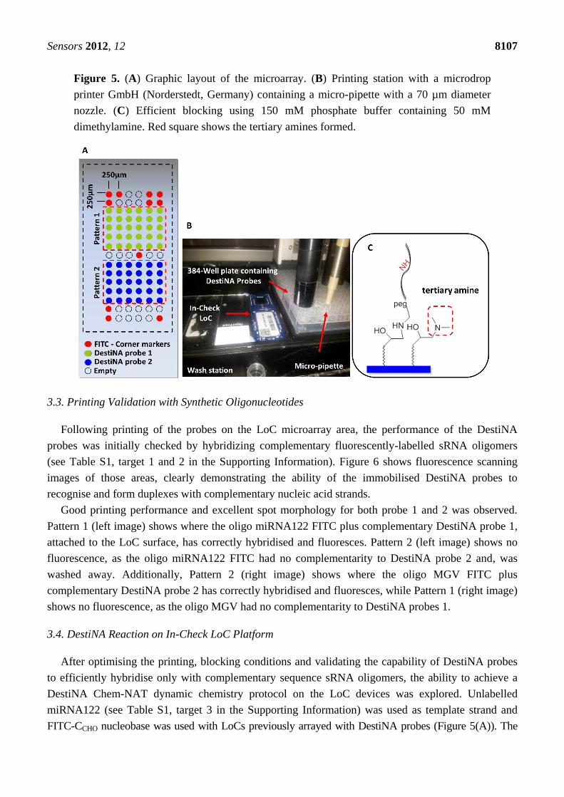
An important stage in the construction of the novel LoCs was defining the microarray probe layout.
The two test probes were printed onto two symmetrical portions of the array (pattern 1 and 2) and
arranged in 15 rows and six columns (90 features in total). Taking into consideration the size of the
microarray of 3.7 mm × 1.45 mm, it was decided to create a spot pitch of 250 µm Figure 5(A). Corner
control FITC-labelled DNA oligomers (see Table S1 in the Supporting Information) were printed to
identify array orientation. Empty positions to evaluate background noise were provided. Overall,
32 positions were allocated to the control probes and 64 to the specific probes Figure 5(A).
Printing was performed using a proprietary non-contact inkjet printing process in which DestiNA
probes were deposited uniformly onto the LoC microarray area. The precise inkjet process enabled the
delivery of extremely small, accurate volumes (picoliters) of the chemicals Figure 5(B).
Efficient blocking of reactive surface groups after arraying was critical for a reduced fluorescence
background. The LoC was blocked with a 150 mM phosphate buffer containing 50 mM dimethylamine.
The tertiary amines engendered prevented reactions with the aldehyde groups of the FITC-CCHO
Figure 5(C). Standard SCHOTT blocking buffer (containing ethanolamine) blocking approach were
excluded as this would give rise to secondary amines on the surface which could react with the
aldehyde groups of the fluorescently-labelled SMART nucleobase.
Sensors 2012, 12 8107
Figure 5. (A) Graphic layout of the microarray. (B) Printing station with a microdrop
printer GmbH (Norderstedt, Germany) containing a micro-pipette with a 70 µm diameter
nozzle. (C) Efficient blocking using 150 mM phosphate buffer containing 50 mM
dimethylamine. Red square shows the tertiary amines formed.
3.3. Printing Validation with Synthetic Oligonucleotides
Following printing of the probes on the LoC microarray area, the performance of the DestiNA
probes was initially checked by hybridizing complementary fluorescently-labelled sRNA oligomers
(see Table S1, target 1 and 2 in the Supporting Information). Figure 6 shows fluorescence scanning
images of those areas, clearly demonstrating the ability of the immobilised DestiNA probes to
recognise and form duplexes with complementary nucleic acid strands.
Good printing performance and excellent spot morphology for both probe 1 and 2 was observed.
Pattern 1 (left image) shows where the oligo miRNA122 FITC plus complementary DestiNA probe 1,
attached to the LoC surface, has correctly hybridised and fluoresces. Pattern 2 (left image) shows no
fluorescence, as the oligo miRNA122 FITC had no complementarity to DestiNA probe 2 and, was
washed away. Additionally, Pattern 2 (right image) shows where the oligo MGV FITC plus
complementary DestiNA probe 2 has correctly hybridised and fluoresces, while Pattern 1 (right image)
shows no fluorescence, as the oligo MGV had no complementarity to DestiNA probes 1.
3.4. DestiNA Reaction on In-Check LoC Platform
After optimising the printing, blocking conditions and validating the capability of DestiNA probes
to efficiently hybridise only with complementary sequence sRNA oligomers, the ability to achieve a
DestiNA Chem-NAT dynamic chemistry protocol on the LoC devices was explored. Unlabelled
miRNA122 (see Table S1, target 3 in the Supporting Information) was used as template strand and
FITC-CCHO nucleobase was used with LoCs previously arrayed with DestiNA probes (Figure 5(A)). The

Sensors 2012, 12 8108
resulting LoC scanning (Figure 7) showed the ability of the DestiNA technology to identify miRNA122
oligomer on In-Check, using a highly selective incorporation of the fluorescently-labelled cytosine.
Figure 6. LoCs scanned after the hybridisation of DestiNA probes with either synthetic
miRNA122-FITC (left) or MGV-FITC (right). LoCs were scanned using a 200 ms
exposure time using emission and detection filters appropriate for FITC.
Figure 7. LoC scanned after DestiNA reaction with FITC-CCHO. Red square (pattern 1)
shows where the oligo miRNA122 plus complementary DestiNA probe 1 attached to the
array has correctly hybridised and fluorescence due to FITC-CCHO incorporation (showed
by black arrow). On the other hand, red square (pattern 2) does not show fluorescence
due to an absence of hybridisation between oligo miRNA122 plus DestiNA probe 2
(FITC-CCHO incorporation not take place).

Sensors 2012, 12 8109
Background was at a very low level, demonstrating excellent blocking and the benefit of the proof
reading step using the DestiNA SMART base protocol.
Selective incorporation of FITC-CCHO into the PNA/RNA hybrid through dynamic chemical
reactions can take place ONLY when miRNA122 forms a perfect duplex with the designed DestiNA
probes (pattern 1). Pattern 2 shows no fluorescence because non-complementary (miRNA122) has not
been hybridised with the PNA probe 2. This demonstrated the selective incorporation of the “correct”
base when the target nucleic acid forms a perfect duplex. The result confirms that the DestiNA
fluorescence detection approach to nucleic acid discrimination reported here could be ported into the
LoC platform, facilitating the analysis of label-free nucleic acid. The result further indicates that the
approach used with miRNA122 herein could be extended to more generic, direct nucleic acid analysis
such as allele-discrimination.
4. Conclusions
In this manuscript we have given an overview of a “Proof of Concept” study for testing the
integration of DestiNA Genomics and STMicroelectronics technologies. All the key proof of concept
objectives were achieved, including effective DestiNA probe printing and DestiNA reactions,
demonstrating successful translation of DestiNA reagents onto the In Check LoC platform. This study
represents a first step in merging the two technologies to create a suite of novel medical diagnostic
assays capable of delivering a novel biochip platform for the rapid detection of nucleic acids with high
sensitivity and specificity.By combining the novel chemical-based methods with the Lab-on-Chip
platform, clinically valuable innovation is achieved through:
1. The unique high affinity and selective hybridisation between DestiNA probes and target DNA or
RNA, superior to standard DNA/DNA or DNA/RNA hybridisations.
2. The innovative DestiNA probes, that contain a “blank”/nucleobase free position within the
probe, enable a unique proof-reading step to occur. A rapid and selective incorporation of
complementary fluorescently labelled SMART nucleobase into the PNA/RNA hybrid can ONLY
occur if the target nucleic acid forms a perfect duplex with the designed DestiNA probe.
3. Flexibility to design “personalized” and low cost multiplex assays for target detection in
individuals able to be undertaken by laboratory technicians.
The integration and combination of DestiNA Genomics and STMicroelectronics technologies are
very promising and potentially suitable for developing a highly innovative next generation system
capable of true diagnostic value and utility for the rapid detection of nucleic acids. Benefits for health
care providers will be cost benefits in term of reliability, time, and ease of use.
Acknowledgements
The authors thank F. R. Bowler for his previous work in developing DestiNA technology, and the
financial support of Scottish Enterprise for Salvatore Pernagallo and Claudia Cavalluzzo. These studies
were approved and supported by DestiNA Genomics and STMicroelectronics.

Sensors 2012, 12 8110
References
1. Hay Burgess, D.C.; Wasserman, J.; Dahl, C.A. Global health diagnostics. Nature 2006, 444, 1–2.
2. Urdea, M.; Penny, L.A.; Olmsted, S.S.; Giovanni, M.Y.; Kaspar, P.; Shepherd, A.; Wilson, P.;
Dahl, C.A.; Buchsbaum, S.; Moeller, G.; et al. Requirements for high impact diagnostics in the
developing world. Nature 2006, 444, 73–79.
3. Mabey, D.; Peeling, R.W.; Ustianowski, A.; Perkins, M.D. Diagnostics for the developing world.
Nat. Rev. Microbiol. 2004, 2, 231–240.
4. Girosi, F.; Olmsted, S.S.; Keeler, E.; Hay Burgess, D.C.; Lim, Y.W.; Aledort, J.E.; Rafael, M.E.;
Ricci, K.A.; Boer, R.; Hilborne, L.; et al. Developing and interpreting models to improve
diagnostics in developing countries. Nature 2006, 444, 3–8.
5. Houpt, E.R.; Guerrant, R.L. Technology in global health: The need for essential diagnostics.
Lancet 2008, 372, 873–874.
6. Smith, C. Tools for drug discovery: Tools of the trade. Nature 2007, 446, 219–222.
7. Miller, M.B.; Tang, Y.-W. Basic concepts of microarrays and potential applications in clinical
microbiology. Clin. Microbiol. Rev. 2009, 22, 611–633.
8. Yager, P.; Domingo, G.J.; Gerdes, J. Point-of-care diagnostics for global health. Annu. Rev.
Biomed. Eng. 2008, 10, 107–144.
9. Sorger, P.K. Microfluidics closes in on point-of-care assays. Nat. Biotechnol. 2008, 26,
1345–1346.
10. McNerney, R.; Daley, P. Towards a point-of-care test for active tuberculosis: Obstacles and
opportunities. Nat. Rev. Microbiol. 2011, 9, 204–213.
11. Horton, B. Nucleic acid testing. Nature 1997, 390, 425–426.
12. Niemz, A.; Ferguson, T.M.; Boyle, D.S. Point-of-care nucleic acid testing for infectious diseases.
Trends Biotechnol. 2011, 29, 240–250.
13. Chin, C.D.; Linder, V.; Sia, S.K. Lab-on-a-chip devices for global health: Past studies and future
opportunities. Lab Chip 2007, 7, 41–57.
14. Daw, R.; Finkelstein, J. Lab on a chip. Nature 2006, 442, 367.
15. Dutse, S.W.; Yusof, N.A. Microfluidics-based lab-on-chip systems in DNA-based biosensing:
An overview. Sensors 2011, 11, 5754–5768.
16. Clarissa, L.; Nathaniel, C.C.; Carl, A.B. Nucleic acid-based detection of bacterial pathogens using
integrated microfluidic platform systems. Sensors 2009, 9, 3713–3744.
17. Qian, F.; Baum, M.; Gu, Q.; Morse, D.E. A 1.5 mL microbial fuel cell for on-chip bioelectricity
generation. Lab Chip 2009, 9, 3076–3081.
18. Mairhofer, J.; Roppert, K.; Erlt, P. Microfluidic systems for pathogen sensing. Sensors 2009, 9,
4804–4823.
19. Palmieri, M.; Alessi, E.; Conoci, S.; Marchi, M.; Panvini, G. Develop the “In-Check” Platform for
Diagnostic Applications. In Microfluidics, BioMEMS, and Medical Microsystems VI; Wang, W.,
Vauchier, C., Eds.; Proceedings of the SPIE: San Jose, CA, USA, 2008; Volume 6886,
pp. 688602-1–688602-14.
20. Petralia, S.; Ventimiglia, G. Stability evaluation of protein coating for sensing: An application to
silicon based lab-on-chip device. Sens. Transducers J. 2012, 137, 215–225.
Sensors 2012, 12 8111
21. Ventimiglia, G.; Petralia, S. Inorganic nanoparticles properties, applications and production
photosynthetic routes. Recent Res. Devel. Photochem. Photobiol. 2011, in press.
22. Consolandi, C.; Severgnini, M.; Frosini, A.; Caramenti, G.; de Fazio, M.; Ferrara, F.; Zocco, A.;
Fischetti, A.; Palmieri, M.; de Bellis, G. Polymerase chain reaction of 2-kb cyanobacterial
gene and human anti-α1-chymotrypsin gene from genomic DNA on the In-Check single-use
microfabricated silicon chip. Anal. Biochem. 2006, 353, 191–197.
23. Foglieni, B.; Brisci, A.; San Biagio, F.; di Pietro, P.; Petralia, S.; Conoci, S.; Ferrari, M.;
Cremonesi, L. Integrated PCR amplification and detection processes on a Lab-on-Chip platform:
A new advanced solution for molecular diagnostics. Clin. Chem. Lab. Med. 2010, 48, 329–336.
24. Teo, J.; di Pietro, P.; San Biagio, F.; Capozzoli, M.; Deng, Y.M.; Barr, I.; Caldwell, N.;
Ong, K.L.; Sato, M.; Tan, R.; et al. VereFlu™: An integrated multiplex RT-PCR and microarray
assay for rapid detection and identification of human influenza A and B viruses using lab-on-chip
technology. Arch. Virol. 2011, 156, 1371–1378.
25. Bowler, F.R.; Reid, P.A.; Boyd, A.C.; Diaz-Mochon, J.J.; Bradley, M. Dynamic chemistry
for enzyme-free allele discrimination in genotyping by MALDI-TOF mass spectrometry.
Anal. Methods 2011, 3, 1656–1663.
26. Bowler, F.R.; Diaz-Mochon, J.J.; Swift, M.D.; Bradley, M. DNA Analysis by dynamic chemistry.
Angew. Chem. Int. Ed. 2010, 49, 1809–1812.
27. Diaz-Mochon, J.J.; Bradley, M. Nucleobase Characterisation. WO Patent WO2009/037473, 2008.
28. Starkey Lewis, P.J.; Dear, J.; Platt, V.; Simpson, K.J.; Craig, D.G.; Antoine, D.J.; French, N.S.;
Dhaun, N.; Webb, D.J.; Costello, E.M.; et al. Circulating microRNAs as potential markers of
human drug-induced liver injury. Hepatology 2011, 54, 1767–1776.
29. Pintó, R.M.; Costafreda, M.I.; Bosch, A. Risk assessment in shellfish-borne outbreaks of hepatitis A.
Appl. Environ. Microbiol. 2009, 75, 7350–7355.
© 2012 by the authors; licensee MDPI, Basel, Switzerland. This article is an open access article
distributed under the terms and conditions of the Creative Commons Attribution license
(http://creativecommons.org/licenses/by/3.0/).