Embed Size (px)
Citation preview

NOVEL FABRICATION OF BILAYERED COMPOSITE SCAFFOLDS FOR OSTEOCHONDRAL TISSUE ENGINEERING APPLICATIONS
43
NOVEL FABRICATION OF BILAYERED COMPOSITE SCAFFOLDS FOR OSTEOCHONDRAL TISSUE ENGINEERING APPLICATIONS
RINGKASAN: Kajian ini memberi tumpuan kepada pembentukan dwi-lapisan perancah (scaffold) dengan menggabungkan kaedah pengeringan beku dan pensinteran perancah (scaffold) 45S5 Bioglass® untuk tujuan penggantian tisu osteochondral. Lapisan konstruk PDLLA (poly-D,L-lactic acid) telah direka menggunakan 7.5 wt/v% PDLLA dalam dimetil karbonat untuk tempoh 3, 5 dan 7 hari pengeringan beku. Ketebalan lapisan PDLLA boleh diubah sekurang-kurangnya 1 cm tebal. Kekuatan mekanikal dan morfologi dwi-lapisan perancah (scaffold) telah melalui teknik percirian seperti UTM (Mesin Tegangan Universal) dan SEM (Pengimbasan Mikroskop Elektron) masing-masing. Kesimpulannya, konstruk dwi lapisan yang dibangunkan dengan menggabungkan pengeringan beku dan kaedah pensinteran perancah berdasarkan komposit Bioglass® telah menghasilkan lapisan bersepadu menggabungkan kedua-dua struktur tulang dan struktur tulang rawan. Bioaktiviti lapisan PDLLA menunjukkan bahawa HA (hidroksiapatit) mineral menurun apabila ketebalan PDLLA meningkat. Kajian “in vitro” mencadangkan bahawa lapisan PDLLA membantu percambahan sel-sel “osteochondral”.
ABSTRACT: This work focuses on designing bilayered constructs by combining a freeze-drying method and Bioglass®-derived scaffolds (BG) for osteochondral tissue replacement materials. PDLLA (poly-D,L-lactic acid) layer of the constructs was fabricated using 7.5 wt/v% PDLLA in dimethyl carbonate for 3, 5 and 7 days freeze-drying duration. The PDLLA layer can be varied at least by 1 cm thickness. Mechanical strength and morphology of the bilayered constructs were characterised by Universal Testing Machine (UTM) and Scanning Electron Microscopy (SEM), respectively. In conclusion, the bilayered constructs developed by combining freeze-drying and sintering method of Bioglass®-based composite scaffolds have produced a well integrated interface combining both the bone structure and cartilage structure constructs. Bioactivity of the PDLLA layer showed that HA mineralisation decreased as the thickness of the PDLLA increased. In vitro studies suggested that PDLLA layer induced osteochondral cells proliferation.
Keywords: Bilayered constructs, Bioglass®, PDLLA.
D.Mohamad Yunos 1*, S.Mat Ghani 1, S.Shamsudin2,
S.Sahid2, W.R. Wan Sulaiman2,
S.Sabudin2
1 IBRC, SIRIM Berhad, 1 Persiaran Dato Menteri, Seksyen 2, 40911, Shah Alam, Malaysia2 AMREC, SIRIM Berhad, Lot 34, Jln Hi-Tech 2/3, Kulim Hi-Tech Park, 09000, Kulim, Malaysia.

NOVEL FABRICATION OF BILAYERED COMPOSITE SCAFFOLDS FOR OSTEOCHONDRAL TISSUE ENGINEERING APPLICATIONS
44
INTRODUCTION
Articular cartilage injuries occur frequently as a result of trauma, osteoarthritis or in some cases due to tumors [Yang and Temenoff, 2009]. Articular (hyaline) cartilage comprises cartilage rich extracellular matrix composed of a complex organization of type II collagen and other minor collagens in combination with hyaluronic acid and cartilage-specific proteoglycans (mainly aggrecan). The term “osteochondral defect” is used to indicate complex damage of articular cartilage and the underlying and adjacent bone i.e. subcondral bone. Osteochondral defects require a unique repair response compared to that of chondral defects because cartilage is avascular, not innervated and has a low capacity for intrinsic repair [Mano and Reiss, 2007].
Formation of fibrocartilage in the defect void [Shapiro et al., 1995] has been often observed after long-term follow-up of repair procedure, made usually by conventional surgical method such as abrasion arthroplasty, micro fracture and subcondral bone drilling [Buckwalter, 1998]. Several studies have shown that tissue engineering (TE) strategies have potential for regeneration of cartilage [Glowacki, 2000, Grande et al., 1999].
For osteochondral tissue engineering both bone and cartilage tissue engineering principles must be combined, for example, by using engineered osteochodral (bone-cartilage) composite scaffolds for predefined pore architecture and cartilage tissue formation. The bone aspect of the engineered osteochondral composite, made for example, from a bioactive ceramic material, may further support anchoring of the graft within the defects since bone-to-bone interfaces bond stronger and faster than cartilage-to-cartilage interfaces [Schaefer et al., 2000]. Indeed, osteochondral tissue engineering can be considered a typical case of the emerging field of “interfacial tissue engineering” as discussed in the recent literature [Moffat et al., 2009]. The tissue engineering of interface refers to the approaches being proposed to regenerate specialized tissue areas that intimately connect two different tissues of different biochemical and mechanical characteristics. The interface area usually plays an important role in transferring mechanical load between the tissues, this being the case in the osteochondral tissue interface. Due to the complex biology and mechanics of the interface, the challenge for osteochondral tissue engineering includes developing scaffolds that integrate with both the surrounding cartilage, and the underlying bone tissue while maintaining the mechanical properties and integrity of the interface.
Unlike traditional scaffolds, bilayered structures incorporate various materials, usually in a sandwich-like or stratified design, to form composite layered morphologies. In this paper, results are presented in which novel bilayered scaffolds based on Bioglass®-derived foams (for the bone aspect) coated with a biodegradable polymer PDLLA (poly-D,L-lactic acid) sponge layer obtained by freeze-drying method (for cartilage construct) have been developed and in vitro test using simulated body fluid (SBF) was used to characterise the bioactivity of the bone and cartilage layer.
NOVEL FABRICATION OF BILAYERED COMPOSITE SCAFFOLDS FOR OSTEOCHONDRAL TISSUE ENGINEERING APPLICATIONS
45
MATERIALS AND METHOD
Materials
Poly(D,L-Lactide) (PDLLA) (Purasorb) was purchased from Purac Biochem, Gorichem, The Netherlands. PDLLA (with density of 1.26 g/cm3 and an inherent viscosity of 2.15 dL/g) was completely amorphous. Dimethyl carbonate (DMC) of > 90 % purity was used as solvent (Sigma-Aldrich, Malaysia). 45S5 Bioglass® powder of mean particle size < 53 μm was used to make 3D porous scaffolds using the foam replica technique developed earlier (Chen et al., 2006). The reticulated polyurethane foam (PU) with 45 ppi (pores per inch) was purchased from Recticel, Belgium. The foam was cut into size of 10 mm width x 10 mm length x 20 mm height. Polyvinyl alcohol (PVA), 99 % hydrolysed (Mw=85,000-124,000) used as a binder was purchased from Sigma-Aldrich, Malaysia.
Fabrication of Bilayered Scaffolds
Polyurethane foam samples of the required dimensions were immersed in a 5 wt/v% PVA-H2O solution containing 66 % wt/v Bioglass® powder for 3 min; the foams were manually retrieved from the suspension as quickly as possible and the extra slurry was removed by squeezing vigorously by hand. The “green bodies” were placed on tissue paper and dried at room temperature for at least 12 h prior to the subsequent heat treatment (sintering) process. Bioglass® Scaffolds (BG) of nominal dimension 10 mm x 10 mm x 20 mm were sintered at 1000 °C and soaked for 2 h with heating rate of 2 °C/min and coated with PDLLA by a dipping process similar to that described elsewhere [Chen et al., 2006]. Briefly scaffolds were slowly immersed in a 5 wt/v% PDLLA-DMC solution and left in suspension for 2 hours. Scaffolds were then taken out and placed on a tissue paper to dry at room temperature for at least 12 h. The process of coating was repeated three times. Finally, the weight of the dried scaffolds was measured using an electronic analytical weighing balance and the dimensions were measured using a digital calipers (Mitutoyo, UK).
The composite scaffolds were then coated with PDLLA sponge by freeze-drying method. To prepare the PDLLA sponge, 7.5 wt/v% PDLLA was dissolved in 25 ml DMC. Once the solution of 7.5 wt/v% PDLLA-DMC dissolved, it was poured into a plastic petri dish at two different volumes of 5 ml (thin PDLLA) and 20 ml (thick PDLLA), wrapped with parafilms and put in the deep freezer at -80 °C for 3 days (3FT). Prior to that, Bioglass® scaffolds prepared above was attached onto the surface of the pre-frozen 7.5 wt/v% PDLLA-DMC after 10 minutes in the deep freezer at -80 °C. After 3 days in the deep freezer, the frozen units of Bioglass® scaffolds attached to different volume of PDLLA-DMC solution was transferred into a freeze-dryer (Labconco, USA) for 3 (3FD), 5 (5FD) and 7 (7FD) days respectively. The bilayered scaffolds consisting of Bioglass® composite scaffolds coated with PDLLA sponge were taken out from the freeze dryer after 3 days (3FT/3FD), 5 days (3FT/5FD) and 7 days (3FT/7FD) prior to further characterisations. Figure 1 shows a diagram of bilayered scaffolds constructs consist of PDLLA sponge and Bioglass® scaffolds.

NOVEL FABRICATION OF BILAYERED COMPOSITE SCAFFOLDS FOR OSTEOCHONDRAL TISSUE ENGINEERING APPLICATIONS
46
Figure 1. Digital picture of the bilayered scaffold constructs composed of a Bioglass® based glass-ceramic scaffolds covered with thin or thick PDLLA sponge obtained by freeze-dryer
Materials Characterization
1) Mechanical Testing [Chen et al., 2006]
The compression strengths of the uncoated and coated Bioglass® scaffolds were determined using Universal Tensile Machine (UTM) with crosshead speed of 2 mm/min with the sample dimension of (10 x 10 x 20) mm. The load was applied until 70 % of the overall dimension. At least five samples were tested to get average results (n=5).
2) Microstructural Analysis
Freeze-dried bilayered construct morphology before and after immersion in simulated body fluid (SBF) was observed by using a Variable Pressure Scanning Electron Microscope (VPSEM) and observed at accelerating pressure of 10-20 kV. EDS analysis was carried out using INCA software connected to the VPSEM operated at an accelerating voltage of 15 kV.
3) Bioactivity Studies in Simulated Body Fluid (SBF)
SBF solution was prepared according to the standard procedure introduced by Takadama et al. (Takadama and Kokubo, 2006). In typical experiments, PDLLA sponge-coated Bioglass® scaffolds were immersed in 30 mL SBF (pH 7.4) in clean centrifuge tubes. These were placed inside an incubator at a controlled temperature of 37 °C. The SBF was replaced twice a week. Samples were extracted from the SBF solution after 7, 14 and 28 days. Once removed from the solution, samples were rinsed gently with deionized water and left to dry at room temperature in a desiccator prior to further characterization. SEM observation and EDS analyses were carried out on the surface of the Bioglass®-based bilayered scaffolds after different SBF immersion times

NOVEL FABRICATION OF BILAYERED COMPOSITE SCAFFOLDS FOR OSTEOCHONDRAL TISSUE ENGINEERING APPLICATIONS
47
to investigate the formation of hydroxyapatite layers on the surface of the bilayered scaffolds. In addition, XRD analyses were done to determine the crystallinity of the formed calcium phosphate layer.
4) Cell Studies using Osteochodral Cell
The proliferation of osteochodral cells in the bilayered scaffolds was assessed with 100 μL Alamar Blue at day 1 and day 4 post seeding with the Alamar Blue dye reduction assay by measuring the florescence intensity at 560 nm and emission at 590 nm at the selected time point.
RESULTS AND DISCUSSION
Physical and Mechanical Studies
The PDLLA sponges coated on the 45S5 Bioglass® scaffolds did not give a significant value to the compressive strength of the bilayered construct. Therefore, the compressive strength of the bilayered construct was measured based on the coated 45S5 Bioglass® scaffolds layer only. The compressive strength of the 45S5 Bioglass® scaffold layer was determined using Universal Testing Machine as shown in Figure 2(A). The compressive strength increased from 0.08 MPa to 0.32 MPa after coating three times with 5 wt/v% PDLLA (3xC) samples sintered at 1000 °C for 2 hours. The increase in the mechanical strength of the coated samples as compared to the sintered scaffolds was due to the polymer infiltration into the micropores of the partially sintered scaffolds as discussed in previous studies (Chen et al., 2006, Yunos et al., 2008). The polymer infiltration increases the toughness of the scaffolds as shown by previous studies (Chen et al., 2006, Yunos et al., 2008). It was also shown that compressive strength of the 45S5 Bioglass® scaffolds coated three times (3xC) was slightly higher than the scaffolds coated once (C). The high strength can be contributed by the thicker layer of polymer coatings. Figure 2(B) shows a typical structure of Bioglass® scaffolds having highly interconnected pores of up to 500 micron in size. These can allow cells to attach and proliferate onto Bioglass® scaffolds (Chen et al., 2006).

NOVEL FABRICATION OF BILAYERED COMPOSITE SCAFFOLDS FOR OSTEOCHONDRAL TISSUE ENGINEERING APPLICATIONS
48
A
BFigure 2. A) Compressive strength of 45S5 Bioglass® scaffold (BG) sintered at 1000 °C for 2 hours and after
coated with PDLLA (n=5, p<0.05) and B) SEM micrograph of 45S5 Bioglass® composite scaffolds
Figure 3 shows SEM images of the PDLLA sponge deposited on Bioglass® composite scaffolds. From the microstructure, it was observed that the sponge structures, were in general, have a longitudinal pore structure orientation for both thick and thin samples for different freeze-drying time of 3, 5, and 7 days respectively. The longitudinal pore structure is common for PDLLA using freeze-drying method. It was also observed that thin samples were less dense than thick samples. Moreover, the pores size of the PDLLA sponges were highly interconnected with pore dimensions of up to ~ 800 micron in sizes for all samples.
NOVEL FABRICATION OF BILAYERED COMPOSITE SCAFFOLDS FOR OSTEOCHONDRAL TISSUE ENGINEERING APPLICATIONS
49
A B
C D
E F
Figure 3. SEM Micrographs showing microstructure of PDLLA sponge layers at different freeze drying conditions of A) 3FT/3FD-PDLLA(thick), B) 3FT/3FD-PDLLA (thin), C) 3FT/5FD-PDLLA
(thick), D) 3FT/5FD-PDLLA (thin), E) 3FT/7FD-PDLLA (thick) and F) 3FT/7FD-PDLLA (thin)
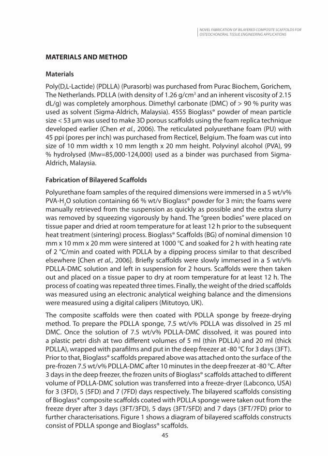
SEM-EDS images and spectra of bilayered construct for thick samples of PDLLA freeze-dried for five days (3FT/5FD-PDLLA) (thick) are shown in Figure 4 for confirmations. EDS spectra of the BG scaffolds coated with PDLLA layer (A) indicates the presence of Ca, P, Si, Na, O, C peaks which is the typical composition of Bioglass (SiO2-45 wt%, Na2O-24.5 wt%, CaO -24.4 wt% and P2O5 -6 wt%). On the other hand, the PDLLA

NOVEL FABRICATION OF BILAYERED COMPOSITE SCAFFOLDS FOR OSTEOCHONDRAL TISSUE ENGINEERING APPLICATIONS
50
sponge layer (B) indicates the presence of C and O peaks from PDLLA composition. From the SEM images, it can be deduced that there is a strong integration between BG scaffolds layer and PDLLA layer at the interface as shown in Figure 4 A, B.
A
B Figure 4. SEM-EDS spectrum of bilayered constructs of 3FT/5FD-PDLLA (thick) for A) 45S5
Bioglass® composite layer and B) PDLLA sponge layer
Assessment of Bilayered Scaffolds after Immersion in SBF
Bilayered constructs consisted of BG composite scaffolds coated with different PDLLA sponge taken out from the freeze dryer after 3 days (3FT/3FD), 5 days (3FT/5FD) and 7 days (3FT/7FD) were immersed in 30 ml solutions of SBF for 7, 14 and 28 days respectively. The bioactivity assessment was characterized by SEM, EDS and XRD.
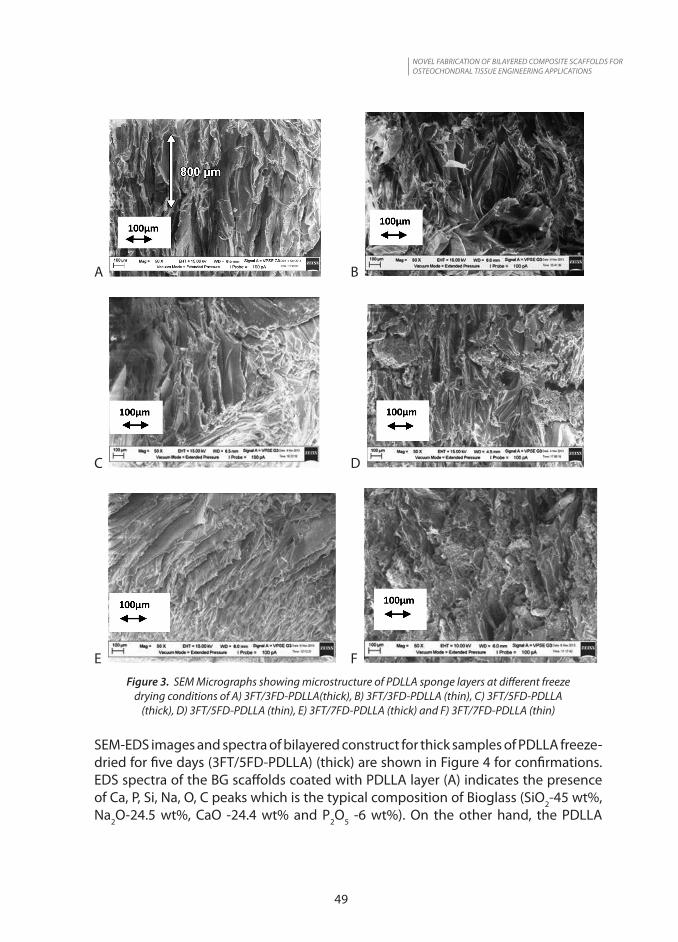
Figure 5 shows the SEM micrograph of the bilayered constructs of sample PDLLA after 3 days freeze- drying time (3FT/3FD) (thick) after immersion in SBF solution for 7 to 28 days. HA formation was not present on the PDLLA layer after immersion in SBF for 7, 14 and 28 days (Figures 5 A, C, E). On the other hand, HA formation on the coated BG scaffolds increased from day 7 to day 28 (Figures 5 B, D, F). These clear formation of HA phases was confirmed using XRD analysis on the surface of the scaffolds as shown in Figure 10. Therefore, it can be concluded that PDLLA sponge deposited on Bioglass®-based scaffolds did not mineralised after immersion in SBF of up to 28 days as confirmed by XRD analysis on the surface of the scaffolds as shown in Figure 11. This is a very important result for the application of the present bilayered constructs
NOVEL FABRICATION OF BILAYERED COMPOSITE SCAFFOLDS FOR OSTEOCHONDRAL TISSUE ENGINEERING APPLICATIONS
51
(Figure 1) in osteochondral tissue engineering, because it is a requirement that the cartilage side does not mineralize, for example, where the PDLLA sponge scaffolds should support the attachment, growth and proliferation of chondrocytes only as shown in the previous study (Yunos et al., 2013). These can be further supported by the SEM-EDS analysis in the discussion below.
A B
C D
E F Figure 5. SEM Micrograph of PDLLA layer and Bioglass®-scaffolds (BG) layer of the bilayered
constructs for sample of 3 days freeze drying condition (3FT/3FD) (thick) after immersion in SBF for7 days (A-B), 14 days( C-D) and 28 days (E-F) respectively

NOVEL FABRICATION OF BILAYERED COMPOSITE SCAFFOLDS FOR OSTEOCHONDRAL TISSUE ENGINEERING APPLICATIONS
52
The results of SEM were supported by EDS analysis as shown by Figures 6, 7, 8 and 9. Figures 6-8 show EDS spectra for (3FT/5FD) (thick) sample bilayered constructs after immersion in SBF for 7, 14 and 28 days. It can be seen that after soaking in SBF for 14 days, PDLLA layer showed the presence of C and O peaks only (Figure 6A and Figure 7A). However, after immersion in SBF for 28 days, EDS spectra of both Bioglass® composite (Figure 8A) and PDLLA layer (Figure 8B) showed the presence of Ca and P peaks indicating formation of HA. PDLLA at the BG interface mineralized when in close contact with the bioactive Bioglass® scaffolds as shown by previous studies (Yunos et al., 2013).
A
B
Figure 6. SEM-EDS spectra for sample bilayered construct 3FT/5FD (thick) after immersion in SBF for 7 days for different location at the interface for A) PDLLA sponge and B) Bioglass composite

NOVEL FABRICATION OF BILAYERED COMPOSITE SCAFFOLDS FOR OSTEOCHONDRAL TISSUE ENGINEERING APPLICATIONS
53
A
B
Figure 7. SEM-EDS spectra for sample bilayered construct 3FT/5FD (thick) after immersion in SBF for 14 days for different location at the interface for A) PDLLA sponge and B) BG composite layer
A
B
Figure 8. SEM-EDS spectra for sample bilayered construct 3FT/5FD (thick) after immersion in SBF for 28 days for different location at the interface for A) BG composite layer and B) PDLLA sponge layer

NOVEL FABRICATION OF BILAYERED COMPOSITE SCAFFOLDS FOR OSTEOCHONDRAL TISSUE ENGINEERING APPLICATIONS
54
For the purpose of comparison, SEM-EDS spectra for thin PDLLA sponge sample of bilayered construct of 3FT/5FD (thin) after immersion in SBF for 14 days at the interface is shown in Figures 9 A, B and C. The EDS spectra of PDLLA layer (Figures 9B-C) show faster HA formation indicated by presence of Ca and P peaks as compared to thick sample as shown in Figure 7A. The SEM images of the bilayered constructs for both (thin and thick) samples at the interfaces were well integrated with each other and were maintained in vitro without delamination over time. (Figures 6, 7, 8 and 9).
A
B
C
Figure 9. SEM-EDS spectra for sample bilayered construct 3FT/5FD (thin) after immersion in SBF for 14 days for different location at the interface
A) BG composite layer, B) and C) PDLLA sponge layer

NOVEL FABRICATION OF BILAYERED COMPOSITE SCAFFOLDS FOR OSTEOCHONDRAL TISSUE ENGINEERING APPLICATIONS
55
Assessment of bioactivity was also carried out with XRD analysis of the 45S5 Bioglass® scaffolds after immersion in SBF for 7-28 days. A significant pattern obtained from XRD spectra was that the crystallinity of the sintered 45S5 Bioglass® scaffolds corresponding to Na2Ca2Si3O9 phase as 2θ = 34° and 35° at 7 days were identified and decreased with increasing immersion time in SBF as shown in Figure 10. The sharp diffraction peaks of Na2Ca2Si3O9 disappeared from the XRD spectra after immersion in SBF for 28 days, leaving a typical broad halo (produced by an amorphous phase) overlapped by the sharp diffraction peaks of the HA phase (Chen et al., 2006). The results of the XRD also show that the coating of the 45S5 Bioglass® scaffolds do not impede the bioactivity as shown by previous studies (Chen et al., 2006). XRD spectra of the PDLLA layer shows an amorphous pattern of polymer layer after soaking up in SBF of up to 28 days as shown in Figure 11 which confirmed that mineralization does not occur here. The clear peak for 7 days at 2-theta 45° could be due to the presence of impurities.
Figure 10. XRD spectrum for sample 3FT/7FD for Bioglass®scaffolds layer at different conditions after immersion in SBF for: 7 days (thin) and (thick), 14 day (thick) and (thin) and 28 days (thick)

NOVEL FABRICATION OF BILAYERED COMPOSITE SCAFFOLDS FOR OSTEOCHONDRAL TISSUE ENGINEERING APPLICATIONS
56
Figure 11. XRD spectra for sample 3FT/7FD for PDLLA layer at different conditions after immersion in SBF for: 7 days (thick); (thin), 14 days (thick) and 28 days (thick)
In vitro cell culture assessment of the bilayered scaffolds.
Figure 12. Cell proliferation data up to 4 days for the Bilayered scaffolds (3FD/5FD/7FD) as compared to BG scaffolds only
Cell proliferation was quantitatively measured using Alamar Blue assay for bilayared scaffolds samples of different freeze-drying times (3FD, 5FD and 7FD) in comparison to BG scaffolds only for 1 and 4 days as shown in Figure 12. The osteochondral cell viability data shows that bilayered scaffold exhibit a significantly higher (p<0.05) cell viability than BG scaffolds at day 4 as compared to day 1. From this results it can be concluded that bilayered scaffolds of the PDLLA constructs induced cell proliferation after 4 days as compared to BG scaffolds layer.

NOVEL FABRICATION OF BILAYERED COMPOSITE SCAFFOLDS FOR OSTEOCHONDRAL TISSUE ENGINEERING APPLICATIONS
57
CONCLUSION
In conclusion, the bilayered constructs developed by combining freeze-drying and sintering method of Bioglass®-based composite scaffolds have produced well integrated interface combining both the bone structure and cartilage contructs. The interfacial properties of thick sample and thin sample of PDLLA sponge deposited on the Bioglass® scaffolds shows mineralization at the Bioglass® scaffolds interface. However, the mineralization of HA at the BG interface decreased as the PDLLA layer thickness increased. The PDLLA layer of the constructs did not mineralized and induced osteochondral cell viability after 4 days while the part on Bioglass® composite scaffolds mineralized after immersion in SBF over time did not induced cell proliferation after 4 days. These bilayered scaffolds criteria are suitable for osteochondral tissue engineering applications.
ACKNOWLEDGEMENT
We would like to thank MOSTI for the provision of Science fund (03-03-02-SF0207) and SIRIM Berhad for allowing to conduct these studies. We acknowledged Dr. Nurul Izza Nordin for her assistance in the cell studies.
REFERENCES
Buckwalter J.A., Articular cartilage: Injuries and potential for healing, The Journal of orthopaedic and sports physical therapy 28 (1998), pp 192-202.
Chen Q.Z. and Boccaccini A.R., Poly (D, L-Lactic acid) coated 45S5 Bioglass based scaffolds: processing and characterization, J Biomed Mater Res 77A (2006), pp 445-457
Glowacki J.J., In vitro engineering of cartilage, Journal of rehabilitation research and development 37 (2000), pp 171-177.
Grande D.E., Breitbart A.S., Mason J., Paulino C., Laser J., and Schwartz R.E., Cartilage tissue engineering: Current limitations and solutions, Clinical orthopedics and related research (1999), pp176-185.
Mano J.F. and Reiss R.L., Osteochondral defects: present situation and tissue engineering approaches, Journal of Tissue engineering and regenerative medicine 1 (2007), pp261-273
Moffat K., Wang I., Rodeo S. and Lu H., Orthopedic interface tissue engineering for the biological fixation of soft tissue grafts, Clin Sports Med 28 (2009), pp 157-175.
Schaefer D., Martin I., Shastri P., Padera R.F., Langer R., Freed L.E., and Vunjak- Novakovic G., In vitro generation of osteochondral composites, Biomaterials 21 (2000), pp 2599-2606.
Schaefer D., Martin I., Jundt, H. Seidel G., Heberer M., Grodzinsky A., Bergin I., Vunjak-Novokovic G., and Freed LE, Tissue-engineered composites for the repair of large osteochondral defects, Arthritis and rheumatism 46 (2002), pp 2524-2534

NOVEL FABRICATION OF BILAYERED COMPOSITE SCAFFOLDS FOR OSTEOCHONDRAL TISSUE ENGINEERING APPLICATIONS
58
Shapiro F., Koide S., and Glimcher M.J., Cell origin and differentiation in the repair of full-thickness defects of articular-cartilage, Journal of Bone and Joint Surgery. American Volume 75A (1993), pp 532-553.
Takadama H., and Kokubo T., How useful is SBF in predicting in vivo bone bioactivity?. Biomaterials 27(2006), pp 2907-2915.
Yang P..J. and Temenoff J.S., Engineering orthopedic tissue interfaces. Tissue Eng Part B 2009; 127-138
Yunos D.M., Bretcanu O., Boccaccini A.R., Polymer-Bioceramic composite for tissue engineering, Journal of Material Science, 43(2008), pp 4433-4442.
Yunos D.M., Ahmad, Z., Salih V., and Boccaccini A.R., Stratified scaffolds for osteochondarl tissue engineering applications: Electrospun PDLLA nanofibre coated Bioglass-derived foams, J Biomater Appl 27(5) (2013), pp 537-551