Embed Size (px)
Citation preview

Introduction
The most convenient and commonly employed route of drug delivery has historically been by oral ingestion (Thanoo et al., 1993). However, water-soluble drugs hav-ing short biological half-lives are easily absorbed from the gastrointestinal tract but eliminated quickly from the blood circulation and require frequent dosing, which leads to the development of oral controlled release dos-age form. In recent years, considerable attention has
been focused on hydrophilic polymers in the design of oral controlled drug delivery systems because of their flexibility to obtain a desirable drug release profile, cost effectiveness, and broad regulatory acceptance (Al-Saidan et al., 2005). These polymeric systems have been the potential candidates to deliver bioactive mol-ecules, particularly in controlled release applications (Itokazu et al., 1997; Kawaguchi, 2000). Such naturally abundant carbohydrate polymers, though exhibiting some limitations in their reactivity and processibility,
Drug Delivery, 2010; 17(7): 508–519
Address for Correspondence: Somasree Ray, Gupta College of Technological Sciences, Ashram More.Asansol-1, West Bengal, India. Tel: (+91)9434189144. E-mail: [email protected]
R E S E A R C H A R T I C L E
Novel interpenetrating network microspheres of xanthan gum–poly(vinyl alcohol) for the delivery of diclofenac sodium to the intestine—in vitro and in vivo evaluation
Somasree Ray1, Subham Banerjee1, Sabyasachi Maiti1, Bibek Laha1, Saikat Barik1, Biswanath Sa2, and Uttam Kumar Bhattacharyya1
1Gupta College of Technological Sciences, Ashram More, Asansol-1, West Bengal, India, and 2Centre for Advanced Research in Pharmaceutical Sciences, Department Of Pharmaceutical Technology, Jadavpur University, Kolkata-700032, India
AbstractXanthan gum (XG), a trisaccharide branched polymer and poly vinyl alcohol (PVA), was used to develop pH-sensitive interpenetrating network (IPN) microspheres by emulsion cross-linking method in the presence of glutaraldehyde as a cross-linker to deliver model anti-inflammatory drug, diclofenac sodium (DS) to the intes-tine. Various formulations were prepared by changing the ratio of XG:PVA, extent of cross-linking in order to optimize the formulation variables on drug encapsulation efficiency, and release rate. Formation of interpen-etrating network and the chemical stability of DS after penetration of microspheres was confirmed by Fourier Transform infrared (FTIR) spectroscopy. Differential scanning calorimetry (DSC) and X-ray diffraction (XRD) analysis were done on the drug loaded microspheres which confirmed molecular dispersion of DS in the IPN. Microspheres formed were spherical with smooth surfaces, as evidenced by scanning electron microscopy (SEM), and mean particle size, as measured by laser light scattering technique ranged between 310.25–477.10 µm. Drug encapsulation of up to 82.94% was achieved as measured by UV method. Both equilibrium and dynamic swelling studies and in vitro release studies were performed in pH 1.2 and 6.8. Release data indicated a Fickian trend of drug release which depends on the extent of cross-linking and the ratio of XG:PVA present in the microsphere. When subjected to in vivo pharmacokinetic evaluation in rabbits, microparticles show slow and prolonged drug release when compared with DS solution. Based on the results of in vitro and in vivo stud-ies it was concluded that these IPN microspheres provided oral controlled release of water-soluble DS.
Keywords: Xanthan gum; poly (vinyl alcohol); diclofenac sodium; microspheres; prolonged release
(Received 08 February 2010; revised 31 March 2010; accepted 01 April 2010)
ISSN 1071-7544 print/ISSN 1521-0464 online © 2010 Informa Healthcare USA, Inc.DOI: 10.3109/10717544.2010.483256 http://www.informahealthcare.com/drd
Drug Delivery
2010
17
7
508
519
08 February 2010
31 March 2010
01 April 2010
1071-7544
1521-0464
© 2010 Informa Healthcare USA, Inc.
10.3109/10717544.2010.483256
DRD
483256
Dru
g D
eliv
ery
Dow
nloa
ded
from
info
rmah
ealth
care
.com
by
CD
L-U
C M
erce
d on
05/
06/1
4Fo
r pe
rson
al u
se o
nly.

Xanthan–poly (vinyl alcohol) hydrogel microspheres 509
have still been used after being modified by cross-linking, blending, etc. The chemical and physical com-bination methods and properties of multipolymers have been of great practical and academic interest for the controlled release of drugs and proteins (Chandy et al., 2002) because they provide a convenient route for the modification of properties to meet specific needs. Many studies have been made in the literature to overcome these shortcomings by chemical and physical altera-tions of such natural carbohydrate polymers. Among the different polymers used to prepare controlled release dosage forms, development of hydrogels and interpen-etrating polymer network (IPN) structures has received greater attention as they increase the phase stability and enhance the mechanical properties of the final product (Burugapalli et al., 2001). Better mechanical properties of IPN make it suitable for microspheres preparation for the controlled delivery of drugs (Rokhade et al., 2007). An IPN is a composite of two polymers, which is obtained when at least one polymer network is synthesized or cross-linked independently in the immediate presence of the other.
On the other hand oral controlled release multiple unit dosage forms are becoming more popular than single unit dosage forms as they spread uniformly throughout the gastrointestinal tract, avoiding the vagaries of gastric emptying and different transit rates, resulting in a more uniform drug absorption and reduced local irritation when compared to single unit dosage forms (Kurkuri & Aminabhavi, 2004). In continuation of our earlier research on the develop-ment of multiple unit sustained release devices uti-lizing carbohydrate polymers, we herein present the work on development of interpenetrating network microspheres of xanthan gum and PVA for the sus-tained release of DS. Xanthan gum is a high molecular weight exopolysaccharide produced by Xanthomonas campestris. XG has been widely used in oral topical formulations as a suspending and stabilizing agent (Wade & Weller, 1994), and a release sustaining agent in hydrophilic matrix tablets (Talukdar & Kinget, 1995; Sujja-areevath et al., 1996), and pellets (Santos et al., 2005).
DS, a non-steroidal anti-inflammatory drug with shorter biological half-life of 1.2–2 h has been used as a model drug. Poly vinyl alcohol (PVA) is a widely used hydrophilic polymer because of its processability, strength, and pH, as well as its temperature stability. Because it is biocompatible and non-toxic, it has a wide variety of pharmaceutical applications (Peppas & Wright, 1998; Kurkuri & Aminabhavi, 2004).
Earlier, PVA-gellan gum (Agnihotri & Aminabhavi, 2005), PVA-guar gum (Soppimath et al., 2000), and PVA-polyacrylic acid IPN (Kurkuri & Aminabhavi, 2004) microspheres were developed for controlled release of
drugs. Although sodium carboxy methyl xanthan gum was used to prepare microparticles (Ray et al., 2008), in the literature of pharmaceutics we are not aware of preparation of microspheres from IPN of XG, which can be safely used in food and cosmetics without any spe-cific quantity limitations (Katzbauer, 1998) and PVA for the controlled release of DS. Our aim was to study the potentiality of XG to form IPN network and highly safe XG may be more effective for the delivery of drugs by forming a novel IPN network of XG and PVA. The micro-spheres prepared have been characterized by different techniques to understand their release characteristics and morphological as well as chemical interactions, and also the pharmacokinetics of IPN microspheres were evaluated to investigate its in vivo performance in animals.
Materials and methods
Materials
The diclofenac sodium was kindly received as a gift sample by C.I laboratories (Kolkata, India). Xanthan gum was a gift sample from Micro Lab (Mumbai, India). Analytical reagent grade absolute alcohol was pur-chased from Bengal Chemicals and Pharmaceuticals Limited (Kolkata, India). Analytical reagent grade sam-ples of poly vinyl alcohol (∼ Mw = 125,000, 98% hydro-lyzed), glutaraldehyde (25%v/v), light liquid paraffin, span 80, acetone, acetonitrile, sodium acetate, and glacial acetic acid were purchased from S.D chemi-cals (Mumbai, India). Double distilled water was used throughout the work.
Preparation of microspheres
Xanthan gum and poly(vinyl alcohol) (XG-PVA) IPN microspheres containing DS were prepared by the emulsion cross-linking method. First PVA was dissolved in hot water at 80°C, and, after cooling to ambient tem-perature, XG was added (total polymer concentration was 4%, w/v) and stirred overnight to get a uniform bubble-free solution. DS (30% w/w of polymer) was dissolved in 1 ml alcohol and then added to the mixture of XG and PVA and the solution was stirred to get a uni-form suspension. This suspension was added to liquid paraffin and 1% w/w span 80 and mixed with a stirrer at 800 rpm for 40 min. Then glutaraldehyde and 1 ml 1(N) H
2SO
4 was added slowly and stirred for 4 h. After
4 h hardened microspheres were formed and they were separated by filtration and washed with acetone and distilled water to remove the oil as surfactant. Finally the microsphres were washed with 0.1 M glycine solu-tion to mask the untreated glutaraldehyde (Hejazi &
Dru
g D
eliv
ery
Dow
nloa
ded
from
info
rmah
ealth
care
.com
by
CD
L-U
C M
erce
d on
05/
06/1
4Fo
r pe
rson
al u
se o
nly.

510 S. Ray et al.
Amiji, 2003) and distilled water to remove the unreacted glutaraldehyde. Then the prepared microspheres were dried at 50°C for 24 h. In total, nine formulations were prepared to study the effect of different formulation variables on the characteristics of IPN microspheres (Table 1).
Scanning electron microscopy (SEM)
Microspheres were mounted onto stubs using double-sided adhesive tape and vacuum coated with gold film using a sputter coater (Edward S150, Watford, UK). The coated surface was observed under SEM (Jeol, SM-5200, Tokyo, Japan) for surface appearance.
Particle size measurements
Particle size and size distributions were measured using a laser light scattering technique (Mastersizer-2000, Malvern, UK). Particle size was measured using the dry sample adopter and volume mean diameter (Vd) was recorded. These data are presented in Table 2.
Drug content
Estimation of drug content was done according to the method adopted by Agnihotri & Aminabhavi (2004a). Microspheres of known weight were soaked in 50 ml of water for 30 min and sonicated using a probe sonica-tor for 10 min to break the microspheres. The whole solution was centrifuged (Remi Equipments Private Limited, Mumbai, India) to remove the polymeric
debris. The polymeric debris was washed twice to extract the drug completely. The clear supernatant solution was analyzed by a UV spectrophotometer (Thermo Spectronic UV-1, UK) at the maximum wave-length, λ
max value of 276 nm. Encapsulation efficiency
was calculated as
%=
Drug EntrapmentEfficiency DEE
Experimental drug conte
( )nnt
Theoretical drug content 100×
Fourier transform infrared (FTIR) spectral studies
FTIR spectral data were taken on an instrument (Spectrum BX, Perkin-Elmer® instruments, USA) to confirm the formation of IPN structure and also to find the chemical stability of the drug in the microspheres. FTIR spectra of the placebo microspheres, drug-loaded microspheres, and pristine DS were obtained. Samples were crushed with KBr to get pellets at 600 kg/cm2 pres-sure. Spectral scanning was done in the range between 4000–500 cm−1.
Differential scanning calorimetric (DSC) study
DSC (Rheometric Scientific, UK) was performed on placebo microspheres, drug-loaded microspheres, and pristine DS. Samples were heated from 25 to 350°C at the heating rate of 10°C/min in a nitrogen atmosphere (flow rate, 20 mL/min).
X-ray diffraction (X-RD) studies
Crystallinity of DS after the encapsulation was evaluated by X-ray diffraction (X-RD) measurements recorded for pristine diclofenac sodium, placebo microspheres, and drug-loaded microspheres using an X-ray diffractom-eter (x-Pert, Philips, UK). Scanning was done up to 2θ of 50°C.
Table 1. Formulation codes and different ingredients used to prepare microspheres.
Ratio of XG:PVA
Glutaraldehyde (×10−3 mL)/mg of polymer
0.833 1.16 1.5
1:1 F1
F4
F7
1:3 F2
F5
F8
1:4 F3 F6 F9
Table 2. Effect of xanthan gum:poly (vinyl alcohol) ratio, glutaraldehyde concentration on particle size, drug entrapment efficiency (DEE), n value, and percentage equilibrium water uptake in pH1.2 and pH 6.8 media.
Formulation code
Gum:polymer ratio
Glutaraldehyde (× 10−3 mL)/mg
of polymerPercentage DEE
(± SD, n = 3) n r2
Volume mean diameter (µm)
Percentage equilibrium liquid uptake in media
pH 1.2 pH 6.8
F1
1:1 0.833 × 10⁻³ 60.85 ± 1.61 0.329 0.99 477.10 240.33 233.66
F2
1:3 0.833 × 10⁻³ 64.29 ± 1.56 0.363 0.97 451.74 207.66 213.00
F3
1:4 0.833 × 10⁻³ 67.73 ± 1.11 0.365 0.9902 411.54 194.00 201.66
F4
1:1 1.16 × 10⁻³ 68.54 ± 1.14 0.344 0.9816 394.65 192.00 226.66
F5
1:3 1.16 × 10⁻³ 70.92 ± 1.62 0.331 0.9761 372.52 178.00 183.66
F6
1:4 1.16 × 10⁻³ 74.83 ± 1.84 0.356 0.9717 352.84 153.00 160.00
F7
1:1 1.50 × 10⁻³ 75.02 ± 1.44 0.356 0.98 345.41 133.66 143.33
F8
1:3 1.50 × 10⁻³ 78.77 ± 1.11 0.368 0.989 329.48 118.00 125.00
F9
1:4 1.50 × 10⁻³ 83.94 ± 1.50 0.369 0.994 310.25 103.66 111.33
Dru
g D
eliv
ery
Dow
nloa
ded
from
info
rmah
ealth
care
.com
by
CD
L-U
C M
erce
d on
05/
06/1
4Fo
r pe
rson
al u
se o
nly.

Xanthan–poly (vinyl alcohol) hydrogel microspheres 511
Swelling studies
The pH-dependent equilibrium swelling of the DS-loaded cross-linked microspheres were studied both in pH 1.2 and 6.8 phosphate buffer. Microspheres were allowed to swell completely for ∼ 24 h to attain equilibrium at 37°C. Adhered liquid droplets on the surface of the particles were removed by blotting with tissue paper, and the swollen microspheres were weighed on an electronic balance. The percentage equilibrium water uptake was calculated as:
Weight of swollenmicrospheres
Weight of drymicrosphe
−
rres
Weight of dry microspheres 100
( )×
In order to understand the transport of water into cross-linked microspheres, dynamic swelling studies were performed by microscopic technique (Soppimath & Aminabhavi, 2002). Microspheres were allowed to swell in pH 1.2 and pH 6.8 and, at different time intervals,
microspheres were withdrawn and changes in diameter were measured with an optical microscope after remov-ing adhered liquid droplets using blotting paper. In this study changes in diameter of microspheres were moni-tored as a function of time. All the experiments were car-ried out in triplicate, but average values were considered for calculation and are shown in Figures 1 and 2.
Drying rates of the microspheres
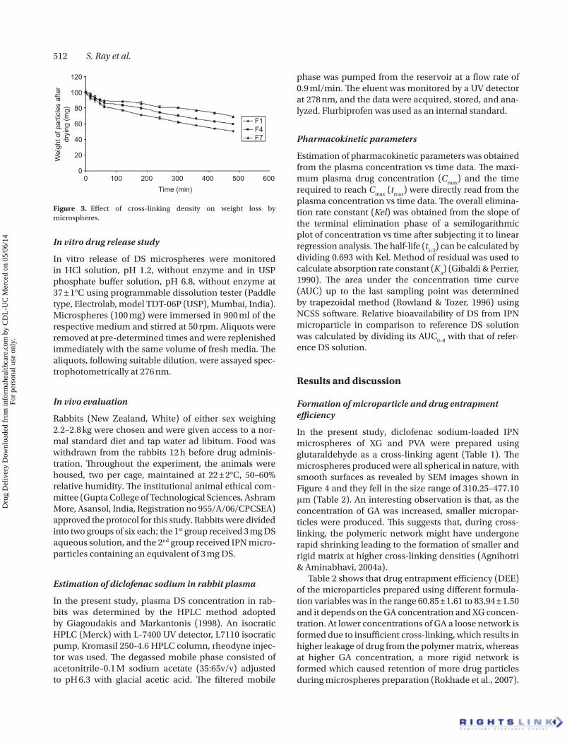
A few samples of the microspheres representative of the batch prepared were allowed to dry in an incubator maintained at 37°C (the initial mass of microspheres should be nearly equal). Microspheres were removed at different intervals of time, and they were weighed on an electronic microbalance (Mettler, Toledo, Switzerland). These measurements were continued until attainment of constant mass, indicating complete dried equilibrium. To obtain reproducible results, experiments were carried out in triplicate and results are displayed in Figure 3.
1.8
1.6
1.4
1.2
1
0.8
Nor
mal
ized
Dia
met
er (
Dt/D
o)
0.6
0.4
0.2
00 50 100
Time (min)
F2
F4(1:1)F5(1:3)
F6(1:4)
F5
F8
150 200
1.8
1.6
1.4
1.2
1
0.8
Nor
mal
ized
Dia
met
er (
Dt/D
o)
0.6
0.4
0.2
00 50 100
Time (min)
150 200
Figure 2. Plot of Dt/D
0 vs time t, in pH 6.8. (a) Effect of extent of cross-linking, and (b) Effect of amount of xanthan gum.
0
1.4
F2F5F8
(a)
1.21
0.80.60.40.2
050 100
Time (min)
Nor
mal
ized
Dia
met
er(D
t/Do)
Nor
mal
ized
Dia
met
er(D
t/Do)
150 200 0
1.4
F4(1:1)F5(1:3)F6(1:4)
(b)
1.2
1
0.8
0.6
0.4
0.2
050 100
Time (min)150 200
Figure 1. Plot of Dt/D
0 vs time t, in pH 1.2. (a) Effect of extent of cross-linking, and (.b) Effect of amount of xanthan gum
Dru
g D
eliv
ery
Dow
nloa
ded
from
info
rmah
ealth
care
.com
by
CD
L-U
C M
erce
d on
05/
06/1
4Fo
r pe
rson
al u
se o
nly.
512 S. Ray et al.
In vitro drug release study
In vitro release of DS microspheres were monitored in HCl solution, pH 1.2, without enzyme and in USP phosphate buffer solution, pH 6.8, without enzyme at 37 ± 1°C using programmable dissolution tester (Paddle type, Electrolab, model TDT-06P (USP), Mumbai, India). Microspheres (100 mg) were immersed in 900 ml of the respective medium and stirred at 50 rpm. Aliquots were removed at pre-determined times and were replenished immediately with the same volume of fresh media. The aliquots, following suitable dilution, were assayed spec-trophotometrically at 276 nm.
In vivo evaluation
Rabbits (New Zealand, White) of either sex weighing 2.2–2.8 kg were chosen and were given access to a nor-mal standard diet and tap water ad libitum. Food was withdrawn from the rabbits 12 h before drug adminis-tration. Throughout the experiment, the animals were housed, two per cage, maintained at 22 ± 2°C, 50–60% relative humidity. The institutional animal ethical com-mittee (Gupta College of Technological Sciences, Ashram More, Asansol, India, Registration no 955/A/06/CPCSEA) approved the protocol for this study. Rabbits were divided into two groups of six each; the 1st group received 3 mg DS aqueous solution, and the 2nd group received IPN micro-particles containing an equivalent of 3 mg DS.
Estimation of diclofenac sodium in rabbit plasma
In the present study, plasma DS concentration in rab-bits was determined by the HPLC method adopted by Giagoudakis and Markantonis (1998). An isocratic HPLC (Merck) with L-7400 UV detector, L7110 isocratic pump, Kromasil 250-4.6 HPLC column, rheodyne injec-tor was used. The degassed mobile phase consisted of acetonitrile–0.1 M sodium acetate (35:65v/v) adjusted to pH 6.3 with glacial acetic acid. The filtered mobile
phase was pumped from the reservoir at a flow rate of 0.9 ml/min. The eluent was monitored by a UV detector at 278 nm, and the data were acquired, stored, and ana-lyzed. Flurbiprofen was used as an internal standard.
Pharmacokinetic parameters
Estimation of pharmacokinetic parameters was obtained from the plasma concentration vs time data. The maxi-mum plasma drug concentration (C
max) and the time
required to reach Cmax
(tmax
) were directly read from the plasma concentration vs time data. The overall elimina-tion rate constant (Kel) was obtained from the slope of the terminal elimination phase of a semilogarithmic plot of concentration vs time after subjecting it to linear regression analysis. The half-life (t
1/2) can be calculated by
dividing 0.693 with Kel. Method of residual was used to calculate absorption rate constant (K
a) (Gibaldi & Perrier,
1990). The area under the concentration time curve (AUC) up to the last sampling point was determined by trapezoidal method (Rowland & Tozer, 1996) using NCSS software. Relative bioavailability of DS from IPN microparticle in comparison to reference DS solution was calculated by dividing its AUC
0–8 with that of refer-
ence DS solution.
Results and discussion
Formation of microparticle and drug entrapment efficiency
In the present study, diclofenac sodium-loaded IPN microspheres of XG and PVA were prepared using glutaraldehyde as a cross-linking agent (Table 1). The microspheres produced were all spherical in nature, with smooth surfaces as revealed by SEM images shown in Figure 4 and they fell in the size range of 310.25–477.10 μm (Table 2). An interesting observation is that, as the concentration of GA was increased, smaller micropar-ticles were produced. This suggests that, during cross-linking, the polymeric network might have undergone rapid shrinking leading to the formation of smaller and rigid matrix at higher cross-linking densities (Agnihotri & Aminabhavi, 2004a).
Table 2 shows that drug entrapment efficiency (DEE) of the microparticles prepared using different formula-tion variables was in the range 60.85 ± 1.61 to 83.94 ± 1.50 and it depends on the GA concentration and XG concen-tration. At lower concentrations of GA a loose network is formed due to insufficient cross-linking, which results in higher leakage of drug from the polymer matrix, whereas at higher GA concentration, a more rigid network is formed which caused retention of more drug particles during microspheres preparation (Rokhade et al., 2007).
0
20
40
60
Wei
ght o
f par
ticle
s af
ter
dryi
ng (m
g) 80
100
0
120
100 200 300Time (min)
400
F1F4F7
500 600
Figure 3. Effect of cross-linking density on weight loss by microspheres.
Dru
g D
eliv
ery
Dow
nloa
ded
from
info
rmah
ealth
care
.com
by
CD
L-U
C M
erce
d on
05/
06/1
4Fo
r pe
rson
al u
se o
nly.

Xanthan–poly (vinyl alcohol) hydrogel microspheres 513
Formulations prepared with a higher amount of XG exhibited lower encapsulation efficiencies due to the formation of a loose network (Rokhade et al., 2007).
An increase in size of microspheres was also observed with the increase in ratio of XG in the microspheres. This could be due to the fact that at higher amounts of XG, the viscosity of the polymer solution increased, thus produc-ing bigger droplets during emulsification that were later hardened in the presence of GA.
Water uptake studies
Equilibrium water uptake of the cross-linked micro-spheres exerts an influence on their release rates (Ritger & Peppas, 1987). The percentage equilibrium water
uptake data of the cross-linked microspheres presented in Table 2 indicate that as the amount of GA in the matri-ces (XG:PVA = 1:1) increases from 0.83 × 10⁻³ ml/mg to 1.583 × 10⁻³ ml/mg, the equilibrium water uptake in pH 1.2 decreases significantly from 240.33% to 133.66%. The reduction in water uptake capacity is due to the formation of a rigid network structure at the higher concentration of cross-linking. Again it was found that formulations containing higher amounts of XG showed higher percentages of equilibrium water uptake than for-mulations containing small amounts of XG. Formulation F
4 (XG:PVA = 1:1) showed higher water uptake capacity
than F5 (XG:PVA = 1:3). Similarly, formulation F
5 exhib-
ited greater swelling than formulation F6 (XG:PVA = 1:4)
due to the hydrophilic nature of XG, thereby leading to higher water uptake capacity.
Dynamic swelling studies
Dynamic swelling studies were performed by monitor-ing the changes in microsphere diameter, as a function of time with the help of an optical microscope. In Figures 1 and 2, a graph is plotted containing normalized diameter, D
t/D
0 (D
0 initial diameter, D
t diameter at time t) of differ-
ent formulations containing different amounts of GA as a function of time. It is evident that in pH 1.2 and 6.8, nor-malized diameter value decreases with the increase in GA concentration, which could be due to the rigid network structure formed at higher amounts of GA (Figures 1a and 2a). Normalized diameter increases with the increase in XG concentration due to the hydrophilic nature of XG,
%T
a)
b)
c)2925.063425.48
3423.97
3735.98
3676.25
3256.04
3556.043841.22
3752.11
2925.26
1387.60
2364.22
1736.03
1623.381420.27
1252.241452.31
1051.22
747.22
668.10
599.92
792.43
872.20
451.96636.21
559.91
531.97
747.08
894.27870.06
844.09715.63
766.13747.23
1044.34
1167.62
1304.33
1400.71575.20
1283.401453.55
1576.63
1560.11
1507.78
1508.21
1656.07
4000.0 3000 2000 1500
cm−1
1000 500 400.0
2925.45
Figure 5. FTIR spectra of (a) physical mixture, (b) PVA, and (c) XG.
20 kv ×150 100 µm 0025 JU-MET
Figure 4. SEM photograph of microparticles.
Dru
g D
eliv
ery
Dow
nloa
ded
from
info
rmah
ealth
care
.com
by
CD
L-U
C M
erce
d on
05/
06/1
4Fo
r pe
rson
al u
se o
nly.

514 S. Ray et al.
which will enhance the water uptake capacity of the microspheres (Figures 1b and 2b).
XRD
X-ray diffractograms of (a) placebo microspheres, (b) drug-loaded microspheres, and (c) pure DS are pre-sented in Figure 6. The diclofenac sodium has shown highly intense sharp peaks at ∼ 2θ of 6.6° (Desai, 2005); however, peaks due to diclofenac sodium crystals are not shown in the drug-loaded microspheres, con-firming that the drug is molecularly dispersed in the microspheres.
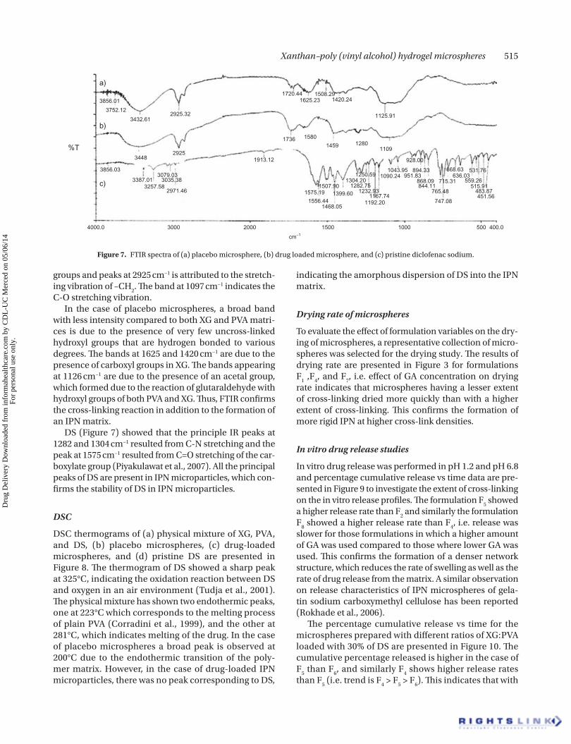
FTIR
FTIR was used to confirm the cross-linking of the IPN matrix. Figures 5 and 7 compare the FTIR spectra of XG, PVA, and physical mixture and placebo microsphere, drug loaded microspheres, and pristine DS, respec-tively. In the case of XG, a broad band which appeared at 3423 cm−1 is attributed to the presence of a hydroxyl group that are hydrogen bonded to various degrees. The bands appearing at 1610 cm−1 and 1420 cm−1 indicate the presence of a carboxylate group. The appearance of peaks at1252cm−1 in the spectra of XG indicates the presence of a C-O-C group. The FTIR spectra of PVA showed a broad peak around 3422 cm−1, indicating stretching of hydroxyl
00
1000
2000
3000
4000
5000
6000
7000
5 10 15 20 25
Degree (2θ)
Line
ar C
ount
s/so
unds
Line
ar C
ount
s/so
unds
Line
ar C
ount
s/so
unds
Degree (2θ)
Degree (2θ)
(a)
30 35 40 45 50
00
1000
2000
3000
4000
5000
6000
7000
5 10 15 20 25
(b)
30 35 40 45 50
00
1000
2000
3000
4000
5000
6000
7000
5 10 15 20 25
(c)
30 35 40 45 50
Figure 6. X-RD diffractograms of (a) Diclofenac sodium, (b) Drug-loaded microspheres, and (c) Placebo microspheres.
Dru
g D
eliv
ery
Dow
nloa
ded
from
info
rmah
ealth
care
.com
by
CD
L-U
C M
erce
d on
05/
06/1
4Fo
r pe
rson
al u
se o
nly.
Xanthan–poly (vinyl alcohol) hydrogel microspheres 515
groups and peaks at 2925 cm−1 is attributed to the stretch-ing vibration of –CH
2. The band at 1097 cm−1 indicates the
C-O stretching vibration.In the case of placebo microspheres, a broad band
with less intensity compared to both XG and PVA matri-ces is due to the presence of very few uncross-linked hydroxyl groups that are hydrogen bonded to various degrees. The bands at 1625 and 1420 cm−1 are due to the presence of carboxyl groups in XG. The bands appearing at 1126 cm−1 are due to the presence of an acetal group, which formed due to the reaction of glutaraldehyde with hydroxyl groups of both PVA and XG. Thus, FTIR confirms the cross-linking reaction in addition to the formation of an IPN matrix.
DS (Figure 7) showed that the principle IR peaks at 1282 and 1304 cm−1 resulted from C-N stretching and the peak at 1575 cm−1 resulted from C=O stretching of the car-boxylate group (Piyakulawat et al., 2007). All the principal peaks of DS are present in IPN microparticles, which con-firms the stability of DS in IPN microparticles.
DSC
DSC thermograms of (a) physical mixture of XG, PVA, and DS, (b) placebo microspheres, (c) drug-loaded microspheres, and (d) pristine DS are presented in Figure 8. The thermogram of DS showed a sharp peak at 325°C, indicating the oxidation reaction between DS and oxygen in an air environment (Tudja et al., 2001). The physical mixture has shown two endothermic peaks, one at 223°C which corresponds to the melting process of plain PVA (Corradini et al., 1999), and the other at 281°C, which indicates melting of the drug. In the case of placebo microspheres a broad peak is observed at 200°C due to the endothermic transition of the poly-mer matrix. However, in the case of drug-loaded IPN microparticles, there was no peak corresponding to DS,
indicating the amorphous dispersion of DS into the IPN matrix.
Drying rate of microspheres
To evaluate the effect of formulation variables on the dry-ing of microspheres, a representative collection of micro-spheres was selected for the drying study. The results of drying rate are presented in Figure 3 for formulations F
1 ,F
4, and F
7, i.e. effect of GA concentration on drying
rate indicates that microspheres having a lesser extent of cross-linking dried more quickly than with a higher extent of cross-linking. This confirms the formation of more rigid IPN at higher cross-link densities.
In vitro drug release studies
In vitro drug release was performed in pH 1.2 and pH 6.8 and percentage cumulative release vs time data are pre-sented in Figure 9 to investigate the extent of cross-linking on the in vitro release profiles. The formulation F
5 showed
a higher release rate than F2 and similarly the formulation
F8 showed a higher release rate than F
4, i.e. release was
slower for those formulations in which a higher amount of GA was used compared to those where lower GA was used. This confirms the formation of a denser network structure, which reduces the rate of swelling as well as the rate of drug release from the matrix. A similar observation on release characteristics of IPN microspheres of gela-tin sodium carboxymethyl cellulose has been reported (Rokhade et al., 2006).
The percentage cumulative release vs time for the microspheres prepared with different ratios of XG:PVA loaded with 30% of DS are presented in Figure 10. The cumulative percentage released is higher in the case of F
5 than F
6, and similarly F
4 shows higher release rates
than F5 (i.e. trend is F
4 > F
5 > F
6). This indicates that with
a)
b)
c)
%T
2925.32
29251913.12
1736 1580
1459 12801109
1125.91
1420.241508.29
1625.231720.44
1507.901575.19
1556.441468.05
1399.60
1192.201167.74
1232.931282.75
1304.201250.59 1090.24
1043.95
928.00
894.33951.83
868.09844.11
765.48
747.08
715.31
451.56483.87
515.91559.26
636.03668.63 531.76
3079.033035.38
2971.46
4000.0 3000 2000 1500 1000 500 400.0
cm−1
3257.583387.01
3856.03
3448
3432.61
3752.12
3856.01
Figure 7. FTIR spectra of (a) placebo microsphere, (b) drug loaded microsphere, and (c) pristine diclofenac sodium.
Dru
g D
eliv
ery
Dow
nloa
ded
from
info
rmah
ealth
care
.com
by
CD
L-U
C M
erce
d on
05/
06/1
4Fo
r pe
rson
al u
se o
nly.

516 S. Ray et al.
the increase in the xanthan gum in the matrix, swelling of the matrix increases due to the hydrophilic nature of XG, which leads to the higher release of DS from the matrix. The release data were further analyzed by Peppas equation. In order to establish a link between drug release rates and molecular transport param-eters, release data were fitted to an empirical equa-tion M
t/M
∞ = Ktn, where M
t and M
∞ are, respectively,
the amount of drug released at time t and at infinite time, K represents a constant incorporating structural and geometrical character of the dosage form, and
n values denote the diffusion exponent indicative of the mechanism of drug release (Ritger & Peppas, 1987). The calculated n values included in Table 2 range from 0.329–0.369, indicating Fickian transport phenomena (Korsmeyer & Peppas, 1981; Agnihotri & Aminabhavi, 2004b). The values of n increase with the increase in cross-linking density and decrease with the increase in ratio of xanthan gum in the IPN matrix. The lower n value indicates the formation of a loosely cross-linked polymer network, which leads to increased swelling. It is evident from Table 2 that the correlation coefficient
70
80
90
100
60
50
40
30
20
10
00 2 31 4 6 75
Time (hour)
Cum
ulat
ive
perc
ent r
elea
sed
8 9
F2,pH1.2F5,pH1.2F8,pH1.2F2,pH6.8F5,pH6.8F8,pH6.8
10 11 12
Figure 9. Effect of cross-linking density on in vitro release profiles in pH 1.2 and pH 6.8.
Temperature (˚C)
223.84˚C
281.57˚C
285.62˚C
328.66˚C
d)
a)
c)
b)
10
0
0 25
Hea
t flo
w E
ndo
dow
n (m
W)
50 75 100 125 150 175 200 225 250 275 300 325 350 375 400 425 450 475 500
2
4
6
8
−24
−22
−20
−18
−16
−14
−12
−10
−8
−6
−4
−2
Figure 8. DSC thermograms of (a) physical mixture containing diclofenac sodium, xanthan gum, PVA (single continuous line), (b) placebo micro-sphere (dotted line), (c) drug-loaded microsphere (bold dotted line), and (d) pristine diclofenac sodium (bold continuous line).
Dru
g D
eliv
ery
Dow
nloa
ded
from
info
rmah
ealth
care
.com
by
CD
L-U
C M
erce
d on
05/
06/1
4Fo
r pe
rson
al u
se o
nly.

Xanthan–poly (vinyl alcohol) hydrogel microspheres 517
values approached unity, suggesting best fit to the Fickian model.
In vivo study
Figure 11 shows the mean plasma concentration time profile attained following oral administration of aque-ous solution of DS and IPN microparticles. The t
max of
diclofenac sodium from oral solution was 2 ± 0.1 h, and the peak concentration (C
max) at that time was
63.2 ± 2.2 ng/ml. In the case of IPN microparticles, the C
max was 58 ± 2.2 ng/ml, which was significantly differ-
ent from that obtained from oral solution. The mean tmax
value after administration of IPN microparticles was 4 ± 0.34 h, which was significantly different from oral solution of DS (Table 3).
The elimination rate constant (kel
) of the drug from solution was 0.449 ± 0.01 h−1, and that obtained from the IPN microparticles was 0.130 ± 0.012 h−1. Thus, the lower C
max, prolonged t
max, and decreased k
el of
diclofenac sodium in rabbit is another important indication on the in vivo performance of sustained release IPN microparticles in providing a prolonged drug delivery. These in vivo pharmacokinetic param-eters were in good agreement with the observed in vitro drug release rate of the drug from microparticles. The extent of absorption (bioavailability) was signifi-cantly higher (p < 0.05) with IPN microparticles, as seen from the AUC
0–8 (296.59 ± 20.7 µghml−1) followed
by the solution of DS (175.4 ± 18.2). When the AUC s obtained from the microparticles were divided by the AUC obtained from the DS solution, the relative bioavailability values for both the formulations were obtained (Table 3). The relative bioavailability of the IPN microparticles was high when compared with the solution.
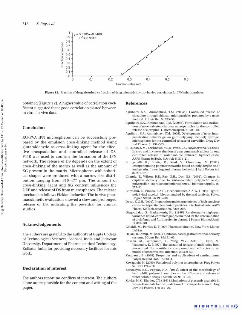
In vitro in vivo correlation (IVIVC)
To assess the viability and validity of the sustaining nature of IPN microparticles, IVIVC study is essential, since prolonged-release products may be specially suited for this kind of study (Kottke & Rhodes, 1991). When the fraction of drug released in pH 6.8 was plotted against the fraction of drug absorbed, a linear correlation was
70
80
90
100
60
50
40
30
20
10
00 2 31 4 6 75
Time (hour)
Cum
ulat
ive
perc
ent r
elea
sed
8 9
F4(1:1),pH6.8F5(1:3),pH6.8F6(1:4),pH6.8F4(1:1),pH1.2F5(1:3),pH1.2F6(1:4),pH1.2
10 11 12
Figure 10. Effect of XG:PVA ratios on in vitro release profiles in pH 1.2 and pH 6.8.
70
60
50
40
30
20
10
0
Drug solutionF9
Pla
sma
drug
con
cent
ratio
n(n
g/m
l)
0 2 4 6Time (hr)
8 10
Figure 11. Serum concentration–time profile of diclofenac sodium in rabbits after oral administration of diclofenac sodium solution and IPN microparticles.
Table 3. Pharmacokinetic parameters of diclofenac sodium after oral administration of diclofenac sodium solution and IPN microparticle (M ± SD, n = 6).
Parameter Solution IPN microparticle
Cmax
(ng/ml) 63.2 ± 2.2 58 ± 1.2
tmax
(h) 2 ± 0.1 4 ± 0.34
AUC (ngh/ml) 175.4 ± 18.2 296.59 ± 20.7
Kel
(h−1) 0.449 ± 0.01 0.13 ± 0.012
t1/2
(h) 1.54 ± 0.03 5.33 ± 0.023
Relative bioavailability 1 1.69
Dru
g D
eliv
ery
Dow
nloa
ded
from
info
rmah
ealth
care
.com
by
CD
L-U
C M
erce
d on
05/
06/1
4Fo
r pe
rson
al u
se o
nly.
518 S. Ray et al.
obtained (Figure 12). A higher value of correlation coef-ficient suggested that a good correlation existed between in vitro–in-vivo data.
Conclusion
XG-PVA IPN microspheres can be successfully pre-pared by the emulsion cross-linking method using glutaraldehyde as cross-linking agent for the effec-tive encapsulation and controlled release of DS. FTIR was used to confirm the formation of the IPN network. The release of DS depends on the extent of cross-linking of the matrix as well as the amount of XG present in the matrix. Microspheres with spheri-cal shapes were produced with a narrow size distri-bution ranging from 310–477 μm. The amount of cross-linking agent and XG content influences the DEE and release of DS from microspheres. The release mechanism follows Fickian behavior. The in vivo phar-macokinetic evaluation showed a slow and prolonged release of DS, indicating the potential for clinical studies.
Acknowledgements
The authors are grateful to the authority of Gupta College of Technological Sciences, Asansol, India and Jadavpur University, Department of Pharmaceutical Technology, Kolkata, India for providing necessary facilities for this work.
Declaration of interest
The authors report no conflicts of interest. The authors alone are responsible for the content and writing of the paper.
References
Agnihotri, S.A., Aminabhavi, T.M. (2004a). Controlled release of clozapine through chitosan microparticles prepared by a novel method. J Contr Rel. 96:245–59.
Agnihotri, S.A., Aminabhavi, T.M. (2004b). Formulation and evalua-tion of novel tableted chitosan microparticles for the controlled release of clozapine. J. Microencapsul. 21:709–18.
Agnihotri, S.A., Aminabhavi, T.M. (2005). Development of novel inter-penetrating network gellan gum-poly(vinyl alcohol) hydrogel microspheres for the controlled release of carvedilol. Drug Dev Ind Pharm. 31:491–503.
Al-Saidan, S.M., Krishnaiah, Y.S.R., Patro, S.S., Satyanaryana, V. (2005). In vitro and in vivo evaluation of guar gum matrix tablets for oral controlled release of water-soluble diltiazem hydrochloride. AAPS Pharm SciTech. 6:Article 5, E14-21.
Burugapalli, K., Bhatia, D., Koul, V., Choudhary, V. (2001). Interpenetrating polymer networks based on poly(acrylic acid) and gelatin. I: swelling and thermal behavior. J Appl Polym Sci. 82:217–27.
Chandy, T., Wilson, R.F., Rao, G.H., Das, G.S. (2002). Changes in cisplatin delivery due to surface-coated poly(lactic acid)-poly(epsilon-caprolactone) microspheres. J Biomater Applic. 16: 275–91.
Corradini, E., Pineda, E.A.G., Hechenleitner, A.A.W. (1999). Lignin-poly (vinyl alcohol) blends studied by thermal analysis. Polym Degrad Stabil. 66:199–208.
Desai, K.G.H. (2005). Preparation and characteristics of high-amylose corn starch/pectin blend microparticles: a technical note. AAPS Pharm. SciTech. 6:Article 30, E202-208.
Giagoudakis, G., Markantonis, S.L. (1998). An alternative high-per-formance liquid-chromatographic method for the determination of diclofenac and flurbiprofen in plasma. J Pharm Biomed Anal. 17:897–901.
Gibaldi, M., Perrier, D. (1990). Pharmacokinetics. New York: Marcel Dekker.
Hejazi, R., Amiji, M. (2003). Chitosan based gastrointestinal delivery systems. J Contr Rel. 89:151–65.
Itokazu, M., Yamemoto, K., Yang, W.Y., Aoki, T., Kato, N., Watanabe, K. (1997). The sustained release of antibiotics from freezedried fibrin-antibiotic compound and efficacies in rat model of osteomyelitis. Infection. 25:359–63.
Katzbauer, B. (1998). Properties and applications of xanthan gum. Polym Degrad Stabil. 59:81–4.
Kawaguchi, H. (2000). Functional polymer microspheres. Prog Polym Sci. 25:1171–210.
Korsmeyer, R.C., Peppas, N.A. (1981). Effect of the morphology of hydrophilic polymeric matrices on the diffusion and release of water-soluble drugs. J Memb Sci. 9:211–27.
Kottke, M.K., Rhodes, C.T. (1991). Limitations of presently available in vitro release data for the prediction of in vivo performance. Drug Dev Ind Pharm. 17:1157–76.
10.90.80.70.60.50.40.30.20.1
00 0.1 0.2 0.3
Fraction released
Frac
tion
abso
rbed
0.4 0.5 0.6
y = 3.3305x−0.8409R2 = 0.9913
Figure 12. Fraction of drug absorbed vs fraction of drug released: in vitro–in vivo correlation for IPN microparticles.
Dru
g D
eliv
ery
Dow
nloa
ded
from
info
rmah
ealth
care
.com
by
CD
L-U
C M
erce
d on
05/
06/1
4Fo
r pe
rson
al u
se o
nly.

Xanthan–poly (vinyl alcohol) hydrogel microspheres 519
Kurkuri, M.D., Aminabhavi, T.M. (2004). Poly(vinyl alcohol) and poly(acrylic acid) sequential interpenetrating network pH sen-sitive microspheres for the delivery of diclofenac sodium to the intenstine. J Contr Rel. 96:9–20.
Peppas, N.A., Wright, S.L. (1998). Drug diffusion and binding in ion-izable interpenetrating networks from poly(vinyl alcohol) and poly(acrylic acid). Eur J Pharm Biopharm. 4:15–29.
Piyakulawat, P., Praphairaksit, N., Chantarasiri, N., Muangsin, N. (2007). Preparation and evaluation of chitosan/carrageenan beads for controlled release of sodium diclofenac. AAPS Pharm.SciTech. 8:Article 97, E1-11.
Ray, S., Maiti, S., Sa, B. (2008). Preliminary investigation on the devel-opment of diltiazem resin complex loaded carboxymethyl xan-than beads. AAPS Pharm Sci Tech. 9:295–300.
Ritger, P.L., Peppas, N.A. (1987). A simple equation for description of solute release. II. Fickian and anomalous release from swellable devices. J Contr Rel. 5:37–42.
Rokhade, A.P., Agnihotri, S.A., Patil, S.A., Mallikarjuna, N.N., Kulkarni, P.V., Aminabhavi, T.M. (2006). Semi-interpenetrating polymer network microspheres of gelatin and sodium car-boxymethyl cellulose for controlled release of ketorolac trometh-amine. Carbohyd Polym. 65:243–52.
Rokhade, A.P., Shelke, N.B., Patil, S.A., Aminabhavi, T.M. (2007). Novel interpenetrating polymer network microspheres of chitosan and methylcellulose for controlled release of theophylline. Carbohyd Polym. 69:678–87.
Rowland, M., Tozer, T.N. (1996). Clinical pharmacokinetics: Concepts and applications. New Delhi, India: BI Waverly Pvt Ltd, 469–72.
Santos, H., Veiga, F., Pina, M.E., Sousa, J.J. (2005). Compaction, com-pression and drug release properties of diclofenac sodium and ibuprofen pellets comprising xanthan gum as a sustained release agent. Int J Pharm. 295:17–27.
Soppimath, K.S., Aminabhavi, T.M. (2002). Water transport and drug release study from crosslinked polyacrylamide-grafted-guar gum hydrogel microspheres for the controlled release application. Eur J Pharm Biopharm. 53:87–98.
Soppimath, K.S., Kulkarni, A.R., Aminabhavi, T.M. (2000). Controlled release of antihypertensive drug from the interpenetrating net-work poly(vinyl alcohol)–guar gum hydrogel microspheres. J Biomater Sci Polym Ed. 11:27–43.
Sujja-areevath, J., Munday, D.L., Cox, P.J., Khan, K.A. (1996). Release characteristics of diclofenac sodium from encapsu-lated natural gum mini-matrix formulations. Int J Pharm. 139: 53–62.
Talukdar, M.M., Kinget, R. (1995). Swelling and drug release behaviour of xanthan gum tablets. Int J Pharm. 120:63–72.
Thanoo, B.C., Sunny, M.C., Jayakrishnan, A. (1993). Oral sustained release drug delivery systems using polycarbonate micro-spheres capable of floating on gastric fluid. J Pharm Pharmacol. 45:21–4.
Tudja, P., Khan, M.Z.I., Městrovíc, E., Horvat, M., Golja, P. (2001). Thermal behaviour of diclofenac sodium: decomposition and melting characteristics. Chem Pharm Bull. 49:1245–50.
Wade, A., Weller, P.J. (1994). Handbook of pharmaceutical excipients. 2nd ed. Washington: American Pharmaceutical Association/London: Pharmaceutical Press, 562–3.
Dru
g D
eliv
ery
Dow
nloa
ded
from
info
rmah
ealth
care
.com
by
CD
L-U
C M
erce
d on
05/
06/1
4Fo
r pe
rson
al u
se o
nly.