Embed Size (px)
Citation preview

RESEARCH ARTICLE Open Access
Novel mutations identified in Chinesefamilies with autosomal dominantcongenital cataracts by targeted next-generation sequencingShan Li1†, Jianfei Zhang2†, Yixuan Cao1, Yi You1 and Xiuli Zhao1*
Abstract
Background: Congenital cataract is a clinically and genetically heterogeneous visual impairment. The aim of thisstudy was to identify causative mutations in five unrelated Chinese families diagnosed with congenital cataracts.
Methods: Detailed family history and clinical data were collected, and ophthalmological examinations wereperformed using slit-lamp photography. Genomic DNA was extracted from peripheral blood of all availablemembers. Thirty-eight genes associated with cataract were captured and sequenced in 5 typical nonsyndromiccongenital cataract probands by targeted next-generation sequencing (NGS), and the results were confirmed bySanger sequencing. Bioinformatics analysis was performed to predict the functional effect of mutant genes.
Results: Results from the DNA sequencing revealed five potential causative mutations: c.154 T > C(p.F52 L) in GJA8 ofFamily 1, c.1152_1153insG(p.S385Efs*83) in GJA3 of Family 2, c.1804 G > C(p.G602R) in BFSP1 of Family 3, c.1532C >T(p.T511M) in EPHA2 of Family 4 and c.356G > A(p.R119H) in HSF4 of Family 5. These mutations co-segregated with allaffected individuals in the families and were not found in unaffected family members nor in 50 controls. Bioinformaticsanalysis from several prediction tools supported the possible pathogenicity of these mutations.
Conclusions: In this study, we identified five novel mutations (c.154 T > C in GJA8, c.1152_1153insG in GJA3, c.1804G >C in BFSP1, c.1532C > T in EPHA2, c.356G > A in HSF4) in five Chinese families with hereditary cataracts, respectively. NGScan be used as an effective tool for molecular diagnosis of genetically heterogeneous disorders such as congenitalcataract, and the results can provide more effective clinical diagnosis and genetic counseling for the five families.
Keywords: Congenital cataract, Next-generation sequencing, Gene mutation, Bioinformatics analysis
BackgroundCongenital cataract is a clinically and genetically hetero-geneous lens disorder, characterized by opacification ofcrystalin lens at birth or during early childhood [1]. Theprevalence of congenital cataracts varies from 1 to 6 per10,000 live births [2]. Approximately one third of thecases have a family history [3]. The cataract may be anisolated anomaly, or part of a multisystem syndrome [4].
Both X-linked and autosomal recessive inheritance pat-terns have been reported for congenital cataract, how-ever autosomal dominant trait is the most prevalentmode [5–7]. Cataracts can be classified as sutural, wholelens, nuclear, lamellar, cortical, polar, cerulean, coralli-form, and other subtypes, according to morphology oflens [8–10].To date, at least 30 pathogenic genes have been found to
link to congenital cataracts. From the reported mutant genesin congenital cataract families, nearly half of the mutationsassociated with crystalin genes [11], including genes codingfor crystalin families (CRYAA, OMIM 604219; CRYAB,OMIM 613763; CRYBA1, OMIM 600881; CRYBB1, OMIM611544; CRYBB2, OMIM 601547; CRYBB3, OMIM 609741;
© The Author(s). 2019 Open Access This article is distributed under the terms of the Creative Commons Attribution 4.0International License (http://creativecommons.org/licenses/by/4.0/), which permits unrestricted use, distribution, andreproduction in any medium, provided you give appropriate credit to the original author(s) and the source, provide a link tothe Creative Commons license, and indicate if changes were made. The Creative Commons Public Domain Dedication waiver(http://creativecommons.org/publicdomain/zero/1.0/) applies to the data made available in this article, unless otherwise stated.
* Correspondence: [email protected]†Shan Li and Jianfei Zhang contributed equally to this work.1Department of Medical Genetics, Institute of Basic Medical Sciences ChineseAcademy of Medical Sciences - School of Basic Medicine Peking UnionMedical College, 5 Dong Dan San Tiao, Dongcheng District, Beijing 100005,People’s Republic of ChinaFull list of author information is available at the end of the article
Li et al. BMC Medical Genetics (2019) 20:196 https://doi.org/10.1186/s12881-019-0933-5

CRYGC, OMIM 604307; CRYGD, OMIM 115700; CRYGS,OMIM 116100), gap junctional proteins (GJA3, OMIM601885; GJA8, OMIM 116200), beaded filament structuralproteins (BFSP1, OMIM 611391; BFSP2, OMIM 611597),and other functional genes (e.g., HSF4, OMIM 116800; MIP,OMIM 615274; PITX3, OMIM 610623; EPHA2, OMIM116600) [7, 9, 12–15].Identification of accurate genetic cause of congenital
cataract is essential for providing precise diagnosis andgenetic counseling [8]. However, due to the high clinicaland genetic heterogeneities, clinical and genetic diagnos-tic of congenital cataract, especially for nonsyndromiccongenital cataracts, are limited by the traditional se-quencing method, by which only few candidate genescan be sequenced at each time [16]. Recently, the nextgeneration sequencing (NGS) combined with targetedgenomic enrichment has proved to be an effective solu-tion to the genetic test of genetically heterogeneous dis-eases and provides a new opportunity for geneticdiagnostics of congenital cataracts [12, 17].In this study, we collected information from five large
Chinese families with congenital cataracts. Then we per-formed targeted enrichment and deep sequencing to de-tect the genetic mutations in these families. Weidentified five novel mutations in the GJA3 (S385Efs*83),GJA8 (F52 L), BFSP1 (G602R), EPHA2 (T511M) andHSF4 (R119H) genes that potentially resulted in the de-velopment of congenital cataract. With Sanger sequen-cing, we confirmed that mutations were co-segregatedwith affected individuals in the five families, whereasmutations were not found in unaffected family membersand normal controls. Bioinformatics analysis, conserva-tive prediction and 3-D protein simulation indicated thatthe five mutations might be the pathogenic mutationsfor congenital cataract families. This study demonstratesthat the targeted gene sequencing can be used as an ef-fective tool for genetics diagnosis of congenital cataract.
Materials and methodsClinical examination and isolation of genomic DNAFive Chinese pedigrees with autosomal dominant heredi-tary cataract were collected from The No.4 hospital (eyehospital) of Zhangjiakou, Hebei, China, and 50 unrelatedsubjects without eye diseases were enrolled as normalcontrols. Informed written consents were obtained fromall adult participants and the legal guardians of childrenunder age 18 and 3–5 mL peripheral blood samples werecollected from all available members. Affected individ-uals were confirmed by histories of cataract surgery orophthalmological examinations, and their clinical pheno-types were recorded by slit-lamp photography. GenomicDNA was extracted from peripheral blood using stand-ard SDS-proteinase K-phenol/chloroform method [18].This study was approved by the Institutional Review
Board (IRB) of the Institute of Basic Medical Sciences,Chinese Academy of Medical Sciences, Beijing, China(015–2015).
Targeted capturing and next generation sequencingA capture array (NimbleGen, Roche) was designed tocapture all exons, splice sites and adjacent introns se-quences of 38 known pathogenic genes associated withinherited cataract diseases based on GeneReviews(NCBI) [12, 19] (Additional file 1: Table S1). GenomicDNA was fragmented ranging from 200 bp to 250 bpand purified, followed by treatment with T4 DNA poly-merase, T4 phosphonucleotide kinase and Klenow frag-ment of DNA polymerase to fill 5′ overhangs and toremove 3′ overhangs. According to standard Illuminaprotocols, terminal A residues were added following abrief incubation with the Klenow 3′-5′ exo-enzyme anddATP. Adapter oligonucleotides from Illumina (singlereads) were ligated to the ends. Subsequently, ligationwas confirmed by four-cycle PCR using a high-fidelitypolymerase with primers containing a custom-synthesized barcode sequence (8 bp) as a sample indexsignature. PCR generated a library for further analysis,and the indexed fragments and DNA adapter-ligatedwere pooled and hybridized to the capture array. Afterhybridization and enrichment, the DNA sample was se-quenced on Illumina HiSeq2000 Analyzers to generatepaired end reads (90 bps) [17]. Raw data was generatedby Illumina Pipeline, followed by imaging analysis andbase calling. Short-reads mapping was then mapped tothe human genome reference from the NCBI database(Build 37) using the Multi-Vision software package ofBurrows Wheeler Aligner. Single nucleotide variants(SNVs) were determined by SOAPsnp, and small inser-tion and deletions (InDels) were identified using theGATK InDel Genotyper. Previously identified SNPs weredetermined using the NCBI dbSNP (http://www.ncbi.nlm.nih.gov/SNP/) or HapMap databases (http://hap-map.ncbi.nlm.nih.gov/). Known disease-causing muta-tions were identified from the Human Gene MutationDatabase HGMD (http://www.hgmd.org/) or from muta-tions reported previously. All reference sequences werebased on the NCBI37/hg19 assembly of the humangenome.
Sanger sequencingTo validate the DNA variants (substitutions or indels) gener-ated from next-generation sequencing, the target sites andtheir flanking sequences were examined by PCR combinedwith Sanger DNA sequencing in the corresponding proband.Genomic DNA reference sequences of GJA8 (NM_005267.4), GJA3 (NM_021954.3), BFSP1 (NM_001195.3),EPHA2 (NM_004431.3) and HSF4 (NM_001538.3) were ob-tained from the University of California, Santa Cruz (UCSC)
Li et al. BMC Medical Genetics (2019) 20:196 Page 2 of 11
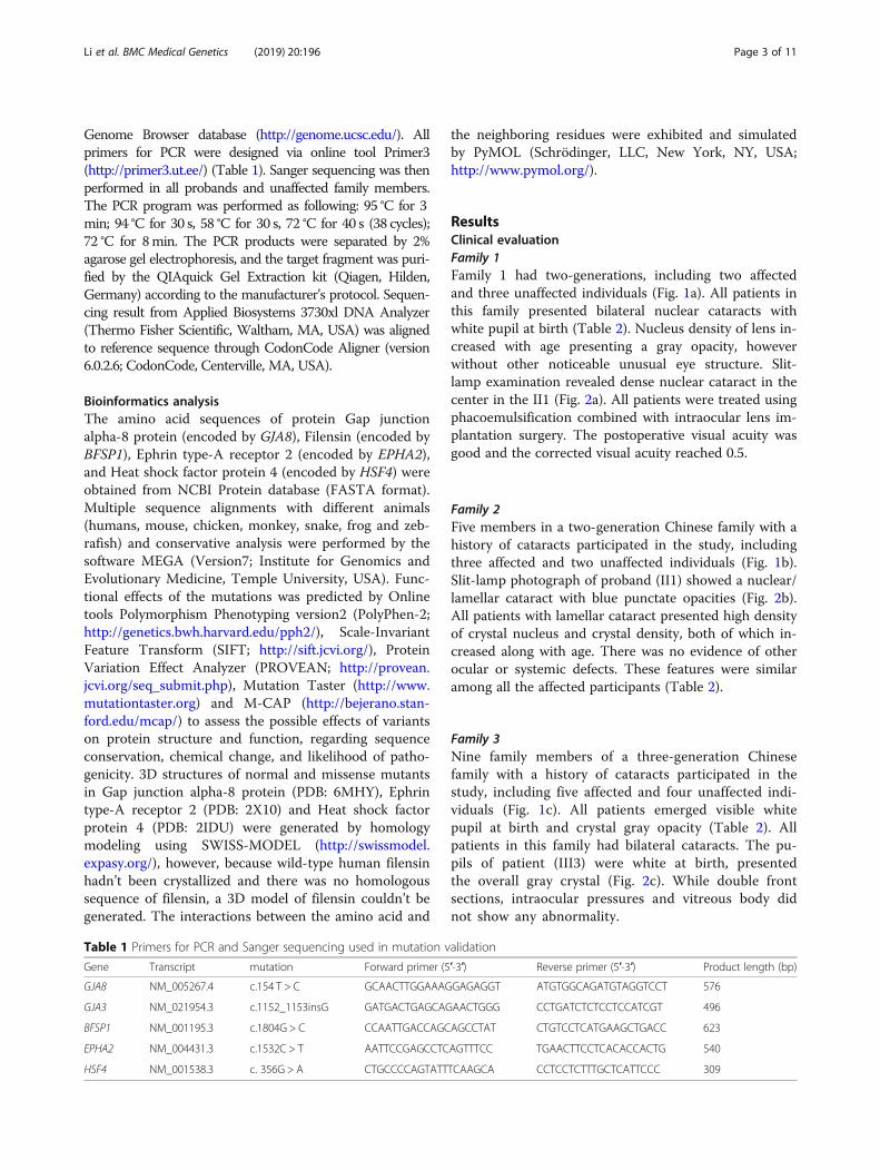
Genome Browser database (http://genome.ucsc.edu/). Allprimers for PCR were designed via online tool Primer3(http://primer3.ut.ee/) (Table 1). Sanger sequencing was thenperformed in all probands and unaffected family members.The PCR program was performed as following: 95 °C for 3min; 94 °C for 30 s, 58 °C for 30 s, 72 °C for 40 s (38 cycles);72 °C for 8min. The PCR products were separated by 2%agarose gel electrophoresis, and the target fragment was puri-fied by the QIAquick Gel Extraction kit (Qiagen, Hilden,Germany) according to the manufacturer’s protocol. Sequen-cing result from Applied Biosystems 3730xl DNA Analyzer(Thermo Fisher Scientific, Waltham, MA, USA) was alignedto reference sequence through CodonCode Aligner (version6.0.2.6; CodonCode, Centerville, MA, USA).
Bioinformatics analysisThe amino acid sequences of protein Gap junctionalpha-8 protein (encoded by GJA8), Filensin (encoded byBFSP1), Ephrin type-A receptor 2 (encoded by EPHA2),and Heat shock factor protein 4 (encoded by HSF4) wereobtained from NCBI Protein database (FASTA format).Multiple sequence alignments with different animals(humans, mouse, chicken, monkey, snake, frog and zeb-rafish) and conservative analysis were performed by thesoftware MEGA (Version7; Institute for Genomics andEvolutionary Medicine, Temple University, USA). Func-tional effects of the mutations was predicted by Onlinetools Polymorphism Phenotyping version2 (PolyPhen-2;http://genetics.bwh.harvard.edu/pph2/), Scale-InvariantFeature Transform (SIFT; http://sift.jcvi.org/), ProteinVariation Effect Analyzer (PROVEAN; http://provean.jcvi.org/seq_submit.php), Mutation Taster (http://www.mutationtaster.org) and M-CAP (http://bejerano.stan-ford.edu/mcap/) to assess the possible effects of variantson protein structure and function, regarding sequenceconservation, chemical change, and likelihood of patho-genicity. 3D structures of normal and missense mutantsin Gap junction alpha-8 protein (PDB: 6MHY), Ephrintype-A receptor 2 (PDB: 2X10) and Heat shock factorprotein 4 (PDB: 2IDU) were generated by homologymodeling using SWISS-MODEL (http://swissmodel.expasy.org/), however, because wild-type human filensinhadn’t been crystallized and there was no homologoussequence of filensin, a 3D model of filensin couldn’t begenerated. The interactions between the amino acid and
the neighboring residues were exhibited and simulatedby PyMOL (Schrödinger, LLC, New York, NY, USA;http://www.pymol.org/).
ResultsClinical evaluationFamily 1Family 1 had two-generations, including two affectedand three unaffected individuals (Fig. 1a). All patients inthis family presented bilateral nuclear cataracts withwhite pupil at birth (Table 2). Nucleus density of lens in-creased with age presenting a gray opacity, howeverwithout other noticeable unusual eye structure. Slit-lamp examination revealed dense nuclear cataract in thecenter in the II1 (Fig. 2a). All patients were treated usingphacoemulsification combined with intraocular lens im-plantation surgery. The postoperative visual acuity wasgood and the corrected visual acuity reached 0.5.
Family 2Five members in a two-generation Chinese family with ahistory of cataracts participated in the study, includingthree affected and two unaffected individuals (Fig. 1b).Slit-lamp photograph of proband (II1) showed a nuclear/lamellar cataract with blue punctate opacities (Fig. 2b).All patients with lamellar cataract presented high densityof crystal nucleus and crystal density, both of which in-creased along with age. There was no evidence of otherocular or systemic defects. These features were similaramong all the affected participants (Table 2).
Family 3Nine family members of a three-generation Chinesefamily with a history of cataracts participated in thestudy, including five affected and four unaffected indi-viduals (Fig. 1c). All patients emerged visible whitepupil at birth and crystal gray opacity (Table 2). Allpatients in this family had bilateral cataracts. The pu-pils of patient (III3) were white at birth, presentedthe overall gray crystal (Fig. 2c). While double frontsections, intraocular pressures and vitreous body didnot show any abnormality.
Table 1 Primers for PCR and Sanger sequencing used in mutation validation
Gene Transcript mutation Forward primer (5′-3′) Reverse primer (5′-3′) Product length (bp)
GJA8 NM_005267.4 c.154 T > C GCAACTTGGAAAGGAGAGGT ATGTGGCAGATGTAGGTCCT 576
GJA3 NM_021954.3 c.1152_1153insG GATGACTGAGCAGAACTGGG CCTGATCTCTCCTCCATCGT 496
BFSP1 NM_001195.3 c.1804G > C CCAATTGACCAGCAGCCTAT CTGTCCTCATGAAGCTGACC 623
EPHA2 NM_004431.3 c.1532C > T AATTCCGAGCCTCAGTTTCC TGAACTTCCTCACACCACTG 540
HSF4 NM_001538.3 c. 356G > A CTGCCCCAGTATTTCAAGCA CCTCCTCTTTGCTCATTCCC 309
Li et al. BMC Medical Genetics (2019) 20:196 Page 3 of 11

Family 4Eight family members of a three-generation Chinesefamily with a history of cataracts participated in thestudy, including four affected and four unaffected indi-viduals (Fig. 1d). The opacities of proband (IV1) werevisible at birth, which had a great influence on visualacuity. She was diagnosed with bilateral total cataractand presented nuclear opacity. These features were simi-lar among all the affected participants (Table 2). Therewas no evidence of other systemic or ocular defects withthe affected family members.
Family 5This family included 9 affected females, 11 affectedmales and 31 unaffected members in a six-generationpedigree (Fig. 1e). All the affected members in this fam-ily were diagnosed as zonular and nuclear cataract
coupled with increased crystal nucleus density and theyhad poor eyesight during the child period of 3~6months(Table 2). There were no other ocular abnormalities norother systematic diseases with the patients. The proband(IV12) showed a nuclear cataract (Fig. 2d) and receivedan operation at age 21 with YAG laser release incision ofposterior capsular. Postoperative vision of the probandreached up to 0.6, and vision condition gradually im-proved after amblyopia training.
Identification of mutationsThirty-eight genes (Additional file 1: Table S1) related withinheritable and congenital cataract were captured and se-quenced by next-generation sequencing. The averagecoverage was approximately 99.2%, and the average mediandepth was 475×. 100% of base pairs with N200× coveragewas successfully detected indicating high capabilities for
Fig. 1 The pedigrees and genotypes of 5 Chinese families with congenital autosomal dominant cataracts. The probands are indicated with anarrow. Squares and circles symbolize male and female individuals respectively. Black symbols indicate affected members and blank symbolsindicate unaffected individuals. Asterisks indicate sequenced samples. “+/+” indicates wild-type and “+/−” indicates heterozygote. (a) Pedigree ofFamily 1, all patients carried the heterozygous mutation c.154 T > C in GJA8. (b) Pedigree of Family 2, all patients carried the heterozygousmutation c.1152_1153insG in GJA3. (c) Pedigree of Family 3, all patients carried the heterozygous mutation c.1804G > C in BFSP1. (d) Pedigree ofFamily 4, all patients carried the heterozygous mutation c.1532C > T in EPHA2. (E) Pedigree of Family 5, all patients carried the heterozygousmutation c.356G > A in HSF4
Li et al. BMC Medical Genetics (2019) 20:196 Page 4 of 11
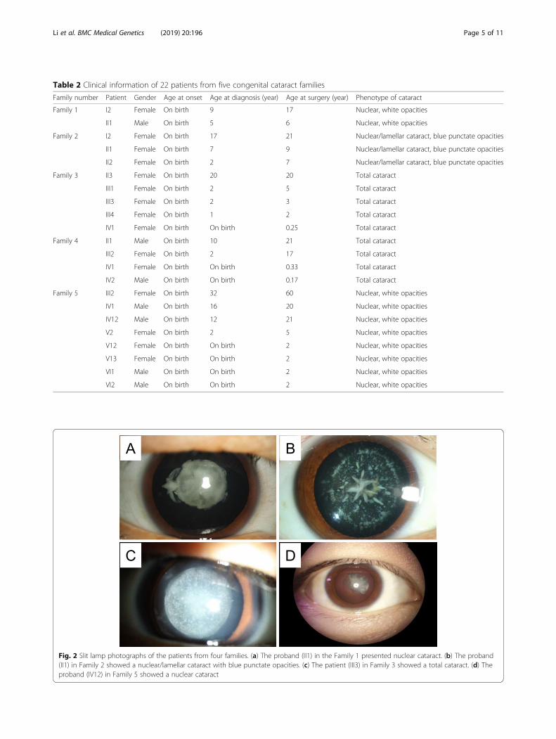
Table 2 Clinical information of 22 patients from five congenital cataract families
Family number Patient Gender Age at onset Age at diagnosis (year) Age at surgery (year) Phenotype of cataract
Family 1 I2 Female On birth 9 17 Nuclear, white opacities
II1 Male On birth 5 6 Nuclear, white opacities
Family 2 I2 Female On birth 17 21 Nuclear/lamellar cataract, blue punctate opacities
II1 Female On birth 7 9 Nuclear/lamellar cataract, blue punctate opacities
II2 Female On birth 2 7 Nuclear/lamellar cataract, blue punctate opacities
Family 3 II3 Female On birth 20 20 Total cataract
III1 Female On birth 2 5 Total cataract
III3 Female On birth 2 3 Total cataract
III4 Female On birth 1 2 Total cataract
IV1 Female On birth On birth 0.25 Total cataract
Family 4 II1 Male On birth 10 21 Total cataract
III2 Female On birth 2 17 Total cataract
IV1 Female On birth On birth 0.33 Total cataract
IV2 Male On birth On birth 0.17 Total cataract
Family 5 III2 Female On birth 32 60 Nuclear, white opacities
IV1 Male On birth 16 20 Nuclear, white opacities
IV12 Male On birth 12 21 Nuclear, white opacities
V2 Female On birth 2 5 Nuclear, white opacities
V12 Female On birth On birth 2 Nuclear, white opacities
V13 Female On birth On birth 2 Nuclear, white opacities
VI1 Male On birth On birth 2 Nuclear, white opacities
VI2 Male On birth On birth 2 Nuclear, white opacities
Fig. 2 Slit lamp photographs of the patients from four families. (a) The proband (II1) in the Family 1 presented nuclear cataract. (b) The proband(II1) in Family 2 showed a nuclear/lamellar cataract with blue punctate opacities. (c) The patient (III3) in Family 3 showed a total cataract. (d) Theproband (IV12) in Family 5 showed a nuclear cataract
Li et al. BMC Medical Genetics (2019) 20:196 Page 5 of 11

identifying variants. Variants in five cataract probands fromthe targeted NGS in Additional file 2: Table S2. The vari-ants were excluded if they presented high frequency in the1000 Genome database or the dbSNP database. Since thefive family pedigrees accorded with autosomal dominant in-heritance, we first focused on heterozygous mutations. Fivepotential pathogenic mutations were confirmed in the fiveprobands associated with congenital cataract: the heterozy-gous mutation c.154 T >C (p.F52 L) in GJA8 in Family 1,c.1152_1153insG (p.S385Efs*83) in GJA3 in Family 2, and c.1804G >C (p.G602R) in BFSP1 in Family 3, c.1532C >T(p.T511M) in EPHA2 in Family 4 and mutation c. 356G >A (p. R119H) in HSF4 in Family 5. The five mutations werenovel and were first identified as associated with congenitalcataract. The mutations were further confirmed by Sangersequencing (Fig. 3), and the five mutations co-segregatedwith the phenotypes in five families (Fig. 1). Additional test-ing proved that mutations were not detected in 50 healthylocal Chinese controls.
Bioinformatics analysis of the mutationsConservation analysis of amino acid located in p.F52of Gap junction alpha-8, p.G602 of Filensin, p.T511
of Ephrin type-A receptor 2, and p.R119 of Heatshock factor protein 4 within different vertebratespecies was performed. The analysis indicated thatthose p.F52 of Gap junction alpha-8, p.T511 ofEphrin type-A receptor 2, and p.R119 of Heat shockfactor protein 4 amino acid sites were highly con-served except p.G602 of Filensin, and the replace-ment of wild type residues might change theirbiological function (Fig. 4). In addition, SIFT,PolyPhen-2, MutationTaster, M-CAP and PROVEANprograms yielded similar outcomes regarding patho-genicity except that MutationTaster and PROVEANpredicted p.G602R was a neutral mutation (Table 3).According to the SWISS-MODEL prediction, substi-tution of phenylalanine into a leucine at position 52of Gap junction alpha-8 protein would influence theconformation of the protein. In addition, the residuewas buried in the core of a domain, and the mutantresidue might disturb the core structure of this do-main (Fig. 5a). Then, simulation predicted that T511interacted via H-bonding with residues N435 andQ515 of Ephrin type-A receptor 2. Substitution ofM511 destroyed the H-bonding as the original wild-
Fig. 3 The potential causative mutations were identified in five Chinese families with congenital cataract. (a) The heterozygous mutation c.154T > C(p.F52 L) in GJA8 was identified in all the affected participants in the Family 1. (b) The heterozygous mutation c.1152_1153insG(p.S385Efs*83)in GJA3 was identified in all the affected participants in the Family 2. (c) The heterozygous mutation c.1804G > C(p.G602R) in BFSP1 was identifiedin all the affected participants in the Family 3. (d) The heterozygous mutation c.1532C > T(p.T511 M) in EPHA2 was identified in all the affectedparticipants in the Family 4. (e) The heterozygous mutation c.356G > A(p.R119H) in HSF4 was identified in all the affected participants in theFamily 5
Li et al. BMC Medical Genetics (2019) 20:196 Page 6 of 11

type residue did. (Fig. 5b). Meanwhile, substitutionof H119 destroyed the H-bonding, with which wild-type R119 interacted with residues L124 of Heatshock factor protein 4 (Fig. 5c). This indicated thatthe substitution would affect protein function.
DiscussionWe reported five novel mutations associated with theautosomal dominance cataract in five Chinese families re-spectively: c.154 T > C in GJA8, c.1152_1153insG in GJA3,c.1804G >C in BFSP1, c.1532C > T in EPHA2 andc.356G >A in HSF4. All of the five mutations werescreened by targeted NGS for the 38 candidate genes ofcongenital cataracts, and verified through Sanger DNA se-quencing. We confirmed that each mutation co-segregated with the disease phenotypes in the correspond-ing family and absent in all the unaffected individuals.Further, bioinformatics analysis, conservative predictionand 3-D protein simulation showed that these mutationsmight be deleterious. According to the ACMG criteria[20], c.1152_1153insG in GJA3 of Family 2 and c.356G >A in HSF4 of Family 5 are clearly pathogenic variants(class V); c.154 T > C in GJA8 of Family 1 is a likely patho-genic variant (class IV); c.1804G >C in BFSP1 of Family 3and c.1532C > T in EPHA2 of Family 4 variants are un-known significance (class III) (Table 3). The unknown sig-nificance variants associated with congenital cataractsmake them interesting candidates for further studies.The lens has developed an extensive cell-cell inter-
action system using connexins to maintain its transpar-ency. Three connexins are expressed in the lens:connexin 43 (Cx43), connexin 46 (Cx46), and connexin50 (Cx50). Cx43 (GJA1) is expressed mainly in epithelialcells of lens, while Cx46 (GJA3) and Cx50 (GJA8) areexpressed in lens fibre cells [21, 22]. GJA8 and GJA3 are
the major connexin of the ocular lens, where gap junc-tions maintain ionic environment, water balance, trans-parency and optical properties of the lens [23]. To date,65 variants in GJA8 and 43 variants in GJA3 have beenreported in the HGMD (Professional 2019.1) to inducegenetic cataracts, which account for about 1/4 of non-syndromic familial cataract cases. The typical structureof connexin includes cytoplasmic NH2- and COOH- ter-minal domain, four transmembrane domains and twoextracellular loops. The two extracellular loops mediatehemichannel docking between connexons and the E1loop, which was also shown to be important for the volt-age required for closure of gap junction pores [24]. Inthis study, we identified an amino acid change (F52 L) atthe first external loop (E1) in GJA8 in family 1. The al-tered protein may disrupt normal interactions betweenthe two connexins, which may reduce resistance of theintercellular channel and lead to the leakage of smallions. Moreover, F52 L is highly conserved among manyspecies, so F52 L is very likely to cause disease. In Family2, frameshift S385Efs*83 in GJA3 resulted from a guan-ine insertion that introduced a premature translationstop codon located in the COOH-terminus, which mayinterfere with the folding of the whole protein and re-sulted in cataract. This insertion mutation (c.1152_1153insG) is similar to the three mutations (c.1137dupC,c.1189dupG, c.1200dupC) reported previously [25–27],thus providing further evidence that the GJA3 C-terminal domain plays an essential role in physiologicalfunction of the gene, and further expanding the muta-tion spectrum of GJA3 in association with congenitalcataract.BFSP1 (filesin) and BFSP2 (phakinin) are major com-
ponents of the beaded filament, which are unique cyto-skeletal lens structures. The biological functions of
Fig. 4 The multiple-sequence alignments from different vertebrate species. (a-d) The amino acid alterations, F52 L of GJA8 in Family 1, T511 M ofEPHA2 in Family 4 and R119H of HSF4 in Family 5 were located in highly conserved region among all vertebrate species and were marked withbox. While G602R of BFSP1 in Family 3 had lower conservative propertys
Li et al. BMC Medical Genetics (2019) 20:196 Page 7 of 11

Table
3Pathog
enicity
pred
ictio
nof
fivevariantsusingbioinformaticstools
Family
(proband
)Gen
eTranscrip
tNucleotide
change
Aminoacid
change
SIFT
Polyph
en2
MutationTaster
M-CAP
PROVEAN
Pathog
enicity
(ACMG)
Eviden
ce(ACMG)
Family
1(II:1)
GJA8
NM_
005267.4
c.154T>C
p.F52L
Deleterious
Possibly
damaging
Disease
causing
Possibly
Pathog
enic
Deleterious
LikelyPathog
enic
PM1PM
2PP3PP4
0.00
0.478
–0.513
−5.81
––
Family
2(II:1)
GJA3
NM_
021954.3
c.1152_
1153insG
p.S385Efs*83
Not
pred
icted
Not
pred
icted
Disease
causing
Not
pred
icted
Not
pred
icted
Pathog
enic
PVS1
PP1PP4BP4
Family
3(IV:1)
BFSP1
NM_
001195.3
c.1804G>C
p.G602R
Deleterious
Possibly
damaging
Polymorph
ism
Possibly
Pathog
enic
Neutral
Uncertain
sign
ificance
PM2PP1PP4
0.02
0.472
–0.308
−1.03
––
Family
4(IV:1)
EPHA2
NM_
004431.3
c.1532C>T
p.T511
MDeleterious
Prob
ably
damaging
Disease
causing
Possibly
Pathog
enic
Deleterious
Uncertain
sign
ificance
PM1PM
2PP1PP3PP4BS1
BS2BP6
0.00
1.00
–0.04
−4.80
––
Family
5(IV:
12)
HSF4
NM_
001538.3
c.356G
>A
p.R119H
Deleterious
Prob
ably
damaging
Disease
causing
Possibly
Pathog
enic
Deleterious
Pathog
enic
PM1PM
2PP1PP2PP3PP4
BP1
0.00
1.00
–0.259
−4.81
––
Li et al. BMC Medical Genetics (2019) 20:196 Page 8 of 11

filesin and phakinin are still not clear, but some evi-dences indicate they play an important role in main-taining lens transparency and homeostasis during fetaldevelopment and fiber cell differentiation [28]. A novelmutation c.1804G > C(p.G602R) in BFSP1 was detectedin Family 3. Alignment of the BFSP1 protein sequenceamong different species revealed that the Gly residue atposition 602 was less conservative. MutationTaster andPROVEAN prediction tools showed the pathogenicityof G602R was neutral. However, M-CAP, SIFT andPolyPhen 2 analysis indicated that G602R was possiblydamaging. Further, mutation was co-segregated withphenotypes in the Family 3 including five affected andfour unaffected individuals and that variant frequencywas 0.000066 in the ExAC browser, indicating that thisvariant was rare event in the human genome. Up tonow, only six BFSP1 mutations have been reported andfour BFSP1 mutations were involved in autosomal re-cessive cataract families [11, 29, 30]. And two muta-tions were found in autosomal dominant cataractfamilies. In 2013, Wang et al. first found a heterozygous
variant c.1042G > A(p.D348N) in BFSP1 in a 5-generation Chinese family in which 15 members hadautosomal dominant nuclear cataract [31]. In 2017,Zhai et al. identified heterozygosity for a splice site mu-tation (c.625 + 3A > G) in BFSP1 in a 4-generation fam-ily co-segregating progressive punctate lamellar cataract[32]. The mutation (G602R) highlighted in this study islocalized at the tail region of filesin, has an importanteffect on beaded filament formation as mutationD348N [31]. Taken together with previous research, theresults of the Family 3 enriched the suspected patho-genicity of the BFSP1 mutation in human autosomaldominant congenital cataract.The protein encoded by EPHA2, Ephrin Receptor
EphA2, is spatially and temporally regulated in the corticallens fiber cells, while its expression is lower in anteriorepithelial cells, and absent in the nuclei of lens [33]. So far,22 mutations of EPHA2 have been reported in the patientswith congenital cataract, and most of them are in theSAM domain. After identification of p.P584L by Daveet al., we reported the second autosomal dominant
Fig. 5 Three missense mutations (F52 L in GJA8 of Family 1, T511 M in EPHA2 of Family 4 and R119H in HSF4 of Family 5) were simulated bymeans of SWISS-MODEL and were represented with Ribbon model. The proteins were colored by element: a-helix = blue, b-stand = purple, turn =pink. Wild and mutated amino acids were labeled in green. The amino acids that interacted with the mutation sites with hydrogen bonding weremarked in yellow. (a). Substitution of F52 L in GJA8 disturbed the core structure domain and influenced the conformation of the protein. (b).Substitutions of T511 M in EPHA2 destroyed the H-bonding between T511 and N435/Q515. (c). Substitution of R119H in HSF4 destroyed the H-bonding between wild-type R119 and L124
Li et al. BMC Medical Genetics (2019) 20:196 Page 9 of 11

mutation p.T511M in the juxta membrane domain of theprotein [34]. The pathogenicity of this mutation wasproved in the following three aspects: (1) Protein sequenceamong different species revealed that the Thr residue atposition 511 was highly conserved; (2) Bioinformatics ana-lysis using five prediction tools indicated that T511M wasa pathogenic change. (3) 3-D protein simulation modelpredicted that amino acids change of M511 T would des-troy H-bonding between T511 interacted via with residuesN435 and Q515 of Ephrin type-A receptor 2. Further-more, this mutation was co-segregated with phenotypes inFamily 4. The mutation T511M identified in EPHA2 geneis a known polymorphism (rs55747232), which raisesdoubt about its pathogenicity. In conclusion, we believethat M511 T in EPHA2 is a potential variation associatedwith congenital cataract.HSF4 belongs to the family of heat-shock transcription
factors that bind heat shock elements and activate down-stream heat-shock response genes under conditions ofstress [35]. It has been reported that HSF4 gene is re-sponsible for both autosomal dominant and autosomalrecessive cataracts [36]. We had screened the affected in-dividuals in Family 5 and identified a missense mutationc.356G > A(p.R119H) in HSF4, and this mutation wasco-segregated with the disease in all the affected individ-uals, but not observed in all the unaffected individuals.Protein sequence among different species revealed thatthe Arginine(R) residue at position 119 is high con-served, and five prediction tools showed p.R119H ispathogenic. 3-D protein simulation predicted that substi-tution of H119 destroyed the H-bonding, with whichwild-type R119 interacted with residues L124 of Heatshock factor protein 4. Above all provided a persuasiveevidence to its pathogenicity of p.R119H in HSF4 ofFamily 5.In summary, we performed genetic analysis in five
Chinese families with congenital dominant cataracts andidentified five novel mutations, including an insertionmutation encoding p.S385Efs*83 in GJA3 and four mis-sense mutations: p.F52 L in GJA8, p.G602R in BFSP1,p.T511M in EPHA2 and p.R119H in HSF4. This workextended the mutation spectrum of congenital cataracts,and would provide more evidences for the precise diag-nosis of the disease.
ConclusionsWe firstly reported five novel mutations associated withautosomal dominant cataracts: c.154 T > C in GJA8,c.1152_1153insG in GJA3, c.1804G > C in BFSP1,c.1532C > T in EPHA2, c.356G > A in HSF4. This studyexpands the mutation spectrum of congenital cataracts,and provide solid evidence for genetic counseling andprenatal gene diagnosis of the cataract families.
Supplementary informationSupplementary information accompanies this paper at https://doi.org/10.1186/s12881-019-0933-5.
Additional file 1: Table S1.. Information of 38 candidate genes forcongenital cataract used in targeted NGS.
Additional file 2: Table S2. Variants in five cataract probands from thetargeted NGS.
AbbreviationsBFSP1: Beaded filament structural protein 1; EPHA2: EPH receptor A2;GJA3: Gap junction protein alpha 3; GJA8: Gap junction protein alpha 8;HGMD: Human Gene Mutation Database; HSF4: Heat shock transcriptionfactor 4; M-CAP: Mendelian Clinically Applicable Pathogenicity; NGS: Nextgeneration sequencing; PCR: Polymerase chain reaction; PolyPhen-2: Polymorphism Phenotyping v2; PROVEAN: Protein variation effect analyzer;SIFT: Scale-Invariant Feature Transform; UCSC: University of California, SantaCruz
AcknowledgementsWe are very grateful to the family members for their participation in thisstudy, as well as the support of CAMS Innovation Fund for Medical Sciences.
Authors’ contributionsSL conducted experiments, data analysis and drafting the manuscript. JFZstudied the patients and collected the clinical samples. YXC and YY analyzedthe data. XLZ designed and supervised the study. All authors read andapproved the final manuscript.
FundingThis study was supported by the CAMS Innovation Fund for MedicalSciences (CIFMS) (2016-I2M-3-003) and the National Key Research andDevelopment Program of China (2016YFE0128400 and 2016YFC0905100).The funding bodies played no role in the design of the study and collection,analysis, and interpretation of data and in writing the manuscript.
Availability of data and materialsThe datasets generated and/or analysed during the current study areavailable in the CNGB Nucleotide Sequence Archive (CNSA: https://db.cngb.org/cnsa; accession number CNP0000764).
Ethics approval and consent to participateThis study was approved by the Institutional Review Board (IRB) of theInstitute of Basic Medical Sciences, Chinese Academy of Medical Sciences,Beijing, China (015–2015). Written consent was obtained from all adultparticipants and the legal guardians of children under age 18 before theyparticipated in this study.
Consent for publicationWritten consent was obtained from all adult participants and the legalguardians of children under age 18 for the publication of this study,including the images in Fig. 2.
Competing interestsThe authors declare no conflicts of interest.
Author details1Department of Medical Genetics, Institute of Basic Medical Sciences ChineseAcademy of Medical Sciences - School of Basic Medicine Peking UnionMedical College, 5 Dong Dan San Tiao, Dongcheng District, Beijing 100005,People’s Republic of China. 2The No.4 hospital (eye hospital) of Zhangjiakou,Zhangjiakou 075000, People’s Republic of China.
Received: 14 May 2019 Accepted: 1 December 2019
References1. Hejtmancik JF. Congenital cataracts and their molecular genetics. Semin Cell
Dev Biol. 2008;19(2):134–49.
Li et al. BMC Medical Genetics (2019) 20:196 Page 10 of 11

2. Lambert SR, Drack AV. Infantile cataracts. Surv Ophthalmol. 1996;40(6):427–58.
3. Zhou Y, Zhai Y, Huang L, Gong B, Li J, Hao F, Wu Z, Shi Y, Yang Y. A novelCRYBB2 Stopgain mutation causing congenital autosomal dominantcataract in a Chinese family. J Ophthalmol. 2016;2016:1–8.
4. Pichi F, Lembo A, Serafino M, Nucci P. Genetics of congenital cataract. DevOphthalmol. 2016;57:1–14.
5. Ionides A, Francis P, Berry V, Mackay D, Bhattacharya S, Shiels A, Moore A.Clinical and genetic heterogeneity in autosomal dominant cataract. Br JOphthalmol. 1999;83(7):802–8.
6. Vanita, Singh JR, Singh D. genetic and segregation analysis of congenitalcataract in the Indian population. Clin Genet. 1999;56(5):389–93.
7. Scott MH, Hejtmancik JF, Wozencraft LA, Reuter LM, Parks MM, Kaiser-KupferMI. Autosomal dominant congenital cataract. Interocular phenotypicvariability. Ophthalmology. 1994;101(5):866–71.
8. Deng H, Yuan L. Molecular genetics of congenital nuclear cataract. Eur JMed Genet. 2014;57(2–3):113–22.
9. Shiels A, Hejtmancik JF. Genetics of human cataract. Clin Genet. 2013;84(2):120–7.10. Hilal L, Nandrot E, Belmekki M, Chefchaouni M, El Bacha S, Benazzouz B,
Hajaji Y, Gribouval O, Dufier J, Abitbol M, et al. Evidence of clinical andgenetic heterogeneity in autosomal dominant congenital ceruleancataracts. Ophthalmic Genet. 2002;23(4):199–208.
11. Ma AS, Grigg JR, Ho G, Prokudin I, Farnsworth E, Holman K, Cheng A, BillsonFA, Martin F, Fraser C, et al. Sporadic and familial congenital cataracts:mutational Spectrum and new diagnoses using next-generationsequencing. Hum Mutat. 2016;37(4):371–84.
12. Min HY, Qiao PP, Asan YZH, Jiang HF, Zhu YP, Du HQ LQ, Wang JW, ZhangJ, et al. targeted genes sequencing identified a novel 15 bp deletion onGJA8 in a Chinese family with autosomal dominant congenital cataracts.Chin Med J. 2016;129(7):860–7.
13. Aoki T, Kunishima S, Yamashita Y, Minamitani K, Ota S.Macrothrombocytopenia with congenital bilateral cataracts: a phenotype ofMYH9 disorder with exon 24 Indel mutations. J Pediatr Hematol Oncol.2018;40(1):76–8.
14. Jiang B, Chen Y, Xu B, Hong N, Liu R, Qi M, Shen L. Identification of a novelmissense mutation of MIP in a Chinese family with congenital cataracts bytarget region capture sequencing. Sci Rep. 2017;7:40129.
15. Chen J, Wang Q, Cabrera PE, Zhong Z, Sun W, Jiao X, Chen Y, GovindarajanG, Naeem MA, Khan SN, et al. Molecular genetic analysis of Pakistani familieswith autosomal recessive congenital cataracts by Homozygosity screening.Invest Ophthalmol Vis Sci. 2017;58(4):2207–17.
16. Chen JH, Qiu J, Chen H, Pang CP, Zhang M. Rapid and cost-effectivemolecular diagnosis using exome sequencing of one proband withautosomal dominant congenital cataract. Eye. 2014;28(12):1511–6.
17. Liu Y, Wei X, Kong X, Guo X, Sun Y, Man J, Du L, Zhu H, Qu Z, Tian P, et al.Targeted next-generation sequencing for clinical diagnosis of 561Mendelian diseases. PLoS One. 2015;10(8):e0133636.
18. Miller SADD, Polesky HF. A simple salting out procedure for extracting DNAfrom human nucleated cells. Nucleic Acids Res. 1988;16(3):1215.
19. Wei X, Ju X, Yi X, Zhu Q, Qu N, Liu T, Chen Y, Jiang H, Yang G, Zhen R, et al.Identification of sequence variants in genetic disease-causing genes usingtargeted next-generation sequencing. PLoS One. 2011;6(12):e29500.
20. Li Q, Wang K. InterVar: clinical interpretation of genetic variants by the 2015ACMG-AMP guidelines. Am J Hum Genet. 2017;100(2):267–80.
21. Lamei Yuan* YG, Junhui Yi†, Jingjing Xiao‡, Jinzhong Yuan†, Wei Xiong†,Hongbo Xu*, Zhijian Yang§, Jianguo Zhang‡, and Hao Deng†: Identificationof a Novel GJA3 Mutation in Congenital Nuclear Cataract. Optometry andVision Science 2015.
22. Ge XL, Zhang Y, Wu Y, Lv J, Zhang W, Jin ZB, Qu J, Gu F. Identification of anovel GJA8 (Cx50) point mutation causes human dominant congenitalcataracts. Sci Rep. 2014;4:4121.
23. Nielsen PA, Baruch A, Shestopalov VI, Giepmans BNG, Dunia I, Benedetti EL,Kumar NM. Lens Connexins alpha 3Cx46 and alpha 8Cx50 interact withzonula occludens protein-1 (ZO-1). Mol Biol Cell. 2003;14(6):2470–81.
24. <Exome sequencing identifies novel and recurrent mutations in GJA8 andCRYGD.pdf>.
25. Mackay D, Ionides A, Kibar Z, Rouleau G, Berry V, Moore A, Shiels A,Bhattacharya S. Connexin46 mutations in autosomal dominant congenitalcataract. Am J Hum Genet. 1999;64(5):1357–64.
26. Zhou D, Ji H, Wei Z, Guo L, Li Y, Wang T, Zhu Y, Dong X, Wang Y, He L,et al. A novel insertional mutation in the connexin 46 (gap junction alpha 3)
gene associated with autosomal dominant congenital cataract in a Chinesefamily. Mol Vis. 2013;19:789–95.
27. Cui XK, Zhu KK, Zhou Z, Wan SM, Dong Y, Wang XC, Li J, Zhang J, Mu HM,Qin L, et al. A novel frameshift mutation in CX46 associated with hereditarydominant cataracts in a Chinese family. Int J Ophthalmol. 2017;10(5):684–90.
28. Song S, Landsbury A, Dahm R, Liu Y, Zhang Q, Quinlan RA. Functions of theintermediate filament cytoskeleton in the eye lens. J Clin Invest. 2009;119(7):1837–48.
29. Ramachandran RD, Perumalsamy V, Hejtmancik JF. Autosomal recessivejuvenile onset cataract associated with mutation in BFSP1. Hum Genet.2007;121(3–4):475–82.
30. Li D, Wang S, Ye H, Tang Y, Qiu X, Fan Q, Rong X, Liu X, Chen Y, Yang J,et al. Distribution of gene mutations in sporadic congenital cataract in aHan Chinese population. Mol Vis. 2016;22:589–98.
31. Wang H, Zhang T, Wu D, Zhang J. A novel beaded filament structuralprotein 1 (BFSP1) gene mutation associated with autosomal dominantcongenital cataract in a Chinese family. Mol Vis. 2013;19:2590–5.
32. Zhai Y, Li J, Yu W, Zhu S, Yu Y, Wu M, Sun G, Gong X, Yao K. Targetedexome sequencing of congenital cataracts related genes: broadening themutation Spectrum and genotype-phenotype correlations in 27 ChineseHan families. Sci Rep. 2017;7(1):1219.
33. Zhang T, Hua R, Xiao W, Burdon KP, Bhattacharya SS, Craig JE, Shang D,Zhao X, Mackey DA, Moore AT, et al. Mutations of the EPHA2 receptortyrosine kinase gene cause autosomal dominant congenital cataract. HumMutat. 2009;30(5):E603–11.
34. Dave A, Laurie K, Staffieri SE, Taranath D, Mackey DA, Mitchell P, Wang JJ,Craig JE, Burdon KP, Sharma S. Mutations in the EPHA2 gene are a majorcontributor to inherited cataracts in south-eastern Australia. PLoS One. 2013;8(8):e72518.
35. Bu L, Jin YP, Shi YF, Chu RY, Ban AR, Eiberg H, Andres L, Jiang HS, ZhengGY, Qian MQ, et al. Mutant DNA-binding domain of HSF4 is associated withautosomal dominant lamellar and Marner cataract. Nat Genet. 2002;31(3):276–8.
36. Forshew T, Johnson CA, Khaliq S, Pasha S, Willis C, Abbasi R, Tee L, Smith U,Trembath RC, Mehdi SQ, et al. Locus heterogeneity in autosomal recessivecongenital cataracts: linkage to 9q and germline HSF4 mutations. HumGenet. 2005;117(5):452–9.
Publisher’s NoteSpringer Nature remains neutral with regard to jurisdictional claims inpublished maps and institutional affiliations.
Li et al. BMC Medical Genetics (2019) 20:196 Page 11 of 11