Embed Size (px)
Citation preview

MiaMi iFaTS 2011 SCiENTiFiC PROGRaM
November 4-6, 2011Eden Roc Renaissance • Miami Beach, Florida

Mark your calendar
International Federation forAdipose Therapeutics and Science
10th AnniversaryQuébec IFATS 2012
October 4-6, 2012Loews Hôtel Le Concorde
Québec City, Québec CANADA
AbstrAct DeADline:Midnight EST, Tuesday, May 1, 2012
The Call for Abstracts will be sent this winter. All members of the IFATS and all registered attendees of the 2011 IFATS Conference will be included in the mailing list. Any others who wish to be reminded to submit papers should contact the IFATS Executive Office.
IFATS Executive Office45 Lyme Road - Suite 304 Hanover, NH 03755 USA
Tel: 1-603-643-2325 • Fax: 1-603-643-1444Email: [email protected] • Web: www.ifats.org
Catherine Foss - Executive Director • [email protected] Cook - Accounting Manager • [email protected] Ambrose - Abstract Coordinator and Marketing Manager • [email protected] Carney - Membership Services Manager • [email protected] DeBeaumont - Special Projects • [email protected]
Loews Hôtel Le Concorde Québec City, Québec CANADA
International Federation forAdipose Therapeutics and Science
MiaMi iFaTS 2011 ConFerenCeNovember 4-6, 2011
Eden Roc Renaissance • Miami Beach, Florida
Recording of any content presented at this educational program either by camera, videocamera, cell phone, audiorecorder, or any other device is strictly prohibited.


Table of Contents
Founders Board & Board of Directors. . . . . . . . . . . . . . . . . . . . . . . . . . . . . . . . . . . . . . . 6
Welcome from the Scientific Program Chair . . . . . . . . . . . . . . . . . . . . . . . . . . . . . . . . . 7
Invited Speakers & Session Moderators . . . . . . . . . . . . . . . . . . . . . . . . . . . . . . . . . . . . . 8
Program in Brief . . . . . . . . . . . . . . . . . . . . . . . . . . . . . . . . . . . . . . . . . . . . . . . . . . . . . . . . 9 - 11
Program Schedule . . . . . . . . . . . . . . . . . . . . . . . . . . . . . . . . . . . . . . . . . . . . . . . . . . . . . . 13 - 37
Paper Presentations . . . . . . . . . . . . . . . . . . . . . . . . . . . . . . . . . . . . . . . . . . . . . . . . . . . . . 39 - 108
Exhibitors . . . . . . . . . . . . . . . . . . . . . . . . . . . . . . . . . . . . . . . . . . . . . . . . . . . . . . . . . . . . . 109 - 114
endorsed by:
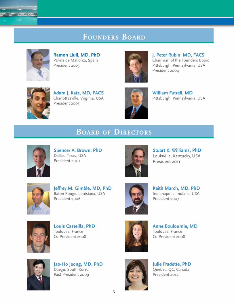
FounderS Board
Board oF direCTorS
Ramon Llull, MD, PhDPalma de Mallorca, Spain President 2003
J. Peter Rubin, MD, FAcSChairman of the Founders Board Pittsburgh, Pennsylvania, USA President 2004
William Futrell, MDPittsburgh, Pennsylvania, USA
Adam J. Katz, MD, FAcSCharlottesville, Virginia, USA President 2005
Spencer A. brown, PhDDallas, Texas, USAPresident 2010
Stuart K. Williams, PhDLouisville, Kentucky, USAPresident 2011
Jeffrey M. Gimble, MD, PhDBaton Rouge, Louisiana, USAPresident 2006
Keith March, MD, PhDIndianapolis, Indiana, USAPresident 2007
Louis casteilla, PhDToulouse, FranceCo-President 2008
Anne bouloumie, MDToulouse, France Co-President 2008
Jao-Ho Jeong, MD, PhDDaegu, South KoreaPast President 2009
Julie Fradette, PhDQuebec, QC, CanadaPresident 2012
6

We are delighted to welcome guests from 26 countries to the 9th Annual Symposium on Adipose Stem Cells and Clinical Application of Adipose Tissue and hope you enjoy your stay in Miami. Both our educational program and the social events we have planned for you are designed to encourage interaction and the exchange of new ideas. We are grateful to our exhibiting companies for their support and encourage you to meet with their representative to learn more about their products and services.
Our program this year includes panel presentations, invited guest speakers, and 111 superb international papers presenting new information from leading scientists in the field of adult adipose stem cells research highlighting the latest scientific, medical, and technological advances in the clinical arena. The abstract review committee had a difficult task selecting the final papers from 145 abstracts submitted from all over the world. The program format will include concurrent sessions in order to accommodate all accepted papers.
As information and clinical applications in stem cell research and clinical fat grafting have gained momentum, so has the IFATS educational program and our membership is growing. This year’s topics include new data on adipocytes, tissue engineering, and preclinical and clinical treatments. Our keynote speakers will provide additional insights into this growing and exciting field.
It is our goal to share new knowledge among clinicians and researchers who are working with adipose tissue and stem cells, and to introduce technologies by developers who are creating new and cost-effective devices, procedures and biological scaffolds to move the application of these cells forward as a regenerative tool.
On behalf of the IFATS Board of Directors, we are delighted to have you with us.
With best wishes,
Stuart K. Williams, PhD2011 Scientific Program Chair
iFaTS Board of Directors
San Hong Baek The Catholic University of Korea South Korea
anne Bouloumie Toulouse University France
Spencer Brown UT Southwestern Medical Center United States
Louis Casteilla Toulouse University France
William Futrell University of Pittsburgh United States
Jae-Ho Jeong Yeungnam University South Korea
adam J. Katz University of Virginia United States
Ramon Llull University of Barcelona Spain
J. Peter Rubin University of PittsburghUnited States
Members at Large
Jeff Gimble Pennington institute United States
Keith March indiana University United States
7
DISCLAImEr
Papers are reprinted as they were submitted.IFATS takes no responsibility for typographical or other errors.
All papers in this Program Book are listed in numerical order.
No one may present more than one paper at any IFATS meeting, although an individual may be an author of more than one paper presented. The paper must be presented by one of the authors. If no alternate presenter is available, the paper will be replaced on the program.
recording of any content presented at this educational program either by camera, video-camera, cell phone, audiorecorder, or any other device is strictly prohibited.
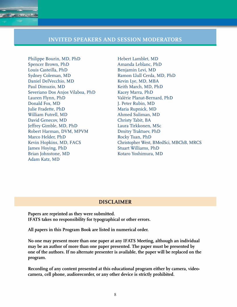
Philippe Bourin, MD, PhDSpencer Brown, PhDLouis Casteilla, PhDSydney Coleman, MDDaniel DelVecchio, MDPaul Dimuzio, MDSeveriano Dos Anjos Vilaboa, PhDLauren Flynn, PhDDonald Fox, MDJulie Fradette, PhDWilliam Futrell, MDDavid Genecov, MDJeffrey Gimble, MD, PhDRobert Harman, DVM, MPVMMarco Helder, PhDKevin Hopkins, MD, FACSJames Hoying, PhDBrian Johnstone, MDAdam Katz, MD
Hebert Lamblet, MDAmanda Leblanc, PhDBenjamin Levi, MDRamon Llull Cerda, MD, PhDKevin Lye, MD, MBAKeith March, MD, PhDKacey Marra, PhDValérie Planat-Bernard, PhDJ. Peter Rubin, MDMaria Rupnick, MDAhmed Suliman, MDChristy Tabit, BALaura Tirkkonen, MScDmitry Traktuev, PhDRocky Tuan, PhDChristopher West, BMedSci, MBChB, MRCSStuart Williams, PhDKotaro Yoshimura, MD
INvITED SPEAkErS AND SESSION mODErATOrS
8

Friday, November 4, 2011
6:30 am - 5:00 pm Registration
7:00 - 7:45 am Continental Breakfast
7:45 - 8:00 am Welcome and Introduction
8:00 - 8:20 am Adipose Tissue and Cells: Past, Present and FutureJ.PeterRubin,MD
8:20 - 9:10 am SESSION 1Clinical SciencesModerators:AdamKatz,MD&SydneyColeman,MD
9:10 - 9:40 am Coffee Break/Exhibits
9:40 - 10:40 am SESSION 1 (continued)Clinical SciencesModerators: AdamKatz,MD&SydneyColeman,MD
10:40 - 11:10 am Coffee Break/Exhibits
11:10 am - 12:20 pm SESSION 2Translational SciencesModerators: PaulDimuzio,MD&SpencerBrown,PhD
12:20 - 1:40 pm Lunch and Exhibits
1:40 - 3:00 pm SESSION 3ConcurrentPodiumPaperSessionsGroup A: Clinical Fat GraftingModerators: AhmedSuliman,MD&KevinHopkins,MD,FACS
Group B: Cell Isolation and Culture Moderators: KaceyMarra,PhD&RobertHarman,DVM,MPVM
3:00 - 3:30 pm Coffee Break/Exhibits
3:30 - 4:40 pm SESSION 4ConcurrentPodiumPaperSessionsGroup A: Adipose Tissue and Cell TransplantationModerators: ChristyTabit,BA&KevinLye,MD,MBA
Group B: Fat Isolation and Processing State of the Art TechnologiesModerators: PhilippeBourin,MD,PhD&DmitryTraktuev,PhD
4:40 - 5:15 pm Coffee Break/Exhibits
5:15 - 6:30 pm Panel 1: Clinical Fat Grafting: State of the ArtModerator:J.PeterRubin,MDPanelists: SydneyColeman,MD,DanielDelVecchio,MD,KotaroYoshimura,MD
RamonLlullCerda,MD
Panel 2: regulatory Pathways for Adipose Technology
Moderators: StuartWillams,PhD&KeithMarch,MD,PhD
7:00 - 9:00 pm Welcome reception
program in brief
9

Saturday November 5, 2011
6:30 am - 5:00 pm Registration
7:00 - 7:30 am Continental Breakfast
7:55 - 8:00 am Opening Remarks
8:00 - 9:40 am SESSION 5 The Biology of Adipose Tissue Therapeutics Moderators: JulieFradette,PhD&JeffGimble,MD,PhD
8:00 - 8:30 am Adipose Tissue mass Can be regulated Through the vasculature Invited Speaker: MariaRupnick,MD
9:10 - 9:40 am Coffee Break/Exhibits
10:10 - 11:10 am SESSION 5 (continued) The Biology of Adipose Tissue Therapeutics Moderators:JulieFradette,PhD&JeffGimble,MD,PhD
11:10 - 11:30 am Coffee Break/Exhibits
11:30 am - 1:10 pm SESSION 6 Translational Sciences Moderators: JamesHoying,PhD&KotaroYoshimura,MD
1:10 - 2:30 pm Lunch and Exhibits
2:30 - 3:20 pm SESSION 7 ConcurrentPodiumPaperSessions Group A: Fat Grafting Techniques Moderators: HebertLamblet,MD&DonaldFox,MD
Group B: Cell Isolation, Culture and Storage Moderators: BenjaminLevi,MD&AmandaLeBlanc,PhD
3:20 - 3:50 pm Coffee Break/Exhibits
3:50 - 4:35 pm SESSION 8 ConcurrentPodiumPaperSessions Group A: Adipogenic Factors Moderators: ChristopherWest,BMedSci,MBChB,MRCS&LauraTirkkonen,MSc
Group B: Stromal vascular Fraction and Tissue responses Moderators: BrianJohnstone,MD&LaurenFlynn,PhD
4:35 - 5:05 pm Coffee Break/Exhibits
5:05 - 6:10 pm SESSION 9 Environmental Factors and Cell Function Moderators: LouisCasteilla,PhD&MarcoHelder,PhD
5:05 - 6:10 pm Panel 3: Clinical Cases - video Presentations Moderator: WilliamFutrell,MD Panelists: RamonLlullCerda,MD,PhD&SeverianoDosAnjosVilaboa,PhD
7:00 - 10:00 pm Gala Dinner
10

Sunday November 6, 2011
6:30 am - 12:00 pm Registration
7:00 - 7:30 am Continental Breakfast
7:55 - 8:00 am Opening Remarks
8:00 - 10:10 am SESSION 10Toward a mechanistic Understanding of Fat and Cell Transplantation
Moderators: RamonLlullCerda,MD,PhD&ValeriePlanat-Benard,PhD
8:00 - 8:30 am Stem Cells, Development and repair Invited Speaker: RockyTuan,PhD
10:10 - 10:40 am Coffee Break
10:40 - 11:45 am SESSION 11 Translational Sciences Adipose Tissue and Cell Transplantation Moderators: J.PeterRubin,MDandDavidGenecov,MD
11:45 am - 12:00 pm Concluding Remarks and Farewell
11

12
13
PrOGrAm SCHEDULE
Recording of any content presented at this educational program either by camera, videocamera, cell phone, audiorecorder, or any other device is strictly prohibited.

Friday, November 4, 2011
6:30 am - 5:00 pm Registration
7:00 - 7:45 am Continental Breakfast
7:45 - 8:00 am Welcome and IntroductionsStuartWilliams,PhD,SpencerBrown,PhD,JulieFradette,PhD
8:00 - 8:20 am Adipose Tissue and Cells: Past, Present and FutureJ.PeterRubin,MD
8:20 - 9:10 am SESSION 1Clinical SciencesModerators:AdamKatz,MD&SydneyColeman,MD
8:20 am 167AUTOLOGOUS BrEAST FAT GrAFTING - CUrrENT OPINIONS AND PrACTICES AmONG NOrTH AmErICAN PLASTIC SUrGEONSPresenter: Ahmed Suliman, MDAffiliation: UCLAAuthors: Suliman A, Fan K, Tanna N, Liao E, Lesavoy MA, Festekjian J
8:30 am 40LONG-TErm OUTCOmES FOLLOWING FAT GrAFTING IN ImPLANT-BASED BrEAST rECONSTrUCTION: A COmPArATIvE ANALYSISPresenter: Akhil K. Seth, MDAffiliation: Northwestern UniversityAuthors: Seth AK, Hirsch EM, Fine NA
8:40 am 84rANDOmIZED CONTrOLLED CLINICAL TrIAL OF FAT GrAFTS SUPPLEmENTED WITH ADIPOSE-DErIvED rEGENErATIvE CELLS FOr PATIENTS WITH HEmIFACIAL mICrOSSOmIAPresenter: Daniela S. Tanikawa, MDAffiliation: University of Sao Paulo School of MedicineAuthors: Tanikawa DS, Aguena M, Bueno DF, Alonso N, Passos-Bueno MR
8:50 am 121FAT GrAFTING TO THE rECONSTrUCTED BrEAST: THE USE OF 3D ImAGING TO EvALUATE vOLUmE rETENTIONPresenter: Kevin Small, MDAffiliation: New York UniversityAuthors: Levovitz C, Small K, Choi M, Karp NS
9:00 am 168BENEFICIAL rOmBErG rECONSTrUCTION - DESPITE POOrEr FATGrAFT TAkE AND mULTIPLE SOFT AND HArD TISSUE PrOCEDUrESPresenter: Christy Tabit, BAAffiliation: UCLAAuthors: Tabit C, Slack G, Andrews B, Kawamoto HK, Bradley JP
9:10 - 9:40 am Coffee Break/Exhibits
14

9:40 - 10:40 am SESSION 1 (continued)Clinical SciencesModerators: AdamKatz,MD&SydneyColeman,MD
9:40 am 153AUTOLOGOUS HIGH vOLUmE FAT GrAFTING FOr COrrECTION OF CONTOUr DEFOrmITIES OF THE BrEAST; TrANSITIONING FrOm THE CONSErvATIvE
Presenter: Andres G. Sarraga, MD Affiliation: University of Massachusetts Authors: Sarraga AG, Noury M, Castle JM, Lalikos JF
9:50 am 147 CLINICAL COmPArISON OF THrEE COmmErCIALLY mADE SvF EXTrACTION mACHINES Presenter: Joel Aronowitz, MD Affiliation: Private Practice Authors: Watson JP, Aronowitz J
10:00 am 164 POWEr ASSISTED BUTTOCk FAT GrAFTING – INTrODUCING A NEW TECHNIQUE FOr ImPrOvED BUTTOCk AUGmENTATION Presenter: Henry A. Mentz III, MD Affiliation: Private Practice Authors: Mentz HA, Newall G
10:10 am 87 CLINICAL EXPErIENCE WITH AUTOLOGOUS FAT GrAFTING IN THE PEDIATrIC PATIENT Presenter: Kevin S. Hopkins, MD, FACS Affiliation: Driscoll Childrens Hospital Authors: Hopkins KS, Dhar PR
10:20 am 5 COmPOSITE BrEAST AUGmENTATION Presenter: Eric M. Auclair, MD Affiliation: Private Practice Authors: Auclair EM, Szpalski C
10:30 am 124 FACE LIFTING ASSISTED BY STrOmAL ENrICHED LIPOGrAFT vErSUS FACE LIFTING ASSISTED BY NON STrOmAL ENrICHED LIPOGrAFT: A CLINICAL STUDY Presenter: Aris Sterodimas, MD, MSc, PhD Affiliation: IASO General Hospital Authors: Sterodimas A, Nicaretta B, Illouz YG
10:40 - 11:10 am Coffee Break/Exhibits
15
cAnceLLeD

11:10 am - 12:20 pm SESSION 2Translational SciencesModerators: PaulDimuzio,MD&SpencerBrown,PhD
11:10 am 62CELLULAr OrIGIN IN ADIPOSE TISSUE rEmODELING AFTEr TrANSPLANTATION: HOST Or DONOr?Presenter: Kentaro Doi, MDAffiliation: University of TokyoAuthors: Doi K, Eto H, Kato H, Yoshimura K
11:20 am 41COmPArISON OF ADIPOSE-DErIvED STrOmAL CELLS AND BONE mArrOW mESENCHYmAL STrOmAL CELLS FrOm THE SAmE HEALTHY INDIvIDUALS - A PHENOTYPIC, FUNCTIONAL, TrANSCrIPTOmIC AND EPIGENETIC STUDYPresenter: Philippe Bourin, MD, PhDAffiliation: STROMALabAuthors: Bourin P, Hebraud B, Chaput B, Peyrafitte JA, Gadelorge M,
Espagnolle N, Huynh A, Roussel M, Attal M, Collas P, Casteilla L, Planat-Benard V
11:30 am 158COmPArISON OF ADIPOSE STrOmAL vASCULAr FrACTION AND PLASTIC ADHErENT ASC POPULATIONS FrOm THE SAmE DONOr IN TWO rODENT mODELS OF CArDIOvASCULAr DISEASESPresenter: Brian Johnstone, MDAffiliation: Indiana University School of MedicineAuthors: Johnstone B, Cook TG, Merfeld S, Motlagh D, Amrani DL, March KL
11:40 am 60COmPArISON OF ADIPOSE-DErIvED STrOmAL CELLS (ASC) AND BONE mArrOW mESENCHYmAL STrOmAL CELLS (Bm-mSC) FrOm THE SAmE HEALTHY INDIvIDUALS – A STUDY OF ImmUNOLOGICAL PrOPErTIESPresenter: Cedric Menard, PharmD, PhDAffiliation: University of Rennes 1Authors: Menard C, Hebraud B, Dulong J, Gadelorge M, Bezier I, Latour M,
Bescher N, Planat-Benard V, Bourin P, Tarte K
11:50 am 115ENCAPSULATED ADIPOGENIC FACTOrS EFFECT IN ADIPOSE GrAFT rETENTIONPresenter: Kacey Marra, PhDAffiliation: University of PittsburghAuthors: Marra KG, Tan H, Rakers A, Rubin JP
16
12:00 pm 161HEPATOCYTE GrOWTH FACTOr/C-mET rECEPTOr AUTOCrINE LOOP
IS ESSENTIAL FOr THE rESISTANCE OF ADIPOSE-DErIvED STEm CELLS TO rEACTIvE OXYGEN SPECIES Presenter: Jie Xie, MD Affiliation: Indiana University School of Medicine Authors: Xie J, Johnstone BH, Feng D, March KL
12:10 pm 126 BEYOND GrAFTING – UNCOvErING THE mOLECULAr mECHANISmS OF HUmAN TISSUE AGING USING PrImArY ADIPOSE TISSUE AS A mODEL Presenter: Ivona Percec, MD, PhD Affiliation: University of Pennsylvania Authors: Percec I, Dierova R, Bucky LP, Chang B, Auman D, Hoover W
12:20 - 1:40 pm Lunch and Exhibits
1:40 - 3:00 pm SESSION 3 ConcurrentPodiumPaperSessions Group A: Clinical Fat Grafting Moderators: AhmedSuliman,MD&KevinHopkins,MD,FACS
1:40 pm 108 CLINICAL APPLICATION OF FAT TrANSFEr IN rECONSTrUCTIvE SUrGErY-SOUTH AFrICAN EXPErIENCE Presenter: Ewa A. Siolo, MD, MBChB, FCS Affiliation: University of KWA Zulu Natal Author: Siolo EA
1:50 pm 17 LIPOmODELLING OF BrEAST rECONSTrUCTION CONTOUr DEFOrmITIES: USE OF STEm-CELL ENrICHED FAT GrAFTS Presenter: Amir Sadri, MD Affiliation: Royal Free Hospital Authors: Akhavani M, Sadri A, Mosahebi A
2:00 pm 136 COrrECTING LOWEr EYELID rETrACTION USING FAT GrAFTING Presenter: Katarina Andjelkov, MD, MS Affiliation: Private Practice Authors: Andjelkov K, Sforza M, Zaccheddu R
2:10 pm 25 BED SIDE ISOLATION OF ADIPOSE DErIvED STEm CELLS WITHIN THE OPErATION rOOm, COLLAGENAGE FrEE, FOr AUTOLOGOUS FAT TISSUE TrANSPLANT: 2 YEArS EXPErIENCE Presenter: Hebert T. Lamblet, MD Affiliation: Vikaara Klinik Author: Lamblet HT
17
2:20 pm 93NONINvASIvE BODY CONTOUrING AND SPOT FAT rEDUCTION BY
LOW LEvEL LASEr THErAPY: EFFICACY OF LIPOLASEr TECHNOLOGY FrOm A SINGLE CENTEr, CONTrOLLED CLINICAL STUDY Presenter: Vinod K. Podichetty, MD ,MS Affiliation: Research Practice Partners Inc Authors: Podichetty VK, LaForge JC, Alibhai H
2:30 pm 112 LIDOCAINE: AN ATTrACTIvE LOCAL ANESTHETIC FOr LIPOASPIrATION PrOCEDUrE IN STEm CELLS rEGENErATIvE mEDICINE Presenter: AnneClaire Girard, PhD Affiliation: Stemcis Authors: Girard A, Loyher PL, Bencharif K, Balat M, Lefebvre d Hellencourt C, Delarue P, Hulard O, Roche R, Festy F, Hoareau L
2:40 pm 46 STEm CELL ENrICHED TISSUE INJECTIONS IN PLASTIC SUrGErY: A NEW WEAPON FOr HOSTILE rECIPIENT ArEAS Presenter: Tunc K. Tiryaki, MD Affiliation: Cellest Plastic Surgery Clinic Authors: Tiryaki TK, Findikli N, Tiryaki D
2:50 pm 97 THE EFFECT OF PrESSUrE IN AUTOLOGOUS FAT GrAFTING Presenter: Jeffrey H. Lee, MD Affiliation: Massachusetts General Hospital Authors: Lee JH, Kirkham JC, McCormack MC, Nicholls AM, Randolph MA, Austen WG
1:40 - 3:00 pm SESSION 3 ConcurrentPodiumPaperSessions Group B: Cell Isolation and Culture Moderators: KaceyMarra,PhD&RobertHarman,DVM,MPVM
1:40 pm 133 ISOLATION, CrYOPrESErvATION AND TrI-LINEAGE DIFFErENTIATION OF ADIPOSE-DErIvED STEm CELLS FrOm HUmAN LIPOASPIrATE Presenter: Kevin Grady Affiliation: Lonza Walkersville Inc Authors: Klarmann GJ, Grady K, Keller J
1:50 pm 75 CASE rEPOrT: OPTImIZATION OF rOCHE LIBErASE IN THE ENZYmATIC DIGESTION OF HUmAN ADIPOSE TISSUE FOr THE ISOLATION OF STEm & rEGENErATIvE CELLS Presenter: Rowena A. Soriano, BS Affiliation: Invitrx Therapeutics Inc Authors: Soriano RA, Torfi H
18
cAnceLLeD
2:00 pm 137ImPACT OF ENZYmE COmPOSITION ON ADIPOSE-DErIvED STrOmAL vASCULAr FrACTION CELL ISOLATION
Presenter: Jacob R. Dale, BS Affiliation: University of Louisville and Jewish Hospital Authors: Dale JR, Breite D, Clayton L, Dwulet F, McCarthy R, Hoying JB, Williams SK
2:10 pm 159 THE USE OF COLLAGENASE IN ADIPOSE STEm CELL ISOLATION: COmPrEHENSIvE LITErATUrE rEvIEW AND mETA-ANALYSIS Presenter: Alexander F. Mericli, MD Affiliation: University of Virginia Authors: Mericli AF, Greyson MA, Katz AJ
2:20 pm 144 DEvICE AND mETHOD FOr EFFICIENT ISOLATION OF ADIPOSE- DErIvED rEGENErATIvE CELLS FrOm mULTIPLE DEPOTS Presenter: Ivone Bruno, PhD Affiliation: InGeneron Inc Authors: Bruno I, Husfeld R, Davis J, French M, Stone G, Stubbers R, Alt E, Coleman M
2:30 pm 73 DEvELOPmENT OF A SErUm-FrEE CHEmICALLY DEFINED HUmAN ADIPOSE DErIvED STEm CELL EXPANSION SYSTEm THAT mAINTAINS mULTIPOTENCY AND ImmUNOPHENOTYPE Presenter: Kirsten Crapnell, PhD Affiliation: Becton Dickinson Authors: Crapnell K, Kelley R, Reyes J, Hastings A, Blaesius R, Brooks J
2:40 pm 19 NOvEL IN vITrO CULTUrE CONDITIONS OF ADIPOSE STEm CELLS FOr CLINICAL CELL THErAPY APPLICATIONS Presenter: Mimmi Patrikainen, MS Affiliation: University of Tampere Authors: Patrikainen M, Juntunen M, Suuronen R, Miettinen S, Mannerstrom B
2:50 pm 152 TACkLING THE mANUFACTUrING CHALLENGES FOr CLINICAL USE OF HUmAN ADIPOSE-DErIvED SvF CELLS: FrOm LOGISTICS, COLLECTION, AND BIOPrOCESSING TO CHArACTErIZATION, CrYOSTOrAGE AND PrODUCT rELEASE Presenter: MaryPat Moyer, PhD Affiliation: INCELL Corporation LLC Author: Moyer MP
3:00 - 3:30 pm Coffee Break/Exhibits
19

3:30 - 4:40 pm SESSION 4 ConcurrentPodiumPaperSessions Group A: Adipose Tissue and Cell Transplantation Moderators: ChristyTabit,BA&KevinLye,MD,MBA
3:30 pm 14 A NEW AND EASY mETHOD FOr LArGE-vOLUmE FAT GrAFTS - THE BEAULI mETHOD Presenter: Klaus Ueberreiter, MD, PhD Affiliation: Asklepios Clinic Berlin Birkenwerder Authors: Ueberreiter K, von Finckenstein JG, Cromme F, Herold C, Tanzella U, Vogt PM
3:40 pm 95 LOW LEvEL LASEr THErAPY FOr BODY CONTOUrING AND SPOT FAT rEDUCTION: CLINICAL rEPOrT OF 222 CASES Presenter: Vinod K. Podichetty, MD, MS Affiliation: Research Practice Partners Inc Authors: Podichetty VK, Bourassa D
3:50 pm 151 BrEAST AUGmENTATION WITH AUTOLOGOUS FAT INJECTION (A rEPOrT OF 105 CASES) Presenter: Facheng Li, MD, PhD Affiliation: Plastic Surgery Hospital Chinese Academy of Medical Science and Peking Union Medical College Authors: Li F, Ma LH
4:00 pm 72 THE FDA APPrOvAL PrOCESS FOr vETErINArY ALLOGENEIC STEm CELL PrODUCTS - YES-STEm CELLS ArE DrUGS IN vETErINArY mEDICINE TOO Presenter: Robert Harman, DVM, MPVM Affiliation: VetStem Authors: Harman R, Black L, Harman S, Smith A
4:10 pm 50 IN vITrO TISSUE GENErATION BY ADULT EQUINE mULTIPOTENT STrOmAL CELLS ON COLLAGEN SCAFFOLDS Presenter: Lin Xie, BS Affiliation: Louisiana State University Authors: Xie L, Zhang Y, Gimble JM, Lopez MJ
20

4:20 pm 78TrEATmENT OF GOAT OSTEOCHONDrAL kNEE DEFECTS WITH ADIPOSE DErIvED STEm CELLS USING A ONE-STEP SUrGICAL PrOCEDUrEPresenter: Marco N. Helder, PhDAffiliation: VU University Medical CenterAuthors: Helder MN, Jurgens WJ, Kroeze RJ, Zandieh Doulabi B, Renders G,
Smit TH, van Milligen FJ, Ritt MP
4:30 pm 67HUmAN ADIPOSE-DErIvED STEm CELLS SUrvIvAL AND IN-vIvO
TrACkING IN ANImAL mODELS Presenter: Hitesh Agrawal, MD Affiliation: University of Virginia Health System Authors: Agrawal H, Shang H, Parker A, Katz AJ
3:30 - 4:40 pm SESSION 4 ConcurrentPodiumPaperSessions Group B: Fat Isolation and Processing State of the Art Technologies Moderators: PhilippeBourin,MD,PhD&DmitryTraktuev,PhD
3:30 pm 65 FAT vIABILITY ASSESSmENT WITH THIrD GENErATION ULTrASOUND ASSISTED LIPOSUCTION Presenter: Mark E. Schafer, PhD Affiliation: Sound Surgical Technologies LLC Authors: Schafer ME, Hicok KC
3:40 pm 55 ANALYSIS OF NEGATIvE PrESSUrES GENErATED BY DIFFErENT CALIBEr SYrINGES USED FOr LIPOSUCTION Presenter: Ricardo L. Rodriguez, MD Affiliation: Cosmeticsurg Authors: Rodriguez RL, Conde Green A, McLenithan J
3:50 pm 155 USE OF A CUSTOm mECHANICAL PrOCESSING DEvICE FOr PrEPArATION OF A rEGENErATIvE CELL ENrICHED mATrIX FrOm LIPOASPIrATE Presenter: Henry A. Mentz III, MD Affiliation: Aesthetic Center for Plastic Surgery Authors: Mentz HA, French M, Stone G, Stubbers R, Alt E, Coleman M
4:00 pm 23 ADIPOGENESIS USING HUmAN ADIPOSE TISSUE-DErIvED CELLS COmBINED WITH COLLAGEN/ GELATIN SCAFFOLD ImPrEGNATED WITH BASIC FIBrOBLAST GrOWTH FACTOr Presenter: Ran Ito, MD Affiliation: Kyoto University Hospital Authors: Ito R, Morimoto N, Tsuji W, Nakamura Y, Kawai K, Suzuki S
21

4:10 pm 150DECELLULArIZED ADIPOSE TISSUE (DAT) AS A BIOmATErIAL FOr SOFT TISSUE rECONSTrUCTIONPresenter: Hulan Shang, MSAffiliation: University of VirginiaAuthors: Shang H, Agrawal H, Flynn L, Katz A
4:20 pm 15DE NOvO ADIPOGENESIS BY ImPLANTATION OF TYPE-I COLLAGEN SPONGE INTO rABBITS’ FAT PADPresenter: Wakako Tsuji, MD, PhDAffiliation: Kyoto University HospitalAuthors: Tsuji W, Inamoto T, Ito R, Morimoto N, Tabata Y, Toi M
4:30 pm 117GENErATION OF AN ADIPOSE-DErIvED EXTrACELLULAr mATrIX SPONGE THAT rETAINS BIOCHEmICAL AND BIOLOGICAL INTEGrITYPresenter: Jerome Connor, PhDAffiliation: Kinetic Concepts IncAuthor: Connor J
4:40 - 5:15 pm Coffee Break/Exhibits
5:15 - 6:30 pm Panel 1: Clinical Fat Grafting: State of the ArtModerator:J.PeterRubin,MDPanelists: SydneyColeman,MD,DanielDelVecchio,MD,KotaroYoshimura,MD
RamonLlullCerda,MD
5:15 - 6:30 pm Panel 2: regulatory Pathways for Adipose TechnologyModerators: StuartWillams,PhD&KeithMarch,MD,PhD
7:00 - 9:00 pm Welcome reception
22
cAnceLLeD
Saturday November 5, 2011
6:30 am - 5:00 pm Registration
7:00 - 7:30 am Continental Breakfast
7:55 - 8:00 am Opening Remarks
8:00 - 9:40 am SESSION 5The Biology of Adipose Tissue TherapeuticsModerators: JulieFradette,PhD&JeffGimble,MD,PhD
8:00 - 8:30 am Adipose Tissue mass Can be regulated Through the vasculature Invited Speaker: MariaRupnick,MD
8:30 am 123 CELL BIOLOGY OF CELL ASSISTED FAT GrAFTING Presenter: Ramon Llull Cerda, MD, PhD Affiliation: Stem Center SL Authors: Llull Cerda R, Dos-Anjos S, Katz A, Futrell W
8:40 am 70 SHOrT- AND LONG-TErm CELLULAr EvENTS IN ADIPOSE TISSUE rEmODELING AFTEr NON-vASCULArIZED GrAFTING Presenter: Harunosuke Kato, MD Affiliation: University of Tokyo Authors: Kato H, Doi K, Eto H, Yoshimura K
8:50 am 128 QUANTIFICATION OF INTErACTIONS OF ADIPOSE DErIvED STrOmAL CELLS AND THE mICrOvASCULATUrE Presenter: James Hoying, MD Affiliation: Cardiovascular Innovation Institute Authors: Krishnan L, Nunes SS, Shumate K, Hoying JB, Williams SK
9:00 am 99 ADIPOSE DErIvED STEm CELL TO AUGmENT vASCULArIZATION AND INCOrPOrATION OF ALLODErm Presenter: Kenneth Fan, BS Affiliation: UCLA Authors: Fan K, Tabit C, Grewal N, Bueno DF, Slack G, Bradley JP
9:10 am 107 DOSE-DEPENDENT EFFECT OF ADIPOSE-DErIvED STrOmAL vASCULAr FrACTION CELLS ImPrOvE ANGIOGENESIS AND ANTI-INFLAmmATION OF HUmAN FAT GrAFT Presenter: LT Li, PhD Affiliation: National Taipei University of Technology Authors: Li LT, Teng SC, Lin YH, Fang HW
23
cAnceLLeD
9:20 am 30STABILIZATION OF vASCULAr NETWOrk FOrmED BY ENDOTHELIAL
CELLS ON mONOLAYEr OF ADIPOSE STrOmAL CELL IS rEGULATED BY TGF Presenter: Dmitry O. Traktuev, PhD Affiliation: Indiana University Authors: Traktuev DO, Merfeld-Clauss S, Feng D, Gollahalli N, March KL
9:30 am 109 IN HUmAN ADIPOSE STEm CELLS TrYPSIN TrEATmENT UPrEGULATES EXPrESSION AND SECrETION OF vEGF IN A mANNEr INDEPENDENT OF HYPOXIA INDUCIBLE FACTOr 1 Presenter: Trine Fink, PhD Affiliation: Aalborg University Authors: Fink T, Rasmussen JG, Riis SE, Lundsted DH, Larsen BF, Frobert O, Kastrup J, Simonsen U, Zachar V
9:40 - 10:10 am Coffee Break/Exhibits
10:10 - 11:10 am SESSION 5 (continued) The Biology of Adipose Tissue Therapeutics Moderators:JulieFradette,PhD&JeffGimble,MD,PhD
10:10 am 110 ADIPOSE-DErIvED STEm CELLS ENHANCED DIABETIC WOUND HEALING vIA rECrUITmENT OF TISSUE ANGIOGENESIS IN A rAT mODEL OF STZ-INDUCED DIABETES Presenter: YurRen Kuo, MD, PhD, FACS Affiliation: Kaohsiung Chang Gung Memorial Hospital Authors: Kuo YR, Wang CT, Chen CC, Kuo YR
10:20 am 10 AGING-rELATED DECrEASE IN HUmAN ASC ANGIOGENIC POTENTIAL IS rEvErSED BY HYPOXIA PrE-CONDITIONING THrOUGH rOS PrODUCTION Presenter: Valerie Planat-Benard, PhD Affiliation: UMR5273 CNRS UPS EFS Inserm U1031 Authors: Planat-Benard V, de Barros S, Dehez S, Casteilla L
10:30 am 89 ANGIOGENESIS OF FAT TISSUE AND ITS rESPONSE TO SEX HOrmONES IN HUmAN FEmALES IS DEPOT-DEPENDENT Presenter: Vinod K. Podichetty, MD, MS Affiliation: Research Practice Partners Inc Authors: Podichetty VK, Greenway FL
24
cAnceLLeD
cAnceLLeD

10:40 am 81ADIPOSE STrOmAL vASCULAr FrACTION CELLS PrESErvE
COrONArY PErFUSION WHEN USED ImmEDIATELY AFTEr ISCHEmIA Presenter: Amanda J. LeBlanc, PhD Affiliation: Jewish Hospital and University of Louisville Authors: LeBlanc AJ, Hoying JB, Williams SK
10:50 am 119 INTrAmYOCArDIAL TrANSPLANTATION OF HUmAN ADIPOSE- DErIvED STrOmAL CELL AND ENDOTHELIAL PrOGENITOr CELL mIXTUrE WAS NOT SUPErIOr TO INDIvIDUAL CELL TYPE TrANSPLANTATION IN ImPrOvING LEFT vENTrICULAr FUNCTION IN rATS WITH mYOCArDIAL INFArCTION Presenter: SoonJun Hong, MD, PhD Affiliation: Korea University Anam Hospital Authors: Hong SJ, Choi SC, Lim DS, Kim JH, March KL
11:00 am 91 EQUIvALENT EFFECTS OF TOPICALLY DELIvErED ADIPOSE-DErIvED STEm CELLS AND DErmAL FIBrOBLASTS IN THE ISCHEmIC rABBIT EAr mODEL FOr CHrONIC WOUNDS Presenter: Jordan P. Steinberg, MD, PhD Affiliation: Northwestern University Authors: Steinberg JP, Hong SJ, Geringer MR, Galiano RD, Mustoe TA
11:10 - 11:30 am Coffee Break/Exhibits
11:30 am - 1:10 pm SESSION 6 Translational Sciences Moderators: JamesHoying,PhD&KotaroYoshimura,MD
11:30 am 8 SINGLE CELL ANALYSIS IDENTIFIES CD105 AS A mArkEr FOr ENrICHmENT OF HUmAN ADIPOSE-DErIvED STrOmAL CELLS TO ENHANCE SkELETAL HEALING Presenter: Benjamin Levi, MD Affiliation: Stanford University Authors: Levi B, Glotzbach JP, Januszyk M, Nelson ER, Hyun J, Quarto N, Li S, Lee M, Gurtner GC, Longaker MT
11:40 am 59 SkELETAL mYOGENIC DIFFErENTIATION OF ADIPOSE-DErIvED STEm CELLS IS ENHANCED BY CYCLIC TENSILE STrAIN Presenter: Vladimir Zachar, MD, PhD Affiliation: Aalborg University Authors: Zachar V, Botha J, Buhl-Christensen O, Bundgaard-Nielsen C, Hahn-Pedersen CJ, Pennisi CP
25

11:50 am 142CHArACTErIZATION OF ADIPOSE DErIvED STEm CELLS COmBINED WITH DEmINErALIZED BONE SUBSTrATES FOr BONE rEGENErATIONPresenter: Yaling Shi, PhDAffiliation: AllosourceAuthors: Shi Y, Niedzinsky JR, Atkinson BL
12:00 pm 96ADIPOSE DErIvED STEm CELLS rESPOND vArIABLY TO A NOvEL OSTEOINDUCTIvE OXYSTErOLPresenter: Sarah C. Sorice, BAAffiliation: David Geffen School of Medicine UCLAAuthors: Sorice SC, Hokugo A, Fan K, Zuk P, Huang W, Miller T, Jarrahy R
12:10 pm 49CELL SUrFACE ImmUNOPHENOTYPE AND IN vITrO DIFFErENTIATION POTENTIAL OF INFrAPATELLAr AND SUBCUTANEOUS ADIPOSE TISSUE IN THE OSTEOArTHrITIC HUmAN kNEEPresenter: Jeffrey M. Gimble, MD, PhDAffiliation: Pennington Biomedical Research CenterAuthors: Gimble JM, Hamel KM, de Carvalho PP, Dasa V, Duarte R,
King AG, Porretta C, Haque M, Dietrick MA, Wu X, Shah F, Burke D, Zhang P, Lopez M, Reis RL
12:20 pm 31OPTImIZATION OF OSTEOGENIC mEDIUm COmPONENTS FOr HUmAN ADIPOSE STEm CELLSPresenter: Laura Tirkkonen, MScAffiliation: University of TampereAuthors: Tirkkonen L, Haimi S, Mannerstrom B, Suuronen R, Miettinen S
12:30 pm 43BONE AUGmENTATION WITH ADIPOSE STEm CELLS AND CALCIUm PHOSPHATE CArrIErS FOr HUmAN mAXILLArY SINUS FLOOr ELEvATION: AN ONGOING PHASE I CLINICAL TrIAL
Presenter: HenkJan Prins, PhD Affiliation: Academic Centre for Dentistry Amsterdam\University of Amsterdam and VU University Amsterdam\VU University Medical Center\Research Institute MOVE Authors: Prins HJ, Helder MN, Overman JR, ten Bruggenkate CM, Schulten EA, Klein-Nulend J
12:40 pm 54 HUmAN ADIPOSE PrECUrSOr CELLS SEEDED ON HYALUrONIC SCAFFOLDS: A PILOT CLINICAL TrIAL Presenter: Maarten Doornaert, MD Affiliation: University Hospital Gent Authors: Doornaert M, Stillaert FB, Di Bartolo C, Hunt J
26
12:50 pm 68rECONSTrUCTION OF BONE DEFECTS USING AUTOLOGOUS ADIPOSE STEm CELLS AND BIOmATErIALS SUPPOrTING OSTEOGENIC DIFFErENTIATIONPresenter: Susanna Miettinen, PhDAffiliation: REGEA Institute of Biomedical Technology University of
TampereAuthors: Miettinen S, Mannerstrom B, Suuronen R
1:00 pm 143DIFFErENTIATION OF ADIPOSE-DErIvED STEm CELLS INTO SmOOTH mUSCLE CELLS FOr THE CrEATION OF A FUNCTIONAL ArTErIAL mEDIAPresenter: Masaya Jimbo, MSAffiliation: Thomas Jefferson UniversityAuthors: Jimbo M, Zhang P, Tulenko TN, Harris LJ, Hall HC, Brody JR,
Shapiro IM, DiMuzio PJ
1:10 - 2:30 pm Lunch and Exhibits
2:30 - 3:20 pm SESSION 7ConcurrentPodiumPaperSessionsGroup A: Fat Grafting TechniquesModerators: HebertLamblet,MD&DonaldFox,MD
2:30 - 2:35 pm Moderator Introduction
2:35 pm 157ADIPOCYTE INSIDE THE DErmIS, THE “STrATEGIC mILITArY” FrONTLINE TO SkIN TrAUmAPresenter: Marco A. Pellon, MDAffiliation: Clinica Sao VicenteAuthor: Pellon MA
2:40 pm 3ENrICHED AUTOLOGOUS FAT TrANSFEr SYSTEmPresenter: Bulent Cihantimur, MDAffiliation: Estetik International ClinicsAuthor: Cihantimur B
2:45 pm 21FAT GrAFT LONGEvITY: rOLE OF FAT PrOCESSING IN mAINTAINING vIABLE CELLSPresenter: Alexandra Conde Green, MDAffiliation: University of Maryland Medical CenterAuthors: Conde Green A, Rodriguez RL, McLenithan J, Slezak S
27
cAnceLLeD
2:50 pm 166CHArACTErISATION OF SECrETIONS FrOm THE STrOmAL vASCULAr FrACTION
Presenter: Sinead Blaber, BBiotech Affiliation: Macquarie University Authors: Blaber S, Webster R, Vesey G, Herbert B
2:55 pm 79 AN INNOvATIvE mATErIAL FOr “mICrO” AUTOLOGOUS FAT GrAFTING: ABOUT 100 CASES Presenter: Jonathan Londner, MD Affiliation: Service Pr Magalon Authors: Londner J, Magalon G, Nguyen P, Ould Ali D, Niddam J
3:00 pm 29 rETAINING AUTOLOGOUS FAT IN FAT GrAFTING: A CADAvEr STUDY OF FACIAL mUSCLE INJECTION Presenter: Donald M. Fox, MD Affiliation: New York Eye and Ear Infirmary Authors: Fox DM, Amar RE, Balin AK
3:05 - 3:15 pm Speaker Discussion and Questions
2:30 - 3:20 pm SESSION 7 ConcurrentPodiumPaperSessions Group B: Cell Isolation, Culture and Storage Moderators: BenjaminLevi,MD&AmandaLeBlanc,PhD
2:30 - 2:35 pm Moderator Introduction
2:35 pm 58 STEPS TOWArD STANDArDIZED PrOTOCOL FOr ADIPOSE-DErIvED mESENCHYmAL STEm CELLS HArvEST OF CLINICAL GrADE Presenter: Nathan Katz, PhD Affiliation: Jointechlabs Inc Authors: Katz N, Koukharenko V, Geldner PD
2:40 pm 61 rEPrODUCIBILITY OF 3D ADIPOGENESIS WITHIN HOLLOW FIBEr- BASED BIOrEACTOrS Presenter: Danielle M. Minteer, BS Affiliation: University of Pittsburgh Authors: Minteer DM, Lin YC, Gerlach JC, Rubin JP, Marra KG
28
cAnceLLeD
2:45 pm 44HUmAN PLATELET LYSATE ImPrOvES HUmAN ADIPOSE DErIvED STEm CELL CULTUrEPresenter: Benno A. Naaijkens, MScAffiliation: VU Medical Center AmsterdamAuthors: Naaijkens BA, Niessen HW, Prins HJ, Krijnen PA, Kokhuis TJ,
de Jong N, van Hinsbergh VW, Kamp O, Helder MN, Musters RJ,van Dijk A, Juffermans LJ
2:50 pm 85CrYOPrESErvATION AND rE-ANImATION OF ADIPOSE AND ADIPOSE
DErIvED rEGENErATIvE CELLS: PrESENT USE IN THE U.S. FOr AESTHETIC AND rECONSTrUCTIvE SUrGErY Presenter: David Genecov, MD Affiliation: Biolife Cell Bank LLC Authors: Genecov D, Barcelo de la Isla CR
2:55 pm 156 PASSAGE-DEPENDENT rEGULATION OF FIBrOTIC AND INFLAmmATOrY GENES IN HUmAN ADIPOSE -DErIvED mESENCHYmAL STEm CELLS Presenter: Joh McLenithan, MD Affiliation: Cosmeticsurg Authors: Bell M, Rodriguez RL
3:00 pm 74 PrOLIFErATIvE AND ADIPOGENIC EFFECTS OF NEUrOPEPTIDE Y ON PrImArY CULTUrED HUmAN ADIPOSE-DErIvED STEm CELLS Presenter: Brian J. Philips, PhD Affiliation: University of Pittsburgh Authors: Philips BJ, Grahovac TL, McAtee J, Bhaumik M, Marra KG, Fernstrom JD, Rubin JP
3:05 pm 39 CrYOPrESErvATION OF ADIPOSE TISSUE (AT) AND ADIPOSE-DErIvED STEm CELLS (ASCS) - NEW PErSPECTIvES Presenter: Henk Snyman, MD Affiliation: CryoSave AG Author: Snyman H
3:10 - 3:20 pm Speaker Discussion and Questions
3:20 - 3:50 pm Coffee Break/Exhibits
3:50 - 4:35 pm SESSION 8 ConcurrentPodiumPaperSessions Group A: Adipogenic Factors Moderators: ChristopherWest,BMedSci,MBChB,MRCS&LauraTirkkonen,MSc
3:50 - 3:55 pm Moderator Introduction
29

3:55 pm 106FrEE FAT TrANSFEr FOr ANAL STrICTUrESPresenter: Susanna C. Kauhanen, MD, PhDAffiliation: Helsinki University HospitalAuthors: Kauhanen S, Salmenkyla S, Tukiainen E
4:00 pm 92ADIPOSE-DErIvED STEm CELL SECrETOmE: EFFECT ON FIBrOBLAST mIGrATION IS ALTErED BY DIABETESPresenter: Lisa J. Gould, MD, PhDAffiliation: James A Haley Veterans HospitalAuthors: Gould LJ, Moor A, Watson J, Cooper DR
4:05 pm 125ADIPOSE TISSUE ENGINEErING IN PLASTIC SUrGErY: CUrrENT AND FUTUrE APPLICATIONSPresenter: Yves Gerard Illouz, MD, PhDAffiliation: IASO General HospitalAuthors: Sterodimas A, Nicaretta B, Illouz YG
4:10 pm 131BrEAST ImPLANT AUGmENTATION COmPLEmENTED BY STrOmAL ENrICHED LIPOGrAFTPresenter: Beatriz Nicaretta, MDAffiliation: IASO General HospitalAuthors: Sterodimas A, Nicaretta B, Illouz YG
4:15 pm 35ADIPO-INDUCTIvE DECELLULArIZED ADIPOSE TISSUE (DAT) mICrOCArrIErS FOr ADIPOSE-DErIvED STEm CELL EXPANSION AND INJECTABLE CELL DELIvErYPresenter: Lauren E. Flynn, PhDAffiliation: Queens UniversityAuthors: Flynn LE, Yu C, Bianco J, Turner AE
4:20 pm 146STrUCTUrAL FAT GrAFTING FOr CrANIOFACIAL TrAUmAPresenter: Tara Grahovac, MDAffiliation: University of PittsburghAuthors: Grahovac T, Philips B, Coleman S, Kaplan D, Haas G, Donnenberg A,
Branstetter B, Hale R, Baer D, Yoo J, Marra KG, Rubin JP
4:25 - 4:35 pm Speaker Discussion and Questions
30

3:50 - 4:35 pm SESSION 8ConcurrentPodiumPaperSessionsGroup B: Stromal vascular Fraction and Tissue responses
Moderators: BrianJohnstone,MD&StuartWilliams,PhD
3:50 - 3:55 pm Moderator Introduction
3:55 pm 127 POTENTIATION OF NEOvASCULArIZATION ACrOSS TISSUE INTErFACES BY STrOmAL vASCULAr FrACTION CELLS IS vEGF DEPENDENT Presenter: Laxminarayanan Krishnan, MBBS, PhD Affiliation: Cardiovascular Innovation Institute Authors: Krishnan L, Nunes SS, Chang CC, Williams SK, Hoying JB
4:00 pm 66 EFFECT OF rHBmP-2 AND ADIPOSE TISSUE-DErIvED STEm CELL ON NEW BONE FOrmATION IN HIGH-SPEED DISTrACTION OSTEOGENESIS OF ADULT rABBIT CrANIUm Presenter: TaeHyun Choi, MD, PhD Affiliation: Seoul National University College of Medicine Authors: Choi TH, Kim S
4:05 pm 148 mESENCHYmAL STrOmAL CELLS ISOLATION FrOm LIPOASPIrATES, LABELING WITH NOvEL FE-NANOPArTICLES AND CELL DETECTION USING mAGNETIC rESONANCE Presenter: Josef Skopalik, MS Affiliation: ACIU Masaryk University Authors: Skopalik J, Michalek J, Polakova K, Svatakova M, Zboril R
4:10 pm 139 IN vITrO EvALUATION OF WOUND PASTE CONTAINING ‘POINT-OF CArE’ ADIPOSE-DErIvED CELLS Presenter: Ning Yang, PhD Affiliation: University of Virginia Authors: Yang N, Shang H, Katz A
4:15 pm 141 FEW HUNDrEDS mILLIGrAmS OF FAT AS SOUrCE OF ADULT mESENCHYmAL PrOGENITOrS FOr CELL-BASED THErAPIES IN rEGENErATIvE mEDICINE AND ONCOLOGY Presenter: Massimo Dominici, MD Affiliation: University Of Modena And Reggio Emilia Authors: Dominici M, Veronesi E, Loschi P, Pignatti M, Grisendi G, Bussolari R, Rasini V, Paolucci P, Conte P, De Santis G, Dominici M
31
cAnceLLeD
cAnceLLeD
4:20 pm 34TOPICAL APPLICATION OF mESENCHYmAL STEm CELLS TO SOmATIC OrGANSPresenter: PK Lam, PhDAffiliation: The Chinese University of Hong KongAuthors: Lam PK, Ng CF, To KF, Ng SS, Mak TW, Chan ES, Lo AW, Lai FM,
Poon WS, Lai PB
4:25 - 4:35 pm Speaker Discussion and Questions
4:35 - 5:05 pm Coffee Break/Exhibits
5:05 - 6:00 pm SESSION 9Environmental Factors and Cell FunctionModerators: LouisCasteilla,PhD&MarcoHelder,PhD
5:05 - 5:10 pm Moderator Introduction
5:10 pm 9IS NErvE GrOWTH WITHIN A CONDUIT ENHANCED BY THE PrESENCE OF ADIPOSE DErIvED STEm CELLS?Presenter: Joel H. Wietfeldt, MDAffiliation: SIU Division of Plastic SurgeryAuthors: Wietfeldt JH, Bueno R, Chambers C, Moore BE, Neumeister M
5:15 pm 77THE ADIPOSE TISSUE EXTrACELLULAr mATrIX rOLE ON ADIPOSE STEm CELL DIFFErENTIATIONPresenter: Casey Roberts, BSAffiliation: Eastern Virginia Medical School and LifeNet HealthAuthors: Roberts C, Ogle RA, Ogle RC
5:20 pm 82ELECTrICALLY CONDUCTIvE POLYPYrrOLE COATING AS A BIOACTIvATOr OF POLYLACTIDE FOr BONE TISSUE ENGINEErING
Presenter: Suvi P. Haimi, PhD Affiliation: University of Tampere Authors: Pelto J, Hamalainen M, Ella V, Suuronen R, Hyttinen J, Miettinen S, Kellomaki M, Haimi S
5:25 pm 94 IN vITrO ANALYSIS OF HUmAN ADIPOCYTE CELL rESPONSE TO LOW LEvEL LASEr THErAPY Presenter: Vinod K. Podichetty, MD, MS Affiliation: Research Practice Partners Inc Authors: Podichetty VK, Greenway FL
32
cAnceLLeD
5:30 pm 24EFFECTIvENESS OF 1064 Nm LASEr LYPOLISYS IN QCW mODE AS AN ADDITIONAL APPrOACH TO TUmESCENT LIPOSUCTION TECHNIQUEPresenter: Dmitry V. Melnikov, MDAffiliation: National Research Center of SurgeryAuthors: Melnikov DV, Sidorenkov DA, Iskornev AA
5:35 pm 129EFFECT OF SECrETIN ON PrE-, DIFFErENTIATING AND mATUrE ADIPOCYTE FUNCTIONSPresenter: Pierre Miegueu, MSAffiliation: Laval UniversityAuthors: Miegueu P, Cianflone K, Denis R, Saint-Pierre DH
5:40 pm 118CO-CULTUrE OF HUmAN ADIPOSE DErIvED STEm CELLS AND NUCLEUS PULPOSUS CELLS FOr INTErvErTEBrAL DISC rEPAIrPresenter: Donna Haworth-Ward, PhDAffiliation: University of PittsburghAuthors: Haworth-Ward D, Oh SJ, Hoyer R, Witt W, Kim KJ, Vo N, Sowa G,
Rubin JP, Marra KG
5:45 pm 32SELECTION AND PrOLIFErATION OF ADIPOSE DErIvED PErI-vASCULAr
STEm CELLS (PSCS) USING HIGH THrOUGHPUT POLYmEr mICrO- ArrAY SCrEENING Presenter: Christopher C. West, BMedSci, MBChB, MRCS Affiliation: The University of Edinburgh Authors: West CC, Medine CN, Wu M, Stewart KJ, Bradley M, Peault B, Hay DC
5:50 - 6:00 pm Speaker Discussion and Questions
5:05 - 6:00 pm Panel 3: Clinical Cases - video Presentations Moderator: WilliamFutrell,MD Panelists: RamonLlullCerda,MD,PhD&SeverianoDosAnjosVilaboa,PhD
7:00 - 10:00 pm Gala Dinner
33
cAnceLLeD
cAnceLLeD

Sunday November 6, 2011
6:30 am - 12:00 pm Registration
7:00 - 7:30 am Continental Breakfast
7:55 - 8:00 am Opening Remarks
8:00 - 10:10 am SESSION 10Toward a mechanistic Understanding of Fat and Cell Transplantation
Moderators: RamonLlullCerda,MD,PhD&ValeriePlanat-Benard,PhD
8:00 - 8:30 am Stem Cells, Development and repair Invited Speaker: RockyTuan,PhD
8:30 am 134 ADIPOSE STEm CELLS INFLUENCE SELF-rENEWAL OF BrEAST CANCEr STEm CELLS Presenter: Riesa M. Burnett, MD Affiliation: Indiana University Authors: Burnett RM, Merfeld-Clauss S, Wooden WA, March KL, Nakshatri H
8:40 am 101 DO ADIPOSE TISSUE DErIvED STEm CELLS (ASCS) PrOmOTE TUmOr GrOWTH? Presenter: Makoto Tokuhara, MD, PhD Affiliation: National Center for Global Health and Medicine Authors: Tokuhara M, Saito Y, Shimizu T, Fukuda S, Ishiguro C, Konno M, Matsuda T, Hamazaki T, Okochi H
8:50 am 122 SOrTING OF FOUr DISTINCT SUB-POPULATIONS FrOm HETErOGENEOUS ADIPOSE DErIvED STEm CELL POOL WITHIN STrOmAL vASCULAr FrACTION FOr PLASTIC AND rECONSTrUCTIvE APPLICATIONS Presenter: Sudheer K. Ravuri, PhD Affiliation: University of Pittsburgh Authors: Ravuri SK, Philips BJ, Li H, Meyer EM, Pfeifer ME, Zimmerlin L, Marra KG, Donnenberg VS, Donnenberg AD, Rubin JP
9:00 am 53 mATrIGEL-INDUCED ADIPOGENESIS IS HOST rATHEr THAN GrAFT DErIvED IN THE mUrINE TISSUE ENGINEErING CHAmBEr Presenter: Filip B. Stillaert, MD Affiliation: University Hospital Gent Authors: Stillaert FB, Abberton K, Morrison WA, Thompson EW
34

9:10 am 88IN vITrO rECONSTrUCTION AND IN vIvO GrAFTING OF TISSUE-ENGINEErED HUmAN ADIPOSE TISSUES PrODUCED BY THE SELF-ASSEmBLY mETHODPresenter: Maryse Proulx, MScAffiliation: Centre LOEX de l Universite LavalAuthors: Proulx M, Vincent C, Lagueux J, Fortin MA, Fradette J
9:20 am 114ImmUNOmODULATOrY mECHANISm OF HUmAN ADIPOSE TISSUE-DErIvED mESENCHYmAL STEm CELLS: rOLE OF SOLUBLE FACTOrSPresenter: Swathi SundarRaj, PhDAffiliation: Stempeutics Research Pvt LtdAuthors: SundarRaj S, Priya N, Gopalakrishnan D
9:30 am 42LL-37 mODULATES HUmAN ADIPOSE-DErIvED STEm CELLS PrOLIFErATION THrOUGH INTErLEUkIN-8 (IL-8)-DEPENDENT mECHANISmPresenter: SaIk Bang, PhDAffiliation: Samsung Medical CenterAuthors: Seon MR, Yang YH, Shim SK, Choi HJ, Cho DH, Bang SI
9:40 am 116THE APPLICATION OF AN ADIPOSE-DErIvED STEm CELL SHEET IN WOUND HEALINGPresenter: YenChih Lin, PhDAffiliation: University of PittsburghAuthors: Lin YC, Grahovac TL, Oh SJ, Rubin JP, Marra KG
9:50 am 69IN vITrO EXPANSION rATES AND mULTI-POTENTIALITY OF ADULT CANINE STIFLE ADIPOSE, SYNOvIUm, AND LIGAmENT mULTIPOTENT STrOmAL CELLSPresenter: Nan Zhang, BSAffiliation: Louisiana State UniversityAuthors: Zhang N, Gimble J, Lopez M
10:00 am 22GrOWTH FACTOr AND CELL ASSISTED LASEr rESUrFACINGPresenter: Robert E. Bowen, MDAffiliation: The Center for Positive AgingAuthors: Bowen RE, McQuillan S, Comella K
10:10 - 10:40 am Coffee Break
35

10:40 - 11:45 am SESSION 11Translational Sciences Adipose Tissue and Cell Transplantation Moderators: J.PeterRubin,MDandDavidGenecov,MD
10:40 - 10:45 am Moderator Introduction
10:45 am 52AUTOLOGOUS STEm CELLS FrOm DEBrIDED HUmAN BUrN SkIN FOr WOUND HEALING APPLICATIONSPresenter: Shanmugasundaram Natesan, PhDAffiliation: US Army Institute of Surgical ResearchAuthors: Natesan S, Wrice NL, Seetharaman S, Zamora DO, Baer DG, Christy RJ
10:55 am 98rOLE OF FGF-2, vEGF-C, AND THEIr COmBINATOrY EFFECT ON THE INDUCTION OF ADIPOSE-DErIvED STEm CELLS TOWArD THE ENDOTHELIAL LINEAGE IN A rODENT mODELPresenter: Stephanie S. Chou, BAAffiliation: UC Davis Medical CenterAuthors: Chou SS, Steigelman MB, Chauviere M, Nolta JA, Sahar DE
11:05 am 26PrOGrESS TOWArDS THE TrANSLATION OF A TISSUE-ENGINEErED vASCULAr GrAFT INTO THE CLINICAL SETTING: THE USE OF
AUTOLOGOUS HUmAN PLASmA IN CULTUrE AND CrYOPrESErvATION Presenter: Ping Zhang, PhD Affiliation: Thomas Jefferson University Authors: Zhang P, Jimbo M, Tulenko T, Hall H, Adams J, Rao A, Eisenberg J, DiMuzio P
11:15 am 149 USE OF AN AUTOmATED, POINT-OF-CArE mEANS OF STrOmAL vASCULAr FrACTION ISOLATION TO PrEPArE AUTOLOGOUS CELL- SODDED vASCULAr BYPASS CONDUITS: CLINICAL TrIAL ENrOLLmENT UPDATE Presenter: Kevin D. Lye, MD, MBA Affiliation: Tissue Genesis Inc Authors: Lye KD, Kosnik PE, Gentzkow GD, Cannon TF, Vossman EM, Ross CB, Morris ME, Williams SK
11:25 am 160 HUmAN ADIPOSE-DErIvED STEm CELLS PrOTECT AGAINST CIGArETTE- SmOkE INDUCED BONE mArrOW HYPOPLASIA THrOUGH PArACrINE FACTOrS Presenter: Jie Xie, MD Affiliation: Indiana University School of Medicine Authors: Xie J, Schweitzer K, Johnstone BH, Albrecht ME, Feng D, Cook TG, Gao Y, Justice MJ, Kamocki K, Cooper SH, Broxmeyer HE, Petrache I, March KL
36

11:35 am 113DEvELOPmENT OF A BILAYErED DErmAL SCAFFOLD WITH A NEW GENErATION NANOCOmPOSITE POLYmEr SEEDED WITH ADIPOSE TISSUE DErIvED STEm CELLSPresenter: Reema Chawla, BScAffiliation: Division of Surgery and Interventional ScienceAuthors: Chawla R, Moieman N, Butler PE, Seifalian AM
11:45 am - 12:00 pm Concluding Remarks and Farewell
37

38

PAPEr PrESENTATIONSinnumericalorder
39
3ENrICHED AUTOLOGOUS FAT TrANSFEr SYSTEmPresenter: Bulent Cihantimur, MDAuthor: Cihantimur BEstetikInternationalClinics
Introduction: Autologous fat transplantation is one of the promising treatments for facial rejuvenation and soft tissue augmentation because it results in no incisional scars or complications associated with foreign materials. However, certain problems remain, such as unpredictability and a low rate of graft survival due to partial necrosis. Therefore we described our experience about how we can have a good volume survival in fat transfer.
materials and methods: We use an aseptic squeezing centrifugation lipotransfer system that increases the density of adipose-derived stem cells and the interstitial structures through the removal of older fat cells and liquid triglycerids. During the whole procedure, fat always stays in the same single-use syringe. During the strong centrifugation the weight-mesh filtering piston squeezes the liposuction aspiration, disrupts the bigger, older and vulnerable fat cells and condenses the fat tissues with simultaneous removal of liquid triglycerides, free oils and impurities. Centrifugation completes in 2500 - 3,000 g intervals for 8 minutes using the same single - use 50 mL syringes used in harvesting. Injection of processed fat is completed using 1,3 or 10 cc luerlock syringes. During the fat injection, we attempted to disperse the fat evenly. We inject into the whole anatomical planes starting from deeper areas.
results: Between 02-2007 and 05-2010, 100 surgeries were performed. From 100 patients, 41 for lower legs, 19 for face, 12 for breasts, 23 for buttocks, 2 for hands, 3 for others with a follow-up time of 3 to 36 months. Postoperative atrophy of injected fat was minimal and did not change substantially after 3 months. Cyst formation or microcalcification was detected in four patients. Almost all patients were satisfied with the soft and natural-appearing augmentation.
Conclusion: Our result suggests that is effective and safe for soft tissue augmentation. Higher condensation of fat tissue through squeezing centrifugation helps surgeons to achieve better results both in volume maintenance and fewer complications.
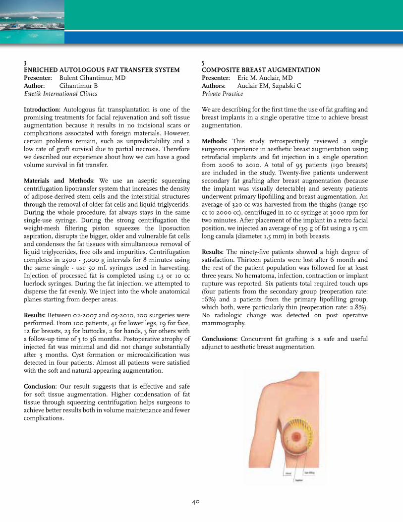
5COmPOSITE BrEAST AUGmENTATIONPresenter: Eric M. Auclair, MDAuthors: Auclair EM, Szpalski CPrivatePractice
We are describing for the first time the use of fat grafting and breast implants in a single operative time to achieve breast augmentation.
methods: This study retrospectively reviewed a single surgeons experience in aesthetic breast augmentation using retrofacial implants and fat injection in a single operation from 2006 to 2010. A total of 95 patients (190 breasts) are included in the study. Twenty-five patients underwent secondary fat grafting after breast augmentation (because the implant was visually detectable) and seventy patients underwent primary lipofilling and breast augmentation. An average of 320 cc was harvested from the thighs (range 150 cc to 2000 cc), centrifuged in 10 cc syringe at 3000 rpm for two minutes. After placement of the implant in a retro facial position, we injected an average of 139 g of fat using a 15 cm long canula (diameter 1,5 mm) in both breasts.
results: The ninety-five patients showed a high degree of satisfaction. Thirteen patients were lost after 6 month and the rest of the patient population was followed for at least three years. No hematoma, infection, contraction or implant rupture was reported. Six patients total required touch ups (four patients from the secondary group (reoperation rate: 16%) and 2 patients from the primary lipofilling group, which both, were particularly thin (reoperation rate: 2.8%). No radiologic change was detected on post operative mammography.
Conclusions: Concurrent fat grafting is a safe and useful adjunct to aesthetic breast augmentation.
40
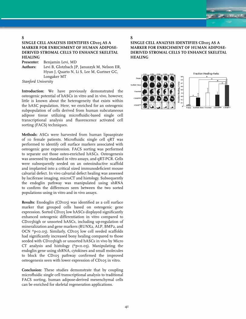
8SINGLE CELL ANALYSIS IDENTIFIES CD105 AS A mArkEr FOr ENrICHmENT OF HUmAN ADIPOSE-DErIvED STrOmAL CELLS TO ENHANCE SkELETAL HEALINGPresenter: Benjamin Levi, MDAuthors: Levi B, Glotzbach JP, Januszyk M, Nelson ER,
Hyun J, Quarto N, Li S, Lee M, Gurtner GC, Longaker MT
StanfordUniversity
Introduction: We have previously demonstrated the osteogenic potential of hASCs in vitro and in vivo, however, little is known about the heterogeneity that exists within the hASC population. Here, we enriched for an osteogenic subpopulation of cells derived from human subcutaneous adipose tissue utilizing microfluidic-based single cell transcriptional analysis and fluorescence activated cell sorting (FACS) techniques.
methods: ASCs were harvested from human lipoaspirate of 10 female patients. Microfluidic single cell qRT was performed to identify cell surface markers associated with osteogenic gene expression. FACS sorting was performed to separate out those osteo-enriched hASCs. Osteogenesis was assessed by standard in vitro assays, and qRT-PCR. Cells were subsequently seeded on an osteoinductive scaffold and implanted into a critical sized immunodeficient mouse calvarial defect. In vivo calvarial defect healing was assessed by luciferase imaging, microCT and histology. Subsequently the endoglin pathway was manipulated using shRNA to confirm the differences seen between the two sorted populations using in vitro and in vivo assays.
results: Enodoglin (CD105) was identified as a cell surface marker that grouped cells based on osteogenic gene expression. Sorted CD105 low hASCs displayed significantly enhanced osteogenic differentiation in vitro compared to CD105high or unsorted hASCs, including up-regulation of mineralization and gene markers (RUNX2, ALP, BMP2, and OCN *p<0.05). Similarly, CD105 low cell seeded scaffolds had significantly increased bony healing compared to those seeded with CD105high or unsorted hASCs in vivo by Micro CT analysis and histology (*p<0.05). Manipulating the endoglin gene using shRNA, cytokines and small molecules to block the CD105 pathway confirmed the improved osteogenesis seen with lower expression of CD105 in vitro.
Conclusion: These studies demonstrate that by coupling microfluidic single cell transcriptional analysis to traditional FACS sorting, human adipose-derived mesenchymal cells can be enriched for skeletal regeneration applications.
8SINGLE CELL ANALYSIS IDENTIFIES CD105 AS A mArkEr FOr ENrICHmENT OF HUmAN ADIPOSE-DErIvED STrOmAL CELLS TO ENHANCE SkELETAL HEALING
41

10AGING-rELATED DECrEASE IN HUmAN ASC ANGIOGENIC POTENTIAL IS rEvErSED BY HYPOXIA PrE-CONDITIONING THrOUGH rOS PrODUCTIONPresenter: Valerie Planat-Benard, PhDAuthors: Planat-Benard V, de Barros S, Dehez S,
Casteilla LUMR5273CNRSUPSEFSInsermU1031
Introduction: Tissue aging is commonly associated with the damaging effects of reactive oxygen species (ROS) accumulation and a decrease in regenerative properties of mesenchymal stem cells in many tissues. However it is also proposed that, redox status may play an important role in stem cell maintenance. Concerning adipose-derived stromal cells (ASC), we previously demonstrated that a 24h treatment with a pro-oxidant fagant improves neovascularisation potential in a mouse model of limb ischemia, suggesting that moderate ROS production improves angiogenic potential of ASC. The present study was designed to compare in vitro and in vivo properties of human ASC during aging as well as the effect of hypoxic pre-conditioning and the role of ROS.
methods: Adipose tissue samples were collected from 20-35 to over 50 years old donors to isolate cells from the stroma-vascular fraction (SVF) and to culture ASC. In vitro ASC properties and in vivo angiogenic potential in a nude mouse model of limb ischemia were estimated. ASC were eventually preconditioned by a 24h exposure to 0.5% O2 before administration.
results: Although, no change in SVF or ASC number, phenotype and proliferation was observed with aging, there is a decrease in ASC ability to differentiate into endothelial cells, to secrete pro-angiogenic and pro-survival factors and to control redox metabolism. In addition, aging impairs the beneficial effect of human ASC in ischemic limb preservation and cutaneous blood flow. Interestingly, a hypoxic-preconditioning that improves redox metabolism and factor secretion was able to restore in vivo angiogenic capacities of ASC from donors aged over 50 years. We demonstrated that the increase in mitochondrial ROS production is crucial in triggering hypoxic preconditioning effect in ASC from aged donors.
Conclusion: Taken together, these results indicate that aging affects oxidative stress of native adipose tissue cells and impairs angiogenic potential of cultured ASC. Nevertheless, short exposure to hypoxia, or at least moderate ROS production reverses adverse effect of aging and improves ASC therapeutic action.
14A NEW AND EASY mETHOD FOr LArGE-vOLUmE FAT GrAFTS - THE BEAULI mETHODPresenter: Klaus Ueberreiter, MD, PhDAuthors: Ueberreiter K, von Finckenstein JG,
Cromme F, Herold C, Tanzella U, Vogt PMAsklepiosClinicBerlinBirkenwerder
Introduction: A new and easy method for large-volume fat grafting to the breast was evaluated in a prospective clinical study with 85 patients in 2 centers in Germany, the overall number of transplantations amounting to 216 treated breasts.
method: Indications were a general lack of breast volume, either genuine or acquired by surgical procedures. The fat was harvested with the BEAULI™ method, which in general consists in harvesting very small fat particles by means of water-jet assisted liposuction (body-jet®, human med AG, Germany) and reinjection of the fat after separation from superfluous water by means of the Lipo-Collector®. All procedures were performed in a standardised protocol, measurements were taken preop, at day 1 postop, after 1 week, 4 weeks, 3 months, 6 months, and then to be continued yearly. Breast MRI`s were taken preop and 6 months postop, the longest follow-up was 30 months.
results: In every case a definite increase of the volume of the fatty layer in the treated areas was observed. The volume control of 35 aesthetic patients by means of BrainLab™ Software and MRI verified a permanent take rate of 76 ± 11% of the grafted fat. In aesthetic patients generally 2 (80%) fat-grafting procedures with an average gain in volume of 1/2 bra cup size or 100 - 150 mL) per procedure per side were required.
Operation time was 1.5 h. No oil cysts and only in 2 cases some palpable subcutaneous nodules which proved to be granulomas were observed. After implant removal, satisfaction was usually reached after only a single procedure, complete reconstruction after cancer surgery required 4 - 5 grafting sessions. An extension of the skin envelope as well as an improvement of existing scars was also observed.
Conclusions: The potential of this method is very promising in cases of definitive implant removal after capsular contracture, after mastectomy, or for corrections of volume deficits.
42
15DE NOvO ADIPOGENESIS BY ImPLANTATION OF TYPE-I COLLAGEN SPONGE INTO rABBITS’ FAT PADPresenter: Wakako Tsuji, MD, PhDAuthors: Tsuji W, Inamoto T, Ito R, Morimoto N, Tabata Y, Toi MKyotoUniversityHospital
Introduction: Adipogenesis for breast reconstruction is expected for patients who have undergone breast surgery. We have confirmed adipogenesis in mice by implanting a type-I collagen sponge with controlled-release FGF2 and human adipose tissue-derived stem cells (Tissue Engineering, Part A, 2009). For clinical application, FGF2 is not always available at present while a larger size of adipose tissue is needed. We aim to regenerate larger amounts of adipose tissue without FGF2 in rabbits.
method: Under general anesthesia, a cage made of polypropylene mesh was implanted into the rabbits’ bilateral fat pad. The size of cage was 2 cm in diameter and 1 cm in height. Minced type-I collagen sponge (PELNAC®, Gunze Co. Ltd., Tokyo, Japan) was injected into the cage. Adipogenesis in the cage was measured with ultrasonography (USG), and the cage were harvested 3, 6, 12 months after the implantation. Histology of the specimen was assessed with H-E stain.
results: With USG, solid mass in the cage gradually increased. Histologically, adipose tissue had regenerated entirely inside the cage (approximately 3.1 mL) 12 months after the implantation.
Conclusions: We found de novo adipogenesis 12 months after the implantation only by implanting a type-I collagen sponge inside the space. USG is a non-invasive and useful method to assess inside the cage. It is suggested that this simple method could be a promising way for clinical application of de novo adipogenesis.
17LIPOmODELLING OF BrEAST rECONSTrUCTION CONTOUr DEFOrmITIES: USE OF STEm-CELL ENrICHED FAT GrAFTSPresenter: Amir Sadri, MDAuthors: Akhavani M, Sadri A, Mosahebi ARoyalFreeHospital
Introduction: Secondary procedures after breast reconstruction are common particularly correction of contour deformities. Autologus fat transfer has been a valuable tool to address this to good effect but needs regular touch up procedures. We compared using ‘stem cell-enriched’ fat grafts to standard fat grafting techniques for volume retention.
method: We audited 20 patients who needed contour defect filling post breast reconstruction for breast cancer by a single surgeon. The patients were randomised to standard fat grafting or stem-cell enriched fat grafts. All patients had 3dimensional volumetric Torso Scans pre-operative and post-operative at 6 weeks and 6 months.
results: There were no detectable difference in the fat volume retained between the two groups at 6 weeks and 6 month post-operative. No complications were detected.
Conclusion: In our series, stem-cell enriched fat graft does not seem to produce a more superior graft take rate when compared with the standard technique. However, larger volumes of fat were injected using the Cytori method which may lead to patients requiring fewer “top-up” procedures.
43

19NOvEL IN vITrO CULTUrE CONDITIONS OF ADIPOSE STEm CELLS FOr CLINICAL CELL THErAPY APPLICATIONSPresenter: Mimmi Patrikainen, MSAuthors: Patrikainen M, Juntunen M, Suuronen R,
Miettinen S, Mannerstrom BUniversityofTampere
Human adipose tissue is an attractive and abundant source of multipotent stem cells. Human adipose stem cells (ASCs) have shown to have therapeutic relevancy in diverse clinical applications. Nevertheless, expansion of ASCs is often necessary prior to performing clinical studies. Standard in vitro cell expansion techniques utilize animal-derived reagents as part of the cell culture workflow which is not recommended in clinical cell therapy applications due to safety issues. Human cells exposed to animal-derived products may trigger a severe immune response in the recipient upon transplantation. By replacing animal-derived cell culture reagents with reagents of non-animal origin, safety and quality of the transplanted stem cells can be enhanced. In the study, an animal-free workflow for the expansion of ASCs was developed and assessed by investigating the stem cell immunophenotype, long-term self-renewal capacity and multilineage differentiation potential into bone-, fat- and cartilage-like cells.
Furthermore, preliminary experiments of immunogenicity were also performed. The results showed, that the new conditions maintained the stem cell characteristics of ASCs by retaining the cells self-renewal capacity, stem cell immunophenotype and differentiation potential. Also, the preliminary results from immunogenic evaluation of ASCs showed that the new conditions elicited a very low immunogenic response in ASCs. The results suggest that ASCs expanded using an animal-free workflow have great potential in clinical cell therapy, but further safety assessments must be performed. If ASCs expanded in the new conditions proved to be non-immunogenic, they could be used as off-the-shelf products in clinical cell therapies which would provide wide possibilities for cell therapy applications. Since clinical cell therapy studies using ASCs are under way, a strong focus on the safety, reproducibility and quality of the stem cells is urgently called for.
21FAT GrAFT LONGEvITY: rOLE OF FAT PrOCESSING IN mAINTAINING vIABLE CELLSPresenter: Alexandra Conde Green, MDAuthors: Conde Green A, Rodriguez RL, McLenithan J,
Slezak SUniversityofMarylandMedicalCenter
Introduction: The use of adipose tissue for treatment of soft tissue asymmetry and depressions is very attractive for being readily available however the results of fat transplantation remain unpredictable. To this day, there is no agreement as to the best way of processing fat to ensure maximum take and viability of the graft. Therefore, to practically understand what happens when we manipulate adipose tissue in the operating room, we studied the most common methods of fat processing in order to see which method yields the highest quantity of viable adipose derived cells.
methods: Fat harvested manually from the lower abdomen of twenty patients was separated and processed by sedimentation, washing and centrifugation at 1,256 g for 3 minutes. The middle layer of each processed sample was analyzed by histological techniques after periodic acid schiff staining for viable adipocyte count. Then, after enzymatic digestion, the middle layer of each lipoaspirate and the stromal vascular fraction (SVF) of centrifuged samples were stained with PI, incubated with monoclonal antibodies and analyzed by flow cytometry and culture for quantification of viable adipose derived stem cells.
results: Intact nucleated adipocyte count was significantly greater in sedimented lipoaspirates, where centrifuged samples showed a great majority of disruptured adipocytes. Quantification of endothelial and mesenchymal stem cells showed a great loss in the middle layer of centrifuged lipoaspirates (usually used for grafting) as compared to washed and sedimented lipoaspirates. Additionally, the SVF collected at the bottom of the centrifuged samples showed the highest concentration.
Conclusion: In our comparative study, washing seemed to be the best processing technique for aspirated fat when needed for grafting, as it maintains the quantity of viable adipocytes, endothelial and mesenchymal stem cells. However if centrifugation were to be used as a way of concentrating the fat, it would be best to collect the stem cells in greater quantity in the SVF and add them to a viable adipose tissue scaffold in order to increase graft survival.
44
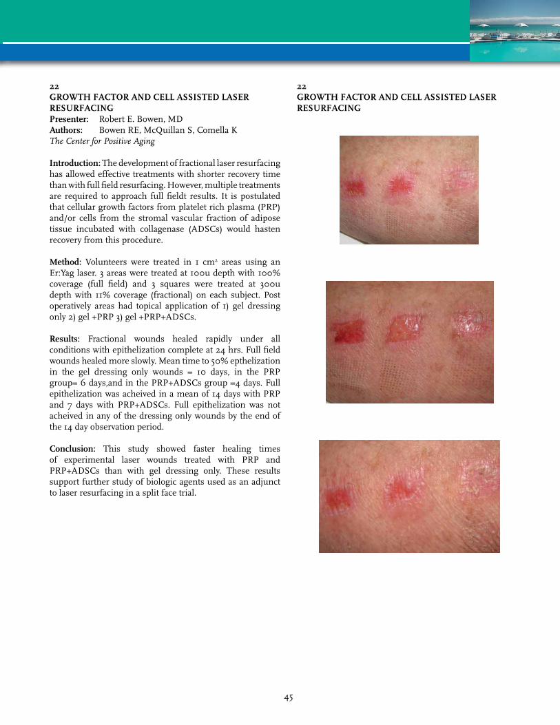
22GrOWTH FACTOr AND CELL ASSISTED LASEr rESUrFACINGPresenter: Robert E. Bowen, MDAuthors: Bowen RE, McQuillan S, Comella KTheCenterforPositiveAging
Introduction: The development of fractional laser resurfacing has allowed effective treatments with shorter recovery time than with full field resurfacing. However, multiple treatments are required to approach full fieldt results. It is postulated that cellular growth factors from platelet rich plasma (PRP) and/or cells from the stromal vascular fraction of adipose tissue incubated with collagenase (ADSCs) would hasten recovery from this procedure.
method: Volunteers were treated in 1 cm2 areas using an Er:Yag laser. 3 areas were treated at 100u depth with 100% coverage (full field) and 3 squares were treated at 300u depth with 11% coverage (fractional) on each subject. Post operatively areas had topical application of 1) gel dressing only 2) gel +PRP 3) gel +PRP+ADSCs.
results: Fractional wounds healed rapidly under all conditions with epithelization complete at 24 hrs. Full field wounds healed more slowly. Mean time to 50% epthelization in the gel dressing only wounds = 10 days, in the PRP group= 6 days,and in the PRP+ADSCs group =4 days. Full epithelization was acheived in a mean of 14 days with PRP and 7 days with PRP+ADSCs. Full epithelization was not acheived in any of the dressing only wounds by the end of the 14 day observation period.
Conclusion: This study showed faster healing times of experimental laser wounds treated with PRP and PRP+ADSCs than with gel dressing only. These results support further study of biologic agents used as an adjunct to laser resurfacing in a split face trial.
22GrOWTH FACTOr AND CELL ASSISTED LASEr rESUrFACING
45

23ADIPOGENESIS USING HUmAN ADIPOSE TISSUE-DErIvED CELLS COmBINED WITH COLLAGEN/ GELATIN SCAFFOLD ImPrEGNATED WITH BASIC FIBrOBLAST GrOWTH FACTOrPresenter: Ran Ito, MDAuthors: Ito R, Morimoto N, Tsuji W, Nakamura Y,
Kawai K, Suzuki SKyotoUniversityHospital
Introduction: We have developed collagen/gelatin scaffold (CGS) that can provide the sustained release of basic fibroblast growth factor (bFGF). We proved that CGS impregnated with the appropriate dosage of bFGF accelerates dermis-like tissue formation two or three times earlier than existing artificial dermis. In this study, we disseminated adipose tissue-derived cells on CGSs impregnated with bFGF.
method: Human adipose tissue-derived cells were primarily isolated from human adipose tissues that were obtained in breast cancer surgery with informed consent at Kyoto University Hospital. Cells were isolated from collagenase digests of adipose tissue. We impregnated CGSs (8 mm in diameter, 3 mm in thickness) with bFGF (1 µg/cm2, 14 µg/cm2) or normal saline. Then, we disseminated cells (passage 3) on CGSs at a seeding density of 1 x 10⁵ cells/cm2 and implanted them into the back subcutis of nude mice. Six weeks after implantation, adipogenesis at the administered site was evaluated.
results: Matured adipose tissue was observed in all groups histologically. The weight of regenerated adipose tissue was largest in the 1 µg/cm2 of bFGF group.
Conclusions: Implantation of collagen sponges disseminated with human adipose tissue-derived cells and controlled release of bFGF was reported to achieve significantly high amounts of adipose tissue newly formed compared with the solution injection of bFGF at a higher dose. In this study, we showed that our CGS could be used as a scaffold that could sustain bFGF with adipose tissue-derived cells for adipogenesis.
24EFFECTIvENESS OF 1064 Nm LASEr LYPOLISYS IN QCW mODE AS AN ADDITIONAL APPrOACH TO TUmESCENT LIPOSUCTION TECHNIQUEPresenter: Dmitry V. Melnikov, MDAuthors: Melnikov DV, Sidorenkov DA, Iskornev AANationalResearchCenterofSurgery
Paper Withdrawn
46

25BED SIDE ISOLATION OF ADIPOSE DErIvED STEm CELLS WITHIN THE OPErATION rOOm, COLLAGENAGE FrEE, FOr AUTOLOGOUS FAT TISSUE TrANSPLANT: 2 YEArS EXPErIENCE Presenter: Hebert T. Lamblet, MDAuthor: Lamblet HTVikaaraKlinik
Goal/Purpose: Besides the fact that fat grafting gained popularity, isolation of these cells within the OR and their immediate use for fat transplantation still remains a challenge. The purpose of this study is to present the possibility of bed side isolation of adipose derived stem cells in combination with fat grafting within the OR, without the use of collagenase.
methods/Technique: Adipose tissue is collected from the abdomem of patients undergoing liposuction. The method consists in washing the aspirated fat with a solution of Dulbecco’s phosfate-buffed saline at equal volume, 3 times and draining the decanted part. The supernatant is reserved and the decanting part that consists the Stromal Vascular Fraction is submitted to the method. The method consists in centrifuging, add red blood cell lysing buffer and use a 100 nm cell stainer to obtain a adipose derived stem cell pellet from the stromal vascular fraction and than add the pellet to the reserved fat, for immediate use. The presence of mesenchymal stem cells isolated in the pellet was confrimed by Indirect Immunofluorescence and Flow Cytometer analysis. These cells expressed several CD marker antigens for mesenchymal linage such as: CD29, CD44, CD71, et and none for hematopoietic linage
results/Complications: From February 2002 to October 2010, 310 patients benefited from autologous fat transplantation in combination with bed side adipose derived stem cell isolation. Average lipoaspirate was 200 ml. The donor site was the abdomen. An average of 40 to 50 millions of mesenchymal cells/100 ml of processed lipoaspirate were isolated with this method. The average follow up was 2 years. The resorption rate after 2 years was approximately 30 to 40% of the injected volume. The whole isolation process lasted around 40 min and was conducted by a trained nurse under supervision.
Conclusion: Up to now, adipose derived stem cells isolation was done exclusively in the laboratory or using expensive processing machines and collagenase. This method has shown to be reproducible and could be an alternative for the pluripotent use of these cells in a more safe and cost effective manner.
26PrOGrESS TOWArDS THE TrANSLATION OF A TISSUE-ENGINEErED vASCULAr GrAFT INTO THE CLINICAL SETTING: THE USE OF AUTOLOGOUS HUmAN PLASmA IN CULTUrE AND CrYOPrESErvATIONPresenter: Ping Zhang, PhDAuthors: Zhang P, Jimbo M, Tulenko T, Hall H,
Adams J, Rao A, Eisenberg J, DiMuzio PThomasJeffersonUniversity
Background: We recently described the success of a tissue-engineered vascular graft (TEVG) created with autologous adipose-derived stem cells (ASC) in an animal model. To translate this work into clinical usage, we investigate the effect of replacing the fetal bovine serum (FBS) with autologous human plasma (HP) within the culture medium as well as the effect of cryopreservation on graft creation and differentiation of ASC.
methods and results: Human ASC were isolated from the peri-umbilical fat from patients undergoing elective vascular surgery (n=6); simultaneously, autologous HP was isolated from the peripheral blood. To assess the effect of replacing the FBS with HP in the culture medium, ASC were grown in Endothelial Growth Medium (EGM2) supplemented with FBS (2%) vs. HP (2%). After 14d, we observed increased proliferation (1.3 fold, NS) in HP over FBS. Endothelial differentiation was evaluated by qPCR (CD31, vWF, CD144), up-take acLDL, and cord formation; in both media, ASC acquired each of these EC characteristics. ASC were subsequently seeded into vascular scaffolds and subjected to increasing shear force within bioreactor (0-9dynes x5d) to evaluate their use in creating a TEVG; confocal microscopy revealed complete luminal coverage of the graft surface and alignment in the direction of flow by cells cultured in both media. Finally, the effect of cryopreservation was assessed. ASC cultured in EGM2/HP x14d were cryopreserved in DMSO (5% + 95% HP) x 4d; subsequent proliferation was equivalent to non-frozen controls. Similarly, TEVG created with ASC cultured in EGM2/HP were cryopreserved x10d; subsequent confocal microscopy revealed a confluent luminal lining aligned in the direction of fluid flow.
Conclusions: The studies suggest: 1) replacing FBS in culture medium with autologous human plasma does not affect endothelial differentiation nor the ability to use ASC as EC substitutes in vascular tissue engineering; and 2) TEVG created with ASC remain intact after cryopreservation, indicating that this method of preservation will be useful in making the graft readily available to implanting surgeons.
47

29rETAINING AUTOLOGOUS FAT IN FAT GrAFTING: A CADAvEr STUDY OF FACIAL mUSCLE INJECTIONPresenter: Donald M. Fox, MDAuthors: Fox DM, Amar RE, Balin AKNewYorkEyeandEarInfirmary
Introduction: Predictable retention of injected fat is a challenge for facial fat grafting, as it is for other areas of the body. Reliable revascularization of free tissue grafts is dependent on a well vascularized graft bed; for facial fat grafting, the muscles of facial expression are the most vascular anatomic structures available. Animal studies have clearly shown high survival rates for fat engrafted into muscle suggesting a strategy for autologous facial fat grafting in human cometic and reconstructive cases. Precisely targeting facial muscles requires a particular approach dictated by anatomic considerations. Avoidance of unnecessary trauma to muscle tissue is as important as accurate cannulation.
method: A blue gel (DAP hair gel + methylene blue) was used to cannulate and inject three facial muscles of a thawed frozen cadaver: corrugator, zygomatic major, and depressor labii inferioris. Dissection was performed immediately after injection to demonstrate the location of the blue gel. The process was video recorded.
results: Facial muscles can be reliably cannulated by beginning the entry at the origin or insertion of the muscle where it is relatively fixed, and passing the cannula within the fascial sheath and in the plane and direction of its fibers: the blue gel was found entirely within the muscle fibers. The injection is guided by visual and tactile cues as well as knowledge of facial anatomy.
Conclusion: Systematic injection of the facial muscles requires a particular approach if their vascular beds are to be used to advantage. Precise targeting and minimalization of trauma can be acheived using an anatomically-based approach, as demonstrated.
30STABILIZATION OF vASCULAr NETWOrk FOrmED BY ENDOTHELIAL CELLS ON mONOLAYEr OF ADIPOSE STrOmAL CELL IS rEGULATED BY TGFPresenter: Dmitry O. Traktuev, PhDAuthors: Traktuev DO, Merfeld-Clauss S, Feng D,
Gollahalli N, March KLIndianaUniversity
Multiple studies have shown that adipose stromal cells (ASC) produce cocktail of angiogenic and anti-inflammatory factors, which potentially are responsible for ASC therapeutic activities. Additionally, ASC possess functional properties of pericytes: support vascular-like network formations (VNF) by endothelial cells (EC) in vitro, establish functional vessels when co-implanted with EC in vivo. We demonstrated in in vitro model that VNF by EC was dependent on direct contact of EC with ASC and was significantly promoted by factors secreted by ASC.
In the present study we evaluated effect of EC-ASC direct contact on ASC paracrine activity. Using in vitro co-culture model we showed that VNF was associated with ASC transformation from ?-smooth muscle actin (?SMA) negative into ?SMA positive cells. In the same time it caused significant decrease in angiogenic potency of ASC: conditioned media (CM) collected from EC-ASC cultures had diminished potency to support VNF, had no effect on EC survival, and prevented EC migration as observed in case of CM collected from ASC monocultures. Analysis of CM revealed significant decrease in VEGF and HGF accumulation in EC-ASC cultures compare to ASC monocultures. The similar effects were observed when ASC were pre-treated for six days with TGF?. Comparative analysis of ASC and EC-ASC CM showed that the concentrations of active TGF? in both cultures were similar, but the concentrations of inactive form of TGF? in EC-ASC CM was more than 2.2 times higher than in ASC CM. We found that the majority of TGF? was produced by EC, where as ASC produced very limited amount of TGF? with no change as a result of interaction with EC. Blocking of serine proteases activities (responsible for TGF? activation) led to significant decrease in ?SMA upregulation.
Based on this study we hypothesize that while co-delivery of EC with ASC can be seen as an opportunity for efficient and timely revascularization of ischemic tissues by providing critical cell components of the mature vessel, this strategy potentially has negative effects: significant decrease in duration and degree of ASC paracrine activity.
48

31OPTImIZATION OF OSTEOGENIC mEDIUm COmPONENTS FOr HUmAN ADIPOSE STEm CELLSPresenter: Laura Tirkkonen, MScAuthors: Tirkkonen L, Haimi S, Mannerstrom B,
Suuronen R, Miettinen SUniversityofTampere
Several in vitro studies have demonstrated that human adipose stem cells (hASCs) undergo osteogenic differentiation when cultured in the presence of inducing components such as L-ascorbic acid 2-phosphate (Asc 2-P), dexamethasone (Dex), and fl-glycerophosphate. However, the osteogenic medium was initially generated for the differentiation of bone marrow derived stem cells and the concentrations of osteogenic supplements may not be optimal for the osteogenic differentiation of hASCs.
The possible risks involved in the use of fetal bovine serum (FBS) have lead to the use of human serum (HS) and the development of different serum-free (SF) and xeno-free (XF) media. Previous studies have reported that hASCs cultured in SF/XF medium or media containing FBS or HS have significantly different effects on hASC proliferation and differentiation, and therefore may require different concentrations of osteogenic components for optimal differentiation. In this study, the effects of three different compositions of osteogenic components (OM1, OM2, and OM3) in FBS, HS, and SF/XF medium on hASC osteogenic differentiation were compared. The concentrations used were 50 µM Asc 2-P and 100 nM Dex for OM1, 150 µM Asc 2-P and 10 nM Dex for OM2, and 250 µM Asc 2-P and 5 nM Dex for OM3. The concentration of ß-glycerophosphate was constant. The results showed that the novel osteogenic medium OM3 with highest Asc 2-P and lowest Dex concentration enhanced alkaline phosphatase activity and mineralization of hASCs in FBS and HS compared to OM1 and OM2. The traditionally used osteogenic medium, OM1, did not support osteogenic differentiation of hASCs more than the control medium in FBS or HS. All OM compositions supported osteogenic differentiation of hASCs cultured in SF/XF medium in some degree, but further optimization is required for serum-free conditions. Our results suggest that in HS and FBS cultures osteogenic medium with high Asc 2-P (250 µM ) and low Dex (5 nM) should be used in order to attain efficient osteogenic differentiation of hASCs in vitro.
32SELECTION AND PrOLIFErATION OF ADIPOSE DErIvED PErI-vASCULAr STEm CELLS (PSCS) USING HIGH THrOUGHPUT POLYmEr mICrO-ArrAY SCrEENINGPresenter: Christopher C. West, BMedSci, MBChB, MRCSAuthors: West CC, Medine CN, Wu M, Stewart KJ, Bradley M, Peault B, Hay DCTheUniversityofEdinburgh
Introduction: Mesenchymal Stem Cells show great promise for therapies in regenerative medicine. Until recently the in vivo location of this heterogenous cell population was poorly understood and remained elusive. Evidence now demonstrates that these stem cells reside in a perivascular niche in all foetal and adult tissues - perivascular stem cells (PSC) - allowing their prospective isolation. Adipose tissue is rich in PSC, and due to its relative abundance and the ease with which it can be harvested, much research has focused on this source and type of stem cell. Before the therapeutic potential of these cells can be utilised, methods for their safe and efficient extraction, purification and expansion need to be realised and optimised. Here we demonstrate that using high throughput screening of a library of FDA approved polymers we identified specific substrates that selectively bind, enhance proliferation and maintain the phenotype of PSCs.
methods: PSCs were isolated from human lipoaspirate using Fluorescence Activated Cell Sorting (FACS) (CD34-, CD45-, CD146+). Polymer micro-array screens were prepared by the printing of microscopic dots of a library of 382 polymers in quadruplicate on to the surface of an agarose coated standard microscope slide. PSCs were cultured on each slide and subsequently fixed at 24 hours (short screen) and 7 days (long screen). Cells were stained with DAPI to demonstrate cell number, and CD146 and ?SMA to demonstrate maintenance of PSC phenotype.
results: Short screens identified specific polymers that rapidly and selectively bound PSCs, long screens demonstrated specific polymers that bound PSCs, aided proliferation and maintained the PSC phenotype in culture (p<0.05).
Conclusions: Our results add to the body of evidence that synthetic polymers can be used in the identification, isolation and proliferation of stem cells. This study identifies unique polymers that show specific bioactive properties in relation to the binding and proliferation of PSCs. Potential uses for this include novel isolation techniques, and synthetic bioactive supports for the in vivo application of PSCs.
49

34TOPICAL APPLICATION OF mESENCHYmAL STEm CELLS TO SOmATIC OrGANSPresenter: PK Lam, PhDAuthors: Lam PK, Ng CF, To KF, Ng SS, Mak TW,
Chan ES, Lo AW, Lai FM, Poon WS, Lai PBTheChineseUniversityofHongKong
Background: Multipotent mesenchymal stem cells (MSCs) have been shown to be capable of repairing damaged organs and tissues in vivo. Migration of MSCs into target tissue, known as homing, is an essential first step of tissue repair and regeneration. Currently homing can be performed in vivo via intravascular injections or direct syringe implantation of MSCs. However their drawbacks include low engraftment rate, direct syringe injury, migration of MSCs to non-targeted sites and thromboembolic risks.
Objective: We aim to study whether mesenchymal stem cells (MSCs) can home to the damaged tissue when the cells are applied to the surfaces of the diseased organs.
methods: MSCs were established from the subcutaneous adipose tissue of transgenic green fluorescent protein (GFP) Sprague-Dawley (SD) rats. Ischemic reperfusion injury (IRI) was induced in the liver, kidney and small intestine of SD rats. Approximately 4 x 10⁶ GFP-MSCs suspension were added to the surfaces of the IRI organs. A thin layer of fibrin was then applied to hold the MSCs in position.
results: Few days after cell transplant, homing of the topically applied MSCs was confirmed by immunohistochemical staining using anti-GFP.
Conclusion: Topically applied MSCs can home to the damaged tissue of the diseased organ.
35ADIPO-INDUCTIvE DECELLULArIZED ADIPOSE TISSUE (DAT) mICrOCArrIErS FOr ADIPOSE-DErIvED STEm CELL EXPANSION AND INJECTABLE CELL DELIvErYPresenter: Lauren E. Flynn, PhDAuthors: Flynn LE, Yu C, Bianco J, Turner AEQueensUniversity
Our group is engineering 3-D scaffolds from the extracellular matrix of human adipose tissue for soft tissue regeneration. We have shown that decellularized adipose tissue (DAT) provides an inductive microenvironment for the adipogenic differentiation of human adipose-derived stem cells (ASCs).
In this study, we developed methods for fabricating porous matrix-derived microcarriers from human DAT, as biodegradable, injectable cell delivery vehicles for culturing human ASCs. In brief, photo-crosslinkable DAT microcarriers were fabricated from pepsin-solubilized DAT using an air-jet droplet technique or electrospraying. In vitro swelling and stability studies over 4 weeks showed that the microcarriers were structurally robust, and injectable through 18-gauge needles. In vitro proliferation studies with human ASCs (Passage 2; 10,000 ASCs/mg microcarriers, 15 RPM) in a CELLSPIN dynamic culturing system (Integra Biosciences) showed greater cell attachment and proliferation on the DAT microcarriers, as compared to gelatin microcarrier controls. Adipogenic differentiation studies were subsequently conducted within the systems.
Interestingly, the adipo-inductive effects of the DAT were preserved, suggesting a compositional effect from the DAT matrix. The ASCs on the DAT microcarriers had the highest levels of glycerol-3-phosphate dehydrogenase (GPDH) enzyme activity, with increasing levels from 72 h to 14 days. End point RT-PCR analysis of adipogenic gene expression demonstrated the highest levels of expression of the master regulators PPARg and CEPBa, as well as lipoprotein lipase (LPL), within the induced DAT microcarrier group. Further, the GPDH enzyme and gene expression results, as well as Oil Red O staining of intracellular lipid, indicate that the DAT microcarriers naturally induce ASC adipogenesis even in the absence of stimulating differentiation factors.
Preliminary in vivo studies of unseeded and ASC-seeded DAT microcarriers in a subcutaneous Wistar rat model confirmed scaffold biocompatibility and indicate promise for applications in injectable ASC delivery and for small volume augmentation procedures.
50

35ADIPO-INDUCTIvE DECELLULArIZED ADIPOSE TISSUE (DAT) mICrOCArrIErS FOr ADIPOSE-DErIvED STEm CELL EXPANSION AND INJECTABLE CELL DELIvErY
39CrYOPrESErvATION OF ADIPOSE TISSUE (AT) AND ADIPOSE-DErIvED STEm CELLS (ASCS) - NEW PErSPECTIvESPresenter: Henk Snyman, MDAuthor: Snyman HCryoSaveAG
Recent technological advances, including controlled rate freezing protocols, the use of cryoprotectant agents and improved quality control parameters, have validated the viability and quality of AT and ASCs after cryopreservation and thawing. This has led to the accreditation and licensing of the first adipose tissue banks where Plastic and Cosmetic Surgeons can now store adipose tissue as well as ASCs. Harvesting of small volume samples can be done under local anesthesia in an outpatient setting or as the initial part of routine liposuction procedures for larger volume samples. The current state of the art for collection, transport, processing and release of samples for re-injection will be discussed. This paper will review the experience to date, the regulatory framework as well as potential benefits and pitfalls. Cryopreservation in vapor phase liquid nitrogen effectively stops the biological clock and thus offers interesting potential for future autologous applications with 51 younger cells.
51

40LONG-TErm OUTCOmES FOLLOWING FAT GrAFTING IN ImPLANT-BASED BrEAST rECONSTrUCTION: A COmPArATIvE ANALYSISPresenter: Akhil K. Seth, MDAuthors: Seth AK, Hirsch EM, Fine NANorthwesternUniversity
Introduction: Autologous fat grafting (FG) has become a common revisional procedure for aesthetic defects following implant-based breast reconstruction. However, the long-term oncologic implications of injecting fat within a previously malignant field have not been reported. This study evaluates and compares long-term outcomes following tissue expander/implant breast reconstruction with and without revisional FG.
methods: Retrospective review of 886 consecutive patients (1202 breasts) undergoing mastectomy with immediate implant reconstruction from 4/1998-8/2008 at one institution was performed. Demographic, operative, oncologic, and postoperative factors were recorded, including the use of revisional FG after reconstruction. Chi-square, Students t-test, and multiple linear regression were used for statistical analysis.
results: There were no significant differences in demographics, operative characteristics, tumor staging, or exposure to radiation therapy between FG (n=90 breasts) and non-FG (n=1112 breasts) patients. FG use did not affect local tumor recurrence or survival when compared to non-FG breasts (1.1% v. 1.5%; 100.0% v. 95.5%, respectively). Overall mean follow-up was 38.4 months, including 24.8 months following FG. Ninety-nine FG procedures were performed an average of 9.7 months following tissue expander/implant breast reconstruction, with no documented complications. Having a poor cosmetic result was an independent predictor of undergoing revisional FG (p<0.0001).
Discussion: Our comparative analysis, the largest to date, suggests that FG after breast reconstruction does not adversely affect the rate of local tumor recurrence or survival on long-term follow-up. Surgeons may continue to utilize autologous FG as an aesthetic adjunct to implant-based reconstruction with minimal postoperative complications. However, these preliminary results also indicate the need for multi-institutional reviews and prospective studies to definitively establish its oncologic safety.
41COmPArISON OF ADIPOSE-DErIvED STrOmAL CELLS AND BONE mArrOW mESENCHYmAL STrOmAL CELLS FrOm THE SAmE HEALTHY INDIvIDUALS - A PHENOTYPIC, FUNCTIONAL, TrANSCrIPTOmIC AND EPIGENETIC STUDYPresenter: Philippe Bourin, MD, PhDAuthors: Bourin P, Hebraud B, Chaput B, Peyrafitte JA,
Gadelorge M, Espagnolle N, Huynh A, Roussel M, Attal M, Collas P, Casteilla L, Planat-Benard V
STROMALab
The relation between ASC and MSC is still a matter of discussion. Many studies had compared those 2 cell types but mostly the cells came from different individuals and/or are cultured in different ways. In the present study we compared ASC and MSC obtained from adipose tissue (AT) and bone marrow (BM) of the same 15 healthy donors and cultured in the same medium.
The cells from the stromal vascular fraction (SVF) were obtained by digestion of AT and centrifugation. Unprocessed BM cells and crude SVF were cultured in alpha-MEM + 10% FCS for 3 passages. The culture conditions were kept as closed as possible for both sources. Before culture and after each passage the CFU-F frequency and population doubling (PD) were evaluated. All the subsequent comparisons were made at an equivalent number of PD. We analyzed the phenotype, the adipose, osteoblastic and chondrocytic differentiation and the hematopoietic supporting activity (HSA). Transcriptomic and promoter methylation analysis was performed with microarrays (Gene 1.0 ST and HG18 RefSeq).
The progenitor frequency as assessed by CFU-F was 330 times higher in the SVF. For the primary culture, the number of PD was statically lower for ASC compared to BM (9 ± 0.6 vs 13 ± 0.3 respectively). There was no significant differences in the osteoblastic differentiation potential, ASC significantly better differentiate in adipocytes and MSC in chondrocytes. The HSA of both cell types was overall the same. While both cell type expressed standard stromal markers (CD73, CD90, CD105), we observed differences in the expression of CD106, CD146, CD49a (MSC>ASC), CD10, CD26, CD157, CD36 (ASC>MSC). The transcriptome clearly differentiate ASC from MSC with more than 450 genes differentially expressed, of them more than 50% corresponded to extracellular region (matrix and secreted) or to membrane associated molecules. Signaling in the immune system and hemostasis are the most enriched pathways. Finally ASC and MSC shared more than 80% of methylated promoters.
In conclusion, despite similarities in phenotype and general properties ASC and MSC are clearly distinct cell types.
52

41COmPArISON OF ADIPOSE-DErIvED STrOmAL CELLS AND BONE mArrOW mESENCHYmAL STrOmAL CELLS FrOm THE SAmE HEALTHY INDIvIDUALS - A PHENOTYPIC, FUNCTIONAL, TrANSCrIPTOmIC AND EPIGENETIC STUDY
42LL-37 mODULATES HUmAN ADIPOSE-DErIvED STEm CELLS PrOLIFErATION THrOUGH INTErLEUkIN-8 (IL-8)-DEPENDENT mECHANISmPresenter: SaIk Bang, PhDAuthors: Seon MR, Yang YH, Shim SK, Choi HJ,
Cho DH, Bang SISamsungMedicalCenter
Introduction: Recent studies have reported that adipose-derived stem cells (ASCs) accelerated wound healing by stimulation of re-epithelialization, angiogenesis, and proliferation. ASCs release cytokines and chemokines, and these factors can promote ASCs proliferation of cutaneous wounds. LL-37, member of the cathelicidin family, induces wound healing, proliferation, and migration of epithelial cells. This study examined the effects of the LL-37 on proliferation of ASCs.
method: Cultured ASCs were treated with LL-37 or interleukin-8 (IL-8) for specific periods of time. The CCK-8 assay was performed to determine the proliferation of ASCs. RT-PCR and ELISA were conducted to analyze the expression of IL-8 levels. Western blot analysis was performed revealed that LL-37-induced proliferation involve MAPK pathway.
results: We demonstrated that LL-37 markedly stimulated ASCs proliferation. LL-37 increased the levels of IL-8 mRNA and protein in dose-dependent manner. In addition, IL-8 increased the proliferation of ASCs in a time- and dose-dependent manner. Functional blocking antibodies for IL-8 suppress the proliferation of LL-37-increased ASCs proliferation. LL-37 increased phosphorylation levels of c-Jun NH2-terminal kinase (JNK) and p38 mitogen-activated protein kinase (p38), and pretreatment of the cells with the JNK inhibitor or the p38 MAPK inhibitor abrogated the LL-37-induced proliferation, and IL-8 production.
Conclusions: We demonstrated that LL-37 may play a role in cutaneous wound healing by enhancing production of IL-8 and proliferation via the activation of JNK and P38 MAPK in human ASCs.
53
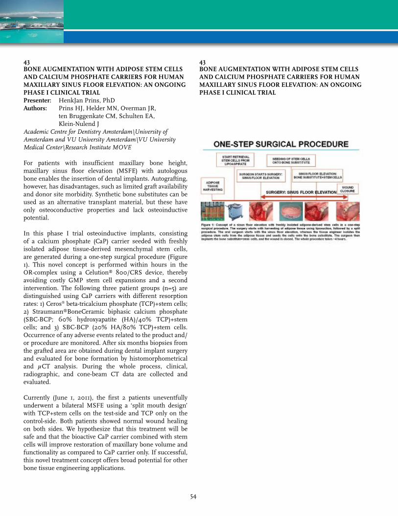
43BONE AUGmENTATION WITH ADIPOSE STEm CELLS AND CALCIUm PHOSPHATE CArrIErS FOr HUmAN mAXILLArY SINUS FLOOr ELEvATION: AN ONGOING PHASE I CLINICAL TrIALPresenter: HenkJan Prins, PhDAuthors: Prins HJ, Helder MN, Overman JR,
ten Bruggenkate CM, Schulten EA, Klein-Nulend J
AcademicCentreforDentistryAmsterdam\UniversityofAmsterdamandVUUniversityAmsterdam\VUUniversityMedicalCenter\ResearchInstituteMOVE
For patients with insufficient maxillary bone height, maxillary sinus floor elevation (MSFE) with autologous bone enables the insertion of dental implants. Autografting, however, has disadvantages, such as limited graft availability and donor site morbidity. Synthetic bone substitutes can be used as an alternative transplant material, but these have only osteoconductive properties and lack osteoinductive potential.
In this phase I trial osteoinductive implants, consisting of a calcium phosphate (CaP) carrier seeded with freshly isolated adipose tissue-derived mesenchymal stem cells, are generated during a one-step surgical procedure (Figure 1). This novel concept is performed within hours in the OR-complex using a Celution® 800/CRS device, thereby avoiding costly GMP stem cell expansions and a second intervention. The following three patient groups (n=5) are distinguished using CaP carriers with different resorption rates: 1) Ceros® beta-tricalcium phosphate (TCP)+stem cells; 2) Straumann®BoneCeramic biphasic calcium phosphate (SBC-BCP; 60% hydroxyapatite (HA)/40% TCP)+stem cells; and 3) SBC-BCP (20% HA/80% TCP)+stem cells. Occurrence of any adverse events related to the product and/or procedure are monitored. After six months biopsies from the grafted area are obtained during dental implant surgery and evaluated for bone formation by histomorphometrical and µCT analysis. During the whole process, clinical, radiographic, and cone-beam CT data are collected and evaluated.
Currently (June 1, 2011), the first 2 patients uneventfully underwent a bilateral MSFE using a ‘split mouth design’ with TCP+stem cells on the test-side and TCP only on the control-side. Both patients showed normal wound healing on both sides. We hypothesize that this treatment will be safe and that the bioactive CaP carrier combined with stem cells will improve restoration of maxillary bone volume and functionality as compared to CaP carrier only. If successful, this novel treatment concept offers broad potential for other bone tissue engineering applications.
43BONE AUGmENTATION WITH ADIPOSE STEm CELLS AND CALCIUm PHOSPHATE CArrIErS FOr HUmAN mAXILLArY SINUS FLOOr ELEvATION: AN ONGOING PHASE I CLINICAL TrIAL
54

44HUmAN PLATELET LYSATE ImPrOvES HUmAN ADIPOSE DErIvED STEm CELL CULTUrEPresenter: Benno A. Naaijkens, MScAuthors: Naaijkens BA, Niessen HW, Prins HJ,
Krijnen PA, Kokhuis TJ, de Jong N, van Hinsbergh VW, Kamp O, Helder MN, Musters RJ, van Dijk A, Juffermans LJ
VUMedicalCenterAmsterdam
Adipose derived stem cells (ASC) are promising candidates for cell therapy, e.g. in myocardial infarction. Commonly, foetal bovine serum (FBS) is used as supplement in ASC culture. However, culturing in FBS-supplemented medium is time-consuming, increases cell size, batch dependent, not constant between batches and, when FBS-cultured ASC are used clinically, is accompanied by risking transmission of pathogens and the presence of antibodies reactive to FBS. In this study we investigated whether FBS can be substituted by human platelet lysate (PL) in ASC culture, without affecting functional capacities of ASC.
FBS- and PL-cultured ASC were compared in early passages. We demonstrate that PL-cultured ASC adhered significantly faster to tissue culture plastic (p<0.01), and had a significant 3-fold increased proliferation rate (p<0.001) compared to FBS-cultured cells. Flow cytometry analysis showed that the cells remained 31.8±2.0% smaller than FBS-cultured ASC (p<0.01). FBS- or PL-cultured ASC showed similar surface marker expression, measured by flow cytometry, with the exception of a higher expression of CD73, CD90 and CD166 in PL-cultured ASC (p<0.05). PL-cultured ASC also showed a significantly higher transmigration rate (33% after 4h) compared to FBS-cultured ASC (22%; p<0.05), as measured in transwell assays. Finally, it was shown that PL-cultured ASC maintained their capacity to differentiate towards cardiomyocytes.
This study thus showed that culturing ASC in PL-supplemented medium has multiple beneficial effects compared to culturing in FBS-supplemented medium. In addition, PL batches show a high constancy of composition and can be directly implemented for clinical applications since it is GMP approved.
In This study thus showed that culturing ASC in PL-supplemented medium has multiple beneficial effects compared to culturing in FBS-supplemented medium. In addition, PL batches show a high constancy of composition and can be directly implemented for clinical applications since it is GMP approved. In conclusion, culturing ASC is more favourable in PL-supplemented medium compared to FBS-supplemented medium.
46STEm CELL ENrICHED TISSUE INJECTIONS IN PLASTIC SUrGErY: A NEW WEAPON FOr HOSTILE rECIPIENT ArEASPresenter: Tunc K. Tiryaki, MDAuthors: Tiryaki TK, Findikli N, Tiryaki DCellestPlasticSurgeryClinic
Autologous fat transplantation is increasingly being used for a variety of cosmetic indications. However, the long-term predictability of volume maintenance remains a limitation of fat transplantation, especially in cases of high volume transplantations. This is particularly valid for injections into areas where the circulation and wound healing capacity is already impaired. To overcome the problems associated with autologous fat transfer into areas with impaired environment for gat graft survival, we use a novel strategy known as stem cell enriched tissue (SET) injection, where autologous ASCs are used to enhance angiogenesis to improve the survival rate of grafts, and to reduce postoperative atrophy.
method: Once obtained, the first part the lipo-aspirated fat is transferred immediately to the isolation facility in the OR. While the cell separation takes place, the major fat aspiration, transfer and tissue reshaping is done simultaneously. After the micro-fat grafting procedure is finished, the patient is sent to the ward. Using routine cell separation techniques described in the literature, the fat and blood components of the lipo-aspirate are eliminated, and the non-stem cells in the SVF are negatively selected through magnetic columns. After 2 hours, the readily separated autologous ADSCs are injected into the fat grafted area, using the recently transferred fat as a living scaffold. Thus, the fat transplant is converted into an ADSC-rich graft with a higher capacity to deal with hostile recipient areas.
results: This technique was applied in selected 105 patients between 2008 and 2011 with a follow-up changing from 8 months to 32 months. The patients either needed large volume fat transfers or underwent previous traditional lipoinjection procedures without satisfactory results as well as for cosmetic reasons. Cell-enriched grafts ranged in volume from 10 to 390 cc per recipient area. No major complication was observed.
Conclusion: The preliminary results suggest that regenerative cell enriched tissue injections might have significant advantages compared to traditional fat transplantation.
55
49CELL SUrFACE ImmUNOPHENOTYPE AND IN vITrO DIFFErENTIATION POTENTIAL OF INFrAPATELLAr AND SUBCUTANEOUS ADIPOSE TISSUE IN THE OSTEOArTHrITIC HUmAN kNEEPresenter: Jeffrey M. Gimble, MD, PhDAuthors: Gimble JM, Hamel KM, de Carvalho PP,
Dasa V, Duarte R, King AG, Porretta C, Haque M, Dietrick MA, Wu X, Shah F, Burke D, Zhang P, Lopez M, Reis RL
PenningtonBiomedicalResearchCenter
Adipose tissue is a metabolic and endocrine organ as well as a critical hematopoietic and immune cell depot. Subcutaneous and visceral adipose tissue inflammation characterized by increased lymphoid and myeloid cell numbers has been documented in human and murine obesity model studies. The hematopoietic cell infiltrates are thought to be contributory factors to diabetes and metabolic syndrome co-morbidities. Similar changes within the infrapatellar fat pad (IPFP) are implicated in knee osteoarthritis. The immunophenotypes of stromal vascular fraction and adipose-derived stem cells of the infrapatellar fat pad and subcutaneous adipose tissue were determined in tissues from osteoarthritic subjects (n = 7) with a Kellgren Lawrence score of 3.4 ± 1.2 (mean ± S.D.) undergoing total knee replacement. Based on flow cytometry, cell populations in the infrapatellar fat pad resembled those within subcutaneous adipose tissue; with the exception of the endothelial marker CD31+ which was significantly greater in cells from subcutaneous tissue. Lower numbers of capillary-like structures and higher numbers of stromal and alkaline phosphotase colony forming units in the infra-patellar fat pad versus subcutaneous tissue support this finding. In conclusion, the infrapatellar fat pad contains an immune cell population similar to that of donor-matched subcutaneous adipose tissue. It remains to be determined if hematopoietic cell populations in adipose depots play an equivalent etiological role in osteoarthritis as they have been shown to play in diabetes associated with obesity.
50IN vITrO TISSUE GENErATION BY ADULT EQUINE mULTIPOTENT STrOmAL CELLS ON COLLAGEN SCAFFOLDSPresenter: Lin Xie, BSAuthors: Xie L, Zhang Y, Gimble JM, Lopez MJLouisianaStateUniversity
Equine adult multipotent stromal cells (MSCs) in combination with biocompatible scaffolds, may augment well-established medical and surgical techniques. Adult equine adipose- (ASC) and bone marrow- (BMSC) derived multipotent stromal cell adipogenesis, osteogenesis and chondrogenesis on commercially available bovine collagen I scaffolds were quantified in this study. Cells were seeded onto scaffolds by perfusion bioreactor and loading efficiency calculated following determination of cell surface marker profiles with flow cytometry. Constructs were assessed immediately after loading and after 7, 14, and 21 days of culture in stromal, adipogenic, chondrogenic, or osteogenic medium. Cell viability and distribution, induction, and number, and was confirmed with confocal laser, light, and scanning electron microscopy (SEM), mRNA levels of of tissue specific genes and DNA quantification. Loading efficiency was 74.6% ± 14.4% and 85.8% ± 9.2% (Mean±SD) for BMSCs and ASCs, respectively. Both cell types were positive for CD29, CD44, and CD105, and BMSCs were additionally positive for CD73 and CD90. Cells were uniformly distributed in the scaffold immediately after loading. The mRNA levels of collagen I and sex determining Region Y-box 9 (Sox9) were significantly higher in BMSC constructs for constructs cultured in osteogenic and chondrogenic medium respectively while lipoprotein lipase (LPL) mRNA levels were significantly higher in ASC constructs for constructs cultured in adipogenic medium at all time points (Figure 1). Tissue specific micro- and ultrastructural changes were evident in cell-scaffold constructs cultured in induction versus stromal medium. Light microscopy showed increased extra cellular matrix formation in cell-scaffold constructs cultured in induction for both cell s types (Figure 2). Scanning electron microscopy of scaffold-cell constructs confirmed specific types of cell formation (Figure 3). The versatility and suitability of type I collagen scaffolds for equine adult MSC tissue regeneration was confirmed by parallel generation of distinct mesenchymal tissues by paired ASCs and BMSCs.
56

50IN vITrO TISSUE GENErATION BY ADULT EQUINE mULTIPOTENT STrOmAL CELLS ON COLLAGEN SCAFFOLDS
52AUTOLOGOUS STEm CELLS FrOm DEBrIDED HUmAN BUrN SkIN FOr WOUND HEALING APPLICATIONSPresenter: Shanmugasundaram Natesan, PhDAuthors: Natesan S, Wrice NL, Seetharaman S,
Zamora DO, Baer DG, Christy RJUSArmyInstituteofSurgicalResearch
Major traumatic injuries to the body, such as large surface area burns, limit the availability of autologous stem cell populations for wound repair. The current standard of care for burn injury involves surgical debridement of necrotic tissue associated with the eschar, followed by the application of skin grafts or tissue-engineered skin substitutes using allogeneic and/or autologous cells. The process of wound debridement typically involves the removal of subcutaneous layers and associated tissue structures, including portions of intact hypodermal adipose tissue. Based on this observation, we hypothesize that the intact adipose tissue associated with the debrided skin could be a source of viable autologous stem cells for use in wound treatments. This report demonstrates that even after severe burn trauma, resident stem cells present within the subcutaneous adipose tissue survive and are available for therapeutic uses. Subcutaneous adipose tissue from the debrided areas yielded ~1.5 x 10⁵ – 2.5 x 10⁵ cells/gram of tissue. Initial immunohistochemistry of the debrided tissue localized PDGFR+ cells to perivascular niches of vascular beds. It was immunophenotypically confirmed that the cell isolates are stem cells (CD54+CD71+CD90+CD105+STRO-1+) and designated as debrided skin adipose-derived stem cells (dsASC). Gene expression analysis of stem cell specific transcripts showed that the dsASC maintained their stemness over serial passages. Furthermore, dsASC were able to differentiate into adipogenic, osteogenic, and vascular cell lineages. Finally, an in vivo excision wound model in athymic rats, demonstrated that the dsASC engrafted within a wound bed after 12 days. These data provide the first evidence that subcutaneous adipose tissue from discarded burned skin contains a viable population of stem cells that can be used for wound repair and skin regenerative therapies.
57
53mATrIGEL-INDUCED ADIPOGENESIS IS HOST rATHEr THAN GrAFT DErIvED IN THE mUrINE TISSUE ENGINEErING CHAmBErPresenter: Filip B. Stillaert, MDAuthors: Stillaert FB, Abberton K, Morrison WA,
Thompson EWUniversityHospitalGent
Previous research has shown that Matrigel®-filled chambers placed around the inferior epigastric pedicle in the mouse groin are highly adipogenic, so long as contact with a source of adipose tissue is guaranteed. This hypothesized that the fat biopsies added to the chambers may provide an adult stem cell source to the Matrigel® which has otherwise been sealed-off from contact with surrounding tissues by the tissue engineering chamber. We have seeded Matrigel®-filled chambers implanted in SCID mice with human adipose tissue grafts (vol. 1 mm3). Those xenografts were taken from the subcutaneous abdominal fat during surgical procedures with appropriate informed consent and ethics approval. After 6 weeks, the in vivo chambers contained a significant amount of healthy, vascularized adipose tissue. The common denominator among the inductive xenografts we used appears to be the presence of a stromal-vascular fraction. The origin of the newly generated adipose tissue was assessed using species-specific probes for immunohistochemistry and fluorescent in situ hybridization (FISH). The differentiated adipocytes and other fibroblast-like cell populations in the chambers seeded with human fat biopsies lacked any human-specific staining for the intermediate filament protein vimentin hypothesizing its murine (host) rather than human (graft) origin. The murine origin was confirmed by the preponderance of mouse-specific Cot-1 DNA hybridization by fluorescent in situ hybridization, and the absence of human Cot-1 labeling. Consistent with these data, Matrigel®-containing chambers seeded with cultured human stromal-vascular fraction (SVF), isolated from different subcutaneous adipose tissue beds, showed prominent human vimentin staining only of those cells which did not adopt an adipocyte phenotype. The abundant, newly formed adipocytes lacked this staining. These data suggest that the xenografts, and specifically the SVF, placed in the chamber have an inductive or Striggering; function in promoting neo-adipogenesis de novo et in vivo rather than simply supply adipocyte-precursor cells to the newly generated adipose tissue.
54HUmAN ADIPOSE PrECUrSOr CELLS SEEDED ON HYALUrONIC SCAFFOLDS: A PILOT CLINICAL TrIALPresenter: Maarten Doornaert, MDAuthors: Doornaert M, Stillaert FB, Di Bartolo C, Hunt JUniversityHospitalGent
Soft tissue reconstruction requires the generation of a long-term stable tissue construct which survives in an equilibrium with adjacent tissue. Histioconductive approaches use biocompatible scaffolds seeded with lineage-specific progenitor cells. Those bio-hybrids can be implanted in deficient sites. The scaffold functions as a temporary nutritional extracellular matrix that guides histiogenesis by coordinating cell-cell and cell-matrix interactions. We tested hyaluronic acid scaffolds for their biocompatibility and efficiency in 3D-adipogenic induction in humans. Twelve volunteers aged 20 to 35 years were enrolled in this clinical trial. Lipoaspirate (10 cc) was obtained and isolated preadipocytes were expanded and seeded (0.5 x 10⁶ cells) on hyaluronic acid-based polymeric scaffolds (HYAFF11®). The engineered bio-hybrid (ADIPOGRAFT®) seeded with autologous preadipocytes, and a non-seeded control scaffold (HYAFF11®) were implanted subcutaneously in the abdominal region. Three time courses (2, 8 and 16 weeks) were included with each group consisting of four volunteers which were followed weekly. Tissue specimens were analyzed microscopically using standard histology. During the postoperative follow-up, no systemic nor local adverse effects were noticed. Cell-seeded and non-seeded scaffolds were palpable and well tolerated. Considerable volume loss of the non-seeded scaffolds compared to the cell-seeded group was observed indicating progressive integration within the surrounding tissue. Histological analysis showed no consistent differences between the ADIPOGRAFT®
samples and the HYAFF11® non-seeded scaffolds. The implanted scaffolds were incompletely infiltrated with cells at 2 weeks but by 8 weeks all void spaces were filled with non-specific cells with matrix deposition. No neo-capillary formation was observed. The clinical trial confirms the biocompatibility of hyaluronic acid scaffolds. They are stable cell-carriers to be used for tissue engineering purposes in humans maintaining their volume. However, there is no evidence that precursor-seeded hyaluronic acid scaffolds support adipogenesis in vivo.
58
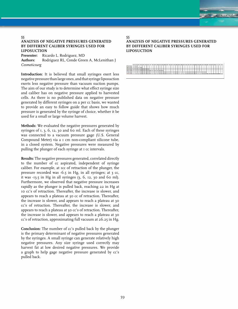
55ANALYSIS OF NEGATIvE PrESSUrES GENErATED BY DIFFErENT CALIBEr SYrINGES USED FOr LIPOSUCTIONPresenter: Ricardo L. Rodriguez, MDAuthors: Rodriguez RL, Conde Green A, McLenithan JCosmeticsurg
Introduction: It is believed that small syringes exert less negative pressure than large ones, and that syringe liposuction exerts less negative pressure than vacuum suction pumps. The aim of our study is to determine what effect syringe size and caliber has on negative pressure applied to harvested cells. As there is no published data on negative pressure generated by different syringes on a per cc basis, we wanted to provide an easy to follow guide that shows how much pressure is generated by the syringe of choice, whether it be used for a small or large volume harvest.
methods: We evaluated the negative pressures generated by syringes of 1, 3, 6, 12, 30 and 60 ml. Each of these syringes was connected to a vacuum pressure gage (U.S. General Compound Meter) via a 1 cm non-compliant silicone tube, in a closed system. Negative pressures were measured by pulling the plunger of each syringe at 1 cc intervals.
results: The negative pressures generated, correlated directly to the number of cc aspirated, independent of syringe caliber. For example, at 1cc of retraction of the plunger, the pressure recorded was -6.5 in Hg, in all syringes; at 3 cc, it was -13.5 in Hg in all syringes (3, 6, 12, 30 and 60 ml). Furthermore, we observed that negative pressure increases rapidly as the plunger is pulled back, reaching 22 in Hg at 10 cc’s of retraction. Thereafter, the increase is slower, and appears to reach a plateau at 30 cc of retraction. Thereafter, the increase is slower, and appears to reach a plateau at 30 cc’s of retraction. Thereafter, the increase is slower, and appears to reach a plateau at 30 cc’s of retraction. Thereafter, the increase is slower, and appears to reach a plateau at 30 cc’s of retraction, approximating full vacuum at 26.25 in Hg.
Conclusion: The number of cc’s pulled back by the plunger is the primary determinant of negative pressures generated by the syringes. A small syringe can generate relatively high negative pressures. Any size syringe used correctly may harvest fat at low desired negative pressures. We provide a graph to help gage negative pressure generated by cc’s pulled back.
55ANALYSIS OF NEGATIvE PrESSUrES GENErATED BY DIFFErENT CALIBEr SYrINGES USED FOr LIPOSUCTION
59

58STEPS TOWArD STANDArDIZED PrOTOCOL FOr ADIPOSE-DErIvED mESENCHYmAL STEm CELLS HArvEST OF CLINICAL GrADEPresenter: Nathan Katz, PhDAuthors: Katz N, Koukharenko V, Geldner PDJointechlabsInc
Paper Withdrawn
59SkELETAL mYOGENIC DIFFErENTIATION OF ADIPOSE-DErIvED STEm CELLS IS ENHANCED BY CYCLIC TENSILE STrAINPresenter: Vladimir Zachar, MD, PhDAuthors: Zachar V, Botha J, Buhl-Christensen O,
Bundgaard-Nielsen C, Hahn-Pedersen CJ, Pennisi CP
AalborgUniversity
Adipose-derived stem cells (ASCs) hold potential for the treatment of skeletal muscle deficits due to their ability to differentiate into the skeletal myogenic lineage. Current approaches to induce in vitro skeletal muscle differentiation require long culturing time and result in a poor yield. Previously, we have established the role of uniaxial cyclic tensile strain (CTS) in driving the assembly and differentiation of skeletal myocytes in vitro using mouse model system. In the current work, we explored the effect of CTS on the skeletal myogenic differentiation of ASCs. ASCs were cultured on flexible-bottomed culture plates (Flexcell). After confluency, the cells were incubated in reduced serum conditions and subjected to 15% uniaxial CTS for 48 h. As a result, the cells were aligned perpendicularly to the axis of strain and multinucleated forms appeared. Immunofluorescence staining revealed the presence of a large percentage of myosin heavy chain positive myotubes. Furthermore, the myofibrillogenesis was confirmed by the presence of actin/myosin cross striations. These results indicate that mechanical stimulation increases the rate of myogenic differentiation of ASCs in vitro. This opens the perspective of using mechanicaly preconditioned ASCs for the treatment of muscular diseases.
60

60COmPArISON OF ADIPOSE-DErIvED STrOmAL CELLS (ASC) AND BONE mArrOW mESENCHYmAL STrOmAL CELLS (Bm-mSC) FrOm THE SAmE HEALTHY INDIvIDUALS – A STUDY OF ImmUNOLOGICAL PrOPErTIESPresenter: Cedric Menard, PharmD, PhDAuthors: Menard C, Hebraud B, Dulong J, Gadelorge M,
Bezier I, Latour M, Bescher N, Planat-Benard V, Bourin P, Tarte K
UniversityofRennes1
ASC and BM-MSC exert powerful anti-inflammatory and immunosuppressive effects that support their clinical use as promising cell therapy approach in both immune disorders and regenerative medicine. Several clinical grade production processes have been validated, which differed not only by the source of stromal cells, i.e. bone marrow versus adipose tissue, but also by culture conditions and did not take into account inter-individual variability.
In this study, we obtained paired ASC and BM-MSC from 5 healthy individuals and tested their immunological properties following standardized procedures. All comparisons were made at an equivalent number of population doubling. We extensively analyzed their immunologic phenotype and evaluated the capacity of stromal cells to inhibit T, NK, and B cell proliferation and to support T, NK, and B cell survival, either in steady-state conditions of after exposure to a combination of TNF-a and IFN-g inflammatory stimuli. Interestingly, ASC were consistently more powerful in inhibiting T-cell proliferation in vitro compared to autologous BM-MSC. We observed no difference in their capacity to reduce NK cell proliferation. In agreement, ASC produced higher levels of the immunosuppressive enzyme Indoleamine-2,3 dioxygenase (IDO) as evaluated by microarray analysis, immunofluorescence study, and quantification of IDO activity. Conversely, BM-MSC promoted more efficiently survival of immune cells, a capacity that could be linked to their overexpression of CD106 adhesion molecule.
Finally, ASC and BM-MSC differentially responded to inflammatory signals. In particular, whereas CD106 was strongly upregulated in BM-MSC after stimulation by TNF-a/IFN-g, it remained poorly expressed by ASC. Conversely, ASC secreted higher levels of TNF stimulated gene protein-6 (TSG-6) in response to inflammatory signals, a property that could be helpful to modulate the activity of macrophages and neutrophils.
In conclusion, our results shed new light on the biological differences between ASC and BM-MSC and support the preferential use of ASC in some inflammation-related clinical applications.
61rEPrODUCIBILITY OF 3D ADIPOGENESIS WITHIN HOLLOW FIBEr-BASED BIOrEACTOrSPresenter: Danielle M. Minteer, BSAuthors: Minteer DM, Lin YC, Gerlach JC, Rubin JP,
Marra KGUniversityofPittsburgh
Background: Recently, our laboratory has developed and optimized a hollow fiber-based bioreactor with the ability to culture adipose tissue reproducibly in three dimensions. Unlike traditional, two-dimensional culture, three-dimensional culture creates a stable system in which long-term culture of adipocytes is possible, providing a model useful for drug discovery in the treatment of diseases such as type 2 diabetes mellitus, which, in 2010, affected 10.9 million US citizens, summing a national cost of $174 billion. Our studies aimed to prove previously established methods of 3D adipogenesis in the bioreactor were reproducible.
methods: Adipose stem cells (ASCs) were isolated from human lipoaspirate and expanded in culture. 80 x 10^6 cells were inoculated into the bioreactor and cultured at physiological conditions for 9 weeks. Weeks 1-2, cells were treated with regular culture medium, differentiation of ASCs into adipose tissue was implemented during weeks 2-6, and adipocytes were maintained within the bioreactor during the final 3 weeks.
results: Daily measurements were taken to ensure stable pH, temperature, glucose and lactate levels within the system. To evaluate mature adipocytes in the system after 9 weeks, immunohistochemical analyses as well as qPCR were performed on the tissue and bioreactor fibers.
Conclusion: Our bioreactor system is stable and reproducible allowing further in vitro diagnostic testing of adipose tissue. Future directions involve utilizing our system to further improve discovering and testing therapeutic strategies for diabetes and obesity research.
61
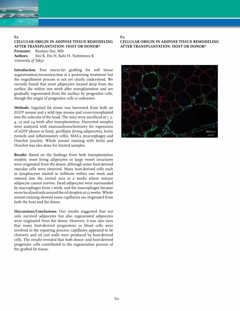
62CELLULAr OrIGIN IN ADIPOSE TISSUE rEmODELING AFTEr TrANSPLANTATION: HOST Or DONOr?Presenter: Kentaro Doi, MDAuthors: Doi K, Eto H, Kato H, Yoshimura KUniversityofTokyo
Introduction: Free micro-fat grafting for soft tissue augmentation/reconstruction is a promising treatment but the engraftment process is not yet clearly understood. We recently found that most adipocytes located deep from the surface die within one week after transplantation and are gradually regenerated from the surface by progenitor cells, though the origin of progenitor cells is unknown.
methods: Inguinal fat tissue was harvested from both an EGFP mouse and a wild type mouse and cross-transplanted into the subcutis of the head. The mice were sacrificed at 1, 2, 4, 12 and 24 week after transplantation. Harvested samples were analyzed with immunohistochemistry for expression of eGFP (donor or host), perillipin (living adipocytes), lectin (vessels and inflammatory cells), MAC2 (macrophage) and Hoechst (nuclei). Whole mount staining with lectin and Hoechst was also done for limited samples.
results: Based on the findings from both transplantation models, most living adipocytes or large vessel structures were originated from the donor, although some host-derived vascular cells were observed. Many host-derived cells such as lymphocytes started to infiltrate within one week and entered into the central area at 2 weeks where mature adipocyte cannot survive. Dead adipocytes were surrounded by macrophages from 1 week, and the macrophages became more localized only around the oil droplets at 12 weeks. Whole mount staining showed some capillaries are originated from both the host and the donor.
Discussions/Conclusions: Our results suggested that not only survived adipocytes but also regenerated adipocytes were originated from the donor. However, it was also seen that many host-derived progenitors or blood cells were involved in the repairing process; capillaries appeared to be chimeric and oil cyst walls were produced by host-derived cells. The results revealed that both donor- and host-derived progenitor cells contributed to the regeneration process of the grafted fat tissue.
62CELLULAr OrIGIN IN ADIPOSE TISSUE rEmODELING AFTEr TrANSPLANTATION: HOST Or DONOr?
62
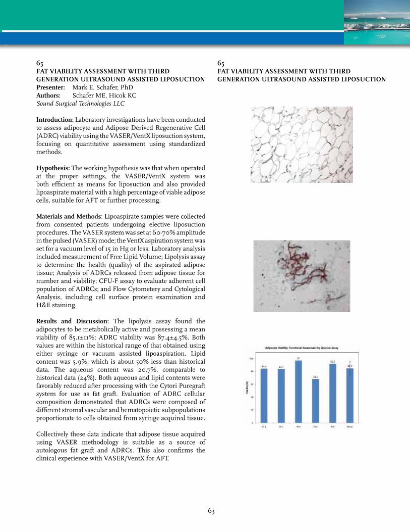
65FAT vIABILITY ASSESSmENT WITH THIrD GENErATION ULTrASOUND ASSISTED LIPOSUCTIONPresenter: Mark E. Schafer, PhDAuthors: Schafer ME, Hicok KCSoundSurgicalTechnologiesLLC
Introduction: Laboratory investigations have been conducted to assess adipocyte and Adipose Derived Regenerative Cell (ADRC) viability using the VASER/VentX liposuction system, focusing on quantitative assessment using standardized methods.
Hypothesis: The working hypothesis was that when operated at the proper settings, the VASER/VentX system was both efficient as means for liposuction and also provided lipoaspirate material with a high percentage of viable adipose cells, suitable for AFT or further processing.
materials and methods: Lipoaspirate samples were collected from consented patients undergoing elective liposuction procedures. The VASER system was set at 60-70% amplitude in the pulsed (VASER) mode; the VentX aspiration system was set for a vacuum level of 15 in Hg or less. Laboratory analysis included measurement of Free Lipid Volume; Lipolysis assay to determine the health (quality) of the aspirated adipose tissue; Analysis of ADRCs released from adipose tissue for number and viability; CFU-F assay to evaluate adherent cell population of ADRCs; and Flow Cytometery and Cytological Analysis, including cell surface protein examination and H&E staining.
results and Discussion: The lipolysis assay found the adipocytes to be metabolically active and possessing a mean viability of 85.1±11%; ADRC viability was 87.4±4.5%. Both values are within the historical range of that obtained using either syringe or vacuum assisted lipoaspiration. Lipid content was 5.9%, which is about 50% less than historical data. The aqueous content was 20.7%, comparable to historical data (24%). Both aqueous and lipid contents were favorably reduced after processing with the Cytori Puregraft system for use as fat graft. Evaluation of ADRC cellular composition demonstrated that ADRCs were composed of different stromal vascular and hematopoietic subpopulations proportionate to cells obtained from syringe acquired tissue.
Collectively these data indicate that adipose tissue acquired using VASER methodology is suitable as a source of autologous fat graft and ADRCs. This also confirms the clinical experience with VASER/VentX for AFT.
65FAT vIABILITY ASSESSmENT WITH THIrD GENErATION ULTrASOUND ASSISTED LIPOSUCTION
63

67HUmAN ADIPOSE-DErIvED STEm CELLS SUrvIvAL AND IN-vIvO TrACkING IN ANImAL mODELSPresenter: Hitesh Agrawal, MDAuthors: Agrawal H, Shang H, Parker A, Katz AJUniversityofVirginiaHealthSystem
Introduction: Adipose Stromal/Stem Cells provide a great deal of promise for regenerative therapies. However, the fate of ASCs after transplantation remain unclear. Our aim was to evaluate the localization and survival of human ASCs implanted as cell suspensions compared to 3-D spheroids in mice, using fluorescent membrane labelling and PCR.
methods: Human ASCs were isolated per standard protocols and culture-expanded in adherent monolayer culture. Cells were DiI labelled and some were formed into self-assembling spheroids. Two strains of mice: immunocompetent (C57BL/6NCr) and immunocompromised (Athymic NCr-nu/nu) were randomly injected with 300,000 cells either in suspension or as spheroids into inguinal fat pads. Non-viable cells produced by overnight incubation at -80°C were implanted in the opposite side in randomized, blinded fashion. 3 sets of mice (each comprising of 12 immunocompetent and 12 immunocompromised) were harvested on days 3, 10 and 21. Grossly visible injection sites were analyzed at the time of sacrifice by fluorescent microscopy, human cell quantification by PCR detection of ERV-3, and by IF staining with Mac-2 for macrophages.
results: Implantation sites were readily identifiable under gross observation as pink blushes but neither direct observation, nor IF microscopic evaluation could identify differences in survival between viable and non-viable groups. ERV-3 quantification showed that nearly 90% of the cells are undetectable by PCR by day 10. Mac2 staining demonstrated that cells were progressively engulfed by macrophages. In this model, there were no statistical differences in survival of cells injected in suspension versus those injected as spheroids.
Conclusions: Based on PCR detection, only 5% of cells remain at the implant site at 21 days, even though a good fluorescent signal is visualized on microscopy. The cells generate a robust macrophage infiltrate that co-localizes to the DiI labelled cells. Our findings suggest that visualization of fluorescent membrane dye does not correspond to cell viability or identity, and may lead to inaccurate conclusions related to cell tracking.
67HUmAN ADIPOSE-DErIvED STEm CELLS SUrvIvAL AND IN-vIvO TrACkING IN ANImAL mODELS
64
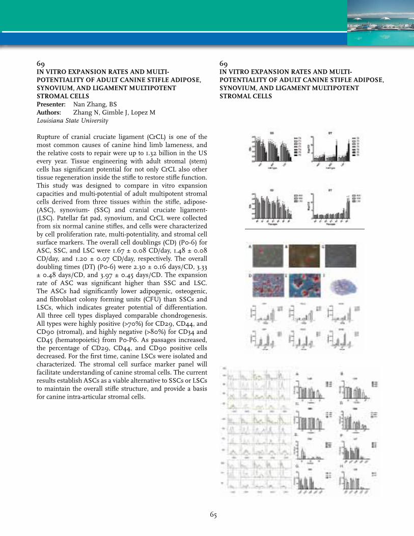
69IN vITrO EXPANSION rATES AND mULTI-POTENTIALITY OF ADULT CANINE STIFLE ADIPOSE, SYNOvIUm, AND LIGAmENT mULTIPOTENT STrOmAL CELLSPresenter: Nan Zhang, BSAuthors: Zhang N, Gimble J, Lopez MLouisianaStateUniversity
Rupture of cranial cruciate ligament (CrCL) is one of the most common causes of canine hind limb lameness, and the relative costs to repair were up to 1.32 billion in the US every year. Tissue engineering with adult stromal (stem) cells has significant potential for not only CrCL also other tissue regeneration inside the stifle to restore stifle function. This study was designed to compare in vitro expansion capacities and multi-potential of adult multipotent stromal cells derived from three tissues within the stifle, adipose- (ASC), synovium- (SSC) and cranial cruciate ligament- (LSC). Patellar fat pad, synovium, and CrCL were collected from six normal canine stifles, and cells were characterized by cell proliferation rate, multi-potentiality, and stromal cell surface markers. The overall cell doublings (CD) (P0-6) for ASC, SSC, and LSC were 1.67 ± 0.08 CD/day, 1.48 ± 0.08 CD/day, and 1.20 ± 0.07 CD/day, respectively. The overall doubling times (DT) (P0-6) were 2.30 ± 0.16 days/CD, 3.33 ± 0.48 days/CD, and 3.97 ± 0.45 days/CD. The expansion rate of ASC was significant higher than SSC and LSC. The ASCs had significantly lower adipogenic, osteogenic, and fibroblast colony forming units (CFU) than SSCs and LSCs, which indicates greater potential of differentiation. All three cell types displayed comparable chondrogenesis. All types were highly positive (>70%) for CD29, CD44, and CD90 (stromal), and highly negative (>80%) for CD34 and CD45 (hematopoietic) from P0-P6. As passages increased, the percentage of CD29, CD44, and CD90 positive cells decreased. For the first time, canine LSCs were isolated and characterized. The stromal cell surface marker panel will facilitate understanding of canine stromal cells. The current results establish ASCs as a viable alternative to SSCs or LSCs to maintain the overall stifle structure, and provide a basis for canine intra-articular stromal cells.
69IN vITrO EXPANSION rATES AND mULTI-POTENTIALITY OF ADULT CANINE STIFLE ADIPOSE, SYNOvIUm, AND LIGAmENT mULTIPOTENT STrOmAL CELLS
65
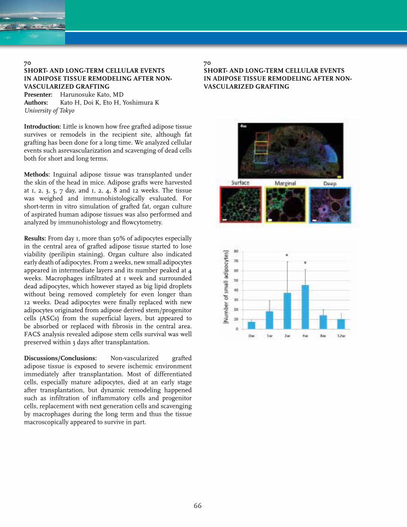
70SHOrT- AND LONG-TErm CELLULAr EvENTS IN ADIPOSE TISSUE rEmODELING AFTEr NON-vASCULArIZED GrAFTINGPresenter: Harunosuke Kato, MDAuthors: Kato H, Doi K, Eto H, Yoshimura KUniversityofTokyo
Introduction: Little is known how free grafted adipose tissue survives or remodels in the recipient site, although fat grafting has been done for a long time. We analyzed cellular events such asrevascularization and scavenging of dead cells both for short and long terms.
methods: Inguinal adipose tissue was transplanted under the skin of the head in mice. Adipose grafts were harvested at 1, 2, 3, 5, 7 day, and 1, 2, 4, 8 and 12 weeks. The tissue was weighed and immunohistologically evaluated. For short-term in vitro simulation of grafted fat, organ culture of aspirated human adipose tissues was also performed and analyzed by immunohistology and flowcytometry.
results: From day 1, more than 50% of adipocytes especially in the central area of grafted adipose tissue started to lose viability (perilipin staining). Organ culture also indicated early death of adipocytes. From 2 weeks, new small adipocytes appeared in intermediate layers and its number peaked at 4 weeks. Macrophages infiltrated at 1 week and surrounded dead adipocytes, which however stayed as big lipid droplets without being removed completely for even longer than 12 weeks. Dead adipocytes were finally replaced with new adipocytes originated from adipose derived stem/progenitor cells (ASCs) from the superficial layers, but appeared to be absorbed or replaced with fibrosis in the central area. FACS analysis revealed adipose stem cells survival was well preserved within 3 days after transplantation.
Discussions/Conclusions: Non-vascularized grafted adipose tissue is exposed to severe ischemic environment immediately after transplantation. Most of differentiated cells, especially mature adipocytes, died at an early stage after transplantation, but dynamic remodeling happened such as infiltration of inflammatory cells and progenitor cells, replacement with next generation cells and scavenging by macrophages during the long term and thus the tissue macroscopically appeared to survive in part.
70SHOrT- AND LONG-TErm CELLULAr EvENTS IN ADIPOSE TISSUE rEmODELING AFTEr NON-vASCULArIZED GrAFTING
66
72THE FDA APPrOvAL PrOCESS FOr vETErINArY ALLOGENEIC STEm CELL PrODUCTS - YES-STEm CELLS ArE DrUGS IN vETErINArY mEDICINE TOOPresenter: Robert Harman, DVM, MPVMAuthors: Harman R, Black L, Harman S, Smith AVetStem
Stem cell therapy has been in the development phase in the pharmaceutical industry for over 20 years. It is a given that allogeneic (donor) stem cell therapy is regulated through the IND (Investigational New Drug) process in the US by the FDA, specifically the Center for Biologics Evaluation and Research (CBER). Not so common knowledge is that the regulation of veterinary drug products is by the fourth branch of the FDA, the Center for Veterinary Medicine (CVM). CVM regulates all veterinary drugs and certain biologics via the INAD (Investigational New Animal Drug) process. The US Department of Agriculture regulates veterinary vaccines and certain other biologics. CVM and USDA have made it quite clear that CVM has the lead in regulation and approval of veterinary stem cell products. This presentation will outline the steps involved in the FDA approval process for a veterinary stem cell product. The key components of a stem cell product approval are the demonstration of purity and potency of the product and the clear demonstration of safety and efficacy in the target species (i.e. dog, horse, cat). The requirments for cGMP (Current Good Manufacturing Practices) production of a veterinary drug are esssentially identical to those for a human drug or biologics approval. CVM requires the demonstration of safety of a stem cell product in pivotal Target Animal Safety studies conducted under GLP (Good Laboratory Practices) conditions. CVM also requires the demonstration of efficacy for any label claims in a series of field studies culminating in one or more pivotal efficacy studies conducted under GCP (Good Clinical Practices). Overall, this process ensures an adequate demonstration of safety and efficacy and protects the consumer and their animals from dangerous or non-effective products entering the marketplace. Yes, stem cells are drugs in veterinary medicine too.
73DEvELOPmENT OF A SErUm-FrEE CHEmICALLY DEFINED HUmAN ADIPOSE DErIvED STEm CELL EXPANSION SYSTEm THAT mAINTAINS mULTIPOTENCY AND ImmUNOPHENOTYPEPresenter: Kirsten Crapnell, PhDAuthors: Crapnell K, Kelley R, Reyes J, Hastings A,
Blaesius R, Brooks JBectonDickinson
Adipose derived stem cells (ASCs) have substantial clinical therapeutic value. Cell therapy and tissue engineering will require solutions to reliably expand and manufacture these primary cells. ASC expansion has traditionally employed serum supplementation of growth media, but serum presents challenges due to its lot-to-lot variation, chemically undefined nature, and potential for transmission of infectious agents.
A novel serum-free expansion medium, BD Mosaic, yields proliferation of ASCs in culture significantly exceeding that of conventional serum-containing media through multiple passages. ASCs isolated in serum and expanded in our serum-free medium reached over 15 population doublings after 15 days in culture. Cells expanded in this serum-free medium maintained their multipotency in in vitro adipogenic, chondrogenic and osteogenic differentiation protocols as measured by lipid accumulation, alkaline phosphatase induction, and Alcian blue staining, respectively. Serum free-expanded cells also maintained immunophenotype (positive for CD13, CD44, CD90, CD105, and CD166; negative for CD31, CD34, CD45, and CD106) through several passages. This chemically defined medium, developed through the application of bioinformatic and screening principles, may thus assist in the progress of ASCs from the laboratory bench to the clinic.
67

74PrOLIFErATIvE AND ADIPOGENIC EFFECTS OF NEUrOPEPTIDE Y ON PrImArY CULTUrED HUmAN ADIPOSE-DErIvED STEm CELLS Presenter: Brian J. Philips, PhDAuthors: Philips BJ, Grahovac TL, McAtee J,
Bhaumik M, Marra KG, Fernstrom JD, Rubin JP
UniversityofPittsburgh
Introduction: A current unmet need for optimizing the long-term viability of soft tissue reconstructive implants is the identification of pharmacologically-active agents that promote adipose-derived stem cell (ASC) growth and differentiation at the transplant site, as well as adequate vascularization of the transplant. Neuropeptide Y (NPY) has been shown to be a promising candidate both in vitro and in vivo as a promoter of pre-adipocyte proliferation and differentiation. However, to our knowledge, its effects have not been studied in primary cultured human ASCs. As a result, we examined the proliferative and adipogenic effects of NPY on primary cultures of human ASCs focusing primarily upon dose-response and patient response variability.
methods: ASCs were isolated from abdominal fat obtained from 20 human, non-diabetic female patients (Age: 30-68, BMI: 22-50) and cultured. The cultures were treated under low-serum conditions with 10-14M-10-6M NPY and assessed for (1) proliferation (3 day) using the CyQUANT Cell Proliferation Assay, and (2) differentiation (12 day) via lipid accumulation using the AdipoRed Assay. Expression of NPY receptor mRNA was analyzed using quantitative Real-Time PCR analyses.
results: NPY treatment increased proliferation in approximately 20% of ASC patient sample cultures, though nearly 60% of ASC cultures were biologically responsive to NPY regarding differentiation. Quantitative PCR analyses indicated variable NPY receptor mRNA expression both within and among primary cultured ASC samples.
Conclusions: Because of their capacity to differentiate into multiple cell lineages and to secrete a variety of growth factors, ASCs offer great potential for clinical applications. Our results indicate that ASC patient variability in response to NPY may be due to differential NPY receptor mRNA expression. For increased understanding of in vivo survival conditions of autologous human transplanted fat ± NPY, studies in athymic mice are currently being conducted. We anticipate such studies will help progress ASC translational usage for the treatment of soft tissue injuries.
75CASE rEPOrT: OPTImIZATION OF rOCHE LIBErASE IN THE ENZYmATIC DIGESTION OF HUmAN ADIPOSE TISSUE FOr THE ISOLATION OF STEm & rEGENErATIvE CELLSPresenter: Rowena A. Soriano, BSAuthors: Soriano RA, Torfi HInvitrxTherapeuticsInc
Paper Withdrawn
68

77THE ADIPOSE TISSUE EXTrACELLULAr mATrIX rOLE ON ADIPOSE STEm CELL DIFFErENTIATIONPresenter: Casey Roberts, BSAuthors: Roberts C, Ogle RA, Ogle RCEasternVirginiaMedicalSchoolandLifeNetHealth
Purpose: To characterize the extracellular environment of the adipose stem cell (ASC) and the mature adipocyte within human adipose tissues and identify the elements of the ASC niche influencing ASC proliferation and differentiation to mature adipocytes. There are many reasons to “engineer” an adipose tissue graft from ASCs delivered in an appropriate carrier. By recreating the adipose tissue niche with ASCs and the adipose tissue matrix, an injectable, living adipose implant may be devised that would maintain its volume and integrity over time. The adipose tissue extracellular matrix (ECM) may be subdivided into the basement membrane (BM) surrounding the mature adipocyte and the interstitial ECM (IM), the ECM is comprised of type I, III, IV, V, and VI collagen (COL I, III, IV, V, or VI), multiple laminin types (Lm), nidogen-1 (NID-1), and heparin sulfate proteoglycan (HSPG). The BM components are expressed in the pericellular region of mature adipocytes and can even be used as markers for adipocyte differentiation.
methods: We fractionated the basement membrane and interstitial matrices of human subcutaneous adipose tissue. The components were identified by electrophoresis and Western blotting. ASCs were grown in vitro on a coating of each type of matrix with and without known required differentiation factors and the fractions supporting growth and adipogenesis in vitro were identified.
results: We found that the adipose tissue ECM extract contained COL I, COL IV, COL VI, Lm chains g1, α±3, α±4, α±5, and Lm type 5, NID-1, and HSPG. The IM extract contained COL I, COL IV, Lm, and HSPG. There is evidence that the adipose tissue ECM supports an increased rate of differentiation to adipocytes when combined with the required differentiation factors.
Conclusion: This data supports the theory that the adipose tissue ECM can facilitate the differentiation of ASCs and possibly contribute to recreating the adipose tissue niche. This could be because the adipose tissue ECM extract has been determined to contain COL I, COL IV, Lms, and HSPG, all of which are known to be able to bind the cells.
78TrEATmENT OF GOAT OSTEOCHONDrAL kNEE DEFECTS WITH ADIPOSE DErIvED STEm CELLS USING A ONE-STEP SUrGICAL PrOCEDUrEPresenter: Marco N. Helder, PhDAuthors: Helder MN, Jurgens WJ, Kroeze RJ, Zandieh
Doulabi B, Renders G, Smit TH, van Milligen FJ, Ritt MP
VUUniversityMedicalCenter
Introduction: The abundant availability and high frequency of adipose derived stem cells (ASC) allow the development of a one-step surgical procedure for the treatment of osteochondral defects. Feasibility of this concept was previously tested in vitro. Here, in vivo safety/efficacy was assessed.
methods: Osteochondral defects were created in knees of eight goats, and filled by either acellular collagen I/III scaffolds or scaffolds seeded with SVF or cultured ASCs. Defects were scored after 4 weeks using macroscopic and immunohistological analysis, whereas after four months evaluation comprised macroscopy, bio- and immunochemistry, and biomechanical and microCT analysis.
results: Four weeks after implantation, macroscopic and microscopic evaluations showed no significant differences between acellular and cell-seeded constructs. After 16 weeks, acellular constructs showed some regeneration, however being inferior to both cell-treated constructs, which exhibited (i) more hyaline like cartilage; (ii) more extensive collagen type II (cartilage) and collagen type I (subchondral bone) stainings; (iii) more subchondral bone filling and a more mature morphology of bone and cartilage (microCT analysis); (iv) higher elastic moduli, and (v) glycosaminoglycan contents better approaching the native tissue.
Conclusions: This pilot-study showed feasibility of a one-step surgical procedure for osteochondral defect regeneration using stem cell seeded constructs. Further studies are warranted to substantiate these results in order to conclusively compare these to the conventional subchondral drilling technique as mimicked by the use of the acellular scaffolds.
69
79AN INNOvATIvE mATErIAL FOr “mICrO” AUTOLOGOUS FAT GrAFTING: ABOUT 100 CASESPresenter: Jonathan Londner, MDAuthors: Londner J, Magalon G, Nguyen P, Ould Ali D,
Niddam JServicePrMagalon
Patients with sclerodermia are good candidates for facial autologous fat grafting. Such cases where the skin is thin or rigid led us to consider a harvesting and injection material with a smaller diameter. We began our work with an experimental study on nude mice which enabled us to define the ideal characteristics and dimensions of a new filler in order to obtain microimplants of adipose tissue of approximately 500 µ in diameter containing several hundred adipocytes. This experimental phase was followed by a clinical phase of 100 cases of microinjection that we will present to you with details of the procedure and the preliminary results with a minimum follow-up of 6 months. Our series was made up of 49 cases of reconstructive surgery (8 cases of sclerodermia, 4 cases of burn sequelae, 5 cases of scars, 3 cases of acne sequelae, 29 cases of posttraumatic depression) and 51 cases of esthetic surgery to provide a trophic and volumizing effect in the face and on the back of the hand (38 cases of facial injection, 3 cases of injection in the hand and 10 cases of facial injection associated with facelift). Filler volume ranged from 12 to 24 cc. The gender ratio was 84 women versus 16 men. The procedure was performed under simple local anesthesia in 89% of the cases.
In conclusion, microinjection multiplies the indications for reconstructive and esthetic surgery, lightens anesthetic procedures and improves tissue viability. It requires innovative single-use infiltration-harvesting and injection cannulae.
81ADIPOSE STrOmAL vASCULAr FrACTION CELLS PrESErvE COrONArY PErFUSION WHEN USED ImmEDIATELY AFTEr ISCHEmIAPresenter: Amanda J. LeBlanc, PhDAuthors: LeBlanc AJ, Hoying JB, Williams SKJewishHospitalandUniversityofLouisville
Purpose: Within adipose tissue is a regenerative cell population known as the stromal vascular fraction (SVF). An epicardial construct was created using SVF and evaluated as a microvascular protection treatment in a rat myocardial infarction (MI) model. Previous research has demonstrated the ability of tissue-engineered constructs to stabilize LV function following acute MI, but the effect of a delayed treatment was unknown.
methods: SVF was isolated from epididymal fat pads through mincing, enzyme digestion, and removal of buoyant adipocytes. SVF (1 x 10(6) cells/cm2) was plated on Vicryl®
and cultured for 14 days. Fischer-344 rats were separated into groups, including Sham, MI, MI + SVF construct, and MI + delayed SVF construct implant. MIs were performed by ligating a branch of the LAD artery. SVF constructs were implanted onto the infarct area either immediately or after two weeks. Four-weeks post-surgery, microperfusion was evaluated by injection of microspheres into the coronary arteries during baseline and maximal blood flow (BF, dobutamine infusion).
results: Immediate implantation of an SVF-laden patch following MI sustained microcirculatory perfusion in the area of infarct during both baseline and dobutamine infusion to levels exhibited by Sham rats. MI only group was associated with a 46% decline in baseline BF to the area of infarct. Compared to Sham rats, this deficiency persisted in when challenged with dobutamine, resulting in approximately 60% less perfusion in the infarct region. Similar to MI only hearts, BF was also impaired after delayed SVF construct implant, indicating that SVF alone is unable to reverse infarct-related maladaptations in LV tissue.
Conclusions: We present a novel strategy to maintain beneficial epicardial microvascular BF after MI through an implantable three-dimensional tissue construct containing adipose-derived SVF. These results indicate that the SVF construct implanted immediately, rather than delayed, following MI can protect microvascular perfusion and function in the infarct area by sustaining coronary BF reserve.
70

82ELECTrICALLY CONDUCTIvE POLYPYrrOLE COATING AS A BIOACTIvATOr OF POLYLACTIDE FOr BONE TISSUE ENGINEErINGPresenter: Suvi P. Haimi, PhDAuthors: Pelto J, Hamalainen M, Ella V, Suuronen R, Hyttinen J, Miettinen S, Kellomaki M, Haimi SUniversityofTampere
Introduction: Polylactide-based polymers have been extensively used in different orthopedic applications, however, lack of bioactivity has limited their use especially in tissue engineering applications. Potential strategy to bioactivate polylactide could be electrically conductive polymer coating. Therefore, for the first time, the combined effect conductive scaffolds and electrical stimulation (ES) on human adipose stem cell (hASC) viability, proliferation and osteogenic differentiation was studied in 3D culture system.
methods: In order to bioactivate polylactide, we coated poly-96L/4D-lactide (PLA) fibers with electrically conductive polypyrrole (PPy) and manufactured non-wowen 3D conductive composite scaffolds. ASCs were exposed to biphasic pulsed voltage stimulation (1 V/cm) of 1 or 100 Hz for 4 hours/day. In each scaffold, 87,500 hASCs of passage 3 were seeded and cultured up to 2 weeks. Viability, proliferation and alkaline phosphatase (ALP) activity were used to analyze the cell response to conductive scaffolds and ES.
results: Live/dead-staining demonstrated that the majority of hASCs were viable and spread homogenously in both scaffold types with and without ES. By qualitative estimation, the number of hASCs was higher in PLA/PPy scaffolds compared to plain PLA scaffolds. This result was confirmed with the quantitative DNA assay showing significantly higher hASC numbers in PLA/PPy scaffolds with and without ES. Furthermore, the ALP activity was higher in PLA/PPy seeded scaffolds, but due to patient variation no statistical differences were detected. No significant differences in osteogenic differentiation were detected between electrically stimulated and non-stimulated hASCs.
Conclusions: PPy-coated scaffolds enhanced hASC adhesion, proliferation and osteogenic differentiation consistently compared to plain PLA scaffolds. This study highlights the future potential of PPy-coated PLA scaffolds in clinical use to repair bone defects. The ES in 3D systems should be further studied for optimal culture conditions for hASCs.
84rANDOmIZED CONTrOLLED CLINICAL TrIAL OF FAT GrAFTS SUPPLEmENTED WITH ADIPOSE-DErIvED rEGENErATIvE CELLS FOr PATIENTS WITH HEmIFACIAL mICrOSSOmIAPresenter: Daniela S. Tanikawa, MDAuthors: Tanikawa DS, Aguena M, Bueno DF,
Alonso N, Passos-Bueno MRUniversityofSaoPauloSchoolofMedicine
Purpose: Although first reports of the clinical use of adipose-derived regenerative cells (ADRC) suggest that this approach may be feasible and effective for soft tissue augmentation, there is a lack of randomized controlled clinical trials in the literature. Using a quantitative analysis for results evaluation, this study aimed to investigate whether a novel protocol for isolation of ADRC and their use in combination with fat tissue improve the long-term retention of the grafts.
methods: Patients with hemifacial microssomia (n=15) were selected and randomly assigned to group 1 (with ADRC) or group 2 (without ADRC). For group 1, ADRC freshly isolated from half of the aspirated fat sample were used to enrich for progenitor cells in the graft. Quantity of transplanted adipose tissue was determined by trying to obtain symmetry with the unaffected side without any overcorrection. Number of viable cells isolated before and after the supplementation of the grafts was calculated, and these cells were examined for mesenchymal cell surface markers using flow cytometry. Computed tomography was performed to assess both hemi-faces preoperatively and at 6 months postoperatively (n=8). Cutaneous thickness at 4 different reference points and face volume were calculated. Overall morbidity was recorded.
results: Mean injected volume was 34 mL. Average number of viable cells isolated before and after supplementation of the grafts was 10.8 x 10⁵ and 19.2 x 10⁵ cells per mL of fat tissue. Flow cytometry analysis revealed that the ADRC were positive for mesenchymal cell markers (>95% for SH2 and CD90). Preoperatively, average thickness ratio at points 1 to 4 was 0.66, 0.80, 0.79, and 0.75; and postoperatively it was 0.87, 0.91, 0.97, and 0.94 in group 1, and 0.70, 0.89, 0.90 and 0.79 in group 2. For group 1 surviving fat volume at 6 months was 89%, and for group 2 it was 61%. No complications were detected.
Conclusions: These results suggest that this strategy for isolation and supplementation of ADRC is effective, safe and superior to conventional lipoinjection for facial recontouring in patients with hemifacial microssomia.
71

84rANDOmIZED CONTrOLLED CLINICAL TrIAL OF FAT GrAFTS SUPPLEmENTED WITH ADIPOSE-DErIvED rEGENErATIvE CELLS FOr PATIENTS WITH HEmIFACIAL mICrOSSOmIA
85CrYOPrESErvATION AND rE-ANImATION OF ADIPOSE AND ADIPOSE DErIvED rEGENErATIvE CELLS: PrESENT USE IN THE U.S. FOr AESTHETIC AND rECONSTrUCTIvE SUrGErYPresenter: David Genecov, MDAuthors: Genecov D, Barcelo de la Isla CRBiolifeCellBankLLC
Cryopreservation of human tissues has been around for over 25 years with great success in the areas of blood banking, fertility sciences and now stem cell applications. Only recently has the cryopreservation of fresh adipose tissue been available to the general population. For the past 16 months, adipose and adipose derived regenerative cell cryopreservation has been effectively performed at a U.S. banking facility. The cryopreservation and thawing processes will be presented. Improvements in tissue transportation and on site thawed cellular therapies will be reviewed. Current efficacy has been seen in both the aesthetic facial and breast procedures as well as facial reconstrucive surgery. Benefits of this technique include single harvest of large volumes of tissue, delayed controlled tissue grafting under local anesthesia with oral sedation, and re-application to the patient’s and physician satisfaction. Case studies with close consideration of patient safety will be presented.
Cryopreservation of adipose and adipose derived regenerative cells can safely be performed with current techniques. As important, the thawing of these tissues with re-injection for facial and breast procedures (aesthetic and reconstructive) has been performed with efficacious outcomes and without complications. The combination of both cryopreservation and delayed re-injection of tissues enhances our abilities to provide natural and autologous alternatives to our patients.
72

87CLINICAL EXPErIENCE WITH AUTOLOGOUS FAT GrAFTING IN THE PEDIATrIC PATIENTPresenter: Kevin S. Hopkins, MD, FACSAuthors: Hopkins KS, Dhar PRDriscollChildrensHospital
Introduction: Autologous fat grafting has been successful in soft tissue augmentation and contouring primarily in the adult population. It is also a viable tool in restoring soft tissue in the pediatric patient for congenital and traumatic defects but there are inherent issues in this population such as the accessibility and amount of available autologous fat, harvesting techniques and modifications in injection cannulas that first need to be addressed.
method: Eight patients (5 female, 3 male) ages 8-17 years (average age 11.8 yrs) presented with six congenital (cleft lip/palate 5; frontonasal dysplasia 1) and two traumatic defects (craniofacial 1; extremity 1). The congenital problems included palatal fistula with severe scarring and fibrosis (3), velopharyngeal insufficiency (VPI) (2); a paucity of soft tissue in a repaired bilateral cleft lip (1) and scarred and attenuated nasal tip soft tissue (1). Traumatic cases included soft tissue deformities of the forehead and arm (1) and traumatic scarring of the calf (1). Fat was harvested manually using the tumescent technique with a 12-hole Khouri cannula, processed using the Coleman centrifuge at 3000 rpm for 3 minutes and then transferred to 1 ml syringes for injection. The volume of fat transferred ranged from 1 to 5.5 ml per site (avg 2.5 ml) Modifications were made to 16 ga and 18 ga epidural needles to facilitate the intraoral delivery of fat to the contours of the palate. Two patients have undergone serial autologous fat transfers (2).
results and Conclusions: Autologous fat can be safely harvested and transferred to correct soft tissue deformities in children. Hypernasality improved significantly in both patients with VPI. Direct observation and serial photographs demonstrate enhanced palatal soft tissue and improved tissue quality in the frontonasal dysplasia and two traumatic cases. Care must be taken to safely harvest fat in these children so as to not create a contour deformity or perforation. It is speculated that there is a higher density of stem cells in this population that may significantly augment healing. More studies are warranted.
88IN vITrO rECONSTrUCTION AND IN vIvO GrAFTING OF TISSUE-ENGINEErED HUmAN ADIPOSE TISSUES PrODUCED BY THE SELF-ASSEmBLY mETHODPresenter: Maryse Proulx, MScAuthors: Proulx M, Vincent C, Lagueux J, Fortin MA,
Fradette JCentreLOEXdelUniversiteLaval
Tissue-engineered adipose tissues represent a promising approach to provide soft tissues for reconstructive surgery. To ensure graft survival, a rapid tissue vascularization must take place after implantation. Our objective was to produce reconstructed human adipose tissues from adipose-derived stromal cells using the self-assembly approach of tissue engineering and to evaluate their vascularization after grafting. Reconstructed adipose tissues containing a preformed capillary network in vitro were also obtained by the addition of human microvascular endothelial cells (ECs) during production. Reconstructed adipose tissues enriched or not in ECs were grafted subcutaneously onto nude mice (n = 6 tissues per group per time-point, 3 repetitions). At day 3, 7 and 14, grafted tissues were excised and the presence of adipocytes as well as vascularization were analyzed on histological cross-sections and by immunofluorescence on frozen sections. Histology revealed that adipose tissues without ECs were vascularized from day 7 after grafting and that large quantities of capillaries were present in ECs enriched tissues at all time-points examined. Immunolabelings revealed the presence of a human capillary network containing murine red blood cells in the grafted tissues enriched in ECs. A pilot study evaluating volume retention of the grafts was also conducted using magnetic resonance imaging. It revealed near 90% volume retention for both types of reconstructed tissues 14 days after grafting when compared to their volume on grafting day (120.04 ± 8.95 mm3). Human adipose tissues produced by the self-assembly method could be promising for soft tissue grafting in reconstructive surgery, especially since they are devoid of exogenous biomaterials. Future experiments will assess the long-term fate of thicker tissues after grafting.
73

89ANGIOGENESIS OF FAT TISSUE AND ITS rESPONSE TO SEX HOrmONES IN HUmAN FEmALES IS DEPOT-DEPENDENTPresenter: Vinod K. Podichetty, MD, MSAuthors: Podichetty VK, Greenway FLResearchPracticePartnersInc
Paper Withdrawn
74

91EQUIvALENT EFFECTS OF TOPICALLY DELIvErED ADIPOSE-DErIvED STEm CELLS AND DErmAL FIBrOBLASTS IN THE ISCHEmIC rABBIT EAr mODEL FOr CHrONIC WOUNDSPresenter: Jordan P. Steinberg, MD, PhDAuthors: Steinberg JP, Hong SJ, Geringer MR,
Galiano RD, Mustoe TANorthwesternUniversity
Skin wound healing involves the orchestration of multiple cell types to produce a biological patch. In chronic wounds, local hypoxia and ischemia-reperfusion injury impede these events. Despite the advent of bioengineered products and growth factors for problem wounds, many prove refractory. For many years, we have employed a model system in the rabbit ear to quantitatively assay wound repair. Based on literature implicating adipose-derived stem cells (ASCs) in wound repair via differentiation and/or angiogenic paracrine effects, we hypothesized that ASCs might improve wound healing in the ischemic rabbit ear. We compared ASCs with rabbit dermal fibroblasts (RDFs) given the lack of appropriate controls in recent studies. Ears were rendered ischemic by ligation of the central and caudal arteries. Six mm dermal punch wounds were made down to bare cartilage, treated with fibrin sealant vehicle ± GFP-labeled ASCs or RDFs, and harvested at 1, 7, and 10 days. When compared with sham ears, ischemic ears showed a significant increase in epithelial and granulation tissue gap and a corresponding decrease in total epithelial and granulation tissue area at 7 days. The deficit in granulation tissue gap and area, but not epithelial gap and area, persisted in ischemic wounds at 10 days. The topical application of 300,000 ASCs in fibrin sealant to ischemic wounds contributed to the development of a neo-granulation tissue layer across the entire wound bed at 7 days. Strong GFP signal was observed throughout this neo-granulation layer, indicating successful engraftment and proliferation of ASCs. To our surprise, a qualitatively and quantitatively similar effect of RDFs was observed in ischemic wounds at both 7 and 10 days. Our results suggest that topically delivered ASCs can successfully engraft in ischemic wounds to a much greater extent than that previously demonstrated in normal wounds. Importantly, ASCs share many properties with RDFs, consistent with recent in vitro reports and supporting the need for thorough characterization of both cell types before translation to clinical therapeutics.
92ADIPOSE-DErIvED STEm CELL SECrETOmE: EFFECT ON FIBrOBLAST mIGrATION IS ALTErED BY DIABETESPresenter: Lisa J. Gould, MD, PhDAuthors: Gould LJ, Moor A, Watson J, Cooper DRJamesAHaleyVeteransHospital
Human adipose-derived stem cells (hADSCs) have the potential to differentiate into multiple cell lineages and release factors that modulate wound healing. Because adipose tissue is an abundant, easily accessible and renewable source of stem cells, hADSCs have significant advantages over other stem cell sources. However, most hADSC research has focused on lipoaspirate derived from young, healthy, predominantly female donors. To characterize hADSCs from clinically relevant donors we compared the impact of ADSC secreted factors from diabetic and non-diabetic obese patients on fibroblast migration.
methods: hADSCs isolated from the stromal vascular fraction of obese diabetic and non-diabetic patients were cultured for 3 passages and validated to express stem cell markers (CD34+/CD45-). Conditioned media was collected after 96 hour exposure to ADSCs and stored at -80. Primary human dermal fibroblasts (HDF) from young and elderly donors were grown to 80% confluence and transferred to basal media for 24 hours. Proliferation was inhibited with Mitomycin C and the monolayers were scratched with a yellow pipette tip. The cultures were treated with ADSC conditioned medium (ADSC-CM) at increasing concentrations (10, 50 and 100%). Cell migration was photographically assessed at 18 hours.
results: CM from non-diabetic, obese hADSCs inhibits fibroblast migration in a dose dependent fashion with near complete inhibition at 50%. A non-statistically significant increase in migration was noted in fibroblasts exposed to low dose diabetic ADSC-CM. Fibroblasts derived from young and old donors responded similarly.
Conclusions: hADSCs from non-diabetic obese patients secrete a factor(s) that markedly impedes migration of young and old fibroblasts in a dose dependent fashion. This factor is not present in ADSC-CM from diabetic patients and in fact, low-dose hADSC-CM from diabetic patients tends to promote fibroblast migration. These novel findings suggest that the co-morbid conditions of patients from which hADSCs are derived significantly affects their ability to promote wound healing. Diabetic ADSCs may promote cell migration.
75

92ADIPOSE-DErIvED STEm CELL SECrETOmE: EFFECT ON FIBrOBLAST mIGrATION IS ALTErED BY DIABETES
93NONINvASIvE BODY CONTOUrING AND SPOT FAT rEDUCTION BY LOW LEvEL LASEr THErAPY: EFFICACY OF LIPOLASEr TECHNOLOGY FrOm A SINGLE CENTEr, CONTrOLLED CLINICAL STUDYPresenter: Vinod K. Podichetty, MD ,MSAuthors: Podichetty VK, LaForge JC, Alibhai HResearchPracticePartnersInc
Introduction: Advances in laser technology, particularly the use of low level laser therapy (LLLT) for body contouring and spot fat reduction has the potential to greatly decrease the need for invasive procedures. We report results from a cohort of patients assessing the efficacy of semiconductor based low level laser device for noninvasive body contouring and abdominal fat reduction.
methods: Eligible healthy adult subjects were enrolled into a study and all patients received nine 30 minute laser treatments utilizing low level laser device at 658 nm wavelength (Lapex BCS, YOLO Medical Inc.), 3 times per week for 3 weeks totaling nine treatment sessions and evaluated over a 9-week period. Efficacy outcomes included reduction of waist circumference, patient satisfaction and photographic assessment by blinded independent observers. Safety was monitoring by study physician.
results: Nineteen healthy men and women between the ages of 23-58 years (average age=39.9 years) formed the study sample (n=19). 4 men and 15 women received a complete nine treatment session protocol which was well tolerated producing a mean waist circumference reduction of 5.7 inches with majority of the inch loss effect achieved within the first week. Responses after three treatment sessions in both genders were comparable, with a mean circumference reduction of 0.9 inches in men and 1.0 inches in women. (p=0.302). Overall at the end of the 9th treatment session, the average circumference reduction above the umbilicus=1.76 inches, at umbilicus =2.05 inches and below the umbilicus =1.98 (average cumulative loss =5.76 inches or 14.6 cms). Results varied between a range of 1 3/8 inch to 9 inches lost over the series of treatments. The effects were sustained at mean follow-up of 3 months post therapy. Independent observers found 75-100% improvement in abdominal contour in 90% of subjects and 50-74% improvement in 10% of subjects. No adverse event was reported.
Conclusions: Low level laser therapy is an effective non-invasive method for reducing subcutaneous fat of the abdomen and photographic evaluation correlates well with clinical findings.
76

93NONINvASIvE BODY CONTOUrING AND SPOT FAT rEDUCTION BY LOW LEvEL LASEr THErAPY: EFFICACY OF LIPOLASEr TECHNOLOGY FrOm A SINGLE CENTEr, CONTrOLLED CLINICAL STUDY
94IN vITrO ANALYSIS OF HUmAN ADIPOCYTE CELL rESPONSE TO LOW LEvEL LASEr THErAPYPresenter: Vinod K. Podichetty, MD, MSAuthors: Podichetty VK, Greenway FLResearchPracticePartnersInc
Background: Low-level laser therapy (LLLT) is commonly used in medical applications, but mechanistic studies by which it causes loss of fat is lacking. The study examined mechanism by which 635–680 nm LLLT (YOLO Medical Inc) acts on adipocyte cell.
methods: In vitro assays on human adipose-derived stem cells obtained from subcutaneous fat during abdominal surgery were conducted to determine evidence of cell lysis, and possibility to increase glycerol and triglyceride release.
results: Experiment 1: Fat cells that came in contact with plasma or plasma with white blood cells were lysed in both laser treated and the control plate, but cells in control wells or in wells with heat-inactivated plasma were not lysed. This indicates that serum complement does lyse fat cells, but the laser does not activate a complement cascade. The mechanism by which fat leaked from the fat cells into the interstitial space is by formation of transitory pores. Experiment 2: Laser does not kill adipocytes. The number of viable cells in the laser-treated or untreated group as determined by the propidium iodide assay were similar, but calcein levels were lower in the laser-treated cells. These findings suggest that laser-treated cells show micropores in the membrane, which presumably contributed to leakage of fat. Experiment 3: Laser increases triglyceride release, but not by lipolysis from adipocytes. The laser-irradiated wells containing serum had significantly greater increases in triglycerides than the non-irradiated wells containing serum (69 ± 1.7 vs. 66.7 ± 1.5 mg/dL, p = 0.004). Similarly, heat-inactivated serum had a significantly greater increase in triglycerides (72.6 ± 1.8 vs. 70.1 ± 1.6 mg/dL, p = 0.008). Baseline glycerol levels were not different in the laser-treated or the non-irradiated groups (0.11 ± 0.01 vs. 011 ± 0.01 mmol/L, p = 0.44). Before and after laser irradiation in the presence of serum, cells continued to appear intact without evidence of lysis.
Conclusion: LLLT increases fat loss from adipocytes by release of triglycerides, without inducing cell lysis.
77

94IN vITrO ANALYSIS OF HUmAN ADIPOCYTE CELL rESPONSE TO LOW LEvEL LASEr THErAPY
95LOW LEvEL LASEr THErAPY FOr BODY CONTOUrING AND SPOT FAT rEDUCTION: CLINICAL rEPOrT OF 222 CASESPresenter: Vinod K. Podichetty, MD, MSAuthors: Podichetty VK, Bourassa DResearchPracticePartnersInc
Introduction: Although Low-level laser therapy (LLLT) has evolved as an efficient tool in aesthetic body contouring and spot fat reduction, there are no large sample studies reporting the effectiveness of this method. The authors have previously presented a large cohort study with 272 patients. The aim of the study is a follow-up and evaluation of clinical effects of LLLT on subcutaneous fat reduction and enhancing body contouring.
methods: Retrospective data review of patients (n=222) treated with LLLT (range=635nm-680nm, center wavelength=658nm, 150mW array/40mW+/-20% diode laser radiation source) during a period of three years. The laser (YOLO Medical Inc) was applied directly on the skin of the abdomen and thighs where undesired fat was present.
results: 197 females, 25 males (range: 19-75 yrs) underwent from 6 to 13 laser treatments (abdominal area) and 6 to 10 (thighs). In abdominal sample, measured loss from single first session treatment in 81% of the sample (n=194) averaged 3.01 cm (range: 0.5-8.8 cm). Overall, 194 patients who completed a minimum of 6, maximum of 29 sessions achieved an average cumulative loss of 8.6 cm. Correspondingly 28 patients who had between 6 and 12 sessions on the thigh had a measurable change of 2.5cm in right and 2.8 cm in left thigh. 77.2% were able to sustain at least 4 cm or more abdominal loss in treatments of 6 or more sessions. Patient satisfaction and photographic assessment demonstrated significant higher score. No significant complications were encountered in the patient population. Using a threshold of at least 75% self reported satisfaction and improvement post procedure, 86% of patients treated at abdominal site and 89.2% of thigh patients were considered responders (p=0.4228).
Conclusion: While there is a high demand for body shaping procedures, effective non-invasive alternatives are few. Low level laser therapy is safe and efficacious method for reducing subcutaneous fat in the abdominal and thigh region where undesired fat is present.
78

95LOW LEvEL LASEr THErAPY FOr BODY CONTOUrING AND SPOT FAT rEDUCTION: CLINICAL rEPOrT OF 222 CASES
96ADIPOSE DErIvED STEm CELLS rESPOND vArIABLY TO A NOvEL OSTEOINDUCTIvE OXYSTErOLPresenter: Sarah C. Sorice, BAAuthors: Sorice SC, Hokugo A, Fan K, Zuk P, Huang W,
Miller T, Jarrahy RDavidGeffenSchoolofMedicineUCLA
Background: Adipose-derived stem cells (ASCs) have been identified as a potential source of cells for use in bone tissue engineering due to their ready availability, ease of harvest, and susceptibility to osteogenic induction. Bone morphogenetic protein (BMP) has been shown to augment the osteogenic potential of ASCs in vitro and in vivo. Clinical applications of BMPs, however, are limited due to its exorbitant cost and side effect profile. We have demonstrated the ability of a novel osteogenic molecule called oxysterol, an oxidative derivative of cholesterol, in inducing osteogenic differentiation in pluripotent rabbit bone marrow stromal cells via the Hedgehog (Hh) signaling pathway with an efficacy similar to that of BMP-2. In this study, we examine the ability of oxysterols to induce osteogenesis in human ASCs.
methods: ASCs were isolated from raw human lipoaspirates from patients. Cells were plated onto conventional tissue culture plates in control medium and harvested between passages 2 and 3. Cells were then incubated with various concentrations of oxysterols or dexamethasone in conventional osteogenic media. Alkaline phosphatase (ALP) activity was performed.
results: ALP activity in ASCs treated with oxysterol and dexamethasone was variable among different cell populations obtained from different patients. ASCs conditioned with higher concentration of oxysterols consistently inhibited osteogenic differentiation.
Conclusion: Oxysterols show a variable ability to induce osteogenic differentiation in human ASCs in vitro, in some cases exceeding ALP activity seen in ASCs cultured in osteogenic media alone. The addition of dexamethasone also altered the ALP activity of ASCs variably. We conclude that oxysterols have some effect on the osteogenic differentiation of human ASCs in vitro and that this effect is potentially modulated by the presence or absence of steroids. Further studies examining the mechanisms of this influence are underway with the aim of determining specific signaling pathways implicated in this process.
79

97THE EFFECT OF PrESSUrE IN AUTOLOGOUS FAT GrAFTINGPresenter: Jeffrey H. Lee, MDAuthors: Lee JH, Kirkham JC, McCormack MC,
Nicholls AM, Randolph MA, Austen WGMassachusettsGeneralHospital
Background: Fat grafting has become more prevalent because it often has low donor site morbidity, low complication rate, and fast recovery time. The optimal technique, however, has not yet been defined. One important variable that is often mentioned is pressure. In this study, we examined the role of aspiration and injection pressure on human fat grafts in our nude mouse model.
methods: Aspiration Pressure: Tumescent liposuction was performed in the laboratory on fresh panniculectomy specimens with a 4mm cannula. Suction pressure was set to -15 inches Hg (-0.5 atmosphere) or -25 inches Hg (-0.83 atmosphere). Lipoaspirate was centrifuged at 1200 G and injected into the flanks of nude mice with a 16 gauge angiocatheter. Injection Pressure: The injection pressure of slow flow rates (0.5-1 cc/second) and fast flow rates (4-5 cc/second) were also measured. Lipoaspirate was centrifuged at 1200 G and injected into nude mice using low or high injection pressures. After 4 weeks, the fat lobules were analyzed for weight and histology.
results: Aspiration Pressure: In vivo, high versus low suction pressures yielded similar weights and histology. Injection Pressure: In a 3 cc syringe, injecting fat at a faster versus slower rate achieved pressures of 2744 mmHg (3.61 atm) versus 549 mmHg (0.722 atm), respectively (p<0.001). In vivo, a low injection pressure yielded a 38% improvement in weight (p<0.001) over high injection pressure. This was also reflected in histology.
Conclusion: Changes in suction pressures did not affect fat grafts in vivo. Lobules injected with high pressure, however, did significantly worse relative to those injected with low pressure. These data suggest that it is injection pressure, and not suction pressure, that significantly affects fat graft survival.
98rOLE OF FGF-2, vEGF-C, AND THEIr COmBINATOrY EFFECT ON THE INDUCTION OF ADIPOSE-DErIvED STEm CELLS TOWArD THE ENDOTHELIAL LINEAGE IN A rODENT mODELPresenter: Stephanie S. Chou, BAAuthors: Chou SS, Steigelman MB, Chauviere M,
Nolta JA, Sahar DEUCDavisMedicalCenter
Introduction: Adipose-derived stem cells (ASCs) have shown to be a promising source of multipotent plastic cells in regenerative medicine. Endothelial induction of ASCs is of special interest in prevascularization for composite engineered tissues. However, the optimal harvest site and induction media for ASCs undergoing this process have not yet been determined. In this study we examined ASCs from different adipose deposit sites in the rat, and their response to selected growth factors.
methods: ASCs were harvested from rat inguinal and epididymal fat pockets. Their growth rate, morphology and CD31 expression were assessed from P1-P3 using flow cytometry. CD31 expression was quantified in inguinal and epididymal ASCs when exposed to angiogenic media consisting of rFGF-2 (10 ng/ml), rVEGF-C (50 ng/ml) or both combined.
results: Freshly isolated ASCs appeared smaller in size than those obtained from successive passages. Inguinal ASCs reached 70% confluency faster than epididymal ASCs. The discrepancy between the growth rates of inguinal and epididymal ASCs became less apparent with subsequent subculture. Upon differentiation, individual ASCs adopted cobblestone morphology as opposed to the fibroblastic morphology observed among undifferentiated cells. Among the undifferentiated cells, CD31 expression level was highest immediately after isolation, but eventually stabilized as passaging directed the SVF toward a more homogenous cell population. Overall, ASCs cultured in angiogenic media supplemented with rFGF-2 exhibited the highest CD31 expression level, followed by the combination of rFGF-2 and rVEGF-C, and the least by rVEGF-C among the treated groups.
Conclusion: Although cells isolated from the inguinal fat pads have a higher response rate to the induction media than those isolated from the epididymal region, their response to the different growth factors in-vitro appears to be similar regardless of their harvest sites. The data suggest the inguinal fat pad can be an advantageous harvest site for ASC isolation and the potential favorable role of rFGF-2 in mediating ASCs toward endothelial differentiation.
80
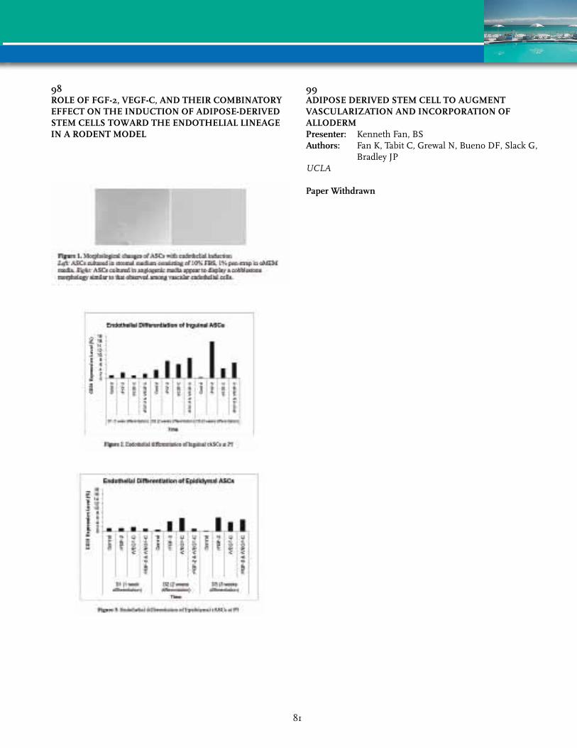
98rOLE OF FGF-2, vEGF-C, AND THEIr COmBINATOrY EFFECT ON THE INDUCTION OF ADIPOSE-DErIvED STEm CELLS TOWArD THE ENDOTHELIAL LINEAGE IN A rODENT mODEL
99ADIPOSE DErIvED STEm CELL TO AUGmENT vASCULArIZATION AND INCOrPOrATION OF ALLODErmPresenter: Kenneth Fan, BSAuthors: Fan K, Tabit C, Grewal N, Bueno DF, Slack G,
Bradley JPUCLA
Paper Withdrawn
81
101DO ADIPOSE TISSUE DErIvED STEm CELLS (ASCS) PrOmOTE TUmOr GrOWTH?Presenter: Makoto Tokuhara, MD, PhDAuthors: Tokuhara M, Saito Y, Shimizu T, Fukuda S,
Ishiguro C, Konno M, Matsuda T, Hamazaki T, Okochi H
NationalCenterforGlobalHealthandMedicine
Introduction: It is commonly known that human adipose tissue-derived stem cells (ASCs) are an attractive cell source for tissue regeneration because of their high accessibility with minimal invasiveness, compared to other somatic stem cells. ASCs may have various possibilities for clinical application, not only for tissue regeneration but also for cell therapy. Nevertheless, it has been reported that ASCs stimulate the proliferation of certain cancer cells. Therefore, we examined whether the implantation of ASCs had positive or negative effect on the proliferation of human pancreatic cancer.
material and methods: ASCs were isolated from subcutaneous adipose tissue of gastric cancer patient. ASCs were labeled with DiI before injection. Six-week old female Balb/c nude mice were injected subcutaneously with pancreatic cancer cells (MiaPaCa-2). The mice were divided into three groups. The first group had a tumor cells injection only, the second group was injected with mixture of tumor cells and ASCs, and third group had injection of ASCs from tail vein 24 hours after subcutaneous tumor cells injection. Tumors were measured every fourth day. At the end of the experiment, mice were sacrificed and tumors were excised for histological examination.
results: Co-implantation of ASCs with pancreatic cancer remarkably increased tumor growth than the cell alone. Histologic examination revealed that the aggregated ASCs were found in tumor. On the other hand, ASCs did not significantly affect on tumor growth in tail vain injection group. ASCs were detected in lung, spleen and liver in tail vain injection group, but almost no ASCs were detected in tumor.
Discussion: Co-implantation of ASCs with pancreatic cancer cell line induced a significant increase in tumor growth. However, significant tumor growth was not observed in intravenous injection of ASCs. This study suggested the paracrine effects of ASCs are associated with tumor growth promotion. The further studies are needed to elucidate the influence of ASCs on tumor growth.
106FrEE FAT TrANSFEr FOr ANAL STrICTUrESPresenter: Susanna C. Kauhanen, MD, PhDAuthors: Kauhanen S, Salmenkyla S, Tukiainen EHelsinkiUniversityHospital
Introduction: Basic research supports the immunomodulative effects of adipose derived stem cells. Therefore free fat transfer (FFT) has been allocated as a mean for treatment of troublesome scars and chronic wounds. Anal strictures hindering defecation is a disabling state.
Patients and methods: Our study comprises 3 patients with severe anal strictures. The strictures were the result of earlier radical heamorroidectomy, sfincterotomies and dilations. The patients were not able to defecate spontaneously. Their age ranged from 71-86 years (mean 78). Patients were treated in collaboration between plastic- and GI surgeons. Free fat was harvested manually from the lower abdomen. Scar tissue was “rigottomized” with a sharp canule. Thereafter fat was injected in a three-dimensional circumferential manner into the scar in the anal canal. The amount of injected fat ranged from 24-60 ml. In the first two cases, an incision at the most tense part of the stricture was performed. In these two cases the skin defect created after incision was reconstructed with a local V-Y flap. The mean follow-up time was 9 months.
results: After the surgery, patients were followed in hospital for 3-6 days. Wounds in the perianal region healed primarely in one case and after minor dehisence in one case. No morbidity was connected to the fat harvest. FFT into the strictured anus softened up the scar in a symmetrical manner. The scar softened to such an extent, that postoperatively defecation was possible without mechanical aid. The continence of the patients was not impaired. The result remained constantly soft and functioning during follow-up.
Conclusion: FFT for the treatment of anal strictures is a multidisciplinary approach to bring n new tool to help this desperate group of patients. Our preliminary results encourage us to treat the next anal stricture patients with FFT alone.
82

107DOSE-DEPENDENT EFFECT OF ADIPOSE-DErIvED STrOmAL vASCULAr FrACTION CELLS ImPrOvE ANGIOGENESIS AND ANTI-INFLAmmATION OF HUmAN FAT GrAFTPresenter: LT Li, PhDAuthors: Li LT, Teng SC, Lin YH, Fang HWNationalTaipeiUniversityofTechnology
Adipose tissue is not only an ideal material for soft tissue filling and augmentation, but also a plentiful source of regenerative cells in Adipose-derived stromal vascular fraction. The combination of fat with Adipose-derived stromal vascular fraction cells (ADSVFCs) can improve the fat graft survival in previously study. Furthermore, we estimated the effects of various dosages of stromal vascular fraction on fat graft quality in this study. Human fat tissue was subcutaneously injected into back of immunologically-compromised nude mice. The injected fat were combined without or with different dosage ADSVFCs. Base on the difference ratio of fat volume for isolate the ADSVFCs and mix to 0.5 cc injected fat tissue, there were divided in to 4 groups; group 1 (4 to 1), group 2 (1 to 1), group 3 (1/4 to 1), group 4 (1/8 to 1), respectively. After 4 weeks implantation, fat graft samples were examined the survival rate and histological evaluation was performed. The survival rate of fat graft sample, only group 1 (71.3 percentage) was slightly increased and group2 (54.8 percentage), group3 (60.1 percentage), group4 (62.3 percentage) were decreased that compared with the control group (68.1 percentage). The histological findings shown control group without ADSVFCs has poor integration that contained few mature, nucleated adipocytes in fat graft tissue. On the other hand, control group has more fibrosis, cyst formation and inflammatory cell infiltration than all groups with ADSVFCs. As the ratio of ADSVFCs in injected fat tissue increased, the fibrosis, cyst formation and inflammation decreased. Angiogenesis was also observed in each group with ADSVFCs. In conclusions, Adipose-derived stromal vascular fraction cells supplemented fat implant can improve angiogenesis, anti-inflammation and the effects of ADSVFCs on fat graft quality were dose-dependent.
108CLINICAL APPLICATION OF FAT TrANSFEr IN rECONSTrUCTIvE SUrGErY-SOUTH AFrICAN EXPErIENCEPresenter: Ewa A. Siolo, MD, MBChB, FCSAuthors: Siolo EAUniversityofKWAZuluNatal
Introduction: Fat transfers are widely used in reconstructive & esthetic surgery. Since 2005 our centre is using fat transfers in all fields of plastic surgery like: craniofacial & cleft surgery, wound healing, burn contractures, scar treatment, reconstruction of breast & posttraumatic contour deformities. This presentation will show broad spectrum of application of this modality.
material & method: This is retrospective overview of 60 cases done over the last 5 years. All cases were done according to Coleman method & using his standard set of instruments. Lipoaspirate from varieties of sites (mainly abdomen & flanks) was spun at 3000 rpm for 1.5 min. All 60 cases were done in broad categories of:Scar & keloid treatment 15Posttraumatic facial reconstruction 13Posttraumatic body contour reconstruction 2Scleroderma/romberg reconstruction 8Postradiation damage 9Breast reconstruction 5Iatrogenic deformities 3Occuloplastic reconstruction 4Hiv related facial lipoatrophy 1Most of the cases require 2-4 sessions of fat transfers repeated every 3-4 months. Number of sessions depended on severity of deformity & volume required.
results: All cases had good, stable results with high patient satisfaction. There were no major complications related to surgery.
Conclusions: Fat grafts are useful tool in reconstructive surgery. Although there is small amount of adipose derived stem cell we could observe full potential of regenerative & healing properties like: increased angiogenesis, regeneration of sensory innervation, increased collagen formation and thickening of dermis. According to above overview more clinical trials should be accepted & perform to establish properties of stem cells in regenerative medicine.
83

109IN HUmAN ADIPOSE STEm CELLS TrYPSIN TrEATmENT UPrEGULATES EXPrESSION AND SECrETION OF vEGF IN A mANNEr INDEPENDENT OF HYPOXIA INDUCIBLE FACTOr 1Presenter: Trine Fink, PhDAuthors: Fink T, Rasmussen JG, Riis SE, Lundsted DH,
Larsen BF, Frobert O, Kastrup J, Simonsen U, Zachar V
AalborgUniversity
Numerous studies indicate that in order to enhance the effect of adipose-derived stem cells (ASCs)-based theraperutic protocols aiming at ischemia-associated conditions, the hypoxic preconditioning to support a pro-angiogenic phenotype is beneficial. The angiogenic effects of ASCs have been suggested to be mediated through secretion of growth factors such as VEGF and IGF. However, while it is well known that hypoxic treatment induces VEGF and IGF-1 expression in ASCs, it has only recently been reported that also treatment with trypsin upregulates the expression of VEGF via activation of the protease-activated receptor 2 (PAR2). To obtain insight into the mechanisms involved in activation of VEGF in ASCs, we determined the effect of hypoxia and trypsin alone and in combination on the transcription, translation, and secretion of VEGF. Our results demonstrated that trypsin and hypoxia each stimulated VEGF expression and that the effects were additive. Secondly, the involvement of PAR2 in the cellular response to trypsin was confirmed through attenuation of VEGF expression by blocking with a specific antibody. Finally, through blocking different intracellular signaling pathways, it was determined that the PAR2 activation resulted in activation of the PI3K and MAPK pathways, and that this activation was independent of hypoxia inducible factor-1 (HIF-1). The possibility of enhancing a pro-angiogenic phenotype in ASCs by preconditioning the cells by hypoxic culture in combination with trypsin treatment, holds interesting prospects for future clinical applications.
112LIDOCAINE: AN ATTrACTIvE LOCAL ANESTHETIC FOr LIPOASPIrATION PrOCEDUrE IN STEm CELLS rEGENErATIvE mEDICINEPresenter: AnneClaire Girard, PhDAuthors: Girard A, Loyher PL, Bencharif K, Balat M,
Lefebvre d Hellencourt C, Delarue P, Hulard O, Roche R, Festy F, Hoareau L
Stemcis
Introduction: Lidocaine is a local anesthetic commonly used for liposuction. Only a few studies have tried to explain the effects of lidocaine on fibroblastic cells, and controversial results exist on preadipocytes. Mechanism of action and effects on adipose tissue, particularly on cells from the stromal vascular fraction (SVF), is not clearly understood. Here, we sought to detect the direct effect of lidocaine on SVF cells.
methods: Human SVF cells were isolated from lipoaspirates and cultured before being treated with different concentrations of lidocaine, for different exposure times. Following short or long exposure, cell viability was measured by lactate dehydrogenase (LDH) activity and by flow cytometry using propidium iodide (PI). Inflammatory status was assessed by interleukin-6 (IL-6) and tumor necrosis factor-alpha (TNFa) secretion (ELISA).
results: LDH cytotoxicity assays showed no difference after short exposure to lidocaine compared to non-treated cells, but cell death could be visualized from 12 hours of treatment in a dose-dependent manner, as confirmed by the increase of PI staining by flow cytometry.
Whatever the dose of lidocaine used and the time of exposure, the level of TNFa and IL-6 secretion remain unchanged. Consequently, no inflammation seems to be induced by lidocaine.
Conclusion: This study provides evidence that short exposure to lidocaine has no effect on SVF cells viability and does not lead to inflammation. However, longer exposure or high dose of lidocaine can directly affect SVF cells by promoting cell death.
Lidocaine seems to be a good local anesthetic that can be used especially for regenerative medicine, provided that the cells do not suffer from abusive concentration. Indeed, time between tissue aspiration and cell extraction usually does not exceed 2 hours during regenerative surgery. Nevertheless, the protocol of SVF cell extraction must include steps of washing before reinjection of these cells in the patient, in order to be sure of the viability after reinjection. Further investigations are necessary to define the actual mechanism of action of lidocaine on SVF cells.
84

113DEvELOPmENT OF A BILAYErED DErmAL SCAFFOLD WITH A NEW GENErATION NANOCOmPOSITE POLYmEr SEEDED WITH ADIPOSE TISSUE DErIvED STEm CELLSPresenter: Reema Chawla, BScAuthors: Chawla R, Moieman N, Butler PE,
Seifalian AMDivisionofSurgeryandInterventionalScience
Background: Despite the myriad of skin substitutes, current gold standard treatment of full-thickness burns remains split-thickness autografts. However, their use cannot be extended to patients with a large % total body surface affected. The objective was to develop a porous bilayered scaffold for dermal replacement from a novel nanocomposite polymer, polyhedraloligomeric-silsesquioxane poly (caprolactone-urea) urethane (POSS-PCL) and compare the properties of the construct to the current industry leader, Integra®; in addition, to seed adipose tissue derived stem cells (ADSC’s) onto developed scaffolds, which enhance wound healing and angiogenesis.
methods: The inner layer was produced via phase separation for a highly porous morphology. A removable outer layer incorporated silver nanoparticles to impart antimicrobial properties. Effects of different pore sizes on chemical and physical properties were established by tensile testing, contact angle, permeability, FTIR and scanning electron microscopy (SEM) analysis. Optimal pore morphology for cell proliferation was elucidated through ADSC culture. Cell viability and apoptosis were tested using an Alamar Blue™ (AB) and LDH assay respectively. All tests were repeated on Integra®.
results: The physical construct was easy to handle and clinically applicable. Results demonstrate the macroporosity and permeability of the scaffolds, which were up to 72% porous; confirmed by SEM. Outer layer contact angle was >100°, indicating hydrophobicity and the inner layer was <70° indicating hydrophilicity of the scaffold. Young’s modulus of scaffolds ranged from 0.406-0.492 MPa. Both results are comparable to skin. AB assay showed cell proliferation onto the scaffold, comparable to that on Integra®. This was confirmed by fluorescent imaging.
Conclusions: In vitro assessment of the dermal scaffold suggests it is a promising alternative to the current industry leader, Integra® and has many desirable properties that could successfully mimic human skin. Future directions involve covalently bonding bioactive molecules (i.e. cyclic RGD) to further enhance cell attachment and proliferation.
113DEvELOPmENT OF A BILAYErED DErmAL SCAFFOLD WITH A NEW GENErATION NANOCOmPOSITE POLYmEr SEEDED WITH ADIPOSE TISSUE DErIvED STEm CELLS
85

114ImmUNOmODULATOrY mECHANISm OF HUmAN ADIPOSE TISSUE-DErIvED mESENCHYmAL STEm CELLS: rOLE OF SOLUBLE FACTOrSPresenter: Swathi SundarRaj, PhDAuthors: SundarRaj S, Priya N, Gopalakrishnan DStempeuticsResearchPvtLtd
Introduction: Adipose tissue-derived stromal cells (ASC) have enormous potential in immunomodulatory cell therapy. ASC inhibit activation, proliferation and function of T-, B-, NK-cells and monocytes, and suppress inflammation. Animal models have shown efficacy of ASC in ameliorating autoimmune disease and promoting allograft tolerance. However, the molecular mechanisms involved in ASC-immunoregulation still remain largely undefined, and formed the focus of our study.
method: ASC were co-cultured with naive, mitogen or allo-antigen stimulated PBMC and the effect on lymphocyte proliferation was measured. Induction of tolerogenic factors by ASC upon co-culture was determined and quantified by gene expression profiling. Requirement of cell contact vs. soluble factors was studied using conditioned medium from native or co-cultured ASC, and by co-culture in transwell systems. Antibody-based array analysis was carried out to study cytokine secretion from the co-cultures.
results: Suppression of PBMC proliferation by ASC is brought about mainly by soluble factors that require direct contact with PBMC or an inflammatory environment for induction. Gene expression profiling identified induction of IDO, PGE-2, LIF, IL-10, galectin-1 and galectin-9 from co-cultured ASC. Cytokine profiling of co-cultures revealed upregulation of GCSF, GM-CSF, GRO-α±, IL-10, VEGF and angiogenin, and a decrease in the inflammatory cytokines IL-1, IL-5, IL-8 and TNF.
Conclusion: We show that immunosuppression by ASC is mediated predominantly by soluble factors. IDO, PGE-2, LIF and IL-10 are known to promote immune tolerance in various physiological settings. We demonstrate for the first time expression of galectin-1 and galectin-9 from ASC and their involvement in immune regulation. Galectins are immune effectors during development and disease, and promote fetal allograft and transplantation tolerance. The secretion of the panel of soluble factors outlined above, combined with the suppression of inflammatory cytokines, highlights the potential of ASC in immunomodulatory cell therapy.
115ENCAPSULATED ADIPOGENIC FACTOrS EFFECT IN ADIPOSE GrAFT rETENTIONPresenter: Kacey Marra, PhDAuthors: Marra KG, Tan H, Rakers A, Rubin JPUniversityofPittsburgh
Background: Adipose tissue retention during fat transfer has been one of the major challenges in plastic and reconstructive surgery. While current reconstructive procedures involve movement of soft tissue from other areas of the body, there remains an unmet need for new modalities that are less invasive and more precise. One strategy involves the controlled delivery of adipogenic factors, such as insulin and dexamethasone, within the fat graft. In this study, both drugs were encapsulated in poly (lactic-co-glycolic acid), (PLGA) microspheres and mixed with lipoaspirate to induce adipogenesis in vivo, using a combined drug therapy approach.
Hypothesis: We hypothesize that the slow release of combined insulin and dexamethasone can enhance adipogenesis and angiogenesis, thus retaining the fat graft volume.
methods: Insulin/dexamethasone-loaded PLGA micospheres (Insulin/Dex MS) were prepared using double emulsion/solvent extraction technique. The bioactivity of the drugs was assessed by mixing the microspheres with human lipoaspirate and injecting subcutaneously into an athymic mouse model. Group A contained 56 mg of insulin MS per 1 ml of human fat with 150 mg dexamethasone MS; group B contained 28 mg MS of insulin and 150 mg dex MS per 1 ml human fat; group C contained 56 mg of insulin MS and 80 mg dex MS per 1 ml of human fat; and group D with 28 mg of insulin and empty MS with the last group E with 80 mg of dex combined with empty MS. Samples were analyzed grossly and histologically after 3, 6 and 12 weeks in vivo.
results: Mass and volume were measured, with the microsphere-containing samples demonstrating an increased mass and volume displacement with an increase MS content. Histological analysis indicates there is increased vascularization within the insulin/dex MS-containing samples, individually, and the combined therapy analysis is underway.
Conclusion: This study demonstrates that the controlled delivery of adipogenic factors via polymer microspheres can significantly affect tissue formation and vascularization. This represents a clinically relevant method of stimulating fat retention.
86

116THE APPLICATION OF AN ADIPOSE-DErIvED STEm CELL SHEET IN WOUND HEALINGPresenter: YenChih Lin, PhDAuthors: Lin YC, Grahovac TL, Oh SJ, Rubin JP,
Marra KGUniversityofPittsburgh
Introduction: There is a clinical need for wound healing that will enhance skin regeneration. Stem cell sheets have been used for bone, ligament and wound tissue-engineering research in recent years. Adipose-derived stem cells (ASCs) have the capacity for self-renewal and the capability of differentiation to various cell lineages. The specific aim of this study was to examine an ASC sheet for wound healing in an athymic nude mice model.
methods: ASCs were isolated from discarded human abdominal subcutaneous adipose tissue, and then ASC cell sheets were created on the surface of fibrin-grafted culture dishes. In vitro experiments investigated the histochemical characterization of ASC sheets. In vivo experiments consisted of implanting single layer sheets, triple layer sheets or empty wounds (control treatment) onto a full-thickness wound defect in nude mice for three weeks.
results: Cell sheets were easily peeled off of the dishes using forceps. The single layer and triple layer ASC sheets showed complete extracellular structure via hematoxylin and eosin staining. As an indicator of would recovery, the injury area was measured 7, 14, and 21 days post treatment. The injury area from the ASC sheet treatment was significantly smaller than the empty control group at all time points. The triple layer ASC sheet treatment significantly enhanced wound healing over the single layer ASC sheet at both 7 and 14 days. Hematoxylin and eosin staining showed that ASC treatment significantly enhanced cell proliferation and migration over control treatment.
Conclusions: From our studies, we conclude ASC sheets represent a potentially viable matrix for full-thickness defect wound healing in a mouse model. Consequently, our ASC sheet technology provides a substantial advance for developing various types of three-dimensional tissues and represents a potentially viable matrix for full-thickness defect wound healing in a mouse model.
117GENErATION OF AN ADIPOSE-DErIvED EXTrACELLULAr mATrIX SPONGE THAT rETAINS BIOCHEmICAL AND BIOLOGICAL INTEGrITYPresenter: Jerome Connor, PhDAuthor: Connor JKineticConceptsInc
Introduction: Intact extracellular matrix (ECM) materials have been demonstrated to be effective connective tissue scaffolding for cellular repopulation and revascularization. The ability to generate an adipose-derived ECM scaffold, which retains biological components, could provide a suitable matrix for fat grafting and delivery of adipose stem cells clinically. This report describes the generation of a biologically competent ECM derived from human adipose that could serve as a biologically intact scaffold for fat grafting.
methods: Human adipose was obtained from human cadavers following all requirements of the Tissue Banking industry. The adipose was processed using sequential mincing steps in conjunction with centrifugation washes to separate the ECM material from cellular components. The resulting ECM was dispersed into templates and freeze-dried. The freeze-dried ECM was mechanically stabilized using protein cross-linking technology. The adipose tissue sponge (ATS) was evaluated for retention of ECM and biochemical components. Electron microscopy was performed to visualize the micro-structure. Biomechanical integrity was measuring by flow dynamics under vacuum. The ATS was implanted into nude rats and evaluated histologically for the in-growth of fibroblast and new vasculature, the deposition of new collagen and the presence of macrophage.
results: The ATS retained intact collagen fibrils in a web-like structure. The sponges contain chondroitin sulfate and hyaluronic acid as well as growth factors. The ATS demonstrated biomechanical integrity measured as constant flow under vacuum. In-vivo, the ATS demonstrated significant, rapid in-growth of fibroblasts, the presence of new vessels and deposition of collagen with a minimal presence of macrophages.
Conclusion: This report demonstrates the generation of a sponge matrix derived from human adipose tissue that retains key biochemical components yielding a positive biological response following implantation. This ATS could serve as a biologically competent scaffold for human fat grafting potentially in conjunction with adipose-derived stem cells.
87
118CO-CULTUrE OF HUmAN ADIPOSE DErIvED STEm CELLS AND NUCLEUS PULPOSUS CELLS FOr INTErvErTEBrAL DISC rEPAIrPresenter: Donna Haworth-Ward, PhDAuthors: Haworth-Ward D, Oh SJ, Hoyer R, Witt W,
Kim KJ, Vo N, Sowa G, Rubin JP, Marra KGUniversityofPittsburgh
Background: Intervertebral disc (IVD) degeneration, caused by loss of disc matrix, leads to severe back pain, which affects 60-80% of the population during their life. Current treatments only address the symptoms of this degeneration, but do not alter the course of the disease process. Stem cell therapy has the potential to reverse the disease course through improving cellular production of matrix. The objective of this study wereto determine the effects of direct co-culture in a 3D environment of adipose derived stem cells (ASCs) and nucleus pulposus (NP) cells with and without TGF- on extracellular matrix content.
methods: ASCs and degenerated NP cells were obtained from human subjects and cultured. ASCs, NP cells, ASCs+NP cells or ASCs+NP cells+TGF- (5 ng/mL) were cultured in direct contact via a pellet culture for 1 and 2 weeks at both 5% and 20% oxygen in chondrogenic differentiation media. Following culture, pellets were assessed for glycosaminoglycan (GAG) content via a dimethylmethylene blue colorimetric assay, DNA content via a PicoGreen assay and were histologically evaluated via H&E, alcian blue and saftranin-O staining.
results: ASCs at 20% oxygen produced the greatest GAG amount per cell (ug GAG/ng DNA) after 1 week of culture, with the remaining groups being showing minimal variation. After 2 weeks of culture, ASCs+NP cells±TGF-showed an increase in 1 week GAG content with ASCs+NP cells+TGF- at 5% oxygen having the greatest increase. H&E results at 1 week show that NP cells at 5% oxygen formed a hollow pellet, while all other pellets were solid spheres. The size of the pellet increased with an increasing amount of NP cells. NP cells at 5% oxygen had the greatest staining for Safranin-O, followed by NP cells at 20% and ASC+NP cells+ TGF- at 20% after 1 week of culture.
Conclusion: These results indicate that the direct co-culture of ASCs and NP cells in a 3D environment in the presence of TGF- has the potential to increase GAG content. These results could lead to a potential therapy where ASCs and TGF- could be directly injected into degenerative IVD for repair.
119INTrAmYOCArDIAL TrANSPLANTATION OF HUmAN ADIPOSE-DErIvED STrOmAL CELL AND ENDOTHELIAL PrOGENITOr CELL mIXTUrE WAS NOT SUPErIOr TO INDIvIDUAL CELL TYPE TrANSPLANTATION IN ImPrOvING LEFT vENTrICULAr FUNCTION IN rATS WITH mYOCArDIAL INFArCTIONPresenter: SoonJun Hong, MD, PhDAuthors: Hong SJ, Choi SC, Lim DS, Kim JH, March KLKoreaUniversityAnamHospital
Background: Both adipose-derived stromal cells (ASCs) and endothelial progenitor cells (EPCs) have high potential for promoting tissue revascularization and functional recovery in acute myocardial infarction (AMI) models. We investigated the functional effects of intramyocardial transplantation of a human ASC and EPC mixture in immunodeficient rats after MI.
methods: MI was induced by ligating left anterior descending coronary artery. Survived rats were randomly assigned to 1 of 4 different groups: the control group (n=10, saline in 100 µL), the ASC group (n=10, 106 ASCs), the EPC group (n=10, 106 EPCs), or the ASC+EPC group (n=10, 2 x 10⁵ ASCs + 8 x 10⁵ EPCs). Left ventricular (LV) function was compared using echocardiography during the 28-day follow-up. GAP43+ nerve sprouting and smooth muscle alpha-actin+ angiogenesis were also compared.
results: Serial changes in LV ejection fraction (EF) and fractional shortening revealed significant increases in the ASC, EPC, and ASC+EPC groups when compared to the control group during the follow-up (49±3%, 49±4%, 47±4%, 39±2%, P<0.001, respectively for LVEF) (33±4%, 32±2%, 31±2%, 23±2%, P=0.002, respectively for fractional shortening). The number of alpha-actin+ arterioles and GAP43+ nerve area were significantly greater in the ASC, EPC, and ASC+EPC groups when compared to the control group in the peri-infarct area (34.4±1.0/mm2, 35.9±1.1/mm2, 35.3±0.9/mm2, 17.4±0.7/mm2, P<0.001, respectively for angiogenesis) (346.2±10.7 µm2/mm2, 357.2±12.8 µm2/mm2, 368.0±9.7 µm2/mm2, 174.6±7.9 µm2/mm2, P<0.001, respectively for nerve sprouting).
Conlusions: Intramyocardial injections of ASCs, EPCs, or ASCs+EPCs are effective modalities for the treatment of myocardial damage in rats and may expand the potential clinical application of ASC or EPC therapy in patients with ischemic heart disease.
88

121FAT GrAFTING TO THE rECONSTrUCTED BrEAST: THE USE OF 3D ImAGING TO EvALUATE vOLUmE rETENTIONPresenter: Kevin Small, MDAuthors: Levovitz C, Small K, Choi M, Karp NSNewYorkUniversity
Introduction: Fat grafting (FG) has emerged as a useful method for breast contouring. However, our ability to judge the overall success of FG remains limited. The following study applies three-dimensional (3D) imaging to assess the stability of breast volume following FG. In this study, 65 patients underwent FG into 97 breasts. They included autologous and implant reconstructions and lumpectomy defects.
methods: FG surgery was performed using a modified Coleman technique. 3D imaging was performed using the Canfield VECTRA system and analyzed using Geomagic software. Preoperative and post-operative 3D scans were obtained on all patients and a calculated volume deficit was determined for each breast.
results: The data stratified patients into 3 groups with statistically significant parameters based on the volume of fat injected. The group that received the largest volume of fat had an average injection volume of 144 cc, lost 2.4% of vol/day during the 1st postoperative week (POW), 1.3% of vol/day over the 2nd and 3rd POW, and experienced volume stability (VS) at an average post-operative day (POD) 67 with a volume retention (VR) of 51%. These findings significantly contrast with the patients receiving smaller volume fat, who had an average injection volume of 47 cc, lost 1.8% of vol/day the 1st POW, 1.6% of vol/day over the 2nd and 3rd POW, and did not experience VS until POD 120 with 39% VR. The third patient group received an intermediate volume of FG with an average injected volume of 95 cc, lost 2.10% of vol/day the 1st POW, 1.30% of vol/day over the 2nd and 3rd POW, and experienced VS at POD 77 with 42% VR. Of note, radiation did not seem to affect the VR rate of any of the three patient groups.
Conclusion: Our data suggests fat retention is volume and time dependent. Patients receiving >110 cc of FG lost the greatest percentage of volume the 1st POW and then the rate of volume loss tapered until VR stabilized to half the total volume injected around 2 months post-operatively. Patients receiving smaller volumes of FG take longer to reach VS and have lower rates of VR. Radiation therapy does not seem to affect FG results.
122SOrTING OF FOUr DISTINCT SUB-POPULATIONS FrOm HETErOGENEOUS ADIPOSE DErIvED STEm CELL POOL WITHIN STrOmAL vASCULAr FrACTION FOr PLASTIC AND rECONSTrUCTIvE APPLICATIONSPresenter: Sudheer K. Ravuri, PhDAuthors: Ravuri SK, Philips BJ, Li H, Meyer EM,
Pfeifer ME, Zimmerlin L, Marra KG, Donnenberg VS, Donnenberg AD, Rubin JP
UniversityofPittsburgh
Introduction: Human adipose derived stem cells (ASCs) may have broad applications to plastic and reconstructive surgery. Limited information is available on highly heterogeneous ASC population and its distribution within the Stromal Vascular Fraction (SVF) of isolated adipose tissue. Hence, this study focused on phenotypic characterization of ASCs by flowcytometry via lentiviral-mediated transduction.
methods: Standard procedures were followed for SVF isolation from human adipose tissue. Distinct cell populations were sorted by flowcytometry based upon CD3, CD31, CD34, CD45, CD90, CD117 and CD146 surface marker profiles. Apoptotic cells and cell clusters were excluded by DAPI staining. Lentiviruses expressing fluorescent genes (GFP, RFP, CFP & YFP) were constructed by transfection of HEK293T cells. Lentivirus transduced ASC populations was mixed with lipoaspirate and implanted into athymic nude mice. Masses and volume of fat tissue explants were analyzed at 3 & 6 weeks using a gas pycnometer. Tissue sections of explants were imaged by confocal microscopy.
results: Flowcytometric analyses showed four distinct sub-populations characterized as weakly proliferative 0.15% Endothelial “mature” (CD31+CD34-CD3-CD45- and 0.4% Pericytes (CD31-CD146+CD3-CD45-) and highly proliferative 0.65% Endothelial “Progenitor” (CD31+CD34+CD3-CD45-) and 4.8% Preadipocytes (CD31-CD34+CD146-CD3-CD45-). Gross morphology inspection showed increased vascularization, masses and volumes in Lipo+ASC explants at 3 & 6 weeks time points. Sections of fat tissue explants showed fluorescence for four (GFP, RFP, CFP & YFP) lentiviral reporter genes under confocal microscopy. Sorted preadipocyte population (CD31-CD34+) showed higher expression of PPAR gamma and observed enhanced adipogenesis with minimum stimulation compared with unsorted ASCs.
Conclusions: We have successfully sorted four distinct ASC sub-populations and individually transduce them with each fluorescent lentivirus to determine the fate of each population. Preadipocyte population showed greatest adipogenic potential. Different scaffolds and cell signaling mechanisms are under evaluation.
89
123CELL BIOLOGY OF CELL ASSISTED FAT GrAFTINGPresenter: Ramon Llull Cerda, MD, PhDAuthors: Llull Cerda R, Dos-Anjos S, Katz A, Futrell WStemCenterSL
Autologous fat transplantation is being successfully used for facial rejuvenation and soft tissue augmentation. The use of cell-assisted lipotransfer has solved the problem of unpredictable results and low rate of fat graft survival. However, the mechanisms mediated by stromal vascular fraction on fat graft survival are not very well understood In this paper we present the results obtained from co-incubation studies of fat grafts and stromal vascular fraction cells. The cells adhere to the fat in a short period of time. Moreover, the cell quantity and/or concentration used is very important, since using small amounts of cells results in death or dedifferentiation of adipocytes, whereas using a significantly higher amount of cells lead to fat graft survival. The cells between fat grafts organize in networks similar to blood vessels in some situations and can also differentiate into adipocytes. In summary the ratio between adipose tissue and cell number is very important to the long-term survival of fat graft, as well as for adipogenic differentiation.
124FACE LIFTING ASSISTED BY STrOmAL ENrICHED LIPOGrAFT vErSUS FACE LIFTING ASSISTED BY NON STrOmAL ENrICHED LIPOGrAFT: A CLINICAL STUDYPresenter: Aris Sterodimas, MD, MSc, PhDAuthors: Sterodimas A, Nicaretta B, Illouz YGIASOGeneralHospital
Paper Withdrawn
90
125ADIPOSE TISSUE ENGINEErING IN PLASTIC SUrGErY: CUrrENT AND FUTUrE APPLICATIONSPresenter: Yves Gerard Illouz, MD, PhDAuthors: Sterodimas A, Nicaretta B, Illouz YGIASOGeneralHospital
Standard approaches to soft-tissue reconstruction include autologous tissue flaps, autologous fat transplantation and alloplastic implants. All of these approaches have disadvantages, including donor-site morbidity; implant migration, and foreign body reaction. Adipose tissue is the most abundant and accessible source of adult stem cells. After the introduction of liposuction, adipose tissue harvesting has become easier. Adipose-derived stem cells (ADSCs), because of their pluripotentiality and unlimited capacity for self-renewal, project great promise for tissue engineering and are expected to allow significant advances for distinct reconstructive procedures.
A brief description of the primary functions and cellular composition of fat, with an emphasis on the adipose-derived stem cell population is presented. Current methods for isolating ADSCs rely on a collagenase digestion followed by centrifugal separation to isolate the stromal/vascular cells from primary adipocytes. Tissue-specific scaffolds and signaling systems are essential in order to differentiate stem cells into the required cells and use them effectively to construct three-dimensional tissues.
The results from some of the main adipose tissue engineering studies are highlighted, and a perspective for the future is provided. In Stromal Enriched Lipograft (SEL), freshly isolated SVF is attached to the aspirated fat, with the fat tissue acting as a living bioscaffold before transplantation. The preliminary results suggest that SEL is effective and safe for soft tissue augmentation and superior to conventional lipoinjection.
The current applications and the future perspectives of adipose tissue engineering unquestionably represent innovative techniques with huge therapeutic potential that must be though balanced against the stringent standards of scientific and clinical investigation.
126BEYOND GrAFTING – UNCOvErING THE mOLECULAr mECHANISmS OF HUmAN TISSUE AGING USING PrImArY ADIPOSE TISSUE AS A mODELPresenter: Ivona Percec, MD, PhDAuthors: Percec I, Dierova R, Bucky LP, Chang B,
Auman D, Hoover WUniversityofPennsylvania
Aging is a complex phenomenon involving thousands of genes. We know that longevity is heritable, caloric restriction increases life span, and epigenetic contributions may be more important than genetic ones. Yet, these findings are derived from in vitro assays or model organisms, questioning their applicability to normal human aging. Fat grafting provided us with critical insight, namely that with age, the viability of adipose tissue decreases, adipose-derived stem cells (ASCs) decrease in number and activity, and anatomic compartments age differently. We hypothesize here that primary human adipose tissue is an excellent model with which to study human aging.
Adipose tissue samples are collected from patients undergoing aesthetic surgery at UPenn. Adipocytes, stromal vascular fraction (SVF) and ASCs are isolated from each specimen. Genome-wide transcriptional differences are characterized using gene chips. An analysis of the Sirtuin gene family (SIRT1-7) investigates the contribution of this pathway, whose role has been well documented in model organism aging, to normal human adipose tissue aging.
Our model of adipose tissue aging demonstrates that primary human adipose tissue can successfully be analyzed via genome-wide approaches. Adipocytes, SVFs and ASCs clearly differ in their molecular aging phenotypes. We have identified transcriptional differences in over 700 differentially expressed genes and establish that transcriptional modifications are more important in distinguishing between cell types than between age-related changes. Furthermore, we demonstrate for the first time that the human Sirtuin genes, which are known to be involved in aging and adipose metabolism in model organisms, regulate human adipose tissue aging in a differential manner via specific effects on cell type and chronological age. These findings are critical for advancing the understanding of tissue aging and demonstrate that adipose tissue is an excellent model for the study of human aging.
91

127POTENTIATION OF NEOvASCULArIZATION ACrOSS TISSUE INTErFACES BY STrOmAL vASCULAr FrACTION CELLS IS vEGF DEPENDENTPresenter: Laxminarayanan Krishnan, MBBS, PhDAuthors: Krishnan L, Nunes SS, Chang CC,
Williams SK, Hoying JBCardiovascularInnovationInstitute
The integration of implanted constructs, engineered or autotransplants in reconstructive surgery, is critically dependent on early vascularization. However, angiogenic neovessels approaching a boundary between tissue structures may not readily grow across such interfaces. While neovascularization strategies centered around augmenting the results of transplants by addition of mesenchymal stem cells have received significant attention, the role of the interface as a deterrent to inosculation between host and implanted vessels has not been investigated. Based on literature evidence relating invasive nature of cells to their extracellular matrix structure, we developed an interface model, using a vascularized collagen construct generated using adipose derived microvessels as the core, surrounded by cell free collagen on the sides, to serve as an in-vitro platform to study microvessel growth across tissue boundaries. We have previously shown that while angiogenesis in the core progresses to form networks, instances of neovessels traversing the core-exterior interface are infrequent; and that addition of stromal vascular fractions cells (SVFs) derived from collagenase digestion of adipose tissue, to the microvessels in the core, dramatically increases crossing events. Here we describe a possible mechanism by which SVFs improve graft performance. We hypothesize that a close association of microvessels with SVFs is necessary, in addition to their global pro-angiogenic role. Using a tissue-insert co-culture model, we show that the SVF secreted soluble factors alone are not sufficient to promote neovessel growth across the interface. Further, a reduction in crossing events (with SVFs) by VEGF blockers indicates that neovessel secretion of VEGF, arguably involved in recruiting SVFs in its proximity to incorporate, is central to this potentiating activity. Given the lack of significant changes in matrix structure at the interface by SVFs, we propose a regulatory modulation of existing microvessels and incorporation of SVF into neovessel sprouts as the underlying mechanism potentiating neovascular growth across boundaries.
128QUANTIFICATION OF INTErACTIONS OF ADIPOSE DErIvED STrOmAL CELLS AND THE mICrOvASCULATUrEPresenter: James Hoying, MDAuthors: Krishnan L, Nunes SS, Shumate K, Hoying JB,
Williams SKCardiovascularInnovationInstitute
Adipose derived cells are commonly used to improve implant or ischemic revascularization. In addition to cytokines produced by these cells, recent studies, including ours, demonstrate a complex interaction between these cells and the neovasculature. Such incorporation as endothelial, smooth muscle, or perivascular cells, suggest a defined role for these cells in the developing microvascular bed, and may be critical in augmenting vascularization and modulating network characteristics. We believe that accurate quantification of such complex relationships will provide opportunities for controlled perturbations on these cells to determine its outcome on the neovasculature. Here we present a method to quantify such interactions based on differential labeling of SVFs and vasculature. Central to the technique is the fluorescent labeling of SVFs (or genetic reporter systems), and a secondary marker for vasculature (Isolectins). In our study, collagen constructs containing SVFs with or without microvessels were implanted subcutaneously in immunocompromised mice. Explants were stained to label the vessels and confocal image stacks were generated at random areas of the explants under 488 nm and 594 nm illumination. These images were corrected for depth errors, median filtered, and deconvolved to improve image quality, and then binarized using a custom threshold value for each stack. The overlap volume, common to both SVFs and vessels was calculated using logical (and) operations. When expressed as a ratio of the total cell or vascular volume, the overlap volume allows estimation of percent vascular volume made up of exogenous SVF. Further, a similar metric called the overlap number, allows estimation of number of cellular structures in contact with vasculature. A high volume overlap is indicative of SVF integration into the neovasculature, while a low volume overlap along with a high overlap number is indicative of perivascular SVF association. This analysis agrees with qualitative observations of SVF incorporation into microvessels and is being improved to identify vasculogenesis from SVF in addition to SVF incorporation.
92
129EFFECT OF SECrETIN ON PrE-, DIFFErENTIATING AND mATUrE ADIPOCYTE FUNCTIONSPresenter: Pierre Miegueu, MSAuthors: Miegueu P, Cianflone K, Denis R,
Saint-Pierre DHLavalUniversity
Paper Withdrawn
131BrEAST ImPLANT AUGmENTATION COmPLEmENTED BY STrOmAL ENrICHED LIPOGrAFTPresenter: Beatriz Nicaretta, MDAuthors: Sterodimas A, Nicaretta B, Illouz YGIASOGeneralHospital
Up to now, adipose tissue injection to the breast or mammary lipoaugmentation has been stuck by two limiting factors. Firstly, fat injection in and around the breast could result in cyst formation, indurations and fat necrosis that could be mistaken as cancerous calcifications. Secondly, the degree of reabsorption of the injected adipose tissue is unpredictable. Fat grafting remains shrouded in the stigma of variable results experienced by most plastic surgeons when they first graft fat.
A perspective study in order to evaluate the outcomes, complications and patient satisfaction of silicone breast augmentation complemented by Stromal Enriched Lipograft (SEL) was performed during November 2008 and January 2010. Twenty patients, aged 24-36 years old, were included in the study. All the patients had a silicone polyurethane implants inserted complemented by SEL. Overall satisfaction with the breast appearance after breast augmentation complemented by SEL was rated on a scale of 1 to 5, whereas 1 is ‘poor’, 2 is ‘fair’, 3 is ‘good’, 4 is ‘very good’ and 5 is ‘excellent’. The evaluation was made at follow-up time of 12 months.
The silicone implant size ranged from 190 to 285 cc. The SEL volume ranged from 63 to 110 ml on each breast. There was no hematoma formation and no case of infection. There was one patient that developed seroma that was treated conservatively. One patient underwent implant exchange because of implant size dissatisfaction. One case of stretch marks in a young nulliparous woman did not need treatment. Three unilateral breast fat necrosis were confirmed as benign by mammographic examination. A meta analysis of the patient satisfaction after silicone breast augmentation complemented by SLE after 1 year of follow up is presented.
Preliminary results of this study shows aesthetically acceptable outcomes following silicone breast augmentation and SEL. Further long term studies need to be performed in order to confirm the favorable results seen in this study.
93
133ISOLATION, CrYOPrESErvATION AND TrI-LINEAGE DIFFErENTIATION OF ADIPOSE-DErIvED STEm CELLS FrOm HUmAN LIPOASPIrATEPresenter: Kevin GradyAuthors: Klarmann GJ, Grady K, Keller JLonzaWalkersvilleInc
Subcutaneous fat is a rich source of multipotent adult stem cells that is attainable in large quantities from lipoaspirate. The therapeutic potential of ADSCs is of great interest to the research and medical communities. We report the isolation and cryopreservation of ADSCs. These cells retain rapid proliferation and differentiation potential after thawing. Stromal-vascular fraction was obtained with enzymatic digestion of fresh lipoaspirate. This cell mixture was grown in flasks with FBS for several days to enrich for ADSCs. Cells were then harvested and frozen at a controlled rate. Upon thawing and replating, flow analysis showed >90% of the cells expressed CD13, CD29, CD44, CD73, CD90, CD105 and CD166, while <5% expressed CD45, CD14, CD31 and CD34. The cells had a doubling time of ~21 hrs and robust post-thaw viability (5 different donors). Adipogenic differentiation was obtained in monolayer culture after ~12 days of growth in Preadipocyte basal medium (PDM-2TM) supplemented with human insulin, dexamethasone (dex), IBMX and indomethacin. Cells had obvious intracellular adipose droplets that were positive by AdipoRedTM staining. The S/N for differentiated cells ranged from 32-57. Osteoblast differentiation was achieved by growth for 21 days on coated plates in the presence of dex, ascorbate and serum. In this process, morphology changed to cuboidal, and mineral deposits were observed in phase contrast. The cells were positive for OsteoImageTM staining and had a S/N ratio of ~35. Chondrogenesis was achieved by growing 500,000 cells in pellet culture for 21 days in the presence of dex, ascorbate, pyruvate, proline, ITS supplement, and TFG-3. Fixed pellets were positive by Alcian blue staining only when the media contained TFG. Differentiation to other lineages is being investigated. Thus we demonstrate that our ADSCS are of high purity and quality and are capable of multilineage differentiation.
134ADIPOSE STEm CELLS INFLUENCE SELF-rENEWAL OF BrEAST CANCEr STEm CELLSPresenter: Riesa M. Burnett, MDAuthors: Burnett RM, Merfeld-Clauss S, Wooden WA, March KL, Nakshatri HIndianaUniversity
Breast reconstruction after breast cancer surgery is a constantly evolving field. Autologous fat transplantation has become a means of achieving satisfactory cosmetic results without the use of synthetic or allograft materials. Given the discovery of multipotent stem cells in a variety of tissues including adipose, and the ability of these stem cells to influence the growth and differentiation of surrounding cells through paracrine effects, it is logical to ask whether transplanted adipose stem cells affect proliferation/survival of any residual/dormant cancer cells when placed in a former tumor bed. An in vitro model was established to test this possibility. Adipose stromal/stem cells (ASCs) isolated from three individuals, “normal” breast epithelial cell line MCF10A, and a SRC oncogene transformed variant of MCF10A (MCF10A-ER-SRC) were examined for their ability to form mammospheres (a surrogate in vitro self-renewal assay) in mammosphere culture media either individually or in combination. MCF10A and MCF10A-ER-SRC cells had a stably integrated green fluorescence protein (GFP) gene to distinguish breast epithelial cells from adipose stromal cells in the spheres. After 7-10 days in culture, plates were examined for the number and size of spheres formed, presence of GFP fluorescence, and for various cell surface markers. Although co-culturing with ASCs did not increase the number of spheres formed by MCF10A or MCF10A-ER-SRC, ASCs significantly increased the size of spheres, indicating a paracrine effect on self-renewal of breast stem cells. Interestingly, ASCs cultured under mammosphere conditions formed spheres (adipospheres). Adipospheres and mammospheres displayed several common cell surface markers except for the epithelial marker EpCAM. These results raise the possibility of engrafted adipose stromal/stem cells reactivating dormant cancer cells. Further studies are required to identify adipose-derived factors that influence self-renewal of breast cancer stem cells and therapeutically target such factors to overcome potential deleterious effects of autologous fat cell transplantation.
94

136COrrECTING LOWEr EYELID rETrACTION USING FAT GrAFTINGPresenter: Katarina Andjelkov, MD, MSAuthors: Andjelkov K, Sforza M, Zaccheddu RPrivatePractice
Introduction: Many surgeons largely accept the use of fat as a natural filler. The filling effect of fat has the potential to be used as a natural spacer, expanding areas with cicatricial retraction. Moreover, the anti-inflammatory properties of fat-derived stem cells are well recognized subsequent to the cell transfer to other areas. Eyelid retraction after blepharoplasty is a severe deformity that could potentially significantly benefit from the anti-inflammatory and expansion effects of fat. We present a retrospective study of 10 patients who had fat injections in order to correct eyelid retraction following lower blepharoplasty.
method: All 10 patients presented eyelid retraction in the early postoperative period. The age range was between 42 and 62 years. In all patients, fat grafting was done within the first month of the original procedure under local anesthetic. The evaluation of lid laxity was done by gently pulling up the lower eyelid. Any patient with an eyelid margin over passing the pupilar level was included in this study. Some patients were initially treated locally with steroid injections. The fat was harvested and processed according to Coleman’s principles. Around 0,5-1 cc of grafted fat was placed carefully in a deep plane; posterior to the orbicularis muscle, controlling at all times the placement and volume of fat given and also the position of the lower-lid margin.
results: The new position of lower eyelid margin was evaluated by measuring the distance between the lower margin and iris and then comparing before and after photos. In all cases, successful elevation of the lower eyelid margin was achieved. The average elevation was 1,1 mm.
Conclusion: The fat-injection procedure is simple, quick, safe and effective in stabilizing and repositioning the lower eyelid. Originally, patients who have undergone blepharoplasty should wait 8-12 weeks for any surgical attempt of correction. This technique has minimal associated risks and complications and has been shown to be helpful in correcting mild lower lid retraction in the early postoperative period when no other surgical treatment is possible.
137ImPACT OF ENZYmE COmPOSITION ON ADIPOSE-DErIvED STrOmAL vASCULAr FrACTION CELL ISOLATIONPresenter: Jacob R. Dale, BSAuthors: Dale JR, Breite D, Clayton L, Dwulet F,
McCarthy R, Hoying JB, Williams SKUniversityofLouisvilleandJewishHospital
The most common approach to harvest adipose-derived cells is by enzymatic digestion. Typically this involves a crude preparation of C. histolyticum enzymes containing collagenases, gelatinases, trypsin-like proteases, neutral proteases and others, in which the proportional activity of each can vary significantly between lots. The use of purified enzymes would provide for less variability and presumably more consistent cell yields. We compared cell isolations from human fat using an effective lot of crude enzyme preparation [collagen degradation activity (CDA) of 25,578 CDA units/g tissue and neutral protease (NP) activity of 21,408 NP units/g tissue] and a purified enzyme blend with comparable CDA and NP activity. The purified blend produced a considerably larger tissue pellet and a smaller adipocyte/lipid cake. While the purified enzyme blend generally released more cells than crude enzyme, there was no statistical difference (1.90 x 10⁶ ± 0.50 x 10⁶ vs. 1.59 x 10⁶ ± 0.12 x 10⁶ cells/ml of fat, respectively). The fraction of harvested cells that adhered to 1% gelatin coated polystyrene was similar between purified and crude enzyme isolates (51% ± 10% vs. 47% ± 9%, respectively). Similarly, the distribution of cell types, as determined by flow cytometry for 6 markers, as well as cell morphology in culture was not different between the two isolates. SVF cell isolations from rat adipose using the crude and pure enzymes gave similar trends in cell yields. However, in culture under endothelial cell differentiation conditions, cells harvested with crude collagenase reached confluence sooner than those isolated with the pure enzyme blend. Also, there was an additional fibroblast-like morphology in the cultures prepared with the pure enzyme blend. These differences in culture behavior were more pronounced when the NP activity was reduced to 60% of normal. Overall, the findings indicate that purification of crude tissue dissociation enzymes doesn’t significantly alter cell yields and cell character, but this may be species-dependent. The findings also suggest that CDA and NP are the key enzymes involved in adipose cell yield.
95
139IN vITrO EvALUATION OF WOUND PASTE CONTAINING ‘POINT-OF CArE’ ADIPOSE-DErIvED CELLSPresenter: Ning Yang, PhDAuthors: Yang N, Shang H, Katz AUniversityofVirginia
Paper Withdrawn
141FEW HUNDrEDS mILLIGrAmS OF FAT AS SOUrCE OF ADULT mESENCHYmAL PrOGENITOrS FOr CELL-BASED THErAPIES IN rEGENErATIvE mEDICINE AND ONCOLOGYPresenter: Massimo Dominici, MDAuthors: Dominici M, Veronesi E, Loschi P, Pignatti M,
Grisendi G, Bussolari R, Rasini V, Paolucci P, Conte P, De Santis G, Dominici M
UniversityOfModenaAndReggioEmilia
The purification of multipotent mesenchymal stromal cell (MSC) from sub-cutaneous adipose tissue (SCAT) represents a promising approach for several clinical applications in regenerative medicine and oncology (Grisendi G et al. 2010). The isolation procedures have been applied in humans only to fairly large amount (>5 g) of harvested SCAT (Zuk PA et al., 2001; Van Harmelen V et al., 2004; Meyerrose TE et al., 2007). Thus, we tested whether it might be possible to isolate MSC from far smaller fat specimens SCAT specimens from healthy donors (n=5; mean weight < 300 mg) were processed with a protocol modified from Zuk et al. 2001. The released cells were filtered, centrifuged and seeded into a serum free expansion medium. Solely small, undifferentiated and adherent proliferating fibroblasts were soon evident in the cultures and cells reached the first confluence in less than 2 weeks. After 40 days of expansion, the SCAT-MSC displayed a robust proliferative potential, with a cell count of over 1x10^8. FACS analyses showed that SCAT-MSC were positive (>95%) for CD90, CD105, CD73 and negative (<1%) for CD45, CD14, HLA-DR, CD31. CD34 antigen was expressed (<50%) in 2 samples. In vitro differentiation assays successfully drove the cells towards osteogenic, adipogenic and chondrogenic phenotypes. Long-term cryopreservation (2 years) did not affect both phenotype and functional properties. Additionally, SCAT-MSC were gene-modified (85±12%) by retroviruses encoding for GFP and subcutaneously injected into NOD/SCID mice. Cells were found in transplanted animals without long-term (6 months) evidence of uncontrolled proliferation. In a different context, SCAT-MSC were gene modified to deliver anti-tumor molecules in NOD/SCID xenotransplants where pancreatic tumor cell lines were effectively treated. We here report a novel and safe approach to isolate multipotent cells from small SCAT specimens to be used as platform for cell based therapies with minimal donors discomfort.
96

142CHArACTErIZATION OF ADIPOSE DErIvED STEm CELLS COmBINED WITH DEmINErALIZED BONE SUBSTrATES FOr BONE rEGENErATIONPresenter: Yaling Shi, PhDAuthors: Shi Y, Niedzinsky JR, Atkinson BLAllosource
Mesenchymal stem cells (MSCs) isolated from cadaveric adipose tissue have been reported in the literature to be capable of inducing bone formation in vivo and ex vivo. The hypothesis tested whether demineralized cancellous bone matrix (DBM) can provide an effective substrate for selection and retention of stem cells derived from the stromal vascular fraction (SVF) of adipose. The objective of this study was to characterize the stem cell-seeded allografts for the potential use as a bone substitute.
Human cadaveric adipose tissue was recovered from a donor and digested with collagenase. Cancellous bone was recovered from the same donor and demineralized. The resulting SVF containing MSCs was seeded onto the bone allografts, after which the non-adherent cells were washed off. The MSCs were characterized using flow cytometer and tri-lineage differentiation (osteogenesis, chondrogenesis and adipogenesis) in vitro. The stem cell-seeded allografts were also characterized for cell number, adherence to DBM, osteogenic activity (alkaline phosphatase, ALP) and bone morphorgenic protein (BMP) quantity.
Flow cytometry identified a mean total of 7.2% MSCs in SVF and 87.2% MSCs after culture. The stem cells showed the capability of differentiating into bone, cartilage and fat. On the stem cell-seeded bone allografts, there were consistent, attached, viable stem cells. An assessment of donor age, gender and body mass index (BMI) revealed no significant differences in cell numbers. MSCs combined with bone produced 6 fold greater osteogenic activities (ALP) as compared to identical scaffolds lacking cells. Enzyme-linked immunosorbent assay revealed the presence of BMP-2 and BMP-7.
This study has successfully demonstrated that SVF can be used directly to seed demineralized bone grafts from the same donor. The resulting stem cell-seeded bone grafts appeared to contain attached stem cells and have consistent cell numbers regardless of donor age, gender and BMI. This bone graft contains 3 import bone regeneration characteristics: a scaffold for stem cells to adhere, an osteoinduction signal and cellular osteogenesis potential.
143DIFFErENTIATION OF ADIPOSE-DErIvED STEm CELLS INTO SmOOTH mUSCLE CELLS FOr THE CrEATION OF A FUNCTIONAL ArTErIAL mEDIAPresenter: Masaya Jimbo, MSAuthors: Jimbo M, Zhang P, Tulenko TN, Harris LJ,
Hall HC, Brody JR, Shapiro IM, DiMuzio PJThomasJeffersonUniversity
Background: We recently described the successful creation of a tissue-engineered vascular graft (TEVG) created with autologous adipose-derived stem cells (ASCs) differentiated towards endothelial lineage. To create a more complete TEVG consisting of not only a functional intimal layer but also a functional medial layer, we investigate the ability of ASCs to differentiate towards smooth muscle cell (SMC) lineage. In addition, we evaluate the ability of differentiated ASCs to seed porcine small intestinal submucosa (SIS), a potential extracellular matrix (ECM) scaffold for the creation of a TEVG.
methods and results: Human ASCs (CD13+29+90+) were isolated from the periumbilical fat of patients undergoing elective vascular surgery. ASCs were cultured for up to 3wk in advanced DMEM supplemented with 10% FBS and 5ng/ml Transforming Growth Factor-β1 (TGFβ1) on culture flasks pre-coated with 100 ng/ml fibronectin (FN). Compared to controls grown in the absence of TGFβ1 and FN pre-coat, treated cells exhibited significant morphological change, strongly resembling SMC. Moreover, treated cells induced the SMC-specific markers calponin (16-fold) and smooth muscle myosin heavy chain (MYH11, 8.5-fold, p<0.01). To examine the contractile SMC phenotype, cells were embedded into type I collagen gels and incubated for 24h. Compared to controls, collagen gels embedded with treated cells demonstrated significantly greater contraction (79.7 ± 3.8% vs. 62.1 ± 3.2%, p<0.01). To evaluate attachment to a vascular scaffold, cells were seeded onto SIS scaffolds and allowed to attach under gravitational force over 24h. Confocal microscopy showed that cells adhered readily to the scaffold surface, with cell spreading and random orientation.
Conclusions: The studies suggest that: 1) TGFβ1 treatment of ASCs, combined with culture on FN pre-coated culture flasks, stimulates differentiation of ASCs toward SMC lineage, as demonstrated by morphology, SMC-specific marker upregulation, and contractile ability; and 2) porcine SIS is a suitable scaffold for the creation of a TEVG with functional medial layer.
97

144DEvICE AND mETHOD FOr EFFICIENT ISOLATION OF ADIPOSE-DErIvED rEGENErATIvE CELLS FrOm mULTIPLE DEPOTSPresenter: Ivone Bruno, PhDAuthors: Bruno I, Husfeld R, Davis J, French M,
Stone G, Stubbers R, Alt E, Coleman MInGeneronInc
Background: Methods to dissociate adipose tissue into individual cells were first described by Rodbell et al. in 1966 and since then have been modified for the isolation of ASC from various species including horses, dogs, and humans. Factors such as patient health status and concurrent surgical intervention may influence choice of tissue donor site. Consequently, a method and device enabling efficient processing of adipose tissue from multiple depots would be optimal. We report results for a custom, single unit, tissue digestion and cell recovery device for the preparation of regenerative cells from adipose tissue.
methods: Adipose tissue was collected from the subcutaneous and omental depots of dogs ranging in age from four to eight years immediately following euthanasia. Tissue was minced manually and samples of 10 g were processed. Viable cell number was determined using histological stains in conjunction with a hemacytometer. Percent of plastic adherent cells was assessed by culturing cells and similarly counting adherent cells 48 hours post-plating.
results: Adipose derived stem cells demonstrated high viability from both tissues, as assessed by fresh cell staining and stromal cell fraction adherence at 48 hours. Total cell yield directly correlated to the efficiency of tissue digestion and not to the source of the tissue processed. At 30 minutes subcutaneous tissue had incomplete digestion and consequently correlated to a 40-50% lower ASC cell yield when compared to the subcutaneous samples for which complete digestion was achieved after a one-hour digestion. Omental tissue appeared fully digested after 30 minutes of incubation and generated similar viability and cell yields compared to one hour digestion of either omental or subcutaneous tissue.
We conclude that the respective technique of tissue processing, and the ability to digest the tissue, but not the source of adipose tissue determine the cell yield from canine adipose tissue.
146STrUCTUrAL FAT GrAFTING FOr CrANIOFACIAL TrAUmAPresenter: Tara Grahovac, MDAuthors: Grahovac T, Philips B, Coleman S, Kaplan D,
Haas G, Donnenberg A, Branstetter B, Hale R, Baer D, Yoo J, Marra KG, Rubin JP
UniversityofPittsburgh
Background: Disfiguring craniofacial trauma is devastating to wounded warriors and precise correction of the soft tissue form remains a challenge for reconstructive surgeons. Current treatments such as tissue flaps can cause tremendous morbidity. Autologous lipoinjection is a promising alternative for soft tissue augmentation, although graft survival is unpredictable. The objectives of this study were to characterize its therapeutic potential and limitations. Adipose stem cells (ASCs) may play an important role in graft survival.
methods: Subjects underwent liposuction and lipoaspirate was grafted to craniofacial defects using standard Coleman techniques. Changes in soft tissue volume and contour were followed with 3D imaging at 2 weeks, 3 months and 9 months post-operatively. Quality of life was assessed with questionnaires. In parallel, excess lipoaspirate from the subjects was used to determine ASC yield, viability, proliferation, differentiation, and characterization. Additionally, athymic mice were injected with this lipoaspirate and grafts were explanted and volumes measured at 8 weeks.
results: Thirteen subjects have undergone successful fat grafting to date. Clinical data analysis is in progress. Flow cytometry revealed an ASC yield ranging from 6.09% to 36.71% and fat retention in mice ranged from 42.9% to 68.6%, demonstrating a correlation coefficient of 0.7.
Conclusions: ASC yield varied significantly among subjects. Higher ASC yields correlated positively with increased fat retention in mice. Fat retention in human subjects is being analyzed. Current results support future studies in which we will concentrate lipoaspirate with ASCs. We hypothesize that ASC-rich fat will better withstand the ischemic insult that results from fat harvesting, leading to improved graft retention and vascularization
98

147CLINICAL COmPArISON OF THrEE COmmErCIALLY mADE SvF EXTrACTION mACHINESPresenter: Joel Aronowitz, MDAuthors: Watson JP, Aronowitz JPrivatePractice
Three commercial devices for the extraction of stromal vascular faction from lipoaspirate are available today, including the P&C Multistation (manual, open system), ChaBiotech’s ChaStation (manual, closed system) Cytori’s Cellution (automatic, closed system). No independent study has compared these 3 systems in a head-to-head comparison, using the same lipoaspirate samples. For this reason, the authors obtained IRB approval from the Cedars Sinai Medical Center to study these 3 systems with donated fat, obtained from cosmetic liposuction patients. The following factors were compared in this laboratory-based study: Ease of medical device use, maximal volume of lipoaspirate processible per run, costs of consumables and labor (per batch processed), total nucleated cell counts, cell viability, time required for SVF extraction, and standardized cell counts/100 cc lipoaspirate. A total of 27 samples of lipoaspirate, obtained via low-vacuum pressure syringe harvest liposuction was used for stromal vascular fraction (SVF) preparation and the three systems were compared. No statisically significant differences were noted in nucleated cell counts, when standardized per 100 cc of lipoaspirate. Cell viability with tryphan blue staining showed no statistical differences in cell viability. The Cytori cellution system showed a significantly higher cost of consumables, even without labor cost (no special technician experience required). The multi station device required the most technical experience to operate and the Cytori system required the least technical experience to operate. All of these systems produced substantial numbers of viable nucleated cells at a cost and time frame consistant with routine clinical use. The multi station produces more cells per cycle at lower cost due to the lost cost of consumables and high volume of lipo aspirate processed.
148mESENCHYmAL STrOmAL CELLS ISOLATION FrOm LIPOASPIrATES, LABELING WITH NOvEL FE-NANOPArTICLES AND CELL DETECTION USING mAGNETIC rESONANCEPresenter: Josef Skopalik, MSAuthors: Skopalik J, Michalek J, Polakova K,
Svatakova M, Zboril RACIUMasarykUniversity
Introduction: Mesenchymal stromal cell (MSC) based therapies have emerged as a promising approach in regenerative medicine of cartilage, bone and myocardium. For advanced cell-based therapy it is necessary to monitor the spatio-temporal distribution of transplanted cells in the target organs. Among the different clinically used imaging techniques, magnetic resonance imaging (MRI) offers several advantages such as noninvasive character and superior spatial resolution. Dextran-coated iron oxide nanoparticles were the first commercial approved contrast agent used for cell labeling and MRI detection, but they showed low intracellular uptake. Other type of nanoparticles (e.g. coated with oleic acid) have problems with affecting of cellular growth. Our study evaluate labeling of MSCs with newly synthesized maghemite nanoparticles FeNV.
methods: MSCs were isolated from lipoaspirates from 6 donors using enzyme Liberase. Cells were washed and seeded into the plastic chamber. MSCs were incubated with different concentration of FeNV nanoparticles (10-200 ug/ml) for 24-62 hours. Viability, proliferation, and nanoparticles uptake efficiency were tested (microscopy count analysis, automatic X-celligence analysis, ferozine 562nm absorption spectroscopy). Gene stability, cell-surface antigen profile and morphology development of MSCs after nanoparticle staining were checked.
results: FeNV particles did not affect the MSCs viability, if we used concentration up to 100 ug/mL. If we used concentration 50 ug/mL and incuabtion time 48 hours, the cellular growth decreased by 10-20% in comparison with control unstained MSCs (depending on the starting cell density). Nanoparticles uptake under these incubation condition was 200 pg/cell. Sufficient MRI contrast of MSC was tested in phantom of cartilage, bone and myocardium using 1,5 T MRI scanner. Gene stability, cell-surface antigen profile and morphology development were not affected by FeNV nanoparticles staining.
Conclusion: FeNV nanoparticle staining affected only speed of cellular growth, FeNV nanoparticle staining methods seems to be safe and have in-vivo applicability .
99
149USE OF AN AUTOmATED, POINT-OF-CArE mEANS OF STrOmAL vASCULAr FrACTION ISOLATION TO PrEPArE AUTOLOGOUS CELL-SODDED vASCULAr BYPASS CONDUITS: CLINICAL TrIAL ENrOLLmENT UPDATEPresenter: Kevin D. Lye, MD, MBAAuthors: Lye KD, Kosnik PE, Gentzkow GD,
Cannon TF, Vossman EM, Ross CB, Morris ME, Williams SK
TissueGenesisInc
Introduction: Replacement of small-diameter blood vessels is often required to address critical limb ischemia (CLI); many patients, however, do not have adequate saphenous veins to do so. Tissue Genesis, Inc. has developed an automated point-of-care system for separating and concentrating cells of the stromal vascular fraction (SVF) from abdominal lipoaspirate. An investigational device exemption clinical trial approved by the United States Food and Drug Administration (FDA) has been initiated wherein the SVF, once isolated, impregnates the luminal surface of a synthetic ePTFE conduit. It is hypothesized that the graft will resemble an autologous vessel, with decreased innate thrombogenicity and incidence of stenosis when used as a bypass in the femoral–tibial position.
methods: A prospective, randomized, controlled, parallel group, blinded feasibility study was designed with a primary objective of evaluating the safety and effectiveness (measured by graft patency) of generating and engrafting an SVF-coated vascular conduit for patients with CLI. A standard, off-the-shelf heparin-bonded ePTFE conduit serves as the control.
results: To date, three patients have been enrolled, each of whom has received a cell-sodded conduit. One patient experienced a peri-anastomotic hematoma unrelated to the experimental conduit which required proximal wound exploration to achieve hemostasis before leaving the operating room. One-month follow-up ultrasound data thus far confirms patency of all conduits. Subjective measures of patient satisfaction and post-procedure improvements in pain assessment remain high.
Conclusions: This clinical trial represents the first FDA-approved use of an automated device to isolate SVF from lipoaspirate at the point of care. Although early results appear promising, primary outcome measures of lower-extremity conduit patency at six months following engraftment have yet to be collected. A subsequent pivotal trial may ultimately confirm the development of automated adipose-derived regenerative medicine techniques to add to the arsenal of the vascular surgeon combating peripheral vascular disease.
150DECELLULArIZED ADIPOSE TISSUE (DAT) AS A BIOmATErIAL FOr SOFT TISSUE rECONSTrUCTIONPresenter: Hulan Shang, MSAuthors: Shang H, Agrawal H, Flynn L, Katz AUniversityofVirginia
Paper Withdrawn
100

151BrEAST AUGmENTATION WITH AUTOLOGOUS FAT INJECTION (A rEPOrT OF 105 CASES)Presenter: Facheng Li, MD, PhDAuthors: Li F, Ma LHPlasticSurgeryHospitalChineseAcademyofMedicalScienceandPekingUnionMedicalCollege
Background: There has been continuous interest in breast augmentation using autologous fat transplantation for cosmetic purpose in China. This study investigated the efficacy and safety of fat grafting to breast which has become a routine procedure due to its simplicity and reproducibility.
methods: Fat was harvested using 20-ml syringe attached to a three-hole blunt-tipped cannula with a diameter 3 mm. After washing with cool saline to remove blood, the fat was managed with open method using cotton towel as a platform for concentration fat cell and separating them from fluids, oil and debris. The same cannula was used to place the fat through 3-mm incision on inframammary fold. The fat was infiltrated into retropectoral and prepectoral space, followed by injection into the breast from deep to superficial subcutaneous plane. Cannula with blunt tip can prevent casual penetration into the firm parenchyma of breast.
results: Since 2002, 105 patients have undergone this procedure. The age distribution of the patients ranged from 18 to 45 years, with a mean of 31.3 years. Grafted fat volume has ranged from 120 to 250 ml (average, 205 ml) per breast. All women had a significant improvement in their breast size and shape postoperatively and the breasts were soft and natural in appearance.
Conclusion: Liposuction and autologous fat transplantation is a suitable approach for augmentation mammaplasty.
152TACkLING THE mANUFACTUrING CHALLENGES FOr CLINICAL USE OF HUmAN ADIPOSE-DErIvED SvF CELLS: FrOm LOGISTICS, COLLECTION, AND BIOPrOCESSING TO CHArACTErIZATION, CrYOSTOrAGE AND PrODUCT rELEASEPresenter: MaryPat Moyer, PhDAuthor: Moyer MPINCELLCorporationLLC
Stromal vascular fraction (SVF) cells have been successfully isolated from human adipose tissues and used clinically. Many devices have been and are being developed for surgical procedures, fat processing and/or cell isolation, but numerous technical, clinical, scientific and regulatory challenges remain. Our team approach has been to tackle logistics, manufacturing, characterization, use and cryostorage components in the context of aseptic bioprocessing methods that meet GCP, GMP, and GTP quality standards and are done in FDA-registered cell and tissue processing facilities. Multi-factor comparative analyses of SVF cells (e.g., number, viability, biomarkers, differentiation potential) post-manufacturing or after removal from cryostorage have shown that many variables can affect outcomes and their interpretation. Examples are: surgical harvesting methods; donor characteristics; starting volumes of fat and tumescent fluid; collection or mixing containers, devices and procedures; rinsing/storage solutions and protocols; enzyme types, concentrations and sources; centrifugation; logistics, such as chain-of-custody and handling short term vs. long-term; laboratory methods validation; bio-banking and cryostorage issues; product release criteria; and measurable increases in cell numbers and multi-lineage potency. Acceptable criteria may have narrow or broad ranges, depending on the test. Viable fat as measured by glycerol-3-phosphate dehydrogenase [GPDH] assay, and lost from the isolated SVF cells, could be compared to live cell ATP bio-luminescence and across the bioprocessing variables over time. Biomarker expression and cell differentiation captured with image analysis software and use of specific monoclonal antibodies (e.g. Stro-1+++, CD44+++, CD34+/-), stains (e.g., oil red O for adipocytes) or culture methods are helping to define the critical paths for product characterization and release, for autologous or allogeneic use. Although they need to be further integrated into well-designed clinical studies, these data provide important guidelines on manufacturing processes and to product safety, quality and release.
101

152TACkLING THE mANUFACTUrING CHALLENGES FOr CLINICAL USE OF HUmAN ADIPOSE-DErIvED SvF CELLS: FrOm LOGISTICS, COLLECTION, AND BIOPrOCESSING TO CHArACTErIZATION, CrYOSTOrAGE AND PrODUCT rELEASE
153AUTOLOGOUS HIGH vOLUmE FAT GrAFTING FOr COrrECTION OF CONTOUr DEFOrmITIES OF THE BrEAST; TrANSITIONING FrOm THE CONSErvATIvEPresenter: Andres G. Sarraga, MDAuthors: Sarraga AG, Noury M, Castle JM, Lalikos JFUniversityofMassachusetts
Background: Recently there has been significant interest in autologous fat grafting for various reconstructive procedures. Advances in the techniques of harvest, transfer and injection of fat have allowed plastic surgeons to become more successful. Breast contour deformities are among the most challenging to correct, particularly in the setting of breast reconstruction. Traditional techniques use low volume grafting and longer processing times. We present a series of patients comparing traditional techniques to newer techniques more amenable to high volume fat grafting.
methods: In a period of 18 months, the charts of 24 female patients undergoing autologous fat transfer for correction of contour deformities were retrospectively reviewed. 92% of patients had a history of breast reconstruction with either autologous and/or expander reconstruction for a variety of mastectomy and lumpectomy defects. 58 % of patients had history of radiation to the chest wall. A total of 31 breasts were fat grafted. In group 1 - 14 patients were contoured through traditional Coleman techniques; group 2 - 12 were grafted through the modified technique as described by DelVecchio. 2 of the first group of patients were regrafted using the latter technique. Volume injected averaged 105 ml per breast in group 1 (r. 35-630 ml) and 225 ml in group 2 (r. 40-600 ml). Mean follow up done at 1and 6 months. Analysis of outcomes was assessed through patient and surgeon satisfaction, number of procedures required, pre- and post-operative photography. Pre-expansion was not performed on any patients in this series.
results: The complication rate was 6.4%. One patient had wound dehiscence and exposure of underlying implant, the second patient had infected fat necrosis; both had a history of radiation and had 2ry procedures along. We are able to improve these deformities with shorter intraoperative times and less procedures.
Conclusions: Use of a modified technique of harvesting and processing of fat allows the injection of higher volumes in less time, theoretically increasing the viability of the fat cells and decreasing secondary procedures.
102

155USE OF A CUSTOm mECHANICAL PrOCESSING DEvICE FOr PrEPArATION OF A rEGENErATIvE CELL ENrICHED mATrIX FrOm LIPOASPIrATEPresenter: Henry A. Mentz III, MDAuthors: Mentz HA, French M, Stone G, Stubbers R,
Alt E, Coleman MAestheticCenterforPlasticSurgery
Background: Autologous fat grafting of processed lipoaspirate is a widely practiced procedure for dermal filling, but the point-of-care processing of lipoaspirate for grafting has not been standardized. Centrifugation is commonly employed and current methods can be characterized as low g force (< 400 x g) aimed at preserving integrity of lipid-filled adipocytes and high g force processing (> 1000 x g) aimed at concentrating regenerative cells and matrix. Recent studies suggest that the regenerative cell fraction of lipoaspirate is an important determinant of graft durability. To facilitate standardization of processing of lipoaspirate a custom device incorporating a proprietary horizontal rotor was designed, constructed, and tested.
methods: Lipoaspirate samples were collected from elective lipoplasty patients with informed consent. Parameters tested for lipoaspirate processing included centrifugal force (400 to 2000 x g), centrifugation time (up to 30 minutes), and repeated extrusion prior to centrifugation. Lipoaspirate was processed in syringes. Immediately after processing and centrifugation the relative proportions of each layer were quantified and then layers were further processed to recover regenerative cells. Lipoaspirate layers were digested with a combination of collagenase and neutral protease followed by filtration and centrifugation to recover regenerative cells. Numbers of viable regenerative cells were quantified for each set of conditions and compared to unprocessed lipoaspirate.
results: Processing yielded fractionation of samples into three distinct layers comprising an upper oil layer, a middle lipoaspirate layer, and a lower aqueous layer that included a cell and debris pellet. Some processing combinations also yielded a visible gradient within the lipoaspirate layer. Regenerative cells were found almost exclusively in the lipoaspirate layer. Centrifugation at > 1200 x g significantly increased the concentration of viable regenerative cells in the lipoaspirate layer by approximately 2 fold, which was further enhanced 1-2 fold by prior repeated extrusion.
156PASSAGE-DEPENDENT rEGULATION OF FIBrOTIC AND INFLAmmATOrY GENES IN HUmAN ADIPOSE -DErIvED mESENCHYmAL STEm CELLSPresenter: Joh McLenithan, MDAuthors: Bell M, Rodriguez RLCosmeticsurg
Adipose-derived mesenchymal stem cells (ADMSCs) have been widely used as a therapeutic tool in regenerative medicine because of their abundance and plasticity. Disease states involving fibrosis and ischemia have been treated successfully using ADMSCs. Cultured ADMSCs represent a easily renewable source of therapeutic stem cells that may be banked and utilized for future treatments. However, ADMSCs may exhibit different growth and secretory patterns with increasing passages in cultuire that may affect their therapuetic potential. To address this question we have measured steady-state mRNA levels of a panel of genes involved in fibrosis and inflammation in passage 1 (P1) and passage 3 (P3) human ADMSCs using Real Time qRT-PCR. P1 ADMSCs exhibited higher expression of pro-inflammatory chemokines and cyotkines such as MCP-1, MIP-1A, and TNF-alpha as well as chemokine receptors. P3 ADMSCs exhibited higher expression of anti-fibrotic genes such as the matrix metalloproteinases MMP2 and 3. Although the profibrotic TGF-beta expression remained constant between P1 and P3 cultures, the TGF-beta sequestering binding protein LTBP-1 was elevated in P3 ADMSCs, thus favoring decreased TGF-bata activity. In summary, early passage ADMSCs (P1) demonstrated a greater pro-inflammatory gene expression profile than later passage cells. Later passage ADMSCs (P3) expressed genes that would contribute to a greater anti-fibrotic profile. Although it is sometimes difficult to extraploate from tissue culture to in vivo studies, these data suggest that later passage ADMSCs could represent a better therapeutic intervention for fibrotic disease.
103
157ADIPOCYTE INSIDE THE DErmIS, THE “STrATEGIC mILITArY” FrONTLINE TO SkIN TrAUmAPresenter: Marco A. Pellon, MDAuthor: Pellon MAClinicaSaoVicente
Introduction: The author describes the presence of a specific type of adipocyte inside the dermis, which in cases the hair ducts, shows their vascular intercommunication and its action in maintenance and restoration of the skin integrity. The presence of these pools of adipocytes inside the dermis and its intercommunication helps the interaction with the skin and hair metabolism and operates like a military strategy by using the cytokines and plasticity of those frontline cells to recovery the damaged structures of the skin. Based on clinical observation of injured skin, studies from Klein (2007), Trayhurn (2008), and others, the author concludes that some cytokines produced by that adipose tissue, like leptin, IGF-1 and the vascular and cellular grow factors, acts in the normal skin metabolism, like hair growth cycle.
methods: The author conducted a clinical trial in post-traumatic and burnt patients in which he removes a partial layer of non-damaged skin (2.5 to 3 mm in thickness, that was utilized at the same time for skin grafts) and made macro and microscopic demonstration of the presence of adipocytes surrounding the hair ducts inside the dermis and its vascular intercommunication.
results: The author observed the formation and multiplication of the vascular and epithelial structures, growing from the peri-ductal adipocytes and ductal keratinocytes, rising in the surface of the wound leading and documented, through micro photographic pictures, the movements of these structures (by chemoattraction, magnetic attraction or both) until the total healing of the skin. A “collateral effect” of excessive hair growth at the adjacent skin was also observed.
Conclusion: When a damage of the skins structures occurs, that information came to dermal and subcutaneous adipocytes via paracrine signaling, extracellular components, and cell-cell interactions, resulting in an increase of adipokines expression, which unleash neovascularization and the great multiplication of keratinocytes necessary to the healing process. The dermal adipocytes are the first barrier to react against a skin trauma.
158COmPArISON OF ADIPOSE STrOmAL vASCULAr FrACTION AND PLASTIC ADHErENT ASC POPULATIONS FrOm THE SAmE DONOr IN TWO rODENT mODELS OF CArDIOvASCULAr DISEASESPresenter: Brian Johnstone, MDAuthors: Johnstone B, Cook TG, Merfeld S, Motlagh D,
Amrani DL, March KLIndianaUniversitySchoolofMedicine
Introduction: The potential clinical use of adipose-derived stem cells (ASC) to treat cardiovascular diseases is currently under evaluation. ASC are contained in the stromal vascular fraction (SVF), which is a mixture of cell types. ASC are selected from SVF by attachment to uncoated tissue culture plastic. Preclinical studies have demonstrated efficacy with both SVF and cultured ASC in models of acute myocardial infarction (AMI) and peripheral vascular disease (PVD); however, comparison of the relative effectiveness of each preparation is difficult due to methodological differences. Moreover, donor effects on the relative potency of the active ASC component cannot be directly compared. Here we conducted a direct comparison of SVF and plated ASC in rodent models of PVD and AMI.
methods: SVF was prepared from human adipose tissues by the standard collagenase digestion and centrifugation method. Portions of SVF were plated overnight. Immunocompromised rats and mice were used. Cell preparations were delivered intravenously after inducing unilateral hindlimb ischemia (HLI) in mice by femoral artery ligation/excision or ligating the left anterior descending coronary artery of rats to induce AMI. Reperfusion of ischemic hindlimbs was monitored by serial laser Doppler perfusion imaging (LDPI). Cardiac function in rats was assessed by serial echocardiography.
results and Discussion: A dose response study in the mouse HLI model demonstrated that potency was equivalent for both preparations. The optimal timing of administration in the HLI model was found to be 1 day. Based on the findings in the mouse HLI model, plated ASC or SVF, adjusted for body surface area, were delivered to rats at 1 day after inducing AMI. Surprisingly, no functional benefit was found with SVF treatment; whereas, cardiac function was robustly preserved in rats treated with plated ASC.
Conclusions: Dramatically different effects were found with SVF and enriched ASC in two rodent models of cardiovascular diseases. Providing this finding translates to human, there may be limits to the clinical effectiveness of fresh preparations of ASC.
104

159THE USE OF COLLAGENASE IN ADIPOSE STEm CELL ISOLATION: COmPrEHENSIvE LITErATUrE rEvIEW AND mETA-ANALYSISPresenter: Alexander F. Mericli, MDAuthors: Mericli AF, Greyson MA, Katz AJUniversityofVirginia
Background: In adipose stem cell research, the zinc metalloproteinase collagenase is most commonly employed to separate the stromal vascular fraction from the extracellular matrix. Nearly all collagenases are all made from C. histolyticum, or are recombinant versions expressing a gene cloned from C. histolyticum. Collagenase digestion is a key step in ASC isolation. There is little consistency in the literature regarding many of the variables associated with the use of collagenase. We hypothesize that variables such as the type of collagenase used, brand, concentration, and digestion time as well as centrifugation force and time, and cell filter size may have effects on ASC phenotype and eventual cell differentiation.
methods: A search of the PubMed electronic database was performed with the following Boolean operators: “adipose stem cells” OR “adipose-derived stem cells” OR “adipose derived stem cells” OR “adipose-derived stromal cells” OR “adipose derived stromal cells”. Studies found using the electronic search were evaluated for the following inclusion criteria: publication date within the last five years (2006-2011), original research article written in English, and adipose stem cells of human origin. The following variables were extracted from the studies meeting inclusion criteria: brand of collagenase, type of collagenase used, concentration of collagenase, digestion time, centrifugation force and time, filter size, and CD expression.
results: Our search yielded 342 original research articles; of these, 43 papers failed to note their collagenase methodology. 11 different brands of collagenase were cited with Sigma-Aldrich as the most common. 9 types of collagenase were recorded; type 1 collagenase was the most common (73 papers). The mean concentration used was .14% ranging from .01%-1%. The average digestion time was 74 min, ranging from 5 min to 120 min.
Conclusions: There is great variability concerning the use of collagenase in ASC isolation. Further research is needed to determine if there is a statistically-significant association before ASCs can be applied to treat human disease.
160HUmAN ADIPOSE-DErIvED STEm CELLS PrOTECT AGAINST CIGArETTE-SmOkE INDUCED BONE mArrOW HYPOPLASIA THrOUGH PArACrINE FACTOrSPresenter: Jie Xie, MDAuthors: Xie J, Schweitzer K, Johnstone BH,
Albrecht ME, Feng D, Cook TG, Gao Y, Justice MJ, Kamocki K, Cooper SH, Broxmeyer HE, Petrache I, March KL
IndianaUniversitySchoolofMedicine
Introduction: Acquired bone marrow (BM) failure is a chronic disease with diverse etiologies but limited therapeutic options. We have previously shown that cigarette smoke (CS) induces BM hypoplasia, and allogeneic murine adipose-derived stromal cells (ASC) effectively restore hematopoiesis. This study was designed to extend this finding to mice receiving xenograft human ASC (intravenous, i.v. or intraperitoneal, i.p.) or ASC-conditioned media i.v., in order to test the hypothesis that ASC exert their marrow protective effects through paracrine mechanisms.
methods: For chronic exposure, mice (NSG) were exposed to CS for 6 months. Human ASC were administered i.v. or i.p. weekly for the last 3 months. For acute exposure, mice were exposed to CS for 1 day, and human ASC or media conditioned by human ASC was administered i.v. simultaneously. At the end of the study, total BM mononuclear cells were collected for colony forming-unit assays to quantify hematopoietic progenitor subsets. BM stromal cells were also collected for anti-human nuclei staining to identify possible engrafted human ASC.
results: CS (6 mos) significantly reduced the number of BM hematopoietic progenitors (vs. control: GM-CFU 50.7%; BFU 40.0%; GEMM-CFU 44.8%, p<0.01). This depletion of BM progenitor cells can be restored by human ASC i.p. (vs. control: GM-CFU 110.6%; BFU 109.2%; GEMM-CFU 123.4%, p>0.05) and i.v. (vs. control: GM-CFU 70.9%; BFU 64.1%; GEMM-CFU 61.3%, p>0.05). One-day CS exposure was also sufficient to significantly decrease bone-marrow-derived HPC (vs. control: GM-CFU 61.6 ± 9.1%, p<0.01), which was fully ameliorated with either human ASC (vs. control: GM-CFU 111.0%; BFU 135.5%; GEMM-CFU 104.6%, p>0.05) or ASC conditioned-media (vs. control: GM-CFU 105.7%; BFU 104.2%; GEMM-CFU 109.1%, p>0.05). No human cells were detected in recipient mouse bone marrow stroma.
Conclusions: NSG mice could be used as a novel model for testing of xenograft human cell therapy for both acute and chronic smoke-induced bone marrow failure. The protective effects of ASC are mediated by paracrine factors rather than cell engraftment or differentiation.
105

160HUmAN ADIPOSE-DErIvED STEm CELLS PrOTECT AGAINST CIGArETTE-SmOkE INDUCED BONE mArrOW HYPOPLASIA THrOUGH PArACrINE FACTOrS
161HEPATOCYTE GrOWTH FACTOr/C-mET rECEPTOr AUTOCrINE LOOP IS ESSENTIAL FOr THE rESISTANCE OF ADIPOSE-DErIvED STEm CELLS TO rEACTIvE OXYGEN SPECIESPresenter: Jie Xie, MDAuthors: Xie J, Johnstone BH, Feng D, March KLIndianaUniversitySchoolofMedicine
Introduction: Adipose-derived stem cells (ASCs) have been increasingly recognized as a promising therapy for ischemic cardiovascular diseases, mainly attributed to paracrine factors they secrete, such as hepatocyte growth factor (HGF). However, the therapeutic efficiency is limited by rapid death of implanted cells exposed to ischemic disease-mediated insults such as elevated levels of reactive oxygen species (ROS). We have previously demonstrated in a murine hindlimb ischemia model, that pretreating ASCs with HGF enhanced the potency leading to significantly improved reperfusion. Conversely, inhibiting its receptor, c-Met, abolished the effect of ASC. The exact mechanism by which c-Met activation affects increased potency remains unknown. This study was designed to test the hypothesis that the HGF/c-Met autocrine loop induces survival of ASCs exposed to toxic levels of ROS.
methods: The selective inhibitors PHA-665752 (10 πM) and LY-294002 (10 πM) were used to inactivate c-Met and its downstream signaling molecule PI3K/Akt, respectively. Control ASC were treated with phosphate buffered saline (PBS). After 24-hour incubation, the cells were washed before exposure to 300 πM hydrogen peroxide (H2O2) for 6 hours. The viability of ASCs both before and after H2O2 treatment was evaluated using the Prestoblue assay.
results: Compared to the PBS control, PHA-665752 significantly reduced the viability of ASCs, as reflected by decreased fluorescent signal upon measurement (80.2 + 4.0% of PBS control, p < 0.01). Cells became more susceptible to H2O2-induced apoptosis (PHA: 59.0 + 2.0% of PBS control; LY: 53.9 + 4.6% of PBS control, normalized by ASC density before H2O2 treatment, p < 0.01) when HGF-cMet-Akt signaling was interrupted by either inhibitors.
Conclusions: These data suggest that ASC possess a functional HGF/c-Met autocrine loop that is critical for survival in adverse environments, and that disruption of this cycle reduces the ability of ASC to withstand stresses associated with the ischemic environment and their ability to affect tissue rescue.
106

164POWEr ASSISTED BUTTOCk FAT GrAFTING – INTrODUCING A NEW TECHNIQUE FOr ImPrOvED BUTTOCk AUGmENTATIONPresenter: Henry A. Mentz III, MDAuthors: Mentz HA, Newall GPrivatePractice
Goals/Purpose: Buttock enhancement through fat grafting is a slow and tedious process. Implant augmentation has been complicated by problems like extrusion, infection, scar, pain, distortion, displacement, and durability. More recently, advances in fat grafting techniques have allowed for more aggressive grafting techniques. The most frequently utilized technique is fat grafting with centrifuge partition and syringe placement. This technique works well but requires extra personnel, equipment and OR time ranging from 1 to 8 hours. We have developed a new mega grafting technique that can be used for large volume fat grafting which reduces OR time, graft exposure time, allows for even graft distribution, even distribution of antibiotics, reduced number of incisions, better control of placement, and requires no additional equipment.
results/Complications: 120 patients have been treated with this fat grafting technique over a 12 month period. Buttock grafting amounts have ranged from 200 ccs to 1200 ccs per side, averaging 687 ccs per side. Most substantially, the grafting has only required one circulating nurse and one tech and the procedure times have ranged from 5 to 10 minutes per side. There were no cases of infection, bleeding, DVT, PE, unevenness, or other complications. There was one case with lumpiness as a result of testing higher pump settings. There was one case which requested further grafting, totaling 3600 cc of fat injected.
Conclusion: This grafting technique is easy to learn, requires no additional equipment, reduces necessary OR personnel and is significantly faster than traditional syringe buttock fat grafting. Since inception it has allowed for easier grafting and thus we have increased our grafting amounts, which has enhanced results. At this time there have been no complications from this technique, but further evaluation and use will allow for more extensive review.
166CHArACTErISATION OF SECrETIONS FrOm THE STrOmAL vASCULAr FrACTIONPresenter: Sinead Blaber, BBiotechAuthors: Blaber S, Webster R, Vesey G, Herbert BMacquarieUniversity
A key focus and central hypothesis of mesenchymal stem cell (MSC) research and clinical use has often been that implanted cells would effect tissue repair by engraftment and differentiation. In the last decade, however, a series of discoveries has completely changed the view of MSCs and their therapeutic use. It is now clear that MSCs exert powerful therapeutic and regenerative effects through the combination of their ability to modulate the immune response to injury and secrete a range of trophic factors, which promote tissue repair. Adipose tissue is easy to harvest and contains approximately 500-1000 times more MSCs per gram than bone marrow. MSCs make up a high percentage of the stromal vascular fraction (SVF) of adipose tissue and a large enrichment of cells including T regulatory cells and macrophages can be obtained by using a collagenase digestion to purify the SVF. It is common to infer the likely therapeutic efficacy of stem cells by in vitro assays of differentiation potential. As discussed above, implanted cells do not repair tissue by direct differentiation, so these assays should not be used as the sole predictor of efficacy. In addition, differentiation assays are necessarily performed on cultured cells and therefore give no information on fresh SVF. Because secreted molecules drive the therapeutic and regenerative effects of MSCs and SVF cells we have implemented Bio-Plex assays to enable direct measurement of immuno-modulatory cytokines and trophic factors, which promote tissue repair. We have observed significant changes in cytokine profiles dependent on the type of tissue processing, the final cell population and during any culture or differentiation. This is likely to be related to the level of cell damage sustained during processing and cell population changes during any subsequent culture and differentiation processes. Future directions for this study will include further modulation of the secretion profile by alterations to culture conditions. In addition, we are exploring the use of flow cytometry and cell sorting to study secretions from discrete cell populations within the SVF.
107

167AUTOLOGOUS BrEAST FAT GrAFTING - CUrrENT OPINIONS AND PrACTICES AmONG NOrTH AmErICAN PLASTIC SUrGEONSPresenter: Ahmed Suliman, MDAuthors: Suliman A, Fan K, Tanna N, Liao E, Lesavoy MA,
Festekjian JUCLA
Purpose: There is International interest in breast fat grafting (BFG). However, this remains controversial for fear of altering breast cancer detection and promoting tumorigenesis. Although there are recent published reports of favorable results and minimal morbidity, there remain no current guidelines, standards, and a lack of consensus. The authors sought to clarify the current attitudes, practices, and outcomes among plastic surgeons regarding BFG.
methods: A 20 question 2 part web-based survey was distributed by e-mail to ASPS members (n=5084). In the first part of the survey, respondents answered questions regarding their attitudes towards BFG. The surgeons that performed fat grafting then completed the 2nd part of the survey regarding their practice and outcomes of BFG. The respondents were then separated into two groups (grafters and nongrafters) and compared with the aid of a statistician.
results: There were 1042 responses (rate 20.5%). The majority of respondents were from CA, NY, and FL. 63.8% of respondents were from private practice. 41.7% had over 20 years of experience. 54.2% of surgeons performed BFG as part of their practice. Surgeons performing BFG were more likely to have <10 yrs experience and practice in an academic and reconstructive setting (p<.05). Only 52% of breast fat grafters had formal training. 12.3% of all surgeons surveyed perform fat grafting for breast augmentation/micromastia. Of all breast fat grafters, 62.9% reported no imaging changes needing biopsy, and only 3 respondents (0.6%) reported that fat injection resulted in a delay or error in breast cancer diagnosis. Although BFG is performed by a significant proportion of plastic surgeons, 86.3% of surgeons did not advertise this modality.
Conclusion: Although controversial, a significant number of surgeons perform autologous breast fat grafting without major adverse events. There is not a unanimous and accepted consensus among practicing plastic surgeons on the role of breast fat grafting for augmentation and the views between surgeons performing fat grafting when compared to those that do not are quite disparate.
168BENEFICIAL rOmBErG rECONSTrUCTION - DESPITE POOrEr FAT GrAFT TAkE AND mULTIPLE SOFT AND HArD TISSUE PrOCEDUrESPresenter: Christy Tabit, BAAuthors: Tabit C, Slack G, Andrews B, Kawamoto HK,
Bradley JPUCLA
Introduction: For the treatment of Romberg disease or progressive hemifacial atrophy, we studied five controversial issues: 1) Optimal timing; 2) Volume retention of autologous fat grafts within diseased regions; 3) Skin changes after fat grafting; 4) Need for skeletal reconstruction; and 5) Need for soft tissue (medial canthus/lacrimal duct) reconstruction.
methods: Patients with Romberg syndrome (>5 year f/up) were divided into two groups: 1) Less than 14 years of age; 2) 14 years or older (n=29). Gender, age, severity of deformity, number of procedures, operative times, augmentation fat volumes were recorded. A digital 3D photogrammetry system were used to determine ‘final fat take’ and symmetry (affected vs. unaffected side). Romberg fat grafting volumes were compared to non-affected cosmetic fat grafted patients. Physician and patient satisfaction surveys (4 points scale) were elicited including skin color/texture, and overall outcome. Pre/postoperative 3D CT scans were also reviewed.
results: Younger patients required more procedures (4.3 vs 2.8) but had higher patient/family satisfaction scores (3.7 vs 3.0). With Romberg patients mean Fat Grafting volume injected per case was 48 cc with total fat injections of 188 ccs and a final measured volume of 101 ccs. Romberg patients had less fat graft take that non-affected grafted patients: Final take 41% vs 81%. Symmetry score improved from 60% preoperatively to 93% after last procedure. Physician and patient skin color/texture showed 3 fold improvement after multiple fat grafting procedures.
Conclusions: Romberg patients required multiple corrective surgeries but showed improvements even when beginning before pueberty. Fat graft take was poorer in diseased regions; soft and hard tissue reconstruction was more involved but all were beneficial.
108
EXHIBITOrS
109
BioLife Cell Bank, LLC12200 Stemmons Freeway, Suite 100Dallas, TX 75234Tel: 1-972-888-4250Fax: 1-972-888-4200Email: [email protected] Cell Bank offers patients a unique way to safely and affordably store their adipose (fat) tissue or their stem and regenerative cells for the future. Until now, this fat has always been thrown away as medical waste. But thanks to our breakthrough technology, we’re able to maintain these youthful cells over the course of an entire lifetime.
BioSpherixPO Box 8719 Demott StreetLacona, NY 13083Tel: 1-315-387-3414Fax: 1-315-387-3415Email: [email protected] will be exhibiting the Xvivo System, the world’s first and only point-of-care cGMP compliant cell and tissue incubation and processing systems for cell and gene therapy. For any application: explants or implants; minimal or maximal manipulation; autologous or allogeneic cells; pre or post biorepository. Stop by the BioSpherix booth.
BioTime, Inc.1301 Harbor Bay ParkwayAlameda, CA 94502Tel: 1-801-230-5087Fax: 1-510-521-3389Email: [email protected], Inc. manufactures and sells injectable, clinical-grade hydrogel matrices for adipose-derived stem cell culture and implantation.
Crescent - Serva - Nordmark2 Oval DriveIslandia, NY 11749Tel: 1-631-348-0333Fax: 1-631-348-0913Email: [email protected] offer SERVA Collagenase NB4 Research Grade and NB6 GMP grade for isolation of stem cells. Both are particularly suitable for isolation and passaging of stem cells. Because these collagenases are manufactured by Nordmark, a pharmaceutical company, stringent quality control, reliable lot-to-lot consistency and excellent performance are guaranteed.
Genesis Biosystems1500 Eagle CourtLewisville, TX 75057Tel: 1-888-577-7335Fax: 1-972-315-7888Email: [email protected] Biosystems is excited to be working with Cryo-Save USA to bring the most sophisticated cryopreservation and stem cell technology to patients and physicians in the U.S. Cryopreservation of SVF and adipose tissue for use today and for future regenerative medicine through an accredited laboratory under stringent controls.
110

Harvest Technologies Corp40 Grissom Road, Suite 100Plymouth, MA 02360Tel: 1-508-732-7500Fax: 1-508-732-0400Web: www.harvesttech.comHarvest manufactures the first-of-its-kind SmartPreP® 2 Cell Concentration System with BMAC® kits for Concentrating Autologous Adult Stem Cells, and APC+® kits for Concentrating Platelets--both at point-of-care in only 15 minutes, and both with small volume 30mL kits available.
human med inc.1860 Crown Drive, Suite 1408Dallas, TX 75234Tel: 1-972-556-9593Fax: 1-972-556-9152Email: [email protected] innovator and leader in water-jet surgery, human med® is the world’s first and foremost manufacturer of water-jet assisted aesthetic devices. Human med’s innovative products, including the body-jet®, harvest-jet®, FillerCollector™ and LipoCollector™, have helped to usher in a fundamentally new approach to lipoplasty and natural fat harvesting. By continuing to explore and develop cutting-edge technologies, human med® is dedicated to playing an integral role in the ongoing evolution of aesthetic medicine.
IntelliCell BioSciences Inc.30 East 76th Street, 6th FloorNew York, NY 10021Tel: 1-212.249.3050Fax: 1-212.249.1482Email: [email protected] BioSciences Inc. is a pioneering regenerative medicine company focused on the medical markets usingadipose stromal vascular fraction. It has acquired the technologies and business processes developed by Steven Victor, MD that target the regenerative, preventative, and curative elements of age related disease states. The adipose stromal vascular fraction, branded “IntelliCells™” has the potential to treat a wide variety of clinical problems such as orthopedic, gastrointestinal, periodontal, and autistic as well as aesthetic conditions.
Jac-cell Medic25 Locust Street, Unit 326Champlain, NY 12919Tel: 1-866-401-4522Fax: 1-514-731-0002Email: [email protected] Medic is a leading distributor of instruments and accessories for cosmetic surgery. We provide value and savings on the highest quality of disposable and reusable products. Breast retractors, microsurgery instruments, precision dissection needles, cautery pens, saline and autoclavable fat transfer systems, aspiration and infiltration tubing and the lowest prices on silicon drains and reservoirs, including the new “ouchless” 2 in 1 round drains.
111

LifeCellOne Millennium WayBranchburg, NJ 08876Tel: 1-908-947-1100Fax: 1-908-947-1087Email: [email protected]™ is a pioneer in the field of regenerative medicine. Both AlloDerm® Regenerative Tissue Matrix and Strattice™ Reconstructive Tissue Matrix support tissue regeneration through rapid revascularization, cell repopulation and white cell migration. Both products can be used in surgical applications including hernia repair, breast reconstruction and breast plastic surgery revisions.
Lonza8830 Biggs Ford RoadWalkersville, MD 21793Tel: 1-301-378-1570Fax: 1-301-845-7900Email: [email protected] provides innovative product and service solutions for cell biology & molecular biology, including Clonetics™ & Poietics™ Primary Cells and Media; Amaxa™ Nucleofector™ Technology, BioWhittaker™ Media and Sera, SeaKem®, MetaPhor™, and NuSieve™ Agarose; FlashGel™ System, PAGEr™ Precast Protein Gels, as well as unique BioAssay Products and Services.
MD Resource5981 Graham CourtLivermore, CA 94550Tel: 1-510-732-9950Fax: 1-510-785-8182Email: [email protected] equipment/supplies, and fat transfer equipment/supplies are our focus. Our AQUAVAGE sterile canister system is the new innovation in small and large volume fat collection for fat grafting and stem cell collection. Mainstay products remain liposuction aspirators, infiltrator/infusion pumps, cannulae, micro-cannulae, garments, canisters, tubing...just about anything needed for liposuction and/or fat grafting.
MicroAire Surgical Instruments LLC1641 Edlich DriveCharlottesville, VA 22911Tel: 1-434-975-8000Fax: 1-434-975-8018Email: [email protected] Aesthetics is a world leader in aesthetic plastic surgery through two groundbreaking products: the PAL® LipoSculptor (power-assisted lipoplasty); and Endotine® bioabsorbable multi-point fixation devices for cosmetic facial procedures. MicroAire Aesthetics also proudly makes EpiCutÔ (epithelium tissue removal), SurgiWire™ and ReleaseWireÔ (subcutaneous dissection). For more information, please visit www.microaire.com
112
Mill Creek Life Sciences LLC221 First Avenue SW, Suite 209Rochester, MN 55902Tel: 1-507-316-2029Email: [email protected] is manufactured following cGMP standards, yielding consistent product, batch-to-batch. It is derived from human platelets providing an optimal culture environment for cell culture and growth. It has been used extensively to culture adult stem cells and other primary cells yielding superior growth to other supplements.
Palomar Medical Technologies, Inc.15 Network DriveBurlington, MA 01803Phone: 800-PALOMAR (800-725-6627)Tel: 1-781-993-2300Fax: 1-781-993-2330Email: [email protected] Medical Technologies, Inc., provides the most advanced energy based and surgical systems for aesthetic applications including true laser body sculpting, fractional skin resurfacing, pigment and vessel clearance, permanent hair reduction, and fat grafting. Palomar systems empower aesthetic providers to offer remarkable results with exceptional versatility, ease of use and comfort. Discover “From Light Comes Beauty” with Palomar
PNC International - North America3600 Wilshire Bl. #1034Los Angeles, CA 90010Tel: 1-213-380-7300Fax: 1-213-380-0266Email: [email protected] International-North America, a division of Advance Bio-Medical Equipment Co. Inc., specializes in medical equipments and products in the field of autologous and regenerative medicine. PNC International-N/A has been constantly researching and developing innovative products so that physicians and surgeons have a choice to provide patients with alternative methods of treatment. Physicians and surgeons will be able to utilize the patient’s own blood or adipose tissue and process them through PNC International-North America product lines to obtain growth factors and regenerative cells.
Shippert Medical6248 S. Troy Circle, Suite ACentennial, CO 80111Tel: 1-303-754-0044Fax: 1-303-754-0318Email: [email protected] Medical’s Tissu-Trans fat collection units offer a simple, non-traumatic harvest of fat and stem cells in a closed system. These sterile units work in-line with any liposuction set-up. FILTRONs filter out waste, retaining fat with proven viability. MEGA collects all material for surgeon to decant and process as wish. Syringe-FILL unit collects all material directly in 60cc syringes. Disposable liposuction cannulas are also available.
Sound Surgical Technologies357 S. McCaslin Blvd., Suite 100Louisville, CO 80027Tel: 1-720-240-2943Fax: 1-720-240-2946Email: [email protected] Surgical Technologies (SST) is a leading manufacturer and distributor of ultrasonic body shaping technologies and related products. Known for the VASER Lipo® System, SST recently launched the non-surgical VASER® Shape and TouchView™ Systems, as well as a new line of traditional liposuction products, the PowerX™ System and Origins™ accessories.
113

Tissue Genesis677 Ala Moana Blvd., Suite 1100Honolulu, HI 96813Tel: 1-808-772-5540Email: [email protected] Genesis, Inc. is an emerging leader in the field of regenerative medicine, targeting adipose derived adult stem cells to repair damaged tissue, coat implants and accelerate healing. Tissue Genesis is a clinical stage company, leveraging its expertise and point of care Cell Isolation System to deliver therapeutic solutions to the global market.
Tulip Medical Products4360 Morena Blvd., Suite 100San Diego, CA 92117Tel: 1-858-270-5900Fax: 1-858-270-5901Email: [email protected]’s commitment to fat as a viable resource for regenerative therapy led to the development of our CellFriendly technology. Find out why Tulip instrumentation is widely recommended by the world’s leading fat transplant surgeons. For delicate work, Tulip’s 0.7mm and 0.9mm CellFriendly micro-fine injectors excel. Tulip - Simply The Best.
VitaCyte LLC1102 Indiana AvenueIndianapolis, IN 46202Tel: 1-317-917-3457Fax: 1-317-917-3459Email: [email protected] manufactures and sells purified & defined tissue dissociation enzymes to isolate cells. CIzymeä AS is designed to isolate human adipose stromal vascular cells. Rigorous characterization leads to consistently manufactured, low endotoxin product, eliminating the need for lot testing prior to use. Achieve superior yields and viability with CIzymeä AS.
Wells Johnson8000 S. Kolb RoadTucson, AZ 85756Tel: 1-800-528-1597Fax: 1-520-885-1189Email: [email protected] EVOLUTIONTM: changing the way the physicians infiltrates, aspirates, harvests and re-injects; effectively shifting the paradigm of how cosmetic surgery is performed. Using the EVOLUTIONTM, the surgeon utilizes precise controls of flow rates, vacuum levels and pressure, leading to a fully integrated, seamless and continuous procedure. Complementing the EVOLUTIONTM is the introduction of the CLCTM cannula, designed with function and longevity in mind featuring a continuous lumen design.
114