Embed Size (px)
Citation preview
NS201C
Anatomy 1: Sensory and Motor Systems
25th January 2017
Peter OharaDepartment of [email protected]
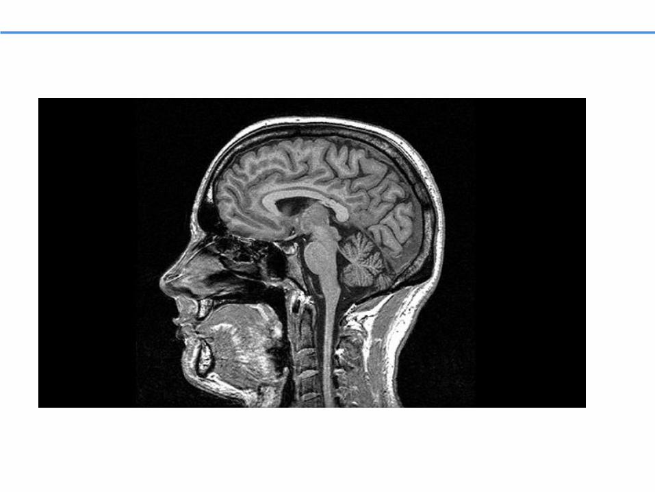
The Subdivisions and Components of the Central Nervous System
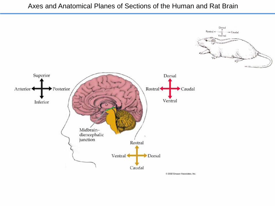
Axes and Anatomical Planes of Sections of the Human and Rat Brain
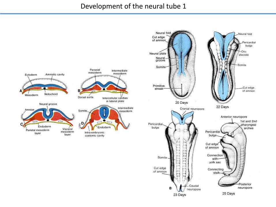
Development of the neural tube 1
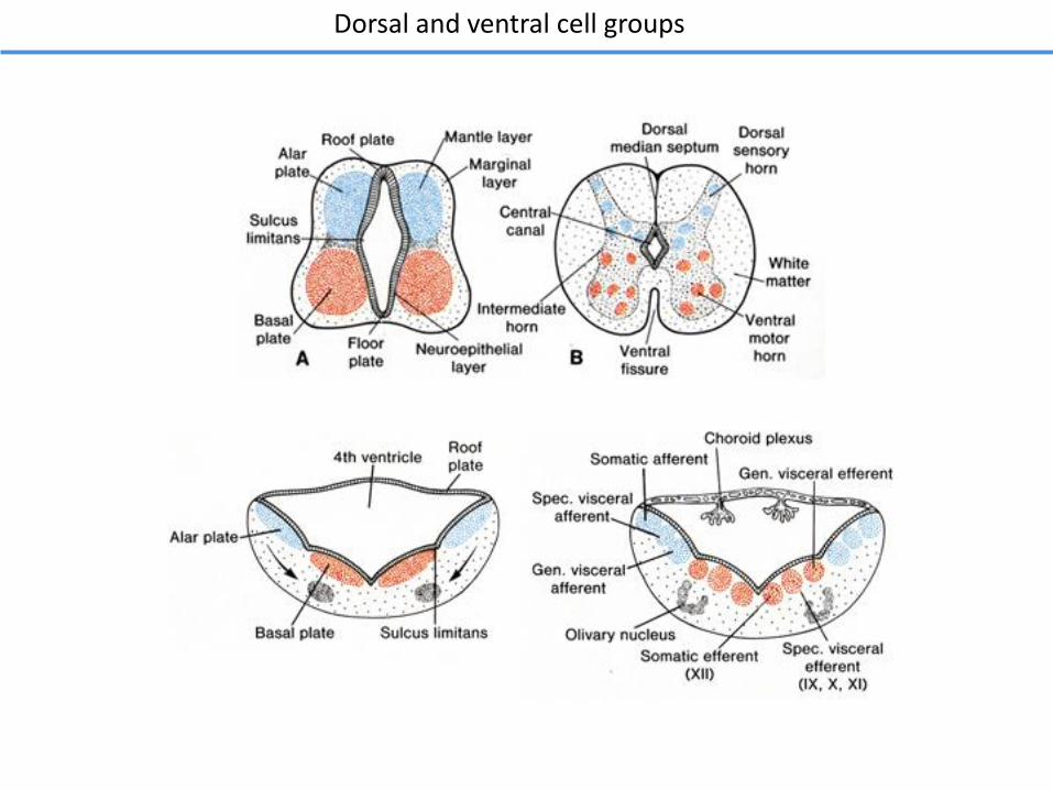
Dorsal and ventral cell groups
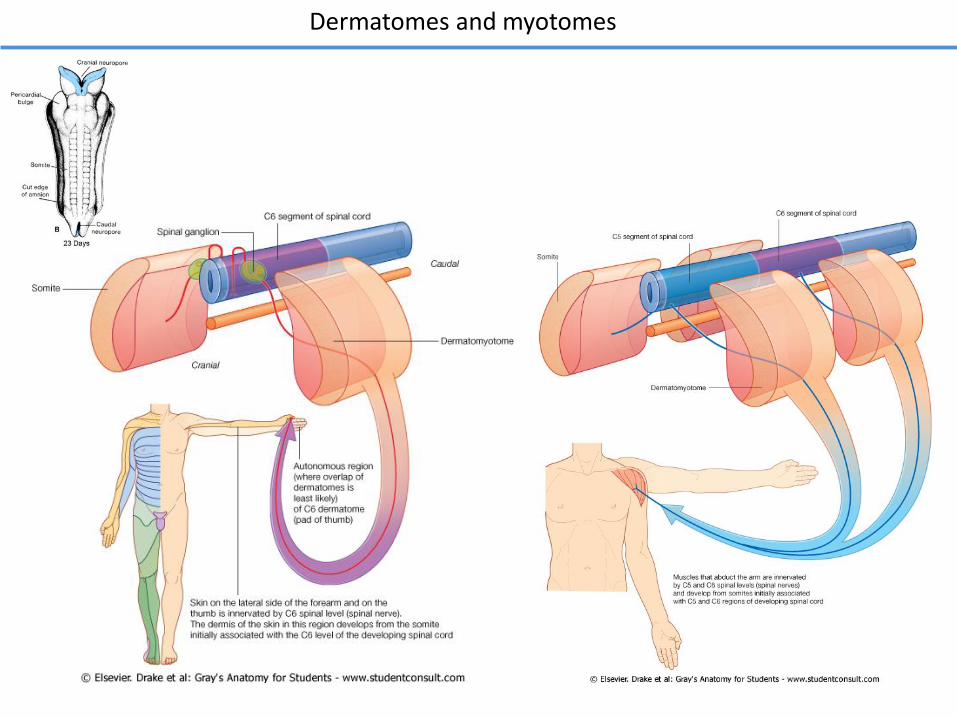
Dermatomes and myotomes
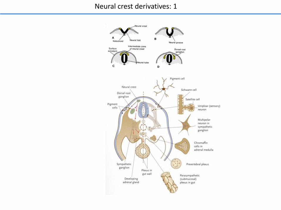
Neural crest derivatives: 1
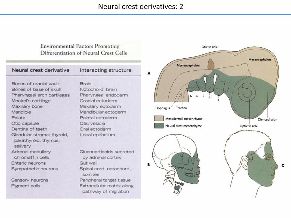
Neural crest derivatives: 2
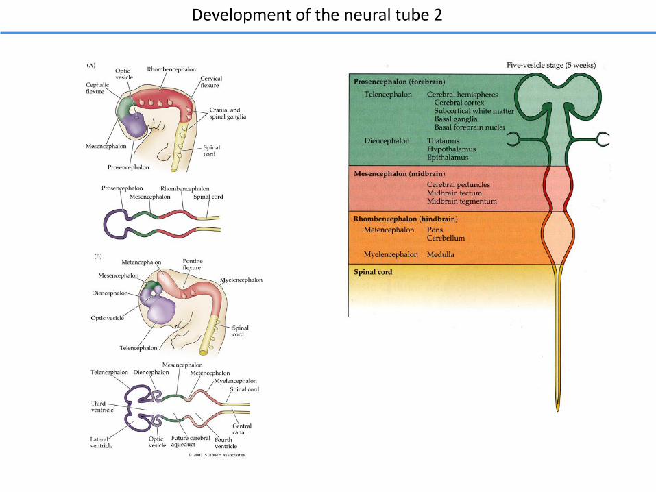
Development of the neural tube 2
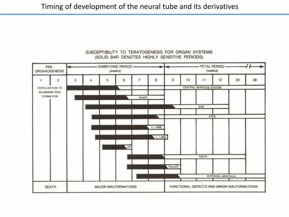
Timing of development of the neural tube and its derivatives
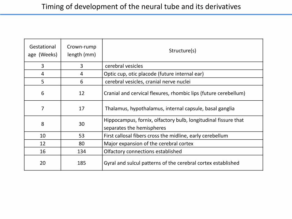
Timing of development of the neural tube and its derivatives
Gestational
age (Weeks)
Crown-rump
length (mm)Structure(s)
3 3 cerebral vesicles
4 4 Optic cup, otic placode (future internal ear)
5 6 cerebral vesicles, cranial nerve nuclei
6 12 Cranial and cervical flexures, rhombic lips (future cerebellum)
7 17 Thalamus, hypothalamus, internal capsule, basal ganglia
8 30Hippocampus, fornix, olfactory bulb, longitudinal fissure that
separates the hemispheres
10 53 First callosal fibers cross the midline, early cerebellum
12 80 Major expansion of the cerebral cortex
16 134 Olfactory connections established
20 185 Gyral and sulcul patterns of the cerebral cortex established
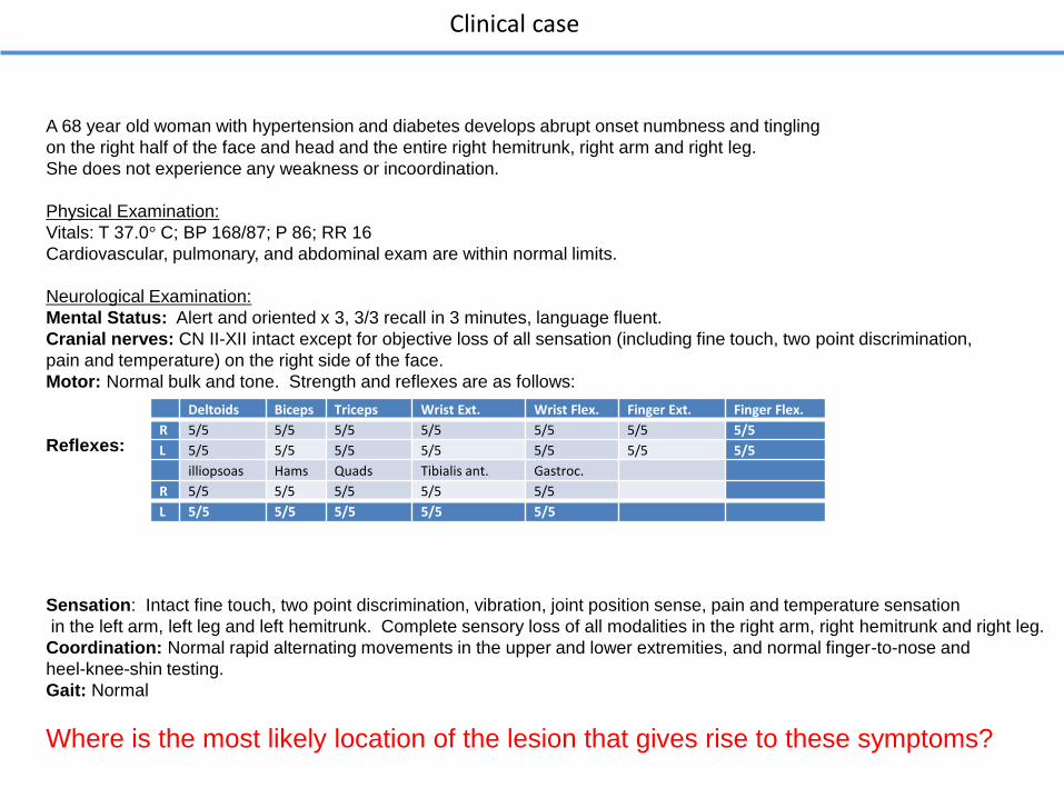
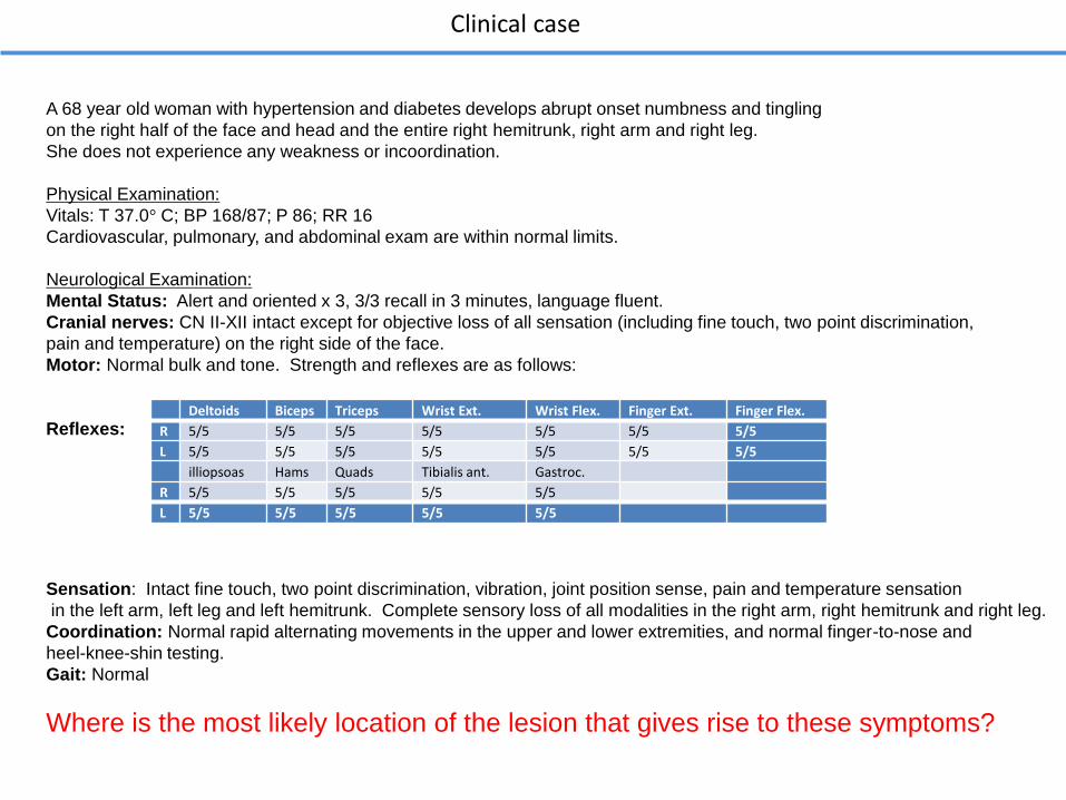
A 68 year old woman with hypertension and diabetes develops abrupt onset numbness and tingling
on the right half of the face and head and the entire right hemitrunk, right arm and right leg.
She does not experience any weakness or incoordination.
Physical Examination:
Vitals: T 37.0° C; BP 168/87; P 86; RR 16
Cardiovascular, pulmonary, and abdominal exam are within normal limits.
Neurological Examination:
Mental Status: Alert and oriented x 3, 3/3 recall in 3 minutes, language fluent.
Cranial nerves: CN II-XII intact except for objective loss of all sensation (including fine touch, two point discrimination,
pain and temperature) on the right side of the face.
Motor: Normal bulk and tone. Strength and reflexes are as follows:
Reflexes:
Sensation: Intact fine touch, two point discrimination, vibration, joint position sense, pain and temperature sensation
in the left arm, left leg and left hemitrunk. Complete sensory loss of all modalities in the right arm, right hemitrunk and right leg.
Coordination: Normal rapid alternating movements in the upper and lower extremities, and normal finger-to-nose and
heel-knee-shin testing.
Gait: Normal
Where is the most likely location of the lesion that gives rise to these symptoms?
Clinical case
Deltoids Biceps Triceps Wrist Ext. Wrist Flex. Finger Ext. Finger Flex.
R 5/5 5/5 5/5 5/5 5/5 5/5 5/5
L 5/5 5/5 5/5 5/5 5/5 5/5 5/5
illiopsoas Hams Quads Tibialis ant. Gastroc.
R 5/5 5/5 5/5 5/5 5/5
L 5/5 5/5 5/5 5/5 5/5
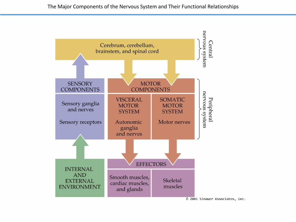
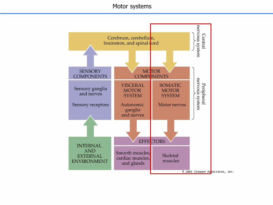
The Major Components of the Nervous System and Their Functional Relationships
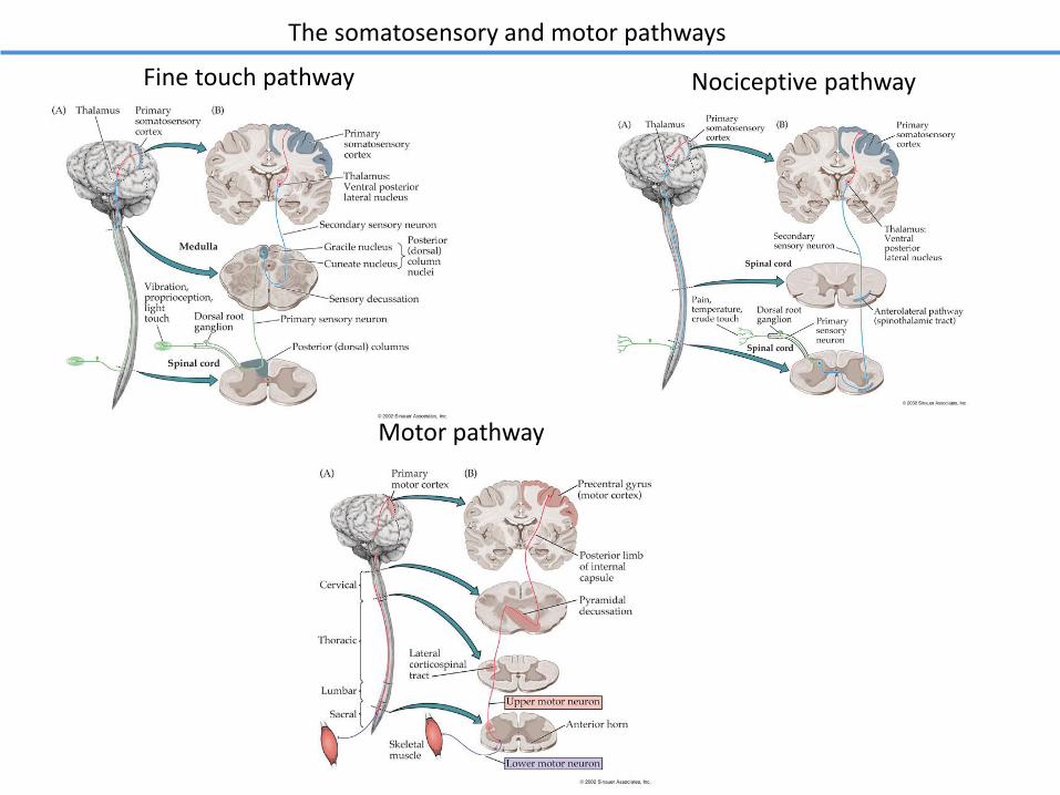
The somatosensory and motor pathways
Fine touch pathway Nociceptive pathway
Motor pathway
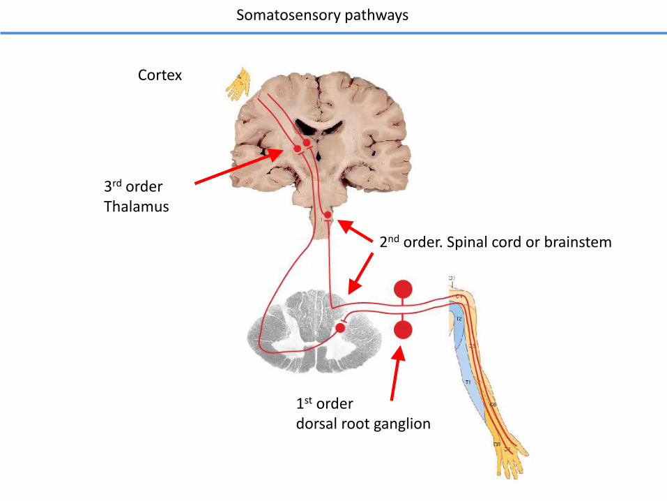
Somatosensory pathways
1st orderdorsal root ganglion
3rd orderThalamus
2nd order. Spinal cord or brainstem
Cortex
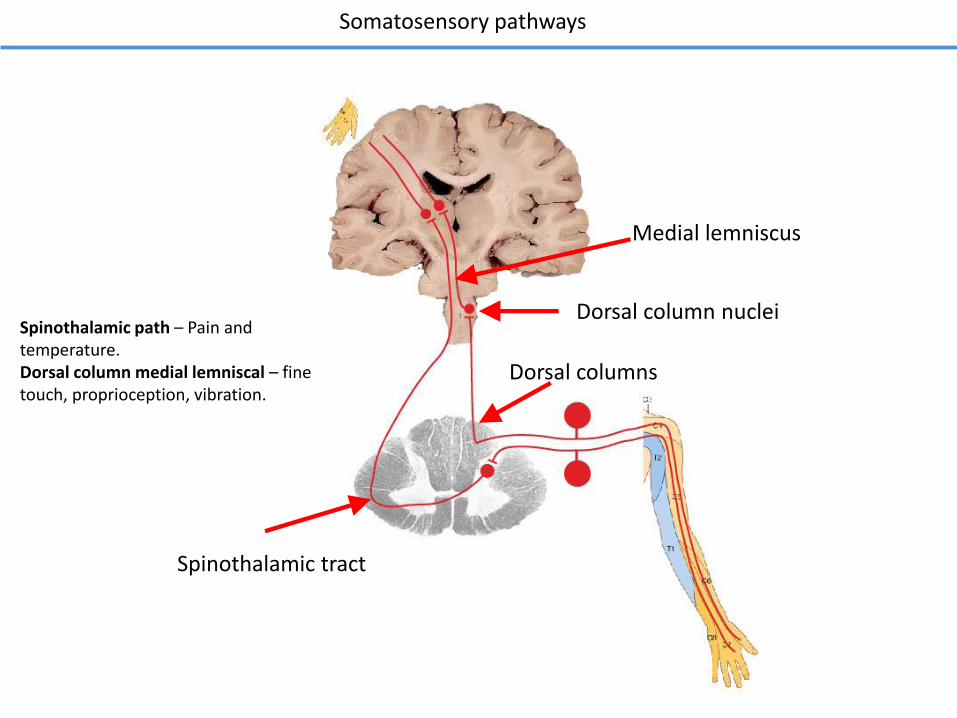
Somatosensory pathways
Dorsal column nuclei
Dorsal columns
Spinothalamic tract
Medial lemniscus
Spinothalamic path – Pain and temperature.Dorsal column medial lemniscal – fine touch, proprioception, vibration.
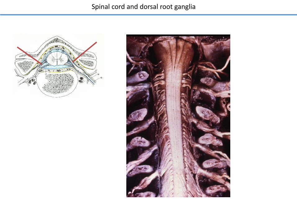
Spinal cord and dorsal root ganglia
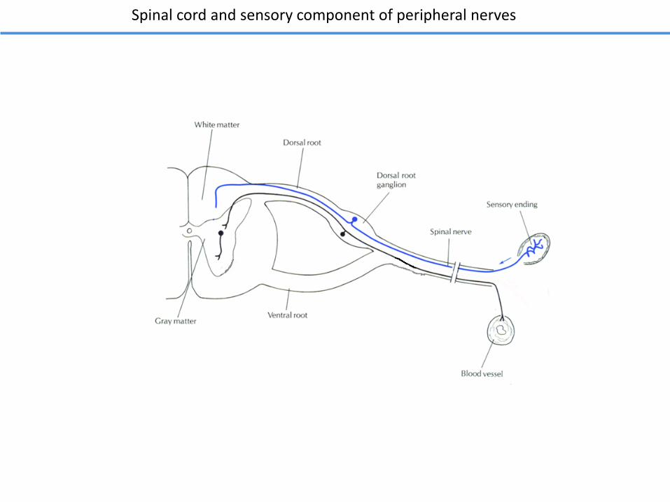
Spinal cord and sensory component of peripheral nerves
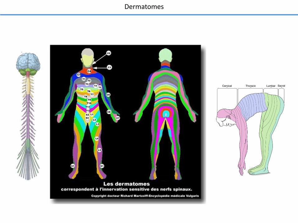
Dermatomes
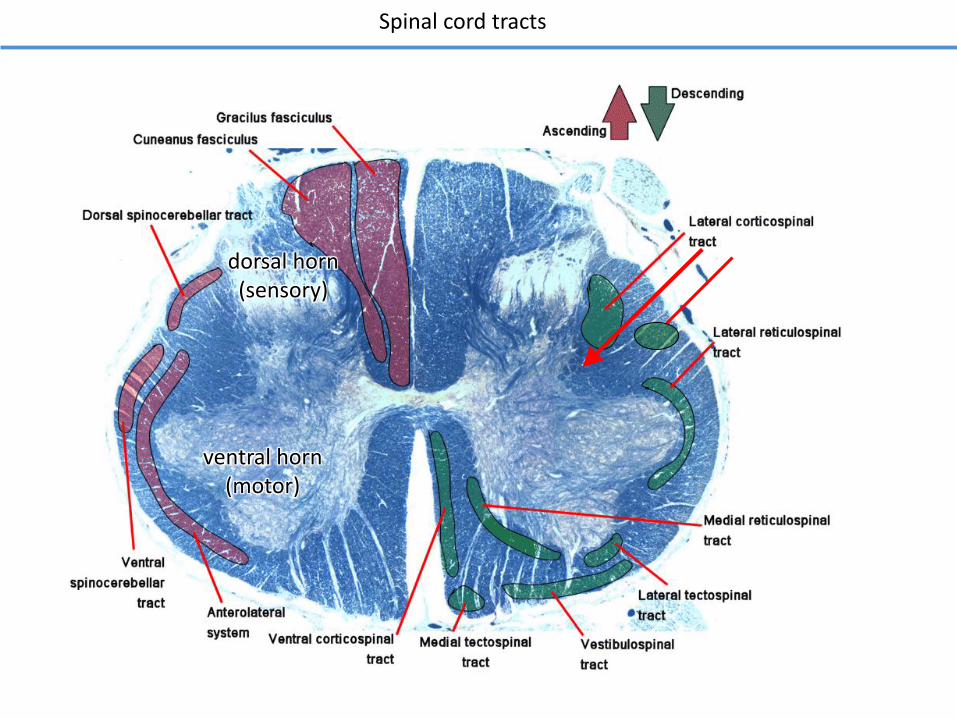
Spinal cord tracts
dorsal horn(sensory)
ventral horn(motor)
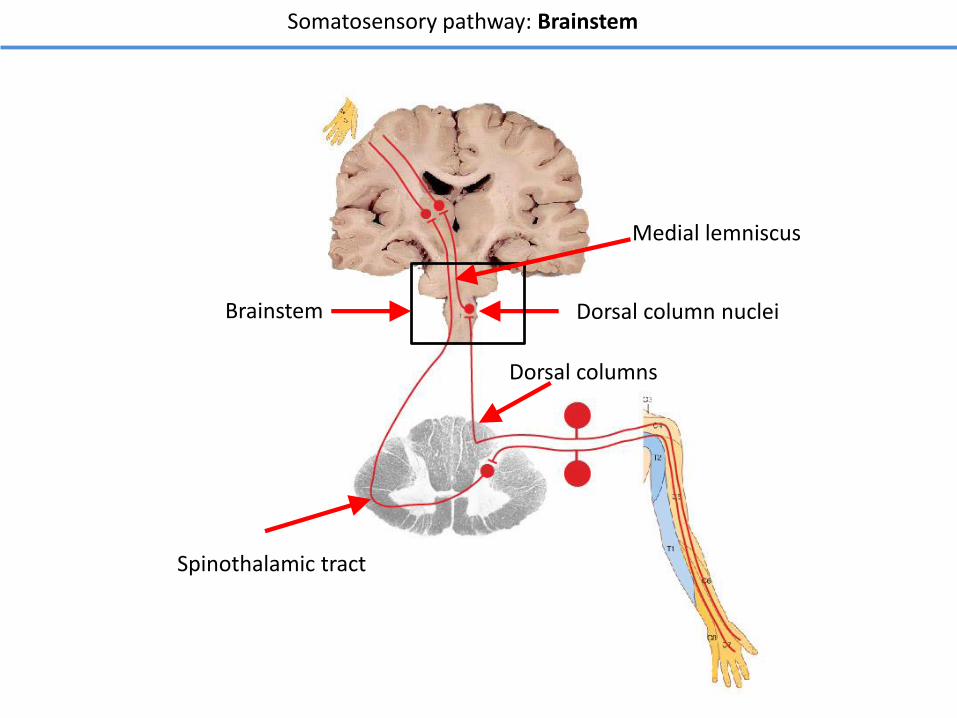
Somatosensory pathway: Brainstem
Dorsal column nucleiBrainstem
Dorsal columns
Spinothalamic tract
Medial lemniscus
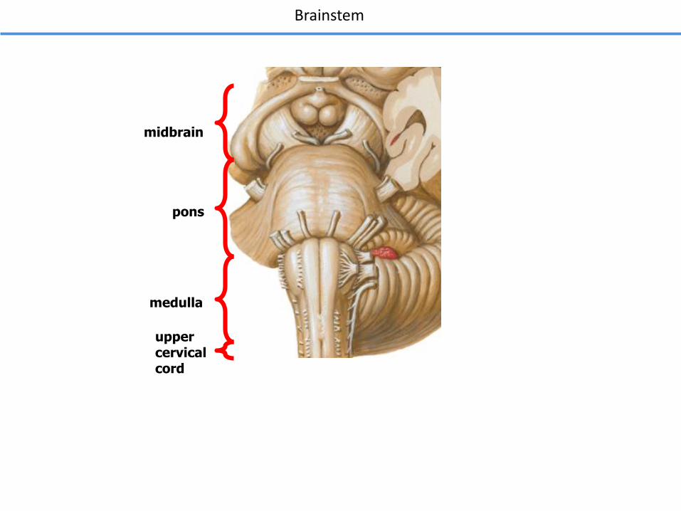
Brainstem
upper cervicalcord
medulla
pons
midbrain
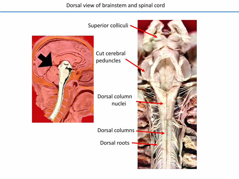
Dorsal view of brainstem and spinal cord
Dorsal columnnuclei
Dorsal columns
Dorsal roots
Superior colliculi
Cut cerebral peduncles
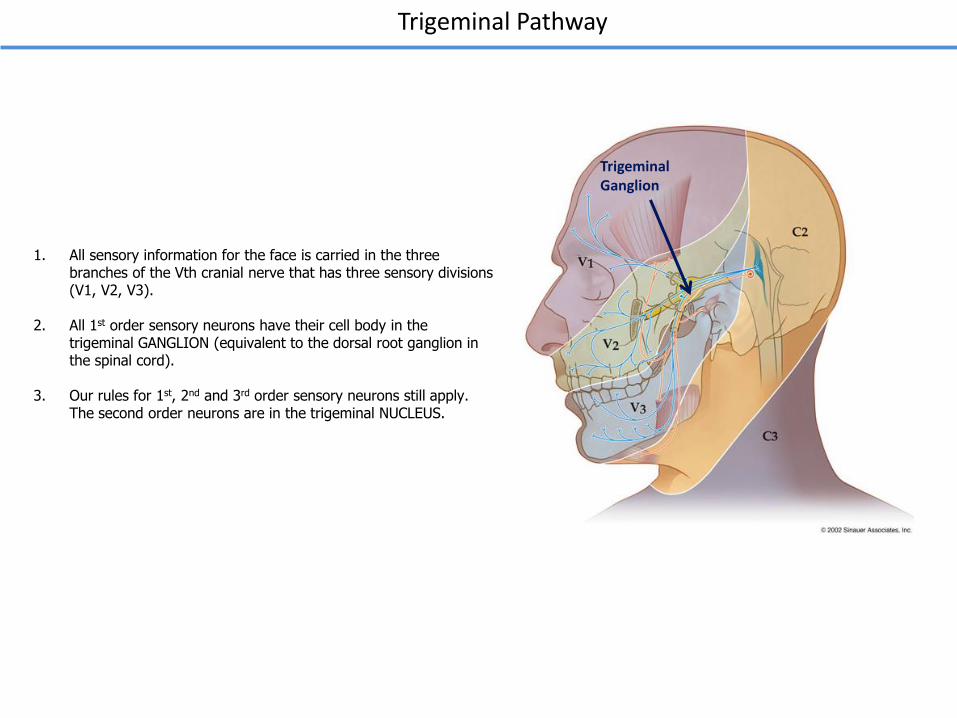
1. All sensory information for the face is carried in the three branches of the Vth cranial nerve that has three sensory divisions (V1, V2, V3).
2. All 1st order sensory neurons have their cell body in the trigeminal GANGLION (equivalent to the dorsal root ganglion in the spinal cord).
3. Our rules for 1st, 2nd and 3rd order sensory neurons still apply. The second order neurons are in the trigeminal NUCLEUS.
Trigeminal Ganglion
Trigeminal Pathway
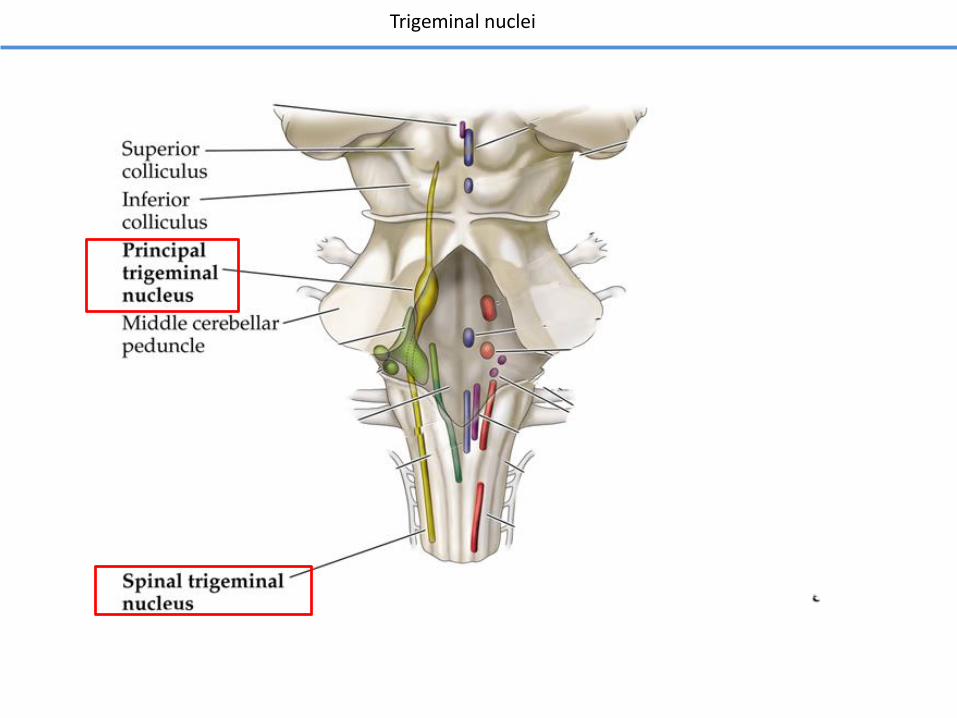
Trigeminal nuclei
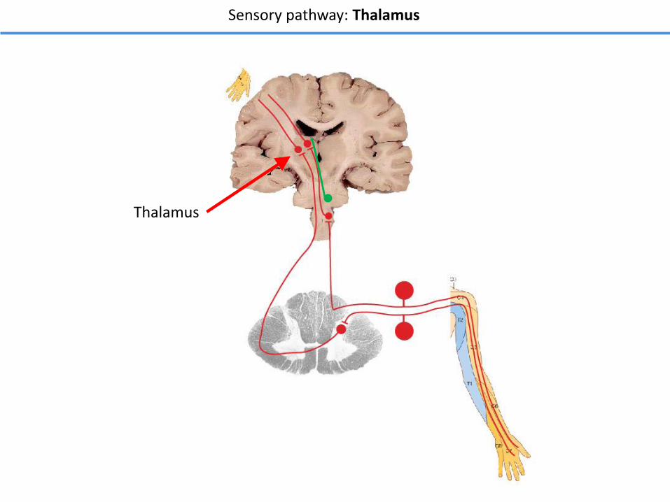
Sensory pathway: Thalamus
Thalamus
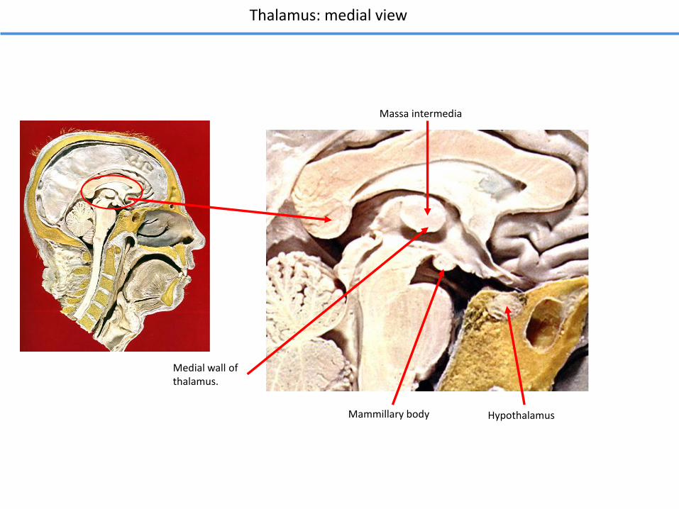
Thalamus: medial view
Medial wall of thalamus.
Massa intermedia
Mammillary body Hypothalamus
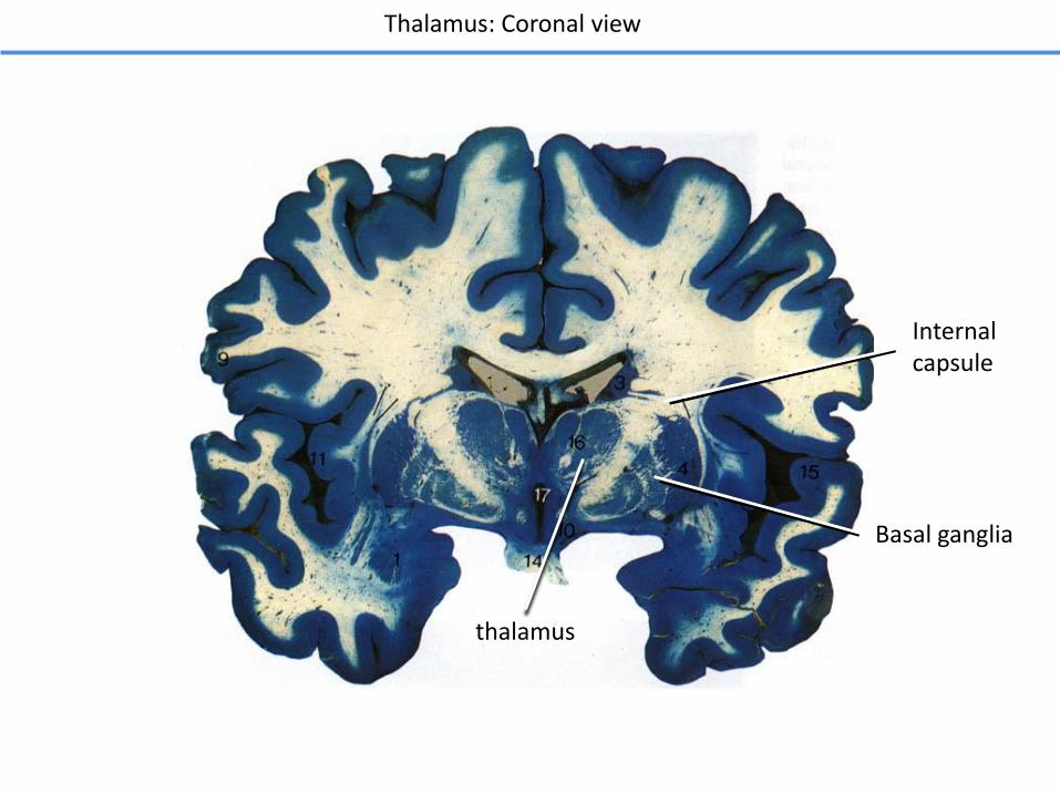
Thalamus: Coronal view
thalamus
Basal ganglia
Internalcapsule
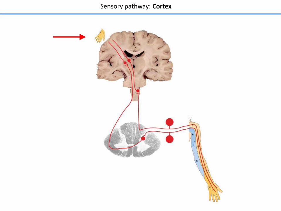
Sensory pathway: Cortex
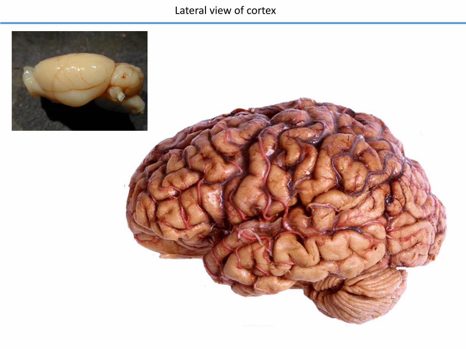
Lateral view of cortex
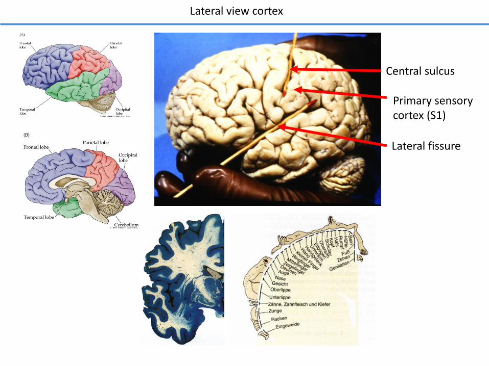
Lateral view cortex
Central sulcus
Lateral fissure
Primary sensory cortex (S1)
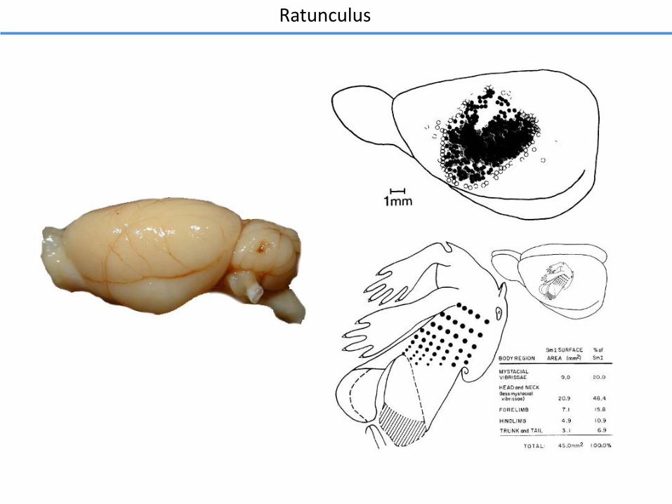
Ratunculus
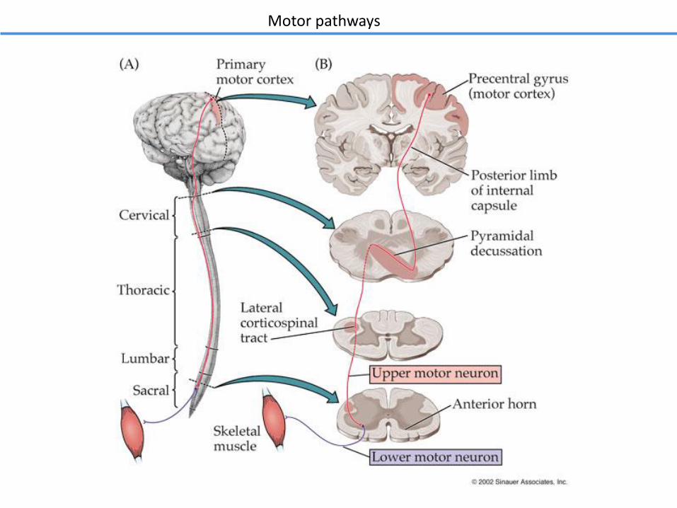
Motor pathways
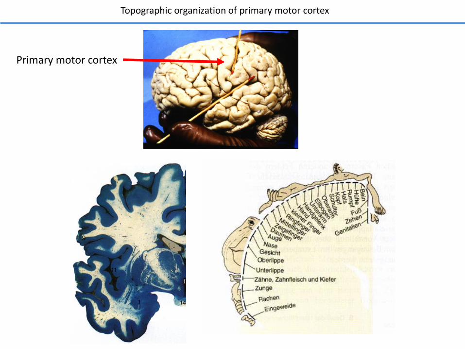
Topographic organization of primary motor cortex
Primary motor cortex
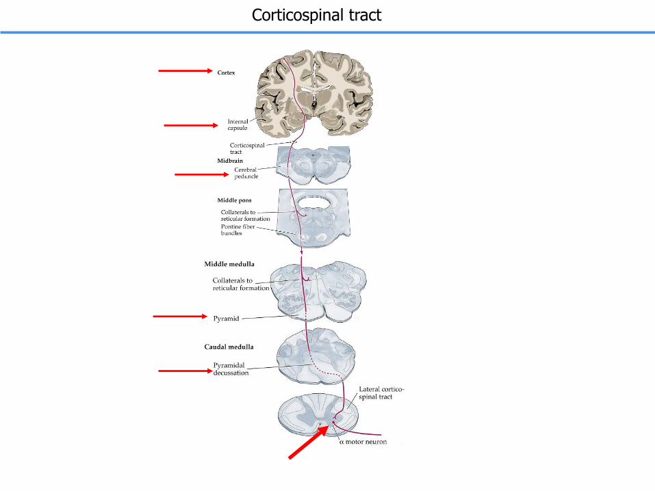
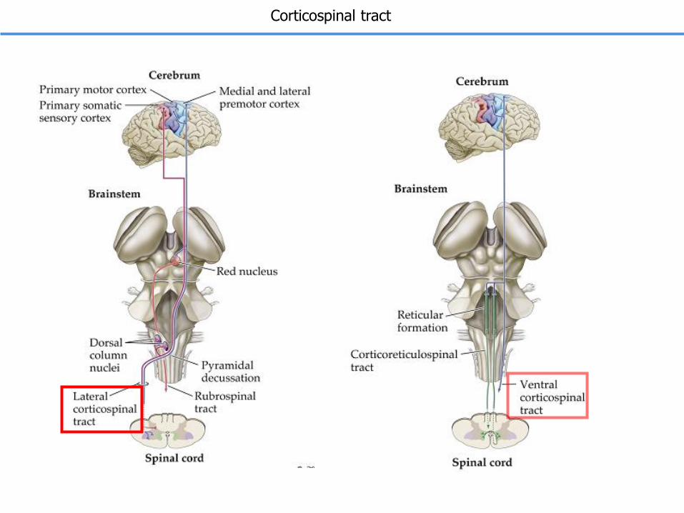
Corticospinal tract
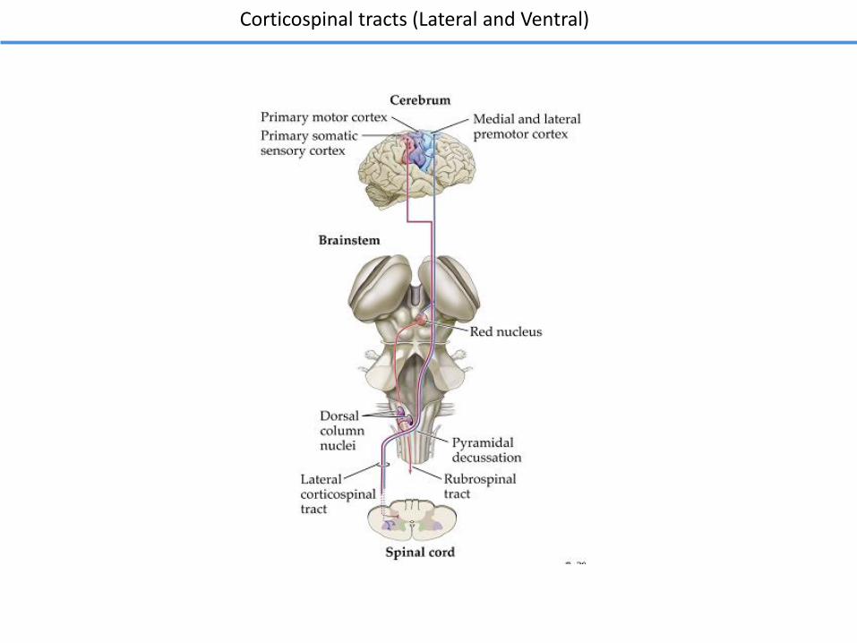
Corticospinal tracts (Lateral and Ventral)
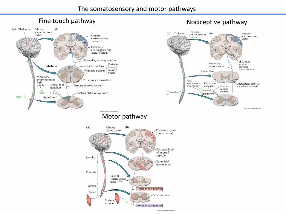
The somatosensory and motor pathways
Fine touch pathway Nociceptive pathway
Motor pathway
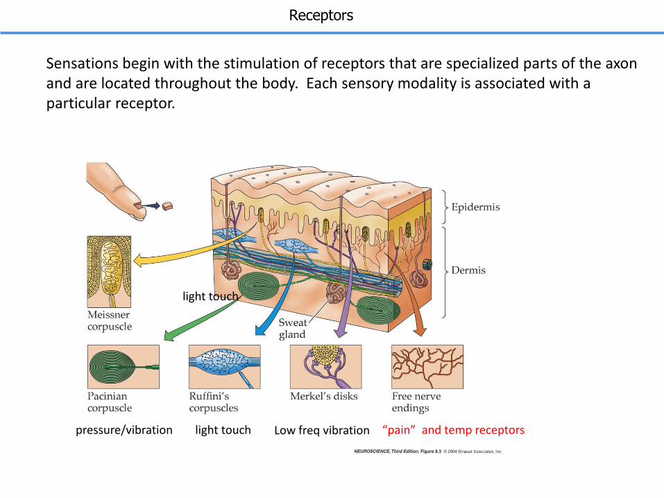
Sensations begin with the stimulation of receptors that are specialized parts of the axon and are located throughout the body. Each sensory modality is associated with a particular receptor.
“pain” and temp receptorspressure/vibration light touch Low freq vibration
light touch
Receptors
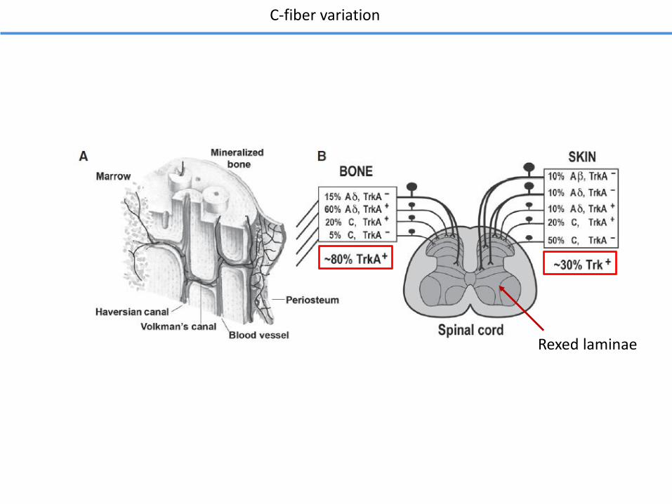
C-fiber variation
Rexed laminae
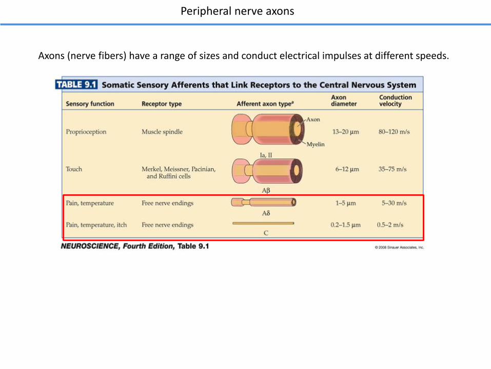
Axons (nerve fibers) have a range of sizes and conduct electrical impulses at different speeds.
Peripheral nerve axons
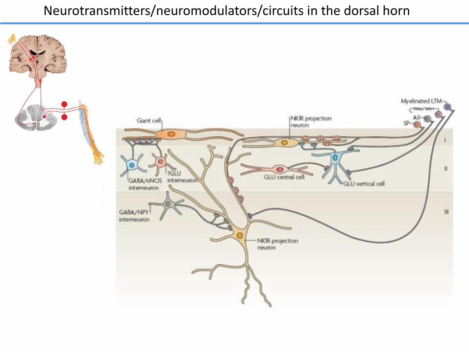
Neurotransmitters/neuromodulators/circuits in the dorsal horn
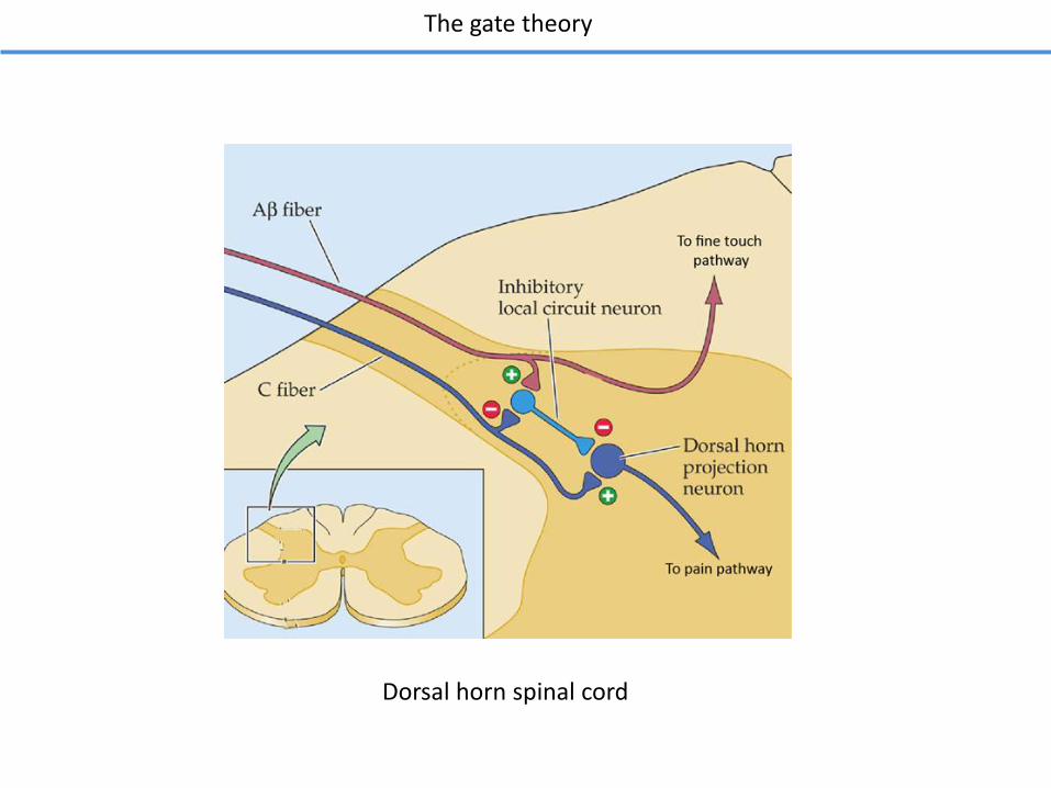
The gate theory
Dorsal horn spinal cord
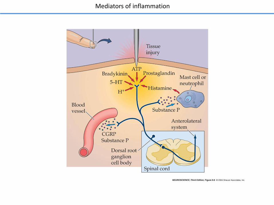
Mediators of inflammation
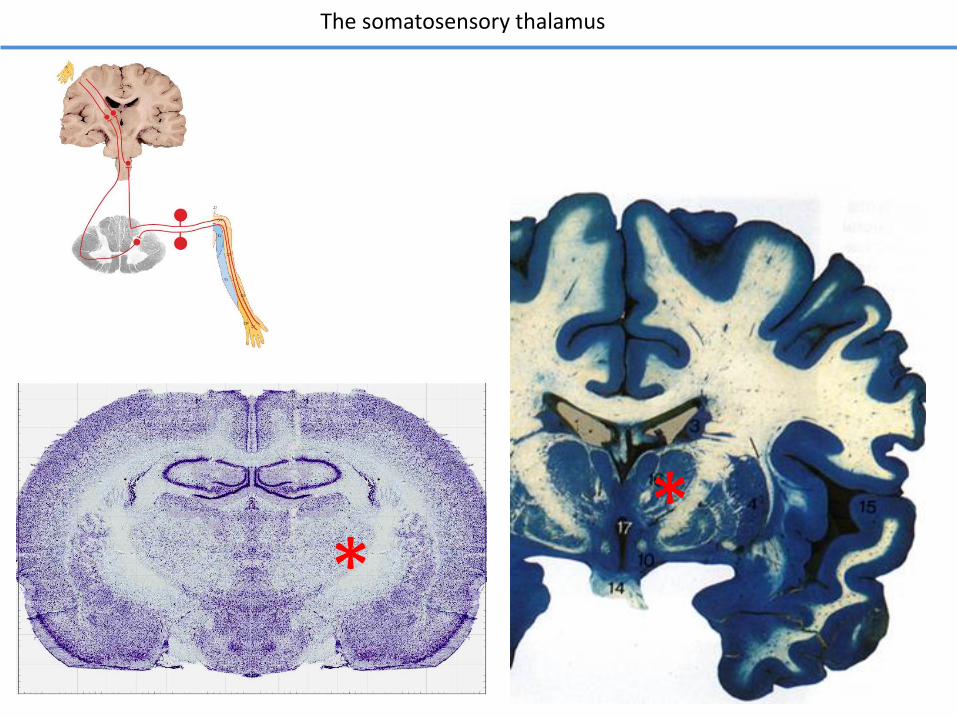
The somatosensory thalamus
**

Somatosensory thalamus topography
Somatotopy (VPL)
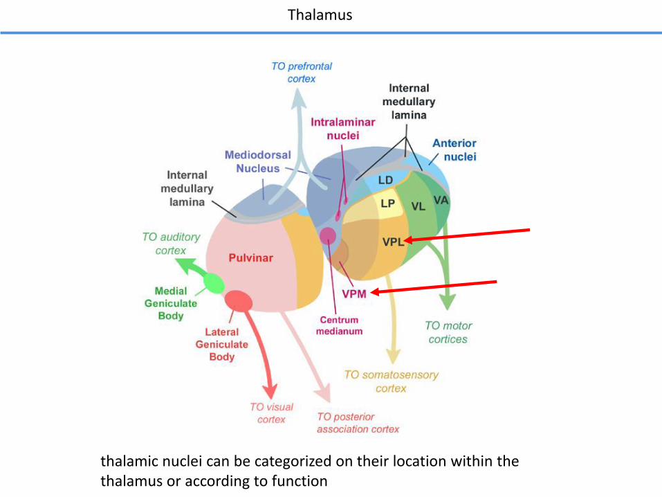
Thalamus
thalamic nuclei can be categorized on their location within the thalamus or according to function
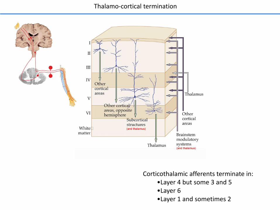
Corticothalamic afferents terminate in:•Layer 4 but some 3 and 5•Layer 6•Layer 1 and sometimes 2
Thalamo-cortical termination
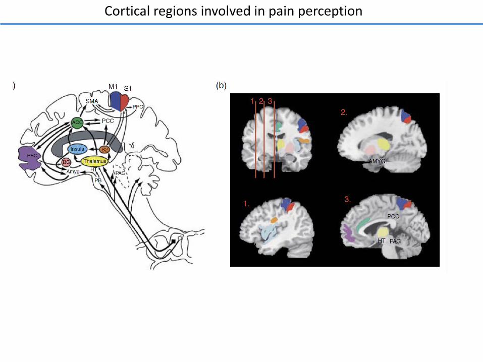
Cortical regions involved in pain perception
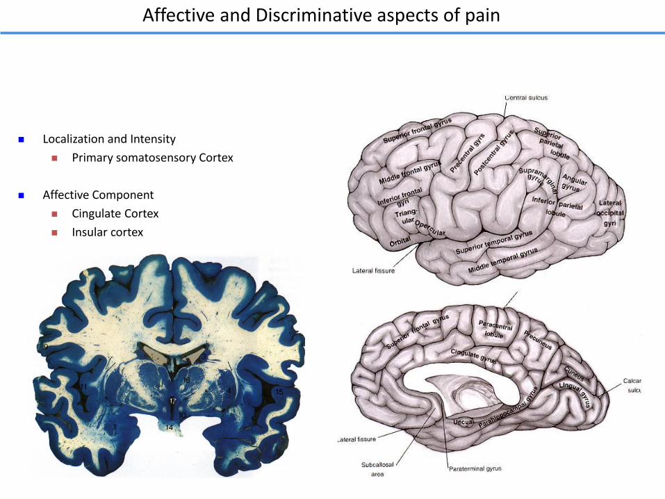
Localization and Intensity
Primary somatosensory Cortex
Affective Component
Cingulate Cortex
Insular cortex
Affective and Discriminative aspects of pain
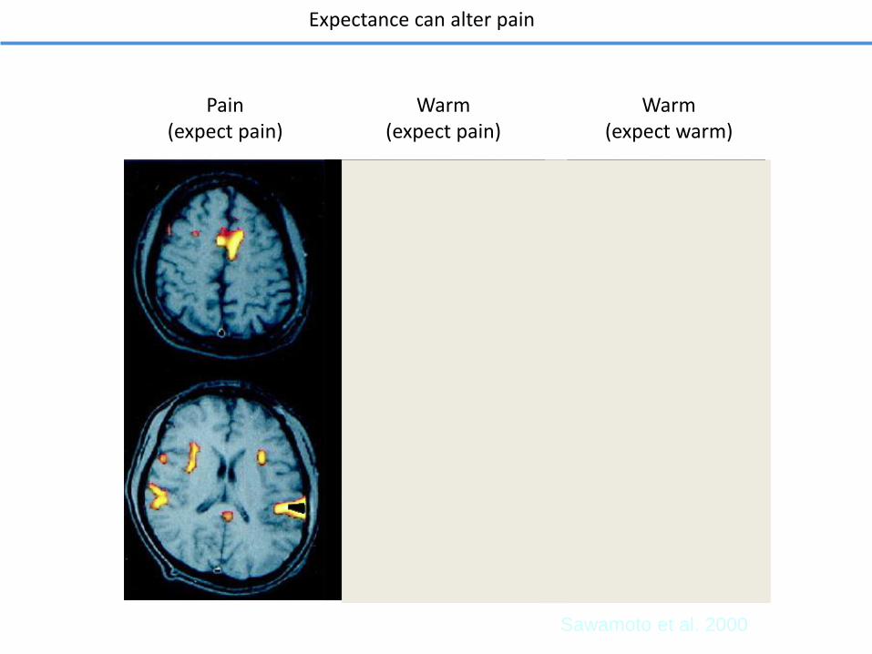
Sawamoto et al. 2000
Pain(expect pain)
Warm(expect pain)
Warm(expect warm)
Expectance can alter pain
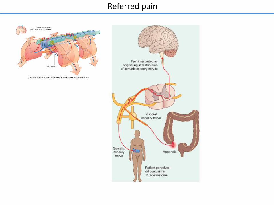
Referred pain
Motor systems
Corticospinal tract
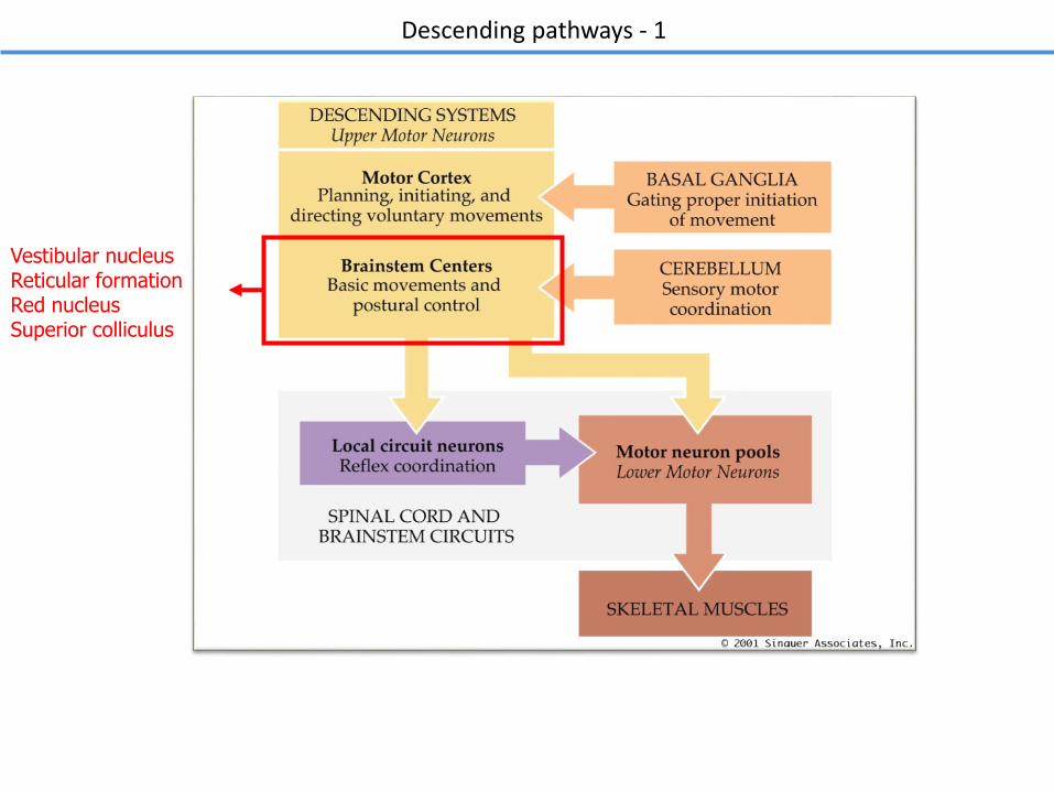
Descending pathways - 1
Vestibular nucleusReticular formation Red nucleusSuperior colliculus
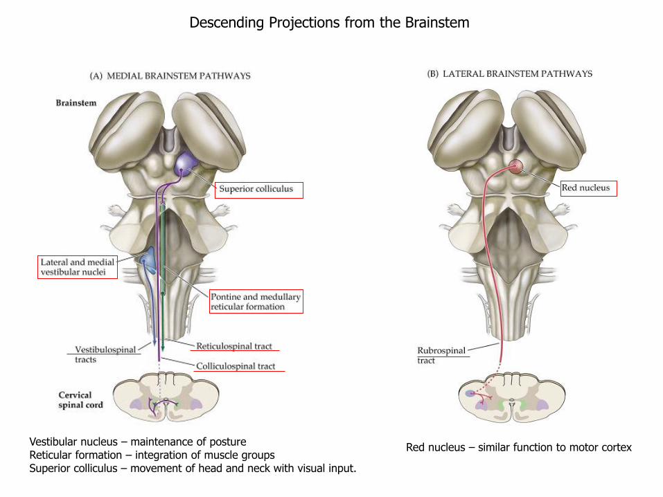
Descending Projections from the Brainstem
Vestibular nucleus – maintenance of postureReticular formation – integration of muscle groupsSuperior colliculus – movement of head and neck with visual input.
Red nucleus – similar function to motor cortex

The motor tracts in the spinal white matter
Rubrospinal tract
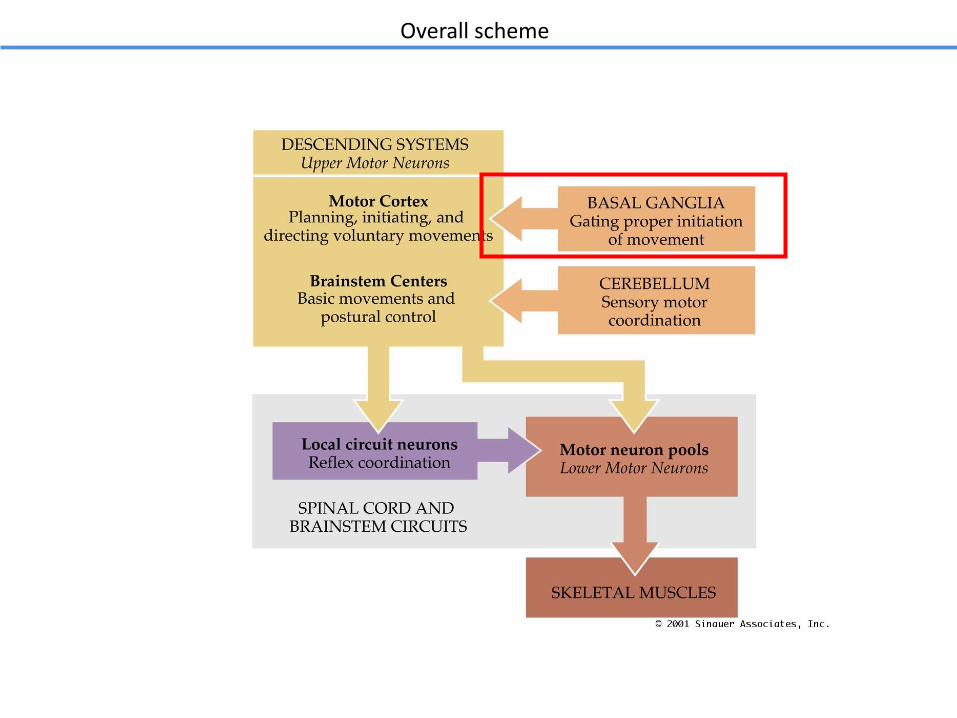
Overall scheme
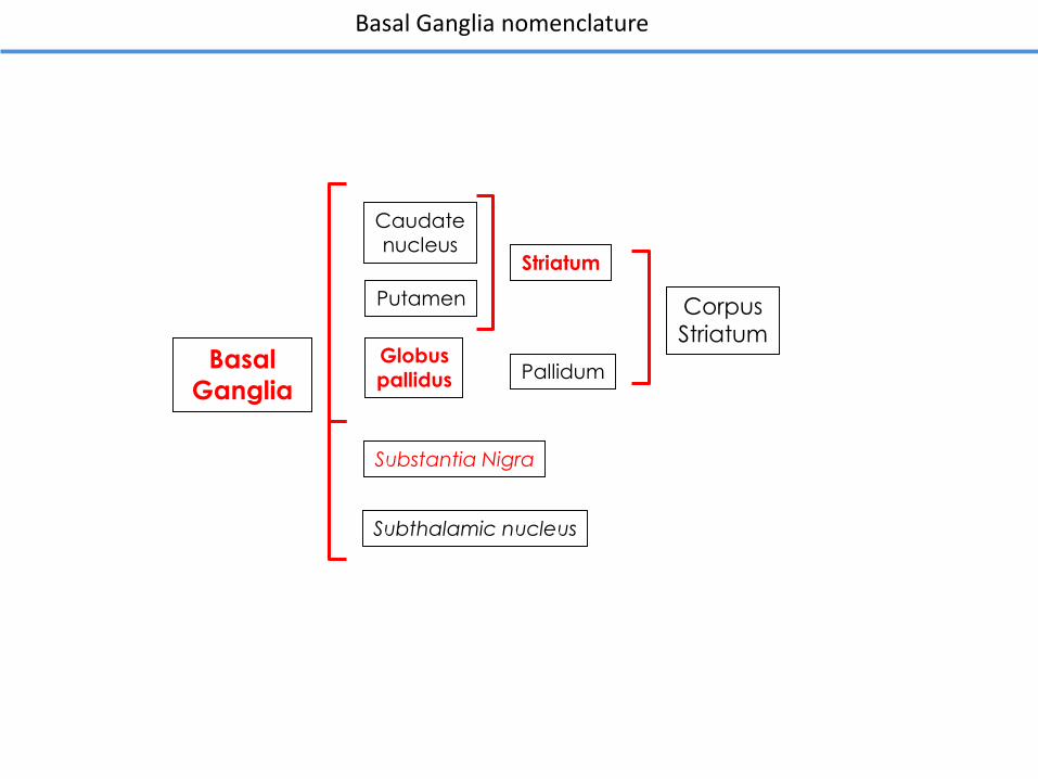
Basal Ganglia nomenclature
Basal
Ganglia
Putamen
Caudatenucleus
PallidumGlobuspallidus
Substantia Nigra
Subthalamic nucleus
Striatum
Corpus
Striatum
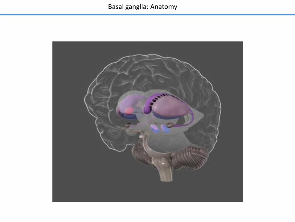
Basal ganglia: Anatomy
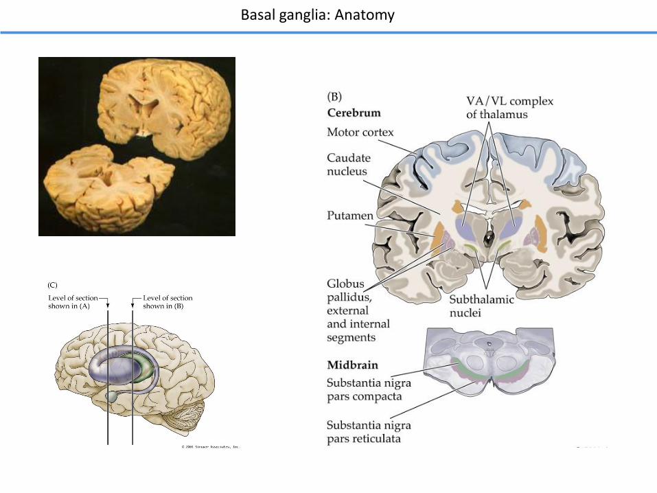
Basal ganglia: Anatomy
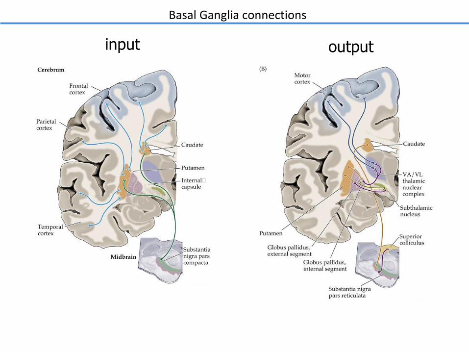
Basal Ganglia connections
input output
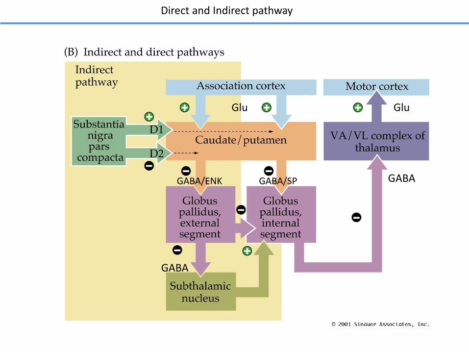
Direct and Indirect pathway
Glu
GABA/SP GABA
GABA
GABA/ENK
Glu
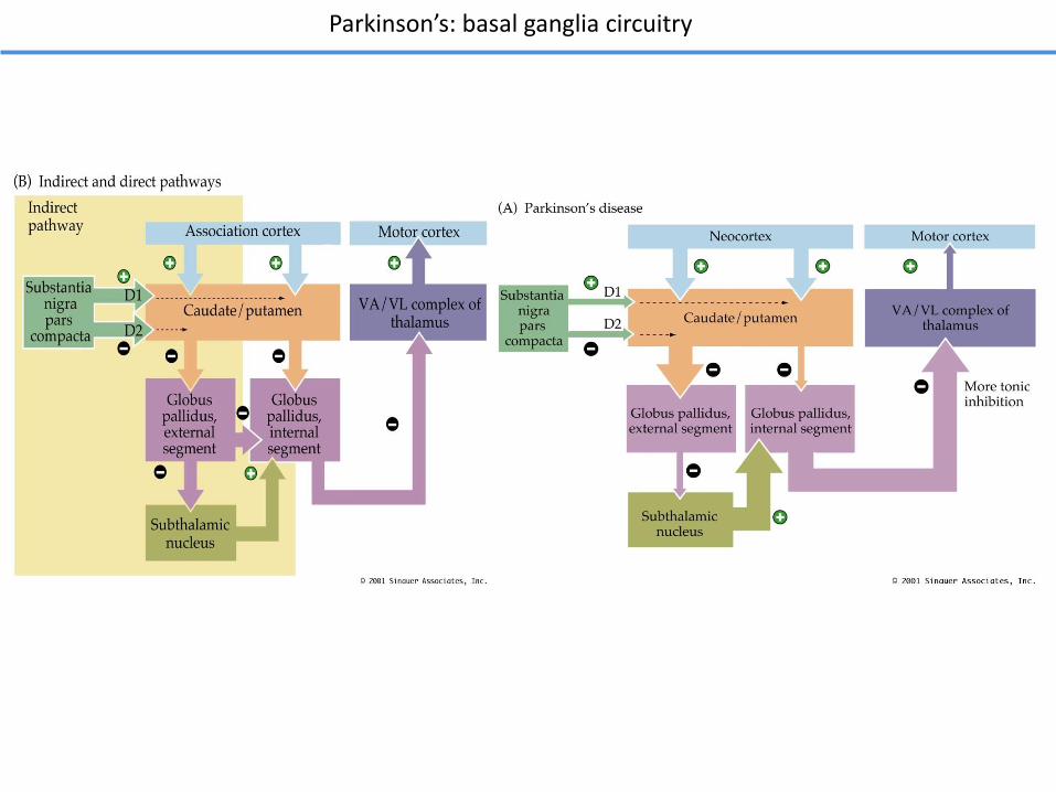
Parkinson’s: basal ganglia circuitry
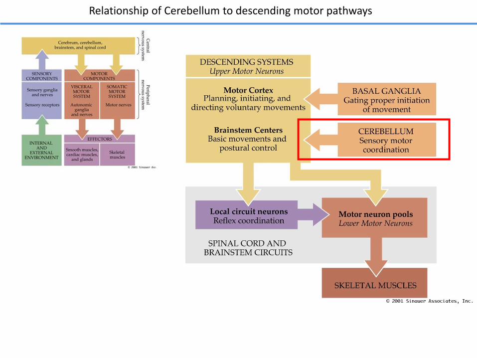
Relationship of Cerebellum to descending motor pathways
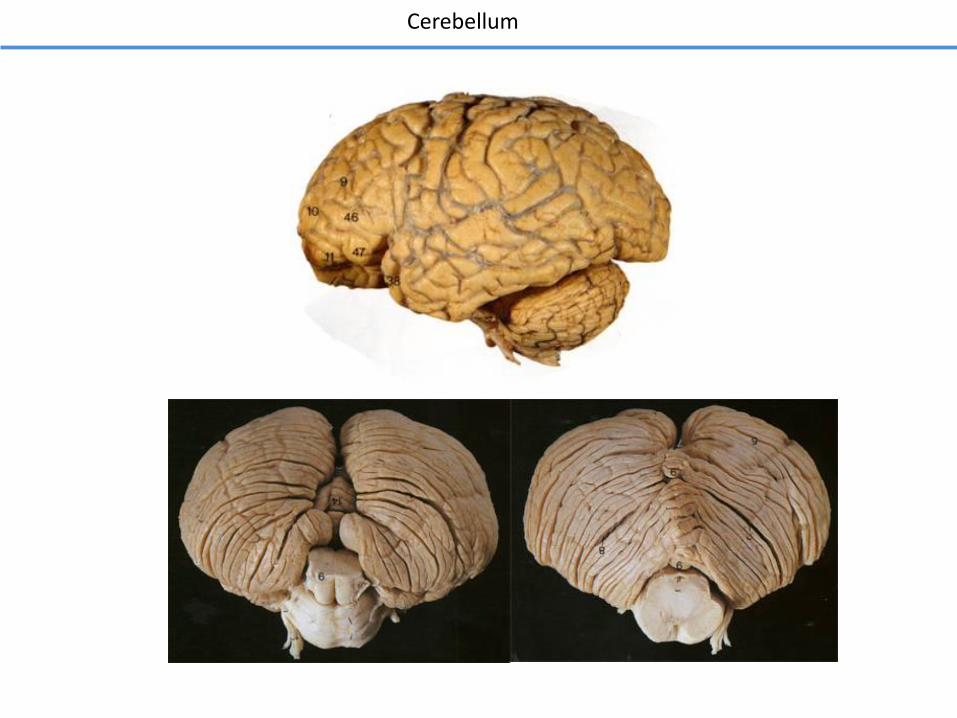
Cerebellum
CEREBELLUM
input output
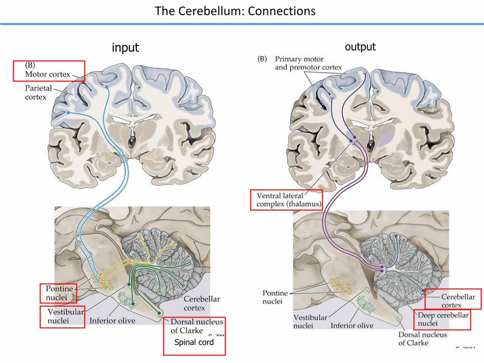
The Cerebellum: Connections
Spinal cord
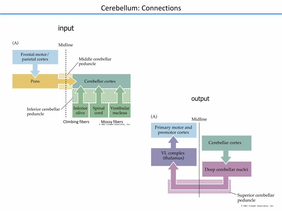
Cerebellum: Connections
input
output
Climbing fibers Mossy fibers
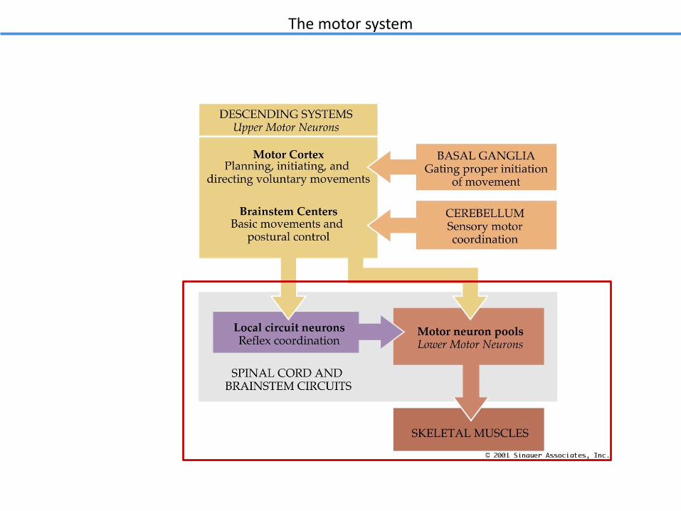
The motor system
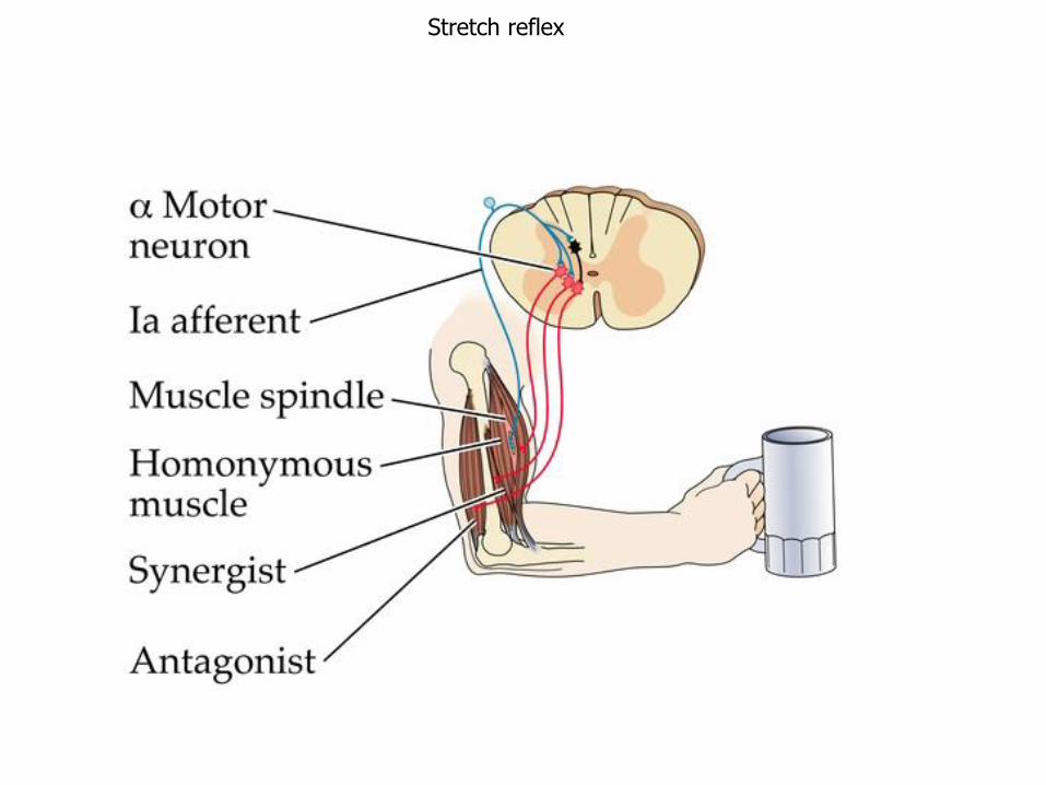
Stretch reflex
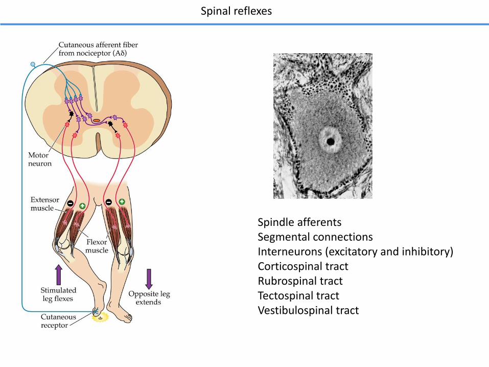
Spinal reflexes
Spindle afferentsSegmental connections Interneurons (excitatory and inhibitory)Corticospinal tractRubrospinal tractTectospinal tractVestibulospinal tract
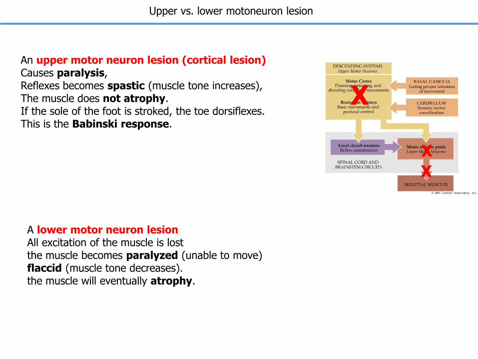
Upper vs. lower motoneuron lesion
A lower motor neuron lesionAll excitation of the muscle is lostthe muscle becomes paralyzed (unable to move) flaccid (muscle tone decreases). the muscle will eventually atrophy.
An upper motor neuron lesion (cortical lesion)Causes paralysis, Reflexes becomes spastic (muscle tone increases), The muscle does not atrophy. If the sole of the foot is stroked, the toe dorsiflexes. This is the Babinski response.
X
X
X
Lesions of the basal ganglia generally lead to hyper- or hypo-kinetic movement and resting tremors.
Lesions of the cerebellum lead to errors in accuracy and coordination of movements and intention tremors
Basal Ganglia and Cerebellum
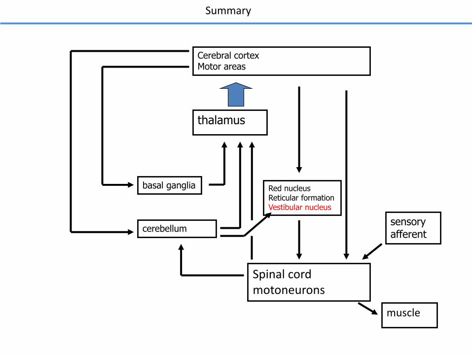
Cerebral cortexMotor areas
thalamus
basal ganglia
cerebellum
Summary
Spinal cordmotoneurons
sensory afferent
muscle
Red nucleusReticular formationVestibular nucleus
A 68 year old woman with hypertension and diabetes develops abrupt onset numbness and tingling
on the right half of the face and head and the entire right hemitrunk, right arm and right leg.
She does not experience any weakness or incoordination.
Physical Examination:
Vitals: T 37.0° C; BP 168/87; P 86; RR 16
Cardiovascular, pulmonary, and abdominal exam are within normal limits.
Neurological Examination:
Mental Status: Alert and oriented x 3, 3/3 recall in 3 minutes, language fluent.
Cranial nerves: CN II-XII intact except for objective loss of all sensation (including fine touch, two point discrimination,
pain and temperature) on the right side of the face.
Motor: Normal bulk and tone. Strength and reflexes are as follows:
Reflexes:
Sensation: Intact fine touch, two point discrimination, vibration, joint position sense, pain and temperature sensation
in the left arm, left leg and left hemitrunk. Complete sensory loss of all modalities in the right arm, right hemitrunk and right leg.
Coordination: Normal rapid alternating movements in the upper and lower extremities, and normal finger-to-nose and
heel-knee-shin testing.
Gait: Normal
Where is the most likely location of the lesion that gives rise to these symptoms?
Clinical case
Deltoids Biceps Triceps Wrist Ext. Wrist Flex. Finger Ext. Finger Flex.
R 5/5 5/5 5/5 5/5 5/5 5/5 5/5
L 5/5 5/5 5/5 5/5 5/5 5/5 5/5
illiopsoas Hams Quads Tibialis ant. Gastroc.
R 5/5 5/5 5/5 5/5 5/5
L 5/5 5/5 5/5 5/5 5/5
As a volunteer working for Doctors Without Borders in a clinic in Jordan, you are asked to evaluate a 14 year-old Iraqi refugee who was injured by a sniper’s bullet 7 weeks ago. The bullet entry hole is obliterated by an apparent attempt at exploratory surgery in the mid-back, and plain x-rays show that the bullet was lodged somewhere in the bony spine.Physical Examination:Vitals: T 37.6° C; BP 112/60; P 64; RR 12Cardiovascular and abdominal exam are within normal limits. Pulmonary exam reveals mild crackles in the upper right lung field.Neurological Examination:Mental Status: AO x 3, 3/3 recall in 3 minutes, language fluent.Cranial nerves: CN II-XII intact.Motor: Normal bulk. Increased tone (spasticity) in the left lower extremity. No pronator drift. Sensation: Markedly decreased pain and temperature sensation on the right side only from the level of the umbilicus down to and including the entire right leg. Vibration and joint position sense normal bilaterally in the upper and lower extremities.Coordination: Normal rapid alternating movements and finger-to-nose in the upper extremities. Slow foot tap in the left leg.Gait: Spastic with impaired movement of the left leg (circumduction of the left leg during swing-through phase of gait).
Q – Diagram a single continuous lesion that can explain these findings.
Clinical Case