Embed Size (px)
Citation preview
NUCLEAR FACTOR-KAPPA B EXPRESSION IN ORAL
LEUKOPLAKIA AND SQUAMOUS CELL CARCINOMA
AN IMMUNOHISTOCHEMICAL STUDY
Dissertation submitted to
THE TAMILNADU Dr.M.G.R.MEDICAL UNIVERSITY
In partial fulfillment for the Degree of
MASTER OF DENTAL SURGERY
BRANCH VI
ORAL PATHOLOGY AND MICROBIOLOGY
APRIL 2011

CERTIFICATE
This is to certify that this dissertation titled “NUCLEAR
FACTOR-KAPPA B EXPRESSION IN ORAL LEUKOPLAKIA
AND SQUAMOUS CELL CARCINOMA AN
IMMUNOHISTOCHEMICAL STUDY” is a bonafide dissertation
performed by P. JEYAPREETHA under our guidance during her
post graduate period between 2008-2011.
This dissertation is submitted to THE TAMIL NADU DR. M.
G. R. MEDICAL UNIVERSITY , in partial fulfilment for the
degree of MASTER OF DENTAL SURGERY IN ORAL
PATHOLOGY AND MICROBIOLOGY, BRANCH VI . It has not
been submitted (partial or full) for the award of any degree or
diploma.
Dr. S. Ramachandran, MDS
Principal
Ragas Dental College & Hospitals
Chennai
Dr. K. Ranganathan, MDS, MS (Ohio),PhD Professor and Head Department of Oral & Maxillofacial Pathology Ragas Dental College & Hospitals , Chennai .
Dr. Elizabeth Joshua, MDS Professor Department of Oral & Maxillofacial Pathology Ragas Dental College & Hospitals , Chennai .

ACKNOWLEDGEMENT
I extend my sincere thanks to Dr. K . Ranganathan , MDS, MS
(Ohio)PhD, Professor and Head, Department of Oral and
Maxillofacial Pathology, Ragas Dental College and Hospital for his
valuable guidance, support, encouragement and all help throughout
my postgraduate curriculum. I thank him for the time spent on going
through my work, giving ideas and correcting all the minute
mistakes without which this thesis could not be presented in the
present form.
Sincere thanks to the Principal, Dr. S. Ramachandran ,
Ragas Dental College and Hospital for his permission to use the
facilities of the institution.
I extend my sincere thanks to Dr. M. Uma Devi , Professor,
Department of Oral and Maxillofacial Pathology, Ragas Dental
College and Hospital, for her guidance, support and help
throughout my postgraduate curriculum, and various advices in
completion of this work.
I earnestly thank Dr. Elizabeth Joshua , Professor,
Department of Oral and Maxillofacial Pathology, Ragas Dental
College and Hospital for her constant encouragement and support
throughout my study.
I thank Dr. Rooban, Assistant Professor, Department of Oral
and Maxillofacial Pathology, Ragas Dental College and Hospital,
who helped me with his guidance throughout my postgraduate
course.

My heartfelt gratitude to our lecturers Dr. K. M. Vidya , Dr.P.
Jayanthi , Dr. Deepu George Mathew , Dr. N. Lavanya , Dr. C.
Lavanya for their valuable guidance and encouragement.
I am extremely grateful to our geneticist Mrs. Kavitha
Wilson , for her continuous help and advice throughout the course of
my study.
I extend my sincere thanks to Bio-statistician Mrs. Deepa ,
Lab Technician Mr. Rajan , Ragas Dental College and Hospital, for
all the patience shown and the constant help they rendered in
completion of this study.
I acknowledge gratefully all my batch mates , Aesha , Divya ,
Revathi , Shahela and Sreeja , all my seniors and juniors, for their
constant help and support throughout my postgraduate life and my
friends for being there whenever I needed them.
I would like to dedicate my work to my beloved parents Dr. S.
Pavunraj and Dr. G. V. Rajambigai and my younger brother
Er . S.P. Jai prasanna sudhan for their sacrifices, abundant
support, faith, understanding and love without which this thesis
could not have been presented.
Last but not least, I thank Lord Almighty , the Supreme Being
without whose will nothing would have been possible.

CONTENTS
1. INTRODUCTION 1
2. AIM AND OBJECTIVES 4
3. MATERIALS AND METHODS 5
4. STATISTICAL ANALYSIS 16
5. REVIEW OF LITERATURE 17
6. RESULTS 35
7. TABLES & GRAPHS 40
8. PHOTOGRAPHS 48
9. DISCUSSION 53
10. SUMMARY AND CONCLUSION 60
11. BIBLIOGRAPHY 61
12. ANNEXURES 73

1
Oral cancer constitutes the sixth most common cancer
worldwide and accounts for approximately 5% of all malignant
tumours worldwide.1 In India and South East Asia, Oral Squamous
Cell Carcinoma (OSCC) is the third most common malignancy
constituting 50% of all malignant tumours.1 OSCC is the most
common type of oral cancer and is often preceded by or associated
with potentially malignant lesions or conditions such as leukoplakia
and oral submucous fibrosis.2
Oral leukoplakia (OL) is “a white patch or plaque that cannot
be characterized clinically or pathologically as any other disease”.¹
It is the most common potentially malignant lesion of the oral
mucosa. Tobacco and areca nut chewing (alone or in combination
such as paan) are the habits that are positively associated with oral
leukoplakia.3
Chewing areca nut containing betel quid without tobacco is an
independent risk factor for developing oral cancer. When betel quid
with tobacco is consumed with alcohol and smoking the relative risk
increases eleven fold. 4
The malignant transformation rate for leukoplakia ranges
from 5-20% and is particularly correlated with the degree of
dysplasia.3 The grading of epithelial dysplasia remains subjective as
it relies on cellular atypia and architectural disturbances. It is
important to identify markers that could help us to ascertain the
2
malignant transformation of potentially malignant lesions to target
them for aggressive treatment. 5
NF-κ B is a family of nuclear transcription factors identified
by Sen and Baltimore in 1986. It is involved in immune response
and comprises of hetero or homo dimers of 5 different subunits
NF-κB1, NF-κB2, REL A, REL B, cREL. NF-κB’s are transiently
activated in response to infection or injury and are aberrantly
activated in cancers contributing to their pathogenesis and
therapeutic resistance.6
NF-κB is bound to inhibitor -kappa B (IκB) and is found in
the cytoplasm of cells in an inactive form known as the canonical or
classical form of NF-κB. In response to stimulus by growth factors
or cytokines, IκB is phosphorylated, ubiquitinated and degraded by
proteosome resulting in the activation of NF-κB which
translocates to the nucleus and regulates target genes involved in
immunoregulation, inflammation, proliferation and apoptosis.7
NF-κB is involved in the regulation of cell-cycle and
apoptosis, thus playing an important role in cell proliferation and
survival. Inappropriate NF-κB activation can mediate oncogenesis
and tumor progression.8 It is known to inhibit apoptosis through the
induction of anti-apoptotic proteins, and to suppress the apoptotic
potential of chemotherapeutic agents, leading to chemoresistance.8

3
The present study was done to evaluate and compare the
expression of NF-κB in formalin fixed paraffin embedded tissues of
oral leukoplakia, oral squamous cell carcinoma and normal oral
mucosa.
4
Aim of the study
To assess NF-κB expression in Oral squamous cell carcinoma,
leukoplakia and normal buccal mucosa.
Hypothesis (null)
There is no difference in the expression of NF-κB in oral
squamous cell carcinoma and leukoplakia when compared with
normal buccal mucosa.
Objectives of the study
A. To evaluate the expression of NF-κB in formalin fixed
paraffin embedded tissues of normal buccal mucosa by
immunohistochemistry.
B. To evaluate the expression of NF-κB in formalin fixed
paraffin embedded tissue of oral squamous cell carcinoma
from patients by immunohistochemistry.
• With areca chewing habit
• With tobacco chewing habit
• With both areca and tobacco chewing
• With both smoking and chewing
C. To evaluate the expression of NF-κB in formalin fixed
paraffin embedded tissue of oral leukoplakia (epithelial
dysplasia) by immunohistochemistry.
D. To compare the expression of NF-κB n oral squamous cell
carcinoma, epithelial dysplasia and normal buccal mucosa.
5
Study Design
A retrospective study was conducted in Department of Oral
and Maxillofacial Pathology, Ragas Dental College & Hospital,
Chennai, using archival paraffin embedded tissues.
Study samples
The study material comprised of 80 formalin fixed, paraffin
embedded tissue specimens (archival blocks).
• Forty (n=40) histopathologically confirmed OSCC tissue
specimens.
• Twenty (n=20) histopathologically confirmed epithelial
dysplasia tissue specimens
• Twenty (n=20) normal buccal mucosa tissue specimens
Study subjects
The study comprised of 3 groups:
Group 1 – (CASES)
Forty tissue blocks from patients diagnosed with OSCC clinically
and confirmed histopathologically.
Group 2- (CASES)
Twenty tissue blocks from patients diagnosed with leukoplakia
clinically and confirmed histopathologically as epithelial dysplasia.
Group 3- (CONTROLS)
Twenty patients who had clinically normal buccal mucosa,
reporting to the outpatient department of oral and maxillofacial

6
surgery for removal of impacted third molar constituted the control
group.
Inclusion Criteria
• They had no habit of smoking, alcohol consumption or
chewing areca nut.
• They were apparently healthy with no systemic disorders.
• They were not on any medications for systemic diseases like
hypertension, diabetes.
Methodology
1. A detailed case history including age, sex and occupation, past
medical and dental history, history of habits, drugs and trauma
were recorded.
2. General examination and intraoral examination was done.
3. Biopsy was done in both cases and controls.
4. The tissue taken was immediately transferred to 10% buffered
formalin
5. After adequate fixation, tissues were embedded in paraffim.
6. From the paraffim embedded blocks 4 micron thick, sections
were cut and used, for routine hematoxylin and eosin (H&E)
staining and Immunohistochemical (IHC) staining.
7. This project was approved by Institutional Review Board (IRB)
of Ragas Dental College & Hospital, Chennai
8. Patient consent was taken for those patients from whom normal
mucosa was obtained.
7
HEMATOXYLIN & EOSIN STAINING
Reagents
• Harry’s hematoxylin
• 1% acid alcohol
• Eosin
Procedure
• The slides were deparaffinised in xylene and hydrated
through graded alcohol to water.
• The sections on the slides were flooded with Harry’s
hematoxylin for 5 minutes.
• The slides were washed in running tap water for 5 minutes.
• The slides were differentiated in 1% acid alcohol for 5
minutes.
• The slides were washed well in running tap water for 5
minutes.
• The tissue sections on the slides were then stained in eosin
for 30 seconds.
• The slides were washed in running tap water for 1 minute.
• The slides were then dehydrated through alcohol, cleared,
mounted and viewed under light microscope (LM).
8
IMMUNOHISTOCHEMISTRY (IHC)
Armamentarium
• Microtome
• Autoclave
• Hot air oven
• Slide warmer
• Couplin jars
• Measuring jar
• Weighing machine
• APES coated slides
• Slide carrier
• Aluminium foil
• Micro-pipettes
• Toothed forceps
• Electronic timer
• Beakers
• Rectangular steel tray with glass rods
• Sterile gauze
• Cover-slips
• Light microscope
9
Reagents used
1. Conc. HCl
2. Laxbro soln
3. APES (3 amino propyl tri ethoxy silane)
4. Acetone
5. Citrate buffer
6. Phosphate Buffer Saline (PBS)
7. 3% H2O2
8. Deionized distilled water
9. Haematoxylin
10. Absolute alcohol
11. Xylene
Antibodies used
1. Primary antibody – NF-κB (p50) Rabbit polyclonal antibody
(SantaCruz).
2. Secondary antibody – SC-2018 Rabbit Avidin Biotin Complex
staining system (SantaCruz).
IHC Procedure
Pretreatment of the slides
• The slides were first washed in tap water for few minutes
• The slides were then soaked in detergent solution for 1 hour
• After 1 hour, each slide was brushed individually using the
detergent solution and were transferred to distilled water.
• The slides were washed in two changes of distilled water.
• The slides were washed in autoclaved distilled water.

10
• The slides were immersed in 1 N HCL (100 ml HCl in 900 ml
distilled water) overnight.
• The following day slides were taken out of acid and washed in
two changes of autoclaved distilled water.
• All the slides were then transferred to slide trays, wrapped in
aluminium foil and baked in hot air oven for 4 hours at 180
degrees centigrade.
APES (3 Amino propyl tri ethoxy silane) coating
Slides first dipped in couplin jar containing acetone for 2 minutes
Dipped in APES for 5 minutes
Dipped in two changes of distilled water for 2 minutes each and
slides were left to dry
Preparation of paraffin sections
After the slides were dry, tissue section of 4µ thickness were
made in a rotary manual microtome. The ribbons of tissue section
were transferred onto the APES coated slide from the tissue float
bath such that two tissue bits come on to the slide with a gap in
between. One of the tissue sections was labeled positive (P) and the
other negative (N).

11
Procedure
The slides with tissue sections were treated with three
changes of xylene to remove paraffin wax. They were put in
descending grades of alcohol and then rehydrated with water. Slides
were then treated with 3% hydrogen peroxide for 30 minutes to
quench endogenous peroxidase activity of cells that would
otherwise result in non – specific staining. Then the slides were
transferred to citrate buffer and autoclaved for antigen retrieval at
15 lbs pressure for 30 minutes. Then the slides were dipped in 3
changes of distilled water for 5 minutes each. Circles were drawn
around the tissues, so that the antibodies added later on do not
spread and are restricted to the circle. The tissues were incubated in
protein blocking serum for one hour in an enclosed hydrated
container. Then the slides were wiped carefully without touching
the tissue section to remove excess of blocking serum. The primary
antibody, rabbit polyclonal antibody, 1:50 dilution was added to
positive tissue on the slide and then to the N, PBS was added. The
slides were incubated for one hour. Then the slides were wiped
carefully without touching the tissue section to remove excess of
antibody and washed with three changes of cold PBS for 5 minutes.
Then a drop of biotin conjugated secondary antibody was added on
both the sections and the slides were incubated for 30 minutes.
Later slides were washed in three changes of cold PBS for 5 minutes
in each. The slides were wiped carefully without touching the tissue
section to remove excess PBS. Then a drop of avidin biotin enzyme
12
reagent was added on both the sections and the slides were
incubated for 30 minutes. The sections were washed in 3 changes of
cold PBS for 5 minutes in each. Then the slides were wiped
carefully without touching the tissue section to remove excess PBS.
Then a drop of freshly prepared DAB (3’–Diaminobenzidine Tetra
Hydrochloride – a substrate chromogen) was added on both sections.
Slides were then washed in distilled water to remove excess DAB
and counter stained with hematoxylin. The slides were placed in a
tray with tap water for bluing. Then the slides were transferred to
70% alcohol, 100% alcohol and one change of xylene. The tissue
sections were mounted with Di-n-butyl phthalate. The slides were
then observed under the microscope. Throughout the procedure care
was taken not to dry the tissues.
Positive Control
A known case of NF-κ B positive oral squamous cell carcinoma
specimen tissue were fixed, processed, embedded, sectioned and
stained in same manner and used as positive control. One positive
control tissue slide was included for each batch of IHC procedure.
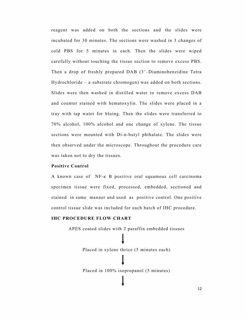
IHC PROCEDURE FLOW CHART
APES coated slides with 2 paraffin embedded tissues
Placed in xylene thrice (5 minutes each)
Placed in 100% isopropanol (5 minutes)

13
Placed in 70% isopropanol (5 minutes)
Washed in distilled water thrice (5 minutes each)
Placed in 3% hydrogen peroxide (20 minutes)
Kept in citrate buffer at pH 6 and autoclaved for antigen retrieval
and bench cooled for 40 minutes
Washed in distilled water thrice (5 minutes each)
Protein blocking serum added and incubated for one hour
Primary antibody added to the specimen and incubated for one hour
Washed in PBS thrice (5 minutes each)
Secondary antibody added and incubated in an enclosed hydrated
container (30 minutes)
Washed in PBS thrice (5 minutes each)
avidin biotin enzyme reagent added and incubated (30 minutes)
Washed in PBS thrice (5 minutes each)
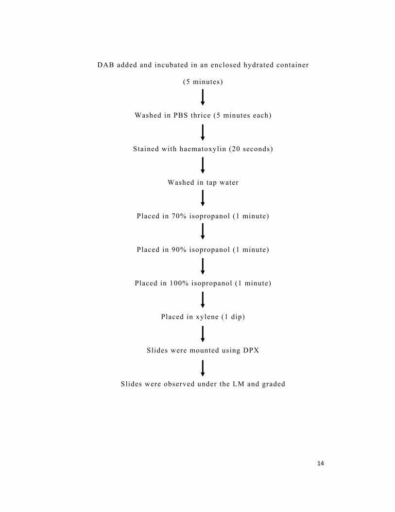
14
DAB added and incubated in an enclosed hydrated container
(5 minutes)
Washed in PBS thrice (5 minutes each)
Stained with haematoxylin (20 seconds)
Washed in tap water
Placed in 70% isopropanol (1 minute)
Placed in 90% isopropanol (1 minute)
Placed in 100% isopropanol (1 minute)
Placed in xylene (1 dip)
Slides were mounted using DPX
Slides were observed under the LM and graded
15
Criteria for evaluation of NF-κ B staining
The following parameters were used to evaluate NF-κ B staining
1. Tissue localization of stain – Location of NF-κ B staining
either in the epithelium and /or connective tissue was
recorded. In the epithelium staining in the basal and /or
suprabasal layers was recorded.
2. Cellular localization of stain – Nucleus and /or cytoplasm
3. Intensity of the stain – In each of the positive case the entire
tissue was graded as no stain (0), mild (+), moderate (++),
and intense (+++).
4. Labelling index (LI) – was calculated by dividing the number
of positive cells by the total number of cells counted in the
slide and expressed as percentage. A minimum of thousand
cells was counted for each slide.
Number of positive cells
LI = X 100
Total number of cells counted

16
Statistical analysis was done using SPSS T M software (version
10.0.5). p value ≤0.05 was considered to be statistically significant.
• Pearson’s Chi-square test was done to compare mean age, the
distribution of gender and habits, t issue localization of stain,
cellular location, nature of stain, intensity of stain and the
percentage of cells stained among the three study groups.
• The inter-observer variability for the intensity of stain was
assessed by 2 investigators and their reports were confirmed with
kappa statistics.
• The mean labeling index between the groups was analysed by
Mann Whitney U test.
17
STRUCTURE AND MOLECULAR MECHANISM OF NF- κ B
NF-κB described in 1986, by Baltimore. D was characterized
initially from B-lymphocytes as a nuclear factor necessary for the
transcription of the immunoglobulin κ l ight chain gene.
Nuclear factor-kappaB (NF-κB) is a collective term that
refers to a small class of dimeric transcription factors for a number
of genes, including growth factors, angiogenesis modulators, cell-
adhesion molecules and antiapoptotic factors.9 Although most NF-
κB proteins promote transcription, some act as inactivating or
repressive complexes. The most common dimer known
“specifically” as NF-κB, is relatively abundant, controls the
expression of numerous genes and exist as an inactive cytoplasmic
complex bound to inhibitory proteins of the NF-κB inhibitory (Iκ B)
family.10
The inactive NF-κB –IκB complex is activated by a variety of
stimuli, including proinflammatory cytokines, mitogens, growth
factors and stress-inducing agents. The release of NF-κB facilitates
its translocation to the nucleus, where it promotes cell survival by
initiating the transcription of genes encoding stress-response
enzymes, cell-adhesion molecules, proinflammatory cytokines and
antiapoptotic proteins.1 1

18
MEMBERS OF THE FAMILY
NF-κB is a family of signal activated transcription factors
comprised of hetero-or homo-dimers from 5 different subunits, NF-
κB1, NF-κB2, RELA, c REL and RELB. NF-κBs normally are
transiently activated in response to infection or injury, but in
cancers are aberrantly activated, regulating a transcriptome of
hundreds of genes and corresponding proteosome that promote
pathogenesis and therapeutic resistance.1 2
NF-κB1 is transcribed as p105 and processed to p50, NF-
κB2 is transcribed as p100 and processed to p52 following
phosphorylation and proteosome-dependent degradation of their
ankyrin repeat –containing c-termini. RELA, RELB and cREL are
synthesized in mature form and often form hetero dimers with NF-
κB1 or NF-κB2.1 3
ACTIVATION OF NF-κB
There are two pathways that lead to the activation of NF- κB.
Canonical / Classical pathway.
Non – canonical / alternate pathway.
NF-κB1 is often associated with RELA (or cREL) and bound
in the cytoplasm in an inactive form by Inhibitor-kappaB (IκB)- α , β
or γ , and known as the canonical, or classical form of NF-κB. This
form is activated by various components of pathogens or cytokines
such as Tumor Necrosis Factor (TNF) or Interleukin-1 (IL-1),
primarily via a trimeric Inhibitor-kappaB Kinase (IKK) comprised
of α , β and γ subunits, that phosphorylates IκB, leading to IκB

19
proteasomal degradation and nuclear translocation and DNA binding
of the NF-κB1/RELA heterodimer.1 4
NF-κB2 often forms a heterodimer with RELB, known as the
non-canonical form of NF-κB. The non-canonical or alternate
pathway is activated by other stimuli such as lymphotoxin B, BAFF
and CD40L, via an NF-κB Inducing Kinase (NIK) and IKKα dimeric
complex.15
Aberrant activation of canonical pathway has been broadly
implicated in development of many cancers, consistent with its
ubiquitous expression and role in promoting cell survival and
growth. Aberrant activation of the non- canonical pathway has been
demonstrated in B lymphoid malignancies, as well as other
cancers.16
INHIBITION OF NF-κB
IκB is a large family of inhibitory molecules that includes
IκBα , IκBβ , IκB , IκBγ and Bcl-3. The IκB are characterized by the
presence of multiple ankyrin repeats and interact with NF- κB via
RHD. The RHD serves several functions; this domain can act as the
dimerization and DNA binding domain of this class of proteins as i t
contains nuclear localization sequence and it is the site for binding
of IκB. NF – κB binds to 9 - 10 base pair DNA sites (κB sites) as
dimers. The activity of NF - κB is primarily regulated by interaction
with inhibitory IκB proteins. 17
IκB kinase (IKK) is present in the cell cytoplasm as an
enzyme with serine-protein-kinase activity that is responsible for

20
IκBα phosphorylation and that links tumor necrosis factor (TNF) –
induced and interleukin-1 (IL-1)-induced kinase cascades to NF-κB
activation. It is a large, multisubunit complex with three known
components. Two of these polypeptide components, IKKα and IKKβ
are catalytic subunits, whereas the third component, IKKα (NEMO),
has a regulatory function. In most cells, NF-κB is present as a
latent, inactive IκB – bound complex in the cytoplasm.1 8 The
stimulation by different pathogens and other inducers including
viruses. Cytokines lead to the activation of signaling cascades that
activates the IκB complex and causes phosphorylation of IκB. The
NF–κB is then released and translocated to the nucleus where they
bind with target genes and regulate their transcription. 1 9
ROLE OF NF-κB IN CELL CYCLE REGULATION
The inactive NF-κB-IκB complex is activated by a variety of
stimuli, including pro inflammatory cytokines, nitrogens, growth
factors and stress-inducing agents. Pro-inflammatory cytokines
produced by macrophages, T cells and other cells exert their actions
on target cells by transactivating NFκB (i.e., by initiating the signal
cascade).2 0 Most cells express receptors for the pro-inflammatory
cytokines, IL-1β and TNF-α , and they also contain the IKK complex
needed for signal transduction. Cells such as leukocytes, vascular
endothelial and smooth muscle cells, cardiomyocytes and fibroblasts
therefore respond to proinflammatory cytokines by NF-κB
activation.2 1

21
NF-κB activation induces the expression of pro-inflammatory
cytokines in a positive feed-back loop. The NF-κB pathway is used
not only by pro-inflammatory cytokines but also by microbial
products. In particular, endotoxins of gram-negative bacteria signal
through NF-κB. Different stimuli initiates different signal-
transduction pathways, which are activated by their respective
ligands.2 2 NF-κB then translocates to the nucleus where, by binding
to κB, it initiates transcription of various cytokines, adhesion
molecules, vascular endothelial growth factor etc. These
transcription products aid in further proliferation of cells.2 3
NF-κB plays a critical role in cell cycle progression during
G0-to-G1 phase. Inhibition of NF-κB caused impairment of cell
cycle progression, retarded G1 to S-phase transition. Passage
through the restriction point in G1 and transit into S phase requires
the sequential activation of cyclin-dependent kinases (CDK4,
CDK6, CDK2) in complex with their particular G1 cyclins (D or E).
The D family of cyclins (D1, D2, D3) in complex with the catalytic
subunits CDK4 and CDK6 are the primary holoenzymes required for
entry and passage through G1. NF-κB promotes cell cycle
progression by regulating the expression of several genes involved
in the cell cycle machinery such as cyclin D1, D2, D3, E and c-
myc.2 4
DUAL ROLE OF NF-κB
NF-κB plays a major role as a mediator in inhibition of
apoptosis in many cell types. Tumor initiation begins with the
22
prolonged survival of a cell and so, NF-κB’s role has an obvious
implication for cancer. An anti-apoptotic role of NF-κB has been
linked to T cell lymphoma, osteoclasts, melanoma, pancreatic
cancer, bladder cancer and breast cancer. Cell types that display an
anti-apoptotic role for NF-κB include B cells, T cells, granulocytes,
macrophages, neuronal cells and smooth muscle cells.25
NF-κB has been shown to play a pro-apoptotic role in
addition to its more common anti-apoptotic role. Different
activation pathways of NF-κB may cause the expression of proteins
that promote apoptosis (eg: Fas, c-myc, p53, IκBα) or inhibit
apoptosis (eg: TRAF2, IAP proteins, Bcl-2 like proteins). NF-κB
activation variably controls the regulation of cell cycle proteins (eg:
cyclin D1 and CDK2 kinase) and the interaction with various
cellular components (eg: p300 and p53) that promote or induce
apoptosis.2 6
ROLE OF NF-κB IN CANCER
NF-κB has been implicated in carcinogenesis because of its
critical roles in cell survival, cell adhesion, inflammation,
differentiation and cell growth. The role of NF-κB in different steps
of tumoriogenesis are:
NF-κ B ACTIVATION AND CELL PROLIFERATION
In normal cells, NF-κB becomes activated only after the
appropriate stimulation and then, upregulates the transcription of its
target genes. In tumor cells, different types of molecular alterations
may result in impaired regulation of NF-κB activation. In such
23
cases, NF-κB loses its transient nature of activation and becomes
constitutively activated. This leads to deregulated expression of NF-
κB controlled genes.2 7
Several genes that mediate cell proliferation are regulated by
NF-κB. These include growth factors such as TNF-α , IL-1β and
interleukin-6 (IL-6). Besides growth factors, certain cell cycle-
regulatory proteins (eg: the cyclin D1 required for transition of
cells from G1 to S phase) are also regulated by NF-κB.28
In some cells, PGE2 has been shown to induce proliferation of
tumor cells. The synthesis of cyclooxygenase-2 (COX-2), which
controls PGE2 production, is also regulated by NF-κB activation. It
has also been shown that growth factors such as EGF and platelet-
derived growth factor (PDGF) induce proliferation of tumor cells
through activation of NF-κB.2 9
ACTIVATION OF NF-κ B PROMOTES SURVIVAL OF TUMOR
CELLS
Gene products that negatively regulate apoptosis in tumor
cells are controlled by NF-κB activation. These include IAP-1, IAP-
2, XIAP, cFLIP, TRAF1, TRAF2 and Bcl-xL. Tumor necrosis
factor- alpha can cause programmed cell death (i .e, apoptosis) and
this is often paralleled by increased NF-κB activation. Inappropriate
activation of NF-κB inhibit apoptosis.3 0

24
NF-κB may upregulate the mitochondrial antiapoptotic factor
Bcl-2, perhaps in a positive feed back loop as Bcl-2 downregulates
IκBα , thus increasing NF-κB activation. NF-κB has been linked to
anti-apoptotic function in tumors such as T-cell lymphoma,
melanoma, pancreatic cancer, bladder cancer and breast cancer and
in tumor-related cell types such as B cells, T cells, granulocytes,
macrophages, neuronal cells, smooth muscle cells and osteoclasts.3 1
NF-κ B MEDIATES THE INVASION OF TUMOR CELLS
NF-κB regulates many genes involved in the promotion of
cancer by clonal expansion, growth, diversification, angiogenesis,
adhesion, extravasation, degradation of extracellular matrix, etc.
The development of cancer is generally categorized into three
stages: tumor initiation, tumor promotion and tumor metastasis.
Proteases influence tumor invasiveness (eg: the matrix
metalloproteinases and the serine protease urokinase- type
plasminogen activator [uPA]) which is regulated by NF-κB.32
Matrix metalloproteinases (MMPs) promote growth of cancer
cells through the interaction of extracellular matrix (ECM)
molecules and integrins, cleaving insulin-like growth factors and
shedding transmembrane precursors of growth factors, including
transforming growth factor-β (TGF-β). uPA is another critical
protease involved in tumor invasion and metastasis. Constitutively
active phosphatidylinositol-3 kinase (P13K) controls cell motility
by regulating the expression of uPA through the activation of NF-
κB. Thus one potential way to block the invasion of tumors is to
25
target NF-κB so that the activation of genes involved in cancer
progression is also blocked.33
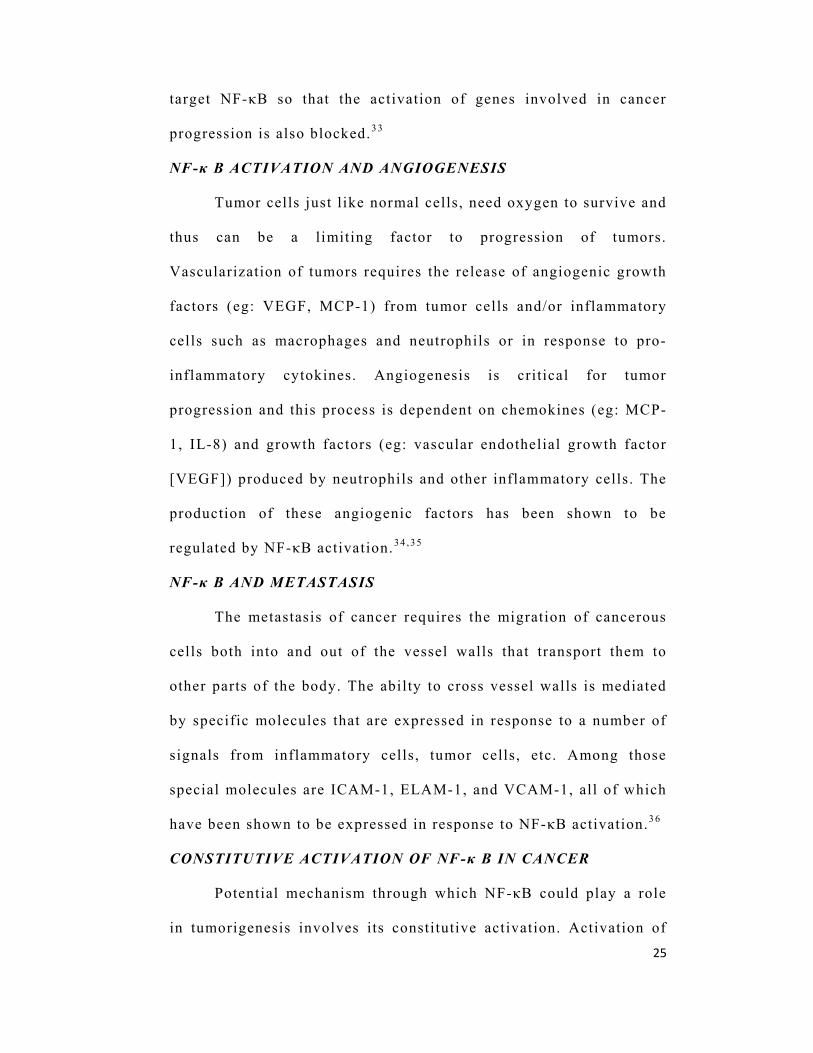
NF-κ B ACTIVATION AND ANGIOGENESIS
Tumor cells just l ike normal cells, need oxygen to survive and
thus can be a limiting factor to progression of tumors.
Vascularization of tumors requires the release of angiogenic growth
factors (eg: VEGF, MCP-1) from tumor cells and/or inflammatory
cells such as macrophages and neutrophils or in response to pro-
inflammatory cytokines. Angiogenesis is critical for tumor
progression and this process is dependent on chemokines (eg: MCP-
1, IL-8) and growth factors (eg: vascular endothelial growth factor
[VEGF]) produced by neutrophils and other inflammatory cells. The
production of these angiogenic factors has been shown to be
regulated by NF-κB activation.3 4 ,35
NF-κ B AND METASTASIS
The metastasis of cancer requires the migration of cancerous
cells both into and out of the vessel walls that transport them to
other parts of the body. The abilty to cross vessel walls is mediated
by specific molecules that are expressed in response to a number of
signals from inflammatory cells, tumor cells, etc. Among those
special molecules are ICAM-1, ELAM-1, and VCAM-1, all of which
have been shown to be expressed in response to NF-κB activation.3 6
CONSTITUTIVE ACTIVATION OF NF-κ B IN CANCER
Potential mechanism through which NF-κB could play a role
in tumorigenesis involves its constitutive activation. Activation of

26
NF-κB occurs as it is transported from the cytoplasm to the nucleus
upon degradation of the inhibitory subunit. In the nucleus it binds to
specific κB sites on the DNA and mediates the expression of a
number of genes involved in the cellular response to various
stresses. Thus when NF-κB is found persistently in the nucleus, it is
referred to as constitutive activation.3 7
While many NF-κB stimuli have been identified, the stimulus
responsible for constitutive activation of NF-κB in most cell types
is not understood. Cells that express constitutively activated NF-κB
are resistant to various chemotherapeutic agents.38
CARCINOGENESIS AND NF-κB
Hideki Nakayama, Tetsuro Ikebe, Mahiro Beppu et al
(2001) used immunohistochemistry to examine the expression of
NF-κB and the signaling molecules leading to NF-κB activation in
36 untreated biopsy specimens from patients with squamous cell
carcinoma (SCC) and in 15 specimens from patients with epithelial
dysplasia of the oral cavity.The results suggest that high expression
levels of p65 and IKKα contribute to malignant behaviour and
antiapoptotic activity in SCC of the oral squamous epithelium.4 4
S. Chen, A. Fribly, and C. Y. Wang et al (2002) used
enzyme linked immunosorbent assay and western blot analysis to
explore the gene therapy approaches relevant to the inhibition of
NF-κB signaling, to determine whether Adv-SR-IκBα inhibited
NF-κB activation in Oral Squamous Cell Carcinoma (OSCC) cells.
The results of the in vitro application of a gene therapy stratergy for

27
oral cancer treatment confirmed that OSCC cells are accessible to
adenovirus-mediated gene transfer and the combination of gene
therapy with IκBα and TNF was efficient in the induction of
apoptosis and OSCC cells. Also inhibition of NF-κB by IκBα gene
transfer sensitizes OSCC cells to TNF killing.39
Asha Nair, Manickam Venkatraman, Tessy T Maliekal et al
(2003) have used several techniques such as immunohistochemistry,
Western blotting, Semiquantitative Reverse Transcriptase –
Polymerase Chain Reaction and Electrophoretic mobility shift
assays, and provided evidences for the constitutive activation of
NF-κB during cervical cancer progression using 106 paraffin-
embedded cervical tissue specimens of different histological grades.
In normal cervical tissue and low grade squamous intraepithelial
lesions, p50, RelA and IκB-α were mainly localized in the cytosol,
whereas in high-grade lesions and squamous cell carcinomas, p50-
RelA heterodimers translocated into the nucleus with a concurrent
decrease in IκB-α protein.4 1
Lan Li, Bharat B. Aggarwal, Shishir Shishodia et al (2004)
studied the ability of curcumin (diferuloylmethane), an agent that is
pharmacologically safe in humans, to modulate NF-κB activity.
Nuclear factor kappa B (NF-κB) has been determined to play a role
in cell survival or proliferation in pancreatic carcinoma. 5-human
pancreatic carcinoma cell lines were examined using electrophoretic
mobility gel-shift assay and immunoblot analysis.

28
Curcumin down-regulated NF-κB and growth control
molecules induced by NF-κB in human pancreatic cells. These
effects were accompanied by marked growth inhibition and
apoptosis. Through these findings, the authors provided a biologic
rationale for the treatment of patients with pancreatic carcinoma
using this nontoxic phytochemical.4 8
Guido M. Sclabas, Tadashi Uwagawa, Christian Schmidt et
al (2005) studied the effects of aspirin on pancreatic carcinoma
prevention and to reveal a possible mechanism of aspirin-mediated
cancer chemoprevention. An orthotopic mouse model with human
pancreatic carcinoma cell lines PANC-1, PANC-1/Puro, and PANC-
1/IκBαM was used to study the inhibitory effects of aspirin on
pancreatic tumor formation. It was concluded that aspirin repressed
tumor formation by PANC-1cells in vivo in a prophylactic setting,
suggesting a possible mechanism for aspirin’s preventive effect in
pancreatic carcinoma through inhibition of NF-κB activation and a
mechanistic link between inflammation and tumorigenesis.4 7
An Immunohistochemical study by Alok Mishra, Alok
Bharti, Bhuder Das C et al (2006) was done to analyze the
activation of NF – κB and its alterations, in the expression of
different NF – κB proteins during oral carcinogenesis in vivo using
tissue biopsies from precancerous and cancerous oral lesions. The
role of high risk human papilloma virus (HPV – 16) which activates
the p50 / p65 NF – κB complex formation, which promotes
differentiation of oral neoplastic cells leading to better prognosis of

29
the oral cancer was also analysed. Here the total biopsy specimens
were 110,out of which 10 cases were normal (n = 10), 34 cases was
oral precancer (n = 34) and 66 cases was cancer (n= 66).Expression
of p50 protein was 1000 in all the normal tissues and 40% of
precancerous lesion & 70% of oral cancers showed moderate to high
expression of NF – κB.1
Ximing Tang, Diane Liu, Shishir Shishodia et al (2006)
showed Immunohistochemical expression of NF-κB p65 in 394 lung
cancers (370 non-small cell lung carcinomas [NSCLC]; and 24
small cell lung carcinomas[SCLC]) and 269 lung normal epithelium
and preneoplastic lesions, including hyperplasias, squamous
metaplasias, dysplasias, and atypical adenomatous hyperplasias.
High levels of nuclear Immunohistochemical expression of NF-κB
p65 were detected in the lung cancers, with significantly higher
levels in SCLCs compared with NSCLCs (P < 0.0001).
The findings indicate that NF-κB activation plays an
important role in lung cancer pathogenesis and NF-κB p65 nuclear
expression is an early and frequent phenomenon in the pathogenesis
of lung cancer.42
Ming Yu, Jason Yeh and Carter Van Waes et al (2006)
showed that CK2 contributes to the activation of IKK and NF-κB in
response to serum factors, which suggests that CK2 and IKK2 are
key candidates for targeting the NF-κB pathway in head and neck
squamous cell carcinoma (HNSCC). NF-κB activation has been
broadly shown and associated with progression in intraepithelial
30
premalignant and malignant squamous neoplasms of the head and
neck as well as uterine cervix.43
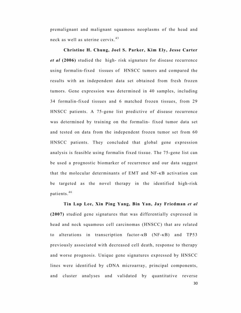
Christine H. Chung, Joel S. Parker, Kim Ely, Jesse Carter
et al (2006) studied the high- risk signature for disease recurrence
using formalin-fixed tissues of HNSCC tumors and compared the
results with an independent data set obtained from fresh frozen
tumors. Gene expression was determined in 40 samples, including
34 formalin-fixed tissues and 6 matched frozen tissues, from 29
HNSCC patients. A 75-gene list predictive of disease recurrence
was determined by training on the formalin- fixed tumor data set
and tested on data from the independent frozen tumor set from 60
HNSCC patients. They concluded that global gene expression
analysis is feasible using formalin fixed tissue. The 75-gene list can
be used a prognostic biomarker of recurrence and our data suggest
that the molecular determinants of EMT and NF-κB activation can
be targeted as the novel therapy in the identified high-risk
patients.4 6
Tin Lap Lee, Xin Ping Yang, Bin Yan, Jay Friedman et al
(2007) studied gene signatures that was differentially expressed in
head and neck squamous cell carcinomas (HNSCC) that are related
to alterations in transcription factor-κB (NF-κB) and TP53
previously associated with decreased cell death, response to therapy
and worse prognosis. Unique gene signatures expressed by HNSCC
lines were identified by cDNA microarray, principal components,
and cluster analyses and validated by quantitative reverse

31
transcription-PCR (RT-PCR) and in situ hybridization.
Bioinformatic analysis of the promoters and ontogeny of these
cluster genes was done. NF-κB promotes expression of a novel NF-
κB – related gene signature and cell survival in HNSCC that weakly
express TP53, a subset previously associated with inactivated wild-
type TP53, greater resistance to chemoradiotherapy, and worse
prognosis.4 5
Zhong Chen, Bin Yan, Carter Van Waes et al (2008) used
immunohistochemistry (IHC), Reverse-phase protein microarray
(RPMA), Western blot analysis and Enzyme linked Immunosorbant
Assay (ELISA) to determine which pathways are activated and how
they correlate with prognosis, select therapy targeting these
pathways and to use changes in phosphorylation, expression of
protein and functional changes such as TUNEL or caspase cleavage
together as early biomarkers of response to the treatment.
Serum cytokines regulated by NF-κB represent biomarkers of
response, recurrence and survival in HNSCC.40
NF-κB AND LEUKOPLAKIA
Squamous cell carcinoma is the most common malignancy of
the oral cavity which is often preceded by premalignant lesions, the
most common of which is leukoplakia.6 0 Epithelial malignancies of
the oral cavity often begin as preneoplastic lesions in the form of
inflammatory lesions such as leukoplakia. Leukoplakia is associated
with tobacco and alcohol use and chronic inflammation with a

32
higher risk of malignant transformation to oral squamous cell
carcinoma.63
Nuclear factor kappa B has been implicated in the
development of head and neck squamous cell carcinoma from
premalignancy, progression to invasion, metastasis and treatment
resistance.5 1 Transcription factor NF-κ B is an early response gene
that is found to be elevated in subjects with tobacco use, chronic
inflammatory conditions of oropharynx and head and neck cancer.
Inflammatory gene expressions are induced by the downstream of
cytokine and growth factor receptors, such as signal transducers and
activators of transcription. The expression of NF-κ B has been
found to be gradually increasing from premalignant lesions to
invasive cancer.6 2
NF-κB AS A POTENTIAL MOLECULAR TARGET FOR
CANCER THERAPY
NF-κB is constitutively activated in a large number of
epithelial and hematologic malignancies, strongly suggest that NF-
κB inhibitors would be useful in cancer therapy. Inhibition of NF-
κB has been found to be an important mechanism of action of
steroids, nonsteroidal anti-inflammatory drugs (NSAID) and natural
and synthetic compounds that show therapeutic and preventive
activity with acceptable safety profiles.4 9
NSAIDS AND CANCER PREVENTION
NSAIDs such as aspirin, sulindac, ibuprofen, celecoxib, have
been shown to inhibit NF-κB activation and arachidonic acid

33
inflammatory pathways upstream and downstream of NF-κB.
Strategy for blocking NF-κB include an upstream strategy and a
NF-κB targeting strategy.50 Blocking the activation of NF-κB
signaling pathway using:
a) proteasome inhibitors (such as PS-341, MG 132);
b) IKK inhibitors (such as NSAIDs, sulfa salazine, arsenic
trioxide, curcumin, thlidomide);
c) cell-permeable peptides (such as SN-50);
d) antioxidants (such as disulfiram, glutathione); or
e) the recombinant adenovirus-mediated overexpression of the
IκBα gene.
On the other hand, the NF-κB targeting strategy includes:
(a) blocking the DNA binding of NF-κB using decoy
oligodeoxynucleotides (ODNs)
(b) blocking the transactivation of NF-κB using glucocorticoids
(c) interfering with mRNA using NF-κB antisense
oligonucleotide (ASO)51 ,5 2
CORTICOSTEROIDS AND CYTOTOXIC AGENTS USED FOR
THERAPY
The cytotoxic effects of corticosteroids in combination with
other DNA-damaging agents led to the use of steroid-based
regimens as a current mainstay of treatment of certain leukemias,
lymphomas and myelomas. Subsequently, corticosteroids were
shown to mediate many of their anti-inflammatory and tumor
cytotoxic effects through inhibition of NF-κB, and these lymphoid

34
malignancies and supporting host responses were found to be
exquisitely dependent on NF-κB regulated survival or inflammatory
mechanisms.5 3 , 5 4
PROTEASOME INHIBITORS
A new class of therapeutic agents under development are
proteasome inhibitors, which regulate degradation of IκB and
inhibit NF-κB, as well as turnover of other cellular proteins.55 ,56
IKK INHIBITORS
IKKβ antagonists that more specifically inhibit IκB and the
NF-κB classic pathway initially implicated in cancers have been the
subject of intensive development and preclinical studies.57 ,58
35
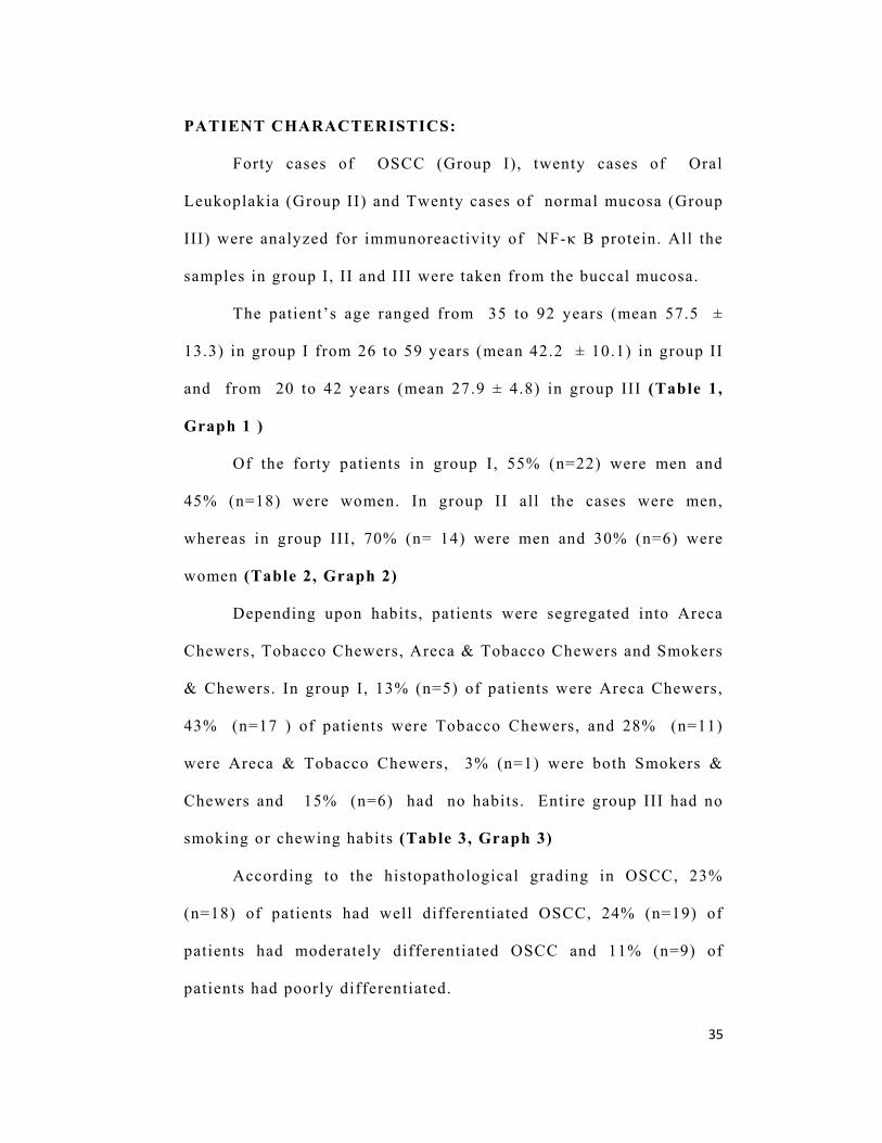
PATIENT CHARACTERISTICS:
Forty cases of OSCC (Group I), twenty cases of Oral
Leukoplakia (Group II) and Twenty cases of normal mucosa (Group
III) were analyzed for immunoreactivity of NF-κ B protein. All the
samples in group I, II and III were taken from the buccal mucosa.
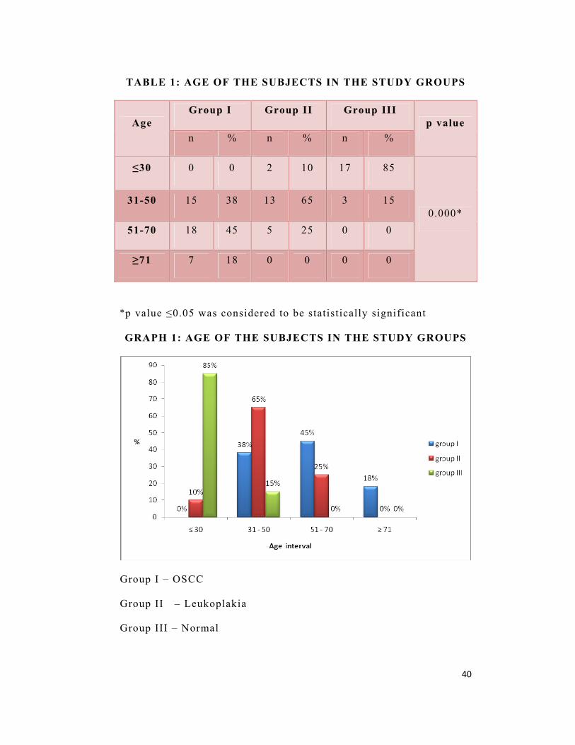
The patient’s age ranged from 35 to 92 years (mean 57.5 ±
13.3) in group I from 26 to 59 years (mean 42.2 ± 10.1) in group II
and from 20 to 42 years (mean 27.9 ± 4.8) in group III (Table 1,
Graph 1 )
Of the forty patients in group I, 55% (n=22) were men and
45% (n=18) were women. In group II all the cases were men,
whereas in group III, 70% (n= 14) were men and 30% (n=6) were
women (Table 2, Graph 2)
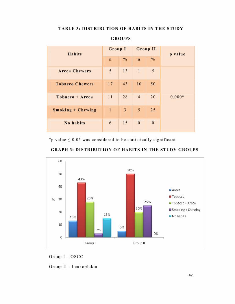
Depending upon habits, patients were segregated into Areca
Chewers, Tobacco Chewers, Areca & Tobacco Chewers and Smokers
& Chewers. In group I, 13% (n=5) of patients were Areca Chewers,
43% (n=17 ) of patients were Tobacco Chewers, and 28% (n=11)
were Areca & Tobacco Chewers, 3% (n=1) were both Smokers &
Chewers and 15% (n=6) had no habits. Entire group III had no
smoking or chewing habits (Table 3, Graph 3)
According to the histopathological grading in OSCC, 23%
(n=18) of patients had well differentiated OSCC, 24% (n=19) of
patients had moderately differentiated OSCC and 11% (n=9) of
patients had poorly differentiated.

36
Distribution of NF-κ B Staining Among 3 Groups:
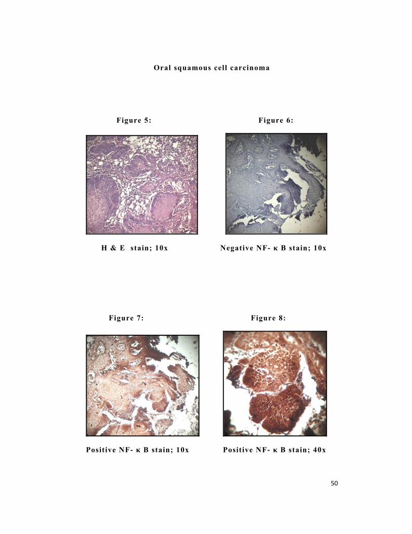
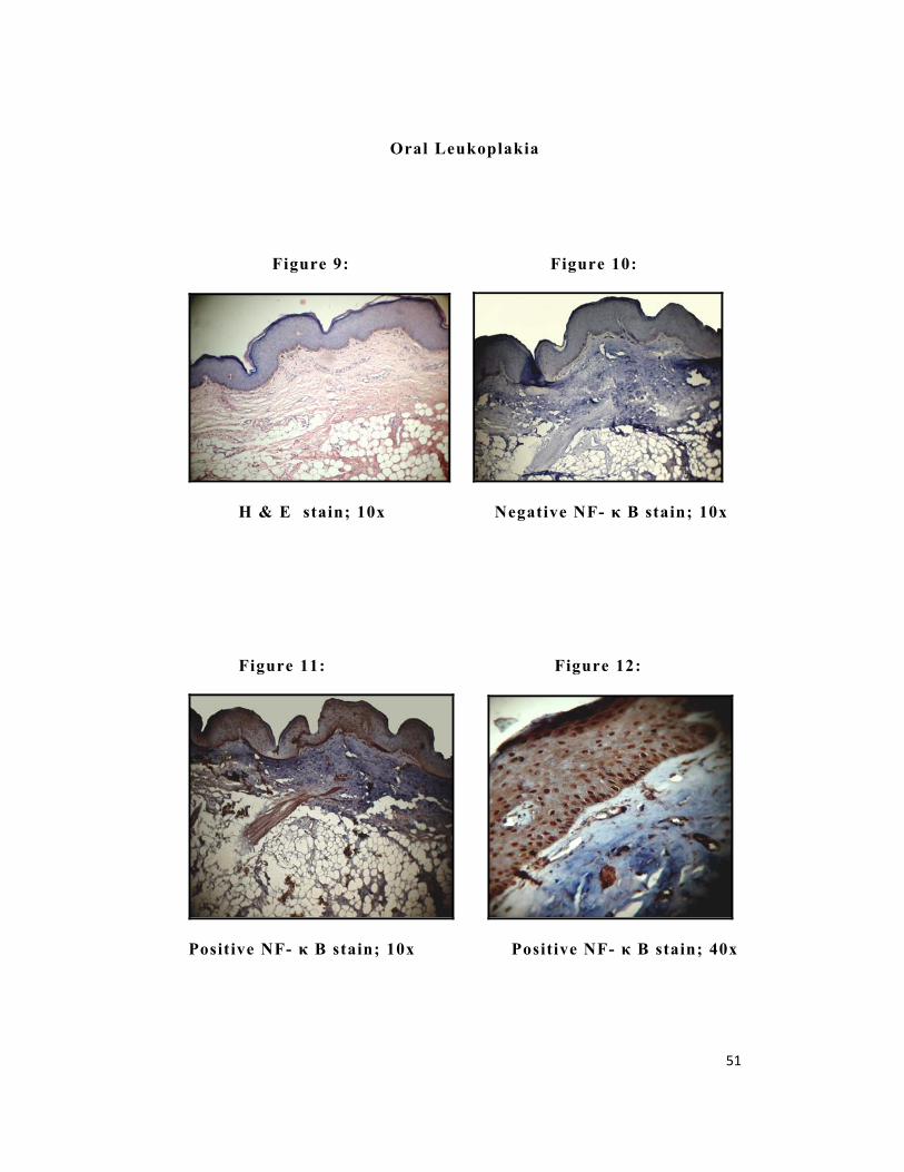
NF-κ B revealed positivity in group I (Figure 7 & 8), group II
(Figure 11& 12) and group III (Figure 15 & 16). In Group II
positive staining was observed in 100 % of the cases, whereas in
Group I and Group III, positive staining was observed in 97% and
80% respectively.
Tissue localization of stain (Table 4, Graph 4)
NF – κB staining was seen both in the epithelium as well as in
the connective tissue. In connective tissue the staining was seen in
lymphocytes, muscle fibres and endothelial cells.
In group I, 65% (n=26) of cases showed nuclear staining in
the epithelium and connective tissue, whereas in 35% (n=14) of
cases showed cytoplasmic staining, In groupII 80% (n=16) showed
nuclear staining while 20% (n=4) showed cytoplasmic staining
and in group III 35% (n=7) showed nuclear staining and 65%
(n=13) cases showed cytoplasmic staining. In connective tissue,
staining was predominantly seen in lymphocytes, muscle f ibres and
endothelial cells. These results were statistically significant
(p=0.011)

37
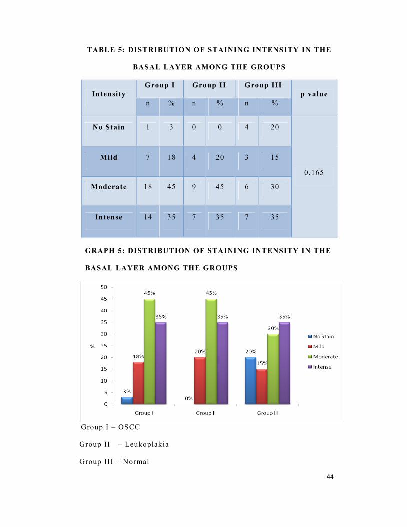
INTENSITY OF STAIN
Distribution of staining intensity in the basal layers among the
groups (Table 5, Graph5)
In the basal layer, mild intensity of staining was seen in 18% (n=7)
of OSCC, 20% (n=4) of leukoplakia and 15% (n=3) of normal cases.
Moderate staining was seen in 45% (n=18) of OSCC , 45% (n=9) of
leukoplakia and 30% (n=6) of normal cases whereas 35% (n=14) of
OSCC, 35% (n=7) of leukoplakia and 35% (n=7) of normal cases
showed intense staining.
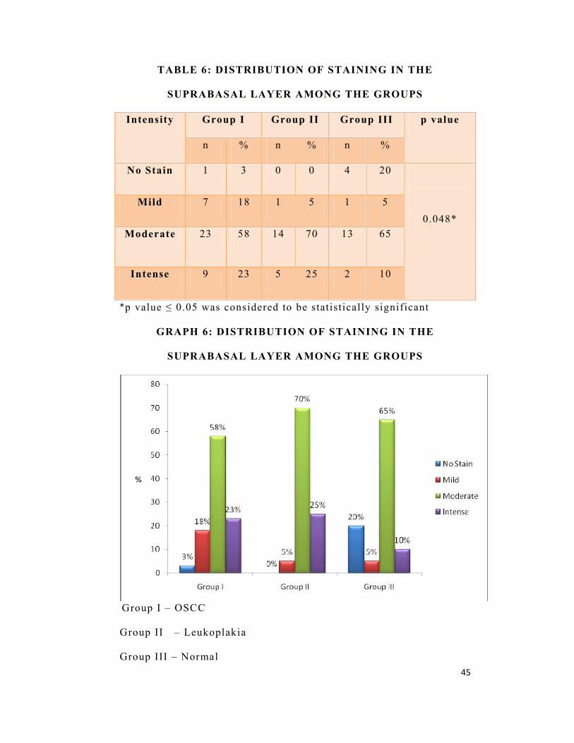
Distribution of staining intensity in the suprabasal layers among
the groups (Table 6, Graph 6)
In the suprabasal layer, mild intensity of staining was seen in
18% (n=7) of OSCC, 5% (n=1) of leukoplakia and 5% (n=1) of
normal cases. Moderate staining was seen in 58% (n=23) of OSCC,
70% (n=14) of leukoplakia and 65% (n=13) of normal cases whereas
23% (n=9) of OSCC, 25% (n=5) of leukoplakia and 10% (n=2) of
normal cases showed intense staining. The results were statistically
significant (p=0.048)
Comparison of staining intensity in the connective tissue among
the groups (Table 7, Graph 7)
In 40 cases of OSCC 23% (n=9) and in normal 50% (n=10)
showed no stain. Mild intensity of staining was seen in 18% (n=7)
of OSCC,10% (n=2) of both leukoplakia and normal cases.
Moderate intensity of staining was seen in 35% (n=14) of OSCC and
70% (n=14) of leukoplakia and 25% (n=5) of normal cases. And
38
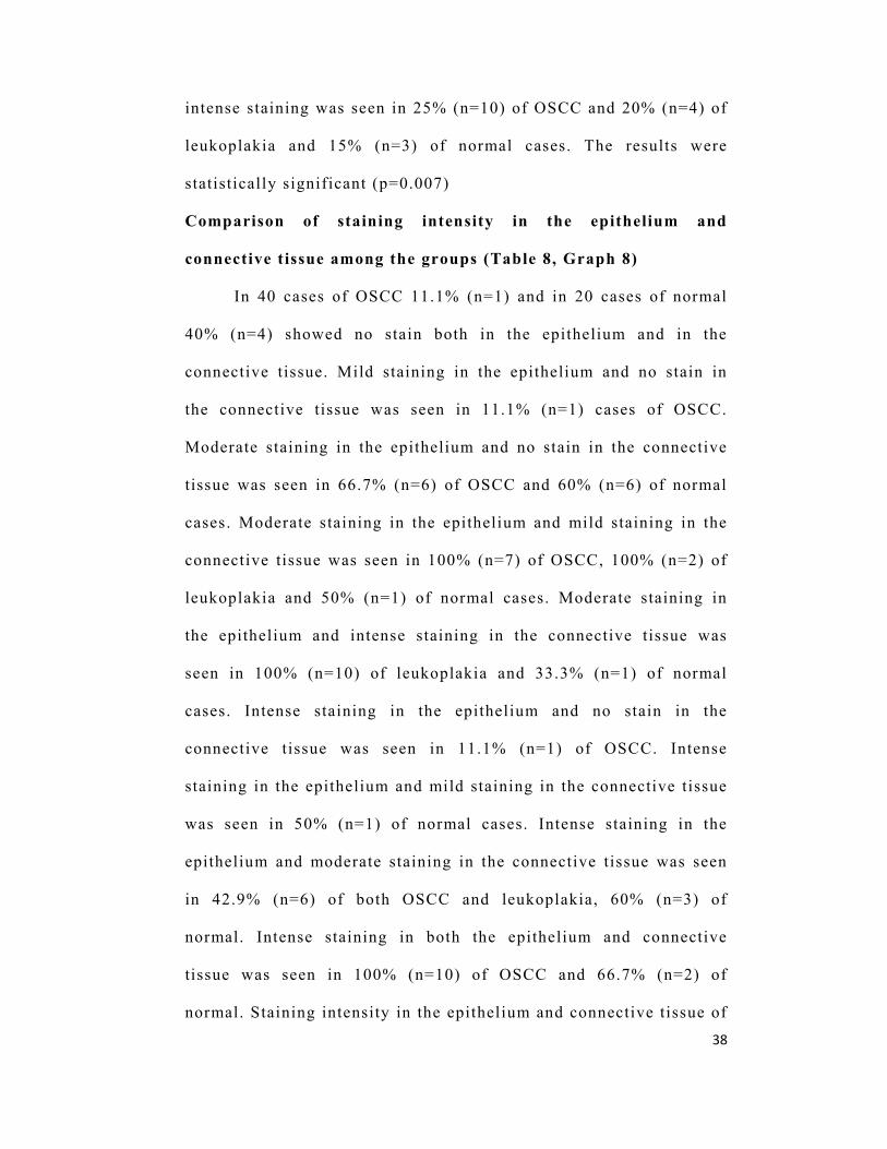
intense staining was seen in 25% (n=10) of OSCC and 20% (n=4) of
leukoplakia and 15% (n=3) of normal cases. The results were
statistically significant (p=0.007)
Comparison of staining intensity in the epithelium and
connective tissue among the groups (Table 8, Graph 8)
In 40 cases of OSCC 11.1% (n=1) and in 20 cases of normal
40% (n=4) showed no stain both in the epithelium and in the
connective tissue. Mild staining in the epithelium and no stain in
the connective tissue was seen in 11.1% (n=1) cases of OSCC.
Moderate staining in the epithelium and no stain in the connective
tissue was seen in 66.7% (n=6) of OSCC and 60% (n=6) of normal
cases. Moderate staining in the epithelium and mild staining in the
connective tissue was seen in 100% (n=7) of OSCC, 100% (n=2) of
leukoplakia and 50% (n=1) of normal cases. Moderate staining in
the epithelium and intense staining in the connective tissue was
seen in 100% (n=10) of leukoplakia and 33.3% (n=1) of normal
cases. Intense staining in the epithelium and no stain in the
connective tissue was seen in 11.1% (n=1) of OSCC. Intense
staining in the epithelium and mild staining in the connective tissue
was seen in 50% (n=1) of normal cases. Intense staining in the
epithelium and moderate staining in the connective tissue was seen
in 42.9% (n=6) of both OSCC and leukoplakia, 60% (n=3) of
normal. Intense staining in both the epithelium and connective
tissue was seen in 100% (n=10) of OSCC and 66.7% (n=2) of
normal. Staining intensity in the epithelium and connective tissue of

39
OSCC and leukoplakia were statistically significant and they were
0.001 and 0.043 respectively.
Comparison of mean labeling index among the study groups
In OSCC, 26/40 cases showed nuclear staining and their mean
labeling index was 25.96. In leukoplakia, 16/20 cases showed
nuclear staining which had the mean labeling index as follows
20.84. In normal, 7/20 cases showed nuclear staining and its mean
labeling index is 30.93, but this difference was not statistically
significant (p=0.262).
Comparison of mean labeling index between OSCC and normal,
Leukoplakia and normal & OSCC and leukoplakia
Comparison of mean labeling index between OSCC (54.34)
and normal (44.97) was done. Comparison of mean labeling index
between leukoplakia (51.17) and normal (44.97) was done. Similarly
the mean labeling index of OSCC (54.34) was compared to
leukoplakia (51.17), but these differences were not statistically
significant.
The inter-observer agreement for the intensity of stain for all
the 3 groups was arrived at kappa values of 0.648
40
TABLE 1: AGE OF THE SUBJECTS IN THE STUDY GROUPS
Age Group I Group II Group III
p value n % n % n %
≤30 0 0 2 10 17 85
0.000* 31-50 15 38 13 65 3 15
51-70 18 45 5 25 0 0
≥71 7 18 0 0 0 0
*p value ≤0.05 was considered to be statistically significant
GRAPH 1: AGE OF THE SUBJECTS IN THE STUDY GROUPS
Group I – OSCC
Group II – Leukoplakia
Group III – Normal

41
TABLE 2: DISTRIBUTION OF GENDER IN THE STUDY
GROUPS
CASES MALE FEMALE
p value n % n %
Group I 22 55 18 45
0.002* Group II 20 100 0 0
Group III 14 70 6 30
*p value ≤ 0.05 was considered to be statistically significant
GRAPH 2: DISTRIBUTION OF GENDER IN THE STUDY
GROUPS
Group I – OSCC
Group II – Leukoplakia
Group III – Normal
42
TABLE 3: DISTRIBUTION OF HABITS IN THE STUDY
GROUPS
Habits Group I Group II
p value n % n %
Areca Chewers 5 13 1 5
0.000*
Tobacco Chewers 17 43 10 50
Tobacco + Areca 11 28 4 20
Smoking + Chewing 1 3 5 25
No habits 6 15 0 0
*p value ≤ 0.05 was considered to be statistically significant
GRAPH 3: DISTRIBUTION OF HABITS IN THE STUDY GROUPS
Group I – OSCC
Group II - Leukoplakia

43
TABLE 4: LOCALIZATION OF NF-κ B STAINING AMONG
THE GROUPS
GROUP I
(%)
GROUP II
(%)
GROUP III
(%)
p
value
NUCLEAR 65 80 35
0.011*
CYTOPLASMIC 35 20 65
*p value ≤ 0.05 was considered to be statistically significant
GRAPH 4: LOCALIZATION OF NF-κ B STAINING AMONG
THE GROUPS
Group I – OSCC
Group II – Leukoplakia
Group III – Normal
44
TABLE 5: DISTRIBUTION OF STAINING INTENSITY IN THE
BASAL LAYER AMONG THE GROUPS
Intensity Group I Group II Group III
p value n % n % n %
No Stain 1 3 0 0 4 20
0.165
Mild 7 18 4 20 3 15
Moderate 18 45 9 45 6 30
Intense 14 35 7 35 7 35
GRAPH 5: DISTRIBUTION OF STAINING INTENSITY IN THE
BASAL LAYER AMONG THE GROUPS
Group I – OSCC
Group II – Leukoplakia
Group III – Normal
45
TABLE 6: DISTRIBUTION OF STAINING IN THE
SUPRABASAL LAYER AMONG THE GROUPS
Intensity Group I Group II Group III p value
n % n % n %
No Stain 1 3 0 0 4 20
0.048* Mild 7 18 1 5 1 5
Moderate 23 58 14 70 13 65
Intense 9 23 5 25 2 10
*p value ≤ 0.05 was considered to be statistically significant
GRAPH 6: DISTRIBUTION OF STAINING IN THE
SUPRABASAL LAYER AMONG THE GROUPS
Group I – OSCC
Group II – Leukoplakia
Group III – Normal

46
TABLE 7: DISTRIBUTION OF STAINING INTENSITY IN THE
CONNECTIVE TISSUE AMONG THE GROUPS
Intensity Group I Group II Group III p value
n % n % n %
No Stain 9 23 0 0 10 50
0.007* Mild 7 18 2 10 2 10
Moderate 14 35 14 70 5 25
Intense 10 25 4 20 3 15
*p value ≤ 0.05 was considered to be statistically significant
GRAPH 7: DISTRIBUTION OF STAINING INTENSITY IN THE
CONNECTIVE TISSUE AMONG THE GROUPS
Group I – OSCC
Group II – Leukoplakia
Group III – Normal
47
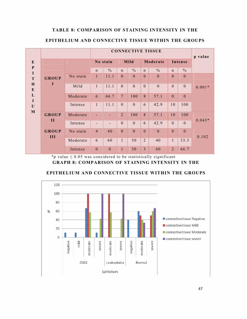
TABLE 8: COMPARISON OF STAINING INTENSITY IN THE
EPITHELIUM AND CONNECTIVE TISSUE WITHIN THE GROUPS
E P I T H E L I U M
CONNECTIVE TISSUE p value
GROUP I
No stain Mild Moderate Intense
n % n % n % n % No stain 1 11.1 0 0 0 0 0 0
0.001* Mild 1 11.1 0 0 0 0 0 0
Moderate 6 66.7 7 100 8 57.1 0 0
Intense 1 11.1 0 0 6 42.9 10 100
GROUP II
Moderate - - 2 100 8 57.1 10 100 0.043*
Intense - - 0 0 6 42.9 0 0
GROUP III
No stain 4 40 0 0 0 0 0 0 0.102
Moderate 6 60 1 50 2 40 1 33.3
Intense 0 0 1 50 3 60 2 66.7
*p value ≤ 0.05 was considered to be statist ically signif icant GRAPH 8: COMPARISON OF STAINING INTENSITY IN THE
EPITHELIUM AND CONNECTIVE TISSUE WITHIN THE GROUPS

48
Figure 1: Armamentarium
Figure 2: Antibodies
49
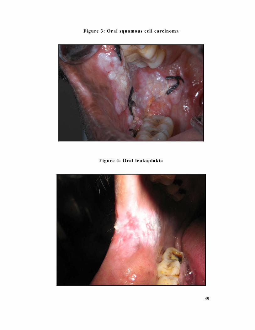
Figure 3: Oral squamous cell carcinoma
Figure 4: Oral leukoplakia
50
Oral squamous cell carcinoma
Figure 5: Figure 6:
H & E stain; 10x Negative NF- κ B stain; 10x
Figure 7: Figure 8:
Positive NF- κ B stain; 10x Positive NF- κ B stain; 40x
51
Oral Leukoplakia
Figure 9: Figure 10:
H & E stain; 10x Negative NF- κ B stain; 10x
Figure 11: Figure 12:
Positive NF- κ B stain; 10x Positive NF- κ B stain; 40x
52
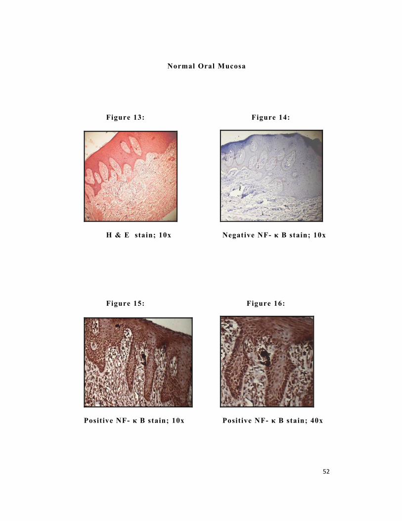
Normal Oral Mucosa
Figure 13: Figure 14:
H & E stain; 10x Negative NF- κ B stain; 10x
Figure 15: Figure 16:
Positive NF- κ B stain; 10x Positive NF- κ B stain; 40x

53
Nuclear factor κ B (NF-κ B) is a transcription factor that
induces the expression of various genes, that influence
inflammatory reactions, embryonic morphogenesis and
antiapoptosis. Intracellular signaling mechanism of NF-κ B
activation is effected by stimulators such as tumor necrosis factor α
(TNF-α) and many other signals.44 An inhibitor of NF-κ B known
as Iκ B, which holds NF-κ B in the cytoplasm gets phosphorylated
and ubiquitinated by ubiquitin ligase and degraded by 26S
proteosome and active NF-κ B translocates from the cytoplasm into
the nucleus and binds with the specific sequence in the promoter of
target genes.37 These genes include pro-inflammatory cytokines,
chemokines and cell survival genes. Activation of NF-κ B plays a
key role by which cells are resistant to TNF- mediated apoptosis,
thus being associated with development and progression and
metastasis of several malignancies including squamous cell
carcinoma.39
Oral dysplasia is often a common precursor of oral cancer.
Progression to cancer varies widely ranging from 5% to 20%. The
assessment and severity of dysplasia is based on architectural
disturbances accompanied by cytological atypia which are objective
in nature. Since NF – κB is a transcription factor that is known to
involve in growth, invasion and anti-apoptotic activity of cancer
cells. In this study we wanted to study expression in OSCC and
leukoplakia and compare its activity with that of normal oral

54
mucosa to ascertain if it could be used as a surrogate marker for
malignant transformation.8
PATIENT CHARACTERISTICS
In our study the mean age in OSCC cases was 57.5 ± 13.3
years. Mishra et al, studied 66 cases of OSCC and reported the
mean age in their study group as 52.9 ± 10.8 years. In the study
done by Nakayama et al, the mean age was 70.2 years in 36 cases
and Yu- Chao chang et al reported age range from 50-59 years with
a mean age of 55 years in 442 cases of OSCC. In a study by
Ranganathan et al , the age ranged from 56-85 years with a mean
age of 71.2 years in 10 cases.
In our study, the age presentation for leukoplakia ranged from
26 to 59 years with a mean of 42.2 ± 10.1 years which is consistent
with the study by Thavaraj et al with a mean age of 36.67years in
500 patients. Yu- Chao chang et al reported an age range from 20-
29 years and with a mean age of 25 years in 344 cases of oral
leukoplakia. In the study by Lumerman et al the age range was 25
to 92 years with a mean age of 59.3 years.
In our study subjects who had OSCC were predominantly males
and they constituted to about 70%. The male to female ratio was
2.2:1 and this finding was consistent with that of Napier et al
where the male: female ratio was 1.9:1 and the male: female ratio
was 3.9:1 with that of Mishra et al .
In our study all the patients who had leukoplakia were males which
is similar to a male: female ratio of 0.7 : 0.4 reported by
55
Saraswathi et al in their study of 2017 patients at our centre
earlier. In a study by Thavaraj et al the male: female ratio was
9.8:1 They also stated that number of males was around five times
more than the females.
In this study, those patients who presented with OSCC, had
the habit of chewing tobacco 17% and 5% of the patient had the
habit of only Areca and about 11% of patients used both areca and
tobacco . This finding is consistent with that of Thavaraj et al who
reported that the majority of patients with OSCC in their study
about 9.8% had the habit of chewing tobacco whereas 5.6% patients
had the habit of only areca.
In this study, of 20 patients who presented with leukoplakia,
10% had the habit of chewing tobacco and 4% had the habit of both
areca nut and tobacco chewing. These findings are consistent with
that of Saraswathi et al who reported that 10% of the patients had
the habit of chewing areca nut and 8% had the habit of betel quid
chewing.
STAINING CHARACTERISTICS OF NF – κB
In our study, cytoplasmic expression of NF – κB was seen in
77.5% (n=31) of OSCC. When we analysed the pattern of
cytoplasmic staining, we observed that 17.5% (n=7) of the cases
showed mild intensity of staining, while 35% (n=14) showed
moderate and 25% (n=10) showed intense staining. Nuclear
expression of NF – κB was seen in 65% (n=26) 0f cases. The

56
nuclear percentage positivity increased with increasing grade of
OSCC.
Dysregulation of NF – κB expression and its activation is
frequently observed in many human cancers. Activated NF – κB
shows nuclear expression and it indicates an early event in
carcinogenesis. Once activated, it controls the expression of
several genes that regulate cell cycle (cyclin D1), differentiation
(p2 1), cell survival (Bcl – 2), growth factors (VEGF) cell adhesion
(VCAM, ECAM) and angiogenesis (MMPs).
Our results were consistent with Nakayama et al who also
reported 78.2% cytoplasmic expression in 36 cases of OSCC. There
are other studies which states 64% of cytoplasmic expression as
observed by Asha Nair et al. The explanation for the absence of
cytoplasmic expression of NF – κB in OSCC has not been indicated
in this study.
In all these studies there was an association between tumor
progression and extent of nuclear translocation of NF – κB. In our
study also the moderately differentiated OSCC exhibited increased
nuclear expression which is consistent with tumor progression.
Nakayama et al suggested that high expression level of NF – κB
contributed to the malignant behavior and anti apoptotic activity of
OSCC. Mishra et al reports that p50 homodimers transcriptionally
regulate anti – apopotic Bcl -2 which is shown to be over expressed
in oral cancer cells and inhibits terminal differentiation of oral
keratinocyte. The exact mechanism by which NF – κB is
57
constitutively activated in OSCC though not fully understood, it has
been suggested that autocrine expression of interleukins and EGFR
may play an important role in the activation of NF – κB.
In inflammation associated cancer, non-genetic stimuli
encourage the survival and proliferation of cells. NF – κB has dual
actions in tumor promotion; first by preventing the death of cells
with malignant potential and second by stimulating the production
of pro-inflammatory cytokines in inflammatory cells in tumor mass.
These cytokines then signals the cell to promote their survival and
proliferation. Classical NF – κB pathway i.e, the IKK – β depedent
NF – κB activation pathway might show the molecular link between
inflammation and tumor promotion48 ,4 9. So, in chronic
inflammatory diseases, pro-inflammatory factors cause
accumulation of DNA damage in dormant pre-malignant cells in
tumor microenvironment to become malignant.44
In our study in leukoplakia, cytoplasmic expression of NF –
κB was seen in 100% (n=20) of the cases. When we analysed the
pattern of staining, mild intensity of staining was observed in 10%
(n=2) of case, 70% (n=14) showed moderate intensity, and 20%
(n=4) showed intense cytoplasmic expression. Our study also
showed nuclear expression of NF – κB in 80% (n=16) of cases.
When we further analysed in order to understand the intensity of
staining we observed that proliferating cells showed more staining
than differentiated cells. We observed that nuclear expression is

58
seen in those cases of leukoplakia which expressed histologically
dysplastic alterations and an increase in inflammatory response.
Nayakama et al observed 4% moderate staining and 11% of
mild staining of NF – κB expression in the epithelium of 15
dysplasia (leukoplakia) samples. They also reported that staining
was seen in both basal and spinous epithelium cells and staining of
NF – κB was seen in the basal epithelial cells of epithelial dysplasia
(leukoplakia).
They suggest that NF – κB is an important mediator in
chronic inflammatory process. Aberrant and persistent tissue
inflammation is believed to play an important role in the occurence
of tissue fibrosis and cancer. One of the etiological factors of
leukoplakia is associated with area nut chewing habit. They propose
that areca nut extract was found to activate NF – κB in human oral
keratinocytes and one of the pathogenic mechanisms of leukoplakia
may be due to increased expression of NF – κB in response to areca
nut. NF – κB activates cytokines such as IL – 1 and TN F – α which
can result in amplification of inflammatory, response and
persistence of chronic inflammation at local site.6 1
So, in leukoplakia we observed nuclear staining in 16% of
the cases. This nuclear expression of NF – κB by the epithelial
cells is correlated with the amount of cytotoxic cell infiltration
suggesting that increased NF – κB activity may represent the basis
of maintenance of the inflammatory response. This can lead to the
expression of genes that are mediated in carcinogens. So, expression

59
of nuclear staining in leukoplakia can be used as an indicator for
carcinogenesis.
The nuclear expression in our cases showed
histopathologically a higher number of inflammatory cells which
could account for its expressivity. We would like to corroborate our
finding by studying more cases.
Compared to the normal cases the cytoplasmic intensity and
nuclear immunoreactivity in OSCC and leukoplakia was increased
and also we could find a statistically significant association
between the two patterns of staining.

60
• The study group comprised of Group I (OSCC n=40), Group II
(leukoplakia n=20) and Group III (Normal n=20).
• In OSCC, there was 98% positivity for NF – κB with 35% of
cytoplasmic expression and 65% of nuclear expression.
• In leukoplakia; there was 100% positivity for NF – κB
expression; with 20% of cytoplasmic expression and 80% of
nuclear staining.
• In normal, there was 80% of positivity for NF – κB
expression within 65% of cytoplasmic expression and 35% of
nuclear expression.
• In OSCC and leukoplakia when the epithelial staining
intensity was compared with the connective t issue staining
intensity there was a statistically significant correlation
(p=0.001) and (p=0.043) respectively.
• Nuclear expression of NF – κB exhibited difference (p=0.011)
between the groups.
In conclusion, our results show that there is increased
expression of NF – κ B in OSCC and leukoplakia when compared to
normal. Although the mean labeling index did not show any
significant difference between OSCC and leukoplakia, further
studies on a larger sample will help in ascertaining the exact role of
NF – κ B expression in leukoplakia samples.
61
1. Mishra. A, Bharti. C.A, Varghese. P, Saluja. D, Das. C. B
Differential expression and activation of NF-κB family proteins
during oral carcinogenesis: Role of high risk human
Papillomavirus infection.
Int.J. Cancer 2006; 119: 2840-2850
2. Smith J, Rattay T, McConkey C, Helliwell T, Mehanna H
Biomarkers in dysplasia of oral cavity: A systemic review
Oral Oncology 2009; 45 : 647-653
3. Mehrotra R, Gupta A, Singh M, Ibrahim R
Application of cytology and molecular biology in diagnosing
premalignant or malignant oral lesions
Mol Cancer 2006; 5(11): 1-10
4. Ni W-F, Tsai C-H, Yang S-F, Yu- Chao Chang
Elevated expression of NF-κB in oral submucous fibrosis-
Evidence for NF-κB induction by safrole in human buccal
mucosal fibroblasts
Oral Oncology 2006:12
5. Ferris RL, Grandis JR
NF-κB Gene Signatures and p53 Mutations in Head and Neck
Squamous Cell Carcinoma
Clin Cancer Res 2007;13(19); 5663-5664
62
6. Panwalkar A, Verstovsek S, Giles F
Nuclear Factor-KappaB Modulation As a Therapeutic Approach
in Hematologic Malignancies
Cancer 2004; 100: 1578-1589
7. Nishikori M
Classical and Alternative NF-κB Activation Pathways and Their
Roles in Lymphoid Malignancies
J Clin Exp Hematopathol 2005; 45: 15-24
8. Sun S-C, Ley SC
New insight into NF-κB regulation and function
Trends in immunology 2008; 29(10): 469-478
9. Khanna S
Immunological and Biochemical Markers in Oral
Carcinogenesis: The Public Health Perspective
Int. J. Environ. Res. Public Health 2008; 5(5): 418-422
10. Matsushima A, Kaisho T, Rennert PD
Essential Role of NF-κB inducing kinase and IκB kinase α in NF-
κB Activation through Lymphotoxin β Receptor, but Not through
TNF Receptor I
J. Exp. Med 2001; 193(5): 631-636
11. Ha PK, Chang SS, Glazer CA, Califano JA
Molecular techniques and genetic alterations in head and neck
cancer
Oral Oncology 2009; 45: 335-339

63
12. Smahi A, Courtois G, Rabia SH, Doffinger R
The NF-κB signalling pathway in human diseases: from
incontinentia pigmenti to ectodermal dysplasias and immune-
deficiency syndromes
Human Molecular Genetics 2002; 11: 2371-2375
13. Birbach A, Gold P, Binder BR, Hofer E, Martin R
Signaling Molecules of the NF-κB Pathway Shuttle
Constitutively between Cytoplasm and Nucleus
The Journal of Biological Chemistry 2002; 18: 10842-10851
14. Sheppard K-A, Rose DW, Haque ZK, Kurokawa R
Transcriptional Activation by NF-κB Requires Multiple
Coactivators
Molecular and Cellular Biology 1999; 19: 6367-6378
15. Sethi G, Sung B, Aggarwal BB
Nuclear Factor-κB Activation: From Bench to Bedside
Exp Biol Med 2008; 233: 21-31
16. Pahan K, Sheikh FG, Liu X, Hilger S
Induction of Nitric-oxide Synthase and Activation of NF-κB by
Interleukin-12 p40 in Microglial Cells
J Biological Chemistry 2001; 276: 7899-7905
17. Shiah H-S, Gao W, Baker DC, Yung-Chi Cheng
Inhibition of cell growth and nuclear factor-κB activity in
pancreatic cancer cell l ines by a tylophorine analogue,
DCB-3503
Mol Cancer Ther 2006; 5(10): 2484-2493

64
18. Garcia-Roman R, Perez-Carreon JI, Marquez-Quinones A
Persistent activation of NF-KappaB related to IkappaB’s
degradation profiles during early chemical hepatocarcinogenesis
Journal of Carcinogenesis 2007; 6: 1-11
19. Tinnell SB, Jacobs-Helber SM, Sterneck E
STAT6, NF-κB and C/EBP in CD23 expression and IgE
production
Int Immunol 1998; 10: 1529-1538
20. Maihofner C, Charalambous MP
Expression of cyclooxygenase-2 parallels expression of
interleukin-1 beta, interleukin-6 and NF-kappaB in human
colorectal cancer
Carcinogenesis 2003; 24: 665-671
21. Williams JL, Ji P, Ouyang N, Liu X
Nitric Oxide-donating aspirin inhibits the activation of NF-κB
in human cancer lines and Min mice
Carcinogenesis 2008; 29: 390-397
22. Niu J, Chang Z, Peng B, Xia Q
Keratinocyte Growth Factor/Fibroblast Growth Factor-7-
regulated Cell Migration and Invasion through Activation of
NF-κB Transcription Factors
J Biological Chemistry 2007; 282: 6001-6011
23. Khanbolooki S, Nawrocki ST, Arumugam T
Nuclear factor-κB maintains TRAIL resistance in human
pancreatic cancer cells

65
Mol Cancer 2006; 5(9): 2251-2260
24. Liljeholm S, Hughes K, Grundstorm T, Brodin P
NF-κB only partially mediates Epstein-Barr virus latent
membrane protein 1 activation of B cells
J General Virology 1998; 79: 2117-2125
25. Francis DA, Sen R, Rice N, Rosthstein TL
Receptor-specific induction of NF-κB components in primary B-
cells
International Immunology 1997; 10(3): 285-293
26. Palumbo R, Galvez BG, Pusterla T
Cells migrating to sites of tissue damage in response to the
danger signal HMGB1 require NF-κB activation
J Cell Biology 2007; 179: 33-40
27. Bentires-Alj M, Hellin A-C, Ameyar M, Chouaib S
Stable Inhibition of Nuclear Factor κB in Cancer Cells Does Not
Increase Sensitivity to Cytotoxic Drugs
Cancer Research 1999; 59: 811-815
28. Vodanovic-Jankovic S, Hari P, Jacobs P
NF-{kappa}B as a target for the prevention of graft-versus –host
disease: comparative efficacy of bortezomib and PS-1145
Blood 2006; 107: 827-834
29. Nakshatri H, Bhat-Nakshatri P, Martin DA
Constitutive Activation of NF-κB during Progression of Breast
Cancer to Hormone-Independent Growth
Molecular and Cellular Biology 1997; 17: 3629-3639
66
30. Niu J, Li Z, Peng B, Chiao PJ
Identification of anAutoregulatory Feedback Pathway Involving
Interleukin-1α in Induction of Constitutive NF-κB Activation in
Pancreatic Cancer Cells
The Journal of Biological Chemistry 2004; 279: 16452-16462
31. Waes CV, Yu M, Nottingham L, Karin M
Inhibitor-κB Kinase in Tumor Promotion and Suppression
During Progression of Squamous Cell Carcinoma
Clin Cancer Res 2007; 13: 4956-4959
32. Scian MJ, Stagliano KE, Anderson MA
Tumor-Derived p53 Mutants Induce NF-κB2 Gene Expression
Molecular and Cellular Biology 2005; 25: 10097-10110
33. Wang Y, Meng A, Lang H, Brown SA
Activation of Nuclear Factor κB In vivo Selectively Protects the
Murine Small Intestine against Ionizing Radiation-Induced
Damage
Cancer Research 2004; 64: 6240-6246
34. Valen G, Yan Z, Hansson GK
Nuclear Factor Kappa-B and the Heart
J Am Coll Cardiol 2001; 38: 307-314
35. Ouchida R, Kusuhara M, Shimizu N
Suppression of NF-κB –dependent gene expression by a
hexamethylene bisacetamide-inducible protein HEXIM1 in
human vascular smooth muscle cells
Genes to Cells 2003; 8: 95-107.

67
36. Kim BY, Kim KA, Kwon O, Kim S
NF-κB inhibition radiosensitizes Ki-Ras-transformed cells to
ionizing radiation
Carcinogenesis 2005; 26: 1395-1403
37. Waes CV
Nuclear Factor- κB in Development, Prevention and Therapy of
Cancer
Clin Cancer 2007; 13(4): 1076-1082
38. Lee CH, Jeon Y-T, Kim S-H, Song Y-S
NF-κB as a potential molecular target for cancer therapy
BioFactors 2007; 29: 19-35
39. Fribley CS, Wang CY
Potentiation of Tumor Necrosis Factor-mediated Apoptosis of
Oral Squamous Cell Carcinoma Cells by Adenovirus-mediated
Gene Transfer of NF-κB inhibitor
J Den Res 2002; 81(2): 98-102
40. Chen Z, Yan B, Waes CV
The Role of the NF-kappaB Transcriptiome and Proteome as
Biomarkers in Human Head and Neck Squamous Cell
Carcinomas.
Biomark Med 2008; 2(4): 409-426
41. Asha Nair, Venkatraman M, Maliekal TT
NF-κB is constitutively activated in high-grade squamous
intraepithelial lesions and squamous cell carcinomas of the
human uterine cervix

68
Oncogene 2003; 22: 50-58
42. Tang X, Liu D, Shishodia S
Nuclear Factor-κB (NF-κB) Is Frequently Expressed in Lung
Cancer and Preneoplastic Lesions
Cancer 2006; 1(107): 2637-2646
43. Yu M, Yeh J, Waes CV
Protein Kinas Casein Kinase 2 Mediates Inhibitor-κB Kinase
and Aberrant Nuclear Factor-κB Activation by Serum Factor(s)
in Head and Neck Squamous Carcinoma Cells
Cancer Res 2006; 66: 6722-6731
44. Nakayama H, Ikebe T, Beppu M
High Expression Levels of Nuclear Factor κB, IκB Kinase α and
Akt Kinase in Squamous Cell Carcinoma of the Oral Cavity
Cancer 2001; 92: 3037-3044
45. Lee TL, Yang XP, Yan B, Friedman J, Duggal P
A Novel Nuclear Factor-κB Gene Signature Is Differentially
Expressed in Head and Neck Squamous Cell Carcinomas in
Association with TP53 Status
Clin Cancer Res 2007;13: 5680-5691
46. Chung CH, Parker JS, Ely K
Gene Expression Profiles Identify Epithelial-to-Mesenchymal
Transition and Activation of Nuclear Factor-κB Signaling as
Charecteristics of a High-risk Head and Neck Squamous Cell
Carcinoma
Cancer Res 2006; 66: 8210-8218

69
47. Sclabas GM, Uwagawa T, Schmidt C
Nuclear Factor Kappa B Activation Is a Potential Target for
Preventing Pancreatic Carcinoma by Aspirin
Cancer 2005; 103 (12) : 2485-2490
48. Li L, Aggarwal BB, Shishodia S
Nuclear Factor-κB and IκB kinase Are Constitutively Active in
Human Pancreatic Cells, and Their Down-Regulation by
Curcumin (Diferuloylmethane) is Associated with the
Suppression of Proliferation and the Induction of Apoptosis
Cancer 2004; 15 (101): 2351-2362
49. Naugler WE, Karin M
NF-κB and Cancer – Identifying Targets and Mechanisms
Curr Opin Genet Dev 2008; 18(1): 19-26
50. Stark LA, Reid K, Sansom OJ
Aspirin activates the NF-κB signaling pathway and induces
apoptosis in intestinal neoplasia in two in vivo models of human
colorectal cancer
Carcinogenesis 2007; 28: 968-976
51. Garg A ,Aggarwal BB
Nuclear transcription factor-κB as a target for cancer drug
development
Leukemia 2002; 16: 1053-1068
52. Baud V, Karin M
Is NF-κB a good target for cancer therapy? Hopes and pitfalls
Nat Rev Drug Discov 2009; 8(1): 33-40

70
53. Wu M, Min C, Wang X, Yu Z, Krisch KH
Repression of BCL2 by the Tumor Suppressor Activity of the
Lysl Oxidase Propeptide Inhibits Transformed Phenotype of
Lung and Pancreatic Cancer Cells
Cancer Res 2007; 67: 6278-6285
54. Zelivianski S, Glowacki R, Lin M-F
Transcriptional activation of the human prostatic acid
phosphatise gene by NF-κB via a novel hexanucleotide-binding
site
Nucleic Acids Research 2004; 32: 3566-3580
55. King M, Chatelain K, Farris D, Jensen D
Oral squamous cell carcinoma proliferative phenotype is
modulated by proanthocyanidins: a potential prevention and
treatment alternative for oral cancer
Complementary and Alternative Medicine 2007; 7(22): 1-12
56. Shaw J, Yurkova N, Zhang T, Gang H
Antagonism of E2F-1 regulated Bnip3 transcription by NF-κB is
essential for basal cell survival.
PNAS 2008; 105: 20734-20739
57. Yoshida A, Yoshida S, Ishibashi T, Kuwano M
Suppression of Retinal Neovascularization by the NF-κB
Inhibitor Pyrrolidine Dithiocarbamate in Mice
IOVS 1999; 40: 1624-1629
71
58. Kishida Y, Yoshikawa H, Myoui A
Parthenolide, a Natural Inhibitor of Nuclear Factor-κB, Inhibits
Lung Colonization of Murine Osteosarcoma Cells
Clin Cancer 2007; 13(1): 59-67
59. Lumerman H, Freedman P, Kerpel S
Oral epithelial dysplasia and the development of invasive
squamous cell carcinoma
OOOE 1995; 79: 321-9
60. Waal IV
Potentially malignant disorders of the oral and oropharyngeal
mucosa; terminology, classification and present concepts of
management
Oral oncology 2009; 45: 317-323
61. Thavaraj R, Rao A, Raman U
Oral lesions of 500 habitual psychoactive substance users in
Chennai, India
Archives of Oral Biology 2006; 51: 512-519
62. Ranganathan K, Bijur G, Susan
Transforming growth factor alpha overexpression in proliferative
verrucous leukoplakia and oral squamous cell carcinoma. An
Immunohistochemical study
OOOE 1996; 82: 69-74

72
63. Saraswathi TR, Ranganthan K, Shanmugam S
Prevalence of oral lesions in relation to habits: Cross- sectional
study in South India
Ind J Den Res 2006;17: 121-125

73
PATHWAY OF NUCLEAR FACTOR-κ B
Courtesy – Gilmore TD, Introduction to NF-KB: players, pathways,
perspectives, Oncogene, 2006, 25:6680-6684.

74
ACTIVATION OF NF- κ B AND GENES INVOLVED
Courtesy – Gilmore TD, Introduction to NF-KB: players, pathways,
perspectives, Oncogene, 2006, 25:6680-6684.
75
ABBREVIATIONS
1. RHD - Rel Homology Domain
2. IκB - Inhibitor of NF-κB
3. IKK - IκB Kinase
4. NEMO - NF-κB Essential Modulator
5. NIK - NF-κB inducing Kinase
6. VCAM - Vascular Cell Adhesion Molecule
7. ICAM - Intercellular Cell Adhesion
Molecule
8. TRAF - TNF – Receptor Associated Factor
9. IAP - Inhibitor of Apoptosis Protein
10. JNK - Jun N-terminal Kinase
11. TNF - Tumor Necrosis Factor
12. IL - Interleukins
13. COX - Cyclo oxygenase
14. INOS - Inducible Nitric Oxide Synthase
15. MMP - Matrix Metallo Proteinase
16. uPA - Serine protease urokinase – type
plasminogen activator