Embed Size (px)
Citation preview
Proc. Natl. Acad. Sci. USAVol. 92, pp. 9279-9283, September 1995Biophysics
Nuclear magnetic dipole interactions in field-oriented proteins:Information for structure determination in solution
(myoglobin/NMR/paramagnetic proteins)
J. R. TOLMAN*, J. M. FLANAGANt, M. A. KENNEDY4, AND J. H. PRESTEGARD**Department of Chemistry, Yale University, New Haven, CT 06520-8107; tBiology Department, Brookhaven National Laboratories, Upton, NY 11937; and*Environmental and Molecular Sciences, Battelle Northwest Laboratory, Richland, WA 99352
Communicated by Frederic M. Richards, Yale University, New Haven, CT, July 3, 1995
ABSTRACT The measurement ofdipolar contributions tothe splitting of 15N resonances of 1H-15N amide pairs inmultidimensional high-field NMR spectra of field-orientedcyanometmyoglobin is reported. The splittings appear assmall field-dependent perturbations of normal scalar cou-plings. Assignment of more than 90 resonances to specificsequential sites in the protein allows correlation of the dipolarcontributions with predictions based on the known suscepti-bility and known structure of the protein. Implications as anadditional source of information for protein structure deter-mination in solution are discussed.
Within the past 15 years, NMR has emerged as a powerfulapproach to the study of protein structure in solution (1, 2).The approach relies on distance constraints extracted fromnuclear Overhauser effects (NOEs) and typically proceeds inthree stages: assignment of backbone resonances to specificsequential sites, identification of secondary structure ele-ments, and determination of a tertiary fold. The latter stage isparticularly demanding because it requires that a large numberof long-range distance constraints be extracted from NOE dataand assigned to specific proton pairs. Because NOEs drop offwith the inverse 6th power of the internuclear distance, thepairs tend to arise from direct side-chain-side-chain contacts,contacts involving protons which are among the most difficultto assign. Opportunities to supplement long-range distanceconstraints with other types of structural data would, there-fore, be welcome.We demonstrate here that NMR spectra of certain proteins,
taken at very high field, may contain data that can usefullycomplement NOEs in determining a tertiary fold. The datacome from residual dipolar contributions to the scalar cou-plings normally seen in high-resolution spectra. These appearwhen the protein has a slightly preferred orientation in amagnetic field. The contributions are angle dependent and canyield constraints for the orientation of one structural elementrelative to another structural element. In our case, we use a17.5-kDa protein, cyanometmyoglobin, which has a very highlyanisotropic paramagnetic susceptibility to achieve preferredorientation. Myoglobin crystals have been previously shown toorient in a magnetic field because of their anisotropy (3).Bothner-By and co-workers (4) also showed years ago thatorientational effects on NMR spectra of single molecules insolution could be observed if fields and resolution were highenough. That these effects can be observed in an isolatedprotein molecule, and that the effects can provide usefulstructural constraints, has awaited higher fields (17.5 T) andmultidimensional NMR experiments for the detection andassignment of 15N resonances in an isotopically labeled protein.
Theory. The dipolar interaction between two spin 1/2 nucleiin the high-field limit is given by the formula
HD = - [yiyjh/(2wr3)]((3COS20- 1)/2)IziZj, [1]
where the -y values are the gyromagnetic ratios for the nuclei,h is Plank's constant, r is the distance between the nuclei, 0 isthe angle between the internuclear vector and the appliedmagnetic field, and the I, values are spin operators for the twonuclei (5). In principle, the interaction would manifest itself asa splitting of NMR resonances into doublets. When nuclearpairs are directly bonded, as in a 'H-15N amide pair, forexample, the interaction would add to the scalar couplingnormally seen in nondecoupled high-resolution NMR spectra.
Variations in scalar couplings due to dipolar effects are notnormally observed in high-resolution spectra. This is becauseof motional averaging denoted by the brackets in Eq. 1. Allmotions occurring on time scales short compared the recip-rocal of the dipolar interaction in Hz (<10-3 s for 1H-15N andr = 1 A) contribute to the averaging, including moleculartumbling in solution. For diamagnetic molecules at moderatefield strengths, molecules have little preference in orientation,the tumbling samples a nearly isotropic distribution, and theterm inside the bracket goes to zero. If, however, a moleculehas a preference in orientation, an average interaction wouldpersist, and the splittings of resonances would vary from theirnominal scalar couplings by an amount that depends on theangle 0.
Actually, most molecules have preferred orientations in thepresence of a magnetic field, because most have anisotropicmagnetic susceptibility tensors, X (6). Different orientationsresult in induced magnetic moments of different sizes and theenergies of interaction of these moments with the magneticfield are different. An expression for the distribution functionthat results from sampling with a Boltzmann distribution in alow-energy limit is given as
P(O',0) = (N)-1{1 + B2(4 kT)-'[AXa(2/3)(3cos201 - 1)+ AXr(sin2O'cos2o)]}, [2]
where B is the applied magnetic field, AXa and AXr, respec-tively, are the axial and rhombic anisotropies of the suscepti-bility tensor, 0' and 4 describe the orientation of the suscep-tibility tensor relative to the magnetic field, and N is anormalization constant. Under many circumstances, the de-parture of P(0',4) from (N)-1 (an isotropic distribution) isinsignificant, showing consistency with most observations insolution. Conditions where departures are significant andresidual interactions are observable include those where theanisotropy of X is large and the applied fields B are large.
It is clear in Eq. 1 that measurement of residual couiplingscan provide powerful structural constraints through theirrelationship to the angle 0 and the internuclear distance r. Theeffects of averaging simply need to be separated. When many
Abbreviations: NOE(SY), nuclear Overhauser effect (spectroscopy);HSQC, heteronuclear single quantum coherence.
9279
The publication costs of this article were defrayed in part by page chargepayment. This article must therefore be hereby marked "advertisement" inaccordance with 18 U.S.C. §1734 solely to indicate this fact.
Dow
nloa
ded
by g
uest
on
Dec
embe
r 3,
202
1
Proc. Natl. Acad. Sci. USA 92 (1995)
interactions exist within the same molecule, and they areaveraged by the same motion, this separation is possible. Theconditions for structural application are therefore well de-fined: large anisotropy of susceptibility, high magnetic field,and numerous interactions, preferably ones where either r or0 is known.Molecular System. Myoglobin does have a highly anisotropic
susceptibility, and any protein has a large number of potentialdipolar interactions, but the choice of a protein, and myoglobinin particular, deserves some further discussion. It is primarilythe ability to produce 15N-enriched forms of proteins bymolecular biological techniques and the ability to study thesemolecules by multidimensional NMR methods that makeproteins a good choice for study. 15N enrichment leads todirectly bonded 1H and 15N pairs in all amide bonds. Thesepairs have substantial dipolar interactions, are well distributedover the molecule, and have an internuclear distance, r, that isreasonably invariant. The resonances arising from these pairsare assignable to specific sequential sites using the variety ofmultidimensional NMR methods that have evolved over thepast several years (1), and couplings are easily resolvable in theindirectly detected 15N dimension of many of these experi-ments.
Myoglobin in particular is a good choice for several reasons.Horrocks et al. (7) have calculated the paramagnetic suscep-tibility tensor for cyanometmyoglobin and Emerson and LaMar (8) have oriented it in the protein. Using this work, andassuming the paramagnetic component of the susceptibilitywill dominate the anisotropy, we predict that there should beresidual dipolar couplings of -3 to +4.5 Hz for 'H-'5N amidepairs at 17.5 T and 303 K. The electron spin in cyanometmyo-globin also relaxes rapidly, making high-resolution NMR ex-periments possible (9). Several groups have now shown thathigh-resolution NMR experiments, including multidimen-sional experiments normally used for resonance assignment,are applicable to certain paramagnetic proteins (10, 11). In thecase of myoglobin, the recent publication of a complete set ofassignments for 'H and 15N resonances in the diamagneticcarboxy form also may aid assignment by allowing the corre-lation of chemical shifts and NOE connectivity patterns in thediamagnetic versus paramagnetic states (12).
MATERIALS AND METHODSExpression and Growth of Myoglobin. Myoglobin was ex-
pressed in Escherichia coli BL26(DE3) directed from theplasmid pET13MB, which was developed for production ofdeuterated myoglobin for neutron diffraction studies (14).Production of 15N-labeled myoglobin was accomplished inMops minimal medium containing 15NH4Cl (99% enrichment;Cambridge Isotope Laboratories, Cambridge, MA) as the solenitrogen source (13). The myoglobin-producing cells weregrown at 37°C to an OD600 of 1.0, at which point the temper-ature was lowered to -23°C and protein production wasinduced by addition of isopropyl f3-D-thiogalactopyranoside toa final concentration of 0.4 mM. The culture was grown anadditional 20 h (OD600 4.0), after which time the cells wereharvested by low-speed centrifugation. Cell pellets were frozenand stored at -70°C until they were used.
Separation of Myoglobin. Myoglobin was separated accord-ing to the procedure described by F. Shu (14). Briefly, 6-10 gof E. coli BL26(DE3)-pET13MB paste was thawed by suspen-sion in 50 ml of 50 mM Tris-HCl, pH 8.0/25% (wt/vol)sucrose/5 mM EDTA/1 mM dithiothreitol (DTT)/0.05 mMphenylmethylsulfonyl fluoride. DNase I (1 mg) and hen eggwhite lysozyme (50 mg) were added and the suspension wasincubated for 30 min on ice. At the end of this period,deoxycholate was added to a final concentration of 0.08%followed by incubation for an additional 15 min. To promotehydrolysis of the DNA, MgCl2 and MnCl2 were added to final
concentrations of 10 and 1 mM, respectively. After a 30-minincubation, the majority of the 260-nm-absorbing material wasprecipitated by dropwise addition of 10% polyethylenimine toa final concentration of 0.25%, and the precipitate was re-moved by centrifugation at 12,000 x g for 30 min. Normally,a second extraction of the pellet with lysis buffer was requiredto recover all of the myoglobin.
In Vitro Heme Reconstitution of Myoglobin. A 2-fold molarexcess of solid hemin (Aldrich) was dissolved in a minimumvolume of 0.1 M NaOH and reconstitution was carried out at4°C by dropwise addition to the slowly stirred lysate. Thedegree of reconstitution was followed by examining the ratioof the absorbance at 280 nm to that at 410 nm throughout thereconstitution procedure. At this stage, a ratio of 1.5 was takenas an indication that the myoglobin was fully reconstituted. Thereconstitution reaction mixture was allowed to sit on ice for anadditional 1 h. Excess solid hemin and any precipitate werethen removed by centrifugation at 12,000 x g for 30 min.Myoglobin Purification Procedure. The pH of the deep red
supernatant was carefully adjusted to pH 6.0 by addition ofconcentrated acetic acid and then loaded onto a 50-ml SPcation-exchange column [Fractogel EMD SP-650(M); EMScience] equilibrated with 50 mM Mes, pH 6.0/1 mM DTT.The column was washed with 100 ml of this buffer and thebound proteins were eluted in a 500-ml linear 0-0.4 M NaClgradient. The peak fractions were pooled and concentrated to-2 ml. The concentrated myoglobin was further purified bysize-exclusion chromatography on a TSK-250 column (600 x21.5 mm) (Bio-Rad) equilibrated with 25 mM Mes, pH 6.0/100mM NaCl.
Preparation of NMR Samples. The pooled fractions fromthe size-exclusion column were exchanged into 90% H20/10%2H20, which was 200 mM NaCl and 25 mM KCN. The pH wasadjusted to 8.6 by addition of solutions of HCl or NaOH, andthe sample, -5 mM myoglobin, was divided between twoidentical 5-mm NMR tubes (Shigemi, Tokyo). The pH istypical of what has been used for analysis of nonexchangeableproton spectra of cyanometmyoglobin in the past (8), but it ishigh for analysis of exchangeable protons such as amideprotons. We nevertheless maintained this pH because ofconcern for the stability of the cyanide complex. We havesubsequently discovered that these concerns are unfounded(9). The oxidation state and degree of proper heme insertionwere verified by observation and integration of hyperfineshifted resonances from the heme group (9). We found that thesample was fully oxidized but only -60% of the heme was inthe proper orientation. Addition of cyanide effectively stopsequilibration of heme orientation in the myoglobin binding siteand cyanide was apparently added too early. Despite theseminor problems, excellent spectra could be acquired.NMR Spectroscopy. Proton-detected heteronuclear NMR
experiments were collected on three different spectrome-ters-a GE Omega PSG operating at 500 MHz, a Varian UnityPlus operating at 600 MHz, and a Varian Unity Plus operatingat 750 MHz. A three-dimensional NOE heteronuclear singlequantum correlation spectrum (NOESY-HSQC) was collectedat 600 MHz for the purpose of spectral assignment. The pulsesequence uses gradient-enhanced water-flipback strategiesintroduced by Grzesiek and Bax (15). These sequences areimportant for operation at high pH, where exchange of amideprotons with water protons is fast. Over a total acquisitionperiod of 52 h, 64 complex t1 points, 128 complex t2 points, and512 complex t3 points were collected at an average recyclingrate of 0.7 s-1 using sweep widths of 1800, 7500, and 9000 Hz,respectively. A NOE mixing time of 50 ms was chosen toprovide a reasonable blend of interresidue and intraresidueconnectivities.A specially designed two-dimensional HSQC experiment
was used to collect data at 750 MHz from which splittings ofthe 15N resonance in the indirect dimension could be measured
9280 Biophysics: Tolman et al.
Dow
nloa
ded
by g
uest
on
Dec
embe
r 3,
202
1
Proc. Natl. Acad. Sci. USA 92 (1995) 9281
(coupling-enhanced HSQC) (J.R.T. and J.H.P., unpublisheddata). The sequence is based on a gradient-enhanced water-flipback HSQC (16) but incorporates an extra 15N 1800 pulseduring t, evolution. The pulse was simultaneous with the usual1800 'H pulse, but both were positioned accordion style toallow pure dipolar splitting evolution for half the period andchemical shift plus dipolar splitting evolution for the other halfthe period. This sequence scaled chemical shift dispersion sothat a smaller spectral width and longer evolution time couldbe used. For the 750-MHz data set, 200 complex t, points werecollected with a total t, acquisition time of 182 ms. The totalexperiment time was 14 h, with a recycling rate of 0.5 s-1. Forthe 500-MHz coupled HSQC data, where chemical shiftresolution proved to be a major concern, a standard gradient-enhanced water-flipback HSQC experiment modified to allowcoupling evolution during t1 was acquired. The total experi-mental duration was 17 h, with a recycle rate of 0.5 s-1; 300complex t, points were acquired for a t1 acquisition time of 210ms.Data Processing. FELIX 3.1 software (Biosym Technologies,
San Diego) was used to process all of the acquired NMR data.For the three-dimensional NOESY-HSQC data set, the datawere zero-filled in order to produce a final data matrix, whichwas 512 x 512 x 128 points. In the Dl (15N) dimension, thesize of the data was extended by 33% via linear prediction.Sinebell window functions, shifted 600, 300, and 900 in the threedimensions, respectively, were applied prior to Fourier trans-formation. The two-dimensional HSQC data sets were zero-
filled in order to produce 2K x 2K data matrices. A sinebellwindow function, which was shifted by 700 and which had a
skew factor of 0.7, was applied in the D2 dimension. In Dl, an
unshifted sinebell was applied prior to Fourier transformationin order to enhance resolution. The software package PIPP,provided by Dan Garrett (National Institutes of Health), was
used to find peak centers in both the coupled HSQC 500-MHzdata set and the coupling enhanced HSQC 750-MHz data set.From these tabulated peak centers, the measured 15N splittingscould readily be computed.
RESULTS
Normally, assignment of resonances to specific sequential sitesin a molecule of unknown structure would have to proceed bythe sequential assignment strategies based on the initial workof Wuthrich and co-workers (17). For myoglobin, the structureis known (18) and assignments for the diamagnetic carboxyform of the protein at pH 5.6 and 35°C are available (12). Wecan, therefore, approach assignment in a slightly different way.Myoglobin is a predominantly a-helical protein from whichone would expect a very characteristic pattern of interresidueNOE connectivities; namely strong amide (i) to amide (i ± 1),and moderate amide (i) to a (i - 1) connectivities. Thesepatterns allow identification of strings of sequential residues.We would also expect major chemical shift differences be-tween corresponding resonances in the carboxy and cyanometforms to be due to pseudocontact shifts. The expected shiftdifferences can be calculated based on the susceptibility tensororientation work of Emerson and La Mar (8) and options forassignment restricted to those consistent with the work ofTheriault et al. (12). Based on these expectations, we were ableto sequentially assign 1H-"5N pairs by just a single three-dimensional NOESY-HSQC experiment.
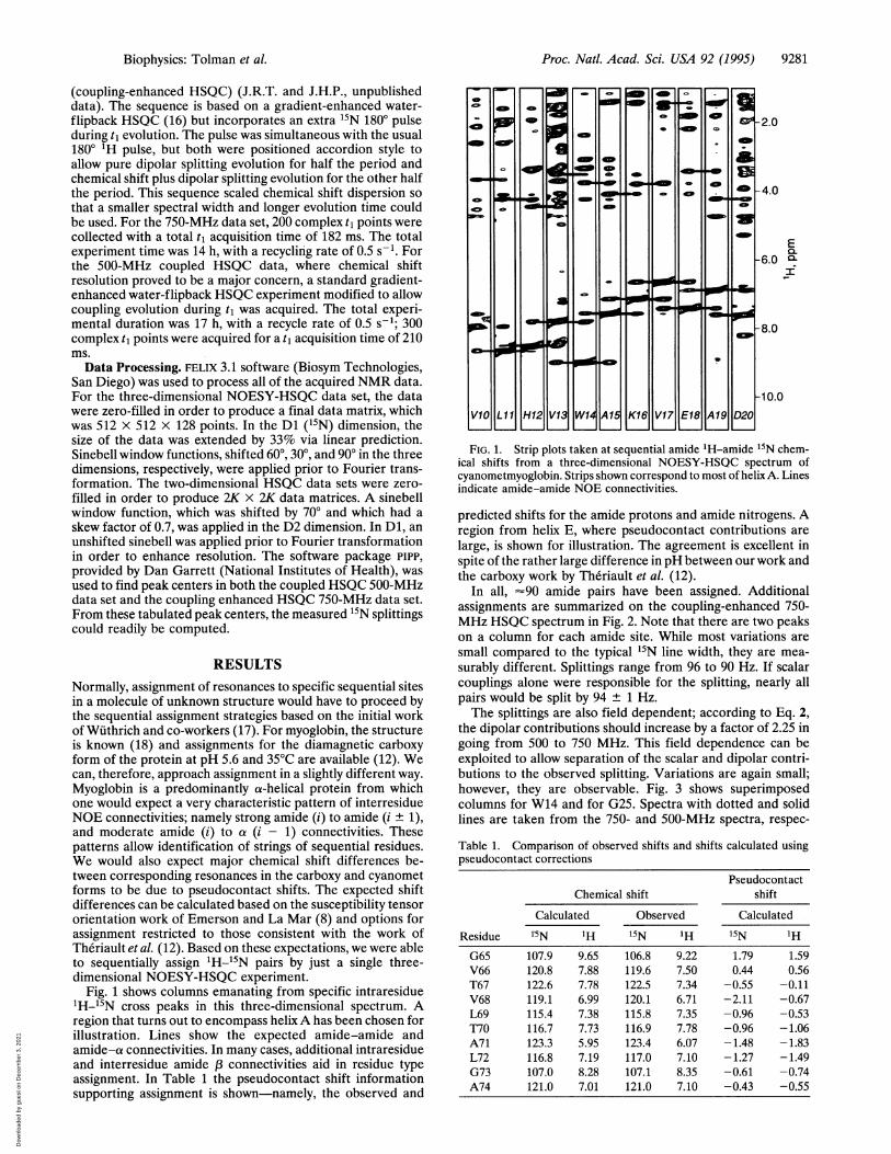
Fig. 1 shows columns emanating from specific intraresidue'H-15N cross peaks in this three-dimensional spectrum. Aregion that turns out to encompass helix A has been chosen forillustration. Lines show the expected amide-amide andamide-a connectivities. In many cases, additional intraresidueand interresidue amide connectivities aid in residue typeassignment. In Table 1 the pseudocontact shift informationsupporting assignment is shown-namely, the observed and
L1u
a
W14 A15 K16 V17 E18 A19 D20
-2.0
-4.0
E-6.0 m
I-
-8.0
-10.0
FIG. 1. Strip plots taken at sequential amide 'H-amide 15N chem-ical shifts from a three-dimensional NOESY-HSQC spectrum ofcyanometmyoglobin. Strips shown correspond to most of helix A. Linesindicate amide-amide NOE connectivities.
predicted shifts for the amide protons and amide nitrogens. Aregion from helix E, where pseudocontact contributions arelarge, is shown for illustration. The agreement is excellent inspite of the rather large difference in pH between our work andthe carboxy work by Theriault et al. (12).
In all, '90 amide pairs have been assigned. Additionalassignments are summarized on the coupling-enhanced 750-MHz HSQC spectrum in Fig. 2. Note that there are two peakson a column for each amide site. While most variations aresmall compared to the typical 15N line width, they are mea-surably different. Splittings range from 96 to 90 Hz. If scalarcouplings alone were responsible for the splitting, nearly allpairs would be split by 94 ± 1 Hz.The splittings are also field dependent; according to Eq. 2,
the dipolar contributions should increase by a factor of 2.25 ingoing from 500 to 750 MHz. This field dependence can beexploited to allow separation of the scalar and dipolar contri-butions to the observed splitting. Variations are again small;however, they are observable. Fig. 3 shows superimposedcolumns for W14 and for G25. Spectra with dotted and solidlines are taken from the 750- and 500-MHz spectra, respec-
Table 1. Comparison of observed shifts and shifts calculated usingpseudocontact corrections
PseudocontactChemical shift shift
Calculated Observed Calculated
Residue 1-5N 'H 1"N 1H 15N IHG65 107.9 9.65 106.8 9.22 1.79 1.59V66 120.8 7.88 119.6 7.50 0.44 0.56T67 122.6 7.78 122.5 7.34 -0.55 -0.11V68 119.1 6.99 120.1 6.71 -2.11 -0.67L69 115.4 7.38 115.8 7.35 -0.96 -0.53T70 116.7 7.73 116.9 7.78 -0.96 -1.06A71 123.3 5.95 123.4 6.07 -1.48 -1.83L72 116.8 7.19 117.0 7.10 -1.27 -1.49G73 107.0 8.28 107.1 8.35 -0.61 -0.74A74 121.0 7.01 121.0 7.10 -0.43 -0.55
4
0
JL
4
)L
p
6
14
L I
I
0
4
I1
c
I
a
PI
-J L
a
4
4
II 0
L
p
I
n
L
Biophysics: Tolman et al.
I'
V1G H12
Dow
nloa
ded
by g
uest
on
Dec
embe
r 3,
202
1

Proc. Natl. Acad. Sci. USA 92 (1995)
9.0 8.0
-105.0
E0.
-108.0 a
z
-111.0
7.01H, ppm
FIG. 2. Expanded region of a coupling-enhanced HSQC spectrum acquired at 750 MHz. Assigned doublets are indicated with a vertical line.
tively. In the case of G25, the splitting increases slightly withfield, indicating a negative contribution from dipolar coupling(the scalar coupling is negative), and in the case of W14 thesplitting decreases with field, indicating a positive dipolarcoupling.A better demonstration of the existence of dipolar contri-
butions can be obtained by using the known structure andsusceptibility of myoglobin to calculate dipolar contributions(Eqs. 1 and 2) and comparing these to dipolar contributionsextracted from the observed field dependence of splittings.Splittings can also be extracted more accurately by fitting peaksin two-dimensional data sets than by visually comparing peaksin a single column. The correlation of observed splittings with
A
0.0
1
0.0
50.0 100.0Hz
50.0 100.0
15( ).0
150.0
calculated splittings is displayed in Fig. 4. Only data from 37doublets that have a high signal/noise ratio and are wellresolved at both 750 and 500 MHz are included. The extracteddipolar contributions are plotted on the abscissa, and thepredicted dipolar contributions are plotted on the ordinate.The correlation coefficient for the best-fit line is 0.7, the slopeis 1.1, and the intercept is -1.2.
DISCUSSIONFrom the above results it is clear that dipolar contributions tothe splittings of 'H-15N pairs in myoglobin are observable. Thecorrelation demonstrated in Fig. 4 is actually quite good giventhe possible sources of error. There will, of course, be someerror associated with our precision of measurement (± 1.0 Hz).Our assumption that all N-H bond lengths are 1.0 A is not
N
0
.0
cu
coCL.5'a:5
.3C.)
0a
Hz
FIG. 3. Superimposed slices taken at the same 'H chemical shiftfrom 500 MHz (solid line) and 750 MHz (dotted line) HSQC data sets.(A) Superposition of slices taken through the doublet assigned to W14.Measured difference in splitting, AD, is approximately -1.5 Hz. (B)Superposition of slices taken through the doublet assigned to G25.Measured difference in splitting, AD, is approximately +0.5 Hz.
4.U -
x2.0-
0.0 - x
-2.0 X
A/ x x
-4.0 X
-6.0 --6.0 -4.0 -2.0 0.0
Measured dipolar contribution, Hz2.0
FIG. 4. Measured dipolar contribution to 15N-'H splittings at 750
MHz plotted against theoretical contributions at 750 MHz calculatedfrom the neutron structure and using Eqs. 1 and 2. J has been separatedfrom D by using the expected theoretical field dependence of D andthe splittings were measured at both 500 and 750 MHz. Line indicatesresulting least-squares fit of the data. Correlation coefficient is 0.70.Note that we consider measured splittings to be positive even thoughthe scalar 15N-'H one bond coupling is negative.
o uo.,w eU
O
G065 * 0129G73 p
GT51
*G259,~~~~d
G150 P0 0
0 S58 aG1250 0 a
49 L98 oF46 0L
6 E410T39 K77 01
9 0 ~~~~~~~~p00 0o@o
o o
1. J+D
I "I
11
AD
.4 L m.
9282 Biophysics: Tolman et al.
.
Dow
nloa
ded
by g
uest
on
Dec
embe
r 3,
202
1

Proc. Natl. Acad. Sci. USA 92 (1995) 9283
strictly accurate. The angles of N-H vectors taken from theneutron diffraction structure will have some imprecision. Andthe assumption that the anisotropy of the paramagnetic sus-ceptibility accurately represents that of the total susceptibilitymay be wrong. The marginally significant nonzero interceptmay also suggest some uniform field-dependent contributionto splittings for which there is currently no theoretical basis.Despite these possibilities, the data in Fig. 4 clearly show thatthe direction and magnitude of the splittings are consistentwith analysis using Eqs. 1 and 2 and the structure of myoglobinas determined from neutron diffraction (18).Given the observability of dipolar contributions and dem-
onstrated utility of Eqs. 1 and 2, a more important question is,If the structure of myoglobin were not known, could measureddipolar splittings provide primary information needed to builda structural model? In these cases, assignment strategies wouldhave to be a little different from that presented above.Calculation of pseudocontact shifts would not be an option,and very likely an experiment based on through-bond connec-tivities would be needed to complete sequential assignments.But once this was dote we would know secondary structurebased on the NOE data; for an a-helical protein we wouldknow how the sequence divided up into more or less rigida-helical segments. At this point, we believe that dipolarconstraints could replace some of the long-range NOE datathat are so essential for determining a tertiary fold. Forexample, using Eqs. 1 and 2 to interpret the data presented inFigs. 3 and 4, we know that the N-H vector for residue 14 mustbe directed nearly along the principal axis of the susceptibilitytensor, because its dipolar coupling contribution to the ob-served splitting is near the negative extreme of the distribution.We also know that the N-H vector for residue 25 must be atan angle of nearly 900 relative to the same axis, because itsdipolar coupling contribution to the observed splitting is nearthe positive extreme of the distribution. These residues areparts of helixA and helix B, respectively. Since N-H vectors aremore or less parallel to helix axes, their direction shouldapproximately reflect the orientation of the helices in which
FIG. 5. Helices A and B along with the heme group taken from the
neutron structure of carboxymyoglobin (18). Principal axis of theparamagnetic susceptibility tensor has been indicated with 01 and 02denoting the angles subtended by this principal axis and selected amideinternuclear vectors from helices A and B, respectively.
they are found. Although the splitting measured for G25provided our only constraint for the B helix, the predictedorientation of helixA was reinforced by measured splittings forfour additional assigned N-H pairs. Fig. 5 depicts helicesA andB as they occur in the neutron diffraction structure. Theorientations, in fact, agree with our predictions.For other helices, the situation is nmre complex because the
inverse of the angular function in Eq. 1 is multivalued and theaxial term in Eq. 2 does not always dominate. However,systematic representation of constraints based on these equa-tions should be possible, and when combined with short-rangeNOE constraints, and covalent constraints from the primarystructure, a more general structure determination protocolshould result. Measurements with the precision presentedabove will clearly be useful in applying this protocol, not somuch because they are accurate, but because a very largenumber of measurements can be made. Also, it is important torealize that the size of the effect, and in some cases theprecision of measurement, will increase with field squared.NMR instruments operating at 17.5 T are currently available,but further advances should occur shortly.
We wish to thank Dr. Fang Shu for making the myoglobin expressionsystem available to us and Dr. Melanie Cocco for advice on samplepreparation. We also wish to thank Dr. Dan Garrett for providing dataanalysis software. This research was supported by Grant GM33225from the National Institutes of Health and benefitted from instru-mentation provided by National Science Foundation Grant NSFCHE9413445.
1. Edison, A. S., Abildgaard, F., Westler, W. M., Mooberry, E. S. &Markley, J. L. (1994) Methods Enzymol. 239, 3-79.
2. Wuthrich, K. (1994) Curr. Opin. Struct. Bio. 4, 93-99.3. Oldfield, E. & Rothgeb, T. M. (1980) J. Am. Chem. Soc. 102,
3635-3637.4. Bastiaan, E. W., Maclean, C., Van Zijl, P. C. M. & Bothner-By,
A. A. (1987) Annu. Rep. NMR Spectrosc. 19, 35-77.5. Abragam, A. (1961) in Principles ofNuclear Magnetism (Claren-
don, Oxford), pp. 103-106.6. Lohman, J. A. B. & Maclean, C. (1979) Mol. Phys. 38, 1255-1261.7. Horrocks, W. D. W., Jr., & Greenberg, E. S. (1973) Biochim.
Biophys. Acta 322, 38-44.8. Emerson, S. D. & La Mar, G. N. (1990) Biochemistry 29, 1556-
1566.9. Lecomte, J. T. J., Johnson, R. D. & La Mar, G. N. (1985) Bio-
chim. Biophys. Acta 829, 268-274.10. Emerson, S. D. & La Mar, G. N. (1990) Biochemistry 29, 1545-
1556.11. Banci, L., Bertini, I. & Luchinat, C. (1994) Methods Enzymol. 239,
485-514.12. Theriault, Y., Pochapsky, T. C., Dalvit, C., Chiu, M. L., Sligar,
S. G. & Wright, P. E. (1994) J. Biomol. NMR 4, 491-504.13. Neidhart, F. C., Bloch, P. L. & Smith, D. F. (1974) J. Bacteriol.
119, 736-747.14. Shu, F., Ramakrishana, V. & Schoenborn, B. P. (1995) in Neu-
trons in Biology, eds. Knott, R. & Schoenborn, B. P. (Academic,New York), in press.
15. Grzesiek, S. & Bax, A. (1993) J. Am. Chem. Soc. 115, 12593-12594.
16. Kay, L. E., Xu, G. Y. & Yamazaki, T. (1994) J. Magn. Reson. Ser.A 109, 129-133.
17. Wuthrich, K., Wider, G., Wagner, G. & Braun, W. (1982) J. Mol,Biol. 155, 311-318.
18. Cheng, X. & Schoenborn, B. P. (1991)J. Mol. Biol. 220, 381-399.
Biophysics: Tolman et al.
Dow
nloa
ded
by g
uest
on
Dec
embe
r 3,
202
1


![Interfacial magnetic coupling between Fe nanoparticles in Fe ...magnetic moment [10] and magnetic interactions [11–13]. Concerning interparticle magnetic interactions, it has been](https://img.pdfslide.net/doc/110x75/60eea974519ccd0158590d85/interfacial-magnetic-coupling-between-fe-nanoparticles-in-fe-magnetic-moment.jpg)


![Electronic Structure and Magnetic Interactions in the ... · Electronic Structure and Magnetic Interactions in the Radical Salt [BEDT-TTF]2[CuCl4] Carmen J. Calzado,*,† Bárbara](https://img.pdfslide.net/doc/110x75/5f8d495dc82f4f36ed3ff20b/electronic-structure-and-magnetic-interactions-in-the-electronic-structure-and.jpg)