Embed Size (px)
Citation preview

88
Departm
Uni
Address
Cen
talle
Nuclear Medicine Tests for Acute GastrointestinalConditionsThomas W. Allen, MD,* and Mark Tulchinsky, MD
0001-2998/12
doi:http://dx.do
ent of Radiolo
versity, Milton
reprint reques
ter, M.C. H06
Acute cholecystitis (AC) and lower-gastrointestinal (GI) bleeding are 2 emergencies commonlyencountered in nuclear medicine. Evidence of AC on hepatobiliary scintigraphy (HBS) allowsfor confident diagnosis and provides support for definitive surgical treatment. Proper patientpreparation is essential for HBS including fasting and the use of pharmacologic adjuncts issometimes required. Pharmacologic adjuncts may also be administered during HBS to shortenthe length of the examination and increase its specificity. In the interpretation of HBS, there areseveral sources of false-positive results to be aware of, most commonly chronic cholecystitis.False-negative results on HBS are usually the result of mistaking another structure, such as adilated cystic duct, for the gallbladder. Abdominal ultrasound is the appropriate initial test inpatients with suspected AC, but HBS is an excellent second tier test for the diagnosis of AC inthe work-up of indeterminate cases by sonography.GI bleeding scintigraphy plays an important role in the evaluation and management of patientswith acute lower-GI bleeding. Scintigraphy serves to localize sites of active GI bleeding andstratify those patients who would benefit from aggressive treatment (surgery or arteriography)vs those who can be managed medically. Pretest involvement of respective services is criticalfor successful bleeding site confirmation and therapy by interventional radiology or surgery orboth. Single photon emission computed tomography/computed tomography erythrocytescintigraphy has demonstrated superior accuracy and precision over planar scintigraphy inthe diagnosis of acute GI bleeding. Additionally, single photon emission computed tomo-graphy/computed tomography scintigraphy of GI bleeding provides useful supplementalanatomical information that benefits patient management.Semin Nucl Med 43:88-101 C 2013 Elsevier Inc. All rights reserved.
The 2 most common acute gastrointestinal (GI) conditionsaddressed by nuclear medicine include acute cholecys-
titis (AC) and GI bleeding. There is urgency for the diagnosis
in both conditions and patient management critically dependson the study results. In patients with suspected AC the
presenting complaint is often the ambiguous abdominal pain,
which can be caused by a number of other diseases. Showingevidence of AC on hepatobiliary scintigraphy (HBS) allows for
confident diagnosis and provides support for definitive
surgical treatment. This high level of confidence in diagnosisis particularly important in severely ill patients who have an
inherently high risk of surgical mortality and morbidity. On
/$-see front matter & 2013 Elsevier Inc. All rights reserved.
i.org/10.1053/j.semnuclmed.2012.11.001
gy, Division of Nuclear Medicine, Pennsylvania State
S. Hershey Medical Center, Hershey, PA.
ts to Thomas W. Allen, MD, Milton S. Hershey Medical
6, 500 University Drive, Hershey, PA 17033. E-mail:
edu
the one hand, given that the best outcome in AC is afforded bya prompt laparoscopic cholecystectomy (LC),1-3 high accu-
racy and expedience of some HBS protocols could improve
those patients’ outcome. On the other hand, for those whobelieve in the effectiveness of conservative treatment,4 the
high specificity of HBS would be reassuring that the other
more ominous causes of abdominal pain have not been
overlooked. A comprehensive review of HBS in AC was
recently published5; therefore, this article focuses on the
practical aspects and nuances that aim at further enhancing
interpretational accuracy as well as improving the clinical
applicability.For more than 30 years, the GI bleeding scan has played an
important role in the evaluation and management of patients
with acute GI bleeding. The purpose of the GI bleeding scan isto locate the site of active GI bleeding and also to identify
those patients who would benefit from aggressive treatment
(surgery or arteriography) vs those who can be managed

Nuclear medicine tests for acute gastrointestinal conditions 89
medically. Once the upper GI source of bleeding has been
excluded, typically by negative nasogastric aspirate or endo-
scopy or both, the GI bleeding scan is typically performed asthe next step in the evaluation of acute GI hemorrhage which
is non-life threatening. Life-threatening GI hemorrhage is
treated surgically. Due to the technique’s superior sensitivityfor detecting small amounts of active GI hemorrhage, a
positive result on a GI bleeding scan generates enough
confidence to proceed with the next step in the diagnosticevaluation (eg arteriography and endoscopy) or even surgical
intervention.
AC: Pathophysiology and ClinicalPresentationAC is typically an acute inflammation of the gallbladder (GB)
that results from obstructing gallstone, acute calculouscholecystitis (ACC), which occurs in over 90% of all AC
cases.6 The obvious result is that obstruction does not allow
for the radioactively marked bile to enter the GB on HBS.The pathophysiology of acute acalculous cholecystitis (AAC)
is not as well understood. Chemical injury from bile stasis
with added insult from microvascular ischemia7 is implicatedas the central catalyst of mucosal injury in AAC, leading
to inflammation and necrosis. It is most likely that the
inflammatory material fills the GB, increasing its pressureto the level that does not allow for newly produced bile
to enter.
The pathologic changes in AC evolve from mild sterileinflammation to subserosal hemorrhage and GB wall swelling.
Although no histologic characteristic distinguishes AAC from
ACC, there are major differences in clinical characteristics andprognostic implications. The predisposing factors for ACC
include gender (females), pregnancy, obesity, hemolytic
process, and many others. Stone dislodgement occurs inabout 70% of the cases with gradual resolution of the acute
inflammatory reaction. About 30% of the patients showed no
improvement without surgical intervention and could pro-gress to complications.4 In 5%-10% of all the cases, ischemia
and necrosis extend to include the entire thickness of the
GB wall, creating a fertile ground for secondary bacterialinfection that is found in 20%-75% of the bile from ACC LC
specimens.8-10 The overall mortality rate of ACC is about
1%.11 The incidence of ACC has declined following popular-ization of elective LC in chronically symptomatic patients
with cholelithiasis, which prevents their progression to
ACC.12
AAC is rare (5%-10%) among AC patients undergoing LC.
It is reasonable to presume that most, if not all of the patients
with AAC would progress if left untreated. Infection isthought to be a secondary event.9 It facilitates the develop-
ment of gangrene, which can progress to abscess or empyema
in AAC, as well as in ACC. The most serious complication ofAC is when the pus traverses the GB wall and finds its way
into the peritoneum or forms a fistulous tract that may lead
into neighboring organs. All the complications are muchmore common in AAC compared with ACC.13 The
population at risk for AAC includes both extremes of age
(especially older males), critically ill (burn, trauma, diabetic,
and immunosuppressed patients), those on total parenteralnutrition, patients with vasculopathy, and women shortly
after childbirth.13 Therefore, it is not surprising that mortality
rates reported in AAC patients have been high, averagingabout 30% with a range from 10% (when diagnosis
is made early) to 90% (when diagnosis is delayed).13
Male patients comprise a higher proportion of the AACpopulation.13
The prevalence of cholelithiasis is approximately 10%among adults in the United States, most of whom areasymptomatic.14 Hence, a stone discovered on any imaging
test is not a specific finding for any associated symptoms.
Annual development of intermittent, biliary-type abdominalpain (colic) is 1%-4%.15,16 There is often a history of periodic
episodes of colicky pain, usually lasting less than 6 hours.
The clinical presentation of ACC is typically an attack ofbiliary colic that worsens with time. When symptoms and
signs progress to generalized pain over the right upper
quadrant, a palpable and painful GB that is commonly madeworse with inspiration or cough to the point that it interrupts
an inspiratory effort (clinical Murphy’s sign), diagnosis of AC
would be suspected. There is usually a low-grade fever,leukocytosis, mild hyperbilirubinemia, and a modest eleva-
tion of serum aminotransferases. However, it is widely
recognized that none of these clinical manifestations, indivi-dually or in combination, provide sufficient diagnostic
certainty for proceeding with management decisions.17,18
Diagnoses of AC that are based on clinical and laboratoryfindings result in 16%-20% error rates.19,20
Acute phase LC is preferred over LC that follows a cooling-
off period of 6-10 weeks because of reduction in morbidity,hospital stay, and time off work.21,22 The preponderance of
evidence indicates that LC is the optimal treatment for AC and
has a lower conversion rate to open LC when done within thefirst 2 days of clinical presentation, as compared with longer
delays.23 In most studies, the perioperative mortality of LC
approaches 0%.23 Therefore, clinicians have a 2-day windowafter initiation of symptoms to make the diagnosis of AC
before referring these patients to surgery. Accepting that there
is always a trade-off between sensitivity and specificity, themost sensitive AC imaging test that would offer the best
chance for early detection may be preferred. However, it is
also important to select an imaging test that can be performedin the shortest time, with the least expense and radiation
exposure.
The most common AC complication is development ofgangrene (20%-30% of AC cases), which can proceed to
perforation in up to 10% of the patients.24-26 The clinical
picture is indistinguishable from uncomplicated AC and thediagnosis of gangrene is seldom made preoperatively.27
Preoperative identification of this complication is highly
desirable as it changes the standard surgical management(open cholecystectomy would be preferred to LC). Making
the clinical diagnosis of AAC is even more challenging than
ACC because it occurs in a population of severely ill patientswho have a complex clinical picture and greater comorbidity.
T.W. Allen and M. Tulchinsky90
Physical examination of severely ill AAC patients is also
problematic—many of them are on ventilators or under heavy
sedation and unable to communicate complaints. The key totimely diagnosis of AAC is maintaining a high index of
suspicion in these patients at high risk.
AC: HBSThe published clinical research on modalities, including the
HBS, has been recently reviewed in this journal.5 The samearticle detailed approach to the description of findings and
their interpretation. HBS is the second line test for AC and
should be preceded by the abdominal ultrasound (AUS)examination.5 This review expands discussion on variety of
approaches to HBS and clinical factors that influence the test
sensitivity and specificity.The diagnostic finding on HBS that signals AC is the
inability of radiolabeled bile to enter the GB, which is
described as GB nonvisualization (non-viz). In ACC this isdue to the blocking stone, whereas in AAC it is due to
maximally filled GB with viscous inflammatory fluid or frank
pus. If the GB is visualized, the study is called negative for AC.In patients with poor or absent radiotracer excretion into the
biliary tree, no comment about AC is possible (indetermi-
nate). It is also indeterminate when biliary excretion is absentsecondary to common bile duct (CBD) obstruction or severe
nonobstructive (intrahepatic) cholestasis.28,29 AC diagnosis
can be pursued by other means (CT or magnetic resonanceimaging (MRI)) in these patients.
There are 2 main approaches used for AC
evaluation—unaided HBS (UA-HBS) and morphine-augmented HBS (MA-HBS). The UA-HBS uses the advantage
of time to allow the patent cystic duct to fill the GB with
radioactive bile. MA-HBS uses the ability of morphine to moreexpeditiously force the radioactive bile through the patent
cystic duct, as explained further in this article. The accuracy
and utility of either HBS approach depends greatly on properpreparation.
Preparation for HBSThe patient should be fasting for 4 hours or longer to avoid
inability of GB to fill with radioactive bile simply because it is
contracted (stimulated).30-32 One study showed GB non-vizon HBS in 53%-64% of the normal volunteers 1 hour after a
meal vs 0% (all visualized the GB) after 4-6 hours of
fasting.33,34 However, prolonged fasting (greater than 24hours) may result in a GB that is maximally filled with bile,
causing false-positive results in some studies.35 The same
mechanism is probably responsible for false-positive HBScases in patients on total parenteral nutrition.36 Patients with
prolonged fasting should be pretreated with sincalide (syn-
thetic cholecystokinin) that contracts and empties the GB.Visualization of the GB is optimized by timing the radioactive
bile excretion to occur during GB relaxation and refilling (the
‘‘turkey baster phenomenon’’).5,37 One study demonstratedthat 50 minutes after 0.02 mcg/kg of sincalide administration,
GB refilling was observed in all normal adults.38 Sincalide was
infused over 3 minutes in that study, which we now recognize
as too fast for consistent, optimal stimulation and emptying ofthe GB.39 A reasonable modification would be to infuse
0.02 mcg/kg of sincalide over 15, 30, or 60 minutes (depend-
ing on logistics dictated by local and individual patientfactors), realizing that longer infusion empties GB best with
the least side effects.40 The administration of the radio-
pharmaceutical and imaging can be started 30 minutes aftersincalide infusion ends.5 However, Flancbaum et al. pub-
lished evidence challenging false-positive results for MA-HBS
in patients with prolonged fasting and total parenteralnutrition.41,42
Ideally, patients should have had no narcotics prior to the
study because those drugs have a profound influence on thebiliary tract. They constrict the sphincter of Oddi, which
raises the pressure in the biliary tract, driving the bile to the
low-pressurereservoir—the GB. This is true in the presence ofpatent cystic duct and healthy GB that is at the time in the
basal (nonstimulated) state. The healthy GB can continue
receiving the incoming bile by dilating and concentrating(dehydrating) it. This mechanism allows the GB to accom-
modate excess bile in a totally obstructed system at the level of
the CBD for 24-48 hours. At the point of saturation, the GBpressure equilibrates with the high pressure of the obstructed
biliary tree and inflow of bile into the GB seizes. At this point
the HBS can be false positive for AC even if the cystic duct ispatent. But prior to this point, increased biliary pressure
would only expedite radiolabeled bile appearance in the GB,
which is the basis of MA-HBS. Therefore, if a patient receivesnarcotics for clinical indications, such as severe abdominal
pain, it is reasonable to start HBS as long as the first dose was
no longer than 24 hours ago. It would be reasonable to giveadditional morphine, as per standard MA-HBS protocol, if the
most recent dose would not amount to the same effect in your
best clinical estimation. In cases without AC (patent cysticduct) there would be rapid tracer appearance in the GB
(Fig. 1). If a patient has been receiving narcotics for over 48
hours, it is reasonable to stop them for 4 half-lives of the drugand conduct MA-HBS after sincalide pretreatment. The 24-48
hours interval is a gray zone and should be handled by
clinicians individually, depending on circumstances.
UA (Without Augmentation)-HBSDuration of the early UA-HBS studies was for up to 1 hour.43-
46 It became obvious early on that extending the imaging to4 hours or longer improves specificity of the study by allowing
GB viz in cases with chronic cholecystitis (CC).47-54 The range
of sensitivity and specificity of this protocol for AC is 95%-97% and 93%-99%, respectively.5 It is the optimal approach
when time allows or when administration of morphine or
both is either undesirable or contraindicated. In patients withintercurrent illness and patent cystic duct, GB non-viz can
persist for longer than 4 hours for a variety of reasons (CC,
slow biliary flow, etc.), but could be visualized clearly on the18-24 hours delayed image.55,56

Figure 1 36-Year-old male developed severe right upper quadrant
pain 2 days after hemorrhoidectomy. (A) Sagittal ultrasound section
showed 7 cm long, ‘‘hourglass shaped’’ gallbladder (GB) with
thickened wall (arrowheads) of distal half (DH) that measured
0.8 cm. Acute cholecystitis (AC) of distal half was suspected. The
patient received a single 4 mg dose of morphine sulfate for pain
2 hours before hepatobiliary scan (HBS). His calculated morphine
dose for the study was 2 mg and when the remaining drug from the
prior injection was estimated to be equivalent or greater, the imaging
was initiated. (B) Image taken between 20 and 24 minutes showed
intense activity in the proximal half (PH) of the GB and in the
common hepatic or bile duct to the duodenal junction (black
arrowhead). There is also reflux of bile to the gastric fundus (arrow).
(C) Image taken between 44 and 48 minutes showed activity
progressing to the DH and AC was excluded. Significant activity
was seen in the gastric body and fundus (arrow), which can be seen
in bilious gastritis.
Nuclear medicine tests for acute gastrointestinal conditions 91
M-A HBSMA-HBS shortens the study and improves its specificity. Choyet al. developed the method and tested it on a prospective
cohort.57 The morphine dose of 0.04 mg/kg was diluted in
10 mL of saline and given over 3 minutes to those with GBnon-viz after 40 minutes of UA-HBS. They found GB
visualization in 36% of those patients within 20 minutes after
morphine. UA-HBS and MA-HBS had the same sensitivity of96%, but MA-HBS outperformed UA-HBS (with 2.5 hours
delay imaging) in specificity—100% vs 88.5%, respectively.
Scintigraphy characteristically showed prominent CBD activ-ity with slowed tracer passage into the duodenum, shortly
after morphine infusion. Subsequent investigations have
confirmed the diagnostic accuracy of MS-HBS, showinga pooled sensitivity of 96% and specificity of 89%.29,42,58-69
The only weakness of this approach is that diagnosis of
CC cannot be made, as that would require 60 minutes of GBnon-viz on UA-HBS (but eventual visualization after
morphine).
There are 3 variations of MA-HBS technique based on the
timing of morphine injection. The first, and the most time-
consuming, variation begins with 1 hour UA-HBS. Thosewho demonstrate GB non-viz proceed to the second phase of
imaging after morphine administration that would typically
continue for 20-30 minutes,29,60-62,67,70,71 and in some otherreports for up to 60 minutes.60,72 If activity appears in the GB
after morphine, the diagnosis of CC can be confidently made.
In cases of GB non-viz, the diagnosis is AC. The maximalimaging time is 80-120 minutes. The second variation
suggested morphine administration at 40 minutes or as soon
as activity is seen in the bowel during UA-HBS (first phase),which starts the morphine (second) phase that lasts from 30-
40 minutes.57,61,64 The criterion for AC is GB non-viz after
morphine injection. The maximal testing time is 60-90minutes. The diagnosis of CC cannot be made, as already
explained above. The third variation was introduced by
Louridas et al. and totally omits the first phase.60 It startswith sincalide pretreatment followed 10 minutes later by
simultaneous injection of morphine and the radiotracer. The
criterion for a positive study is GB non-viz at 60 minutes post-morphine injection. The whole study is done in 70 minutes or
less. Because sincalide pretreatment does not improve the
accuracy of MA-HBS,72-74 the test can be further shortened to60 minutes by omitting it. Again, diagnosis of CC cannot be
made. Although some may be concerned that morphine
infusion could compound CBD obstruction,29,66 there is noevidence to support this concern and the experience of
Louridas et al. supports the safety of morphine
pretreatment.60
There are 4 variations on morphine dosing. The original
one was developed by Choy et al. and has been described
already.57 The second variation uses the same formula, but tothe maximum dose limit of 2 mg.62 The third variation is a
fixed dose of 2 mg,64,67 which defies the basic pharmacologic
principals of adjusting a total dose to an individual’s bodymass. There is concern that the fixed dose of 2 mg may be
ineffective in heavier patients, causing false-positive stu-
dies.75,76 Finally, Flancbaum et al. used 0.05-0.1 mg/kg ofmorphine, but offered no rationale for it or guidance on how
to navigate this dose range.63 At the present time, it makes the
best pharmacologic sense to use the weight-adjusted0.04 mg/kg dose of morphine.77
Although true hypersensitivity to morphine is rare, it
constitutes an absolute contraindication, if confirmed duringpatient interview.78 It is important to realize that most patients
with a history of ‘‘allergic reaction’’ to morphine are inappro-
priately labeled and actually experienced either a pseudoal-lergy (for which premedication with antihistamine would
suffice) or simply had one of the mild, common morphine
side effects, such as vomiting, agitation, or vision changes (inwhich case a single administration of a small morphine dose
for HBS would be acceptable).79,80 Morphine is absolutely
contraindicated in patients with respiratory depression oracute asthma, that is, status asthmaticus. Morphine is also
contraindicated in patients with circulatory shock and in
patients whose ability to maintain blood pressure has alreadybeen compromised by hypovolemia. Finally, morphine is
T.W. Allen and M. Tulchinsky92
absolutely contraindicated in patients with paralytic ileus.
Caution is advised in administering morphine for a long list of
GI and other medical conditions (see the package insert), butone 0.04 mg/kg dose of morphine can be safely administered
as it would be highly unlikely to compound any of them.
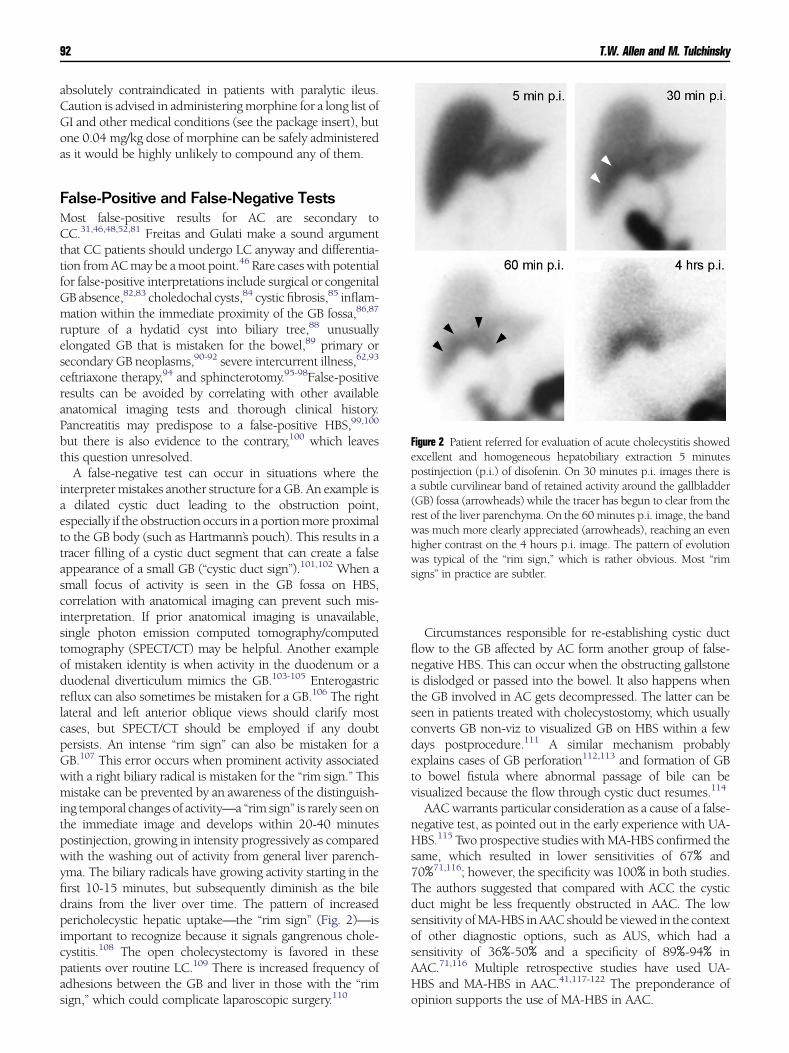
Figure 2 Patient referred for evaluation of acute cholecystitis showed
excellent and homogeneous hepatobiliary extraction 5 minutes
postinjection (p.i.) of disofenin. On 30 minutes p.i. images there is
a subtle curvilinear band of retained activity around the gallbladder
(GB) fossa (arrowheads) while the tracer has begun to clear from the
rest of the liver parenchyma. On the 60 minutes p.i. image, the band
was much more clearly appreciated (arrowheads), reaching an even
higher contrast on the 4 hours p.i. image. The pattern of evolution
was typical of the ‘‘rim sign,’’ which is rather obvious. Most ‘‘rim
signs’’ in practice are subtler.
False-Positive and False-Negative TestsMost false-positive results for AC are secondary toCC.31,46,48,52,81 Freitas and Gulati make a sound argument
that CC patients should undergo LC anyway and differentia-
tion from AC may be a moot point.46 Rare cases with potentialfor false-positive interpretations include surgical or congenital
GB absence,82,83 choledochal cysts,84 cystic fibrosis,85 inflam-
mation within the immediate proximity of the GB fossa,86,87
rupture of a hydatid cyst into biliary tree,88 unusually
elongated GB that is mistaken for the bowel,89 primary or
secondary GB neoplasms,90-92 severe intercurrent illness,62,93
ceftriaxone therapy,94 and sphincterotomy.95-98False-positive
results can be avoided by correlating with other available
anatomical imaging tests and thorough clinical history.Pancreatitis may predispose to a false-positive HBS,99,100
but there is also evidence to the contrary,100 which leaves
this question unresolved.A false-negative test can occur in situations where the
interpreter mistakes another structure for a GB. An example is
a dilated cystic duct leading to the obstruction point,especially if the obstruction occurs in a portion more proximal
to the GB body (such as Hartmann’s pouch). This results in a
tracer filling of a cystic duct segment that can create a falseappearance of a small GB (‘‘cystic duct sign’’).101,102 When a
small focus of activity is seen in the GB fossa on HBS,
correlation with anatomical imaging can prevent such mis-interpretation. If prior anatomical imaging is unavailable,
single photon emission computed tomography/computed
tomography (SPECT/CT) may be helpful. Another exampleof mistaken identity is when activity in the duodenum or a
duodenal diverticulum mimics the GB.103-105 Enterogastric
reflux can also sometimes be mistaken for a GB.106 The rightlateral and left anterior oblique views should clarify most
cases, but SPECT/CT should be employed if any doubt
persists. An intense ‘‘rim sign’’ can also be mistaken for aGB.107 This error occurs when prominent activity associated
with a right biliary radical is mistaken for the ‘‘rim sign.’’ This
mistake can be prevented by an awareness of the distinguish-ing temporal changes of activity—a ‘‘rim sign’’ is rarely seen on
the immediate image and develops within 20-40 minutes
postinjection, growing in intensity progressively as comparedwith the washing out of activity from general liver parench-
yma. The biliary radicals have growing activity starting in the
first 10-15 minutes, but subsequently diminish as the biledrains from the liver over time. The pattern of increased
pericholecystic hepatic uptake—the ‘‘rim sign’’ (Fig. 2)—is
important to recognize because it signals gangrenous chole-cystitis.108 The open cholecystectomy is favored in these
patients over routine LC.109 There is increased frequency of
adhesions between the GB and liver in those with the ‘‘rimsign,’’ which could complicate laparoscopic surgery.110
Circumstances responsible for re-establishing cystic duct
flow to the GB affected by AC form another group of false-negative HBS. This can occur when the obstructing gallstone
is dislodged or passed into the bowel. It also happens when
the GB involved in AC gets decompressed. The latter can beseen in patients treated with cholecystostomy, which usually
converts GB non-viz to visualized GB on HBS within a few
days postprocedure.111 A similar mechanism probablyexplains cases of GB perforation112,113 and formation of GB
to bowel fistula where abnormal passage of bile can be
visualized because the flow through cystic duct resumes.114
AAC warrants particular consideration as a cause of a false-
negative test, as pointed out in the early experience with UA-
HBS.115 Two prospective studies with MA-HBS confirmed thesame, which resulted in lower sensitivities of 67% and
70%71,116; however, the specificity was 100% in both studies.
The authors suggested that compared with ACC the cysticduct might be less frequently obstructed in AAC. The low
sensitivity of MA-HBS in AAC should be viewed in the context
of other diagnostic options, such as AUS, which had asensitivity of 36%-50% and a specificity of 89%-94% in
AAC.71,116 Multiple retrospective studies have used UA-
HBS and MA-HBS in AAC.41,117-122 The preponderance ofopinion supports the use of MA-HBS in AAC.

Nuclear medicine tests for acute gastrointestinal conditions 93
AC: Other Diagnostic ModalitiesNuclear Medicine ApproachesInflammation imaging with labeled white blood cells can be
useful in AAC where HBS has lower sensitivity.123-125 Thelong delay between injection and imaging (typically 24 hours
for In-111 labeled white blood cells) is problematic given the
urgency of AAC diagnosis.71Tc-99m-labeled white cell ima-ging is faster126 and demonstrated promising sensitivity and
specificity for AC—94% and 100%, respectively.127 Imaging
inflammation directly in ACC and particularly in AAC may berevisited when faster in vivo radiolabeling of leukocytes
becomes available. There are few reports of increased GB
uptake on F-18 FDG-PET/CT in cases with AC,128-130 but thisapproach lacks substantive literature evidence.
Non-Isotopic ApproachesAUS is the initial imaging test of choice for patients with
suspected AC because of lack of radiation exposure, ability to
perform the test at the bedside, expedience, and lower cost. Itallows exquisite anatomical evaluation of the GB, and is
capable of evaluating other abdominal organs that may be
responsible for patients’ pain. AUS alone is sufficient for thediagnosis of AC in 80% of the patients, leaving about 20% for
HBS and other diagnostic tests to clarify.131 The positive
predictive value of demonstrating stones and a positivesonographic Murphy sign (pain elicited by pressing on the
GB with transducer) was 92%, and that of stones and
thickening of the GB wall was 95%.131 The negative predictivevalue of the absence of stones combined with either a normal
GB wall thickness or a negative sonographic Murphy sign was
95%. Striated GB wall edema is a rare finding, but is highlyspecific of gangrenous AC.132,133 Finding gas in the GB
lumen or the wall signals emphysematous AC, which con-
stitutes a surgical emergency.134
MRI and magnetic resonance cholangiopancreatography
can demonstrate gallstones,135 but demonstration of cystic
duct patency is very difficult. Given no radiation exposure,the technique is particularly well suited for the evaluation of
acute right upper abdominal pain in a pregnant patient.136 In
general, the technique is most helpful in evaluation of CBD forstones where sensitivity and specificity exceed 90%.137-140
Cystic duct and common duct stones may be more easily
detected with MRI as compared with ultrasound.141,142 TheGB wall and adjacent region pericholecystic fluid and tissue
irregularities are well seen with MRI.27,143-147 Contrast agent
administration can be helpful in demonstrating pericholecys-tic enhancement that may be seen with complicated AC,148
which is similar to the ‘‘rim sign’’ on HBS.
CT is commonly employed in hospitalized and emergencyroom patients that can show AC related findings and its
complications. All of the clinical investigations are retro-
spective with a reported negative predictive value for AC ofapproximately 89%.149 CT can visualize only 65%-75% of
gallstones and has no advantages for primary diagnosis. CT
evidence of perfusion defects in the GB wall (discontinuity ordecreased enhancement of the GB wall) is highly specific for
gangrene,150 whereas transmural defects suggest perfora-
tion.151 The combination of CT findings that include perfu-
sion defects or pericholecystic stranding or lack of gallstonesis 94% sensitive and 75% specific in predicting GB gangrene,
which may be CT’s key advantage over other modalities.
ConclusionHBS is excellent for the diagnosis of AC, but the above-discussed advantages of AUS make it the most appropriate
first tier test for these patients. HBS remains an excellent
second tier test in the work-up of indeterminate cases by AUS,as initially suggested in the algorithm proposed by Ralls
et al.131 In a pregnant patient, HBS should be substituted in
this algorithm with abdominal MRI/magnetic resonancecholangiopancreatography.136
Acute GI Bleeding:Pathophysiology and ClinicalPresentationAcute GI bleeding is a common emergency that occurs with
an age-dependent incidence and results in an annual hospi-
talization rate of approximately 25 per 100,000 in the UnitedStates.152 The clinical presentation of acute GI bleeding is
quite variable with the majority of patients, approximately
85%, recovering spontaneously without any further specificintervention. However, it is essential to identify the condition
in the remaining 15% where it can be quite devastating and
life threatening. In particular, GI bleeding is a major cause ofmorbidity, especially in the elderly and those with associated
comorbidities. In hospitalized patients, the published mor-
tality rate of acute GI bleeding is as high as 10%-14%.152-154
Clinical manifestations of acute GI bleeding are often
unreliable in identifying the ultimate source of GI bleeding.
History and physical examination findings only achieve acorrect final diagnosis in less that 40% of the patients.155
Because of transient pooling and retrograde peristalsis that
may occur in the bowel after GI bleeding, clinically evident GIbleeding lacks the temporal resolution needed for accurate
diagnosis and treatment. Furthermore, the clinical evidence of
GI bleeding often does not coincide with active GI bleeding.Acute GI bleeding is typically classified according to its site
of origin as either upper GI bleeding, arising from a source
proximal to the ligament of Treitz, or lower GI bleeding,arising from a source distal to the ligament of Treitz. GI
bleeding may also be classified as obscure or occult if not
otherwise categorized.Causes for upper GI bleeding include esophageal varices,
vascular malformations, esophagitis, Mallory-Weiss tear, gas-
tritis, gastric and duodenal ulcers, and neoplasm. Causes foracute lower GI bleeding (ALGIB) include vascular malforma-
tions such as angiodysplasia, diverticula, adenomatous polyps
and neoplasms, inflammation, and, in children, Meckel’sdiverticulum.

T.W. Allen and M. Tulchinsky94
Esophagogastroduodenoscopy (EGD) is the method of
choice for evaluating the upper GI tract for bleeding with
an accuracy of greater than 90%. Colonoscopy of the lower GItract only has an accuracy of about 70% in confirming or
excluding a site of GI hemorrhage. Endoscopy and arterio-
graphy may be used to localize and control a lower GIbleeding site but often fail due to intermittent bleeding.156
Although it has no therapeutic role, scintigraphy plays a
complementary role to arteriography and endoscopy in theevaluation of lower GI bleeding.
Acute GI Bleeding: GI BleedingScintigraphyNuclear medicine techniques with varying sensitivities for the
detection of acute GI bleeding have been well described in the
literature,157-161 and their role in GI bleeding has beencomprehensively reviewed by others in this journal160,162
This section focuses on the preparation, interpretation,
nuances, and potential pitfalls aimed at further enhancingthe clinical utility of nuclear medicine evaluation of acute GI
bleeding. Additionally, the novel application of hybrid
SPECT/CT imaging technology to the clinical problem ofacute GI bleeding has been discussed.
Radiopharmaceuticals for GIBleeding ScintigraphyHistorically, the 2 primary radiopharmaceuticals that have
been employed for evaluation of ALGIB are 99mTc-sulfur
colloid (99mTc SC) and 99mTc red blood cells (99mTc RBCs).Alavi introduced 99mTc-99m SC, an agent rapidly cleared
from the vascular compartment, to investigate acute GI
bleeding. In a canine experimental model, bleeding rates aslow as 0.05-0.1 mL/min were detected with 99mTc-99m
SC.163 Because of the rapid extraction by the reticuloendothe-
lial elements within the liver, spleen, and bone marrow, sulfurcolloid offers a high target to background ratio between a
bleeding site and the surrounding soft tissues resulting in a
theoretical increase in sensitivity. The sulfur colloid techniquecontinues to have it proponents today.164 Advantages of Tc-
99m SC for acute GI bleeding scintigraphy are the rapid
uptake of the compound by the reticuloendothelial systemwith rapid blood pool clearance, which facilitates the detec-
tion of very small amounts of extravasation at a bleeding site.
Disadvantages of sulfur colloid include uptake within the liverand spleen that obscures bleeding sites in the hepatic and
splenic flexures of the colon, limiting visualization in the
upper abdomen. Another significant limitation of this tech-nique is rapid clearance of this tracer from the vascular
compartment—less than 10% of the tracer remains in the
vascular compartment at 7 minutes.165 This is a significantlimitation in the dynamic evaluation of intermittent acute GI
bleeding.
Bunker et al. compared the performance of 99mTc SC andin-vitro labeled 99mTc RBCs with regard to their relative
sensitivities for the detection and accuracy in localizing of
acute GI bleeding. In a prospective multicenter study of 100
patients, both agents were evaluated under identical clinicalconditions with superior performance by the labeled RBCs.
Bleeding sites were identified in 38 patients with labeled RBCs
compared with detection of only 5 bleeding sites with sulfurcolloid. Labeled RBCs were diagnostically superior in all cases
with a sensitivity of 95%, a specificity of 93%, and an overall
accuracy of 94%. The performance of 99mTc SC was inferior tolabeled RBCs with a sensitivity of 12%, a specificity of 100%,
and an overall accuracy of 62%.166
The lowest detectable bleeding rate for 99mTc RBCs is0.04 mL/min in an anesthetized animal model167 and is
0.1 mL/min in a clinical study.168 The SNM procedure
guideline for GI bleeding indicates that labeled RBCs candetect bleeding at rates as low as 0.1-0.35 mL/min.159
Although the subject of a vigorous and interesting debate
during the development of GI bleeding scintigraphic techni-ques, the preferred radiopharmaceutical for GI bleeding scans
is in vitro labeled RBCs. Labeled RBCs remain intravascular for
hours providing the ability to image dynamically for up to 90minutes and then reimage out to 24 hours after administra-
tion. This is significant because of the intermittent nature of
most acute GI bleeding that is evaluated in the nuclearmedicine department.156
Preparation for GI BleedingScintigraphyPatients who present with acute GI bleeding should undergoinitial triage and resuscitation, a complete history and physical
examination, appropriate laboratory testing, and nasogastric
lavage. The nasogastric lavage results are used to stratify thebleeding into either a suspected upper or lower GI source. If
an upper GI source is suspected, then EGD is performed and
therapy is initiated based on positive EGD results. If a lowerGI bleeding source is suspected or if an upper GI source has
been excluded, then erythrocyte scintigraphy should be
performed as the next step in the evaluation.169
Prior to a patient’s arrival in the nuclear medicine laboratory
for erythrocyte scintigraphy, it is essential for the referring
physician to have a follow-up management plan in place andbe prepared for implementation. Because of its intermittent
nature, the majority of GI bleeding that is referred to nuclear
medicine for evaluation ceases spontaneously.156 Temporaldelays caused by a lack of prior communication between
clinical services may contribute to discordance between a
positive RBC bleeding scan and a subsequent negativearteriogram. Pretest involvement of respective services is
critical for successful bleeding site confirmation and therapy
by interventional radiology or surgery or both.
Interpretion Criteria for GIBleeding ScinitigraphyRequisite interpretation criteria for a positive GI bleedingstudy include: (1) extravasation of radiotracer from the

Figure 4 Red blood cell (RBC) bleeding scan showed curvilinear area
of increased activity over the left common iliac vessel. Eight minutes
later the focus extended to the right in a loopy shape across the
middle of the pelvis. By 20 minutes, the activity reached (arrow) and
accumulated in a larger bulbous structure in the right pelvis
(arrowhead), which is typical of the cecum. On the 24-minute
image, it extended cranially in a linear fashion (arrowheads), typical
of large bowel pattern, consistent with ascending colon. At 48
minutes, some of the right colon activity briskly moved antegrade to
the left colon (arrowheads with concave base). By 90 minutes the
small bowel activity in the mid-lower pelvis appears in typical loopy
pattern (arrows). Activity moved antegrade to visualize the right
(arrowheads with straight base) and left (arrowheads with concave
base) colon that is connected by a slightly redundant transverse
colon. The source of bleeding was correctly diagnosed in the distal
ileum and angiography identified bleeding vessel branch of ileocolic
artery that was successfully embolized.
Figure 3 91-Year-old female presented with bright red blood per
rectum to the hospital. The immediate image of the bleeding scan
showed moderately intense focal tracer activity in the left lower
abdominal quadrant (black arrow). There was another subtle round
focus of activity in the left superior abdomen (straight-base arrow-
head). Splenic activity was absent (concave-base arrowhead). The
same image at 90 minutes after tracer injection showed the
unchanged findings and tracer excretion in the urinary bladder
(B). There was no active bleeding during the 90 minutes of imaging
because the focal activity has not changed shape, intensity, or
configuration. CTof the abdomen (shown in sagittal plane) demon-
strated findings of splenectomy (not shown) and 2 splenules that
corresponded to the 2 focal findings on the bleeding scan with the
lower one (white arrow) more anterior and larger than the upper
(white arrowhead), which explains its greater intensity on the
bleeding scan.
Nuclear medicine tests for acute gastrointestinal conditions 95
vascular compartment and (2) movement of the extravasatedblood which act as a cathartic within an identifiable bowel
segment, either an anterograde or retrograde direction. Once
a GI bleeding site is identified on scintigraphy, it is essential tocontinue the examination for sufficient time to definitively
identify the bleeding bowel segment. Large bowel bleeding
typically drapes around the periphery of the abdomen andhas an elongated trajectory (Fig. 3). Whereas, small bowel
activity is centrally located in the abdomen, propagates in
curvilinear loops, and may lessen in intensity over time as theradiotracer becomes diluted in the fluid containing small
bowel (Fig. 4).
Several studies have demonstrated that computerizedcinematic acquisition and cinematic display of scintigraphic
images results in improved detection and localization
for acute GI bleeding.170 O’Neill et al. demonstratedcinematic erythrocyte scintigraphy to be a sensitive and
noninvasive alternative to mesenteric angiography that
was accurate enough to direct selective surgicalintervention.169
False-Positive Tests and PitfallsStatic areas of extravascular radiotracer accumulation on RBC
bleeding scans rarely represent acute GI hemorrhage but aremore commonly due to aneurysms, varices, inflammation, or
tumors. Heyman et al. reported a patient who, on sulfurcolloid abdominal scan performed to evaluate abdominal
bleeding, had a focal abnormality in the upper abdomen that
was interpreted as consistent with a small bowel bleedingsource; however, an accessory spleen was discovered to be the
source of uptake at surgery.171. Others have detected an
unsuspected splenic rupture with ongoing intraperitonealhemorrhage on erythrocyte scintigraphy performed on a
patient with suspected GI bleeding.172 A splenule can appear
as a fixed focus of RBC accumulation on erythrocytescintigraphy (Fig. 3) and cause a false-positive interpretation.
Other sources of false-positive results include use of cath-
artics, bowel irritation or inflammation, or a recent endoscopyprocedure. Fundamental to avoiding misinterpretation is
investigating nonbleeding causes (by correlation with CT or
performing SPECT/CT) when there is lack of movement ondynamic images (Fig. 3).
False-positive interpretations on erythrocyte scintigraphy
may also result from misinterpretation of excreted freepertechnatate that accumulates within the renal upper tracts.

Figure 5 Ayoung male patient with rectal bleeding was studied. (A) There is physiological activity on the immediate red
blood cell (RBC) bleeding scan. (B) At 16 minutes postinjection (p.i.) there is intense rounded uptake in the lower
midpelvis (arrow) that can represent either the penis base or rectal bleeding. (C) It changed to elongated structure
(arrowhead) with intense activity by 24 minutes. There is expected subtle accumulation in the urinary bladder (u). (D) It
decreased in intensity by 40 minutes. (E) At 90 minutes p.i., it was rounded and low in intensity. (F) Left lateral image at
that time was not helpful to differentiate penile from rectal activity. (G) Posterior image showed intense uptake in the
kidneys that were poorly seen on anterior images, but no rectal activity to suggest bleeding. Penile activity was suspected
with changing intensity due to intermittent erection. (H) Patient was asked to position the penis pointing horizontally to
the right and the anterior image was repeated, confirming that it was the source of the finding, and active rectal bleeding
was excluded.
T.W. Allen and M. Tulchinsky96
The most common false positive results from mistaking penile
activity for rectal bleeding (Fig. 5). Obtaining various spotpelvic views and positional manipulations are simple yet
effective methods to facilitate accurate diagnosis (Fig. 5).
Those views can also occasionally disclose a rectal bleed that isbeing obscured by the bladder.
ApplicationofNewTechnology toAcute GI Bleeding: SPECT/CTIt is intuitive that the advent of hybrid SPECT/CT fusion
imaging provides an opportunity to increase the sensitivity
and accuracy of erythrocyte scintigraphy for the detection andlocalization of GI bleeding. SPECT is well known to increase
contrast resolution over conventional planar imaging by as
much as 10%-15%, augmenting the ability to detect low-volume sites of GI bleeding. Investigators have found that
hybrid SPECT/CT functional anatomical imaging provides a
high degree of specific anatomic information when planarscintigraphy is positive for GI bleeding.173 The supplemental
anatomical information obtained by SPECT/CT can precisely
localize a source of abdominal bleeding. SPECT/CT helps toavoid the common pitfalls seen in erythrocyte scintigraphy
(Fig. 6).
Yama et al. reported the first case of localization of smallintestinal bleeding using the fusion of separate erythrocyte
scintigraphy SPECT images and X-ray CT images.174 In a
study of 81 consecutive patients with various clinical condi-tions and radiopharmaceuticals, Schialli and colleagues found
that functional anatomical mapping with SPECT/CT allowed
a more precise interpretation compared with SPECT and thatfused SPECT/CT images improved the diagnostic accuracy of
SPECT imaging in several clinical situations; this included
2 patients in this series with GI bleeding.173 In a subsequentprospective investigation, Schillaci and colleagues demon-
strated the feasibility and accuracy of SPECT/CT imaging in
acute lower GI erythrocyte scintigraphy.175 In this study,erythrocyte scintigraphy with SPECT/CT had a significant
positive impact on 7 of 19 patients (36.8%) by precisely
localizing the site of hemorrhage in 6 patients whose bleedingsites could not be identified on conventional planar erythro-
cyte scintigraphy. In 1 patient from this series, a site of
suspected active bleeding identified on planar imaging wassuccessfully excluded. Overall, this study showed erythrocyte
scintigraphy with SPECT/CT to be a feasible, more precise,
and accurate method of diagnosing ALGIB compared withplanar erythrocyte scintigraphy. Furthermore, SPECT/CT
augmented erythrocyte scintigraphy in this series provided
supplemental anatomical information that was useful insubsequent patient treatment. Turgeon et al. also have
reported the use SPECT/CT to successfully identify the cause
of abdominal pain and obscure overt GI bleeding in a patientwith a Meckel’s diverticulum.176

Figure 6 A patient with liver cirrhosis developed melena. Nasogastric tube aspirate was negative. (A) The red blood cell
(RBC) bleeding scan immediately postinjection (p.i.) showed photopenic rim from the right heart border where it is
clearly seen between the liver edge (white arrow) and warm activity along the parietal peritoneum (black arrow). This
photopenic rim descends to the pelvis and from there it ascends along the left paracolic gutter to the splenic(s) border.
This is typical appearance of peritoneal effusion. (B) At 24 minutes p.i. there is subtle new activity along the left medial
liver edge (white arrowhead). (C) It becomes clearly defined at 54 minutes p.i., but without significant progression.
(D) On 90 minutes p.i. image, the same finding is even more distinct, but stable in configuration. Differential diagnosis
included gastric fundus hyperemia vs slow bleeding. (E) SPECT/CT fusion image showed the activity in the gastric
fundus lumen with some exiting into the gas-distended, more distal portion (arrowhead with concave base), favoring
slow bleeding. (F) CT showed ascites (A) in the same areas as on the immediate RBC bleeding scan. The liver edges are
nodular, consistent with cirrhosis. (G) Sagittal SPECT/CTconfirms activity in the fundus lumen that extends into the gas-
filled part of the gastric body (arrowhead with concave base). Endoscopy discovered a blood-oozing polyp that was
removed and showed benign features.
Nuclear medicine tests for acute gastrointestinal conditions 97
ConclusionErythrocyte scintigraphy has an important role in the evalua-
tion and management of patients with acute lower GIhemorrhage. It helps to localize sites of active GI bleeding
and stratify those patients who would benefit from aggressive
treatment (surgery or arteriography) vs those who can bemanaged medically. Pretest involvement of respective services
is critical for successful bleeding site confirmation and therapy
by interventional radiology or surgery, or both. Erythrocytescintigraphy with SPECT/CT has superior accuracy and
precision over planar erythrocyte scintigraphy in the diag-
nosis of acute GI bleeding. Additionally, erythrocyte scinti-graphy with SPECT/CT can provide useful supplemental
anatomical information that benefits patient management.
References1. Chang TC, Lin MT, Wu MH, et al: Evaluation of early versus delayed
laparoscopic cholecystectomy in the treatment of acute cholecystitis.
Hepatogastroenterology 2009;56:26-28
2. Gurusamy KS, Samraj K, Fusai G, et al: Early versus delayed
laparoscopic cholecystectomy for biliary colic. Cochrane Database Syst
Rev 2008. CD007196
3. Lau H, Lo CY, Patil NG, et al: Early versus delayed-interval laparoscopic
cholecystectomy for acute cholecystitis: A metaanalysis. Surg Endosc
2006;20:82-87
4. Vetrhus M, Soreide O, Nesvik I, et al: Acute cholecystitis: Delayed
surgery or observation. A randomized clinical trial. Scand J Gastro-
enterol 2003;38:985-990
5. Tulchinsky M, Colletti PM, Allen TW: Hepatobiliary scintigraphy in
acute cholecystitis. Semin Nucl Med 2012;42:84-100
6. Strasberg SM: Clinical practice. Acute calculous cholecystitis. N Engl J
Med 2008;358:2804-2811
7. Hakala T, Nuutinen PJ, Ruokonen ET, et al: Microangiopathy in acute
acalculous cholecystitis. Br J Surg 1997;84:1249-1252
8. Claesson BE: Microflora of the biliary tree and liver—clinical correlates.
Dig Dis 1986;4:93-118
9. Claesson BE, Holmlund DE, Matzsch TW: Microflora of the gallbladder
related to duration of acute cholecystitis. Surg Gynecol Obstet
1986;162:531-535
10. Kuo CH, Changchien CS, Chen JJ, et al: Septic acute cholecystitis.
Scand J Gastroenterol 1995;30:272-275
11. Andersson KL, Friedman LS: Acalculous biliary pain, acalculous
cholecystitis, cholesterolosis, adenomyomatosis, and polyps of the
gallbladder. In: Feldman M, Friedman LS, Brandt LJ, (eds): Sleisenger
and Fordtran’s Gastrointestinal and Liver Disease: Pathophysiology/
Diagnosis/Management. Philadelphia, PA: Saunders Elsevier; 2010.
p. 1139-1152
12. Urbach DR, Stukel TA: Rate of elective cholecystectomy and the
incidence of severe gallstone disease. Can Med Assoc J 2005;172:
1015-1019
13. Huffman JL, Schenker S: Acute acalculous cholecystitis: A review. Clin
Gastroenterol Hepatol 2010;8:15-22
14. Schirmer BD, Winters KL, Edlich RF: Cholelithiasis and cholecystitis.
J Long Term Eff Med Implants 2005;15:329-338
T.W. Allen and M. Tulchinsky98
15. Friedman GD, Raviola CA, Fireman B: Prognosis of gallstones with
mild or no symptoms: 25 Years of follow-up in a health maintenance
organization. J Clin Epidemiol 1989;42:127-136
16. McSherry CK, Ferstenberg H, Calhoun WF, et al: The natural history of
diagnosed gallstone disease in symptomatic and asymptomatic
patients. Ann Surg 1985;202:59-63
17. Schofield PF, Hulton NR, Baildam AD: Is it acute cholecystitis? Ann R
Coll Surg Engl 1986;68:14-16
18. Trowbridge RL, Rutkowski NK, Shojania KG: Does this patient have
acute cholecystitis? J Am Med Assoc 2003;289:80-86
19. Essenhigh DM: Management of acute cholecystitis. Br J Surg
1966;53:1032-1038
20. Halasz NA: Counterfeit cholecystitis, a common diagnostic dilemma.
Am J Surg 1975;130:189-193
21. Lai PB, Kwong KH, Leung KL, et al: Randomized trial of early versus
delayed laparoscopic cholecystectomy for acute cholecystitis. Br J Surg
1998;85:764-767
22. Lo CM, Liu CL, Fan ST, et al: Prospective randomized study of early
versus delayed laparoscopic cholecystectomy for acute cholecystitis.
Ann Surg 1998;227:461-467
23. Willsher PC, Sanabria JR, Gallinger S, et al: Early laparoscopic
cholecystectomy for acute cholecystitis: A safe procedure. J Gastrointest
Surg 1999;3:50-53
24. Jeffrey RB, Laing FC, Wong W, et al: Gangrenous cholecystitis:
Diagnosis by ultrasound. Radiology 1983;148:219-221
25. Croley GG : Gangrenous cholecystitis: Five patients with intestinal
obstruction. Am J Surg 1992;58:284-292
26. Reiss R, Nudelman I, Gutman C, et al: Changing trends in surgery for
acute cholecystitis. World J Surg 1990;14:567-570. [discussion 570–
561]
27. Bennett GL, Rusinek H, Lisi V, et al: CT findings in acute gangrenous
cholecystitis. AJR Am J Roentgenol 2002;178:275-281
28. Bednarz GM, Kalff V, Kelly MJ: Hepatobiliary scintigraphy. Increasing
the accuracy of the preoperative diagnosis of acute cholecystitis. Med J
Aust 1986;145:316-318
29. Fink-Bennett D, Balon H, Robbins T, et al: Morphine-augmented
cholescintigraphy: Its efficacy in detecting acute cholecystitis. J Nucl
Med 1991;32:1231-1233
30. Down RH, Arnold J, Goldin A, et al: Comparison of accuracy of 99mTc-
pyridoxylidene glutamate scanning with oral cholecystography and
ultrasonography in diagnosis of acute cholecystitis. Lancet
1979;2:1094-1097
31. Araki T: Cholecystitis: A comparison of real-time ultrasonography and
technetium-99m hepatobiliary scintigraphy. Clin Radiol 1980;31:
675-679
32. Tulchinsky M, Ciak BW, Delbeke D, et al: SNM practice guideline for
hepatobiliary scintigraphy 4.0. J Nucl Med Technol 2010;38:210-218
33. Baker RJ, Marion MA: Biliary scanning with Tc-
99mpyridoxylideneglutamate—the effect of food in normal subjects:
Concise communication. J Nucl Med 1977;18:793-795
34. Klingensmith WC , Spitzer VM, Fritzberg AR, et al: The normal fasting
and postprandial diisopropyl-IDA Tc 99m hepatobiliary study. Radi-
ology 1981;141:771-776
35. Larsen MJ, Klingensmith WC , Kuni CC: Radionuclide hepatobiliary
imaging: Nonvisualization of the gallbladder secondary to prolonged
fasting. J Nucl Med 1982;23:1003-1005
36. Shuman WP, Gibbs P, Rudd TG, et al: PIPIDA scintigraphy for
cholecystitis: False positives in alcoholism and total parenteral nutri-
tion. AJR Am J Roentgenol 1982;138:1-5
37. Eikman EA, Cameron JL, Colman M, et al: A test for patency
of the cystic duct in acute cholecystitis. Ann Intern Med 1975;82:
318-322
38. Mesgarzadeh M, Krishnamurthy GT, Bobba VR, et al: Filling, post-
cholecystokinin emptying, and refilling of normal gallbladder: Effects
of two different doses of CCK on refilling: Concise communication. J
Nucl Med 1983;24:666-671
39. Ziessman HA, Muenz LR, Agarwal AK, et al: Normal values for sincalide
cholescintigraphy: Comparison of two methods. Radiology 2001;221:
404-410
40. Ziessman HA, Tulchinsky M, Lavely WC, et al: Sincalide-stimulated
cholescintigraphy: A multicenter investigation to determine optimal
infusion methodology and gallbladder ejection fraction normal values. J
Nucl Med 2010;51:277-281
41. Flancbaum L, Alden SM, Trooskin SZ: Use of cholescintigraphy with
morphine in critically ill patients with suspected cholecystitis. Surgery
1989;106:668-673. [discussion 673–664]
42. Flancbaum L, Choban PS: Use of morphine cholescintigraphy in the
diagnosis of acute cholecystitis in critically ill patients. Intensive Care
Med 1995;21:120-124
43. Weissmann HS, Frank MS, Bernstein LH, et al: Rapid and accurate
diagnosis of acute cholecystitis with 99mTc-HIDA cholescintigraphy.
AJR Am J Roentgenol 1979;132:523-528
44. Szlabick RE, Catto JA, Fink-Bennett D, et al: Hepatobiliary scanning in
the diagnosis of acute cholecystitis. Arch Surg 1980;115:540-544
45. Stadalnik RC, Kraus JF, Matolo NM, et al: The validity of 99mTc-
pyridoxylideneglutamate (P.G.) cholescintigraphy as a diagnostic test
for cholecystitis. Clin Nucl Med 1978;3:142-146
46. Freitas JE, Gulati RM: Rapid evaluation of acute abdominal pain by
hepatobiliary scanning. J Am Med Assoc 1980;244:1585-1587
47. Zeman RK, Burrell MI, Cahow CE, et al: Diagnostic utility of
cholescintigraphy and ultrasonography in acute cholecystitis. Am J
Surg 1981;141:446-451
48. Worthen NJ, Uszler JM, Funamura JL: Cholecystitis: Prospective
evaluation of sonography and 99mTc-HIDA cholescintigraphy. AJR
Am J Roentgenol 1981;137:973-978
49. Weissmann HS, Badia J, Sugarman LA, et al: Spectrum of 99m-Tc-IDA
cholescintigraphic patterns in acute cholecystitis. Radiology
1981;138:167-175
50. Samuels BI, Freitas JE, Bree RL, et al: A comparison of radionuclide
hepatobiliary imaging and real-time ultrasound for the detection of
acute cholecystitis. Radiology 1983;147:207-210
51. Ralls PW, Colletti PM, Halls JM, et al: Prospective evaluation of 99mTc-
IDA cholescintigraphy and gray-scale ultrasound in the diagnosis of
acute cholecystitis. Radiology 1982;144:369-371
52. Mauro MA, McCartney WH, Melmed JR: Hepatobiliary scanning with
99mTc-PIPIDA in acute cholecystitis. Radiology 1982;142:193-197
53. Freitas JE, Coleman RE, Nagle CE, et al: Influence of scan and
pathologic criteria on the specificity of cholescintigraphy: Concise
communication. J Nucl Med 1983;24:876-879
54. Duch G, Nielsen A, Axelsson CK: The value of 99mTc-
cholescintigraphy as compared with infusion cholecystography for
diagnosing acute cholecystitis. Scand J Gastroenterol 1981;16:993-997
55. Drane WE, Nelp WB, Rudd TG: The need for routine delayed
radionuclide hepatobiliary imaging in patients with intercurrent
disease. Radiology 1984;151:763-769
56. Smathers RL, Harman PK, Wanebo HJ, et al: Hepatobiliary scan with
delayed gallbladder visualization in a case of acute appendicitis. Clin
Nucl Med 1982;7:222-224
57. Choy D, Shi EC, McLean RG, et al: Cholescintigraphy in acute
cholecystitis: Use of intravenous morphine. Radiology 1984;151:
203-207
58. Grund FM, Reinke DB, Larson BW, et al: Hepatobiliary imaging: The
diagnostic use of intravenous morphine in fasting patients. Am J
Physiol Imaging 1986;1:26-32
59. Kim EE, Pjura G, Lowry P, et al: Morphine-augmented cholescinti-
graphy in the diagnosis of acute cholecystitis. AJR Am J Roentgenol
1986;147:1177-1179
60. Louridas G, Botha JR, Esser JD, et al: Role of morphine administration
with 99m-technetium-labelleddi-isopropyl iminodiacetic acid in the
diagnosis of acute cholecystitis. S Afr J Surg 1987;25:148-150
61. Vasquez TE, Greenspan G, Evans DG, et al: Clinical efficacy of
intravenous morphine administration in hepatobiliary imaging for
acute cholecystitis. Clin Nucl Med 1988;13:4-6
62. Fig LM, Wahl RL, Stewart RE, et al: Morphine-augmented hepatobiliary
scintigraphy in the severely ill: Caution is in order. Radiology
1990;175:467-473
63. Flancbaum L, Alden SM: Morphine cholescintigraphy. Surg Gynecol
Obstet 1990;171:227-232

Nuclear medicine tests for acute gastrointestinal conditions 99
64. Kistler AM, Ziessman HA, Gooch D, et al: Morphine-augmented
cholescintigraphy in acute cholecystitis. A satisfactory alternative to
delayed imaging. Clin Nucl Med 1991;16:404-406
65. Flancbaum L, Wilson GA, Choban PS: The significance of a positive test
of morphine cholescintigraphy in hospitalized patients. Surg Gynecol
Obstet 1993;177:227-230
66. Kim CK, Juweid M, Woda A, et al: Hepatobiliary scintigraphy:
Morphine-augmented versus delayed imaging in patients with sus-
pected acute cholecystitis. J Nucl Med 1993;34:506-509
67. Kim CK, Tse KK, Juweid M, et al: Cholescintigraphy in the diagnosis of
acute cholecystitis: Morphine augmentation is superior to delayed
imaging. J Nucl Med 1993;34:1866-1870
68. Flancbaum L, Choban PS, Sinha R, et al: Morphine cholescintigraphy
in the evaluation of hospitalized patients with suspected acute
cholecystitis. Ann Surg 1994;220:25-31
69. Oates E, Selland DL, Chin CT, et al: Gallbladder nonvisualization with
pericholecystic rim sign: Morphine-augmentation optimizes diagnosis
of acute cholecystitis. J Nucl Med 1996;37:267-269
70. Chatziioannou SN, Moore WH, Ford PV, et al: Hepatobiliary scinti-
graphy is superior to abdominal ultrasonography in suspected acute
cholecystitis. Surgery 2000;127:609-613
71. Prevot N, Mariat G, Mahul P, et al: Contribution of cholescintigraphy to
the early diagnosis of acute acalculous cholecystitis in intensive-care-
unit patients. Eur J Nucl Med 1999;26:1317-1325
72. Yen TC, King KL, Chang SL, et al: Morphine-augmented versus CCK-
augmented cholescintigraphy in diagnosing acute cholecystitis. Nucl
Med Commun 1995;16:84-87
73. Chen CC, Holder LE, Maunoury C, et al: Morphine augmentation
increases gallbladder visualization in patients pretreated with chole-
cystokinin. J Nucl Med 1997;38:644-647
74. Kim CK: Pharmacologic intervention for the diagnosis of acute
cholecystitis: Cholecystokinin pretreatment or morphine, or both? J
Nucl Med 1997;38:647-649
75. Kim CK, Lim JK, Machac J: Variable bile retention on cholescintigraphy
after morphine administration. Eur J Nucl Med 1996;23:1464-1467
76. Kim CK, Yun M, Lim JK, et al: Refinement of the positive predictive
value of gallbladder nonvisualization after morphine administration for
acute cholecystitis based on the temporal pattern of common bile duct
activity. Clin Nucl Med 2000;25:603-607
77. Dedrick DF, Tanner WW, Bushkin FL: Common bile duct pressure
during enflurane anesthesia. Effects of morphine and subsequent
naloxone. Arch Surg 1980;115:820-822
78. Kardaun SH, de Monchy JG: Acute generalized exanthematous
pustulosis caused by morphine, confirmed by positive patch test and
lymphocyte transformation test. J Am Acad Dermatol 2006;55:S21-S23
79. Gibar PJ, Ridge AM: Inappropriate labeling of patients as opioid
allergic. J Oncol Pharm Pract 2004;10:177-182
80. Cupp M. Opioid intolerance decision algorithm. Analgesic options for
patients with allergic-type opioid reactions. Pharmacist’s Letter/Pre-
scriber’s Letter. /http://www.paindr.com/Opiodintolerance.pdfS;
2006 Accessed September 2011.
81. Suarez CA, Block F, Bernstein D, et al: The role of H.I.D.A./P.I.P.I.D.A.
scanning in diagnosing cystic duct obstruction. Ann Surg
1980;191:391-396
82. Dickinson CZ, Powers TA, Sandler MP, et al: Congenital absence of the
gallbladder: Another cause of false-positive hepatobiliary image. J Nucl
Med 1984;25:70-72
83. Warshauer DM, Sulzer JL: Agenesis of the gallbladder. A rare cause for a
false-positive hepatobiliary image. Clin Nucl Med 1988;13:468
84. Kao PF, Huang MJ, Tzen KY, et al: The clinical significance of gall-
bladder non-visualization in cholescintigraphy of patients with chole-
dochal cysts. Eur J Nucl Med 1996;23:1468-1472
85. Lane JI, Sacks D, Winn RS, et al: A false-positive hepatobiliary scan in a
patient with cystic fibrosis. Clin Nucl Med 1988;13:331-333
86. Gordon L, Ellis MC, Schabel SI: Unusual causes for abnormal
hepatobiliary scans. Clin Nucl Med 1983;8:337-339
87. Weiss PE, Lanzafame RJ, Hinshaw JR: Abscess adjacent to gallbladder.
A new cause of false-positive DISIDA scan. Clin Nucl Med
1985;10:615-616
88. Nagler A, Enat R, Brenner B, et al: Hydatid cyst of the liver rupturing
into the biliary tract—mimicking acute cholecystitis on hepatobiliary
scanning. Am J Gastroenterol 1985;80:819-821
89. Yang H, Alavi A, Zhuang H: Significantly elongated sagging gallbladder
can mimic intestine in hepatobiliary scintigraphy and cause false-
positive interpretation for acute cholecystitis. Clin Nucl Med
2006;31:292-294
90. Lecklitner ML, Rosen PR, Nusynowitz ML: Cholescintigraphy: Gall-
bladder nonvisualization secondary to neoplasm. J Nucl Med
1981;22:699-700
91. Nagle CE, Freitas J, Dworkin HJ: Cholescintigraphy in cholecystic
cancer. Clin Nucl Med 1983;8:220-222
92. Saxton CR: Unusual presentation of carcinoid tumor as acute chole-
cystitis. South Med J 1983;76:947-948
93. Kalff V, Froelich JW, Lloyd R, et al: Predictive value of an abnormal
hepatobiliary scan in patients with severe intercurrent illness. Radiology
1983;146:191-194
94. Lorberboym M, Machado M, Glajchen N, et al: Transient false-positive
hepatobiliary scan associated with ceftriaxone therapy. Clin Nucl Med
1996;21:4-7
95. Chandramouli B, Gupta SM, Cohen GE: Scintigraphic evaluation of
bile dynamics before and after endoscopic sphincterotomy. Clin Nucl
Med 1994;19:800-802
96. Holbrook RF, Jacobson FL, Pezzuti RT, et al: Biliary patency imaging
after endoscopic retrograde sphincterotomy with gallbladder in situ.
Clinical impact of nonvisualization. Arch Surg 1991;126:738-741.
[discussion 741-732]
97. Kao CH: Nonvisualization of gallbladder after endoscopic retrograde
sphincterotomy. Semin Nucl Med 1999;29:82-84
98. Lai KH, Peng NJ, Cheng JS, et al: Gallbladder function and recurrent
stones of the biliary tract in patients after endoscopic sphincterotomy.
Scand J Gastroenterol 1996;31:612-615
99. Edlund G, Kempi V, van der Linden W: Transient nonvisualization of
the gallbladder by Tc-99m HIDA cholescintigraphy in acute pancrea-
titis: Concise communication. J Nucl Med 1982;23:117-120
100. Neoptolemos JP, Fossard DP, Berry JM: A prospective study of radio-
nuclide biliary scanning in acute pancreatitis. Ann R Coll Surg Engl
1983;65:180-182
101. Achong DM, Oates E: The cystic duct sign during morphine-
augmented cholescintigraphy. Clin Nucl Med 1991;16:627-629
102. Coleman RE, Freitas JE, Fink-Bennett DM, et al: The dilated cystic duct
sign. A potential cause of false-negative cholescintigraphy. Clin Nucl
Med 1984;9:134-136
103. Brown JF, Buchanan JW, Wagner HN : Pitfalls in technetium-99m
HIDA biliary imaging: Duodenal diverticulum simulating the gallblad-
der. J Nucl Med 1981;22:747-748
104. Gupta S, Rajagopal S, Chander R, et al: Giant duodenal diverticulum: A
cause of false-positive findings of magnetic resonance imaging,
cholangiopancreatography, and hepatobiliary scintigraphy. Clin Nucl
Med 2000;25:1037-1038
105. Shaffer PB, Olsen JO: Differentiation of the gallbladder from the
duodenum on cholescintigrams by dynamic display. Radiology
1982;145:217-218
106. Subramanian KS, Freeman ML, Reznikov I, et al: Enterogastric reflux
mimicking gallbladder visualization in acute cholecystitis. J Nucl Med
1985;26:961-962
107. Arose B, Shreeve WW, Baim RS, et al: Phantom gallbladder. Avariant of
the rim sign. Clin Nucl Med 1987;12:457-460
108. Brachman MB, Tanasescu DE, Ramanna L, et al: Acute gangrenous
cholecystitis: Radionuclide diagnosis. Radiology 1984;151:209-211
109. Cox MR, Wilson TG, Luck AJ, et al: Laparoscopic cholecystectomy
for acute inflammation of the gallbladder. Ann Surg 1993;218:
630-634
110. Brachman MB, Goodman MD, Waxman AD: The rim sign in acute
cholecystitis. Comparison of radionuclide, surgical, and pathologic
findings. Clin Nucl Med 1993;18:863-866
111. Borly L, Stage JG, Gronvall S, et al: Cholescintigraphy in patients with
acute cholecystitis before and after percutaneous gallbladder drainage.
Eur J Gastroenterol Hepatol 1995;7:1093-1097
T.W. Allen and M. Tulchinsky100
112. Achong DM, Newman JS, Oates E: False-negative morphine-augmen-
ted cholescintigraphy: A case of subacute gallbladder perforation. J
Nucl Med 1992;33:256-257
113. Shih WJ, Magoun S, Mills BJ, et al: Bile leak from gallbladder
perforation mimicking bowel activity and a false-negative result
in a morphine-augmented cholescintigraphy. J Nucl Med 1993;34:
131-133
114. Ripley SD, Fink-Bennett D: Enterobiliary fistulae: A potential cause of a
false-negative hepatobiliary study in the diagnosis of acute cholecystitis.
Eur J Nucl Med 1985;10:167-168
115. Shuman WP, Rogers JV, Rudd TG, et al: Low sensitivity of sonography
and cholescintigraphy in acalculous cholecystitis. AJR Am J Roentgenol
1984;142:531-534
116. Mariat G, Mahul P, Prevt N, et al: Contribution of ultrasonography and
cholescintigraphy to the diagnosis of acute acalculous cholecystitis in
intensive care unit patients. Intensive Care Med 2000;26:1658-1663
117. Puc MM, Tran HS, Wry PW, et al: Ultrasound is not a useful screening
tool for acute acalculous cholecystitis in critically ill trauma patients.
Am J Surg 2002;68:65-69
118. Kalliafas S, Ziegler DW, Flancbaum L, et al: Acute acalculous
cholecystitis: incidence, risk factors, diagnosis, and outcome. Am J
Surg 1998;64:471-475
119. Shapiro MJ, Luchtefeld WB, Kurzweil S, et al: Acute acalculous
cholecystitis in the critically ill. Am J Surg 1994;60:335-339
120. Mirvis SE, Vainright JR, Nelson AW, et al: The diagnosis of acute
acalculous cholecystitis: A comparison of sonography, scintigraphy, and
CT. AJR Am J Roentgenol 1986;147:1171-1175
121. Swayne LC: Acute acalculous cholecystitis: Sensitivity in detection
using technetium-99m iminodiacetic acid cholescintigraphy. Radiology
1986;160:33-38
122. Ramanna L, Brachman MB, Tanasescu DE, et al: Cholescintigraphy in
acute acalculous cholecystitis. Am J Gastroenterol 1984;79:650-653
123. Mountford PJ, Kettle AG, O’Doherty MJ, et al: Comparison of
technetium-99m-HM-PAO leukocytes with indium-111-oxine leuko-
cytes for localizing intraabdominal sepsis. J Nucl Med
1990;31:311-315
124. Fink-Bennett D, Clarke K, Tsai D, et al: Indium-111-leukocyte imaging
in acute cholecystitis. J Nucl Med 1991;32:803-804
125. Pounds TR, Hattner RS: Normal cholescintigram in In-111 WBC
positive acute acalculous cholecystitis. The converse photopenic ‘rim’
sign. Clin Nucl Med 1994;19:483-485
126. Southee AE, Lee KJ, McLaughlin AF, et al: Tc-99m white cell
scintigraphy in suspected acute infection. Clin Nucl Med
1990;15:71-75
127. Lantto E, Jarvi K, Laitinen R, et al: Scintigraphy with 99mTc-HMPAO
labeled leukocytes in acute cholecystitis. Acta Radiol 1991;32:
359-362
128. Kao CH: Ring-like FDG uptake in acute cholecystitis. Clin Nucl Med
2003;28:162-163
129. Kitazono MT, Colletti PM: FDG PET imaging of acute cholecystitis. Clin
Nucl Med 2006;31:23-24
130. Nunez R, Yeung HW, Swanston NM, et al: FDG uptake in gall bladder
in the PET and PET/CT scan of three oncology patients. Rev Esp Med
Nucl 2007;26:234-236
131. Ralls PW, Colletti PM, Lapin SA, et al: Real-time sonography in
suspected acute cholecystitis. Prospective evaluation of primary and
secondary signs. Radiology 1985;155:767-771
132. Teefey SA, Baron RL, Bigler SA: Sonography of the gallbladder:
Significance of striated (layered) thickening of the gallbladder wall.
AJR Am J Roentgenol 1991;156:945-947
133. Teefey SA, Baron RL, Radke HM, et al: Gangrenous cholecystitis: New
observations on sonography. J Ultrasound Med 1991;10:603-606
134. Bloom RA, Libson E, Lebensart PD, et al: The ultrasound spectrum of
emphysematous cholecystitis. J Clin Ultrasound 1989;17:251-256
135. Watanabe Y, Nagayama M, Okumura A, et al: MR imaging of acute
biliary disorders. Radiographics 2007;27:477-495
136. Oto A, Ernst RD, Ghulmiyyah LM, et al: MR imaging in the triage of
pregnant patients with acute abdominal and pelvic pain. Abdom
Imaging 2009;34:243-250
137. Adamek HE, Albert J, Weitz M, et al: A prospective evaluation of
magnetic resonance cholangiopancreatography in patients with sus-
pected bile duct obstruction. Gut 1998;43:680-683
138. Halefoglu AM: Magnetic resonance cholangiopancreatography: A
useful tool in the evaluation of pancreatic and biliary disorders. World
J Gastroenterol 2007;13:2529-2534
139. Kaltenthaler EC, Walters SJ, Chilcott J, et al: MRCP compared to
diagnostic ERCP for diagnosis when biliary obstruction is suspected: A
systematic review. BMC Med Imaging 2006;6:9
140. Soto JA, Barish MA, Alvarez O, et al: Detection of choledocholithiasis
with MR cholangiography: Comparison of three-dimensional fast
spin-echo and single- and multisection half-Fourier rapid acquisition
with relaxation enhancement sequences. Radiology 2000;215:
737-745
141. Adusumilli S, Siegelman ES: MR imaging of the gallbladder. Magn
Reson Imaging Clin N Am 2002;10:165-184
142. Park MS, Yu JS, Kim YH, et al: Acute cholecystitis: Comparison of MR
cholangiography and US. Radiology 1998;209:781-785
143. Boland GW, Slater G, Lu DS, et al: Prevalence and significance of
gallbladder abnormalities seen on sonography in intensive care unit
patients. AJR Am J Roentgenol 2000;174:973-977
144. Jung SE, Lee JM, Lee K, et al: Gallbladder wall thickening: MR imaging
and pathologic correlation with emphasis on layered pattern. Eur
Radiol 2005;15:694-701
145. Pedrosa I, Guarise A, Goldsmith J, et al: The interrupted rim sign in
acute cholecystitis: A method to identify the gangrenous form with
MRI. J Magn Reson Imaging 2003;18:360-363
146. Sood B, Jain M, Khandelwal N, et al: MRI of perforated gall bladder.
Australas Radiol 2002;46:438-440
147. Yamashita K, Jin MJ, Hirose Y, et al: CT finding of transient focal
increased attenuation of the liver adjacent to the gallbladder in acute
cholecystitis. AJR Am J Roentgenol 1995;164:343-346
148. Demachi H, Matsui O, Hoshiba K, et al: Dynamic MRI using a surface
coil in chronic cholecystitis and gallbladder carcinoma: Radiologic and
histopathologic correlation. J Comput Assist Tomogr 1997;21:643-651
149. Harvey RT, Miller WT : Acute biliary disease: Initial CT and follow-up
US versus initial US and follow-up CT. Radiology 1999;213:831-836
150. Wu CH, Chen CC, Wang CJ, et al: Discrimination of gangrenous from
uncomplicated acute cholecystitis: Accuracy of CT findings. Abdom
Imaging 2011;36:174-178
151. Singh AK, Sagar P: Gangrenous cholecystitis: Prediction with CT
imaging. Abdom Imaging 2005;30:218-221
152. Longstreth GF: Epidemiology and outcome of patients hospitalized
with acute lower gastrointestinal hemorrhage: A population-based
study. Am J Gastroenterol 1997;92:419-424
153. Palmer K: Management of haematemesis and melaena. Postgrad Med J
2004;80:399-404
154. Longstreth GF: Epidemiology of hospitalization for acute upper
gastrointestinal hemorrhage: A population-based study. Am J Gastro-
enterol 1995;90:206-210
155. Ell PJ, Gambhir SS: Nuclear Medicine in Clinical Diagnosis and
Treatment.ed 3 Edinburgh: Churchill Livingstone; 2004
156. Sos TA, Lee JG, Wixson D, et al: Intermittent bleeding from minute to
minute in acute massive gastrointestinal hemorrhage: Arteriographic
demonstration. AJR Am J Roentgenol 1978;131:1015-1017
157. Winzelberg GG, Froelich JW, McKusick KA, et al: Scintigraphic
detection of gastrointestinal bleeding: A review of current methods.
Am J Gastroenterol 1983;78:324-327
158. Howarth DM, Tang K, Lees W: The clinical utility of nuclear medicine
imaging for the detection of occult gastrointestinal haemorrhage. Nucl
Med Commun 2002;23:591-594
159. Ford PV, Bartold SP, Fink-Bennett DM, et al: Procedure guideline for
gastrointestinal bleeding and Meckel’s diverticulum scintigraphy. J Nucl
Med 1999;40:1226-1232. Society of Nuclear Medicine
160. Zuckier LS: Acute gastrointestinal bleeding. Semin Nucl Med
2003;33:297-311
161. Marignani M, Angeletti S, Filippi L, et al: Occult and obscure bleeding,
iron deficiency anemia and other gastrointestinal stories. Int J Mol Med
2005;15:129-135. (Review)

Nuclear medicine tests for acute gastrointestinal conditions 101
162. Howarth DM: The role of nuclear medicine in the detection of acute
gastrointestinal bleeding. Semin Nucl Med 2006;36:133-146
163. Alavi A, Dann RW, Baum S, et al: Scintigraphic detection of acute
gastrointestinal bleeding. Radiology 1977;124:753-756
164. Ponzo F, Zhuang H, Liu FM, et al: Tc-99m sulfur colloid and Tc-99m
tagged red blood cell methods are comparable for detecting lower
gastrointestinal bleeding in clinical practice. Clin Nucl Med
2002;27:405-409
165. McKusick KA, Froelich J, Callahan RJ, et al: 99mTc red blood cells for
detection of gastrointestinal bleeding: Experience with 80 patients. AJR
Am J Roentgenol 1981;137:1113-1118
166. Bunker SR, Lull RJ, Tanasescu DE, et al: Scintigraphy of gastrointestinal
hemorrhage: Superiority of 99mTc red blood cells over 99mTc sulfur
colloid. AJR Am J Roentgenol 1984;143:543-548
167. Thorne DA, Datz FL, Remley K, et al: Bleeding rates necessary for
detecting acute gastrointestinal bleeding with technetium-99m-labeled
red blood cells in an experimental model. J Nucl Med
1987;28:514-520
168. Smith R, Copely DJ, Bolen FH: 99mTc RBC scintigraphy: Correlation of
gastrointestinal bleeding rates with scintigraphic findings. AJR Am J
Roentgenol 1987;148:869-874
169. O’Neill BB, Gosnell JE, Lull RJ, et al: Cinematic nuclear scintigraphy
reliably directs surgical intervention for patients with gastrointestinal
bleeding. Arch Surg 2000;135:1076-1081. [discussion 1081–1072]
170. Maurer AH, Rodman MS, Vitti RA, et al: Gastrointestinal bleeding:
Improved localization with cine scintigraphy. Radiology
1992;185:187-192
171. Heyman S, Sunaryo FP, Ziegler MM: Gastrointestinal bleeding: An
accessory spleen causing a false-positiveTc-99m-sulfur colloid study.
Clin Nucl Med 1982;7:38-40
172. Champagne C, Powe JE: Incidental detection of ruptured spleen during
Tc-99m RBC gastrointestinal bleeding study. Clin Nucl Med
1992;17:404-405
173. Schillaci O, Filippi L, Danieli R, et al: Single-photon emission
computed tomography/computed tomography in abdominal diseases.
Semin Nucl Med 2007;37:48-61
174. Yama N, Ezoe E, Kimura Y, et al: Localization of intestinal bleeding
using a fusion of Tc-99m-labeled RBC SPECTand X-ray CT. Clin Nucl
Med 2005;30:488-489
175. Schillaci O, Spanu A, Tagliabue L, et al: SPECT/CT with a hybrid
imaging system in the study of lower gastrointestinal bleeding with
technetium-99m red blood cells. Q J Nucl Med Mol Imaging
2009;53:281-289
176. Turgeon DK, Brenner D, Brown RK, et al: Possible role of Meckel’s scan
fused with SPECT CT imaging: Unraveling the cause of abdominal pain
and obscure-overt gastrointestinal bleeding. Case Rep Gastroenterol
2008;2:83-90