Embed Size (px)
Citation preview
DMD #26005
1
Nuclear translocation of Ad/EYFP-hCAR: a novel tool for screening
human CAR activators in human primary hepatocytes
Haishan Li, Tao Chen, John Cottrell and Hongbing Wang
Department of Pharmaceutical Sciences (H.L., T.C., H.W.), University of Maryland
School of Pharmacy, 20 Penn Street, Baltimore MD 21201; Department of Pathology
(J.C.), University of Maryland School of Medicine, Baltimore MD 21201
DMD Fast Forward. Published on February 5, 2009 as doi:10.1124/dmd.108.026005
Copyright 2009 by the American Society for Pharmacology and Experimental Therapeutics.
This article has not been copyedited and formatted. The final version may differ from this version.DMD Fast Forward. Published on February 5, 2009 as DOI: 10.1124/dmd.108.026005
at ASPE
T Journals on June 14, 2018
dmd.aspetjournals.org
Dow
nloaded from

DMD #26005
2
Running title: Nuclear translocation of hCAR in human hepatocytes
Corresponding author: Hongbing Wang, Department of Pharmaceutical Sciences,
University of Maryland School of Pharmacy, 20 Penn. Street, Baltimore MD 21201
E-mail [email protected], Tel. 410-706-1280, Fax 410-706-5017
The number of text pages: 33
The number of tables: 1
The number of Figures: 6
The number of references: 35
The number of words in the Abstract: 239
The number of words in the Introduction: 726
The number of words in the Discussion: 1180
Abbreviations: 3MC, 3-methylcholanthrene; Ad/EYFP-hCAR, adenoviral-enhanced
yellow fluorescent protein-human constitutive androstane receptor; ART, artemisinin;
BHA, butylated hydroxyanisole; CAR, Constitutive androstane receptor; CDCA,
chenodeoxycholic acid; CITCO, 6-(4-chlorophenyl) imidazo[2,1-b][1,3]thiazole-5-
carbaldehyde-O-(3,4- dichlorobenzyl) oxime; CLZ, clotrimazole; CMZ, carbamezapine;
CPZ, chlorpromazine; DMSO, dimethyl sulfoxide; DZP, diazepam; EFV, efavirenz; FLU,
fluconazole; HOC, 22(R)-hydroxycholesterol; HPH, human primary hepatocytes; MCB,
myclobutanil; MLZ, meclizine; NVP, nevirapine; OA, okadaic acid; PB, phenobarbital;
PHN, phenytoin; PK11195, 1-(2-chlorophenyl-N-methylpropyl)-3-isoquinoline-
carboxamide; PXR, pregnane X receptor; RT-PCR, Reverse transcriptase - polymerase
chain reaction; RIF, rifampicin; TCPOBOP, 1,4-bis[2-(3,5-dichlorpyridyloxy)]benzene
This article has not been copyedited and formatted. The final version may differ from this version.DMD Fast Forward. Published on February 5, 2009 as DOI: 10.1124/dmd.108.026005
at ASPE
T Journals on June 14, 2018
dmd.aspetjournals.org
Dow
nloaded from
DMD #26005
3
Abstract
The constitutive androstane receptor (CAR; NR1I3) is a hepatic transcription factor
that controls the expression of numerous drug-metabolizing enzymes and transporters in
response to xenobiotic exposures. In primary hepatocytes and intact liver, CAR resides in
the cytoplasm under basal condition and translocates to the nucleus upon exposure to
inducers. However, CAR spontaneously accumulates in the nucleus of immortalized cell
lines and exhibits constitutive activation in the absence of activators, which makes the
identification of CAR activators extremely challenging. Here, we have established an
efficient screening method for determining the nuclear translocation of human (h) CAR
in human primary hepatocytes (HPH). Our results demonstrated that adenoviral-enhanced
yellow fluorescent protein-tagged hCAR (Ad/EYFP-hCAR) infects HPH with high
efficiency, and the majority of Ad/EYFP-hCAR (>80%) is expressed in the cytoplasm of
non-induced HPH and is translocated to the nucleus in response to activators and
antagonists of hCAR. Furthermore, 22 compounds including known hCAR activators,
non-activators, CYP2B inducers, as well as deactivators were evaluated in this system.
Our results indicated that chemical-mediated Ad/EYFP-hCAR translocation in HPH
significantly correlated with hCAR activation and target gene induction. Compared with
cell-based reporter assay in cell lines and in vitro ligand binding assays, the established
Ad/EYFP-hCAR translocation assay in HPH exhibits apparent advantages such as
sensitivity to chemical activators, and responding to both direct and indirect hCAR
activators. Thus, nuclear translocation of Ad/EYFP-hCAR in HPH represents an efficient
means for in vitro prediction of chemical-mediated hCAR nuclear accumulation.
This article has not been copyedited and formatted. The final version may differ from this version.DMD Fast Forward. Published on February 5, 2009 as DOI: 10.1124/dmd.108.026005
at ASPE
T Journals on June 14, 2018
dmd.aspetjournals.org
Dow
nloaded from

DMD #26005
4
Introduction
Predominantly expressed in the liver, the constitutive androstane receptor (CAR,
NR1I3) is defined as an important xenobiotic-sensor that transfers endogenous and
exogenous stimuli into cellular responses by regulating the expression of numerous
hepatic genes. Upon xenobiotic stimulation, CAR and its closest relative pregnane X
receptor (PXR, NR1I2) coordinate the cellular defensive response by enhancing the
expression of a broad and overlapping set of drug-metabolizing enzymes (DME) and
transporters (Honkakoski et al., 2003; Stanley et al., 2006). In addition to xenobiotic
detoxification, activation of CAR is also involved in other hepatic functions, such as
gluconeogenesis, fatty acid oxidation, biotransformation and clearance of steroid
hormones and bilirubin (Sugatani et al., 2001; Ueda et al., 2002; Huang et al., 2003;
Kodama et al., 2004; Tien and Negishi, 2006), as well as chemical-mediated tumor
promotion (Yamamoto et al., 2004; Huang et al., 2005). Therefore, it is of great interest
to develop an efficient screening method for identifying drug candidates as human (h)
CAR activators at an early stage of drug development.
In contrast to PXR, CAR expresses a high level of constitutive transcriptional
activity in immortalized cell lines and can be accumulated spontaneously into nuclei of
these cells without the presence of xenobiotic activators (Baes et al., 1994; Wang and
Negishi, 2003). Moreover, CAR could be activated through either direct ligand binding,
such as the selective hCAR agonist 6-(4-chlorophenyl) imidazo[2,1-b][1,3]thiazole-5-
carbaldehyde-O-(3,4-dichloro-benzyl)oxime (CITCO) and the antimalarial artemisinin
(ART), or ligand-independent (indirect) mechanisms, such as phenobarbital (PB)-type
This article has not been copyedited and formatted. The final version may differ from this version.DMD Fast Forward. Published on February 5, 2009 as DOI: 10.1124/dmd.108.026005
at ASPE
T Journals on June 14, 2018
dmd.aspetjournals.org
Dow
nloaded from

DMD #26005
5
activators (Yamamoto et al., 2003; Simonsson et al., 2006; Merrell et al., 2008). These
characteristics of CAR significantly decreased the value of the cell-based reporter assay
and the in vitro ligand binding assays for assessing xenobiotic-mediated CAR activation.
Nonetheless, CAR is sequestered primarily in the cytoplasm of non-induced hepatocytes
in vivo and in primary culture conditions, and undergoes a two-step activation process
after exposure to activators. The initial step in response to chemical activators is the
translocation of CAR from the cytoplasm to the nucleus (Kawamoto et al., 1999).
Significant efforts have been centered on elucidating the molecular mechanisms
underlying the chemical-mediated nuclear translocation of CAR. Recently, it has been
proposed that the CAR cytoplasmic complex is composed of heat shock protein 90
(Hsp90), cytoplasmic CAR retention protein (CCRP), membrane-associated subunit of
protein phosphatase 1 (PPP1R16A), and other unknown proteins (Kobayashi et al., 2003;
Sueyoshi et al., 2008). Apparently, direct ligand binding seems not essential for a drug to
stimulate CAR nuclear translocation. For example, PB does not bind to either mouse (m)
or hCAR, but stimulates translocation of both receptors to the nucleus (Kawamoto et al.,
1999; Moore et al., 2000; Tzameli et al., 2000). Although the exact mechanisms of
nuclear translocation are yet elusive, cytoplasmic retention of CAR in the primary
hepatocyte cultures and accumulation in the nucleus following CAR activation, provide a
valuable in vitro system for investigating the signaling pathway involved in the activation
of CAR.
To date, mounting evidence indicates that identification of hCAR activators has
been extremely complicated due to the significant species differences in CAR activation.
This article has not been copyedited and formatted. The final version may differ from this version.DMD Fast Forward. Published on February 5, 2009 as DOI: 10.1124/dmd.108.026005
at ASPE
T Journals on June 14, 2018
dmd.aspetjournals.org
Dow
nloaded from

DMD #26005
6
For instance, 1,4-bis [2-(3,5-dichlorpyridyloxy)] benzene (TCPOBOP) activates mouse
but not human CAR, while CITCO activates human but not mouse CAR. Several
compounds including androstanol, progesterone, and testosterone showed potent
repression of the constitutive activity of mCAR in cell lines. However, such chemical
tools pertaining to hCAR are limited. Recently, we showed that 1-(2-chlorophenyl-N-
methylpropyl)-3-isoquinoline-carboxamide (PK11195), a known ligand for peripheral
benzodiazepam receptor, exhibits potent and selective repression of hCAR activity in
HepG2 cells, but the repressed hCAR activity was only reestablished in the presence of
direct activators such as CITCO and ART not by indirect activators such as PB and
phenytoin (PHN) (Li et al., 2008). Given that a large number of hCAR activators function
through the ligand-independent mechanisms, antagonistic repressors of hCAR only
provide limited value in screening hCAR activation.
In this report, we generated a functional adenoviral-enhanced yellow fluorescent
protein-hCAR (Ad/EYFP-hCAR) construct. Taking the advantage of human primary
hepatocytes (HPH) in which CAR resides in the cytoplasm before activation, we have
established a method for assessing the activation of hCAR in adenoviral-transduced HPH.
Our results showed that Ad/EYFP-hCAR is primarily expressed in the cytoplasm of HPH,
and the chemical-mediated Ad/EYFP-hCAR nuclear accumulation correlated well with
hCAR activation and target gene induction in HPH. This method exhibits clear
advantages over the cell-based reporter assays and in vitro ligand-binding assays in
determining hCAR activation.
This article has not been copyedited and formatted. The final version may differ from this version.DMD Fast Forward. Published on February 5, 2009 as DOI: 10.1124/dmd.108.026005
at ASPE
T Journals on June 14, 2018
dmd.aspetjournals.org
Dow
nloaded from

DMD #26005
7
Materials and Methods
Chemicals and biological reagents
PB, PK11195, TCPOBOP, PHN, rifampicin (RIF), ART, carbamezapine (CMZ),
Wy-14643, chenodeoxycholic acid (CDCA), 22(R)-hydroxycholesterol (HOC), 3-
methylcholanthrene (3MC), butylated hydroxyanisole (BHA), clotrimazole (CLZ),
diazepam (DZP), meclizine (MLZ), and chlorpromazine (CPZ) were purchased from
Sigma-Aldrich (St. Louis, MO). Okadaic acid (OA) was purchased from Calbiochem
(Gibbstown, NJ). CITCO was obtained from BIOMOL Research Laboratories (Plymouth
Meeting, PA). Efavirenz (EFV) was purchased from Toronto Research Chemicals
(Toronto, ON, Canada), and nevirapine (NVP) was purchased from US Pharmacopeia
(Rockville, MD). Fluconazole (FLU) and myclobutanil (MCB) were purchased from
LKT Laboratories (St. Paul, MN). Oligonucleotide primers were synthesized by
Integrated DNA technologies (Coralville, IA). The Dual-Luciferase Reporter Assay
System was purchased through Promega (Madison, WI). FuGENE® 6 transfection reagent
was obtained from Roche (Basel, Switzerland). Matrigel, insulin and ITS+ were obtained
from BD Biosciences (Bedford, MA). Other cell culture reagents were purchased from
Invitrogen (Calsbad, CA) or Sigma-Aldrich.
Plasmids and generation of adenovirus-EYFP tagged hCAR
The pCR3-hCAR and the enhanced yellow fluorescent protein tagged hCAR
(EYFP-hCAR) expression plasmids were kindly provided by Dr. Masahiko Negishi
(National Institute of Environmental and Health Sciences, National Institutes of Health,
Research Triangle Park, NC). As reported previously, the CYP2B6-2.2kb firefly
This article has not been copyedited and formatted. The final version may differ from this version.DMD Fast Forward. Published on February 5, 2009 as DOI: 10.1124/dmd.108.026005
at ASPE
T Journals on June 14, 2018
dmd.aspetjournals.org
Dow
nloaded from
DMD #26005
8
luciferase construct is composed of the PBREM containing 1.8 kb of the native promoter,
and the 400 bp distal XREM region (Wang et al., 2003). The pRL-TK Renilla luciferase
plasmids used to normalize firefly luciferase activities were from Promega. The EYFP
tagged hCAR was sub-cloned into pShuttle-CMV expression vector at SaI I and Xba I
sites. The linearized shuttle vector and AdEasy vector were then cotransformed into
BJ5183 cells. Positive recombinant Ad/EYFP-hCAR plasmids were selected, and
transduced into HEK293 cell cultures for virus packaging and amplification. Viruses
were purified by using ViraBindTM Adenovirus Purification kit (Cell Biolabs, San Diego,
CA).
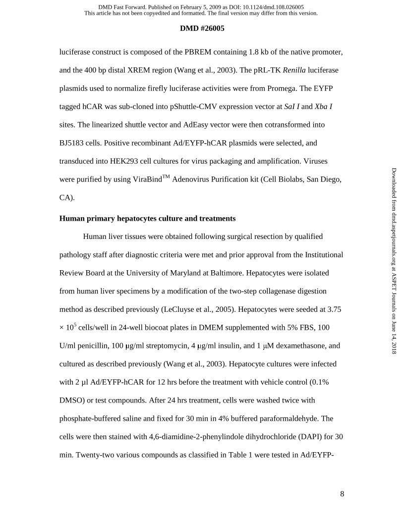
Human primary hepatocytes culture and treatments
Human liver tissues were obtained following surgical resection by qualified
pathology staff after diagnostic criteria were met and prior approval from the Institutional
Review Board at the University of Maryland at Baltimore. Hepatocytes were isolated
from human liver specimens by a modification of the two-step collagenase digestion
method as described previously (LeCluyse et al., 2005). Hepatocytes were seeded at 3.75
× 105 cells/well in 24-well biocoat plates in DMEM supplemented with 5% FBS, 100
U/ml penicillin, 100 μg/ml streptomycin, 4 μg/ml insulin, and 1 μM dexamethasone, and
cultured as described previously (Wang et al., 2003). Hepatocyte cultures were infected
with 2 µl Ad/EYFP-hCAR for 12 hrs before the treatment with vehicle control (0.1%
DMSO) or test compounds. After 24 hrs treatment, cells were washed twice with
phosphate-buffered saline and fixed for 30 min in 4% buffered paraformaldehyde. The
cells were then stained with 4,6-diamidine-2-phenylindole dihydrochloride (DAPI) for 30
min. Twenty-two various compounds as classified in Table 1 were tested in Ad/EYFP-
This article has not been copyedited and formatted. The final version may differ from this version.DMD Fast Forward. Published on February 5, 2009 as DOI: 10.1124/dmd.108.026005
at ASPE
T Journals on June 14, 2018
dmd.aspetjournals.org
Dow
nloaded from

DMD #26005
9
hCAR infected HPH. In separate experiments, HPH seeded in 6-well biocoat plates were
treated with BHA (100 μM), DZP (50 μM), FLU (50 μM), MCB (50 μM), RIF (10 μM),
or 0.1% DMSO as vehicle control for real-time PCR and Western blotting analysis.
Confocal laser scanning microscopy imaging
Confocal laser scanning microscopy was performed with a Nikon C1-LU3
instrument based on an inverted Nikon Eclipse TE2000 microscope. The confocal system
was equipped with three fluorescence detection channels (photomultipliers) and a
nonconfocal transmitted light detector. One of the photomultipliers was used to collect
fluorescence signals from the green and yellow region of the fluorescence emission, and
the nonconfocal transmitted light detector was used to collect brightfield images.
Real-time PCR analysis
Total RNA was isolated from treated hepatocytes using the RNeasy Mini Kit
(Qiagen) and reverse transcribed using High Capacity cDNA Archive kit (Applied
Biosystems, Foster, CA) following the manufacturers’ instructions. CYP2B6 and
CYP3A4 mRNA expression was normalized against that of glyceraldehyde-3-phosphate
dehydrogenase (GAPDH). Real-time PCR assays were performed in 96-well optical
plates on an ABI Prism 7000 Sequence Detection System with SYBR Green PCR Master
Mix (Applied Biosystems). Primers for CYP2B6, CYP3A4, and GAPDH mRNA
detection are as follows: CYP2B6 1215 to 1319 bp, 5’- AGACGCCTTCAATCCTGACC
-3’ (forward), and 5’- CCTTCACCAAGACAAATCCGC - 3’ (reverse); CYP3A4 213 to
462 bp, 5’-GTGGGGCTTTTATGATGGTCA-3’ (forward), and 5’-
GCCTCAGATTTCTCACCAACACA - 3’ (reverse); and GAPDH 217 to 501 bp, 5’-
CCCATCACCATCTTCCAGGAG -3’ (forward), and 5’-
This article has not been copyedited and formatted. The final version may differ from this version.DMD Fast Forward. Published on February 5, 2009 as DOI: 10.1124/dmd.108.026005
at ASPE
T Journals on June 14, 2018
dmd.aspetjournals.org
Dow
nloaded from
DMD #26005
10
GTTGTCATGGATGACCTTGGC - 3’ (reverse). Fold induction values were calculated
according to the equation 2ΔΔCt, where ΔCt represents the differences in cycle threshold
numbers between the target gene and GAPDH, and ΔΔCt represents the relative change
in these differences between control and treatment groups.
Western Blot Analyses
Homogenate proteins (20 µg) from treated HPH were resolved on SDS -
polyacrylamide gels, and electrophoretically transferred onto Immobilon-P
polyvinylidene difluoride membranes. Subsequently, membranes were incubated with
specific antibodies against CYP2B6 or CYP3A4 (Millipore-Chemicon, CA) diluted
1:4000 and 1:5000, respectively. β-actin (Sigma-Aldrich. St. Louis, MO) was used as
internal control. Blots were washed and incubated with horseradish peroxidase goat anti-
rabbit IgG antibody diluted 1:4000. Blots were developed using ECL Western blotting
detection reagent (GE Healthcare).
Transfection assays in cell lines
HepG2 cells in 24-well plates were transfected with hCAR expression vector, and
CYP2B6-2.2kb reporter construct using Fugene 6 Transfection Kit following the
manufacturer’s instruction. Twenty four hours after transfection, cells were treated with
solvent (0.1% DMSO) or test compounds at indicated concentrations (Fig. 6D) in the
absence or presence of PK11195 (5 µM). Twenty-four hours later, cell lysates were
assayed for firefly activities normalized against the activities of cotransfected Renilla
luciferase using Dual-Luciferase Kit (Promega, WI). Data were represented as mean±S.D.
of three individual transfections.
Statistical analysis
This article has not been copyedited and formatted. The final version may differ from this version.DMD Fast Forward. Published on February 5, 2009 as DOI: 10.1124/dmd.108.026005
at ASPE
T Journals on June 14, 2018
dmd.aspetjournals.org
Dow
nloaded from

DMD #26005
11
Experimental data are presented as a mean of triplicate determinations ± S.D.
unless otherwise noted. Statistical comparisons were made using one-way analysis of
variance (ANOVA). The statistical significance was set at p values <0.05 (*), or <0.01
(**).
This article has not been copyedited and formatted. The final version may differ from this version.DMD Fast Forward. Published on February 5, 2009 as DOI: 10.1124/dmd.108.026005
at ASPE
T Journals on June 14, 2018
dmd.aspetjournals.org
Dow
nloaded from

DMD #26005
12
Results
Localization of exogenous hCAR in HepG2 cells and HPH
Spontaneous nuclear accumulation and activation of CAR has been one of the
major obstacles in studying CAR activation in immortalized cell lines. As demonstrated
in Fig. 1, our recombinant adenoviral EYFP-human CAR (Ad/EYFP-hCAR) infected
both HepG2 cells and HPH with high efficiency (Fig. 1A). Consistent with previous
observations (Kawamoto et al., 1999; Kanno et al., 2005), the expression of Ad/EYFP-
hCAR was primarily observed in the nuclei of HepG2 cells (nucleus = 98%; mixed = 2%).
In contrast, the Ad/EYFP-hCAR has been visualized predominantly in the cytoplasm of
human primary hepatocytes prior to activation (nucleus = 4%; cytoplasm = 87%; mixed =
9%). Together, the unique characteristics of hCAR distribution, the direct visualization of
EYFP-hCAR, plus the high efficiency of Ad/EYFP-hCAR infection of HPH offer an
attractive avenue for in vitro identification of hCAR activators.
Ad/EYFP-hCAR response to known hCAR activators
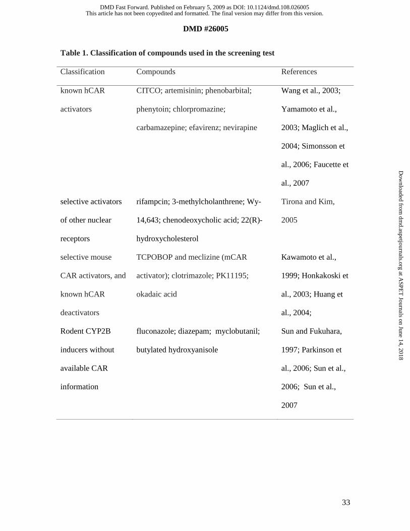
A series of 22 compounds have been chosen to evaluate the correlation between
chemical-mediated hCAR nuclear accumulation and target gene induction in current
studies. These compounds include known hCAR activators, hCAR deactivators, selective
mouse CAR activators, selective activators of other nuclear receptors, and known or
suspected CYP2B inducers for which information regarding hCAR activation is not
available (Table 1). With no activation, the profusely expressed Ad/EYFP-CAR was
observed primarily in the cytoplasm of cultured hepatocytes (84.5%), while around
15.5% infected cells displayed nucleus or mixed (nucleus + cytoplasm) allocation (Fig.
2A, and 2B). On the contrary, all the eight known hCAR activators resulted in
This article has not been copyedited and formatted. The final version may differ from this version.DMD Fast Forward. Published on February 5, 2009 as DOI: 10.1124/dmd.108.026005
at ASPE
T Journals on June 14, 2018
dmd.aspetjournals.org
Dow
nloaded from
DMD #26005
13
remarkable nuclear accumulation after 24 hrs treatment, where nucleus and mixed hCAR
distribution accounts for approximately 90% of the infected hepatocytes (Fig. 2C).
Representative images of hCAR localization were demonstrated in Fig. 2A and 2B
following the treatment of vehicle control (0.1% DMSO), the typical hCAR activator PB
(1 mM) or CITCO (1 µM). Notably, the selective hCAR agonist, CITCO revealed a
unique pattern of hCAR translocation in which up to 64.5% of infected cells exhibit
mixed hCAR distribution (Fig. 2B).
Ad/EYFP-hCAR response to selective activators of other nuclear receptors
Liver is enriched with various transcription factors that govern the regulation of
both constitutive and inducible DME expression (Tirona and Kim, 2005). To determine
the specificity of chemical-mediated hCAR translocation, several typical activators of
other nuclear receptors including RIF (10 µM) for PXR, CDCA (50 µM) for farnesoid X
receptor (FXR), HOC (10 µM) for liver X receptor (LXR), 3MC (5 µM) for aryl
hydrocarbon receptor (AhR), and Wy-14,643 (50 µM) for peroxisome proliferator-
activated receptor α (PPARα) were characterized in HPH infected with Ad/EYFP-hCAR
(Fig. 3). Representative images of hCAR localization were demonstrated in Fig. 3A and
3B following the treatment of vehicle control (0.1% DMSO), the typical hPXR activator
RIF (10 µM) or AhR activator 3MC (5 µM). As demonstrated in Fig. 3C, none of the
selective activators of these other nuclear receptors caused significant shift of Ad/EYFP-
hCAR from the cytoplasm to the nucleus compared with the vehicle control, indicating
the chemical-selectivity of Ad/EYFP-hCAR nuclear accumulation in HPH.
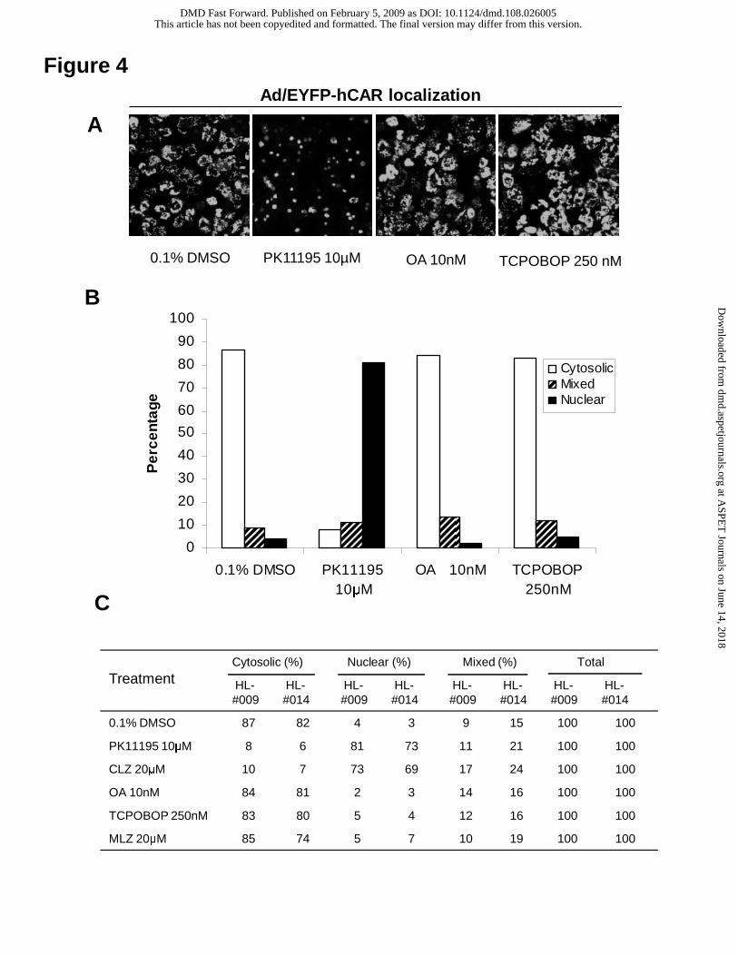
Translocation of Ad/EYFP-hCAR by mCAR agonist and hCAR antagonists
TCPOBOP is a known selective and potent mCAR agonist, and robustly induces
This article has not been copyedited and formatted. The final version may differ from this version.DMD Fast Forward. Published on February 5, 2009 as DOI: 10.1124/dmd.108.026005
at ASPE
T Journals on June 14, 2018
dmd.aspetjournals.org
Dow
nloaded from

DMD #26005
14
Cyp2b10 expression in mouse liver and cultured hepatocytes (Honkakoski et al., 2003).
In order to assess the species-specificity of Ad/EYFP-hCAR translocation, infected HPH
were treated with TCPOBOP (250 nM) for 24 hrs. As shown in Fig. 4, treatment of
TCPOBOP was not associated with nuclear accumulation of Ad/EYFP-hCAR in human
hepatocytes. Intriguingly, a recent study showed that the typical PPARα ligand, Wy-
14,643 remarkably translocated mCAR from the cytoplasm to the nucleus in mouse liver
(Guo et al., 2007), whereas our results revealed that Wy-14,643 neither enhanced the
nuclear accumulation of hCAR in HPH (Fig. 3) nor induced the expression of CYP2B6 in
HPH (data not shown). Together, these data underscore the species-selectivity of
Ad/EYFP-hCAR translocation in HPH. Conversely, two recently reported hCAR
antagonists, CLZ and PK11195 resulted in a significant nuclear accumulation of
Ad/EYFP-hCAR in cultured human hepatocytes (Fig. 4C), signifying the complexity of
the mechanisms underlying CAR activation (Li et al., 2008; Moore et al., 2000). In a
parallel experiment, the indirect CAR deactivator, OA (10 nM) alone exhibited no effects
on the translocation of Ad/EYFP-hCAR (Fig. 4A). Intriguingly, after 1 hr pretreatment of
OA (10 nM), the cotreatment of OA + PB (0.5 or 1 mM) failed to inhibit PB-mediated
hCAR nuclear translocation in HPH (Supplemental Fig. 1).
Correlation between hCAR translocation and CYP2B6 induction
The CYP2B genes are typical CAR targets in different species. Although other
nuclear receptors such as PXR also mediate CYP2B induction, activation of CAR is
closely associated with the induction of CYP2B in a species-specific manner. In the
current study, a number of known or suspected CYP2B inducers without available hCAR
activation data have been evaluated by the Ad/EYFP-hCAR translocation assays. These
This article has not been copyedited and formatted. The final version may differ from this version.DMD Fast Forward. Published on February 5, 2009 as DOI: 10.1124/dmd.108.026005
at ASPE
T Journals on June 14, 2018
dmd.aspetjournals.org
Dow
nloaded from
DMD #26005
15
compounds include four reported rodent CYP2B inducers MCB, BHA, DZP, and FLU
(Sun and Fukuhara, 1997; Parkinson et al., 2006; Sun et al., 2006; Sun et al., 2007). As
shown in Fig. 5, three out of four compounds (BHA, DZP, and MCB) exhibited
remarkable capacity of translocating hCAR to the nucleus of HPH. However, one of the
four rodent CYP2B inducers, FLU did not shift Ad/EYFP-hCAR towards the nucleus of
treated HPH.
To determine whether the hCAR nuclear accumulation correlates with its target
induction, HPH were treated with the four rodent CYP2B inducers as described in
Material and Methods. Total RNA and proteins were prepared from the treated cells for
determining the induction of CYP2B6 and CYP3A4 in human liver. The results
demonstrated that DZP and MCB significantly enhanced the expression of CYP2B6 and
CYP3A4 at both mRNA and protein levels, while BHA exhibited selective induction of
CYP2B6 over CYP3A4 (Fig. 6A, 6B and 6C). In contrast, FLU (50 µM) had little, if any
induction effects on either CYP2B6 or CYP3A4. Taken together, these data indicate that
nuclear translocation of Ad/EYFP-hCAR correlates well with its target gene inducible
expression.
Activation of hCAR in cell-based reporter assays
HepG2 cells transfected with hCAR expression and CYP2B6 reporter constructs
exhibited high basal reporter activity and were not sensitive to chemical-mediated
activation, as expected (Kawamoto et al., 1999). On the other hand, we previously
observed that the potent hCAR deactivator, PK11195 significantly inhibited the
constitutive activity of hCAR in HepG2 cells, and the inhibited hCAR activity could only
be reactivated by direct activator CITCO but not by indirect activators such as PB (Li et
This article has not been copyedited and formatted. The final version may differ from this version.DMD Fast Forward. Published on February 5, 2009 as DOI: 10.1124/dmd.108.026005
at ASPE
T Journals on June 14, 2018
dmd.aspetjournals.org
Dow
nloaded from
DMD #26005
16
al., 2008). In the current reporter experiment, little or no reactivation was observed after
the cotreatment with PK11195, and BHA, DZP, or MCB at 50 µM /each (Fig. 6D).
Combined with the observed hCAR translocation data in HPH, these results indicate that
BHA, DZP, and MCB most likely activate hCAR through PB-type indirect mechanisms.
This article has not been copyedited and formatted. The final version may differ from this version.DMD Fast Forward. Published on February 5, 2009 as DOI: 10.1124/dmd.108.026005
at ASPE
T Journals on June 14, 2018
dmd.aspetjournals.org
Dow
nloaded from

DMD #26005
17
Discussion
Although there is an emerging need for efficient screening of hCAR activators at
the early stage of drug development, CAR is constitutively activated in all the
immortalized cell lines independent of xenobiotic stimulation. In addition, CAR displays
unique activation mechanisms compared with other nuclear receptors, where CAR could
be activated through either direct ligand binding or indirect phosphorylation/
dephosphorylation-related signaling pathways (Kawamoto et al., 1999; Qatanani and
Moore, 2005). These CAR features significantly lowered the value of cell-based reporter
and in vitro ligand binding assays, making the investigation of CAR activation much
more challenging. In contrast to the observations in immortalized cells, CAR is primarily
compartmentalized in the cytoplasm of primary cultured hepatocytes and intact liver in
vivo, and only accumulates in the nucleus upon chemical-mediated activation. Nuclear
translocation in hepatocytes appears to be the essential first step of xenobiotic-induced
CAR activation, and may offer a novel avenue for predicting CAR activation. However,
in vitro detection of hCAR translocation has been difficult and time-consuming, partly
due to the quiescent nature of human primary hepatocyte cultures. In this report, we have
generated an Ad/EYFP-hCAR construct that infects HPH with high efficiency and
maintains hCAR distribution characteristics in a physiologically relevant manner.
Several lines of evidence indicate that activation of CAR is a multi-step process,
with nuclear accumulation as the essential first stride (Kawamoto et al., 1999; Sueyoshi
et al., 2002). To determine the value of Ad/EYFP-hCAR transduced HPH as a tool for
screening hCAR activators in vitro, the current study evaluated 22 compounds including
known hCAR activators, deactivators, typical activators of other nuclear receptors,
This article has not been copyedited and formatted. The final version may differ from this version.DMD Fast Forward. Published on February 5, 2009 as DOI: 10.1124/dmd.108.026005
at ASPE
T Journals on June 14, 2018
dmd.aspetjournals.org
Dow
nloaded from
DMD #26005
18
mCAR activators, and known or suspected CYP2B inducers without available CAR
activation data for the correlation between hCAR nuclear translocation and target gene
induction. Thirteen compounds resulted in significant nuclear accumulation of Ad/EYFP-
hCAR in HPH. Among them, eight compounds (PB, CITCO, ART, PHN, EFV, NVP,
CPZ, and CMZ) are known hCAR activators and CYP2B6 inducers (Sueyoshi et al.,
1999; Maglich et al., 2003; Wang et al., 2004; Faucette et al., 2007), and three
compounds (BHA, DZP, and MCB) are newly established human CYP2B6 inducers by
the current studies, which display a total of 85% (11/13) correlation of Ad/EYFP-hCAR
nuclear translocation with hCAR target gene induction (Sun and Fukuhara, 1997;
Parkinson et al., 2006; Sun et al., 2007). In contrast, we also observed that two reported
hCAR deactivators (CLZ and PK11195) remarkably translocated Ad-EYFP-hCAR to the
nucleus of HPH, which are consistent with our previous reports (Li et al., 2008; Wang
and Tompkins, 2008). Similarly, Guo et al (Guo et al., 2007) reported that the typical
PPARα agonist Wy-14,643 acts as an antagonist of mCAR and stimulates mCAR nuclear
translocation without accompanied transcriptional enhancement of CAR target gene in
mouse liver. Conversely, the known indirect CAR deactivator, OA alone has no effects
on Ad/EYFP-hCAR translocation, and moreover, cotreatment of OA with PB didn’t
inhibit the PB-mediated hCAR nuclear accumulation in HPH (supplemental Fig. 1). This
observation is in contrast with an early report from Dr. Negishi and colleagues, where
OA treatment inhibited PB-mediated mCAR translocation in mouse primary hepatocytes
(Kawamoto et al., 1999). Although the species-specificity of hCAR vs. mCAR might be
one of the reasons for the controversial effects of OA between human and mouse,
detailed explanations are beyond the scope of the current studies. Given the antagonistic
This article has not been copyedited and formatted. The final version may differ from this version.DMD Fast Forward. Published on February 5, 2009 as DOI: 10.1124/dmd.108.026005
at ASPE
T Journals on June 14, 2018
dmd.aspetjournals.org
Dow
nloaded from

DMD #26005
19
nature of CLZ and PK11195, along with the agonistic property of CITCO, it is reasonable
to speculate that ligand binding itself may trigger CAR nuclear accumulation regardless
of its agonistic or antagonistic nature.
Although hCAR shares some common characteristics with its rodent counterparts,
apparent species-specific activation between human and rodent CARs render the
necessity for evaluating hCAR activation in the physiologically relevant in vitro system-
HPH. In the current study, TCPOBOP, the most effective mCAR agonist identified thus
far, exhibited negative nuclear translocation of hCAR in HPH. Notably, our results also
showed that Wy-14,643 failed to reallocate hCAR to the nucleus of HPH (Fig. 3B),
which are in contrast to the observation made by Guo et al (Guo et al., 2007) in
adenoviral-mCAR infected mouse liver, emphasizing the species-selectivity of hCAR
activation. To date, accumulating evidence has revealed that liver is abundant with
xenobiotic receptors that coordinately regulate the expression of their target genes
through cross-talking. For instance, PB activates both hCAR and hPXR, while RIF
selectively actives hPXR. Activation of hCAR and hPXR induces the expression of a
broad spectrum of DMEs and transporters in the liver. To gain insight into the chemical-
specificity of Ad/EYFP-hCAR nuclear translocation, five selective activators of other
xenobiotic receptors have been evaluated in Ad/EYFP-hCAR infected HPH. Without
exception, typical activators of PXR (RIF), AhR (3MC), LXR (HOC), FXR (CDCA), and
PPARa (Wy-14, 643) all failed to significantly relocate Ad/EYFP-hCAR from the
cytoplasm to the nucleus in HPH. Together, current evidence suggests that the Ad/EYFP-
hCAR translocation assay in HPH exhibits both species-specific and chemical-specific
selectivity in hCAR activation.
This article has not been copyedited and formatted. The final version may differ from this version.DMD Fast Forward. Published on February 5, 2009 as DOI: 10.1124/dmd.108.026005
at ASPE
T Journals on June 14, 2018
dmd.aspetjournals.org
Dow
nloaded from

DMD #26005
20
Due to the apparent difficulties associated with identifying hCAR activators,
particularly in a higher throughput fashion, the numbers of known hCAR activators thus
far, are relatively small. In the current study, we also evaluated the hCAR nuclear
translocation by four reported rodent CYP2B (FLU, BHA, DZP, and MCB) inducers that
have no available information regarding hCAR activation. Notably, three out of the four
compounds exhibited significant nuclear translocation of the Ad/EYFP-hCAR in HPH.
Through a combination of direct and indirect experimental approaches, our results
showed that BHA, DZP, and MCB mediated hCAR nuclear translocation is closely
associated with the actual induction of their target gene CYP2B6 and CYP3A4. To our
knowledge, this is the first report utilizing hCAR translocation assay in HPH to
successfully identify novel CYP2B6 and CYP3A4 inducers. Notably, although FLU is a
potent inducer of CYP2B1 in rat (Sun et al., 2006), there is barely visible induction of
either CYP2B6 or CYP3A4, and FLU did not enhance the nuclear translocation of
Ad/EYFP-hCAR in HPH. These data further highlight the species-specificity and the
value of the Ad/EYFP-hCAR translocation assay in HPH.
In summary, our data suggest that Ad/EYFP-hCAR infection of HPH provides a
valuable tool for efficiently identifying hCAR activators in vitro. Because of the unique
characteristics of hCAR activation, Ad/EYFP-hCAR translocation assay in HPH exhibits
several apparent advantages over the cell-based reporter assays in cell lines and in vitro
ligand binding assays, regarding the sensitivity to chemical stimulation and
responsiveness to both direct and indirect activators. Meanwhile, we do realize that one
limitation of this screening test is the incapability of discerning the agonist and antagonist
of hCAR. Nonetheless, we have demonstrated recently that the constitutive activation of
This article has not been copyedited and formatted. The final version may differ from this version.DMD Fast Forward. Published on February 5, 2009 as DOI: 10.1124/dmd.108.026005
at ASPE
T Journals on June 14, 2018
dmd.aspetjournals.org
Dow
nloaded from
DMD #26005
21
hCAR in HepG2 cells was efficiently repressed by hCAR antagonist PK11195, and was
reactivated by hCAR agonist CITCO but not by indirect activators such as PB and PHN
(Li et al., 2008). Utilizing this reactivation assay, our results indicated that all three novel
hCAR activators identified in the current study are most likely indirect activators (Fig.
6D). Overall, the combination of Ad/EYFP-hCAR translocation assay in HPH with the
hCAR reactivation assay in HepG2 cells offers a valuable avenue for the identification of
hCAR activators in vitro.
This article has not been copyedited and formatted. The final version may differ from this version.DMD Fast Forward. Published on February 5, 2009 as DOI: 10.1124/dmd.108.026005
at ASPE
T Journals on June 14, 2018
dmd.aspetjournals.org
Dow
nloaded from
DMD #26005
22
Acknowledgement
The authors would like to thank Dr. Masahiko Negishi (National Institute of
Environmental and Health Sciences, National Institutes of Health, RTP, NC) for the
provision of hCAR expression plasmids. We also gratefully acknowledge the technical
support by Dr. Xilin Niu (University of North Carolina at Chapel Hill, NC) in our
preparation of the Ad/EYFP-hCAR. Human liver tissues were obtained from The
University of Maryland Medical Center (Baltimore, MD).
This article has not been copyedited and formatted. The final version may differ from this version.DMD Fast Forward. Published on February 5, 2009 as DOI: 10.1124/dmd.108.026005
at ASPE
T Journals on June 14, 2018
dmd.aspetjournals.org
Dow
nloaded from
DMD #26005
23
References
Baes M, Gulick T, Choi HS, Martinoli MG, Simha D and Moore DD (1994) A new
orphan member of the nuclear hormone receptor superfamily that interacts with a
subset of retinoic acid response elements. Mol Cell Biol 14:1544-1552.
Faucette SR, Zhang TC, Moore R, Sueyoshi T, Omiecinski CJ, LeCluyse EL, Negishi M
and Wang H (2007) Relative activation of human pregnane X receptor versus
constitutive androstane receptor defines distinct classes of CYP2B6 and CYP3A4
inducers. J Pharmacol Exp Ther 320:72-80.
Guo D, Sarkar J, Suino-Powell K, Xu Y, Matsumoto K, Jia Y, Yu S, Khare S, Haldar K,
Rao MS, Foreman JE, Monga SP, Peters JM, Xu HE and Reddy JK (2007)
Induction of nuclear translocation of constitutive androstane receptor by
peroxisome proliferator-activated receptor alpha synthetic ligands in mouse liver.
J Biol Chem 282:36766-36776.
Honkakoski P, Sueyoshi T and Negishi M (2003) Drug-activated nuclear receptors CAR
and PXR. Ann Med 35:172-182.
Huang W, Zhang J, Chua SS, Qatanani M, Han Y, Granata R and Moore DD (2003)
Induction of bilirubin clearance by the constitutive androstane receptor (CAR).
Proc Natl Acad Sci U S A 100:4156-4161.
Huang W, Zhang J, Washington M, Liu J, Parant JM, Lozano G and Moore DD (2005)
Xenobiotic stress induces hepatomegaly and liver tumors via the nuclear receptor
constitutive androstane receptor. Mol Endocrinol 19:1646-1653.
This article has not been copyedited and formatted. The final version may differ from this version.DMD Fast Forward. Published on February 5, 2009 as DOI: 10.1124/dmd.108.026005
at ASPE
T Journals on June 14, 2018
dmd.aspetjournals.org
Dow
nloaded from
DMD #26005
24
Kawamoto T, Sueyoshi T, Zelko I, Moore R, Washburn K and Negishi M (1999)
Phenobarbital-responsive nuclear translocation of the receptor CAR in induction
of the CYP2B gene. Mol Cell Biol 19:6318-6322.
Kobayashi K, Sueyoshi T, Inoue K, Moore R and Negishi M (2003) Cytoplasmic
accumulation of the nuclear receptor CAR by a tetratricopeptide repeat protein in
HepG2 cells. Mol Pharmacol 64:1069-1075.
Kodama S, Koike C, Negishi M and Yamamoto Y (2004) Nuclear receptors CAR and
PXR cross talk with FOXO1 to regulate genes that encode drug-metabolizing and
gluconeogenic enzymes. Mol Cell Biol 24:7931-7940.
LeCluyse EL, Alexandre E, Hamilton GA, Viollon-Abadie C, Coon DJ, Jolley S and
Richert L (2005) Isolation and culture of primary human hepatocytes. Methods
Mol Biol 290:207-229.
Li L, Chen T, Stanton JD, Sueyoshi T, Negishi M and Wang H (2008) The peripheral
benzodiazepine receptor ligand 1-(2-chlorophenyl-methylpropyl)-3-isoquinoline-
carboxamide is a novel antagonist of human constitutive androstane receptor. Mol
Pharmacol 74:443-453.
Maglich JM, Parks DJ, Moore LB, Collins JL, Goodwin B, Billin AN, Stoltz CA,
Kliewer SA, Lambert MH, Willson TM and Moore JT (2003) Identification of a
novel human constitutive androstane receptor (CAR) agonist and its use in the
identification of CAR target genes. J Biol Chem 278:17277-17283.
Merrell MD, Jackson JP, Augustine LM, Fisher CD, Slitt AL, Maher JM, Huang W,
Moore DD, Zhang Y, Klaassen CD and Cherrington NJ (2008) The Nrf2 activator
This article has not been copyedited and formatted. The final version may differ from this version.DMD Fast Forward. Published on February 5, 2009 as DOI: 10.1124/dmd.108.026005
at ASPE
T Journals on June 14, 2018
dmd.aspetjournals.org
Dow
nloaded from

DMD #26005
25
oltipraz also activates the constitutive androstane receptor. Drug Metab Dispos
36:1716-1721.
Moore LB, Parks DJ, Jones SA, Bledsoe RK, Consler TG, Stimmel JB, Goodwin B,
Liddle C, Blanchard SG, Willson TM, Collins JL and Kliewer SA (2000) Orphan
nuclear receptors constitutive androstane receptor and pregnane X receptor share
xenobiotic and steroid ligands. J Biol Chem 275:15122-15127.
Parkinson A, Leonard N, Draper A and Ogilvie BW (2006) On the mechanism of
hepatocarcinogenesis of benzodiazepines: evidence that diazepam and oxazepam
are CYP2B inducers in rats, and both CYP2B and CYP4A inducers in mice. Drug
Metab Rev 38:235-259.
Qatanani M and Moore DD (2005) CAR, the continuously advancing receptor, in drug
metabolism and disease. Curr Drug Metab 6:329-339.
Simonsson US, Lindell M, Raffalli-Mathieu F, Lannerbro A, Honkakoski P and Lang
MA (2006) In vivo and mechanistic evidence of nuclear receptor CAR induction
by artemisinin. Eur J Clin Invest 36:647-653.
Stanley LA, Horsburgh BC, Ross J, Scheer N and Wolf CR (2006) PXR and CAR:
nuclear receptors which play a pivotal role in drug disposition and chemical
toxicity. Drug Metab Rev 38:515-597.
Sueyoshi T, Kawamoto T, Zelko I, Honkakoski P and Negishi M (1999) The repressed
nuclear receptor CAR responds to phenobarbital in activating the human CYP2B6
gene. J Biol Chem 274:6043-6046.
This article has not been copyedited and formatted. The final version may differ from this version.DMD Fast Forward. Published on February 5, 2009 as DOI: 10.1124/dmd.108.026005
at ASPE
T Journals on June 14, 2018
dmd.aspetjournals.org
Dow
nloaded from

DMD #26005
26
Sueyoshi T, Moore R, Pascussi JM and Negishi M (2002) Direct expression of
fluorescent protein-tagged nuclear receptor CAR in mouse liver. Methods
Enzymol 357:205-213.
Sueyoshi T, Moore R, Sugatani J, Matsumura Y and Negishi M (2008) PPP1R16A, the
membrane subunit of protein phosphatase 1beta, signals nuclear translocation of
the nuclear receptor constitutive active/androstane receptor. Mol Pharmacol
73:1113-1121.
Sugatani J, Kojima H, Ueda A, Kakizaki S, Yoshinari K, Gong QH, Owens IS, Negishi
M and Sueyoshi T (2001) The phenobarbital response enhancer module in the
human bilirubin UDP-glucuronosyltransferase UGT1A1 gene and regulation by
the nuclear receptor CAR. Hepatology 33:1232-1238.
Sun B and Fukuhara M (1997) Effects of co-administration of butylated hydroxytoluene,
butylated hydroxyanisole and flavonoids on the activation of mutagens and drug-
metabolizing enzymes in mice. Toxicology 122:61-72.
Sun G, Grindstaff RD, Thai SF, Lambert GR, Tully DB, Dix DJ and Nesnow S (2007)
Induction of cytochrome P450 enzymes in rat liver by two conazoles,
myclobutanil and triadimefon. Xenobiotica 37:180-193.
Sun G, Thai SF, Lambert GR, Wolf DC, Tully DB, Goetz AK, George MH, Grindstaff
RD, Dix DJ and Nesnow S (2006) Fluconazole-induced hepatic cytochrome P450
gene expression and enzymatic activities in rats and mice. Toxicol Lett 164:44-53.
Tien ES and Negishi M (2006) Nuclear receptors CAR and PXR in the regulation of
hepatic metabolism. Xenobiotica 36:1152-1163.
This article has not been copyedited and formatted. The final version may differ from this version.DMD Fast Forward. Published on February 5, 2009 as DOI: 10.1124/dmd.108.026005
at ASPE
T Journals on June 14, 2018
dmd.aspetjournals.org
Dow
nloaded from

DMD #26005
27
Tirona RG and Kim RB (2005) Nuclear receptors and drug disposition gene regulation. J
Pharm Sci 94:1169-1186.
Tzameli I, Pissios P, Schuetz EG and Moore DD (2000) The xenobiotic compound 1,4-
bis[2-(3,5-dichloropyridyloxy)]benzene is an agonist ligand for the nuclear
receptor CAR. Mol Cell Biol 20:2951-2958.
Ueda A, Hamadeh HK, Webb HK, Yamamoto Y, Sueyoshi T, Afshari CA, Lehmann JM
and Negishi M (2002) Diverse roles of the nuclear orphan receptor CAR in
regulating hepatic genes in response to phenobarbital. Mol Pharmacol 61:1-6.
Wang H, Faucette S, Moore R, Sueyoshi T, Negishi M and LeCluyse E (2004) Human
constitutive androstane receptor mediates induction of CYP2B6 gene expression
by phenytoin. J Biol Chem 279:29295-29301.
Wang H, Faucette S, Sueyoshi T, Moore R, Ferguson S, Negishi M and LeCluyse EL
(2003) A novel distal enhancer module regulated by pregnane X
receptor/constitutive androstane receptor is essential for the maximal induction of
CYP2B6 gene expression. J Biol Chem 278:14146-14152.
Wang H and Negishi M (2003) Transcriptional regulation of cytochrome p450 2B genes
by nuclear receptors. Curr Drug Metab 4:515-525.
Wang H and Tompkins LM (2008) CYP2B6: New Insights into a Historically
Overlooked Cytochrome P450 Isozyme. Curr Drug Metab 9:598-610.
Yamamoto Y, Kawamoto T and Negishi M (2003) The role of the nuclear receptor CAR
as a coordinate regulator of hepatic gene expression in defense against chemical
toxicity. Arch Biochem Biophys 409:207-211.
This article has not been copyedited and formatted. The final version may differ from this version.DMD Fast Forward. Published on February 5, 2009 as DOI: 10.1124/dmd.108.026005
at ASPE
T Journals on June 14, 2018
dmd.aspetjournals.org
Dow
nloaded from
DMD #26005
28
Yamamoto Y, Moore R, Goldsworthy TL, Negishi M and Maronpot RR (2004) The
orphan nuclear receptor constitutive active/androstane receptor is essential for
liver tumor promotion by phenobarbital in mice. Cancer Res 64:7197-7200.
This article has not been copyedited and formatted. The final version may differ from this version.DMD Fast Forward. Published on February 5, 2009 as DOI: 10.1124/dmd.108.026005
at ASPE
T Journals on June 14, 2018
dmd.aspetjournals.org
Dow
nloaded from

DMD #26005
29
Footnotes
This research was supported by National Institute of Health [Grant DK061652].
Reprint requests should be sent to:
Dr. Hongbing Wang
Department of Pharmaceutical Sciences
University of Maryland, School of Pharmacy
20 Penn Street, Baltimore MD 21201
This article has not been copyedited and formatted. The final version may differ from this version.DMD Fast Forward. Published on February 5, 2009 as DOI: 10.1124/dmd.108.026005
at ASPE
T Journals on June 14, 2018
dmd.aspetjournals.org
Dow
nloaded from

DMD #26005
30
Figure Legends
Fig. 1. Localization of Ad/EYFP-hCAR in HepG2 cells and human primary hepatocytes
(HPH). HepG2 cells and HPH (HL-#009, HL-#014) were infected with Ad/EYFP-hCAR
as described in Material and Methods. (A) Confocal images depict the localization of
Ah/EYFP-hCAR in HepG2 and HPH. The left panels are Ad/EYFP-hCAR expression
(green); the middle panels represent the nuclear staining (red); and the merged images are
on the right. (B, C) 100 hCAR-expressing cells from each group were counted and
classified by cytosolic, nuclear, or mixed (cytosolic + nuclear) hCAR cellular
localizations.
Fig. 2. Known hCAR activators promote nuclear translocation of Ad/EYPF-hCAR in
HPH. HPH (HL-#009, HL-#014) were infected with Ad/EYFP-hCAR described in
Materials and Methods and treated with vehicle (0.1% DMSO), or eight known hCAR
activators at the indicated concentrations. Following 24 hrs of treatment, hepatocytes
were DAPI stained and subjected to confocal microscopy. (A) Representative Ad/EYFP-
hCAR localizations from vehicle control, PB (1 mM) or CITCO (1 µM) treated HPH.
Three panels are shown for each treatment, with the left, Ad/EYFP-hCAR (green); the
middle, nuclear staining (red); and the right, the merged image. (B, C) For each treatment,
100 hCAR-expressing cells were counted and classified based on cytosolic, nuclear, or
mixed (cytosolic + nuclear) hCAR cellular localizations.
Fig. 3. Ad/EYFP-hCAR was not translocated by typical activators of other nuclear
receptors. HPH (HL-#009, HL-#014) were infected with Ad/EYFP-hCAR as described
in Materials and Methods and treated with vehicle (0.1% DMSO), or five typical
This article has not been copyedited and formatted. The final version may differ from this version.DMD Fast Forward. Published on February 5, 2009 as DOI: 10.1124/dmd.108.026005
at ASPE
T Journals on June 14, 2018
dmd.aspetjournals.org
Dow
nloaded from

DMD #26005
31
activators of other nuclear receptors at the indicated concentrations. Following 24 hrs of
treatment, hepatocytes were subjected to confocal microscopy. (A) Representative
Ad/EYFP-hCAR localizations from vehicle control, RIF (10 µM) and 3MC (5 µM)
treated HPH. (B, C) For each treatment, 100 hCAR-expressing cells were counted and
classified based on cytosolic, nuclear, or mixed (cytosolic + nuclear) hCAR localizations.
Fig. 4. Translocation of Ad/EYFP-hCAR in HPH following the treatment with mCAR
activator and hCAR deactivators. HPH (HL-#009, HL-#014) were infected with
Ad/EYFP-hCAR as described in Materials and Methods and treated with vehicle (0.1%
DMSO), or tested compounds at indicated concentrations. Following 24 hrs of treatment,
hepatocytes were subjected to confocal microscopy analysis. (A) Representative
Ad/EYFP-hCAR localizations from vehicle control, PK1195 (10 µM), OA (0.01 µM), or
TCPOBOP (250 nM) treated HPH. (B, C) For each treatment, 100 hCAR-expressing
cells were counted and classified based on cytosolic, nuclear, or mixed (cytosolic +
nuclear) hCAR localizations.
Fig. 5. Ad/EYFP-hCAR localization in HPH following the treatment with CYP2B
inducers. HPH (HL-#009, HL-#014) were infected with Ad/EYFP-hCAR as described in
Materials and Methods and treated with vehicle (0.1% DMSO), or tested compounds at
indicated concentrations. Following 24 hrs of treatment, hepatocytes were subjected to
confocal microscopy analysis. (A) Representative images illustrate Ad/EYFP-hCAR
localizations in HPH treated with vehicle control, FLU (50 µM), BHA (100 µM), DZP
(50 µM), or MCB (50 µM). (B, C) For each treatment, 100 hCAR-expressing cells were
This article has not been copyedited and formatted. The final version may differ from this version.DMD Fast Forward. Published on February 5, 2009 as DOI: 10.1124/dmd.108.026005
at ASPE
T Journals on June 14, 2018
dmd.aspetjournals.org
Dow
nloaded from

DMD #26005
32
counted and classified based on cytosolic, nuclear, or mixed (cytosolic + nuclear) hCAR
localizations.
Fig. 6. Effects of FLU, BHA, DZP, and MCB on CYP2B6/CYP3A4 expression in HPH,
and hCAR reactivation in HepG2 cells. HPH (HL-#012, HL-#014) were treated with
FLU (50 µM), BHA (100 µM), DZP (50 µM), MCB (50 µM), RIF (10 µM), CITCO (1
µM) or vehicle control (0.1% DMSO). Total RNA extracted from 24 hrs treatment was
subjected to real-time PCR analysis (A, B). Homogenates (20 µg) harvested from 72 hrs
treatments were prepared for CYP2B6 and CYP3A4 immunoblot analysis (C). In a
separate experiment, HepG2 cells were transfected with CAR expression plasmid along
with CYP2B6-2.2kb reporter plasmid as described under Materials and Methods. Cells
were treated with vehicle (0.1% DMSO) and test compounds alone or in combination
with PK11195 (5 µM). Luciferase activities were determined and expressed relevant to
control (D). Data represent the mean ± SD of three independent transfections. (*p < 0.05;
**p < 0.01).
This article has not been copyedited and formatted. The final version may differ from this version.DMD Fast Forward. Published on February 5, 2009 as DOI: 10.1124/dmd.108.026005
at ASPE
T Journals on June 14, 2018
dmd.aspetjournals.org
Dow
nloaded from
DMD #26005
33
Table 1. Classification of compounds used in the screening test
Classification Compounds References
known hCAR
activators
CITCO; artemisinin; phenobarbital;
phenytoin; chlorpromazine;
carbamazepine; efavirenz; nevirapine
Wang et al., 2003;
Yamamoto et al.,
2003; Maglich et al.,
2004; Simonsson et
al., 2006; Faucette et
al., 2007
selective activators
of other nuclear
receptors
rifampcin; 3-methylcholanthrene; Wy-
14,643; chenodeoxycholic acid; 22(R)-
hydroxycholesterol
Tirona and Kim,
2005
selective mouse
CAR activators, and
known hCAR
deactivators
TCPOBOP and meclizine (mCAR
activator); clotrimazole; PK11195;
okadaic acid
Kawamoto et al.,
1999; Honkakoski et
al., 2003; Huang et
al., 2004;
Rodent CYP2B
inducers without
available CAR
information
fluconazole; diazepam; myclobutanil;
butylated hydroxyanisole
Sun and Fukuhara,
1997; Parkinson et
al., 2006; Sun et al.,
2006; Sun et al.,
2007
This article has not been copyedited and formatted. The final version may differ from this version.DMD Fast Forward. Published on February 5, 2009 as DOI: 10.1124/dmd.108.026005
at ASPE
T Journals on June 14, 2018
dmd.aspetjournals.org
Dow
nloaded from

0
20
40
60
80
100
120
HepG2 HPH
Per
cen
tag
e
CytosolicMixedNuclear
Figure 1
AHepG2
HPH
B
EYFP-hCAR Nucleus Merge
This article has not been copyedited and form
atted. The final version m
ay differ from this version.
DM
D Fast Forw
ard. Published on February 5, 2009 as DO
I: 10.1124/dmd.108.026005
at ASPET Journals on June 14, 2018 dmd.aspetjournals.org Downloaded from

0
10
20
30
40
50
60
70
80
90
100
0.1% DMSO PB 1mM CITCO 1μM
Per
cen
tag
e
CytosolicMixedNuclear
Figure 2
A 0.1%
DMSO
PB
1mM
CITCO
1µM
EYFP-hCAR Nucleus Merge
B
Cytosolic (%) Nuclear (%) Mixed (%) Total
HL- #009 HL- #014 HL- #009 HL- #014 HL- #009 HL- #014 HL- #009 HL- #014
0.1% DMSO 87 82 4 3 9 15 100 100
PB 1mM 6 4 87 79 7 17 100 100
CITCO 1μM 12 15 21 23 67 62 100 100
ART 50μM 10 9 59 67 31 24 100 100
PHN 50μM 7 5 71 83 22 12 100 100
EFV 25μM 11 8 36 49 53 43 100 100
NVP 50μM 14 6 29 51 57 43 100 100
CPZ 50μM 8 10 57 62 35 28 100 100
CMZ 30μM 10 8 53 70 37 22 100 100
C Treatment
This article has not been copyedited and form
atted. The final version m
ay differ from this version.
DM
D Fast Forw
ard. Published on February 5, 2009 as DO
I: 10.1124/dmd.108.026005
at ASPET Journals on June 14, 2018 dmd.aspetjournals.org Downloaded from

0
10
20
30
40
50
60
70
80
90
100
0.1% DMSO RIF 10μM 3MC 5μM
Per
cen
tag
e
CytosolicMixedNuclear
Figure 3
A
0.1% DMSO RIF 10µM 3MC 5µM
B
TreatmentCytosolic (%) Nuclear (%) Mixed (%) Total
HL-#009
HL-#014
HL-#009
HL-#014
HL-#009
HL-#014
HL-#009
HL-#014
0.1% DMSO 87 82 4 3 9 15 100 100
RIF10μM 89 82 3 6 8 12 100 100
3MC 5μM 85 80 4 3 11 17 100 100
Wy-14643 50μM 77 82 5 3 18 15 100 100
CDCA 50μM 81 72 3 9 16 19 100 100
HOC 10μM 78 76 3 12 19 12 100 100
C
Ad/EYFP-hCAR localization
This article has not been copyedited and formatted. The final version may differ from this version.DMD Fast Forward. Published on February 5, 2009 as DOI: 10.1124/dmd.108.026005
at ASPE
T Journals on June 14, 2018
dmd.aspetjournals.org
Dow
nloaded from
0
10
20
30
40
50
60
70
80
90
100
0.1% DMSO PK1119510μM
OA 10nM TCPOBOP250nM
Per
cent
age
CytosolicMixedNuclear
Figure 4
A
TCPOBOP 250 nMOA 10nM0.1% DMSO PK11195 10µM
B
C
Cytosolic (%) Nuclear (%) Mixed (%) Total
HL-#009
HL-#014
HL-#009
HL-#014
HL-#009
HL-#014
HL-#009
HL-#014
0.1% DMSO 87 82 4 3 9 15 100 100
PK11195 10μM 8 6 81 73 11 21 100 100
CLZ 20μM 10 7 73 69 17 24 100 100
OA 10nM 84 81 2 3 14 16 100 100
TCPOBOP 250nM 83 80 5 4 12 16 100 100
MLZ 20μM 85 74 5 7 10 19 100 100
Treatment
Ad/EYFP-hCAR localization
This article has not been copyedited and formatted. The final version may differ from this version.DMD Fast Forward. Published on February 5, 2009 as DOI: 10.1124/dmd.108.026005
at ASPE
T Journals on June 14, 2018
dmd.aspetjournals.org
Dow
nloaded from

Figure 5
A
FLU 50µM BHA 100µM DZP 50µM0.1% DMSO MCB 50µM
B
Cytosolic(%) Nuclear (%) Mixed (%) Total
HL-#009
HL-#014
HL-#009
HL-#014
HL-#009
HL-#014
HL-#009
HL-#014
0.1% DMSO 87 82 4 3 9 15 100 100
FLU 50μM 76 81 6 8 18 11 100 100
BHA 100μM 1 5 87 81 12 14 100 100
MCB 50nM 3 4 79 84 18 12 100 100
DZP 50μM 2 3 84 81 14 16 100 100
C
Treatment
0
10
20
30
40
50
60
70
80
90
100
0.1%DMSO
FLU50μM
BHA100μM
DZP50μM
MCB50μM
Per
cen
tag
e
CytosolicMixedNuclear
Ad/EYFP-hCAR localization
This article has not been copyedited and formatted. The final version may differ from this version.DMD Fast Forward. Published on February 5, 2009 as DOI: 10.1124/dmd.108.026005
at ASPE
T Journals on June 14, 2018
dmd.aspetjournals.org
Dow
nloaded from

0
0.2
0.40.6
0.8
11.2
1.4
0.1%
DM
SO
CIT
CO
1μ
M
PB
1m
M
FL
U 5
0μ
M
BH
A 5
0μ
M
DZ
P 5
0μ
M
MC
B 5
0μ
M
PK
1119
5 5μ
M
CIT
CO
1
μ
M
PB
1m
M
FL
U 5
0μ
M
BH
A 5
0μ
M
DZ
P 5
0μ
M
MC
B 5
0μ
M
Rel
ativ
e L
uc
Act
ivit
y
0
2
4
6
8
10
12
14
16
18
20
0.1%DMSO
FLU50μM
BHA100μM
DZP50μM
MCB50μM
RIF10μM
CITCO1μM
Fo
ld t
o c
on
tro
l
HL-#012
HL-#014
0
1
2
3
4
5
6
7
8
9
0.1%DMSO
FLU50μM
BHA100μM
DZP50μM
MCB50μM
RIF10μM
CITCO1μM
Fo
ld t
o c
on
tro
l
HL-#012
HL-#014
Figure 6
A B
0.1% FLU BHA DZP MCB RIF CITCODMSO 50µM 100µM 50µM 50µM 10µM 1µM
CYP2B6
CYP3A4
β-Actin
C
PK11195 5µM
D
CYP2B6 CYP3A4
****
** **
**
**
****
****
** ** **
**
**
****
This article has not been copyedited and form
atted. The final version m
ay differ from this version.
DM
D Fast Forw
ard. Published on February 5, 2009 as DO
I: 10.1124/dmd.108.026005
at ASPET Journals on June 14, 2018 dmd.aspetjournals.org Downloaded from