Embed Size (px)
Citation preview

THE JOURNAL OF BIOLOGICAL CHEMISTRY 0 1985 by The American Society of Biological Chemists, Inc
Vol. 260, No. 15, Issue of July 25, pp. 8676-8679, 1985 Printed in U. S. A.
Nucleotide Sequence of Escherichia coli K12*
the Gene Encoding GMP Synthetase of
(Received for publication, February 1, 1985)
Amelia A. TiedemanS, John M. Smith$$, and Howard ZalkinT From the $Department of Biochemistry and Molecular Biology, Louisiana State University School of Medicine, Shreveport, Louisiana 71 130 and the llDepartment of Biochemistry, Purdue University, West Lafayette, Indiana 47907
GMP synthetase (EC 6.3.4.1), a glutamine amido- transferase encoded by the guaA gene, catalyzes the syqthesis of GMP from XMP. The guaA gene was subcloned from the Clarke and Carbon (Clarke, L., and Carbon, J. (1976) Cell 9, 91-99) plasmid pLC34-10, and the nucleotide sequence was determined. The structural gene encodes a protein of 525 amino acid residues having a calculated M, of 58,604. The amino acid sequence of the NH2 terminus of GMP synthetase was determined and used to verify the translation start site determined from the DNA sequence. A 68-base pair intercistronic region separates guaA from the upstream guaB gene in the polycistronic guaBA ope- ron. The 3‘ end of the guaA mRNA was determined by S1 nuclease mapping. The 3’ end of guaA mRNA is 36-37 nucleotides downstream of the translation stop codon within a region of dyad symmetry that resembles a p-independent transcription termination site.
~~~
GMP synthetase (EC 6.3.4.1) is a glutamine amidotransfer- ase that catalyzes the glutamine or NHB-dependent synthesis of GMP from XMP (1,2) as shown by the following equations.
XMP + ATP + glutamine -+ GMP (1) + AMP + PPi + glutamate
XMP + ATP + NH3 + GMP + AMP + PPi (2)
GMP synthetase has been purified and studied from bacteria (3-5), pigeon liver (6), calf thymus (7), Ehrlich ascites cells (8), and human fibroblasts (9). The Escherichia coli enzyme has been intensively studied. Homogeneous E. coli GMP synthetase is a dimer of identical subunits of M, 60,000 (1) or 63,000 (3). GMP synthetase shares several common features with other glutamine amidotransferases including distinct sites for the glutamine and NH3-dependent reactions (lo), inhibition by glutamine analogs (1, 2, 4, 10) and hydroxyl- amine (5, l l ) , and an active site cysteine required for gluta- mine amide transfer function (4, 10). In contrast, however, GMP synthetase is uniquely inhibited by the adenine glyco- side antibiotic psicofuranine (12,13). In E. coli and Salmonella typhimurium, GMP synthetase is encoded by guaA, the distal gene of the guaBA operon (14, 15).
In this paper, we report the cloning and DNA sequence of
AI 20068 (to J. M. S.) and GM24658 (to H. Z.). The costs of * This work was supported by National Institutes of Health Grants
publication of this article were defrayed in part by the payment of page charges. This article must therefore be hereby marked “aduer- tisement” in accordance with 18 U.S.C. Section 1734 solely to indicate this fact.
To whom correspondence should be addressed.
a 1.7-kilobase pair BanII-PuuII fragment containing the E. coli guaA structural gene. A single open reading frame of 1575 nucleotides encoding GMP synthetase was found. The de- duced amino acid sequence contains 525 residues having a calculated subunit M, of 58,604. The amino-terminal residue was determined to be the initiator methionine instead of the previously reported arginine (3). Additionally, a 51-amino acid residue peptide was identified in the sequence.
The 3‘ end of the guaA mRNA, as determined by S1 nuclease mapping, was 36-37 bp’ beyond the guaA termina- tion codon. A region of dyad symmetry with potential second- ary structure was found in the 68-bpguaB-guaA intercistronic region.
EXPERIMENTAL PROCEDURES~
RESULTS AND DISCUSSION
Nucleotide Sequence of the DNA Fragment Encoding guu.4-The entire sequence of a 1.764-kilobase pair BanII- PvuII fragment that contained the guuA gene was determined for both strands from overlapping DNA fragment^.^ A detailed restriction map and the specific DNA fragments sequenced are shown in Fig. 2. The DNA sequence shown is from the BanII site to the PuuII site and is numbered from the first nucleotide of the BanII site as the 5’ end. The data in Fig. 3 show a 68-bp intercistronic region from the translation stop codon of guaB to the translation start codon of guaA and 108 bp of sequence distal to the guaA translation stop codon. The BanII site has been determined to be in the 3‘ end of guaB (48). A potential Shine-Dalgarno sequence (45) that obeys Rule 3 of Stormo et al. (46) is located at nucleotide position 66-68 (Fig. 3).
Derived Amino Acid Sequence-The DNA sequence shown in Fig. 3 contains a single open reading frame of 1575 nucleo- tides. The primary translation product contains 525 amino acid residues and has a calculated M, of 58,604. The following evidence supports the identification of the GMP synthetase coding region.
(i) The amino acid analysis of GMP synthetase was com- pared to the amino acid composition derived from the DNA
The abbreviations used are: bp, base pair; kb, kilobase pair. Portions of this paper (including “Experimental Procedures” and
Figs. 1, 2, and 4) are presented in miniprint at the end of this paper. Miniprint is easily read with the aid of a standard magnifying glass. Full size photocopies are available from the Journal of Biological Chemistry, 9650 Rockville Pike, Bethesda, MD 20814. Request Doc- ument No. 85M-310, cite the authors, and include a check or money order for $3.60 per set of photocopies. Full size photocopies are also included in the microfilm edition of the Journal that is available from Waverly Press.
The DNA sequence data reported here have been submitted to GenBank”.
8676
Nucleotide Sequence of g d 8677
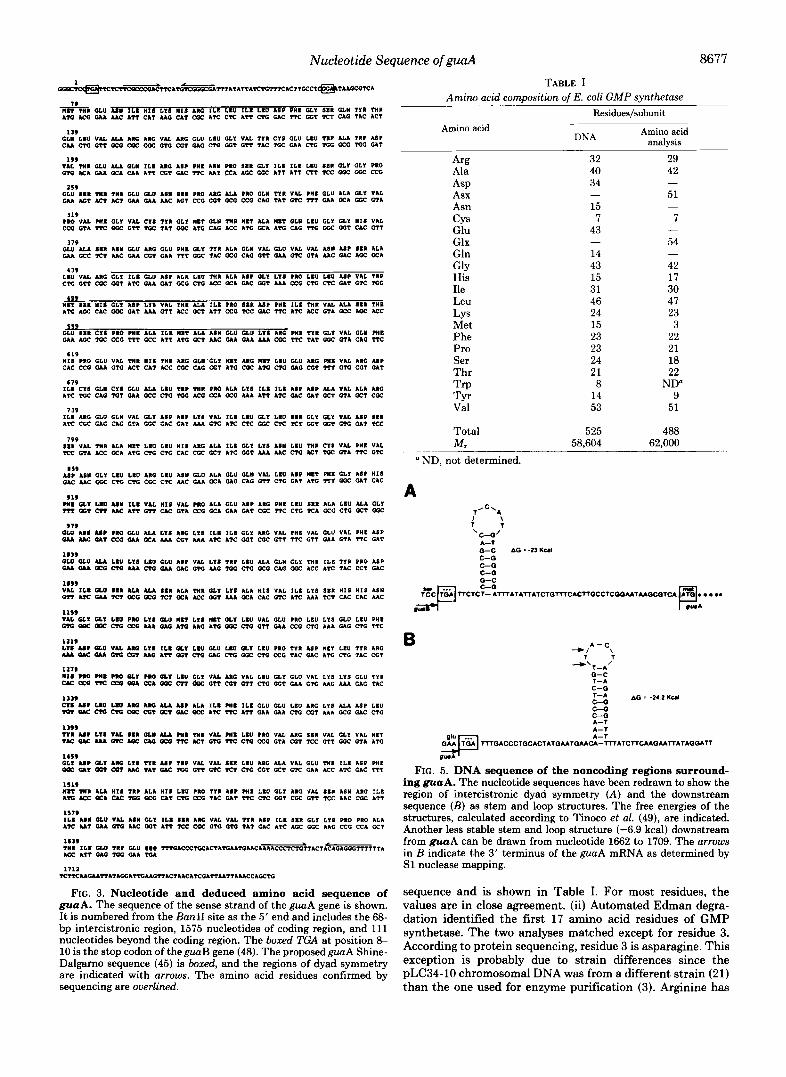
m R GLU A 8 1 I L I HIS LIS H I S ARG I L I LEU I L I LEU ASP P H I GLY SIR GLY TYR THR ATG r(i W M C A T T CAT M G CAT CGC ATC CTC ATT c+G GAC TTC GGT TCT CAG T I C ACT
79
139 GLM LEU VAL ALA ARC ARC VAL ARC GLU LEU GLY VAL TYR CY8 GLU L I U TIP ALA TRP ASP C M CTG GTT OCG EQ: Coc 010 CGT GAG CTG GGT G T T T I C TCC GAA CTG xic OEG TGC GAT
VAL THR GLU ALA GLM I L I ARC ASP P H I A I M P M SER GLY ILI ILC LIU SIR GLY GLY PRO GTG ACA W OCA CALL A T T CGT GAC TTC U T -A AGC GCC AT? ATT CTT KC vf GCC CCG
199
259 GLU S I R THR THR GLU GLU ASN SIR PRO ARC ALA PRO GLM TYR VAL PHI GLU ALA GLY VAL GM ACT ACT ACT GAA GAA MC AGT CCG CGT GCG CCG CAG TAT GTC nr GAA OCA 0 0 ~ GTA
PRO VAL PHI G L ~ VAL CYS Trn GLY MET GLW mu MI? ALA MET GLY LEU GLY oLr HIS VAL 119
CCG CTA UC UX 011 TCC TAT vf ATG CAG Ur ATG GCA ATG CAG TTG uic GGT CAC GTT
119 O W ALA S I R A S I GLU ARG GLU PHI GLY TYR ALA GLI VAL GLU VAL V U ASW ASP 811 ALA W ccf TCT M C W CGT GAA TTT uic TAC GCG CAG 022 GAA GTC GTA M C GAC AGC GCA
4 39 L I U VAL ARC G L 1 I L I GLU ASP ALA LEU T H l ALA ASP GLY LYE P M LIU LIU A8P VAL TIP CTG GTT Coc GGT A T C GAA GAT GCG C C ACC GCA GAC GGT MA CCG CTG Cn: GAT GTC C G
499 MET SIR HIS GLY ASP Lrn VAL run ALA XLI PRO SIR ABP pnr XLI THR VAL ALA SSI rnn ATG AOC CAC UX GAT AM GTT ACC GCT ATT CCG TCC WC TTC ATC ACC GTA OEC AGC ACC
3 5 9 OW asn c1s PRO PHI ALA I L I WIT ALA ABW GLU GLU LYE ARC WI WR GLY VAL GLY PHI O M AGC TGC CCG TTT OEC ATT A M i GCT M C W A GAA AM Coc TTC TAT GCC GTA CAG TTC
HIS PRO GLU VAL THR H I S THR ARC GLM'GLY MET ARC MET L IU GLU ARC PHI VAL 110 ASP CAC CCG GAA GTG ACT CAT ACC CGC CAG 001 ATG CGC ATG CTG GAG COT U T GTG COT GAT
619
679
AX TGC CAG TGT GAA occ CTG TUG ACG CCA OEG AM ATT ATC GAC GAT a r GTA OET CGC I L I CY8 GLN CY8 CLU ALA LEU TUP THR PRO ALA LY8 XLI I L I ASP ASP ALA VAL ALA ARC
739
ATC Eu: GAG CAG GTA GCC GAC GAT M A oic &TC CTC GCC CTC TCT ffiT OGT Oto GAT TCC 1x.I ARC GLU GLII VAL GLr ASP A#P LXI VAL ILI LEU GLY LIU EIR oLr GLK VAL ASP sau
799 S I R VAL THR ALA MET LEU L I U H I S ARC ALA I L I GLY LYS ASY LEU THR C I S VAL P H I VAL TCC GTA Ur Q A A C CTG CTG CAC Coc GCT ATC GGT M A M C CTG ACT TUC GTA TTC 0%
919 wII GLY Lm ASM ILI VAL HIS VAL PRO ALA GLU ASP ARG PHI LEU SIR ALA LEU ALA GLY m a r cn MC ATT GTT CAC GTA CCG GCA GAA GAT ccc l t c CTG TCA KC CTG GCT uic
919 OUI AI# ur PM a u ALA Lra ARG LYS m e ILE oLr ARC VAL PHE VAL GLU VAL PHI ASP GAA M C 011 @X W OCA MA CGT M A ATC ATC ffiT COC GTT TTC GTT GAA GTA C I C CAT
GLU GLO ALA LRU Lr8 LIU GLU AIP VAL LYE TRP Ltu ALA GLY GLr THR ILI WR PRO ASP lS39
GAA W OCC CTG MA CTG GAA GAC OIG M C TUG CTG OCG CAC GCC ACC ATC TAC CCT GAC
1#99 VAL ILI GLU SI. ALA ALA S I R ALA THR GLY LYS ALA H I S VAL I L E LYS S K I H I S HIS ASN O+t A S GAA +El OCG OCG TCT OCA ACC f f iT M A GCA CAC GTC ATC M A U T CAC CAC AAC
rrn U P Lru VAL ssn GLI ALA PHI THR VAL PHI LIU PRO VAL ARC SIR VAL cLr VAL MET 1199
TAC OIC Lu O X .oC ub OCG UC ACT GTG TTC C C CCC GTA CGT TCC GTT CGC CTA A C
1419
QoE OLT QDT CQT AAG TAT 0s TGG GTT GTC +El CTG COT GCT CTC GAL ACC ATC GAC TTT
M R ?HI A W I HIS TI? ALA HI1 LEU ?RC TYR AIP PHI LRU GLY ARG VAL S I X ASY ARG I L I 1519
Am Icc OC1 UC TUG OCG CAT CTG CCG TAC GAT UC CIC GGT CGC GTT TCC M C CGC ATT
1179 111 A I M GLU VAL A I M GL1 ILI S I R ARC VAL VAL W R ASP I L I SIR CLY LYS PRO PRO ALA ATC U T W Oto M E GGT A T T TCC CGC GTG OIG TAT GAC ATC AGC GCC AAG CCG CCA GCT
GLI u r GLY ARC ~ 1 1 WR ASP TRP v u VAL SIR LEU ARC ALA VAL GLU THR ILI ASP PHI
~ M C A A T T A T A G G A I I G M G T T A C T A A C A T C G A T T M T T A M C C A G C T G 1112
FIG. 3. Nucleotide and deduced amino acid sequence of guaA. The sequence of the sense strand of the guuA gene is shown. It is numbered from the Ban11 site as the 5' end and includes the 68- bp intercistronic region, 1575 nucleotides of coding region, and 111 nucleotides beyond the coding region. The bored TGA at position 8- 10 is the stop codon of the guaB gene (48). The proposedguaA Shine- Dalgarno sequence (45) is boxed, and the regions of dyad symmetry are indicated with arrows. The amino acid residues confirmed by sequencing are overlined.
TABLE I Amino acid comDosition of E. coli GMP svnthetase
Amino acid
Arg Ala ASP Asx Asn CYS Glu Glx Gln GlY His Ile Leu LY s Met Phe Pro
Thr Ser
Trp TYr Val
Total M .
ND, not determined.
A I \ 1 , \C"O'
A-T Q-C AG : -23 K u I C--0 C--0 C--0
-
-0°C
Residues/subunit
DNA
32 40 34
15 7
43
14 43 15 31 46 24 15 23 23 24 21 8
14 53
525 58,604
-
-
Amino acid analysis
29 42
51
7
54
42 17 30 47 23 3
22 21 18 22
ND" 9
51
488 62,000
-
-
-
-
B " I \
A-C 7 T
"\f--*' Q-C 1-A C-G l z t AG = -24.2 Uul
C--0 C--0 A-T A-T A-T
G$@ ~ Q A C C C T Q C A C T A T G M T G M ~ - ~ A l C T f C M Q M T f A T A O Q A T f
V U *
FIG. 5. DNA sequence of the noncoding regions surround- ing guaA. The nucleotide sequences have been redrawn to show the region of intercistronic dyad symmetry ( A ) and the downstream sequence ( B ) as stem and loop structures. The free energies of the structures, calculated according to Tinoco et al. (49), are indicated. Another less stable stem and loop structure (-6.9 kcal) downstream from guaA can be drawn from nucleotide 1662 to 1709. The arrows in B indicate the 3' terminus of the guaA mRNA as determined by S1 nuclease mapping.
sequence and is shown in Table I. For most residues, the values are in close agreement. (ii) Automated Edman degra- dation identified the first 17 amino acid residues of GMP synthetase. The two analyses matched except for residue 3. According to protein sequencing, residue 3 is asparagine. This exception is probably due to strain differences since the pLC34-10 chromosomal DNA was from a different strain (21) than the one used for enzyme purification (3). Arginine has
8678 Nucleotide Sequence of guaA
been previously identified as the NH2-terminal residue (3). Our DNA sequence predicts that methionine is the NH2- terminal residue if no processing occurs and was confirmed by the amino acid sequence of purified GMP synthetase (Fig. 3). (iii) A 51-residue peptide of GMP synthetase had been previously sequenced (47). We found an exact match between the peptide sequence and our DNA-derived amino acid se- quence at nucleotide position 448-600. (iv) The molecular weight of the protein encoded by the open reading frame was predicted to be 58,604. This value is in good agreement with the two reported molecular weights of purified GMP synthe- tase of 60,000 (1) and 63,000 (3). Based on these criteria, we believe that the DNA sequence of the guaA locus is correct.
The GMP synthetase sequence shown in Fig. 3 has been used (50) to support a model for the evolution of glutamine amidotransferases from ancestral NH3-dependent enzymes (51). By computer analysis, the first 198 residues of GMP synthetase were homologous to trpG-encoded anthranilate synthase Component I1 and pabA-encoded p-aminobenzoate synthase Component I1 (50). The latter two proteins supply glutamine amide transfer function to anthranilate synthase andp-aminobenzoate synthase, respectively. Thus, GMP syn- thetase residues 1-198 constitute the glutamine amide trans- fer domain in this enzyme. Accordingly, residues 199-525 are predicted to function in the NH3-dependent synthesis of GMP (Equation 2). We visualize that residues 199-525 reflect an ancestral NH,-dependent GMP synthetase that was modified by fusion of a trpG-derived glutamine amide transfer domain at the NH, terminus (50). This fusion permits GMP synthe- tase to function when the concentration of NH3 is low.
By sequence alignment of GMP synthetase with anthranil- ate synthase Component I1 and p-aminobenzoate synthase Component 11, cysteine 86 was identified as the active site residue that functions to form the covalent intermediate with glutamine (50). Cysteine 86 is thus the likely residue alkylated when GMP synthetase is inactivated by chloroketone and 6- diazo-5-oxonorleucine (10). Unexpectedly, cysteine 163 was sufficiently reactive to be alkylated by iodoacetamide (47).
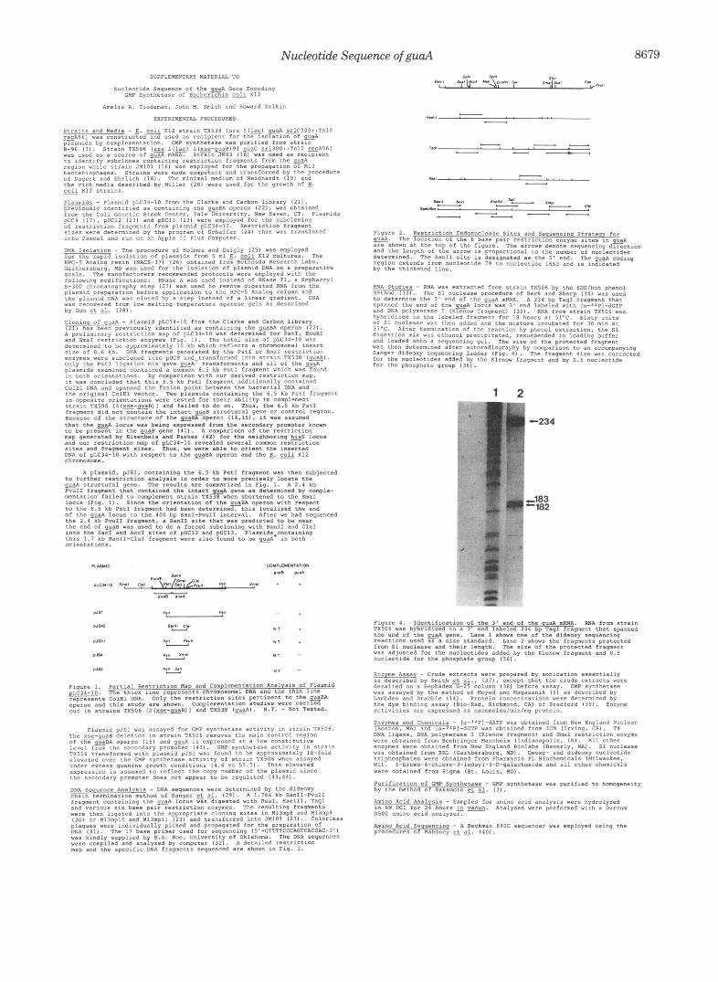
RNA Flanking Regions: SI Nuclease Studies-The 3' end of the guaA mRNA was determined by S1 nuclease mapping. A 234-bp TaqI fragment that spanned the end of the guaA gene was 3' end-labeled with DNA polymerase I (Klenow fragment) and [cx-~*P]~GTP. E. coli RNA extracted from strain TX506 was hybridized to the labeled TaqI fragment. After S1 nuclease treatment, the protected fragment was sized on a DNA sequencing gel using a Sanger dideoxy sequencing ladder as a standard. Two protected fragments of 182 and 183 nucleotides were detected on the sequencing gel (Fig. 4). The length of the protected fragments places the end of the guaA mRNA at nucleotide positions 1692 and 1693, 36-37 bp be- yond the end of the structural gene (Fig. 5). This position is within the loop at a region of dyad symmetry that resembles a p-independent transcription termination site (52). It is possible that transcription actually terminates between nu- cleotides 1705 and 1710 and is then processed back to nucleo- tides 1692-1693, although we have no direct evidence for such a mechanism.
The 68-bp intercistronic region between guaB and guaA contains a palindromic sequence that could form a hairpin loop with the characteristics of a p-independent terminator (Fig. 5). A stability value of -23 kcal/mol was calculated according to Tinoco et al. (49). Most guaA expression is mediated by a promoter upstream of guaB, and thus transcrip-
tion termination, if it occurs in the intercistronic region, should be modest. Measurement of mRNA will be required to determine whether the palindrome functions in a step down for guaA expression.
Acknowledgments-We are indebted to Mark Hermodson for amino acid sequence analyses. We thank B. A. Roe for invaluable advice on sequencing. The skillful typing of this manuscript by Carol Ann Ardoin is gratefully appreciated.
REFERENCES 1. Lee, B. H., and Hartman, S. C. (1974) Biochem. Biophys. Res. Commun.
2. Patel, N., Moyed, H. S., and Kane, J. F. (1975) J. Biol. Chem. 260,2609- 60,918-925
261 .? 3. Sakamoto, N., Hatfield, G. W., and Moyed, H. S. (1972) J. Bwl. Chem.
4. Patel, N., Moyed, H. S., and Kane, J. F. (1977) Arch. Biochern. Biophys.
5. Moyed, H. S., and Magasanik, B. (1957) J. Biol. Chem. 235,351-363 6. Lagerkvist, U. (1958) J. Biol. Chem. 233,143-149 7. Abrams, R., and Bentley, M. (1959) Arch. Biochem. Biophys. 79,91-110 8. Spector, T. (1975) J. Biol. Chem. 260,7372-7376 9. Page, T., Bakay, B., and Nyhan, W. L. (1984) Int. J. Biochem. 16, 117-
247,5880-5887
178,65241
1 zn 10. Z a k , H., and Truitt, C. D. (1977) J. Bwl. Chem. 262,5431-5436 11. Fukuyama, T. T., and Donovan, K. L. (1968) J. Bwl. Chem. 243, 5798-
12. Slechta, L. (1960) Biochern. Biophys. Res. Commun. 3,596-598 13. Fukuyama, T. T. (1966) J. Biol. Chem. 241,47454749 14. Lambden, P. R., and Drabble, W. T. (1973) J. Bacteriol. 116,992-1002 15. Vales, L. D., Chase, J. W., and Murphy, J. B. (1979) J. Bacteriol. 139,320-
16. Messin J (1979) Recomb. DNA Tech. Bull. 2.43-48 17. Vieira, f., and Messing, J. (1982) Gene (Amst.) 19, 259-268 18. Dagert, M., and Ehrlich, S. D. (1979) Gene (Amst.) 6,23-28 19. Neidhardt, F. C., Bloch, P. L., and Smith, D. F. (1974) J . Bacterid. 119,
20. Miller, J. H. (1972) Experiments in Molecular Genetics, pp. 431-435, Cold
21. Clarke, L., and Carbon, J. (1976) Cell 9,91-99 22. Clarke, L., and Carbon, J. (1979) Methods Enzymol. 68,396-408 23. Messing, J. (1983) Methods Enzymol. 101,ZO-78 24. Schaffer, H. E., and Sederoff, R. R. (1981) Anal. Biochem. 115, 113-122 25. Holmes, D. S., and Quigley, M. (1981) Anal. Biochem. 114, 193-197 26. Thompson, J. A., Blakesley, K., Doran, K., Hough, C. J., and Wells, D.
(1983) Methods Enzymol. 100,368-399 27. Norgard, M. V. (1981) Anal. Biochem. 113,34-42 28. Guo, L., Yang, R. C. A., and Wu, R. (1983) Nucleic Acids Res. 11, 5521-
29. Sanger, F., Nicklen, S., and Coulson, A. R. (1977) Pmc. Natl. Acod. Sci. U.
30. Messing, J., and Vieira, J. (1982) Gene (Amst.) 19, 269-276 31. Sanger, F., Coulson, A. R., Barrell, B. G., Smith, A. J. H., and Roe, B. A.
32. Larson, R., and Messing, J. (1983) DNA ( N . Y.) 2, 31-35 33. Saker, W., Gesteland, R. F., and Bolle, A. (1967) Nature 216,588-591 34. Berk, A. J., and Sharp, P. A. (1978) Pmc. Natl. Acod. Sci. U. S. A. 76,
35. Maniatis, T., Fritsch, E. F., and Sambrook, J. (1982) MolecLllor Cloning,
36. Sollner-Webb, B., and Reeder, R. H. (1979) Cell 18,485-499 37. Smith, J. M., Smith, F. J., and Umbarger, H. E. (1979) Mol. Gen. Genet.
5801
322
736-747
Spring Harbor Laboratory, Cold Spring Harbor, NY
5540
S. A. 74,5463-5467
(1980) J. Mol. Biol. 143. 161-178
1274-1278
Cold Spring Harbor Laboratory, Cold Spring Harbor, NY
148. 111-13.4 38. Houlberg, U., Hove-Jensen, B., Jochimsen, B., and Nygaard, P. (1983) J. 39. Bradford, M. (1976) Anal. Bmhem. 71,248-254 40. Mahoney, W. C., Hogg, R. W., and Hermodson, M. A. (1981) J . Biol. Chem.
41. Fukumaki, Y., Shimada, K., and Takagi, Y. (1977) J. Bacteriol. 131,685-
- - -, - - - - - - Bacterrol. 164,1485-1488,
266,4350-4356
688 42. Eisenbeis, S. J., and Parker, J. (1981) Mol. Gen. Genet. 183, 115-122 43. Shimada, K., Fukumaki, Y., and Takagi, Y. (1976) Mol. Gen. Genet. 147,
44. Fukumaki, Y., Shimada, K., and Takagi, Y. (1977) Biochim. Biophys. Acta.
45. Shine, J., and Dalgarno, L. (1974) Proc. Natl. A d . Sei. U. S. A. 71.1342-
46. Stormo, G. D., Schneider, T. D., and Gold, L. M. (1982) Nucleic Acids Res.
47. Truitt, C. D., Hermodson, M. A., and Zalkin, H. (1978) J . Biol. Chern. 263,
48. Tiedeman, A. A., and Smith, J. M. (1985) Nucleic Acids Res. 13, 1303-
49. Tinoco, I., Borer P. N., Den ler, B., Levin, M. D., Uhlenbeck, 0. C.,
50. Zalkin, H., Argos, P., Narayana, S. V. L., Tiedeman, A. A,, and Smith, J.
51. Nagano, H., Zalkin, H., and Henderson, E. J. (1970) J . Biol. Chem. 246,
52. Holmes, W. M., Platt, T., and Rosenberg, M. (1983) Cell 32,1029-1032
203-208
478,90-98
1346
10, 2971-2996
8470-8473
1315
Crothera, D. MI, and Gralla, 5. (1973) Nature New Biol. 246,40-41
M. (1985) J. Biol. Chem. 260,3350-3354
3810-3820
Nucleotide Sequence of guaA 8679
SUPPLEMENTARY MATERIAL TO
Nucleotide Sequence Of the EA Gene Encoding GMP Synthetase of ESCher1Chla coll X12
Amella A. Tiedeman. John M. Smith and Howard Zalkln
EXPERIMENTAL PROCEDURES
Strains and Media - E . coli X12 StTaln TX538 [ara A I & ) E.4 ~ C 3 0 0 : : T n l O
plaamlds by complementatron. GMP synthetase was purlfied from Stram iecA561 was constructed and used as recipient for the 1SOlatiOn of EA
was used a s a source of ~ , 4 mRNA. Strarn JM83 1161 was used as zeclplent 8-96 131. Strain TX506 I= AI&) A l ~ - ~ B l 9 1 EC ~ 3 0 0 : : T n l O =A561
to ldentlfy subclones conrammg r e s t r ~ c t m n fragments from the & region vhlle strain JM101 1161 was employed for the propagation Of M13 bacteriophages. S t r a m s were made competent and transformed by the procedure of Dmert and Ehrlich 1181. The minlmal medium Of Nerdhardt I191 and
-
the rich media described by Miller 1201 were used for the growth Of E . coli K12 EtIalnS.
Plasmids - Plasmid pLC34-10 from the Clarke and Carbon library (211. -ly xdenrified as conrainlng the EBA operon 1221, was obtained from the Coll Genetlc Stock Center, Yale UnlVerSlty, New Haven. CT. Plasmids puc9 1171. puC12 1231 and pUCl3 123) were employed for the subcloning of restriction fragments from plasmid pLC34-10. ReScriCtlOn fragment sizes were determined by the program of Schaffer 1241 that was translated into Pascal and run on an Apple I1 Plus Computer.
DNA ISolatLOn - The procedure of Holmes and Quigly 1251 Was employed for the rapid isolation of plasmids from 5 ml E . solr K12 cultures. The Galthersburg, MD was used for the isolation Of plasmid DNA on a preparative RPC-5 Analog resin INACS-371 126) Obtalned from Bethesda Research Labs,
scale. The manufacturers recommended protoco1s were employed With the followzng modifications: RNase A was used instead of RNase T1, a Sephacryl 5 - 3 0 0 ChrOmatOmraDhv Sten 1271 was used to remove dlaested RNA from the plasmid preparation before applicatlon to the RPC-5 Analog column and the plasmid DNA was eluted by a Step instead Of a linear gradlent. DNA was recovered from low meltxng temperature agarose gels a6 described by GUO et e. 1281.
~ . . .
determmed to be approxkately li kb which reflects a chromosomal insert
enzymes were subcloned into pUC9 and transformed Into Strain TX538 (*AI size of 8.6 kb. DNA fragments generated by the PstI or SmaI restrictlon
Only the PStI llgatlon mix gave =A* transformants and a l l of the =A+ '
plasmrds examined contained a common 6.5 kb PStI fragment which was found in both orlentations. By comparison with our derived restriction map, it was concluded that thls 6.5 kb PstI frament additionally contained ColEl DNA and spanned the fusion point bet&" the bacteriai DNA and the origmal ColEl vector. Tvo plasmids containing the 6 . 5 kb PStI fragment xn opposite orientatmns were tested for thelr abilLty to complement stram TX506 [ A l ~ - ~ B l l and falled to do so. Thus, the 6 . 5 kb PstI fragment dld not Contain the intact E B PtrUCtUT'a1 gene Or Control region. Because of the StruCfUee of the w B A opero.7 114,151, it was assumed that the locus was being expressed from the secondary promoter known
map generated by Eieenbeis and Parker I421 for the neighbring G S locus to be present in the SB gene 1411 . A comparison of the rePtrICtion
and OUT restriction map of pLC34-10 revealed several common reatrlction slteb and fragment sires. Thus. we were able to orient the inserted DNA of pLC34-10 vlth respect to the -81 operon and the E . toll x12 chromosome.
A plasmid. pJS1, containing the 6.5 kb PStI fragment was then subjected
EA Structural gene. The results are sunmarired in Fig. 1. A 2.4 kb to further restriction analyeis in order to more precisely locate the
PvuII fragment that contained the intact & gene a s determined by comple- mentation failed to complement strain TX538 when shortened to the SmaI locus Iflg. 11. Since the orientation of the EBA operon with respect to the 6.5 kb PstI fragment had been determined. this locallred the end of the 1 0 ~ ~ 8 to the 400 bp Srnal-PvuII rnterval. After we had sequenced the 2.4 kb PvuII fragment, a Ban11 Slte that was predicted to be near the end Of E B was used to do a forced subclonlng with Ban11 and ClaI
this 1.7 kb BanII-ClaI fragment were also found to be &+ in both into the Sac1 and ACCI sites of puC12 and pVC13. Plasmids containing
Orientations.
* I . Y I - U T -
Figure 1. Partial Restrlstion Ma and Corn lementation Anal ais of Plasmid
represents COlEl DNA. Only the restrictlon sires pertinent to the E B A pLC34-10. x operon and this study are shown. Complementation studies were carrled our I" strains TX506 [ A l ~ - ~ B I ] and TX538 ( e l . N.T. - Not Tested.
The x s e - w B deletlon In Straln TX506 removes the main Control region of t h e s B A operon 1151 and EA is. expressed at a low ConStitUtlVe
TXSO6 transformed wlth plaamld pJS1 was found to be approximately 10-fold l e v e l from the secondary promoter (431. GMP synthetase aCtlYlty ln strain
elevated over the GMP synthetase activity Of Strain TX506 when assayed under excess guanine growth conditions ( 4 . 6 YS 55.51. This elevated expressmn LE assumed to reflect the copy number of the plasmrd since the secondary promoter does not appear to be regulated 143.441.
Plas-id pJsl was assayed for GMP synthetase activity in Strain TX506.
DNA Sequence Analys~s - DNA sequences were determined by the dideoxy chaln termination method Of Sanger et &. 1291. A 1.764 kb BanII-PvuII framnent containin. the auaA locus was digested wlth RsaI, HaeIII, Tag1 and*varmue six base pair restriction enzcmes. The resulting fragments were then ligated m t o the appropriate c l o n m g Sites ln M13mp8 and Ml3mp9 1301 or M13mp10 and M13mpll 1231 and transformed Into JMlOl 1231. Colorless plaques were lndivldually picked and propagated for the preparatmn Of
was kmdly supplied by B.A. Roe, University Of Oklahoma. The DNA sequences DNA 1311. The 17 base primer used for sequenclnq I5*-GTTTTCCCAGTCACGAC-3'1
were compiled and analyzed by computer 1321. A detalled restriction map and the speclfic DNA fragments Sequenced are shown in F q . 2.
_ -
Figure 2. Restriction Endonuclease Sites and Sequencinq Strateqy for gual\. The location Of the 6 base palr restrlctlon enzyme sites ~n EA are shown at the top Of the figure. The arrovs denote seqvenclnq direction
determmed. The Ban11 site is designated a s the 5 ' end. The & codmq and the length of the arrow is proportional to the number of nwleotldes
by the thickened line. reglon extends from nucleotrde 79 to nucleotlde 1653 and is lndlcated
RNA Studies - RNA was extracted from strain TX506 by the SDSlhot phenol method 1331. The 51 nuclease procedure of Berk and Sharp (341 was used to determlne the 3' end Of the mRNA. A 234 bp TaqI fragment that
and DNA polymerase I IXlenOw fragment) 1351 . RNA from stram TX506 was spanned the end of the & locus was 3 ' end labeled with lo-"PI-dGTP
of 51 nuclease was then added and the mixture Incubated for 30 mi" at hybrldlzed to the labeled fragment for 18 hours at 51'C. Sixty U m t S
37'C. After termlnatlon Of the reaction by phenol extzactmn the 5 1 digestion mix was ethanol precipitated, resuspended rn loading buffer was then determined after autoradiography by comparison to an accompanying and loaded Onto a sequencing gel. The sire of the protected fragment
Sanger dideoxy sequencing ladder Ifig. 4 1 . The fragment sire was corrected for the nucleotides added by the Klenow fragment and by 0 . 5 nucleotlde for the phosphate group 1361.
-183 -182
Figure 4. Identification Of the 3' end Of the 9uaA mRNA. RNA from strain TXSO6 was hybrldized to a 3 ' end labeled 234 bp TaqI fragment that spanned the end of the EA gene. Lane 1 shows one of the dideoxy sequencing reactions used as a sire standard. Lane 2 shows the fragments protected
was adjusted for the nucleotides added by the Xlenow fragment and 0 . 5 from 51 nuclease and their length. The size of the protected fragment
nucleotide for the phosphate group 1361.
Enzyme Assay - Crude extracts were prepared by sonrcation essentially desalted on a Sephadex G-25 column 1381 before assay. GMP synthetase as described by Smlth e., 1371, except that the crude extracts were
was assayed by the method of MOyed and Magas.miL 151 as descrzbed by Lambden and Drabble ( 1 4 1 . Protein COnCentZatlOnS were determined by
activities are expressed as nanomoleslminlmg proteln. the dye bindmg assay IBio-Rad. Richmond, CAI Of Bradford 1 3 9 1 . Enzyme
Enzymes and Chemicals - lo-"PI-dATP was obtained from New England Nuclear
DNA ligase, DNA polymerase I IXlenOw fragment) and SmaI restriction enzyme lB05tOn. MI and I O - ~ ~ P I - ~ G T P was obtained from ICN (Irving, CAI. TI
were obtained from Eoehringer Mannheim IIndianapolis, IN). All Dthez
was Obtained from BRL IGaithersburg, MDI. Deoxy- and dideoxy nucleotide enzymes were obtained from Nev England B101ab6 IBeYerly, MI. 51 nuclease
WII. 5-b~~m0-4-chlo~0-3-indoyl-B-D-g~l*=t~~id~ and a l l other chemicals triphosphates were obtained from Pharmacia. PL Blochemlcals IMilwaukee,
were Obtained from Sigma (St. Louis, Mol.
Purification Of GMP Synthetase - GMP synthetase was purified to homogeneity by the method of sakamoto et e. 131. Amino Acid Analysis - Samples for amino acid analysis were hydrolyzed 0500 ammo acid analyzer. 2" 6N HC1 for 24 hours =. Analyses were performed with a Dvrrum
Amino Acid sequencinq - A Beckman 89OC sequencer was employed using the procedures of Mahoney et dl. ( 4 0 1 .